Preprint
Communication

This version is not peer-reviewed.

Development of Anti-Human Cadherin-26 Monoclonal Antibody, Ca26Mab-6, for Flow Cytometry

Submitted:

08 August 2025

Posted:

12 August 2025

You are already at the latest version

Abstract

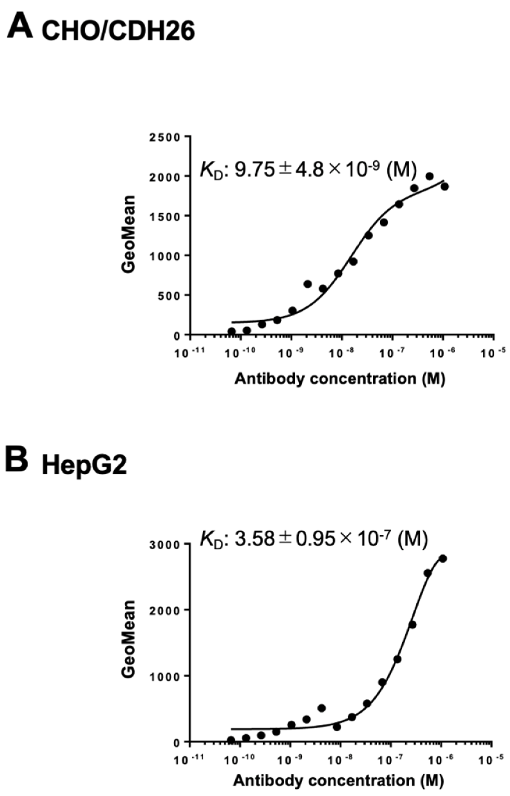
Cadherin 26 (CDH26) is a recently identified member of the cadherin superfamily. Although CDH26 gene expression has been reported in association with allergic inflammatory responses, the protein expression levels and the signaling pathways mediated through its interactions with other proteins remain poorly understood. This is primarily due to the lack of monoclonal antibodies (mAbs) that can recognize the intact, cell surface-expressed form of CDH26. In this study, we developed an anti-human CDH26 mAb, Ca26Mab-6 (IgM, kappa), using the Cell-Based Immunization and Screening (CBIS) method. Ca26Mab-6 demonstrated high sensitivity and specificity for CDH26 in flow cytometry, and did not bind to CHO-K1 cells, which overexpress any of the other type I or type II cadherins. Ca26Mab-6 successfully detected endogenous CDH26 protein expression in HepG2, U-87 MG, MCF7, and 293FT cells. The dissociation constant of Ca26Mab-6 was determined to be 9.8 ± 4.8 × 10⁻⁹ M for CDH26-overexpressed CHO-K1 (CHO/CDH26) cells and 3.6 ± 1.0 × 10⁻⁷ M for HepG2 cells. The detection of CDH26 expression in cancer cells may offer new insights into the potential relationship between inflammatory responses and malignant transformation. Therefore, Ca26Mab-6, developed using the CBIS method, is expected to facilitate functional studies of CDH26 and contribute to the development of CDH26-targeted antibody-based therapies.

Keywords: 
;  ;  ;  

1. Introduction

Cadherins are defined as cell surface glycoproteins responsible for calcium-dependent cell–cell adhesion [1,2,3,4,5]. Large-scale comparative genome sequencing projects have identified over 20,000 cadherin protein sequences, which are now classified into a cadherin major branch, subdivided in two families and 8 subfamilies, and a cadherin-related major branch, subdivided in four families and 11 subfamilies [6]. The major branch includes 21 classical human cadherins, which are further classified into type I and type II cadherins based on their similar structures [6]. These cadherins share a common architecture comprising five extracellular cadherin repeat domains with conserved calcium-binding motifs, a single-pass transmembrane domain, and a highly conserved cytoplasmic domain [6,7]. The extracellular repeat domains mediate homophilic cell–cell interactions, while the cytoplasmic domain interacts with β-catenin and p120-catenin to mediate intracellular signaling [7]. In addition to type I and type II cadherins, cadherin 26 (CDH26) is a relatively newly identified member of the cadherin family and is classified as a non-classical cadherin [6]. CDH26 has five extracellular cadherin repeat domains but its cytoplasmic domain is unique. Phylogenetic analysis of its amino acid sequence, based on the extracellular repeat domains of cadherins, reveals that CDH26 forms a distinct branch, diverging after the desmogleins and before the desmocollins and type I cadherins [6].
Cadherins play a crucial role in cell recognition and adhesion processes, and cadherin-mediated adhesion plays a major role in development and tissue morphogenesis [8,9]. Their function has been reported as maintaining tissue architecture [8,10], stabilizing intercellular adhesion [11], participating in Wnt/β-catenin signaling [12], and being essential for the development of the nervous [13] and cardiovascular systems [14]. Additionally, cadherins are associated with cancer invasion and metastasis [15,16] and are involved in inflammatory and allergic diseases [17]. In this context, the inhibition of cadherin signaling has significant implications for various diseases and has been investigated as a therapeutic target [18,19,20,21].
CDH26 has been reported to be expressed in human allergic gastrointestinal tissues, such as those from patients with eosinophilic gastritis and eosinophilic esophagitis [17]. Moreover, CDH26 mRNA expression is significantly upregulated (approximately 115-fold) in the esophageal tissues of patients with active eosinophilic esophagitis compared to controls [17]. Immunohistochemical staining using anti-CDH26 polyclonal antibodies has shown that CDH26 is localized almost exclusively to the surface and glandular epithelial cells in the gastric tissues of patients with eosinophilic gastritis [17]. In addition, CDH26 has been implicated in the inflammatory responses associated with allergic rhinitis and asthma [22,23]. CDH26 is expressed in airway epithelial cells and has been implicated in actin cytoskeleton maintenance and bone edge polarity [24]. Although these findings have been derived primarily from knockdown experiments [22,24] and RNA sequencing analyses [23], the protein expression levels of CDH26 and signal transduction elicited by CDH26 interactions with proteins remain poorly understood. Therefore, the development of monoclonal antibodies (mAbs) that can recognize the intact form of CDH26 on the cell surface is urgently needed.
In this study, we developed anti-CDH26 mAbs by the Cell-Based Immunization and Screening (CBIS) method [25]. The obtained mAb, Ca26Mab-6 (IgM, kappa) showed clear response of CDH26 expression in flow cytometry. This mAb is anticipated to open new avenues from basic studies of CDH26 function to the development of therapeutic applications.

2. Materials and Methods

2.1. Cell Lines

Chinese hamster ovary (CHO)-K1, mouse myeloma P3X63Ag8U.1 (P3U1), human liver carcinoma HepG2, and human glioblastoma U-87 MG cells were obtained from the American Type Culture Collection (ATCC, Manassas, VA, USA). The human breast cancer cell line MCF7 was obtained from the Cell Resource Center for Biomedical Research, Institute of Development, Aging and Cancer, Tohoku University (Miyagi, Japan). The human embryonic kidney cell line 293FT was purchased from Thermo Fisher Scientific Inc. (Waltham, MA, USA). All cell lines were maintained as previously described [25].

2.2. Plasmid Construction and Establishment of Stable Transfectants

Stable CHO-K1 transfectants that overexpressed human type I cadherins, including CHO/CDH1, CHO/PA16-CDH2 (CHO/CDH2), CHO/CDH3, CHO/PA16-CDH4 (CHO/CDH4), and CHO/PA16-CDH15 (CHO/CDH15) cells, were previously established.[25] CHO-K1 transfectants that overexpressed human type II cadherins and CDH26 were also established. Genes encoding human CDH6 (NM_004932.4), CDH7 (NM_004361.5), CDH17 (NM_004063.4), CDH19 (NM_021153), CDH24 (NM_022478) were purchased from OriGene Technologies, Inc. (Rockville, MD, USA). Genes encoding CDH8 (NM_001796.5), CDH12 (NM_004061), CDH13 (NM_001257), CDH16 (NM_004062), CDH18 (NM_004934), CDH26 (NM_177980) were obtained from the RIKEN BioResource Research Center (Ibaraki, Japan). Genes encoding CDH5 (NM_001795.5), CDH9 (NM_016279.4), CDH10 (NM_006727.5), CDH11 (NM_001797), CDH20 (NM_031891), CDH22 (NM_021248), were synthesized by Eurofins Genomics KK (Tokyo, Japan).
The cDNAs of CDH5, CDH7, CDH8, CDH9, CDH10, CDH11, CDH12, CDH13, CDH15, CDH16, CDH18, CDH19, CDH20, CDH22, CDH24, and CDH26 , with the signal sequence and pro-peptide removed, were subcloned into the pCAG-Ble vector (FUJIFILM Wako Pure Chemical Corporation, Osaka, Japan) with an N-terminal PA16 tag (GLEGGVAMPGAEDDVV) [26]. Additionally, the cDNA of CDH26 with an N-terminal MAP16 tag (PGTGDGMVPPGIEDKI) [27] was constructed. The cDNAs of CDH6 and CDH17 were supplied in pCMV6 vectors containing a C-terminal mycDDK tag (OriGene Technologies, Inc.). These plasmids were transfected into CHO-K1 or LN229 cells and stable transfectants were sorted using an anti-CDH6 mAb (clone 427909, Thermo Fisher Scientific Inc.), an anti-CDH11 mAb (clone 16G5, BioLegend, San Diego, CA, USA), an anti-CDH17 mAb (clone 2618, Novus Biologicals, Centennial, CO, USA), an anti-PA16 tag mAb (clone NZ-1) [26], and an anti-MAP16 tag mAb (PMab-1) [27] using a SH800 cell sorter (Sony Corporation, Tokyo, Japan). The CDH-overexpressed stable transfectants were established, including CHO/PA16-CDH5 (CHO/CDH5), CHO/CDH6-mycDDK (CHO/CDH6), CHO/PA16-CDH7 (CHO/CDH7) , CHO/PA16-CDH8 (CHO/CDH8), CHO/PA16-CDH9 (CHO/CDH9), CHO/PA16-CDH10 (CHO/CDH10), CHO/PA16-CDH11 (CHO/CDH11), CHO/PA16-CDH12 (CHO/CDH12), CHO/PA16-CDH13 (CHO/CDH13), CHO/PA16-CDH15 (CHO/CDH15), CHO/PA16-CDH16 (CHO/CDH16), CHO/CDH17-mycDDK (CHO/CDH17), CHO/PA16-CDH18 (CHO/CDH18), CHO/PA16-CDH19 (CHO/CDH19), CHO/PA16-CDH20 (CHO/CDH20), CHO/PA16-CDH22 (CHO/CDH22), CHO/PA16-CDH24 (CHO/CDH24), CHO/PA16-CDH26 (CHO/CDH26), and LN229/MAP16-CDH26 (LN229/CDH26) cells. To confirm the expression of CDHs in these transfectants, 1 μg/mL of commercially available mAbs or 0.1 μg/mL of NZ-33,[28] a derivative of NZ-1, was used.

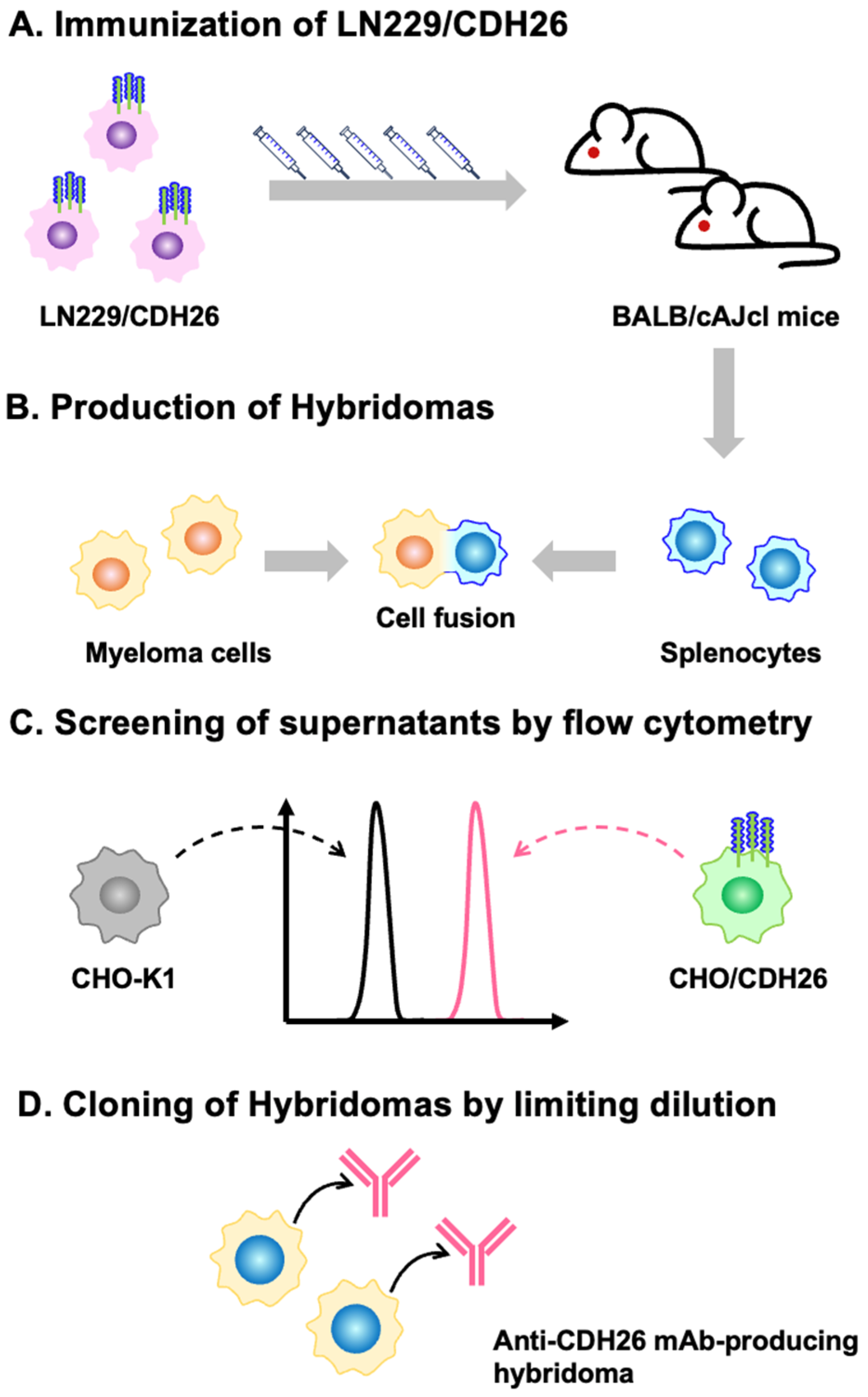
2.3. Hybridoma Production

All animal experiments were conducted in accordance with the guidelines for animal care and use and were approved by the Animal Care and Use Committee of Tohoku University (Permit No. 2022MdA-001). Every effort was made to minimize animal suffering and distress throughout the study. Animals were housed under specific pathogen-free conditions. Two 6-week-old female BALB/cAJcl mice [obtained from CLEA Japan (Tokyo, Japan)] were intraperitoneally immunized with LN229/CDH26 (1 × 10⁸ cells/mouse). with Alhydrogel adjuvant 2% (InvivoGen, San Diego, CA, USA). Hybridomas were generated as described previously [25].

2.4. Flow Cytometry

Cells were detached using 1 mM ethylenediaminetetraacetic acid (EDTA; Nacalai Tesque, Inc., Kyoto, Japan) to prevent enzymatic degradation of surface proteins. The cells were washed with 0.1% bovine serum albumin (BSA) in phosphate-buffered saline (PBS) (blocking buffer) and incubated with mAbs at 4°C for 30 minutes. The cells were stained with anti-mouse IgG (H+L)-Alexa Fluor 488 conjugate (1:2,000 dilution) (Cell Signaling Technology, Inc., Danvers, MA, USA). Data were acquired using the SA3800 Cell Analyzer (Sony Corporation, Tokyo, Japan) and analyzed as described previously [25].

2.5. Determination of the Binding Affinity by Flow Cytometry

CHO/CDH26 cells were treated with serial dilutions of Ca26Mab-6 (100 to 0.0012 μg/mL). The cells were stained with anti-mouse IgG (H+L)-Alexa Fluor 488 conjugate (1:200 dilution). The dissociation constant (KD) values of Ca26Mab-6 were determined using GraphPad Prism 6 software (GraphPad Software, Inc., La Jolla, CA, USA), as described previously [25].

3. Results

3.1. Development of Anti-CDH26 mAbs

Two female BALB/cAJcl mice were immunized with LN229/CDH26 cells (Figure 1A). Splenocytes from the immunized mice were then fused with P3U1 myeloma cells to generate hybridomas (Figure 1B). These hybridomas were seeded into 96-well plates, and their culture supernatants were screened using a flow cytometry-based high-throughput screening to identify supernatants that were positive for CHO/CDH26 cells but negative for parental CHO-K1 cells (Figure 1C). Positive clones were subjected to limiting dilution to establish mAbs, resulting in the isolation of Ca26Mab-6 (mouse IgM, kappa) (Figure 1D).

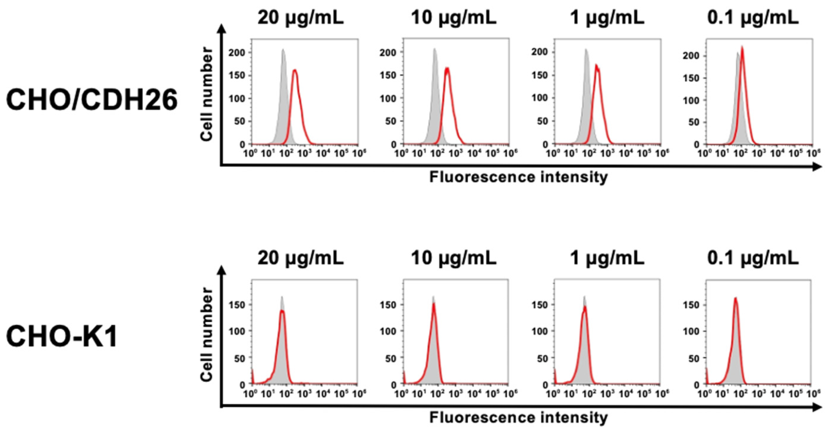
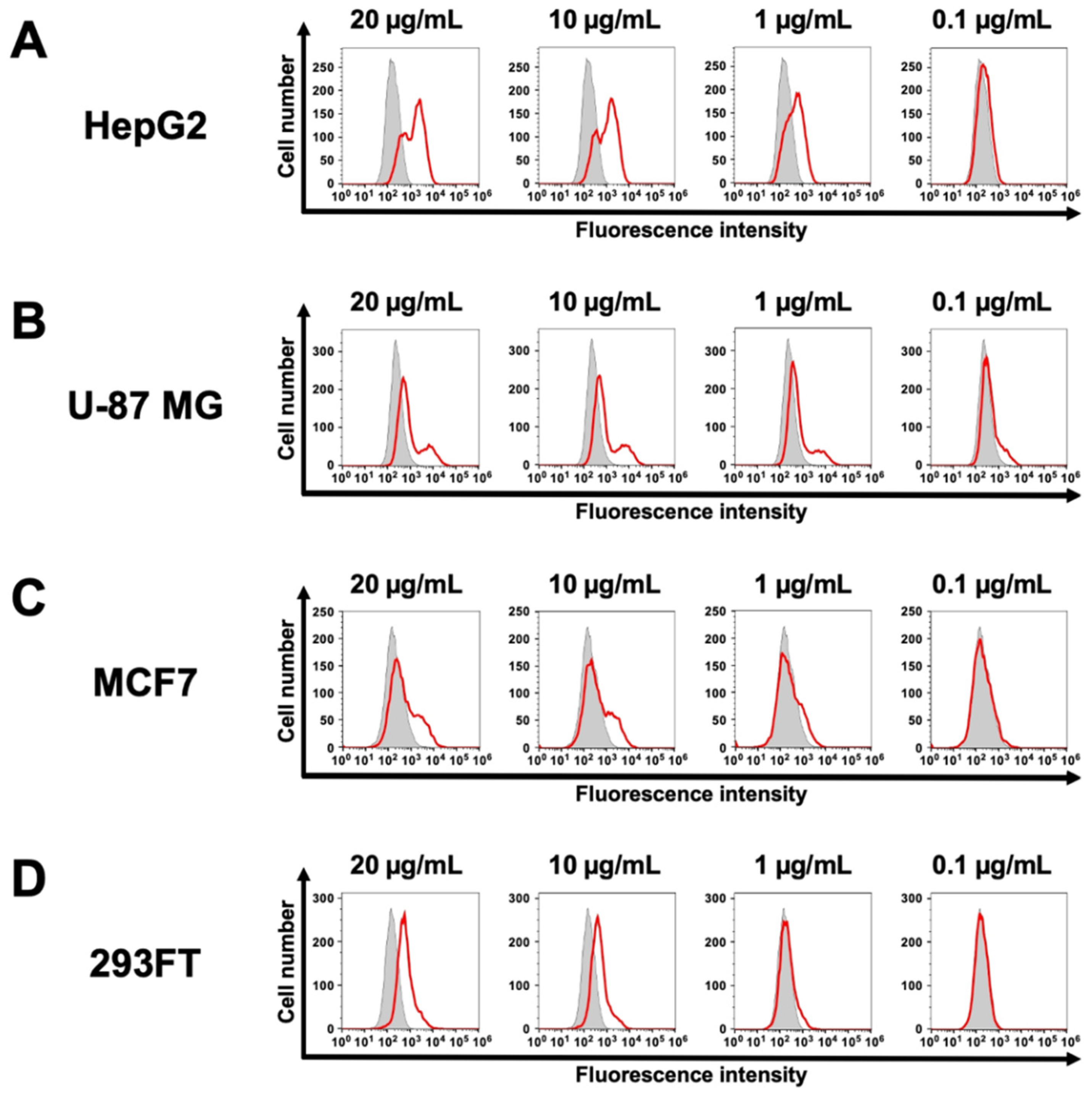
3.2. Investigation of the Reactivity of Ea4Mab-3 Using Flow Cytometry

The binding of purified Ca26Mab-6 to CHO/CDH26 and CHO-K1 cells was analyzed by flow cytometry. Ca26Mab-6 exhibited dose-dependent reactivity with CHO/CDH26 cells at concentrations ranging from 20 to 0.1 μg/mL but showed no binding to CHO-K1 cells at any concentration (Figure 2). These results indicate that Ca26Mab-6 specifically recognizes CDH26 on the cell surface. Furthermore, Ca26Mab-6 bound to HepG2 cells in a dose-dependent manner (Figure 3A), indicating that this cell endogenously expresses CDH26. Binding to U-87 MG, MCF7, and 293FT cells was also observed as a weak but significant signal (Figure 3B–D). These findings suggest that various types of cancer cells and immortalized cells may endogenously express CDH26.

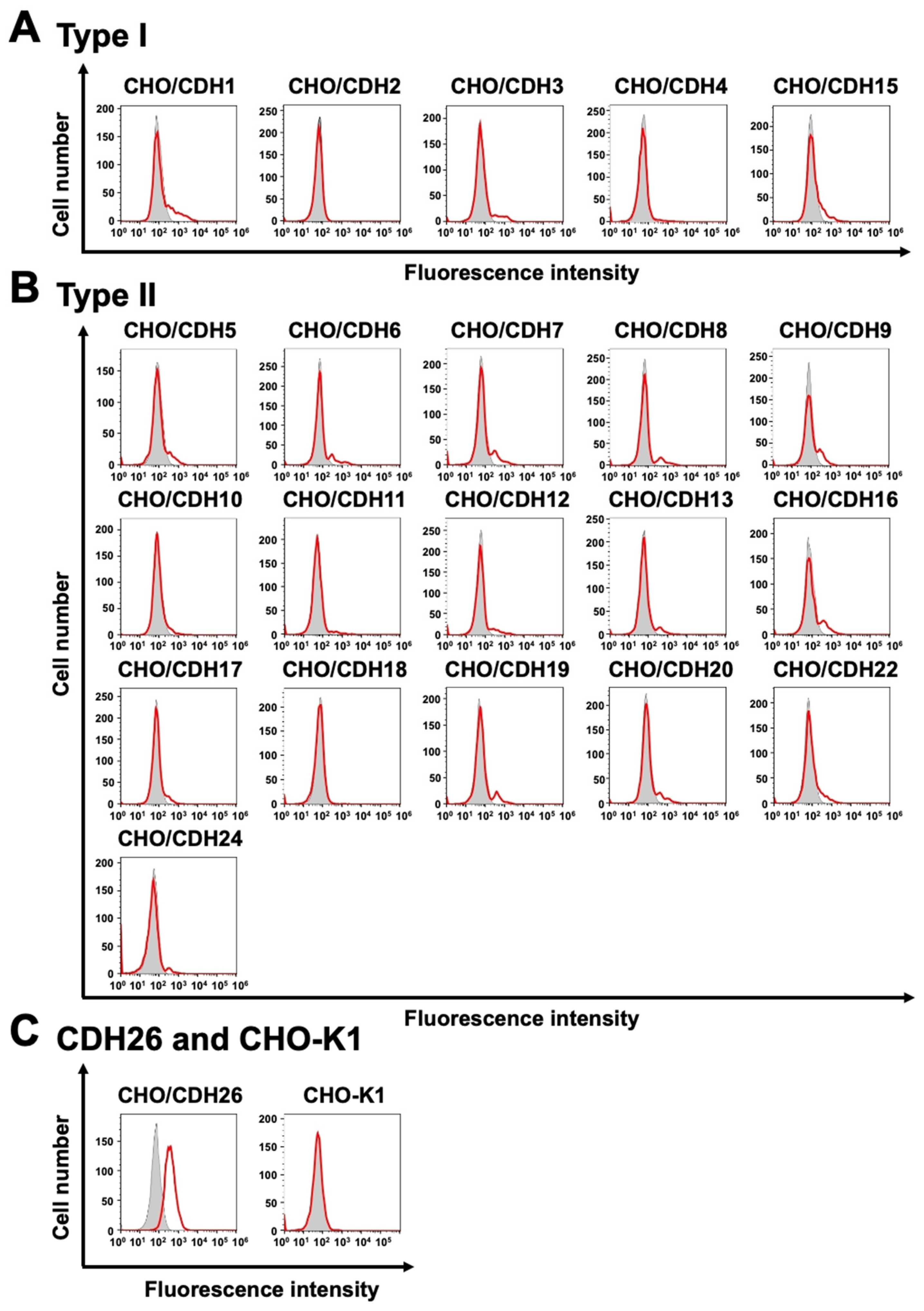
3.3. Analysis of the Specific-Reactivity of Ca26Mab-6 to CDH26 Using CHO-K1 Cells Expressed Various Cadherins

We previously established CHO-K1 cells expressed type I cadherins (Supplementary Figure 1A).[25] Furthermore, we generated stable CHO-K1 transfectants that overexpressed type II cadherins and CDH26 (Supplementary Figure 1B and C). Among these CHO-K1-derived cells expressed human cadherins, Ca26Mab-6 showed no binding to any other cadherins (Figure 4A and B) and recognized only CHO/CDH26 cells (Figure 4C), indicating the specificity of Ca26Mab-6.

3.4. Determination of KD Values of Ca26Mab-6 by Flow Cytometry

The binding affinity of Ca26Mab-6 was evaluated by flow cytometry using serial dilutions ranging from 100 to 0.0012 μg/mL. The KD value of Ca26Mab-6 was 9.75 ±4.8 × 10⁻9 M for CHO/CDH26 cells (Figure 5A and Supplementary Figure 2A) and was 3.58 ± 0.95× 10⁻7 M for HepG2 cells (Figure 5B and Supplementary Figure 2B).

4. Discussion

We established anti-CDH26 mAbs and demonstrated that Ca26Mab-6 is suitable for flow cytometry (Figure 2). Ca26Mab-6 did not bind to any CHO-K1 cells that overexpressed type I or type II cadherins other than CDH26 (Figure 4), indicating its high specificity for CDH26. Considering the absence of previous reports describing mAbs that recognize cell surface-expressed CDH26, our results provide a valuable mAb for further basic research on CDH26 expression and for the development of diagnostic and therapeutic agents.
The expression of CDH26 in various cancer cells suggests that the CDH26 expression may play a role in carcinogenesis and that anti-CDH26 mAbs have potential as therapeutic agents. CDH26 expression has been reported in allergic gastrointestinal tissues [17] and in allergic inflammatory conditions such as rhinitis and asthma [22,23], however, its role in cancer remains unclear. Since chronic inflammation is a major risk factor for cancer development and progression [18], the correlation between CDH26 expression and malignant transformation of cells may be elucidated in future studies.
In HepG2 cells, the flow cytometry histogram displayed two distinct peaks (Figure 3A). This may be attributed to the aneuploidy of HepG2 cells, which are known to exhibit high chromosomal instability [29], potentially affecting CDH26 protein expression levels. Similarly distinct or broadened peaks were observed in U-87 MG and MCF7 cells (Figure 3B and C), suggesting that similar ploidy variations of U-87 MG [30] and MCF7 [31] may influence the expression level of CDH26.
Ca26Mab-6 also bound to 293FT (Figure 3D). This cell is a derivative of HEK293, an immortalized epithelial cell derived from human embryonic kidney, suggesting that CDH26 is expressed not only in cancer cells but also in non-cancer cells. Since Ca26Mab-6 is suitable for flow cytometry but is not applicable to western blot or immunohistochemistry, it remains challenging to comprehensively evaluate the relationship between CDH26 expression levels and tumor malignancy using tissue sections. To advance research into the role of CDH26 in cancer, the development of additional anti-CDH26 mAbs that are compatible with multiple applications is essential.
In the context of developing antibody-based therapeutics targeting CDH26, the expression of CDH26 in normal cells raises concerns about potential off-target effects. To eliminate this risk, the development of cancer-specific mAbs (CasMabs) is a promising strategy [32]. CasMabs are designed to recognize aberrant conformational epitopes of overexpressed and misfolded proteins found specifically in cancer cells [33]. Therefore, the development of anti-CDH26 CasMabs is also anticipated to enhance the specificity and safety of CDH26-targeted therapies.
In conclusion, Ca26Mab-6 is a sensitive mAb suitable for basic research and holds promise for proof-of-concept studies in preclinical models aimed at developing CDH26-targeted antibody therapies.

Supplementary Materials

The following supporting information can be downloaded at the website of this paper posted on Preprints.org.

Author Contributions

Hiroyuki Satofuka : Investigation, Funding acquisition. Mika K. Kaneko: Conceptualization Hiroyuki Suzuki: Investigation, Writing – original draft, Yukinari Kato: Conceptualization, Funding acquisition, Project administration, Writing – review and editing.

Funding

This research was supported in part by Japan Agency for Medical Research and Development (AMED) under Grant Numbers: JP25am0521010 (to Y.K.), JP25ama121008 (to Y.K.), JP25ama221339 (to Y.K.), and JP25bm1123027 (to Y.K.), and by the Japan Society for the Promotion of Science (JSPS) Grants-in-Aid for Scientific Research (KAKENHI) grant no. 25K10553 (to Y.K.).

Institutional Review Board Statement

The animal study protocol was approved by the Animal Care and Use Committee of Tohoku University (Permit number: 2022MdA-001, approved on April 1, 2022) for studies involving animals.

Data Availability Statement

All related data and methods are presented in this paper. Additional inquiries should be addressed to the corresponding authors.

Conflicts of Interest

The authors declare no conflict of interest involving this article.

References

  1. Colas-Algora, N.; Millan, J. How many cadherins do human endothelial cells express? Cell Mol Life Sci 2019;76(7): 1299-1317. [CrossRef]
  2. Takeichi, M. Historical review of the discovery of cadherin, in memory of Tokindo Okada. Dev Growth Differ 2018;60(1): 3-13. [CrossRef]
  3. Takeichi, M. Self-organization of animal tissues: cadherin-mediated processes. Dev Cell 2011;21(1): 24-26. [CrossRef]
  4. Oda, H.; Takeichi, M. Evolution: structural and functional diversity of cadherin at the adherens junction. J Cell Biol 2011;193(7): 1137-1146. [CrossRef]
  5. Hyafil, F.; Babinet, C.; Jacob, F. Cell-cell interactions in early embryogenesis: a molecular approach to the role of calcium. Cell 1981;26(3 Pt 1): 447-454. [CrossRef]
  6. Hulpiau, P.; van Roy, F. Molecular evolution of the cadherin superfamily. Int J Biochem Cell Biol 2009;41(2): 349-369. [CrossRef]
  7. Oas, R.G.; Nanes, B.A.; Esimai, C.C.; et al. p120-catenin and beta-catenin differentially regulate cadherin adhesive function. Mol Biol Cell 2013;24(6): 704-714. [CrossRef]
  8. Gumbiner, B.M. Cell adhesion: the molecular basis of tissue architecture and morphogenesis. Cell 1996;84(3): 345-357. [CrossRef]
  9. Halbleib, J.M.; Nelson, W.J. Cadherins in development: cell adhesion, sorting, and tissue morphogenesis. Genes Dev 2006;20(23): 3199-3214. [CrossRef]
  10. Leckband, D.; Prakasam, A. Mechanism and dynamics of cadherin adhesion. Annu Rev Biomed Eng 2006;8: 259-287. [CrossRef]
  11. Troyanovsky, R.B.; Laur, O.; Troyanovsky, S.M. Stable and unstable cadherin dimers: mechanisms of formation and roles in cell adhesion. Mol Biol Cell 2007;18(11): 4343-4352. [CrossRef]
  12. Nelson, W.J.; Nusse, R. Convergence of Wnt, beta-catenin, and cadherin pathways. Science 2004;303(5663): 1483-1487. [CrossRef]
  13. Polanco, J.; Reyes-Vigil, F.; Weisberg, S.D.; Dhimitruka, I.; Bruses, J.L. Differential Spatiotemporal Expression of Type I and Type II Cadherins Associated With the Segmentation of the Central Nervous System and Formation of Brain Nuclei in the Developing Mouse. Front Mol Neurosci 2021;14: 633719. [CrossRef]
  14. Breier, G.; Breviario, F.; Caveda, L.; et al. Molecular cloning and expression of murine vascular endothelial-cadherin in early stage development of cardiovascular system. Blood 1996;87(2): 630-641. [CrossRef]
  15. Takeichi, M. Cadherins in cancer: implications for invasion and metastasis. Curr Opin Cell Biol 1993;5(5): 806-811. [CrossRef]
  16. Canel, M.; Serrels, A.; Frame, M.C.; Brunton, V.G. E-cadherin-integrin crosstalk in cancer invasion and metastasis. J Cell Sci 2013;126(Pt 2): 393-401. [CrossRef]
  17. Caldwell, J.M.; Collins, M.H.; Kemme, K.A.; et al. Cadherin 26 is an alpha integrin-binding epithelial receptor regulated during allergic inflammation. Mucosal Immunol 2017;10(5): 1190-1201. [CrossRef]
  18. Yu, W.; Yang, L.; Li, T.; Zhang, Y. Cadherin Signaling in Cancer: Its Functions and Role as a Therapeutic Target. Front Oncol 2019;9: 989. [CrossRef]
  19. Mrozik, K.M.; Blaschuk, O.W.; Cheong, C.M.; Zannettino, A.C.W.; Vandyke, K. N-cadherin in cancer metastasis, its emerging role in haematological malignancies and potential as a therapeutic target in cancer. BMC Cancer 2018;18(1): 939. [CrossRef]
  20. Song, Y.; Ye, M.; Zhou, J.; Wang, Z.W.; Zhu, X. Restoring E-cadherin Expression by Natural Compounds for Anticancer Therapies in Genital and Urinary Cancers. Mol Ther Oncolytics 2019;14: 130-138. [CrossRef]
  21. Vieira, A.F.; Paredes, J. P-cadherin and the journey to cancer metastasis. Mol Cancer 2015;14: 178. [CrossRef]
  22. Liu, T.; Sun, Y.; Guo, Z.; Bai, W. Knockdown of Cadherin 26 Prevents the Inflammatory Responses of Allergic Rhinitis. Laryngoscope 2023;133(7): 1558-1567. [CrossRef]
  23. Esnault, S.; Dill-McFarland, K.A.; Altman, M.C.; et al. Identification of bronchial epithelial genes associated with type 2 eosinophilic inflammation in asthma. J Allergy Clin Immunol 2025. [CrossRef]
  24. Lachowicz-Scroggins, M.E.; Gordon, E.D.; Wesolowska-Andersen, A.; et al. Cadherin-26 (CDH26) regulates airway epithelial cell cytoskeletal structure and polarity. Cell Discov 2018;4: 7. [CrossRef]
  25. Ubukata, R.; Suzuki, H.; Tanaka, T.; Kaneko, M.K.; Kato, Y. Development of an anti-CDH15/M-cadherin monoclonal antibody Ca15Mab-1 for flow cytometry, immunoblotting, and immunohistochemistry. Biochemistry and Biophysics Reports 2025;43: 102138. [CrossRef]
  26. Fujii, Y.; Kaneko, M.; Neyazaki, M.; et al. PA tag: a versatile protein tagging system using a super high affinity antibody against a dodecapeptide derived from human podoplanin. Protein Expr Purif 2014;95: 240-247. [CrossRef]
  27. Fujii, Y.; Kaneko, M.K.; Kato, Y. MAP Tag: A Novel Tagging System for Protein Purification and Detection. Monoclon Antib Immunodiagn Immunother 2016;35(6): 293-299. [CrossRef]
  28. Fujisawa, S.; Yamamoto, H.; Tanaka, T.; et al. 2025.
  29. Zhou, B.; Ho, S.S.; Greer, S.U.; et al. Haplotype-resolved and integrated genome analysis of the cancer cell line HepG2. Nucleic Acids Res 2019;47(8): 3846-3861. [CrossRef]
  30. Pokorna, M.; Hudec, M.; Jurickova, I.; et al. All-Trans Retinoic Acid Fosters the Multifarious U87MG Cell Line as a Model of Glioblastoma. Brain Sci 2021;11(6). [CrossRef]
  31. Ben-David, U.; Siranosian, B.; Ha, G.; et al. Genetic and transcriptional evolution alters cancer cell line drug response. Nature 2018;560(7718): 325-330. [CrossRef]
  32. Kaneko, M.K.; Suzuki, H.; Ohishi, T.; et al. A Cancer-Specific Monoclonal Antibody against HER2 Exerts Antitumor Activities in Human Breast Cancer Xenograft Models. Int J Mol Sci 2024;25(3). [CrossRef]
  33. Arimori, T.; Mihara, E.; Suzuki, H.; et al. Locally misfolded HER2 expressed on cancer cells is a promising target for development of cancer-specific antibodies. Structure 2024;32(5): 536-549 e535. [CrossRef]
Figure 1. Schematic illustration of anti-CDH26 mAb production. (A) Two female BALB/cAJcl mice were immunized by intraperitoneal injection of LN229/CDH26 cells. (B) Following immunization, splenocytes were collected and fused with P3U1 cells to produce hybridomas. (C) The hybridoma culture supernatants were screened for anti-CDH26 mAbs by flow cytometry using CHO/CDH26 and parental CHO-K1 cells. (D) Hybridomas producing antigen-specific mAbs were cloned by limiting dilution.
Preprints 171710 g001
Figure 2. Flow cytometry analysis of an anti-CDH26 mAb against CHO/CDH26 and CHO-K1 cells. CHO/CDH26 and CHO-K1 cells were treated with (red lines) or without (gray-filled lines) Ca26Mab-6 at the indicated concentrations. After treatment, the cells were washed and incubated with Alexa Fluor 488-conjugated anti-mouse IgG. Fluorescence data were acquired using the SA3800 Cell Analyzer.
Preprints 171710 g002
Figure 3. Flow cytometry analysis of an anti-CDH26 mAb against cells endogenously expressing CDH26. HepG2 (A), U-87 MG (B), MCF7 (C), and 293FT (D) cells were treated with (red lines) or without (gray-filled lines) Ca26Mab-6 at the indicated concentrations. After treatment, the cells were washed and incubated with Alexa Fluor 488-conjugated anti-mouse IgG. Fluorescence data were acquired using the SA3800 Cell Analyzer.
Preprints 171710 g003
Figure 4. Flow cytometry analysis of cross-reactivity among cadherin-expressed cells. CHO-K1 cells overexpressing type I cadherins (A), type II cadherins (B), CDH26 and parental CHO-K1 cells (C) were analyzed. The cells were treated with (red lines) or without (gray-filled lines) 10 μg/mL of Ca26Mab-6. After treatment, the cells were washed and incubated with Alexa Fluor 488-conjugated anti-mouse IgG. Fluorescence data were acquired using the SA3800 Cell Analyzer.
Preprints 171710 g004
Figure 5. Measurement of the binding affinity of Ca26Mab-6. CHO/CDH26 (A) and HepG2 (B) cells were treated with serial dilutions of Ca26Mab-6 (100 to 0.0012 μg/mL). After treatment, the cells were washed and incubated with Alexa Fluor 488-conjugated anti-mouse IgG. Fluorescence data were acquired using the SA3800 Cell Analyzer. The KD values were calculated by GraphPad PRISM 6.
Preprints 171710 g005
Disclaimer/Publisher’s Note: The statements, opinions and data contained in all publications are solely those of the individual author(s) and contributor(s) and not of MDPI and/or the editor(s). MDPI and/or the editor(s) disclaim responsibility for any injury to people or property resulting from any ideas, methods, instructions or products referred to in the content.
Copyright: This open access article is published under a Creative Commons CC BY 4.0 license, which permit the free download, distribution, and reuse, provided that the author and preprint are cited in any reuse.
Prerpints.org logo

Preprints.org is a free preprint server supported by MDPI in Basel, Switzerland.

Subscribe

Disclaimer

Terms of Use

Privacy Policy

Privacy Settings

© 2025 MDPI (Basel, Switzerland) unless otherwise stated