Submitted:
09 August 2025
Posted:
13 August 2025
You are already at the latest version
Abstract
Nucleic acid aptamers are single-stranded DNA or RNA molecules that can bind to a target with high specificity and affinity, as screened by Systematic Evolution of Ligands by Exponential Enrichment (SELEX). In recent years, SELEX technologies have been significantly advanced for screening of aptamers for a variety of target molecules, cells, and even bacteria and viruses. By integrating recent advances of emerging technologies with SELEX, novel screening technologies for nucleic acid aptamers have emerged with improved screening efficiency, reduced production costs and enhanced aptamer performance for a wide range of applications in medical diagnostics, drug delivery and environmental monitoring. Aptasensors utilize aptamers to detect a wide range of analytes, allowing accurate identification and determination of small molecules, proteins, and even whole cells with remarkable specificity and sensitivity. Further optimization of the aptasensor can be achieved by aptamer truncation, which not only maintains the high specificity and affinity of the aptamer binding with the target analytes, but also reduces the manufacturing cost. Predictive models also demonstrate the powerful capability of determination of the minimal functional sequences by simulation of aptamer-target interaction processes, thus effectively shortening the aptamer screening procedure and reducing the production costs. This paper summarizes the research progress of protein-targeted aptamer screening in recent years, introduces several typical aptasensors at present, discusses the optimization methods of aptasensors by combining efficient SELEX with advanced predictive algorithms or post-SELEX processes, as well as the challenges and opportunities faced by aptasensors.
Keywords:
nucleic acid aptamers
; systematic evolution of ligands by exponential enrichment (SELEX)
; aptasensors
; post-SELEX optimization
; aptamer truncation
; predictive algorithms
1. Introduction
Aptamers typically are short, single-stranded DNA or RNA oligonucleotides that bind to specific targets which include metal ions, small molecules, peptides, and even proteins expressed on the surface of bacteria, virus, or human cells, and are difficult to obtain high-affinity antibodies [1,2,3]. This unique property has turned aptamers into versatile tools for target molecule identification and detection in biological samples, playing an important role in synthetic biology and healthcare industries [4,5,6,7,8,9,10,11]. While having similar binding affinity and specificity to antibodies, nucleic acid aptamers can be chemically synthesized and demonstrate many advantages over antibodies, such as easy chemical modification, better stability and low production costs. Moreover, due to smaller size (typically 20-80 nucleotides in length or 10-20 kDa in weight), aptamers have good tissue permeability, are easily accessible to cells and do not elicit an immune response from cells [12]. Aptamers also have the ability to flexibly fold to recognize the target modules which may be large or small in the biological samples and need to be bonded with the aptamer sensing elements of biosensors [13,14]. In addition, aptamers are easy to preserve and can be kept in a suitable environment for a long time, which makes the application of aptamers as molecular recognition elements in bioassays. As such, aptamers have been extensively investigated for biosensors, as shown in Figure 1. The publications found by searching PubMed with the keywords of either aptamer or aptasensor have increased every year since 2014 up to 20 May 2025.
2. Nucleic Acid Aptamer
2.1. Nucleic Acid Aptamer
As aptamers are short, single-stranded DNA or RNA molecules, they can change shapes dramatically. The binding of the nucleic acid aptamer to the target molecule is achieved through its unique three-dimensional conformation, which is characterized by the shape of hairpins, inner loops, pseudoknots, bulges or G-quadruplexes [15]. A binding between the nucleic acid aptamer and the target molecule occurs through van der Waals forces, hydrogen bonding, and electrostatic forces [16,17]. When a nucleic acid aptamer binds with a target molecule, if the target molecule is small, the nucleic acid aptamer will cover the target molecule and wrap around the surface of its molecule through its unique helical structure. If the target molecule is large, the nucleic acid aptamer will form adaptive-like structures in the clefts and gaps on the surface of the large molecule. Nucleic acid aptamers have a good folding ability, which allows the nucleic acid aptamer to fully adapt to the size of the target molecule [18]. Nucleic acid aptamers can fully bind to target molecules and adapt to numerous target molecules of different shapes. The targets of nucleic acid aptamers include, but are not limited to peptides [19], proteins [20,21], small organic molecules [22], various metal ions [23], and even molecules on the surface of bacteria [24], viruses [25] or human cells [26].
2.2. SELEX Technology
Most aptamers are discovered through a library selection process called Systematic Evolution of Ligands by Exponential Enrichment (SELEX). The working principle of the SELEX is to chemically synthesize a library of single-stranded oligonucleotides as a starting library to incubate with the desired target molecule in vitro to screen best aptamer-target binding [27]. The binding of the target with the nucleic acid sequences in the mixture eliminates the nucleic acid sequences that are not bound to the target. Nucleic acid sequences that bind to the target are isolated and amplified using polymerase chain reaction (PCR) [28,29]. The PCR amplification creates a new pool of nucleic acid sequences, which are then subjected to the subsequent round of the selection process. The initial rounds of screening usually take longer time, and their screening conditions are more lenient compared to later ones [30]. After the first few rounds of screening, the buffer conditions are usually altered to control the volume of the target-aptamer complex and the binding time of the target to the aptamer to achieve the conditions for creating an aptamer with high affinity for the target. In addition, the presence of non-specific nucleic acid sequences in the mixture favors the selection of nucleic acid aptamers with high affinity. At the same time, for some target molecules, the presence of monovalent or divalent cations in buffer solutions can greatly reduce non-specific binding [31]. It is also important to remove non-specific binding sequences from the library using pre-negative selection by incubating the library with the selection matrix only in the absence of target molecules. After repeated screening and amplification, some nucleic acids that do not bind or have low affinity for the target reactants are eliminated, and the remaining nucleic acids with high affinity for the target are isolated. After several rounds of SELEX processes, the purity of nucleic acids with high affinity for the target is enriched until they occupy the majority of the nucleic acid pool (>90%). As shown in Figure 2, after 5 - 20 rounds of SELEX selection processes, the selected nucleic acid aptamers with high affinity to the target were cloned into appropriate vectors and sequenced.
Four factors are critical in selecting nucleic acid sequences that bind to target molecules: the type of randomization, the length of the random sequence region, the chemistry of the nucleic acid library, and the utility of the constant region [32]. During the selection process, the nucleic acid library is processed with the target in the appropriate buffer and at a suitable temperature for the desired application. In vitro SELEX procedures have been widely used to study the nature, function and structure of nucleic acid aptamers. This technology plays an important role in studying molecular recognition and molecular evolution [33]. In order to quickly find residues in the target molecule that cannot be changed without altering the function of the target, in vitro SELEX technologies using DNA and RNA ligands has been proved as a good approach [34]. One of the most important steps in SELEX technology is the strategies of separating the nucleic acid sequences that are not bound to the target from the sequence library. Therefore, the advancement of the SELEX technologies predominantly relies on the improvement of the efficiency and accuracy of separation (or partitioning) of target-bound and non-binding nucleic acid sequences from the oligonucleotide library.
2.3. Separation of Target-Bonded and Non-Binding Aptamers
Main separation techniques for conventional aptamer screening technologies are capillary electrophoresis (CE) SELEX, Cell-SELEX, nitrocellulose membrane filtration SELEX, microbead/ magnetic bead SELEX, microfluidic microarray SELEX, microcolumn SELEX and so on. In this part, we will focus on CE-SELEX, Microfluidic SELEX, and Cell-SELEX technologies.
2.3.1. Capillary Electrophoresis SELEX Technology
Capillary electrophoresis SELEX technology is one of the most important methods applied to screen high-affinity nucleic acid aptamers [35]. The principle of CE-SELEX technology is to incubate a nucleic acid library with a target protein and then pass it through a high-voltage electric field within a capillary, causing the unbound nucleic acid sequence to separate from the bound nucleic acid-target protein complex due to the difference in migration rates. Bound nucleic acid-target protein complexes were collected as the end of the capillary, subsequently amplified by PCR to enrich the bound aptamers for the subsequent screening round. Usually the CE-SELEX requires one to four rounds of screening to obtain high-affinity nucleic acid aptamers, which significantly shortens selection process from weeks to days and are much more effective than the routine SELEX that normally requires several months for 8-15 or more rounds [36]. In CE-SELEX technology, the environment for target proteins and nucleic acid libraries is more liberalized, thus creating a free environment for target proteins and nucleic acid aptamers. This condition makes the probability of non-specific binding of target proteins to nucleic acids lower and not affected by spatial site-blocking during their interaction. CE-SELEX technology was subsequently improved, resulting in the non-equilibrium capillary electrophoresis of equilibrated mixtures (NECEEM) SELEX technology, equilibrium capillary electrophoresis of equilibrated mixtures (ECEEM) SELEX technology, and non-SELEX technology [37,38]. Recently, one-round pressure controllable selection (OPCS) based on the conventional CE-SELEX technology has also been developed [39]. OPCS-SELEX technology enables simultaneous incubation of nucleic acid libraries with two target proteins, which can provide competitive pressure on each other. Subsequently the complexes formed by these two proteins and the nucleic acid library were separated and collected by high-resolution CE. The enhancement of the efficiency and effectiveness of the OPCS-SELEX technology is achieved by adjusting the concentration ratio of target proteins to improve the affinity and specificity of the nucleic acid aptamer. In addition, the improvement can also be achieved by introducing high concentrations of predatory proteins. The great advantage of OPCS-SELEX technology is that it simultaneously obtains nucleic acid aptamers for two proteins with high affinity and high specificity in a single round, while CE-SELEX technology requires three rounds under the same conditions for nucleic acid aptamers to each protein target [40].
2.3.2. Microfluidic SELEX Technology
Microfluidic technology enables precise manipulation and control of small fluid volumes within microfabricated devices, making it possible to develop highly efficient and sensitive analytical screening platforms. Integrating microfluidics with aptamer selection technology, Microfluidic SELEX, can precisely control fluids and temperature and complete mixing, incubation and partitioning from starting aptamer library to subsequent round on a single microdevice. It offers advantages over conventional SELEX like reduced sample volume, improved target-binding capacity, enhanced selection stringency and automated processing for improved throughput These advantages have led to various microfluidic-based SELEX techniques that further improve efficiency and speed of aptamer screening. Further information can be found in latest review articles [41,42]. Despite these, some great challenges remain for microfluidic SELEX, such as technical complexity of integration of microfluidic platforms and its scaleup and standardization for real-world applications.
2.3.3. Cell-SELEX Technology
Cell surfaces have proteins or glycans, which allows nucleic acid aptamers to be screened with purified proteins to recognize target proteins in living cells. Cell-SELEX technology typically uses cells with characteristic protein expression as target cells, while cells with no or less expression are used for negative selection [43,44,45,46,47]. Cell-SELEX is easy to perform in most biological labs and does not require specialized tools. Cells can be easily partitioned by centrifugation, leaving unbound aptamers in suspension. The cell-bound aptamers are eluted by incubating at high temperatures then separating by centrifugation. Aside from its simple protocol, cell-SELEX is advantageous because of its authentic target presence on the cell surface and variety of targets (e.g., cancel cells, T cells, bacteria, and viruses). However, the nucleic acid aptamers screened by Cell-SELEX usually do not meet the requirements due to the low specificity of the nucleic acid aptamers. The reason for this is that such cells used for positive and negative screening usually differ in addition to the target protein. To address this, great efforts have been made to improve cell-SELEX technology. Among them, an approach method proposed by Pleiko et al. [48] is particularly remarkable. They utilized the functional genomics FASTAptamer toolbox and the bioinformatics tool edgeR to investigate binding variability between nucleic acid libraries and positive and negative screening cells. Based on the informative metrics about the selection process achieved with the toolbox FASTAptamer and the tool edgeR, all the sequences in the final nucleic acid library interact with the live cells were selected for cell-SELEX to realize a fast and high-throughput aptamer screening protocol against live cells.
3. Aptasensors
3.1. Aptasensors
In general, a conventional biosensing device can be broadly defined as a device that converts physical, chemical or biological events into measurable signals. Biosensors consist of three main components: the biomolecular recognition element, the sensor, and the signal processing and data analysis system. A biomolecular recognition (sensing) element, usually borne by aptamers, antibodies, enzymes, proteins, cellular receptors, tissues, microorganisms, etc., is a biologically derived material or biomimetic element that provides selectivity for a target molecule or a target substance. Sensors can convert molecular recognition events into measurable signals through different transduction mechanisms. After receiving these measurable signals, electronic data analysis systems process the signals accordingly and visualize the data obtained from these processes [49]. Aptasensors act as molecular recognition elements through aptamers because aptamers selectively bind to target molecules [50,51,52]. As shown in Figure 3, when a target molecule in a sample binds to an aptamer, this binding process is converted into a detectable signal through various mechanisms, which is then quantified by the sensor element. Subsequently the presence of the target molecule in the sample or its specific concentration can be determined [53].
In aptasensors, molecular recognition is a key step in their functioning. The recognition elements for the aptasensors at the early stages were obtained by natural isolation from the biological system. With advances in SELEX technology, molecular recognition elements for aptasensors can be synthesized in the laboratory [54]. The use of aptamers as probes in biosensing devices makes aptasensors uniquely suited for applications, such as blood testing and pathogen detection [55].
Aptasensors have better properties than traditional biosensors that use antibodies as recognition elements [56]. Aptasensors have higher affinity and specificity for target molecules and target substances. Aptamers can be chemically modified with conformational changes in the target molecule [57]. In addition, some aptamers can be used to obtain specific target molecules by adding specific substances to change their properties. The unique advantage that aptamers can undergo multiple changes in properties allows aptamers to be used in a wider range of applications without compromising high affinity at the same time [58,59,60,61,62,63]. Aptamers can be easily chemically modified and labelled, enabling aptasensors to be widely used in clinical medicine and medical diagnostics and mass-produced and reused [64]. Aptasensors are extremely stable compared with antibodies, and can be used for multiple times when specific substances are added to regenerate the functionality of immobilized biological components [65]. In addition, aptasensors can be developed for a wide range of target molecules or target substances, and multiple transduction mechanisms can be applied. By applying different transduction mechanisms, aptasensors can be classified into optoelectronic [66,67,68,69,70,71,72], field-effect transistor (FET) [73,74,75,76,77,78,79,80,81,82,83], electrochemical [84,85,86,87,88,89,90,91,92,93,94] aptasensors, and other types of sensors. Aptasensors based on optical, field-effect transistor, and electrochemical transduction mechanisms will be discussed in the following section.
3.2. Optical Aptasensors
Optical aptasensors are new types of biosensors based on the use of nucleic acid aptamers as recognition elements, combined with the development of optical detection technology. Optical aptasensors work by capturing signals generated by the interaction of a biometric element with a target molecule and converting the signals into optical signals that are subsequently detected and analyzed [95]. Optical aptasensors use an optoelectronic transduction which allows for rapid and highly sensitive detection, and have been widely used in the fields of clinic diagnosis, environmental monitoring, and food safety [96,97,98,99]. Below will be focused on fluorescent aptasensors, colorimetric aptasensors and surface plasmon resonance (SPR) aptasensors.
3.2.1. Fluorescent Aptasensors
Fluorescence-based optical aptasensors are constructed in such a way that when a nucleic acid aptamer binds specifically to a target molecule or a target substance, the aptamer-the target binding causes a change in fluorescence intensity of the fluorescent material, which is used for monitoring the interactions between the aptamer and the target analytes. Fluorescence-based detection is easy to implement among other optical detection techniques. One of the common fluorescence techniques is Forster resonance energy transfer (FRET). The principle of FRET involves a radiationless energy transfer process in which an energy-excited fluorophore (donor) transfers energy to another molecule (acceptor) over long distance of typically 1nm and up to 10nm via dipole-dipole interaction [100]. FRET is particularly effective in the detection of interacting membrane proteins and has been used to monitor dynamic process of protein-protein interactions in living organisms, which is not possible with other conventional monitoring methods. FRET has also been used with monoclonal antibodies, which has led to better understanding of protein structures in solution, biofilms and cell surface mapping on immune cells [101]. Moreover, FRET has been widely exploited in DNA sequencing and polymerase chain reaction [102] together with a variety of novel materials for sensing. Among the most widely used materials in fluorescent aptasensors, graphene oxide (GO) has a strong light quenching ability, good dispersion and biocompatibility [103,104]. GO-based aptasensors has demonstrated great sensitivity and selectivity for DNA analysis by effectively quenching the fluorescence of quantum dots (QDs) when interacting with single-stranded DNA [105,106]. Because of simple operation, high sensitivity, and non-destruction of the target substance, fluorescent aptasensors have been widely used in a variety of detection and analysis of DNAs and proteins, such as the quantitative analysis of protein biomarkers [107].
3.2.2. Colorimetric Aptasensors
Colorimetric aptasensors are a type of aptasensors based on colorimetric bioassay and have received more attention in recent years. The principle of colorimetric bioassay is the detection of the target substances by a color change using the naked eye and simple instruments. Colorimetric aptasensors have demonstrated rapid diagnostic capabilities without complicated instrumentation [108]. Colorimetric bioassays are widely used in point-of-care diagnostics and field testing because of their simplicity, low-cost and effectiveness [109,110]. With the advances of microfabrication and electronic technologies, applications of colorimetric aptasensors have been dramatically expanded with the advent of smart electronic devices, and color detection of aptasensors has been successfully integrated with commonly used devices such as smartphones, digital cameras and flatbed scanners [111,112,113]. However, colorimetric aptasensors still face some challenges. For example. colorimetric aptasensors are not sensitive and accurate as other detection methods in the detection of cancer cells in blood samples [114,115]. In addition, colorimetric aptasensors are highly influenced by the background illumination of the sample matrix and take a long time to be fabricated [116,117]. In clinical diagnostics, colorimetric aptasensors are currently difficult to perform simultaneous multiple targets assays [118].
3.2.3. Surface Plasmon Resonance Aptasensors
The surface plasmon resonance (SPR) aptasensors measure any change in a refractive index proportional to the amount of the target analyte over the process of its binding with the aptamer. The principle of SPR technology is to monitor the change of resonance angle at the metal surface caused by the specific binding between the substance (e.g., aptamers) fixed on the metal surface and the target molecule in a sample for biological characterization [119]. Evanescent waves are formed when light waves travelling in a medium undergo total internal reflection at the interface of the metal and medium, and free electrons (so-called surface plasmons) are generated on the surface of the metal. When the frequency and wave number of two waves are equal, the two resonate, the incident light is absorbed, and the energy of the reflected light decreases sharply, and a resonance peak will appear on the reflection spectrum. When the number of target molecules adsorbed on the metal surface or the configuration of the target molecule changes, the change of the peaks on the reflectance spectra can be used for the detection and real-time monitoring of intermolecular interactions without labelling [120,121,122,123]. SPR aptasensors are now widely used in biological research, environmental monitoring, food safety, and clinical diagnosis [124,125,126,127,128]. However, SPR aptasensors also have the disadvantage of being more expensive to manufacture, as a layer of metallic gold usually needs to be deposited to cover the surface of the device [129]. SPR aptasensors is also highly affected by temperature changes.
3.3. Field-Effect Transistor Aptasensors
Field-effect transistor aptasensors are aptasensors constructed based on field-effect transistor. The FET is a semiconductor device that uses the electric field effect of the input loop to control the output loop current. The source and drain are connected by semiconductor channels to form a typical FET aptasensors. Any adsorption behavior of a target molecule located on the surface of a channel causes a change in the electric field. Adjusting the gate potential at this point can cause the drain current in the FET channel to change. FET aptasensors are used to detect and analyze target molecules by monitoring changes in drain current [130,131,132,133,134,135,136,137,138]. FET aptasensors are inexpensive to manufacture compared to aptasensors for other transduction mechanisms, especially SPR aptasensors, and can be integrated with existing manufacturing processes on a large scale. In addition, FET aptasensors have other advantages, such as the ability to operate at low power, the ever-shrinking design size, the lack of labelling and the rapid detection, which makes them promising [139]. Although FET aptasensors have been rapidly optimized over the past few years, there are still significant challenges in improving their detection sensitivity as they can be affected by various factors [140].
In FET aptasensors, aptamer functionalization methods are essential, and can be categorized into physical and chemical appraaoches. The aptamer physical functionalization approach is based on physical processes of non-covalent interactions such as hydrogen bonding, van der Waals forces, electrostatic interactions and hydrophobic interactions. The aptamer chemical functionalization methods are based on chemical processes of covalent interactions, such as covalent bonding and crosslinking [141,142,143]. In the physical functionalization method of aptamers, the aptamers directly adsorb onto the surface of the channel and then combine with the target molecules. The operation process is relatively simple, which reduces the possibility of aptamer denaturation and maintains the electrical properties of the channel material. However, in the physical functionalization method of aptamers, the binding force between aptamers and channel materials is relatively weak and could be difficult to control, which can lead to aptamer dissociation and reduce the detection repeatability and long-term stability of FET aptasensors. When FRT aptasensors are chemically functionalized, the channel surfaces normally need to be activated at first with carboxylation, hydroxylation [144], amination, or gold deposition [145]. Subsequently, one end of the linker molecule is covalently bonded to the channel, and the other end of the linker molecule is covalently bonded to the aptamer. During the chemical functionalization, aptamers are immobilized onto channel surfaces through covalent bonds which are strong and can improve the detection repeatability and long-term stability of FET aptasensors. However, the aptamer chemical functionalization methods require additional sequence design and purification based on specific target molecules and target substances. Furthermore, during the chemical functionalization process of aptamers, the conformational stability of aptamers could be subject to unpredictable interference [146]. Therefore, it is necessary to select the appropriate aptamer functionalization method based on the specific circumstances. Because of high selectivity, good portability and low manufacturing cost, FET aptasensors have been well developed, and will be further exploited in more fields with better standardization and commercialization [147].
3.4. Electrochemical Aptasensors
Electrochemical aptasensors use the electrode surface as a platform to immobilize the biosensing aptamer, convert the aptamer recognition event into an electrical signal, and detect changes in current, potential, or impedance to achieve rapid detection of target molecules or target tissues [148,149]. The advantages of the electrochemical transduction mechanism are the simplicity of its conversion phenomenon and the possibility of using label-free and reusable detection systems. Electrochemical transduction is extremely sensitive and can be enhanced in practice by amplifying the detection signal by attaching a biocatalytic tag to the aptamer-target complex. In practical applications, electrochemical aptasensors are produced at low cost and are universally applicable [150,151]. The following section will focus on electrochemical impedance spectroscopy (EIS) aptasensors, voltammetric aptasensors and amperometric aptasensors.
3.4.1. Electrochemical Impedance Spectroscopy Aptasensors
EIS is a label-free and non-destructive monitoring techniques, and achieved by applying a small amplitude sinusoidal perturbation signal to the system in equilibrium [152,153]. EIS has excellent performance in measuring the molecular interactions of electrochemically inactive compounds occurring at the electrode surface [154]. It can detect any subtle changes at the interface induced by the interaction between the nucleic acid aptamer and the target molecule. EIS is commonly used to determine organic compounds, such as protein biomarkers, and can also be used to determine electrochemical changes occurring on the surface of modified electrodes due to biorecognition. Various biomolecules such as enzymes, antibodies, nucleic acids and cells are immobilized on the surface of the electrodes and used as detection elements to develop EIS aptasensors [155,156]. Moreover, EIS aptasensors are inexpensive to manufacture and can be mass produced and reused.
In EIS aptasensors, the total impedance can be divided into real component and imaginary component, corresponding to resistivity component and capacitance component respectively. The resistivity component is caused by the obstruction of the electrode surface to the current flow, while the capacitance component reflects the storage of electric charge in the system when the voltage is applied [157]. EIS applications in aptasensors can be classified into two types depending on the nature of the measured signal: Faraday and non-Faraday types. In Faraday-type EIS aptasensors, the electrode surface is partially or completely covered by a non-insulating layer or partially covered by an insulating layer. Impedance is generated when electrons are transferred to the electrodes through redox reactions. In non-Faraday type EIS aptasensors, the electrode surface is completely covered by a dielectric layer and the entire electrode assembly behaves as an insulator. The generation of impedance is based on direct current impedance, at which point no charge is transferred to the electrode surface, but current can still flow, so the system behaves like a capacitor [158,159]. Therefore, the non-Faraday type EIS aptasensors are also known as the capacitive EIS aptasensors. Faraday-type EIS aptasensors are generally considered to have higher sensitivity than capacitive ones, so they can be used for determination of target molecules, rather than simply detecting their presence [160]. Moreover, Faraday type EIS aptasensors can produce a more pronounced signal change at the same concentration of the target, thus having a better sensitivity.
Unlike other conventional biosensors, the EIS aptasensors directly use aptamers as the core, which come into contact with the electrolyte. Therefore, if EIS aptasensors are to be more widely applied in other fields, the aptamer-based electrodes selected must have long-term stability. Compared with other biosensors, EIS aptasensors have better stability, higher sensitivity, and are applicable to multiple media rather than a single medium, which requires complex and efficient electrode designs to immobilize the aptamer on the electrode surface [161]. This is to ensure that the aptamer and the electrode can be effectively connected, utilizing the characteristics of aptamers as recognition elements, which can generate stable signals for detection purposes. How to better construct and optimize EIS aptasensors is also one of the main research directions in the future.
3.4.2. Voltammetric Aptasensors
In addition, there are electrochemical aptasensors based on voltammetry. Voltammetry is the oldest and one of the most commonly used electrochemical techniques. The voltammetric transduction mechanism involves scanning the potential on the working electrode from one preset value to another and recording the electrochemical current as a function of the applied potential [162]. The use of voltammetry for electrochemical aptasensors has been made more widespread with the continuous advancement of computerized techniques for controlling and measuring potentials and currents in constant potentiostats [163]. Voltammetric aptasensors are widely used for the detection of protein biomarkers because of their extreme sensitivity and the ease with which the experiments can be performed and reproduced [164,165].
Voltammetric aptasensors obtain information about target molecules by measuring the changes in current signals within a variable potential range, and simultaneously detect changes in potential and current [166,167]. Voltammetry methods applied in aptasensors can be classified as cyclic voltammetry (CV), differential pulse voltammetry (DPV), square wave voltammetry (SWV), alternating current voltammetry (ACV), linear sweep voltammetry (LSV), based on different technical principles [168]. These different voltammetric techniques, when combined with aptasensors, can detect various types of target molecules, thereby increasing the selectivity and application range of voltammetric aptasensors. Voltammetry generally uses a standard three-electrode system (working electrode, counter electrode and reference electrode) by applying a certain form of electrical potential which triggers the redox reaction of the electroactive substance on the working electrode and then recording the current changes within a specified time range. Voltammetric method reflects the electrochemical characteristics of target molecules from different angles through potential, current, and time functions. The voltammogram records the current curves of the oxidation and reduction processes of the detected target substance with the electroactive substances in the working electrode at a specific potential. In the voltammogram, data such as the peak value, peak potential, and peak width of the curve reflect the electrochemical characteristics of the target molecules or target substances.
Voltammetric aptasensors have low noise characteristics during operation and can detect multiple target substances with different peak potentials in a single scan. Voltammetric aptasensors can also characterize multiple target molecules as a whole and can be integrated into the sensing system to form a voltammetric detection system [169]. Even in the presence of some interfering substances, the voltammetric aptasensors can still detect single or multiple groups of target substances simultaneously. In voltammetric aptasensors, the limit current is affected by testing temperature, so it should be kept at a constant temperature during detection to ensure the accuracy of the results obtained [170]. Voltammetric aptasensors have great commercial potential due to their low manufacturing cost in addition to their high sensitivity.
3.4.3. Amperometric Aptasensors
Amperometric detection is another commonly used transduction mechanism for electrochemical aptasensors. The amperometric aptasensors is a relatively self-contained electrochemical device. It works by converting the biorecognition changes that occur as a result of oxidation or reduction of an electroactive substance into a current signal, which is subsequently detected and quantitatively analyzed [171].
Amperometric aptasensors obtain information about target molecules by measuring the changes in current signals at a constant potential. The magnitude of the current generated by oxidation or reduction reactions can enable accurate quantitative analysis by the amperometric aptasensors. The main difference between amperometric and voltammetric aptasensors is that amperometric aptasensors applied a constant potential. In amperometric aptasensors, the potential is directly adjusted to the specific value and maintained, only allowing the target substance to be reacted on the surface of the electrodes, after which the output current is recorded and used for detection and analysis. The higher output current of the amperometric aptasensors represents the higher concentration of the electroactive substance in the target substance sample being detected [172,173,174].
One of the most crucial aspects in development of amperometric aptasensors is selection of the strategies to improve their selectivity, sensitivity, and detection range. For example, to address issues with disparity of the concentrations of the targets existed in different conditions, great efforts have been made to optimize the designs of amperometric aptosensors. It has been observed that the affinity of the aptamers has an influence on the detection range of the amperometric aptasensors, that the aptamers with stronger affinity possess a larger detection range than those with weaker affinity [175]. Therefore, improving the affinity of the aptamer is proposed to an effective approach to extend the detection range of the amperometric aptasensors. Aptamer affinity can be improved for amperometric aptasensors through sequence optimization, aptamer structure stabilization and the introduction of hydrophobic groups [176]. In addition, a multi-aptamer strategy can also be adopted by incorporating two or more aptamers with different affinities to simultaneously measuring the concentrations of different target substances for the purpose of expanding the detection range [177]. The properties of the linkers between the aptamer and the electrode also influences the detection of the amperometric aptasensors, affecting the sensitivity and detection range of the amperometric aptasensors. Generally, the rigid linkers are used as flexible linkers can comprise sensing performance of amperometric aptasensors under certain circumstances [178]. However, when a rigid linker and a flexible linker are simultaneously introduced into multi-aptamer amperometric aptasensors, it is interestingly noticed that the multi-aptamer sensors exhibited higher sensitivity, greater selectivity and broader detection range than either single-aptamer amperometric aptasensors or multi-aptamer sensors using a single linker approach [179]. In addition, a passivation layer can be added to the electrode surface to suppress background noise from the electrode itself [180].
4. Post-SELEX Optimization Process of Aptamers
In recent years, there has been a rapid development of nucleic acid aptamers, which has led to a wide range of applications in a variety of fields such as biosensors, medical diagnostics, food safety and environmental monitoring [181,182,183,184,185,186,187]. However, the overall performance of nucleic acid aptamers screened by SELEX still needs further improvement. One of the key reasons for the limited performance of nucleic acid aptamers is their poor stability and reproducibility [188]. Nucleic acid aptamers screened by various SELEX technology have drawbacks that limit their use in the target molecule monitoring under complicated conditions [189]. In addition, for the real-world applications, nucleic acid aptamers have to fulfil other requirements such as reduced production costs, integration with existing manufacturing processes and sufficient bioavailability [190]. In order to achieve performance improvement and cost reduction of nucleic acid aptamer applications, a variety of post-SELEX optimization methods have been developed. As shown in Figure 4, the commonly used post-SELEX optimization methods include truncation, mutagenesis, extension, chemical modification and bivalent or multivalent aptamer construction. With the advances of machine learning and AI tools, predictive models and computational tools are also being adopted for aptamer screening. These predictive models and computational tools incorporating machine learning and deep learning are essential in the post-SELEX optimization process to simulate the affinity of a nucleic acid aptamer for a target molecule, to simulate the dynamic binding process, and to determine the minimum sequence that binds to the target with high affinity and specificity [191,192,193,194]. In this section, we will focus on truncation methods and their associated prediction algorithms and computational tools.
4.1. Truncation
The truncation method is based on the principle of partial nucleotide removal to improve aptamer performance and reduce manufacturing costs. Nucleic acid aptamers screened by SELEX typically consist of a random region of 30 to 50 nucleotides with constant primer sequences on both ends and are used for PCR amplification [195]. However, not all nucleotides will interact directly with the target molecule or the target substance, and those that are not necessary will instead lead to a more efficient and less costly synthesis of nucleic acid aptamers. Typically, the key nucleotides of the constant primer sequence and the aptamer binding site are retained, and nucleotides outside the constant primer sequence and the aptamer binding site are truncated.
The structure of the nucleic acid aptamer has a great impact on the effectiveness of the truncation technique, so if the secondary structure of the nucleic acid aptamer can be predicted, it can make the truncation optimization more effective [196,197]. Computational tools such as Mfold, NUPACK and RNAstructure have been developed to predict the secondary structure of nucleic acid aptamers [198,199,200]. After the secondary structure of the nucleic acid aptamer is predicted by these computational tools, the information on the secondary structure is used to perform truncation optimization, thus maximizing the nucleotide length to improve the affinity of the nucleic acid aptamer. In a subsequent study, the structures of the aptamers is analyzed using Mfold and optimized the truncation based on the predicted structures, ultimately screening three aptamers from cancer cells. Upon further analysis, it was found that several of the aptamer primer sequences obtained were structurally similar and that the truncated primer sequences had a greater affinity than the original primer sequences [201]. In 2017, stepping libraries were created and SPR was used as an effective screening and evaluation method to obtain the minimized aptamer In27 in only 35 sequences. The aptamer In27 has only one loop but retains full binding affinity, which demonstrates that efficient post-SELEX truncation strategies are gradually being adopted [202]. a novel aptamer truncation method has also been demonstrated using molecular docking technology, which help successfully obtain aptamer primer sequences that bind to the target molecules with increased affinities [203].
4.2. Other Post-SELEX Optimization Methods
The principle of mutagenesis starts from the existing aptamer primer sequences and uses predictive models and computational tools to accelerate aptamer optimization through modifying nucleotides at key positions [204]. Site-directed mutagenesis is the process of changing the nucleotide composition of an aptamer by mutating a single or multiple nucleotides to obtain an optimized aptamer with better specificity as well as affinity for the target molecule. Mutagenesis method can also change the spatial structure of the aptamer to achieve enhanced performance [205]. The extension method is to extend the aptamer primer sequence to introduce some nucleotide fragments with special structure into the aptamer, to achieve improved performance of the aptamer. This method enhances the originally weaker properties of the aptamer and turns the disadvantage of the original aptamer into an advantage, allowing the aptamer to be used in a wider range of applications [206,207]. Chemical modification technology is used to modify nucleotides by adding chemical groups to enhance the interaction between the aptamer and the target molecule and to improve the properties of the aptamer. Chemical modifications can be classified as modifications of the sugar ring, modifications of bases and modifications of the linkage [208]. Bivalent or multivalent aptamer construction methods combine multiple aptamers through covalent bonds to overcome the short retention time and lack of cross-linking of monovalent aptamers on target molecules [209]. Multivalent aptamers have many advantages over monovalent aptamers, such as higher sensitivity, higher conformational stability and higher binding affinity [210,211,212]. In addition, multivalent aptamer construction methods can link different kinds of aptamers together, even if these aptamers recognize different target molecules or target substances [213,214,215]. The multivalent aptamers constructed by this method can recognize target substances containing different ligands and can also recognize multiple different target substances simultaneously [216,217,218,219].
5. Conclusion and Outlook
This review provides a detailed description of nucleic acid aptamers and SELEX technologies for in vitro screening of nucleic acid aptamers, and it also describes the principles and advantages and disadvantages of three main SELEX technologies developed based on separation and partition of target-bound sequences from non-binding ones. In addition, the working principle and design concept of aptasensors were also summarized with their limitations and remaining challenges discussed for three important aptasensors namely optical aptasensors, field-effect transistor (FET) aptasensors and electrochemical aptasensors. Finally, this review details post-SELEX optimization methods that combine advanced prediction algorithms and computational tools with efficient SELEX techniques on the basis of practical applications, describing their methodological principles and technical features.
Research on nucleic acid aptamers has continued since the development of SELEX technology and efficient SELEX technologies and their optimization methods are constantly being advanced. With the development of machine learning and AI technology, predictive models and computational tools will be adopted for shortening aptamer discovery while enhancing their affinity and specificity simultaneously. With the increasing specificity and sensitivity of nucleic acid aptamers and the improvement of SELEX technology, low-cost and high-performance aptasensors can be achieved for medical diagnostics, environmental monitoring, and food safety applications. The research of nucleic acid aptamers and aptasensors is still of great significance in advancing the development of innovative diagnostic tools in nucleic acid aptamer-based bioassays.
Although remarkable achievements have been made in nucleic acid aptamers and aptasensors, some limitations and great challenges are to be addressed:.
- (1)
- It is more difficult to screen aptamers for small molecule targets than for large ones. It is worthy of highlighting a new type of SELEX technology, named as Capture-SELEX, can be used in the field of screening aptamers for small molecule targets, despite that few SELEX technologies applied in this field [220].
- (2)
- Point-of-care (POC) diagnostic systems become more and more demanded in healthcare and clinical diagnosis, aptamer-based biosensing systems have proven their feasibility, but they are still in their infancy. There is still a significant gap in affordability, standardization and commercialization [221,222,223].
- (3)
- Wearable aptasensors are a brand-new field that combines flexible materials, artificial intelligence, machine learning and aptasensors, but it is still in its infancy at present. In the future, there are still huge challenges ahead in improving the consumption of wearable devices, collection of detection data and storage of wearable aptasensors under various physiological conditions and in complex external environments [224].
- (4)
- (5)
- The discovery of aptamers and their applications in sensing have become an interdisciplinary research field across physics, chemistry, biology, materials science and computer science, and several recent aptasensors designs have demonstrated that deep learning and predictive models can effectively enhance the performance of aptasensors while significantly shorten the discovery time of the aptamers as well as running costs [227,228,229,230,231,232,233]. In the future, the development and adoption of advanced predictive algorithms and computational tools are expected to play significant impact on the development of high-performance and low-cost aptasensors,
In conclusion, as evidenced by the aforementioned, there are still some great challenges for wide exploitation of nucleic acid aptamers for sensing applications. With the aid of emerging machine learning and AI tools, better understanding of aptamer-molecule target interaction and configuration changes in real time would enable us to fully exploration of nucleic acid aptamers and aptasensors.
References
- Ellington, A.D. and Szostak, J.W. (1990). In vitro selection of RNA molecules that bind specific ligands. Nature, 346(6287), pp.818–22. [CrossRef]
- Tuerk, C. and Gold, L. (1990). Systematic evolution of ligands by exponential enrichment: RNA ligands to bacteriophage T4 DNA polymerase. Science, 249(4968), pp.505–510. [CrossRef]
- Farid, S. et al. (2023). Aptamer-based optical and electrochemical sensors: A Review. Chemosensors, 11(12), p. 569. [CrossRef]
- Lan, Y., Farid, S., Meshik, X., Xu, K., et al. (2018). Detection of immunoglobulin E with a graphene-based field-effect transistor aptasensor. Journal of Sensors, 2018, pp.1–8. [CrossRef]
- Famulok, M., Mayer, G. and Blind, M. (2000). Nucleic acid aptamers from selection in vitro to applications in vivo. Accounts of Chemical Research, 33(9), pp.591–599. [CrossRef]
- Wang, Y., Ye, Z., Ping, J., Jing, S. and Ying, Y. (2014). Development of an aptamer-based impedimetric bioassay using microfluidic system and magnetic separation for protein detection. Biosensors and Bioelectronics, 59, pp.106–111. [CrossRef]
- Lamberti, I., Scarano, S., Esposito, C.L., Antoccia, A., et al. (2016). In vitro selection of RNA aptamers against CA125 tumor marker in ovarian cancer and its study by optical biosensing. Methods, 97, pp.58–68. [CrossRef]
- Villalonga, A., Pérez-Calabuig, A.M. and Villalonga, R. (2020). Electrochemical biosensors based on nucleic acid aptamers. Analytical and Bioanalytical Chemistry, 412(1), pp. 55–72. [CrossRef]
- Zhao, Y., Yavari, K. and Liu, J. (2022). Critical evaluation of aptamer binding for biosensor designs. TrAC Trends in Analytical Chemistry, 146, p.116480. [CrossRef]
- Sanford, A.A., Rangel, A.E., Feagin, T.A., Lowery, R.G., Argueta-Gonzalez, H.S. and Heemstra, J.M. (2021). RE-SELEX: restriction enzyme-based evolution of structure-switching aptamer biosensors. Chemical Science, 12(35), pp.11692–11702. [CrossRef]
- Mok, W. and Li, Y. (2008). Recent progress in nucleic acid aptamer-based biosensors and bioassays. Sensors, 8(11), pp.7050–7084. [CrossRef]
- Zhao, L., Yang, G., Zhang, X. and Qu, F. (2020). Development of aptamer screening against proteins and its applications. Chinese Journal of Analytical Chemistry, 48(5), pp.560–572. [CrossRef]
- Zheng, G., Zhao, L., Yuan, D., Li, J., et al. (2022). A genetically encoded fluorescent biosensor for monitoring ATP in living cells with heterobifunctional aptamers. Biosensors and Bioelectronics, 198, p.113827. [CrossRef]
- F. Kleinjung, Klussmann, S., Erdmann, V.A., Scheller, F.W., Fürste, J.P. and Bier, F.F. (1998). High-affinity RNA as a recognition element in a biosensor. Analytical Chemistry, 70(2), pp.328–331. [CrossRef]
- Eriksson, E.S.E., Joshi, L., Billeter, M. and Eriksson, L.A. (2014). De novo tertiary structure prediction using RNA123—benchmarking and application to Macugen. Journal of Molecular Modeling, 20(8). [CrossRef]
- Hayashi, T., Oshima, H., Tsukasa Mashima, Nagata, T., Masato Katahira and Kinoshita, M. (2014). Binding of an RNA aptamer and a partial peptide of a prion protein: crucial importance of water entropy in molecular recognition. Nucleic Acids Research, 42(11), pp.6861–6875. [CrossRef]
- Kwame Sefah, Phillips, J.A., Xiong, X., Meng, L., et al. (2009). Nucleic acid aptamers for biosensors and bio-analytical applications. Analyst, 134(9), pp.1765–1765. [CrossRef]
- Byun, J. (2021). Recent progress and opportunities for nucleic acid aptamers. Life, 11(3), p.193. [CrossRef]
- Michael J.M. Fischer, Schmidt, J.T., Stanislav Koulchitsky, Klussmann, S., Vater, A. and Meßlinger, K. (2018). Effect of a calcitonin gene-related peptide-binding L-RNA aptamer on neuronal activity in the rat spinal trigeminal nucleus. Journal of Headache and Pain, 19(1). [CrossRef]
- Ruckman, J., Green, L.S., Beeson, J., Waugh, S., et al. (1998). 2′-Fluoropyrimidine RNA-based aptamers to the 165-amino acid form of vascular endothelial growth factor (VEGF165). Inhibition of receptor binding and VEGF-induced vascular permeability through interactions requiring the exon 7-encoded domain. Journal of Biological Chemistry, 273(32), pp.20556–20567. [CrossRef]
- Savla, R., Taratula, O., Garbuzenko, O. and Minko, T. (2011). Tumor targeted quantum dot-mucin 1 aptamer-doxorubicin conjugate for imaging and treatment of cancer. Journal of Controlled Release, 153(1), pp.16–22. [CrossRef]
- Mann, D., Reinemann, C., Stoltenburg, R. and Strehlitz, B. (2005). In vitro selection of DNA aptamers binding ethanolamine. Biochemical and Biophysical Research Communications, 338(4), pp.1928–1934. [CrossRef]
- Kawakami, J., Hirofumi Imanaka, Yokota, Y. and Sugimoto, N. (2000). In vitro selection of aptamers that act with Zn2+. Journal of Inorganic Biochemistry, 82(1-4), pp.197–206. [CrossRef]
- Hamula, C., X. Chris Le and Li, X. (2011). DNA Aptamers Binding to Multiple Prevalent M-Types of Streptococcus pyogenes. Analytical Chemistry, 83(10), pp.3640–3647. [CrossRef]
- Tang, Z., Parekh, P., Turner, P., Moyer, R.W. and Tan, W. (2009). Generating Aptamers for Recognition of Virus-Infected Cells. Clinical Chemistry, 55(4), pp.813–822. [CrossRef]
- Chen, F., Hu, Y., Li, D., Chen, H. and Zhang, X.-L. (2009). CS-SELEX Generates High-Affinity ssDNA Aptamers as Molecular Probes for Hepatitis C Virus Envelope Glycoprotein E2. PLoS ONE, 4(12), p.e8142. [CrossRef]
- Gold, L. (1995). Oligonucleotides as research, diagnostic, and therapeutic agents. Journal of Biological Chemistry, 270(23), pp. 13581–13584. [CrossRef]
- Zhang, Y., Lai, B. and Juhas, M. (2019). Recent advances in aptamer discovery and applications. Molecules, 24(5), p.941. [CrossRef]
- Fraser, L.A., Cheung, Y.K., Kinghorn, A., Guo, W., et al. (2019). Microfluidic Technology for Nucleic Acid Aptamer Evolution and Application. 3(5), pp.1900012–1900012. [CrossRef]
- Gopinath, S.C.B. (2006). Methods developed for SELEX. Analytical and Bioanalytical Chemistry, 387(1), pp.171–182. [CrossRef]
- Binkley, J., Allen, P., Brown, D.M., Green, L., Tuerk, C. and Gold, L. (1995). RNA ligands to human nerve growth factor. Nucleic Acids Research, 23(16), pp.3198–3205. [CrossRef]
- Marshall, K.A. and Ellington, A.D. (2000). In vitro selection of RNA aptamers. Methods in enzymology on CD-ROM/Methods in enzymology, pp.193–214. [CrossRef]
- Uphoff, K.W., Bell, S.D. and Ellington, A.D. (1996). In vitro selection of aptamers: the dearth of pure reason. Current Opinion in Structural Biology, 6(3), pp.281–288. [CrossRef]
- Famulok, M. and Szostak, J.W. (1992). In Vitro Selection of Specific Ligand-binding Nucleic Acids. Angewandte Chemie International Edition in English, 31(8), pp.979–988. [CrossRef]
- Mendonsa, S.D. and Bowser, M.T. (2004). In Vitro Evolution of Functional DNA Using Capillary Electrophoresis. Journal of the American Chemical Society, 126(1), pp.20–21. [CrossRef]
- Zhu, C. et al. (2019). Evolution of multi-functional capillary electrophoresis for high-efficiency selection of Aptamers. Biotechnology Advances, 37(8), p. 107432. [CrossRef]
- Yu, X. and Yu, Y. (2014). A Mathematical Analysis of the Selective Enrichment of NECEEM-Based Non-SELEX. Applied Biochemistry and Biotechnology, 173(8), pp.2019–2027. [CrossRef]
- Tok, J., Lai, J., Leung, T. and Li, S.F.Y. (2010). Selection of aptamers for signal transduction proteins by capillary electrophoresis. Electrophoresis, 31(12), pp.2055–2062. [CrossRef]
- Yang, G., Zhu, C., Zhao, L., Li, L., Huang, Y., Zhang, Y. and Qu, F. (2020). Pressure controllable aptamers picking strategy by targets competition. Chinese Chemical Letters, 32(1), pp.218–220. [CrossRef]
- Zhu, C. et al. (2024). Recent progress of Selex methods for screening nucleic acid aptamers. Talanta, 266, p. 124998. [CrossRef]
- Chung, Y., Tsai, Y., Wang, C., and Lee, G. (2025). Aptamer slection via versatile microfluidic platforms and their diverse applications. Lab Chip, 25, pp.1047–1080. [CrossRef]
- Zhu, C., Feng, Z. Qin, H., Chen, L., Yan, M., Li, L., and Qu, F. (2024). Recent progress of SELEX methods for screening nucleic acid aptamers. Talanta, 266, pp.124998. [CrossRef]
- Wu, Y.-Y., Hsieh, I-Shan., Tung, C.-H., et al. (2022). A novel DNA aptamer targeting lung cancer stem cells exerts a therapeutic effect by binding and neutralizing Annexin A2. Molecular Therapy - Nucleic Acids, 27, pp.956–968. [CrossRef]
- Sun, X., Xie, L., Qiu, S., Li, H., et al. (2022). Elucidation of CKAP4-remodeled cell mechanics in driving metastasis of bladder cancer through aptamer-based target discovery. Proceedings of the National Academy of Sciences, 119(16). [CrossRef]
- Ren, M., Zhou, J., Song, Z., Mei, H., Zhou, M., Fu, Z.F., Han, H. and Zhao, L. (2021). Aptamer and RVG functionalized gold nanorods for targeted photothermal therapy of neurotropic virus infection in the mouse brain. Chemical Engineering Journal, 411, pp.128557–128557. [CrossRef]
- Zhu, H. et al. (2023) Development of an aptamer-based molecular tool for specifically targeting microglia via the CD64 protein. [CrossRef]
- Sun, S. et al. (2023). Selection and identification of a novel ssDNA aptamer targeting human skeletal muscle. Bioactive Materials, 20, pp. 166–178. [CrossRef]
- Pleiko, K., Saulite, L., Parfejevs, V., Miculis, K., Vjaters, E. and Riekstina, U. (2019). Differential binding cell-SELEX method to identify cell-specific aptamers using high-throughput sequencing. Scientific Reports, 9(1). [CrossRef]
- Khan, N.I. and Song, E. (2020). Lab-on-a-Chip Systems for Aptamer-Based Biosensing. Micromachines, 11(2), p.220. [CrossRef]
- O’Sullivan, C.K. (2001). Aptasensors – the future of biosensing? Analytical and Bioanalytical Chemistry, 372(1), pp.44–48. [CrossRef]
- Hosseinzadeh, L. and Mazloum-Ardakani, M. (2020). Advances in Aptasensor Technology. Advances in Clinical Chemistry, pp. 237–279. [CrossRef]
- Sassolas, A., Blum, Loïc J. and Leca-Bouvier, Béatrice D. (2009). Electrochemical Aptasensors. Electroanalysis, 21(11), pp.1237–1250. [CrossRef]
- Sequeira-Antunes, B. and Ferreira, H.A. (2023). Nucleic acid aptamer-based biosensors: A Review. Biomedicines, 11(12), p. 3201. [CrossRef]
- Radi, A.-E. (2011). Electrochemical Aptamer-Based Biosensors: Recent Advances and Perspectives. International Journal of Electrochemistry, 2011, pp.1–17. [CrossRef]
- Yao, W., Shi, J., Ling, J., Guo, Y., Ding, C. and Ding, Y. (2020). SiC-functionalized fluorescent aptasensor for determination of Proteus mirabilis. Microchimica Acta, 187(7). [CrossRef]
- Song, S., Wang, L., Li, J., Fan, C. and Zhao, J. (2008). Aptamer-based biosensors. TrAC Trends in Analytical Chemistry, 27(2), pp.108–117. [CrossRef]
- Hong, P., Li, W. and Li, J. (2012). Applications of Aptasensors in Clinical Diagnostics. Sensors (Basel, Switzerland), 12(2), pp.1181–1193. [CrossRef]
- Wang, Z., Wilkop, T., Xu, D., Dong, Y., Ma, G. and Cheng, Q. (2007). Surface plasmon resonance imaging for affinity analysis of aptamer–protein interactions with PDMS microfluidic chips. Analytical and Bioanalytical Chemistry, 389(3), pp.819–825. [CrossRef]
- So, H.-M., Won, K., Yong Hwan Kim, Kim, B.-K., Beyong Hwan Ryu, Pil Sun Na, Kim, H. and Jun Young Lee (2005). Single-Walled Carbon Nanotube Biosensors Using Aptamers as Molecular Recognition Elements. Journal of the American Chemical Society, 127(34), pp.11906–11907. [CrossRef]
- Polsky, R., Gill, R., Kaganovsky, L. and Willner, I. (2006). Nucleic Acid-Functionalized Pt Nanoparticles: Catalytic Labels for the Amplified Electrochemical Detection of Biomolecules. Analytical Chemistry, 78(7), pp.2268–2271. [CrossRef]
- Zayats, M., Huang, Y., Gill, R., Ma, C. and Willner, I. (2006). Label-Free and Reagentless Aptamer-Based Sensors for Small Molecules. Journal of the American Chemical Society, 128(42), pp.13666–13667. [CrossRef]
- Phillips, J.A., Xu, Y., Xia, Z., Fan, Z.H. and Tan, W. (2008). Enrichment of Cancer Cells Using Aptamers Immobilized on a Microfluidic Channel. Analytical Chemistry, 81(3), pp.1033–1039. [CrossRef]
- Balamurugan, S., Obubuafo, A., Soper, S.A. and Spivak, D.A. (2007). Surface immobilization methods for aptamer diagnostic applications. Analytical and Bioanalytical Chemistry, 390(4), pp.1009–1021. [CrossRef]
- Ulrich, H. and Wrenger, C. (2009). Disease-specific biomarker discovery by aptamers. Cytometry Part A, 75A(9), pp.727–733. [CrossRef]
- Jayasena, S.D. (1999). Aptamers: An Emerging Class of Molecules That Rival Antibodies in Diagnostics. Clinical Chemistry, 45(9), pp.1628–1650. [CrossRef]
- Yildirim, N., Long, F., Gao, C., He, M., Shi, H.-C. and Gu, A.Z. (2012). Aptamer-Based Optical Biosensor For Rapid and Sensitive Detection of 17β-Estradiol In Water Samples. Environmental Science & Technology, 46(6), pp.3288–3294. [CrossRef]
- Dittmer, W.U., Reuter, A. and Simmel, F.C. (2004). A DNA-Based Machine That Can Cyclically Bind and Release Thrombin. Angewandte Chemie International Edition, 43(27), pp.3550–3553. [CrossRef]
- Pavlov, V., Xiao, Y., Shlyahovsky, B. and Willner, I. (2004). Aptamer-Functionalized Au Nanoparticles for the Amplified Optical Detection of Thrombin. Journal of the American Chemical Society, 126(38), pp.11768–11769. [CrossRef]
- Ho, H.-A. and Leclerc, M. (2004). Optical Sensors Based on Hybrid Aptamer/Conjugated Polymer Complexes. Journal of the American Chemical Society, 126(5), pp.1384–1387. [CrossRef]
- Feng, C., Dai, S. and Wang, L. (2014). Optical aptasensors for quantitative detection of small biomolecules: A review. Biosensors and Bioelectronics, 59, pp.64–74. [CrossRef]
- Zahra, Q. ul ain, Khan, Q.A. and Luo, Z. (2021). Advances in Optical Aptasensors for Early Detection and Diagnosis of Various Cancer Types. Frontiers in Oncology, 11. [CrossRef]
- Ng, S., Lim, H.S., Ma, Q. and Gao, Z. (2016). Optical Aptasensors for Adenosine Triphosphate. Theranostics, 6(10), pp.1683–1702. [CrossRef]
- Mao, S., Yu, K., Lu, G. and Chen, J. (2011). Highly sensitive protein sensor based on thermally-reduced graphene oxide field-effect transistor. Nano Research, 4(10), pp.921–930. [CrossRef]
- Ghosh, S., Khan, N.I., Tsavalas, J.G. and Song, E. (2018). Selective Detection of Lysozyme Biomarker Utilizing Large Area Chemical Vapor Deposition-Grown Graphene-Based Field-Effect Transistor. Frontiers in Bioengineering and Biotechnology, 6. [CrossRef]
- Ohno, Y., Maehashi, K. and Matsumoto, K. (2010). Label-Free Biosensors Based on Aptamer-Modified Graphene Field-Effect Transistors. Journal of the American Chemical Society, 132(51), pp.18012–18013. [CrossRef]
- Park, S.J., Lee, J., Seo, S.E., Kim, K.H., et al. (2020). High-Performance Conducting Polymer Nanotube-based Liquid-Ion Gated Field-Effect Transistor Aptasensor for Dopamine Exocytosis. Scientific Reports, 10(1). [CrossRef]
- Wang, J., Chen, D., Huang, W., Yang, N., Yuan, Q. and Yang, Y. (2023). Aptamer-functionalized field-effect transistor biosensors for disease diagnosis and environmental monitoring. Exploration, 3:20210027, pp1-19. [CrossRef]
- Nguyen, T.TH., Nguyen, C.M., Huynh, M.A. et al. (2023) Field effect transistor based wearable biosensors for healthcare monitoring. J Nanobiotechnol 21, 411. [CrossRef]
- Vu, C.-A. and Chen, W.-Y. (2020). Predicting future prospects of aptamers in field-effect transistor biosensors. Molecules, 25(3), p.680. [CrossRef]
- Jae Do Kwon, Lee, Y.-J., Taek Seung Lee and Ahn, J.-H. (2020). Aptamer-based field-effect transistor for detection of Avian Influenza Virus in chicken serum. Analytical Chemistry, 92(7), pp.5524–5531. [CrossRef]
- Tran, T.-T. and Mulchandani, A. (2016). Carbon nanotubes and graphene nano field-effect transistor-based biosensors. TrAC Trends in Analytical Chemistry, 79, pp.222–232. [CrossRef]
- Sung Gun Kim, Jun Seop Lee, Ji Hae Jun, Dong Hoon Shin and Jang, J. (2016). Ultrasensitive bisphenol A field-effect transistor sensor using an aptamer-modified multichannel carbon nanofiber transducer. ACS Applied Materials & Interfaces, 8(10), pp.6602–6610. [CrossRef]
- Kwon, O.S., Park, S.J., Hong, J.-Y., Han, A-Reum., Lee, J.S., Lee, J.S., Oh, J.H. and Jang, J. (2012). Flexible FET-type VEGF aptasensor based on nitrogen-doped graphene converted from conducting polymer. ACS Nano, 6(2), pp.1486–1493. [CrossRef]
- Grabowska, I., Sharma, N., Vasilescu, A., Iancu, M., et al. (2018). Electrochemical aptamer-based biosensors for the detection of cardiac biomarkers. ACS Omega, 3(9), pp.12010–12018. [CrossRef]
- Khan, N.I., Maddaus, A.G. and Song, E. (2018). A low-cost inkjet-printed aptamer-based electrochemical biosensor for the selective detection of lysozyme. Biosensors, 8(1), p.7. [CrossRef]
- Crulhas, B.P., Karpik, A.E., Delella, F.K., Castro, G.R. and Pedrosa, V.A. (2017). Electrochemical aptamer-based biosensor developed to monitor PSA and VEGF released by prostate cancer cells. Analytical and Bioanalytical Chemistry, 409(29), pp.6771–6780. [CrossRef]
- Rohrbach, F., Karadeniz, H., Erdem, A., Famulok, M. and Mayer, G. (2012). Label-free impedimetric aptasensor for lysozyme detection based on carbon nanotube-modified screen-printed electrodes. Analytical Biochemistry, 421(2), pp.454–459. [CrossRef]
- Cheng, A., Ge, B. and Yu, H.-Z. (2007). Aptamer-based biosensors for label-free voltammetric detection of lysozyme. 79(14), pp.5158–5164. [CrossRef]
- Li, L.-D., Chen, Z.-B., Zhao, H.-T., Guo, L. and Mu, X. (2010). An aptamer-based biosensor for the detection of lysozyme with gold nanoparticles amplification. Sensors and Actuators B: Chemical, 149(1), pp.110–115. [CrossRef]
- Lian, Y., He, F., Mi, X., Tong, F. and Shi, X. (2014). Lysozyme aptamer biosensor based on electron transfer from SWCNTs to SPQC-IDE. Sensors and Actuators B Chemical, 199, pp.377–383. [CrossRef]
- Liang, G. et al. (2016). Aptamer-based biosensor for label-free detection of ethanolamine by electrochemical impedance spectroscopy. Analytica Chimica Acta, 936, pp. 222–228. [CrossRef]
- Ikebukuro, K., Kiyohara, C. and Sode, K. (2004). Electrochemical detection of protein using a double aptamer sandwich. Analytical Letters, 37(14), pp.2901–2909. [CrossRef]
- Rodriguez, M.C., Kawde, A.-N. and Wang, J. (2005). Aptamer biosensor for label-free impedance spectroscopy detection of proteins based on recognition-induced switching of the surface charge. Chemical Communications, (34), p.4267. [CrossRef]
- Zhang, Z., Yang, W., Wang, J., Yang, C., Yang, F. and Yang, X. (2009). A sensitive impedimetric thrombin aptasensor based on polyamidoamine dendrimer. Talanta, 78(4-5), pp.1240–1245. [CrossRef]
- Wei, Y., Li, B., Wang, X. and Duan, Y. (2014). Magnified fluorescence detection of silver(I) ion in aqueous solutions by using nano-graphite-DNA hybrid and DNase I. Biosensors and Bioelectronics, 58, pp.276–281. [CrossRef]
- Ji, G. et al. (2022). Optical biosensor based on graphene and its derivatives for detecting biomolecules. International Journal of Molecular Sciences, 23(18), p. 10838. [CrossRef]
- Lafleur, J.P., Jönsson, A., Senkbeil, S. and Kutter, J.P. (2016). Recent advances in lab-on-a-chip for biosensing applications. Biosensors and Bioelectronics, 76, pp.213–233. [CrossRef]
- Kong, R.-M., Zhang, X., Ding, L., Yang, D. and Qu, F. (2017). Label-free fluorescence turn-on aptasensor for prostate-specific antigen sensing based on aggregation-induced emission–silica nanospheres. Analytical and Bioanalytical Chemistry, 409(24), pp.5757–5765. [CrossRef]
- Zhang, M. and Guo, X. (2022). Emerging strategies in fluorescent aptasensor toward food hazard aflatoxins detection. Trends in Food Science & Technology, 129, pp.621–633. [CrossRef]
- Sahoo, H. (2011). Förster resonance energy transfer – A spectroscopic nanoruler: Principle and applications. Journal of Photochemistry and Photobiology C: Photochemistry Reviews, 12(1), pp.20–30. [CrossRef]
- Szöllosi, J., Damjanovich, S. and Mátyus, L. (1998). Application of fluorescence resonance energy transfer in the clinical laboratory: Routine and Research. Cytometry, 34(4), pp. 159–179. [CrossRef]
- Kocjan, B.J., Seme, K. and Poljak, M. (2008). Detection and differentiation of human papillomavirus genotypes HPV-6 and HPV-11 by FRET-based real-time PCR. Journal of Virological Methods, 153(2), pp.245–249. [CrossRef]
- Lu, C.-H., Li, J., Qi, X.-J., Song, X.-R., Yang, H.-H., Chen, X. and Chen, G.-N. (2011). Multiplex detection of nucleases by a graphene-based platform. Journal of Materials Chemistry, 21(29), p.10915. [CrossRef]
- Park, Y., Dang, T.V., Jeong, U., Kim, M.I. and Kim, J. (2022). Comparison of Optical and Electrical Sensor Characteristics for Efficient Analysis of Attachment and Detachment of Aptamer. Biosensors, 12(11), p.979. [CrossRef]
- He, S., Song, B., Li, D., Zhu, C., Qi, W., Wen, Y., Wang, L., Song, S., Fang, H. and Fan, C. (2010). A Graphene Nanoprobe for Rapid, Sensitive, and Multicolor Fluorescent DNA Analysis. Advanced Functional Materials, 20(3), pp.453–459. [CrossRef]
- Chou, S.S., De, M., Luo, J., Rotello, V.M., Huang, J. and Dravid, V.P. (2012). Nanoscale graphene oxide (nGO) as artificial receptors: implications for biomolecular interactions and sensing. Journal of the American Chemical Society, 134(40), pp.16725–16733. [CrossRef]
- Xi, G., Chen, T. and Wang, X. (2016). A reduced graphene oxide-based fluorescence resonance energy transfer sensor for highly sensitive detection of matrix metalloproteinase 2. International Journal of Nanomedicine, p.1537. [CrossRef]
- Shaban, S.M. and Kim, D.-H. (2021). Recent advances in aptamer sensors. Sensors, 21(3), p.979. [CrossRef]
- Marín, M.J., Schofield, C.L., Field, R.A. and Russell, D.A. (2014). Glyconanoparticles for colorimetric bioassays. The Analyst, 140(1), pp.59–70. [CrossRef]
- Omid Heydari Shayesteh and Ghavami, R. (2019). A novel label-free colorimetric aptasensor for sensitive determination of PSA biomarker using gold nanoparticles and a cationic polymer in human serum. Spectrochimica Acta Part A Molecular and Biomolecular Spectroscopy, 226, pp.117644–117644. [CrossRef]
- Zhao, Y., Liu, X., Li, J., Qiang, W., Sun, L., Li, H. and Xu, D. (2015). Microfluidic chip-based silver nanoparticles aptasensor for colorimetric detection of thrombin. Talanta, 150, pp.81–87. [CrossRef]
- Cate, D.M., Adkins, J.A., Mettakoonpitak, J. and Henry, C.S. (2014). Recent developments in paper-based microfluidic devices. Analytical Chemistry, 87(1), pp.19–41. [CrossRef]
- Doeven, E.H., Barbante, G.J., Kerr, E., Hogan, C.F., Endler, J.A. and Francis, P.S. (2014). Red–Green–Blue electrogenerated chemiluminescence utilizing a digital camera as detector. Analytical Chemistry, 86(5), pp.2727–2732. [CrossRef]
- Zhou, W., Liang, W., Li, X., Chai, Y., Yuan, R. and Xiang, Y. (2015). MicroRNA-triggered, cascaded and catalytic self-assembly of functional ‘DNAzyme ferris wheel’ nanostructures for highly sensitive colorimetric detection of cancer cells. Nanoscale, 7(19), pp.9055–9061. [CrossRef]
- Lou, B., Zhou, Z., Du, Y. and Dong, S. (2015). Resistance-based logic aptamer sensor for CCRF-CEM and Ramos cells integrated on microfluidic chip. Electrochemistry Communications, 59, pp.64–67. [CrossRef]
- Yue, F., Li, F., Kong, Q., Guo, Y. and Sun, X. (2021). Recent advances in aptamer-based sensors for aminoglycoside antibiotics detection and their applications. Science of The Total Environment, 762, p.143129. [CrossRef]
- Zhang, N., Liu, B., Cui, X., Li, Y., Tang, J., Wang, H., Zhang, D. and Li, Z. (2020). Recent advances in aptasensors for mycotoxin detection: On the surface and in the colloid. Talanta, 223, pp.121729–121729. [CrossRef]
- Ghorbani, F., Abbaszadeh, H., Dolatabadi, J.E.N., Aghebati-Maleki, L. and Yousefi, M. (2019). Application of various optical and electrochemical aptasensors for detection of human prostate specific antigen: A review. Biosensors and Bioelectronics, 142, p.111484. [CrossRef]
- Wei, X., Yin, M., Zhang, L., Lin, H., Wang, J., Xie, W. and Xu, D. (2022). Surface Plasmon Resonance (SPR) biosensor for detection of mycotoxins: A review. Journal of Immunological Methods, 510, pp.113349–113349. [CrossRef]
- Myers, F.B. and Lee, L.P. (2008). Innovations in optical microfluidic technologies for point-of-care diagnostics. Lab on a Chip, 8(12), p.2015. [CrossRef]
- Cooper, M.A. (2002). Optical biosensors in drug discovery. Nature Reviews Drug Discovery, 1(7), pp.515–528. [CrossRef]
- Canoa, P., Simón-Vázquez, R., Popplewell, J. and África González-Fernández (2015). A quantitative binding study of fibrinogen and human serum albumin to metal oxide nanoparticles by surface plasmon resonance. Biosensors and Bioelectronics, 74, pp.376–383. [CrossRef]
- Omar, N., Fen, Y., Saleviter, S., Daniyal, W., Anas, N., Ramdzan, N. and Roshidi, M. (2019). Development of a graphene-based surface plasmon resonance optical sensor chip for potential biomedical application. Materials, 12(12), p.1928. [CrossRef]
- Green, R.J., Frazier, R.A., Shakesheff, K.M., Davies, M.C., Roberts, C.J. and Tendler, S.J.B. (2000). Surface plasmon resonance analysis of dynamic biological interactions with biomaterials. Biomaterials, 21(18), pp.1823–1835. [CrossRef]
- HomolaJ., Yee, S.S. and Gauglitz, G. (1999). Surface plasmon resonance sensors: review. Sensors and Actuators B: Chemical, 54(1-2), pp.3–15. [CrossRef]
- Kazuki Inamori, Masahiro Kyo, Yoshiaki Nishiya, Inoue, Y., Sonoda, T., Kinoshita, E., Koike, T. and Katayama, Y. (2005). Detection and quantification of on-chip phosphorylated peptides by surface plasmon resonance imaging techniques using a phosphate capture molecule. Analytical Chemistry, 77(13), pp.3979–3985. [CrossRef]
- Spadavecchia, J., Manera, M.G., Quaranta, F., Siciliano, P. and Rella, R. (2005). Surface plamon resonance imaging of DNA based biosensors for potential applications in food analysis. Biosensors and Bioelectronics, 21(6), pp.894–900. [CrossRef]
- Naoki Kanoh, Masahiro Kyo, Kazuki Inamori, Ando, A., Asami, A., and Aiko Nakao and Osada, H. (2006). SPR Imaging of photo-cross-linked small-molecule arrays on gold. Analytical Chemistry, 78(7), pp.2226–2230. [CrossRef]
- Myers, F.B. and Lee, L.P. (2008). Innovations in optical microfluidic technologies for point-of-care diagnostics. Lab on a Chip, 8(12), p.2015. [CrossRef]
- Lu, G., Ocola, L.E. and Chen, J. (2009). Reduced graphene oxide for room-temperature gas sensors. Nanotechnology, 20(44), p.445502. [CrossRef]
- Huang, Y.-W., Wu, C.-S., Chuang, C.-K., Pang, S.-T., Pan, T.-M., Yang, Y.-S. and Ko, F.-H. (2013). Real-time and label-free detection of the prostate-specific antigen in human serum by a polycrystalline silicon nanowire field-effect transistor biosensor. Analytical chemistry, 85(16), pp.7912–8. [CrossRef]
- Zhou, G., Chang, J., Cui, S., Pu, H., Wen, Z. and Chen, J. (2014). Real-time, selective detection of Pb2+ in water using a reduced graphene oxide/gold nanoparticle field-effect transistor device. ACS Applied Materials & Interfaces, 6(21), pp.19235–19241. [CrossRef]
- Mao, S. and Chen, J. (2017). Graphene-based electronic biosensors. Journal of Materials Research, 32(15), pp.2954–2965. [CrossRef]
- Mao, S., Chang, J., Pu, H., Lu, G., He, Q., Zhang, H. and Chen, J. (2017). Two-dimensional nanomaterial-based field-effect transistors for chemical and biological sensing. Chemical Society Reviews, 46(22), pp.6872–6904. [CrossRef]
- Wadhera, T., Kakkar, D., Wadhwa, G. and Raj, B. (2019). Recent advances and progress in development of the field effect transistor biosensor: A review. Journal of Electronic Materials, 48(12), pp.7635–7646. [CrossRef]
- Sedki, M., Chen, Y. and Mulchandani, A. (2020). Non-carbon 2D materials-based field-effect transistor biosensors: Recent advances, challenges, and future perspectives. Sensors, 20(17), p.4811. [CrossRef]
- Vu and Chen (2019). Field-effect transistor biosensors for biomedical applications: Recent advances and future prospects. Sensors, 19(19), p.4214. [CrossRef]
- Nehra, A. and Krishna Nand Singh (2015). Current trends in nanomaterial embedded field effect transistor-based biosensor. Biosensors and Bioelectronics, 74, pp.731–743. [CrossRef]
- Poghossian, A., Yoshinobu, T., Simonis, A., Ecken, H., Lüth, H. and Schöning, M.J. (2001). Penicillin detection by means of field-effect based sensors: EnFET, capacitive EIS sensor or LAPS? Sensors and Actuators B: Chemical, 78(1-3), pp.237–242. [CrossRef]
- Zhou, Y., Feng, T., Li, Y., Ao, X., Liang, S., Yang, X., Wang, L., Xu, X. and Zhang, W. (2024). Recent advances in enhancing the sensitivity of biosensors based on field effect transistors. Advanced Electronic Materials. [CrossRef]
- Feng, X., Li, P., Xiao, M., Li, T., Chen, B., Wang, X. and Wang, L. (2023). Recent advances in the detection of pathogenic microorganisms and toxins based on field-effect transistor biosensors. Critical Reviews in Food Science and Nutrition, 64(25), pp.9161–9190. [CrossRef]
- Syedmoradi, L., Ahmadi, A., Norton, M.L. and Omidfar, K. (2019). A review on nanomaterial-based field effect transistor technology for biomarker detection. Mikrochimica Acta, 186(11), p.739. [CrossRef]
- Luo, X. and Davis, J.J. (2013). Electrical biosensors and the label free detection of protein disease biomarkers. Chemical Society Reviews, 42(13), p.5944. [CrossRef]
- Klinghammer, S., Voitsekhivska, T., Licciardello, N., Kim, K., Baek, C.-K., Cho, H., Wolter, K.-J., Kirschbaum, C., Baraban, L. and Cuniberti, G. (2020). Nanosensor-based real-time monitoring of stress biomarkers in human saliva using a portable measurement system. ACS Sensors, 5(12), pp.4081–4091. [CrossRef]
- Estrela, P., Paul, D., Song, Q., Stadler, L.K.J., Wang, L., Huq, E., Davis, J.J., Ferrigno, P.K. and Migliorato, P. (2010). Label-free sub-picomolar protein detection with field-effect transistors. Analytical Chemistry, 82(9), pp.3531–3536. [CrossRef]
- Costa, L., Brissos, V., Lemos, F., Ribeiro, F.R. and Cabral, J.M.S. (2008). Comparing the effect of immobilization methods on the activity of lipase biocatalysts in ester hydrolysis. Bioprocess and biosystems engineering, 31(4), pp.323–7. [CrossRef]
- Sadighbayan, D., Hasanzadeh, M. and Ghafar-Zadeh, E. (2020). Biosensing based on field-effect transistors (FET): Recent progress and challenges. TrAC Trends in Analytical Chemistry, 133, p.116067. [CrossRef]
- Asai, K., Yamamoto, T., Nagashima, S., Ogata, G., Hibino, H. and Einaga, Y. (2020). An electrochemical aptamer-based sensor prepared by utilizing the strong interaction between a DNA aptamer and diamond. The Analyst, 145(2), pp.544–549. [CrossRef]
- Chen, Z., Xie, M., Zhao, F. and Han, S. (2022). Application of nanomaterial modified aptamer-based electrochemical sensor in detection of heavy metal ions. Foods, 11(10), p.1404. [CrossRef]
- Willner, I. and Zayats, M. (2007). Electronic aptamer-based sensors. Angewandte Chemie International Edition, 46(34), pp.6408–6418. [CrossRef]
- Lee, J.-O., So, H.-M., Jeon, E.-K., Chang, H., Won, K. and Kim, Y.H. (2007). Aptamers as molecular recognition elements for electrical nanobiosensors. Analytical and Bioanalytical Chemistry, 390(4), pp.1023–1032. [CrossRef]
- Bott, A.W. (2001). Electrochemical techniques for the characterization of redox polymers. Current Separation, 19, (3), pp. 71-75.
- Katz, E. and Willner, I. (2003). Probing biomolecular interactions at conductive and semiconductive surfaces by impedance spectroscopy: routes to impedimetric immunosensors, DNA-sensors, and enzyme biosensors. Electroanalysis, 15(11), pp.913–947. [CrossRef]
- Farzin, L., Shamsipur, M., Samandari, L. and Sheibani, S. (2018). Recent advances in designing nanomaterial based biointerfaces for electrochemical biosensing cardiovascular biomarkers. Journal of Pharmaceutical and Biomedical Analysis, 161, pp.344–376. [CrossRef]
- Guan, J.-G., Miao, Y.-Q. and Zhang, Q.-J. (2004). Impedimetric biosensors. Journal of Bioscience and Bioengineering, 97(4), pp.219–226. [CrossRef]
- Lisdat, F. and Schäfer, D. (2008). The use of electrochemical impedance spectroscopy for biosensing. Analytical and Bioanalytical Chemistry, 391(5), pp.1555–1567. [CrossRef]
- Leva-Bueno, J., Peyman, S.A. and Millner, P.A. (2020). A review on impedimetric immunosensors for pathogen and biomarker detection. Medical Microbiology and Immunology, 209(3), pp.343–362. [CrossRef]
- Magar, H.S., Hassan, R.Y.A. and Mulchandani, A. (2021). Electrochemical impedance spectroscopy (EIS): principles, construction, and biosensing applications. Sensors, 21(19), p.6578. [CrossRef]
- Prodromidis, M.I. (2010). Impedimetric immunosensors - A review. Electrochimica Acta, 55(14), pp.4227–4233. [CrossRef]
- Kokkinos, C., Economou, A. and Prodromidis, M.I. (2016). Electrochemical immunosensors: Critical survey of different architectures and transduction strategies. TrAC Trends in Analytical Chemistry, 79, pp.88–105. [CrossRef]
- Randviir, E.P. and Banks, C.E. (2022). A review of electrochemical impedance spectroscopy for bioanalytical sensors. Analytical Methods, 14(45), pp.4602–4624. [CrossRef]
- Edmonds, T.E. (1988). Voltammetric and amperometric transducers. Chemical Sensors, pp. 193–213. [CrossRef]
- Chen, A. and Shah, B. (2013). Electrochemical sensing and biosensing based on square wave voltammetry. Analytical Methods, 5(9), p.2158. [CrossRef]
- Waree Boonmee, Kritsada Samoson, Janjira Yodrak, et al. (2021). Adsorptive cathodic stripping voltammetry for quantification of Alprazolam. Molecules, 26(10), pp.2958–2958. [CrossRef]
- Lu, Y., Liang, X., Niyungeko, C., Zhou, J., Xu, J. and Tian, G. (2018). A review of the identification and detection of heavy metal ions in the environment by voltammetry. Talanta, 178, pp.324–338. [CrossRef]
- Kaur, H. and Shorie, M. (2019). Nanomaterial based aptasensors for clinical and environmental diagnostic applications. Nanoscale Advances, 1(6), pp.2123–2138. [CrossRef]
- Singh, A., Sharma, A., Ahmed, A., Sundramoorthy, A.K., Furukawa, H., Arya, S. and Khosla, A. (2021). Recent Advances in Electrochemical Biosensors: Applications, Challenges, and Future Scope. Biosensors, 11(9), p.336. [CrossRef]
- Ronkainen, N.J., Halsall, H.B. and Heineman, W.R. (2010). Electrochemical biosensors. Chemical Society Reviews, 39(5), p.1747. [CrossRef]
- Liu, J., Xu, Y., Liu, S., Yu, S., Yu, Z.-R. and Sze Shin Low (2022). Application and progress of chemometrics in voltammetric biosensing. Biosensors, 12(7), pp.494–494. [CrossRef]
- Kissinger, P.T. and Heineman, W.R., 1983. Cyclic voltammetry. Journal of chemical education, 60(9), p.702.
- Grieshaber, D., MacKenzie, R., Vörös, J. and Reimhult, E. (2008). Electrochemical biosensors - sensor principles and architectures. Sensors, 8(3), pp.1400–1458. [CrossRef]
- Soni, D.K., Ahmad, R. and Dubey, S.K. (2018). Biosensor for the detection of Listeria monocytogenes: emerging trends. Critical Reviews in Microbiology, 44(5), pp.590–608. [CrossRef]
- Xu, Q., Pan, Y., Li, W. and Yang, Z. (2023). Amperometric sensors. Elsevier eBooks, pp.123–145. [CrossRef]
- Baracu, A. and Livia Alexandra Gugoaşă (2021). Review - Recent advances in microfabrication, design and applications of amperometric sensors and biosensors. Journal of The Electrochemical Society, 168(3), pp.037503–037503. [CrossRef]
- Ricci, F., Vallée-Bélisle, A., Simon, A., Alessandro Porchetta and Plaxco, K.W. (2016). Using Nature’s ‘Tricks’ to rationally tune the binding properties of biomolecular receptors. Accounts of Chemical Research, 49(9), pp.1884–1892. [CrossRef]
- Hasegawa, H., Savory, N., Abe, K. and Ikebukuro, K. (2016). Methods for improving aptamer binding affinity. Molecules, 21(4), p.421. [CrossRef]
- Pan, J., Xu, W., Li, W., Chen, S., Dai, Y., Yu, S., Zhou, Q. and Xia, F. (2023). Electrochemical aptamer-based sensors with tunable detection range. Analytical chemistry (Washington), 95(1), pp.420–432. [CrossRef]
- Chen, X., Xie, Y., Zhang, Y., Li, C. and Xu, W. (2020). Programmable 3D rigid clathrate hydrogels based on self-assembly of tetrahedral DNA and linker PCR products. Chemical Communications, 56(86), pp.13181–13184. [CrossRef]
- Qu, L., Xu, J., Tan, X., Liu, Z., Xu, L. and Peng, R. (2014). Dual-aptamer modification generates a unique interface for highly sensitive and specific electrochemical detection of tumor cells. ACS Applied Materials & Interfaces, 6(10), pp.7309–7315. [CrossRef]
- Jolly, P., Formisano, N., Tkáč, J., Kasák, P., Frost, C.G. and Estrela, P. (2015). Label-free impedimetric aptasensor with antifouling surface chemistry: A prostate specific antigen case study. Sensors and Actuators B: Chemical, 209, pp.306–312. [CrossRef]
- Duan, N., Ding, X., He, L., Wu, S., Wei, Y. and Wang, Z. (2013). Selection, identification and application of a DNA aptamer against Listeria monocytogenes. Food Control, 33(1), pp.239–243. [CrossRef]
- Lai, Y.-T. and DeStefano, J.J. (2012). DNA aptamers to human immunodeficiency virus reverse transcriptase selected by a primer-free SELEX method: characterization and comparison with other aptamers. Nucleic Acid Therapeutics, 22(3), pp.162–176. [CrossRef]
- Chamorro-Garcia, A., Parolo, C., Ortega, G., Idili, A., Green, J., Ricci, F. and Plaxco, K.W. (2022). The sequestration mechanism as a generalizable approach to improve the sensitivity of biosensors and bioassays. Chemical Science, 13(41), pp.12219–12228. [CrossRef]
- Ning, Y., Hu, J. and Lu, F. (2020). Aptamers used for biosensors and targeted therapy. Biomedicine & Pharmacotherapy, 132, p.110902. [CrossRef]
- Clark, V., Waters, K., Orsburn, B., Bumpus, N.N., Kundu, N., Sczepanski, J.T., Ray, P. and Arroyo-Currás, N. (2022). Human Cyclophilin B Nuclease activity revealed via nucleic acid-based electrochemical sensors. Angewandte Chemie, 134(45). [CrossRef]
- Yoo, H., Jo, H. and Oh, S.S. (2020). Detection and beyond: challenges and advances in aptamer-based biosensors. Materials Advances, 1(8), pp.2663–2687. [CrossRef]
- Jarczewska, M. and Malinowska, E. (2020). The application of antibody–aptamer hybrid biosensors in clinical diagnostics and environmental analysis. Analytical Methods, 12(25), pp.3183–3199. [CrossRef]
- Oliveira, R., Pinho, E., Sousa, A.L., DeStefano, J.J., Azevedo, N.F. and Almeida, C. (2022). Improving aptamer performance with nucleic acid mimics: de novo and post-SELEX approaches. Trends in Biotechnology, 40(5), pp.549–563. [CrossRef]
- Yu, H., Zhu, J., Shen, G., Deng, Y., Geng, X. and Wang, L. (2023). Improving aptamer performance: key factors and strategies. Mikrochimica Acta, 190(7). [CrossRef]
- Wang, T., Chen, C., Larcher, L.M., Barrero, R.A. and Veedu, R.N. (2019). Three decades of nucleic acid aptamer technologies: Lessons learned, progress and opportunities on aptamer development. Biotechnology Advances, 37(1), pp.28–50. [CrossRef]
- Fallah, A., Havaei, S.A., Sedighian, H., Kachuei, R. and Fooladi, A.A.I. (2024). Prediction of aptamer affinity using an artificial intelligence approach. Journal of Materials Chemistry B, 12(36), pp.8825–8842. [CrossRef]
- Lee, S.J., Cho, J., Lee, B.-H., Hwang, D. and Park, J.-W. (2023). Design and prediction of aptamers assisted by In Silico methods. Biomedicines, 11(2), p.356. [CrossRef]
- Le, T.T., Chumphukam, O. and Cass, A.E.G. (2014). Determination of minimal sequence for binding of an aptamer. A comparison of truncation and hybridization inhibition methods. RSC Adv., 4(88), pp.47227–47233. [CrossRef]
- Nguyen, M.-D., Osborne, M.T., Prevot, G.T., Churcher, Z.R., Johnson, P.E., Simine, L. and Philippe Dauphin-Ducharme (2024). Truncations and in silico docking to enhance the analytical response of aptamer-based biosensors. Biosensors and Bioelectronics, 265, pp.116680–116680. [CrossRef]
- Radom, F., Jurek, P.M., Mazurek, M.P., Otlewski, J. and Jeleń, F. (2013). Aptamers: molecules of great potential. Biotechnology Advances, 31(8), pp.1260–1274. [CrossRef]
- Bing, T., Yang, X., Mei, H., Cao, Z. and Shangguan, D. (2010). Conservative secondary structure motif of streptavidin-binding aptamers generated by different laboratories. Bioorganic & Medicinal Chemistry, 18(5), pp.1798–1805. [CrossRef]
- Akitomi, J., Kato, S., Yoshida, Y., Horii, K., Furuichi, M. and Waga, I. (2011). ValFold: Program for the aptamer truncation process. Bioinformation, 7(1), pp.38–40. [CrossRef]
- Zuker, M. (2003). Mfold web server for nucleic acid folding and hybridization prediction. Nucleic Acids Research, 31(13), pp. 3406–3415. [CrossRef]
- Kilgour, M., Liu, T., Walker, B.D., Ren, P. and Simine, L. (2021). E2EDNA: simulation protocol for DNA aptamers with ligands. Journal of Chemical Information and Modeling, 61(9), pp.4139–4144. [CrossRef]
- Reuter, J.S. and Mathews, D.H. (2010). RNAstructure: software for RNA secondary structure prediction and analysis. BMC Bioinformatics, 11(1). [CrossRef]
- Dihua Shangguan, Tang, Z., Prabodika Mallikaratchy, Xiao, Z. and Tan, W. (2007). Optimization and Modifications of Aptamers Selected from Live Cancer Cell Lines. ChemBioChem, 8(6), pp.603–606. [CrossRef]
- He, X., Guo, L., He, J., Xu, H. and Xie, J. (2017). Stepping library-based Post-SELEX strategy approaching to the minimized aptamer in SPR. Analytical Chemistry, 89(12), pp.6559–6566. [CrossRef]
- Ma, P., Ye, H., Guo, H., Ma, X., Yue, L. and Wang, Z. (2022). Aptamer truncation strategy assisted by molecular docking and sensitive detection of T-2 toxin using SYBR Green I as a signal amplifier. Food Chemistry, 381, p.132171. [CrossRef]
- Zhu, C., Feng, Z., Qin, H., Chen, L., Yan, M., Li, L. and Qu, F. (2023). Recent progress of SELEX methods for screening nucleic acid aptamers. Talanta, pp.124998–124998. [CrossRef]
- Zheng, X., Hu, B., Gao, S.X., Liu, D.J., Sun, M.J., Jiao, B.H. and Wang, L.H. (2015). A saxitoxin-binding aptamer with higher affinity and inhibitory activity optimized by rational site-directed mutagenesis and truncation. Toxicon, 101, pp.41–47. [CrossRef]
- Kimoto, M., Nakamura, M. and Hirao, I. (2016). Post-ExSELEX stabilization of an unnatural-base DNA aptamer targeting VEGF165 toward pharmaceutical applications. Nucleic Acids Research [Preprint]. [CrossRef]
- Zhao, L., Qi, X., Yan, X., Huang, Y., Liang, X., Zhang, L., Wang, S. and Tan, W. (2019). Engineering aptamer with enhanced affinity by triple helix-based terminal fixation. Journal of the American Chemical Society, 141(44), pp.17493–17497. [CrossRef]
- Gao, S., Zheng, X., Jiao, B. and Wang, L. (2016). Post-SELEX optimization of aptamers. Analytical and Bioanalytical Chemistry, 408(17), pp.4567–4573. [CrossRef]
- Prabodhika Mallikaratchy, Ruggiero, A., Gardner, J.R., Vitaly Kuryavyi, Maguire, W.F., Heaney, M.L., McDevitt, M.R., Patel, D.J. and Scheinberg, D.A. (2010). A multivalent DNA aptamer specific for the B-cell receptor on human lymphoma and leukemia. Nucleic Acids Research, 39(6), pp.2458–2469. [CrossRef]
- Sheng, W., Chen, T., Tan, W. and Fan, Z.H. (2013). Multivalent DNA Nanospheres for Enhanced Capture of Cancer Cells in Microfluidic Devices. ACS Nano, 7(8), pp.7067–7076. [CrossRef]
- Kim, Y.-M., Cao, Z. and Tan, W. (2008). Molecular assembly for high-performance bivalent nucleic acid inhibitor. 105(15), pp.5664–5669. [CrossRef]
- Kwon, P.S., Ren, S., Kwon, S.-J., Kizer, M.E., Kuo, L., Xie, M., Zhu, D., Zhou, F., Zhang, F., Kim, D., Fraser, K., Kramer, L.D., Seeman, N.C., Dordick, J.S., Linhardt, R.J., Chao, J. and Wang, X. (2019). Designer DNA architecture offers precise and multivalent spatial pattern-recognition for viral sensing and inhibition. Nature Chemistry, 12(1), pp.26–35. [CrossRef]
- Qin, W., Chen, L., Wang, Z., Li, Q., Fan, C., Wu, M. and Zhang, Y. (2020). Bioinspired DNA nanointerface with anisotropic aptamers for accurate capture of circulating tumor cells. Advanced Science, 7(19). [CrossRef]
- Dou, B., Xu, L., Jiang, B., Yuan, R. and Xiang, Y. (2019). Aptamer-functionalized and gold nanoparticle array-decorated magnetic graphene nanosheets enable multiplexed and sensitive electrochemical detection of rare circulating tumor cells in whole blood. Analytical Chemistry, 91(16), pp.10792–10799. [CrossRef]
- Qi, L., Liu, S., Jiang, Y., Lin, J.-M., Yu, L. and Hu, Q. (2020). Simultaneous detection of multiple tumor markers in blood by functional liquid crystal sensors assisted with target-induced dissociation of aptamer. Analytical Chemistry, 92(5), pp.3867–3873. [CrossRef]
- Dai, Z., Gao, Q., Cheung, M.C., Leung, H.M., Chi, T., Sleiman, H.F., Wai, K. and Lo, P.K. (2016). A highly versatile platform based on geometrically well-defined 3D DNA nanostructures for selective recognition and positioning of multiplex targets. Nanoscale, 8(43), pp.18291–18295. [CrossRef]
- Duan, Y., Zhang, C., Wang, Y. and Chen, G. (2022). Research progress of whole-cell-SELEX selection and the application of cell-targeting aptamer. Molecular Biology Reports, 49(8), pp.7979–7993. [CrossRef]
- Ma, P., Duan, N., Ye, H., Xia, Y., Ding, Z. and Wang, Z. (2022). Selection, truncation and fluorescence polarization based aptasensor for Weissella viridescens detection. Talanta, 246, p.123499. [CrossRef]
- Kim, Y.S., Chung, J., Song, M.Y., Jurng, J. and Kim, B.C. (2014). Aptamer cocktails: enhancement of sensing signals compared to single use of aptamers for detection of bacteria. Biosensors & Bioelectronics, 54, pp.195–198. [CrossRef]
- Lam, S.Y., Lau, H.L. and Kwok, C.K. (2022). Capture-SELEX: Selection strategy, aptamer identification, and biosensing application. Biosensors, 12(12), pp.1142–1142. [CrossRef]
- Villalonga, A., Mayol, B., Villalonga, R. and Vilela, D. (2022). Electrochemical aptasensors for clinical diagnosis. A review of the last five years. Sensors and Actuators B: Chemical, 369, pp.132318–132318. [CrossRef]
- Ansari, M.A., Verma, D., Mohd-Akmal Hamizan, Mukherjee, M.D., Noor Faizah Mohd-Naim and Ahmed, M.U. (2025). Trends in aptasensing and the enhancement of diagnostic efficiency and accuracy. ACS Synthetic Biology, 14(1), pp.21–40. [CrossRef]
- Liu, L.S., Wang, F., Ge, Y. and Lo, P.K. (2020). Recent developments in aptasensors for diagnostic applications. ACS Applied Materials & Interfaces, 13(8), pp.9329–9358. [CrossRef]
- Rabiee, N. and Rabiee, M. (2024). Wearable aptasensors. Analytical Chemistry, 96, pp19160-19182. [CrossRef]
- Naresh, Varnakavi. and Lee, N. (2021). A Review on biosensors and recent development of nanostructured materials-enabled biosensors. Sensors, 21(4), p.1109. [CrossRef]
- Lino, C., Barrias, S., Chaves, R., Adega, F., Martins-Lopes, P. and Fernandes, J.R. (2022). Biosensors as diagnostic tools in clinical applications. Biochimica et Biophysica Acta (BBA) - Reviews on Cancer, 1877(3), p.188726. [CrossRef]
- Zahra Khoshbin, Mohammad Reza Housaindokht, Izadyar, M., Mohammad Reza Bozorgmehr and Asma Verdian (2021). Recent advances in computational methods for biosensor design. 118(2), pp.555–578. [CrossRef]
- Douaki, A., Garoli, D., Inam, A.K.M.S., Angeli, M.A.C., Cantarella, G., Rocchia, W., Wang, J., Petti, L. and Lugli, P. (2022). Smart approach for the design of highly selective aptamer-based biosensors. Biosensors, 12(8), p.574. [CrossRef]
- Darmostuk, M., Rimpelova, S., Gbelcova, H. and Ruml, T. (2015). Current approaches in SELEX: An update to aptamer selection technology. Biotechnology Advances, 33(6), pp.1141–1161. [CrossRef]
- Nor Azlina Ahmad, Razauden Mohamed Zulkifli, Hussin, H. and Muhammad Helmi Nadri (2021). In silico approach for Post-SELEX DNA aptamers: A mini-review. Journal of Molecular Graphics & Modelling, 105, pp.107872–107872. [CrossRef]
- Emami, N., Pakchin, P.S. and Ferdousi, R. (2020). Computational predictive approaches for interaction and structure of aptamers. Journal of Theoretical Biology, 497, p.110268. [CrossRef]
- Cataldo, R., Leuzzi, M. and Alfinito, E. (2018). Modelling and development of electrical aptasensors: A short review. Chemosensors, 6(2), pp.20–20. [CrossRef]
- Wei, X., Ma, P., Mahmood, K.I., Zhang, Y. and Wang, Z. (2022). A review: Construction of aptamer screening methods based on improving the screening rate of key steps. Talanta, 253, pp.124003–124003. [CrossRef]
Figure 1.
The number of published research papers on aptamers or aptasensors each year in the past decade.
Figure 1.
The number of published research papers on aptamers or aptasensors each year in the past decade.
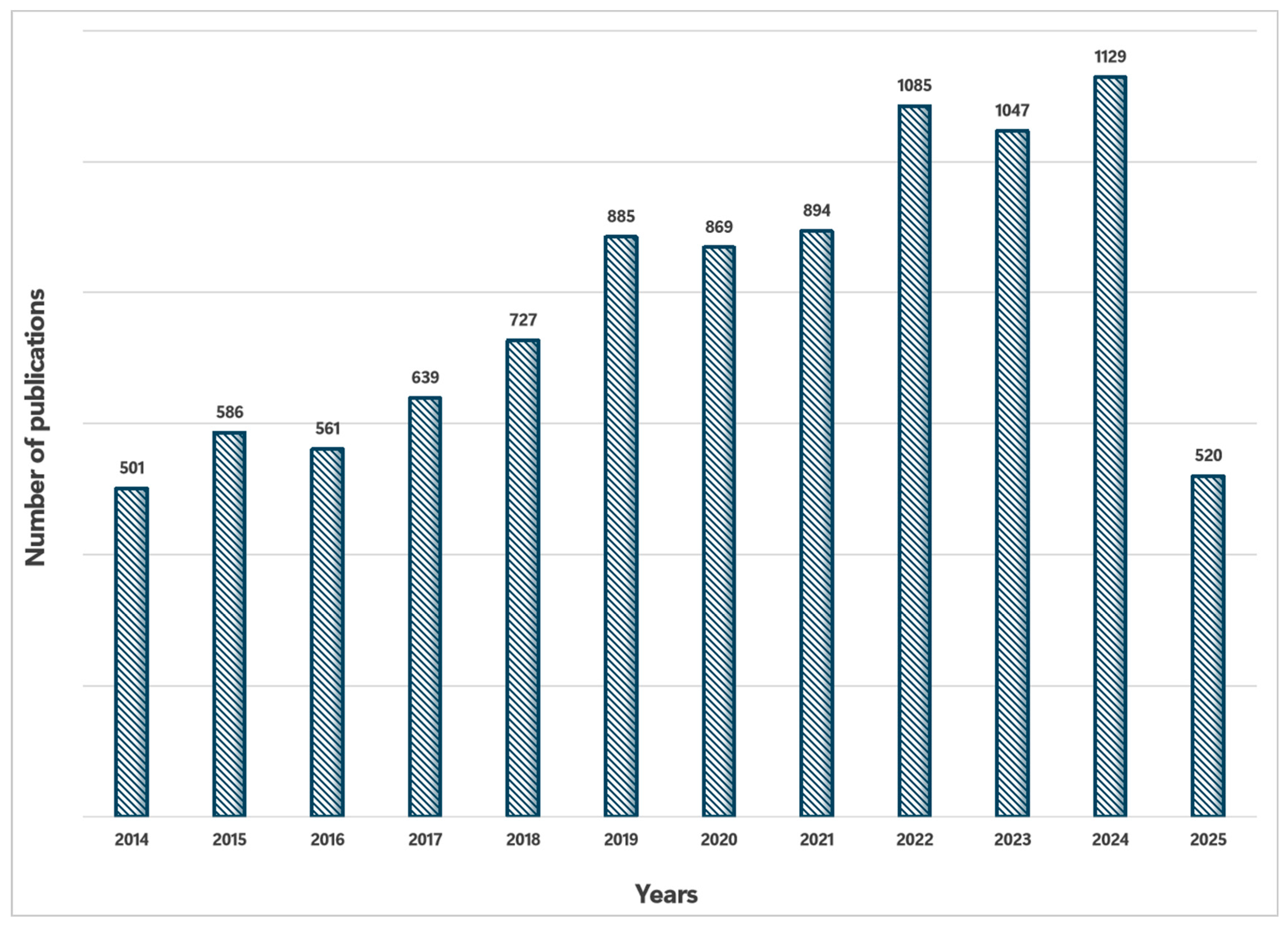
Figure 2.
Schematic procedure of the SELEX protocols.
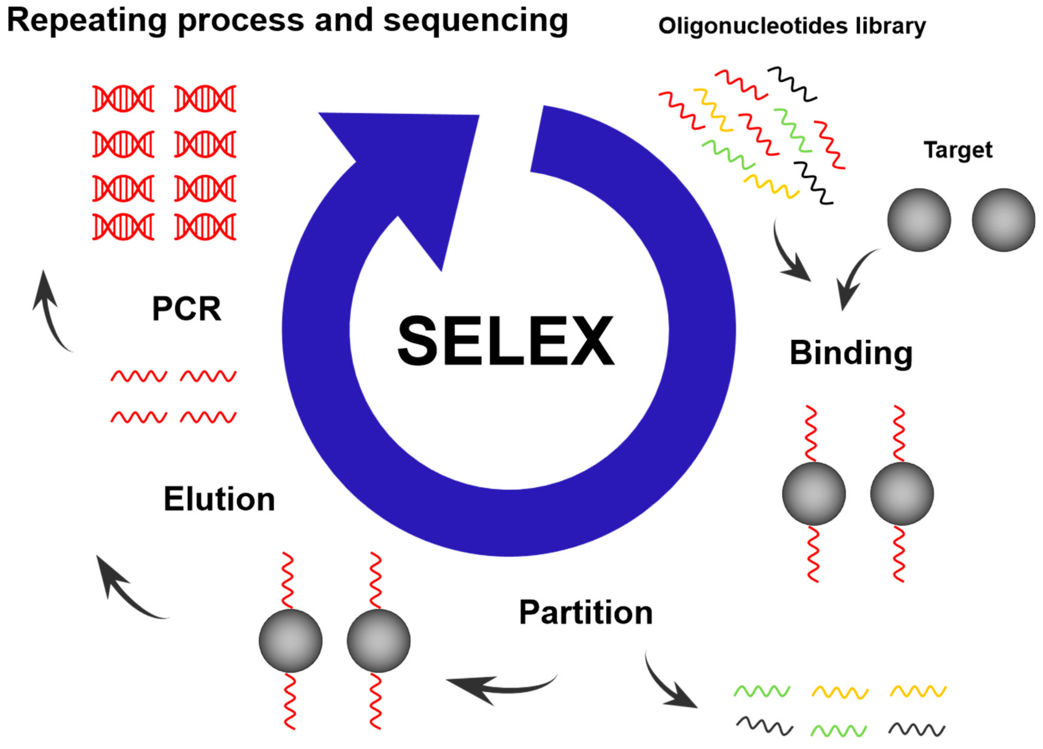
Figure 3.
Schematic diagram of the working principle of the aptasensor.
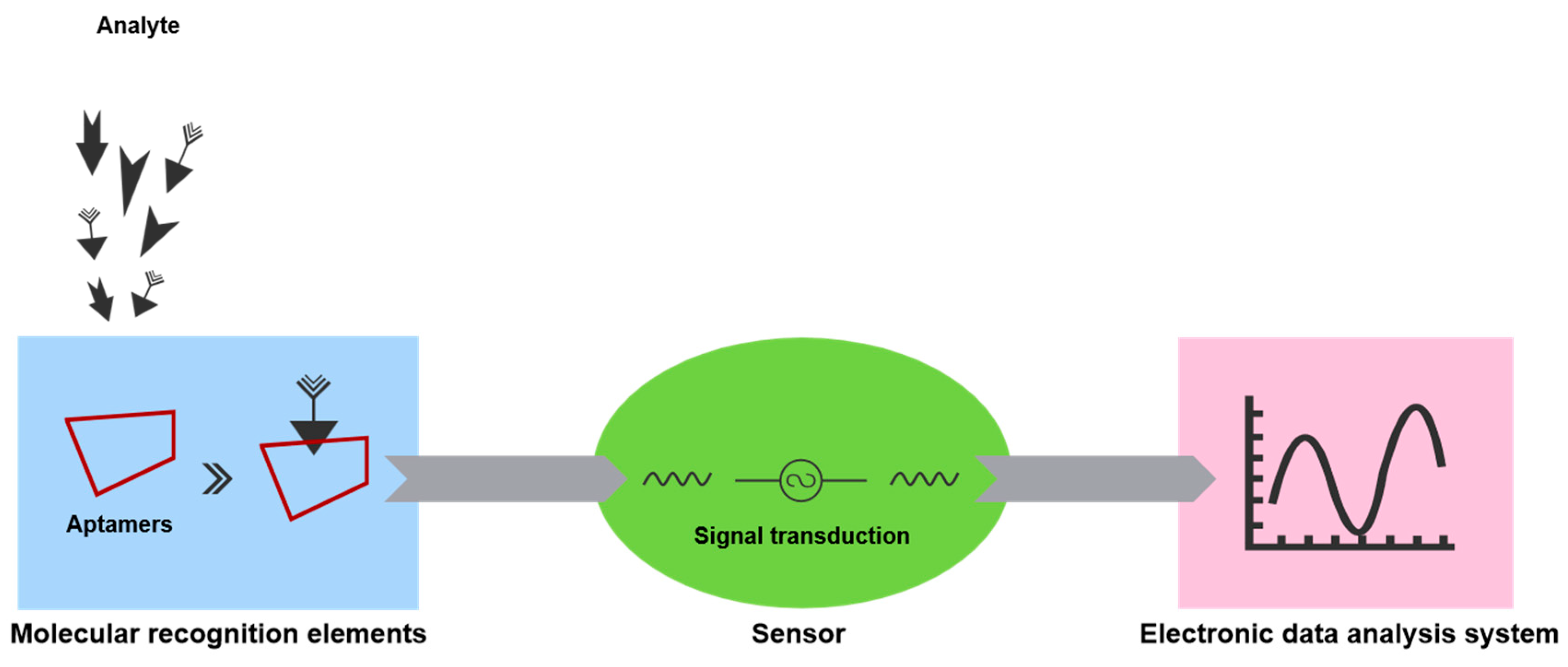
Figure 4.
Commonly used post-SELEX optimization methods.

Disclaimer/Publisher’s Note: The statements, opinions and data contained in all publications are solely those of the individual author(s) and contributor(s) and not of MDPI and/or the editor(s). MDPI and/or the editor(s) disclaim responsibility for any injury to people or property resulting from any ideas, methods, instructions or products referred to in the content. |
© 2025 by the authors. Licensee MDPI, Basel, Switzerland. This article is an open access article distributed under the terms and conditions of the Creative Commons Attribution (CC BY) license (http://creativecommons.org/licenses/by/4.0/).
Copyright: This open access article is published under a Creative Commons CC BY 4.0 license, which permit the free download, distribution, and reuse, provided that the author and preprint are cited in any reuse.


