
Submitted:
07 August 2025
Posted:
11 August 2025
You are already at the latest version
Abstract
Accurate modeling of cardiac excitation propagation requires detailed representation of both the myocardial structure and the ventricular conduction system. Traditional imaging methods like MRI and CT provide anatomical data but fail to capture the anisotropic properties essential for realistic simulations. This work presents an integrated modeling and simulation environment, HE-SU (Heart Excitation Simulation Utility), which employs pre-processed Diffusion Tensor Imaging (DTI) data to reconstruct myocardial fiber architecture and conduction pathways. The simulation is based on the Monodomain reaction-diffusion equation using the Aliev-Panfilov adaptation of the FitzHugh-Nagumo model. The software enables the adjustment of conductivity parameters and visualizes the propagation of excitation in 3D, supporting exportable reports for further analysis. This tool is valuable for researchers studying cardiac electrophysiology and the impact of anisotropic tissue properties on body surface potential mapping (BSPM).
Keywords:
cardiac excitation modeling
; diffusion tensor imaging (DTI)
; monodomain equation
; myocardial fibers
; conduction system
; Aliev-Panfilov model
; cardiac electrophysiology
1. Introduction
The myocardial and the conduction system are the two main structures that need to be taken into account while simulating the excitation propagation in the heart. MRIs and CT scans are two common medical imaging methods used to determine the anatomical structure of the heart. However, tissue anisotropy, which is essential for precise modeling, cannot be captured by these traditional imaging modalities. The myocardium has strong anisotropic properties that have a major impact on the heart’s electrical and mechanical operations. Although some models assume the heart as an isotropic material [1,2,3,4], most acknowledge that it is anisotropic because anisotropy has a significant impact on the Body Surface Potential Map (BSPM) simulation results.
Determining the myocardial fiber architecture is crucial in anisotropic heart models and can be done in a number of ways. The work of Streeter et al. [5] is an example of a popular technique that uses dissection-based data to assign fiber orientations to each voxel in the anatomical heart model [6,7]. On the other hand, models such as Nielson et al. [8] specify fiber directions using mathematical formulations [9]. In more recent advances, fiber architectures are mapped using Diffusion Tensor Imaging (DTI). While DTI data is used in some models to solve the Forward and Inverse Problems [10,11], DTI is used mainly for geometric reconstruction in other models [12,13].
Before modeling, there may be some pre-processing that involves removing the heart from the surrounding tissues, such as blood vessels and adipose tissues. In order to ensure that the majority of the heart tissue is included in the model, it may be necessary to improve the masking pictures [14] before using clustering methods or manual segmentation.
The models outlined by Tawara [15], Massing et al. [16], and Durrer et al. [17] are the most widely used to identify the ventricular conduction system. Ventricular conduction systems are modeled by either constructing a network based on anatomical structure and activation isochrones [18,19,20] or assigning the early activation sites based on Durrer et al. data [21].
2. Methods
The excitation propagation of the heart is modeled based on the Monodomain reaction-diffusion equation in its normalized form namely [22]:
where u and t represent the normalized transmembrance potential and the normalized time respectively, D is the normalized effective conductivity tensor of the heart material, f is the function that represent the reaction term due to ions exchange, and finally g represents the external applied input.
The electrical stimulation of the heart is modeled at the cellular level or for the entire tissue. FitzHugh and Nagumo’s (FHN) most popular model for heart excitation [23] depicts the excitation by two variables that stand for the cell membrane’s depolarization and repolarization. Aliev and Panfilove [24] updated the normalized form of the FHN model to incorporate the effect of Action Potential Duration (APD), however this model is simplified and provided in several forms [25,26].
such that
and
where for the heart tissues, k=8, a=0.15, =0.002, =0.2 and =0.3.
3. Software Description
3.1. Main Form Interface
Figure 1 depicts the HE-SU’s primary form. A pre-processed DTI heart model [27] with an extracted conduction network can be loaded by the user. The PMN file contains a heart that has been resized using the Moving Weighted Effective Sphere (MWES) Filter [28] from 256x256x110 to 55x55x55. The left branch bundle is represented by the red voxels, the right branch bundle by the blue voxels, and the myocardium by the gray voxels.
In addition to the speed factor of the propagation of excitation in the conduction network in the longitudinal direction relative to the myocardial, the user can set the normalized conductivity of the myocardium in both longitudinal and transverse directions (the defaults are 1 and 0.25, respectively). Based on information from the literature, the ventricles’ stated activation point, or His-Bundle, is set up for this heart.
After choosing the parameters, the simulator will be launched by selecting the RDE button, creating the excitation’s wave front (Figure 2).
3.2. Reports Manager
4. Conclusions
The simulation of excitation propagation in the heart must account for the anisotropic nature of myocardial tissue and the detailed structure of the ventricular conduction system. The HE-SU utility offers a comprehensive and customizable simulation environment, integrating DTI-derived fiber orientations and conduction pathways into a reaction-diffusion framework. By enabling user control over key parameters and providing exportable results, this tool facilitates both fundamental research and clinical investigations in cardiac electrophysiology. Future improvements may focus on integrating patient-specific DTI data and coupling electrical simulation with mechanical modeling for a more holistic representation of heart function. Download Link: https://github.com/Ihab-ELAFF/Heart-Excitation-Simulation-Utility-HESU.
- License
This project is licensed under the MIT License for academic and educational use only. If you use HE-SU software, please cite the following papers:
Acknowledgments
Dr. Patrick A. Helm and Dr. Raimond L. Winslow at the Centre for Cardiovascular Bioinformatics and Modelling of John Hopkins University and Dr. Elliot McVeigh at the National Institute of Health for provision of DT-MRI data. SharpDevelop– IDE used for development (GNU GPL). OpenTK– Open Toolkit Library for OpenGL graphics.
References
- A.E. Pollard and R.C. Barr "The Construction of an Anatomically Based Model of the Human Ventricular Conduction System" IEEE Trans. Biom. Eng. (1990); 37(12): 1173-1185.
- X. Zhang, I. Ramachandra, Z. Liu, B. Muneer, S.M. Pogwizd, and B. He "Noninvasive three-dimensional electrocardiographic imaging of ventricular activation sequence" Am J Physiol Heart Circ Physiol (2005); 289: H2724–H2732.
- T. Berger, G. Fischer, B. Pfeifer, R. Modre, F. Hanser,T. Trieb, F. X. Roithinger, M. Stuehlinger, O. Pachinger,B. Tilg, and F. Hintringer "Single-Beat Noninvasive Imaging of Cardiac Electrophysiology of Ventricular Pre-Excitation" J. Am. Coll. Cardiol. (2006);48:2045-2052. [CrossRef]
- Elaff, I. “Modeling of the Human Heart in 3D Using DTI Images”, World Journal of Advanced Engineering Technology and Sciences, 2025, 15(02), 2450-2459. [CrossRef]
- F.P. Mall "On The Muscular Architecture of the Ventricles of the Human Heart" The American Journal of Anatomy (1911); vol. 11(3):211-266.
- K. Simeliusa, J. Nenonena, M. Horácekb "Modeling Cardiac Ventricular Activation" Inter. J. of Bioelectromagnetism, (2001); 3(2):51 - 58.
- B.H. Smaill, I.J. LeGrice, D.A. Hooks, A.J. Pullan, B.J. Caldwell and P.J. Hunter "Cardiac structure and electrical activation: Models and measurement" Proc. of the Australian Physiological and Pharmacological Society, (2004); 34: 141-149.
- P.M.F. Nielsen, I.J. Le Grice, B.H. Smaill, and P.J. Hunter "Mathematical model of geometry and fibrous structure of the heart" The Am. Phys. Soc. (1991); 260: H1365-H1378.
- L.W. Wang, H.Y. Zhang, P.C. Shi "Simultaneous Recovery of Three-dimensional Myocardial Conductivity and Electrophysiological Dynamics: A Nonlinear System Approach" Computers in Cardiology, (2006);33:45-48.
- J.M. Peyrat, M. Sermesant, X. Pennec, H. Delingette, C. Xu, E. McVeigh and N. Ayache "Statistical Comparison of Cardiac Fibre Architectures", FIMH (2007); 4466: 413-423.
- L. Zhukov, A.H. Barr "Heart-Muscle Fibre Reconstruction from Diffusion Tensor MRI" VIS 2003. IEEE, (2003); Conf. Proc.: 597-602.
- R.L. Winslow, D.F. Scollan, J.L. Greenstein, C.K. Yung, W. Baumgartner, G. Bhanot, D.L. Gresh and B.E. Rogowitz "Mapping, modeling,and visual exploration of structure-function relationships in the heart" IBM Sys J.,(2001); 40(2):342-359.
- M. Sermesant , H. Delingette, and N. Ayache "An Electromechanical Model of the Heart for Image Analysis and Simulation" IEEE Trans. Med. Imag. (2006); 25(5): 612-625.
- Elaff, I. “Medical Image Enhancement Based on Volumetric Tissue Segmentation Fusion (Uni-Stable 3D Method)”, Journal of Science, Technology and Engineering Research, vol. 4, no. 2, pp. 78–89, 2023. [CrossRef]
- [S.Tawara "The Conduction System of the Mammalian Heart" English Ed., World Scientific Pub. co., (1998), ISBN: 981023502X.
- [G.K. Massing, and T.N. James "Anatomical Configuration of the His Bundle and Bundle Branches in the Human Heart" Circ. (1976); 53(4):609-621.
- D. Durrer, R.TH. Van Dam, G.E. Freud, M.J. Janse, F.L. Meijler and R.C. Arzbaecher "Total Excitation of the Isolated Human Heart" Circulation, (1970); 41(6):899-912.
- El-Aff, I.A.I. "Extraction of human heart conduction network from diffusion tensor MRI" The 7th IASTED International Conference on Biomedical Engineering, 217-22.
- Elaff, I. “Modeling the Human Heart Conduction Network in 3D using DTI Images”, World Journal of Advanced Engineering Technology and Sciences, 2025, 15(02), 2565–2575. [CrossRef]
- O. Berenfeld and J. Jalife "Purkinje-Muscle Reentry as a Mechanism of Polymorphic Ventricular, Arrhythmias in a 3-Dimensional Model of the Ventricles" Circ. Res., (1998);82;1063-1077.
- D.S. Farina, O. Skipa, C. Kaltwasser, O. Dossel and W.R. Bauer "Personalized Model of Cardiac Electrophysiology of a Patient" IJBEM (2005);7(1): 303-306.
- Elaff, I. “Modeling of The Excitation Propagation of The Human Heart”, World Journal of Biology Pharmacy and Health Sciences, 2025, 22(02): 512–519. [CrossRef]
- R. Fitzhugh "Impulses and Physiological States in Theoretical Models of Nerve Membrane" Biophysical J., 1961; 1: 445-466.
- R.R. Aliev, A.V. Panfilov "A simple two-variable model of cardiac excitation." Chaos, Solitons and Fractals, 1996; 7(3): 293-301.
- A. V. Panfilov, P. Hogeweg "Spiral breakup in a modified FitzHugh—Nagumo model" Phys. Let. A, 1993; 176: 295—299.
- A.T. Winfree "Varieties of spiral wave behavior: An experimentalist’s approach to the theory of excitable media" CHAOS ,(1991) ;1(3): 303-334.
- The Center for Cardiovascular Bioinformatics and Modeling, John Hopkins University Site "http://www.ccbm.jhu.edu/research/DTMRIDS.php", July 2011.
- Elaff, I. "Modeling of 3D Fibers Structure of Human Heart Using DTI Images and Moving Weighted Effective Sphere (MWES) Filter" ,PrePrints,2025. [CrossRef]
Figure 1.
Main Form of HE-SU application showing the original image view.
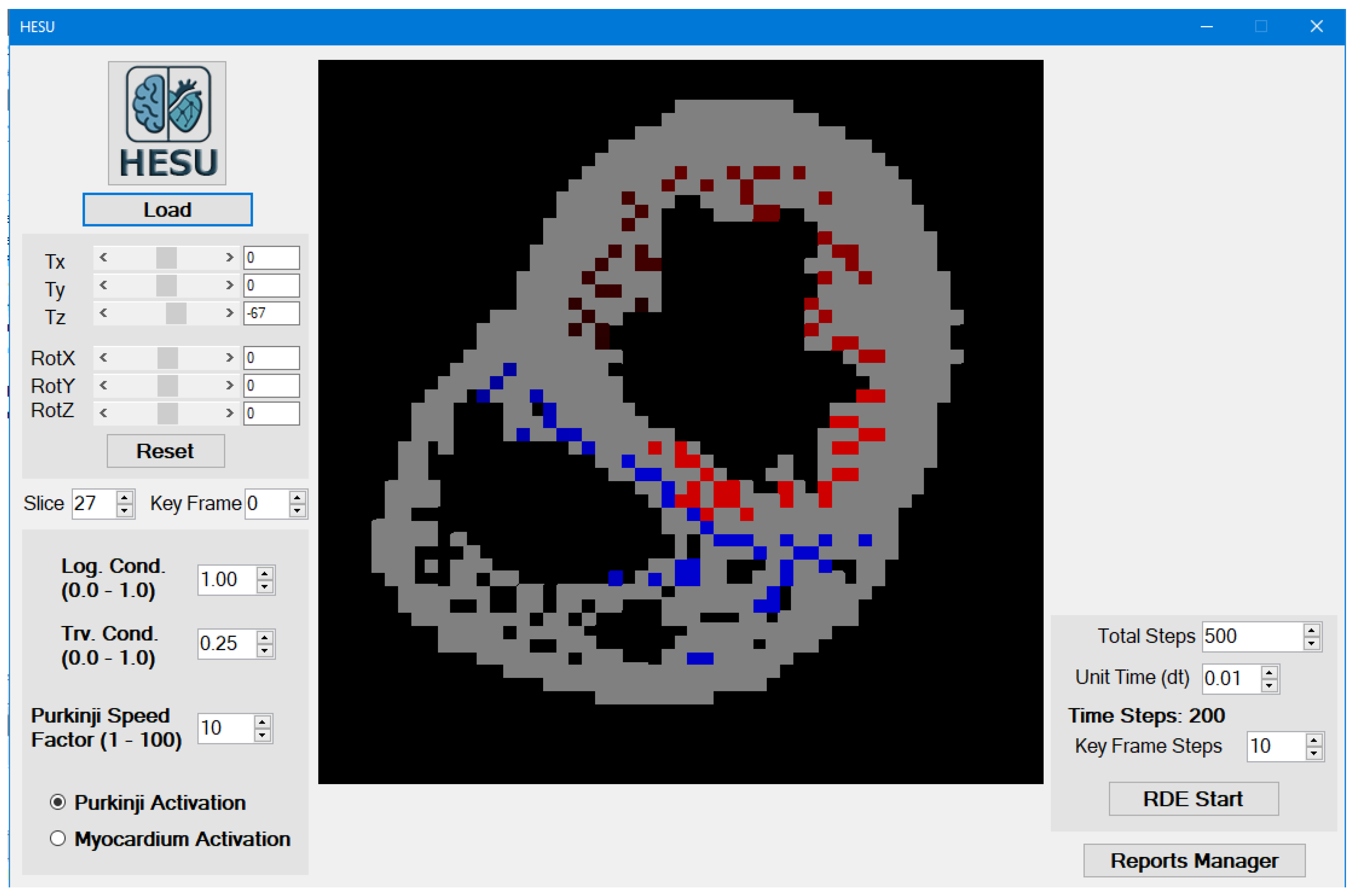
Figure 2.
Excitation Propagation.
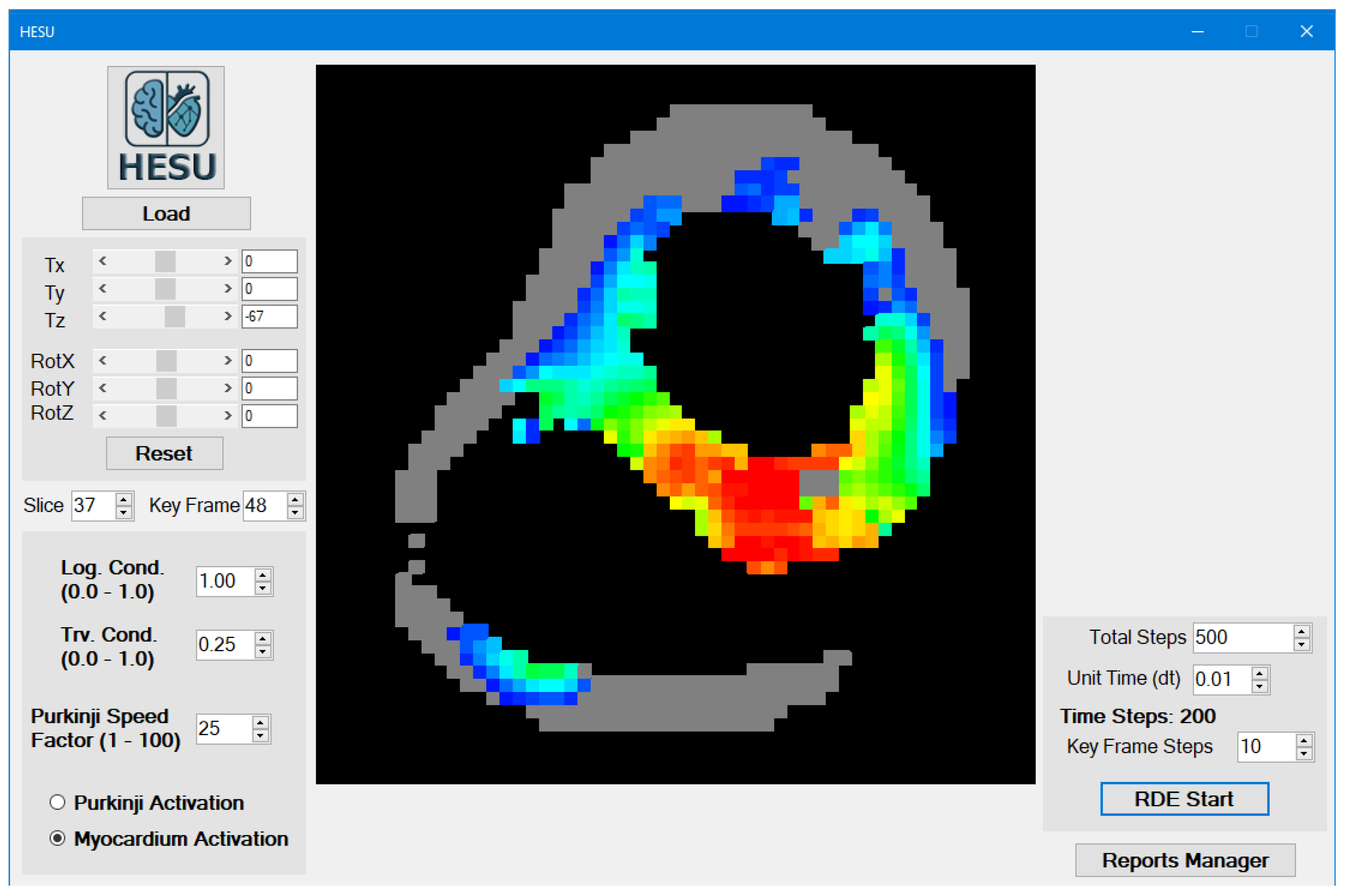
Figure 3.
The Reports Manager.
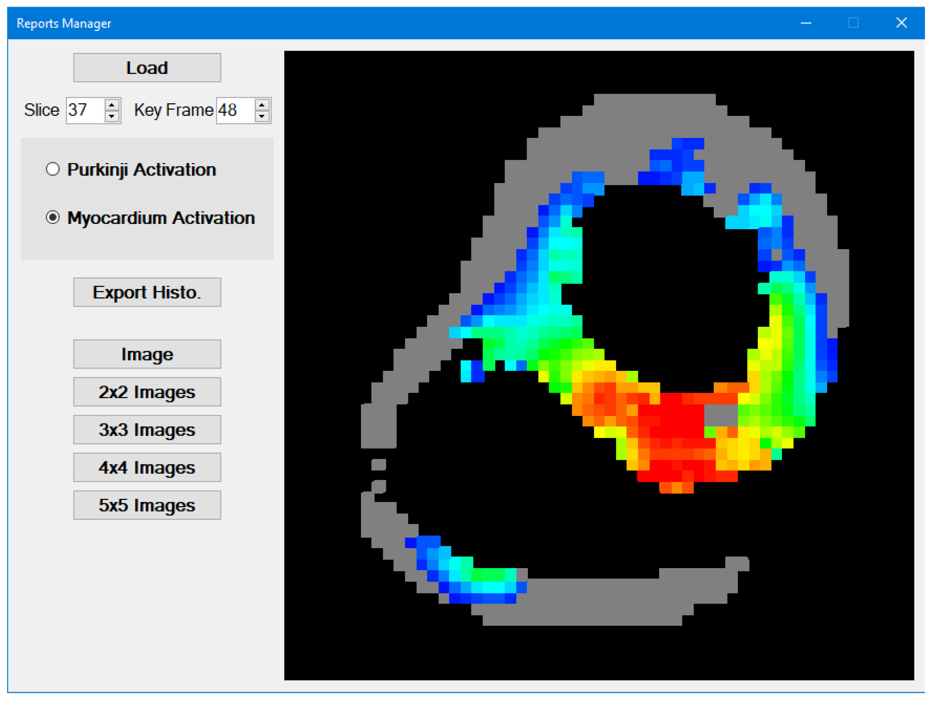
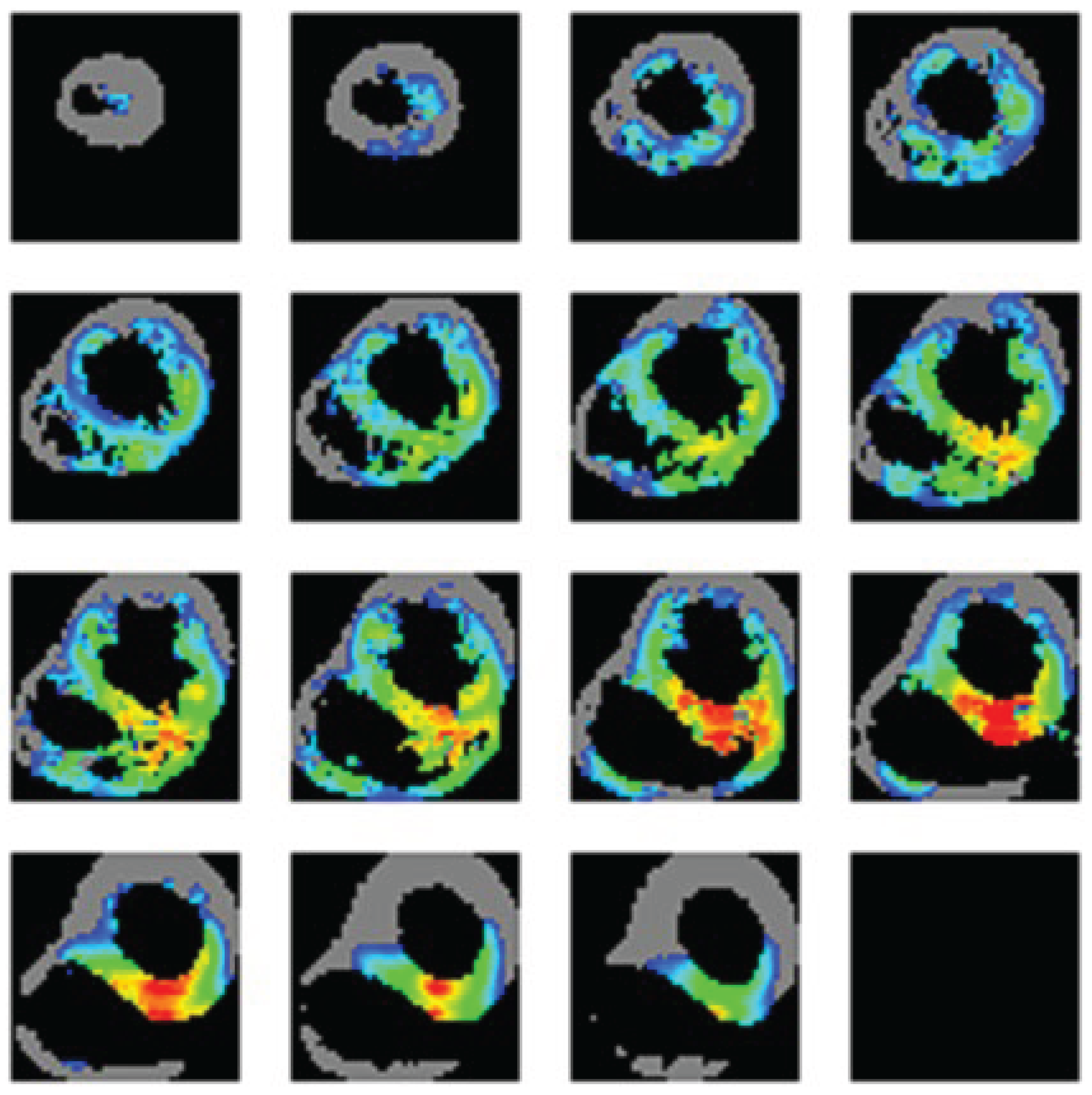
Figure 4.
Sample output of the wave front of the heart excitation.
Disclaimer/Publisher’s Note: The statements, opinions and data contained in all publications are solely those of the individual author(s) and contributor(s) and not of MDPI and/or the editor(s). MDPI and/or the editor(s) disclaim responsibility for any injury to people or property resulting from any ideas, methods, instructions or products referred to in the content. |
© 2025 by the authors. Licensee MDPI, Basel, Switzerland. This article is an open access article distributed under the terms and conditions of the Creative Commons Attribution (CC BY) license (http://creativecommons.org/licenses/by/4.0/).
Copyright: This open access article is published under a Creative Commons CC BY 4.0 license, which permit the free download, distribution, and reuse, provided that the author and preprint are cited in any reuse.


