
Submitted:
09 June 2025
Posted:
09 June 2025
You are already at the latest version
Abstract
Accurately modeling cardiac electrical activity within the human torso is complex due to the need to simulate various physiological and anatomical factors, including organ anisotropy and conduction network architecture. This study compares the effect of two different ventricular conduction network models on the Body Surface Potential Map (BSPM) in a realistically shaped human torso derived from CT scans. One network is manually constructed based on trabecular muscle structure, while the other is derived using diffusion volume data. Activation isochrones and BSPMs are simulated and validated against ECGSIM and clinical ECG data. Results indicate that even minor differences in conduction network modeling can lead to significant changes in BSPM accuracy, highlighting the sensitivity and importance of precise network representation in cardiac simulations.
Keywords:
cardiac electrophysiology
; body surface potential map (BSPM)
; excitation isochrones
; ECG
1. Introduction
The reliability of its computations on all factors makes it difficult to develop a realistic simulator for cardiac activation. It is possible to model the body as either an inhomogeneous or homogeneous volume conductor. It is assumed that all organs, including the blood volume inside ventricles, have the same physical characteristics (permeability, conductivity) for a homogenous volume conductor. An inhomogeneous volume-conductor, on the other hand, has characteristics specific to each organ. Gulrajani and Mailloux [1] presented the idea that the generated surface potential is impacted by the body's inhomogeneity.
In actuality, every organ is made of anisotropic materials. While some tissues, like the liver and lungs, have nearly isotropic qualities, others, like the skeletal muscles, have considerable anisotropic qualities [2]. An enormous quantity of information on the makeup of each organ is needed to account for organ anisotropy. It is okay to treat the organs as an isotropic material, nevertheless, as this would make modeling easier [2]. The realistic torso shape is the most commonly used model, however some models define the volume conductor (the body) in terms of an estimated shape [3]. Either a homogeneous volume conductor, as reported in the literature [4,5,6], or the popular inhomogeneous volume conductor, as described in [7,8,9,10], are the realistic torso form models.
Ventricular conduction networks are modeled by either constructing a network based on anatomical structure and activation isochrones [14,15,16] or allocating the early activation locations based on Durrer et al. observations [11,12,13]. It was also observed that choosing the right excitation sites is a very sensitive task, since a tiny variance in conduction sites will result in a big variation in results. Creating such a network is always accomplished by trial and error. Since they employ certain experimental pacing sites (primary sources) in their models, other model groups do not model any conduction systems [17,18,19,20]. The impact of two conduction networks on the Body Surface Optional Map (BSPM) is compared in this study.
2. Methods
2.1. The Human Torso and the Human Heart Modeling
2.2. The Conduction Network Modeling
Two conduction networks are analyzed (Figure 2) in order to determine how the conduction network structure affects the Body Surface Potential Map (BSPM) [23, 24]. The locations of the trabecular muscles are used to manually construct the first model (Model 1), and the Diffusion Volume quantity is used to extract the second model (Model 2). It is evident that the second model is more accurate than the first.
2.3. Activation Isochrones Modeling
2.4. The Body Surface Potential Map (BSPM) Calculation
3. Results
3.1. Body Surface Potential Map Validation
The BSPM generated by the Forward Model of both Conduction Systems (Model 1 and Model 2) is comparable to a reference model derived from the General Public Licensed (GUN) simulator ECGSIM [29] (Figure 7); however, the Model 2 conduction network's results are superior to Model 1 since they are significantly closer to the reference model. This suggests that even little modifications to the conduction network result in notable variations in the BSPM that is produced.
3.2. Validation of the ECG
4. Conclusion
This study demonstrates that the structure and accuracy of the ventricular conduction network play a critical role in the fidelity of body surface potential maps. Comparing two models, it was shown that the network derived from diffusion volume data (Model 2) produces BSPMs that more closely match reference data from ECGSIM, underscoring the importance of anatomically and functionally accurate conduction networks in cardiac modeling. These findings suggest that realistic conduction system modeling is essential for developing reliable forward simulation models in cardiac electrophysiology.
References
- Gulrajani, R.M.; Mailloux, G.E. A simulation study of the effects of torso inhomogeneities on electrocardiographic potentials, using realistic heart and torso models. Circ. Res. 1983, 52, 45–56. [Google Scholar] [CrossRef] [PubMed]
- Malmivuo, J.; Plonsey, R. Bioelectromagnetism: Principles and Applications of Bioelectric and Biomagnetic Fields, 1st Ed. ed; Oxford Univ. Press, 1995; ISBN 0195058232. [Google Scholar]
- Soundararajan, V.; Besio, W.G. Simulated Comparison of Disc and Concentric Electrode Maps During Atrial Arrhythmias. IJBEM 2005, 7, 217–220. [Google Scholar]
- Simeliusa, K.; Nenonena, J.; Horácekb, M. Modeling Cardiac Ventricular Activation. Inter. J. of Bioelectromagnetism 2001, 3, 51–58. [Google Scholar]
- Jazbinsek, V.; Hren, R.; Trontelj, Z. High resolution ECG and MCG mapping: simulation study of single and dual accessory pathways and influence of lead displacement and limited lead selection on localisation results. Bulletin of the Polish Academy of Sciences, Technical Sciences, 2005, 53, 195–205. [Google Scholar]
- Wang, L.W.; Zhang, H.Y.; Shi, P.C. Simultaneous Recovery of Three-dimensional Myocardial Conductivity and Electrophysiological Dynamics: A Nonlinear System Approach. Computers in Cardiology 2006, 33, 45–48. [Google Scholar]
- Farina, D.S.; Skipa, O.; Kaltwasser, C.; Dossel, O.; Bauer, W.R. Personalized Model of Cardiac Electrophysiology of a Patient. IJBEM 2005, 7, 303–306. [Google Scholar]
- Seger, M. Modeling the Electrical Function of the Human Heart. Ph.D. Thesis, Institute of Biomedical Engineering, University for Health Sciences, Medical Informatics and Technology.
- Liu, Z.; Liu, C.; He, B. Noninvasive reconstruction of three-dimensional ventricular activation sequence from the inverse solution of distributed equivalent current density. IEEE Trans. Med Imaging 2006, 25, 1307–1318. [Google Scholar] [CrossRef]
- He, B.; Liu, C. Three-Dimensional Cardiac Electrical Imaging From Intracavity Recordings. IEEE Trans. Biomed. Eng. 2007, 54, 1454–1460. [Google Scholar] [CrossRef]
- Pollard, A.E.; Barr, R.C. The Construction of an Anatomically Based Model of the Human Ventricular Conduction System. IEEE Trans. Biom. Eng. 1990, 37, 1173–1185. [Google Scholar] [CrossRef]
- Scollan, D.F. Reconstructing The Heart: Development and Application of Biophysically Based Electrical Models of Propagation in Ventricular Myocardium Reconstructed from DTMRI. Ph.D. Thesis, Johns Hopkins University, 2002. [Google Scholar]
- Bernus, O.G. Development of a realistic computer model of the human ventricles for the study of reentrant arrhythmias. Ph.D. Thesis, University of Gent, Belgium, 2003. [Google Scholar]
- Simeliusa, K.; Nenonena, J.; Horácekb, M. Modeling Cardiac Ventricular Activation. Inter. J. of Bioelectromagnetism 2001, 3, 51–58. [Google Scholar]
- Berenfeld, O.; Jalife, J. Purkinje-Muscle Reentry as a Mechanism of Polymorphic Ventricular, Arrhythmias in a 3-Dimensional Model of the Ventricles. Circ. Res. 1998, 82, 1063–1077. [Google Scholar] [CrossRef] [PubMed]
- Farina, D.S.; Skipa, O.; Kaltwasser, C.; Dossel, O.; Bauer, W.R. Personalized Model of Cardiac Electrophysiology of a Patient. IJBEM 2005, 7, 303–306. [Google Scholar]
- Winslow, R.L.; Scollan, D.F.; Greenstein, J.L.; Yung, C.K.; Baumgartner, W.; Bhanot, G.; Gresh, D.L.; Rogowitz, B.E. Mapping, modeling,and visual exploration of structure-function relationships in the heart. IBM Sys J. 2001, 40, 342–359. [Google Scholar] [CrossRef]
- Smaill, B.H.; LeGrice, I.J.; Hooks, D.A.; Pullan, A.J.; Caldwell, B.J.; Hunter, P.J. Cardiac structure and electrical activation: Models and measurement. Proc. of the Australian Physiological and Pharmacological Society 2004, 34, 141–149. [Google Scholar] [CrossRef]
- Knisley, S.B.; Trayanova, N.; Aguel, F. Roles of Electric Field and Fibre Structure in Cardiac Electric Stimulation. Biophysical Journal 1999, 77, 1404–1417. [Google Scholar] [CrossRef] [PubMed]
- Berger, T.; Fischer, G.; Pfeifer, B.; Modre, R.; Hanser, F.; Trieb, T.; Roithinger, F.X.; Stuehlinger, M.; Pachinger, O.; Tilg, B.; et al. Single-Beat Noninvasive Imaging of Cardiac Electrophysiology of Ventricular Pre-Excitation. Circ. 2006, 48, 2045–2052. [Google Scholar] [CrossRef]
- ELAFF. Modeling of 3D Inhomogeneous Human Body from Medical Images. World Journal of Advanced Engineering Technology and Sciences 2025, 15, 2010–2017. [Google Scholar] [CrossRef]
- Elaff, I. Modeling of the human heart in 3D using DTI images. World J. Adv. Eng. Technol. Sci. 2025, 15, 2450–2459. [Google Scholar] [CrossRef]
- IAI El-Aff. Extraction of human heart conduction network from diffusion tensor MRI. The 7th IASTED International Conference on Biomedical Engineering, 217–222.
- Elaff, I. Modeling the human heart conduction network in 3D using DTI Images. World J. Adv. Eng. Technol. Sci. 2025, 15, 2565–2575. [Google Scholar] [CrossRef]
- ELAFF. Modeling of realistic heart electrical excitation based on DTI scans and modified reaction diffusion equation. Turkish Journal of Electrical Engineering and Computer Sciences 2018, 26, 2. [Google Scholar] [CrossRef]
- Elaff, I. Modeling of the excitation propagation of the human heart. World J. Biol. Pharm. Heal. Sci. 2025, 22, 512–519. [Google Scholar] [CrossRef]
- Elaff, I. Effect of the material properties on modeling of the excitation propagation of the human heart. World J. Biol. Pharm. Heal. Sci. 2025, 22, 088–094. [Google Scholar] [CrossRef]
- ELAFF. Modeling of the Body Surface Potential Map for Anisotropic Human Heart Activation. Research Square, 2025.
- ECGSIM [online] https://www.ecgsim.org/.
- Hampton, J.R. The ECG made easy, 5th Ed.; Pearson Prof., 1997.
Figure 1.
Human Torso as surfaces model from CT scans.
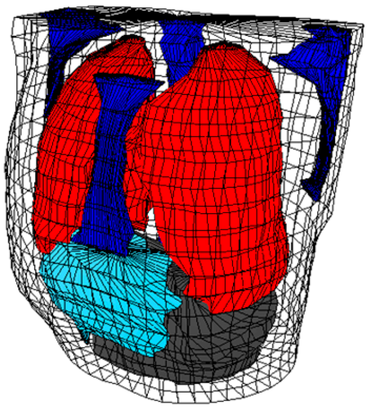
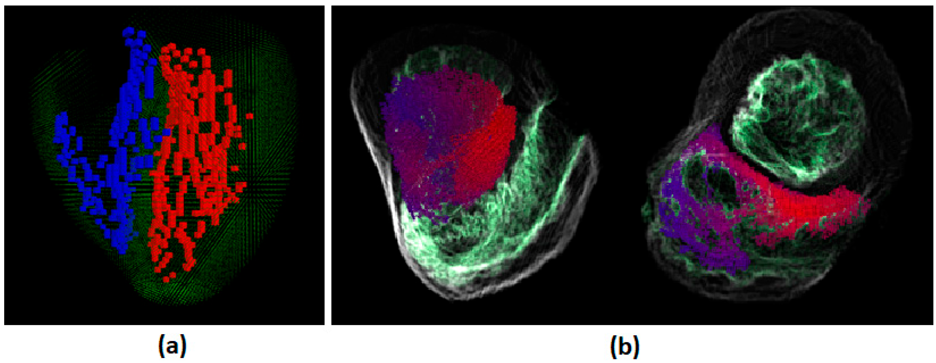
Figure 3.
Excitation Isochrones of Normal Activation using Model 1 of conduction network [27].
Figure 3.
Excitation Isochrones of Normal Activation using Model 1 of conduction network [27].

Figure 4.
Excitation Isochrones of Normal Activation using Model 2 of conduction network [27].
Figure 4.
Excitation Isochrones of Normal Activation using Model 2 of conduction network [27].
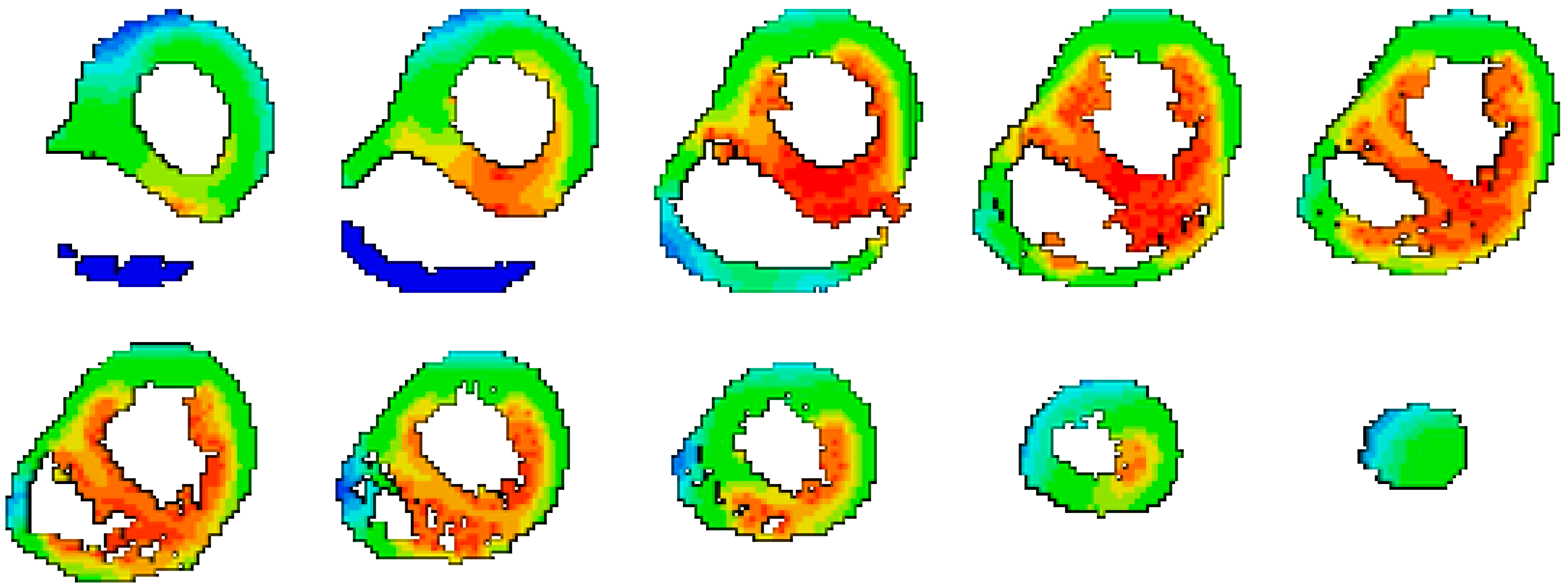
Figure 5.
BSPM of Normal Activation using Model 1 of conduction network.
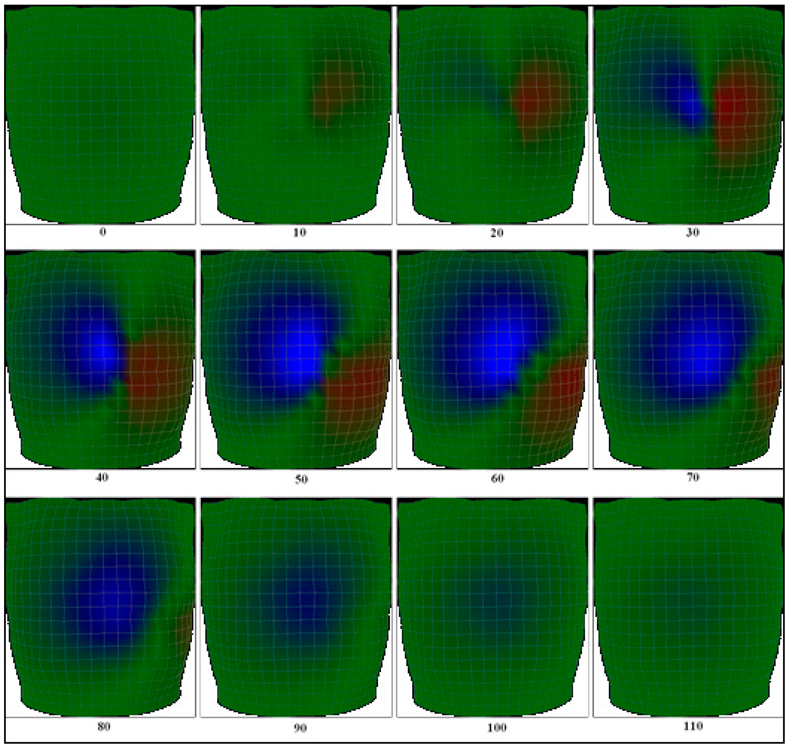
Figure 6.
BSPM of Normal Activation using Model 2 of conduction network.
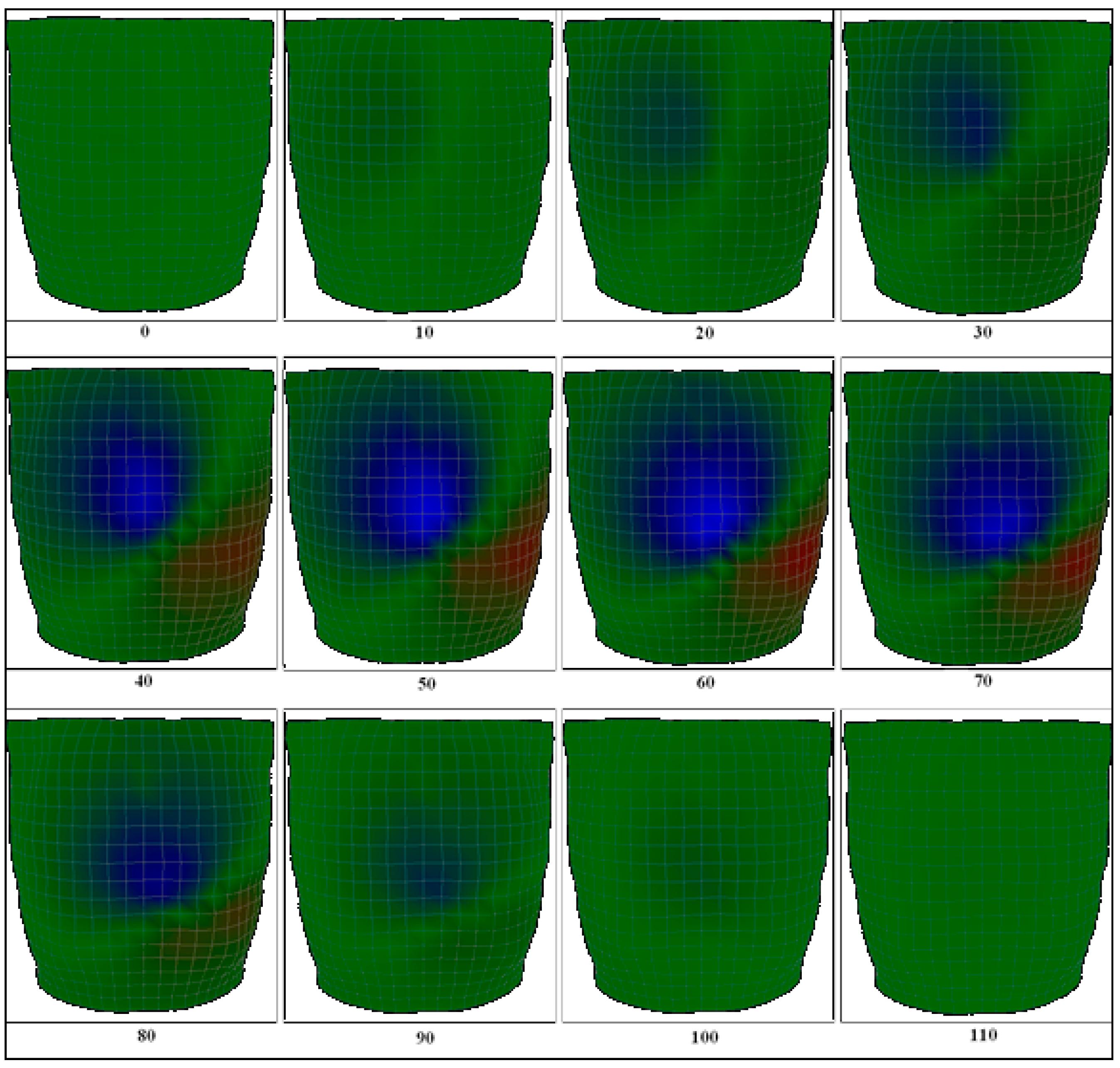
Figure 7.
Reference BSPM [29].
Figure 7.
Reference BSPM [29].
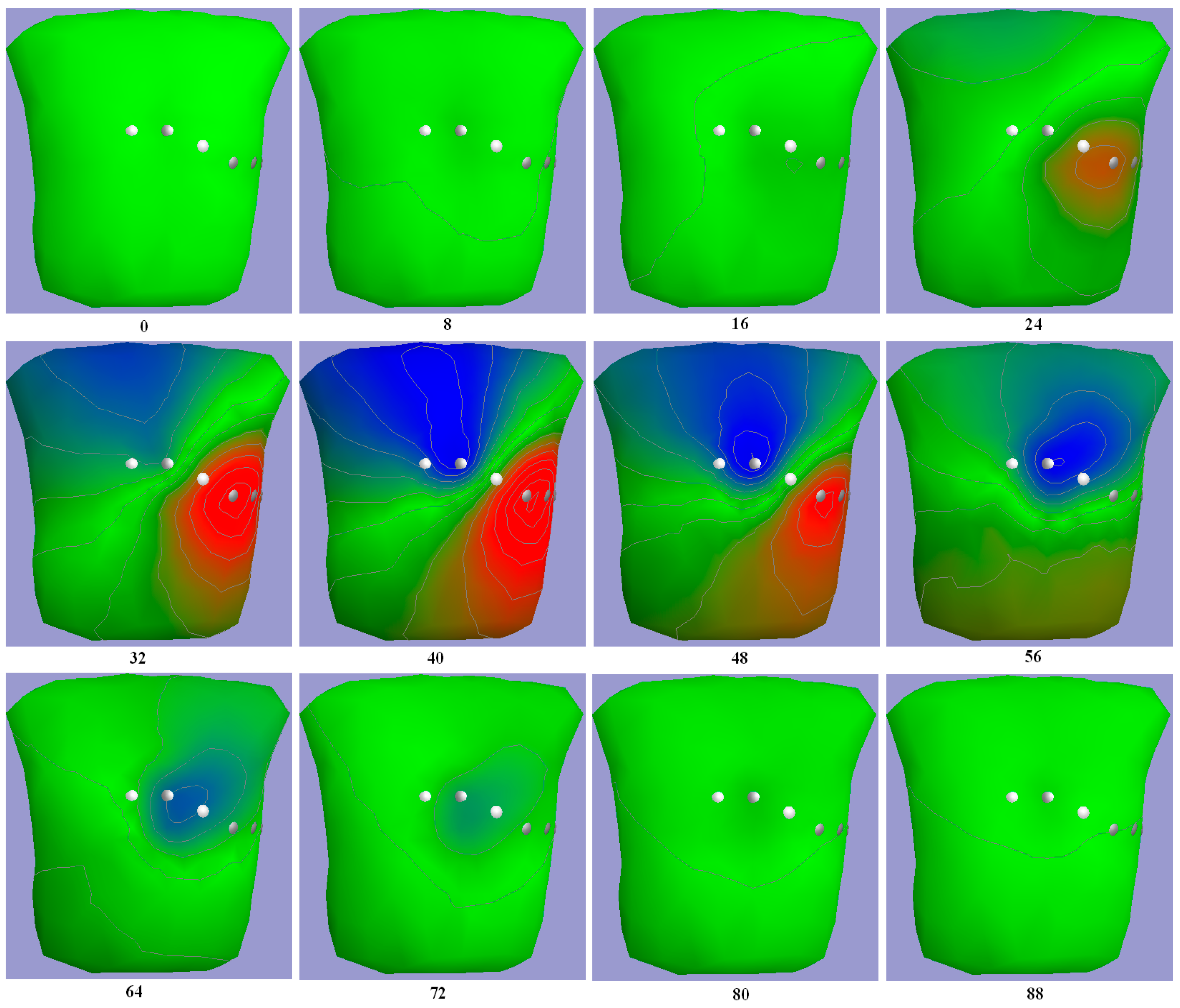
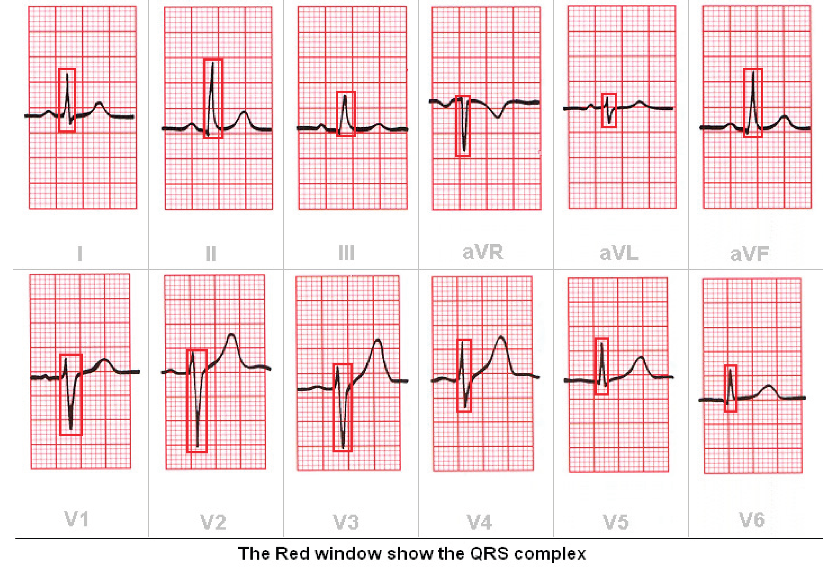
Figure 8.
(a) QRS complex of Model 1 (b) QRS complex of Model 2.
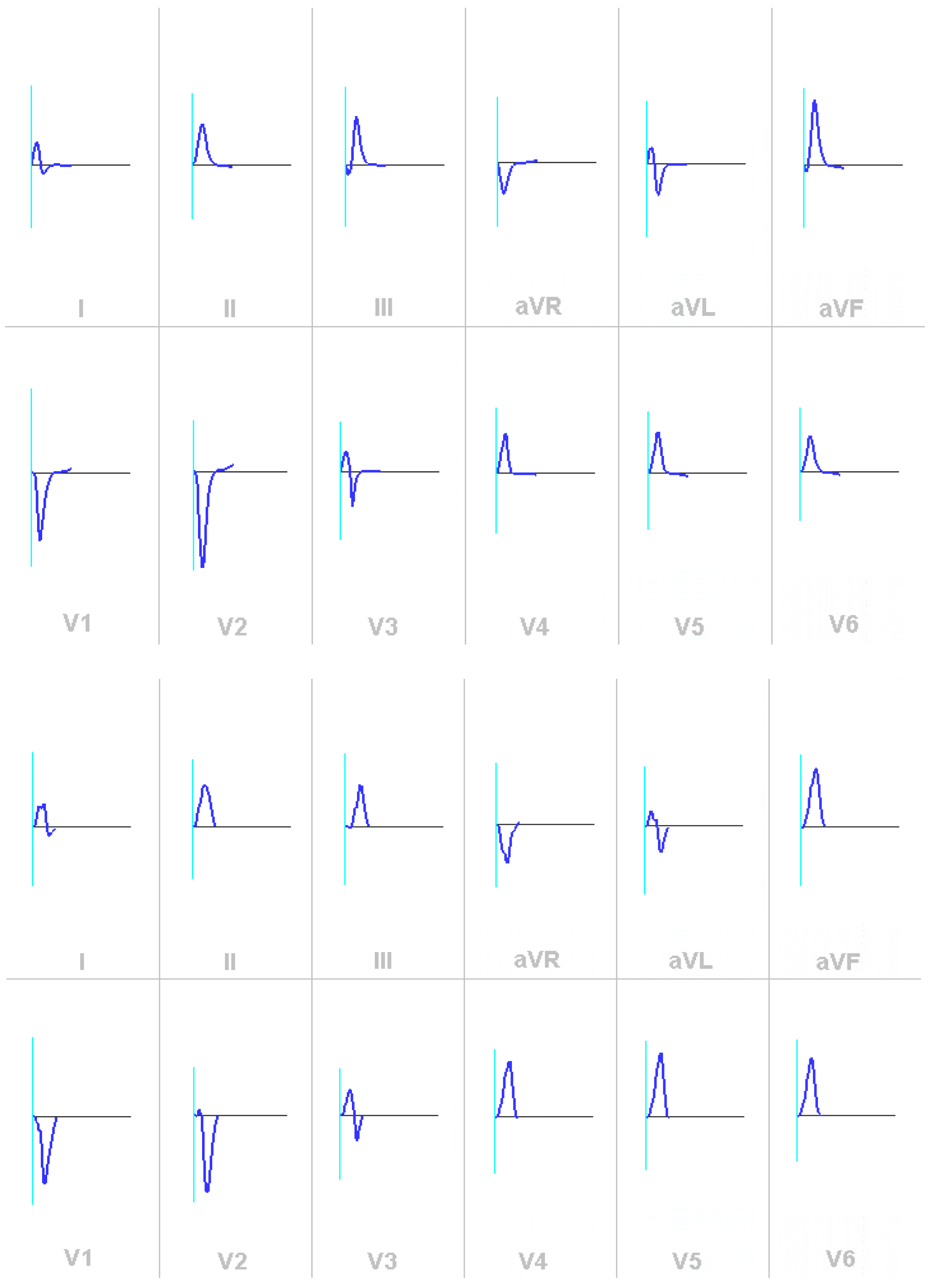
Figure 9.
Reference ECG of normal heart activation [30].
Figure 9.
Reference ECG of normal heart activation [30].
Disclaimer/Publisher’s Note: The statements, opinions and data contained in all publications are solely those of the individual author(s) and contributor(s) and not of MDPI and/or the editor(s). MDPI and/or the editor(s) disclaim responsibility for any injury to people or property resulting from any ideas, methods, instructions or products referred to in the content. |
© 2025 by the authors. Licensee MDPI, Basel, Switzerland. This article is an open access article distributed under the terms and conditions of the Creative Commons Attribution (CC BY) license (http://creativecommons.org/licenses/by/4.0/).
Copyright: This open access article is published under a Creative Commons CC BY 4.0 license, which permit the free download, distribution, and reuse, provided that the author and preprint are cited in any reuse.


