Submitted:
06 August 2025
Posted:
07 August 2025
Read the latest preprint version here
Abstract
Background: The increasing consumption of the western diet, rich in highly refined fats and carbohydrates, is becoming the primary culprit of dietary hepatic lipid accumulation in adults and youngsters This storage, if uncontrolled, induces hepatocyte cell death and sustains inflammation and fibrosis, which are responsible for the increasing prevalence of non-alcoholic fatty liver disease (NAFLD), the gate to cirrhosis and cancer. Due to a lack of targeted therapies, dietary regimens rich in functional foods by providing complexes of antioxidants may be therapeutically advantageous. Objective: Lycopene supplementation and tomato-rich diets are under scrutiny since they have shown promising clinical outcomes with no toxicities, large availability at contained costs. In the present report we have investigated whether a novel whole tomato-based food supplement (WTFS), endowed with potent antioxidant activity and capable of interfering with multiple metabolic pathways, can modulate mechanisms fostering the progression of NAFLD. Methods: Lipidomic assays and proteomic analysis were performed in the HepG2 human cell line, treated with WTSF. Results: WTFS induces a marked reduction in triglyceride and cholesterol ester content, a decrease in the relative levels of diacylglycerols, lysophosphatidylcholine, lysophosphatidylethanolamines, phosphatidylethanolamines, and decreased expression of transforming growth factor-α, tumor necrosis factor-like weak inducer of apoptosis (TWEAK), and Fms-related tyrosine kinase 3 ligand (FLT3LG), modulating signaling relevant to NAFLD progression. Conclusions: WTFS may represent a potential candidate for clinical trials in supplementing the hard-to-follow Mediterranean diet, the presently first-line preventive and therapeutic dietary option for NAFLD.
Keywords:
antioxidants
; functional foods
; lipids
; NAFLD
; tomato
1. Introduction
Epidemiological and experimental evidence increasingly supports the protective role of dietary antioxidants on fat liver accumulation, generating oxidative stress [1,2] lipid peroxidation, mitochondrial dysfunction, and inflammation [3,4,5,6].
Standing the dietary origins of non-alcoholic fatty liver disease (NAFLD) [7] and suggested by the results of in vitro/vivo models, functional nutrients and foods [8] capable of modulating fat metabolism and associated inflammation, are currently undergoing scrutiny since they may represent variably efficient, reversible inhibitors, with low toxicity, contained costs, thus available to large populations fractions. In this regard, increasing attention has been focused on the polyene lycopene, potent antioxidant of the carotenoid family, which through the induction of other endogenous antioxidants [9], possesses anti-inflammatory [10] and lipid-lowering properties [11], which are shared by its metabolites [12]. In this context, it should be underlined that the lycopene hypolipidemic activity relies on the dual mechanism of HMG-CoA reductase inhibition and downregulation of PCSK-9 mRNA synthesis [13].
Because of its hepatic accumulation [14], where active metabolites, i.e. apo-lycopenals and apo-lycopenones are generated [15], the mitigating effects of lycopene and lycopene-containing foods are attractive candidates to exploit dietary tools of wide acceptance to control the development and progression of NAFLD, a disease of increasing incidence and prevalence [16,17].
To acquire the broad spectrum of its healthy biological properties, the trans isomeric form of the naturally occurring lycopene needs to be modified, by the digestive processing [18], in the cis configuration the only biologically active isomer [19] and by the concomitant uptake of other tomato micronutrients, such other carotenoid [20] and nutrients generated by the cooking of the berry [21]. Indeed, the consumption of whole fruits has been shown to result in healthier effects than single lycopene supplementation, as demonstrated in laboratory [16] and clinical studies [22,23]. Therefore, the unique combination of antioxidant and anti-inflammatory nutrients [24] with converging biological activities of the berry [25] advocates the choice of whole tomato dietary consumption as a functional food for equitable and sustainable healthy diets [26]. Along this line of investigation, a novel whole tomato (98%) food supplement (WTFS) [27], enriched with olive waste water (2%) food in the form of an additives/ excipients-free powder [28], has been recently described which is characterized by a multinutrient composition [29], capable of interfering with metabolic pathways sustaining both inflammation and neoplastic transformation [30].
This improved functional food may be a candidate for further testing to mitigate the incidence of lipid accumulation-related liver diseases. To test this possibility, we have performed a lipidomic and proteomic analysis on HepG2 human liver cells exposed to the WTFS.
2. Materials and Methods
2.1. Cell Cultures
The HepG2 human hepatoblastoma cells, a reference target to study liver function and hepatotoxicity [31], obtained from the American Type Culture Collection (ATCC, Manassas, VA, USA), were plated at 1×10⁶ in T-75 cell culture flasks in minimum Essential Media supplemented with 10% heat-inactivated fetal calf serum containing 2 mmol/L L-glutamine, 100 IU/mL penicillin, 100 mg/mL streptomycin, 2.2 mg/L sodium bicarbonate, and 1 mmol/L sodium pyruvate, under a humidified atmosphere of 95% air/5% CO2 at 37° C, as previously descrided [32]. WTSF preparation in dimethyl sulfoxide (DMSO) and cells treatment followed the conditions described by Rubini et al. [33], and HepG2 cells cultured with only DMSO were used as control (CTRL). Cells were collected as a dry pellet following washing with phosphate-buffered saline and subsequent counting. A precipitation solvent composed of water-saturated butanol and 20 mM ammonium acetate in methanol (in a 1:1 volume ratio) was added to achieve a concentration of 3,500 cells/µL. The solution was then sonicated and centrifuged for 5 minutes at 4,500 rpm at room temperature. The supernatant was then frozen at -80° C for later lipidomic analysis.
2.2. Lipidomic Analysis
Lipidomic analysis was conducted using a 5500 QTRAP LC-MS/MS system (AB Sciex, Framingham, MA, USA) equipped with an electrospray ionization source and coupled with an ExionLC HPLC system (AB Sciex). Samples were injected onto an Xbridge BEH C18 precolumn (3.5 µm, Waters Corporation, Milford, MA, USA) and subsequently separated using an analytical column, Xbridge C18 (3.5 µm, 2.1 x 100 mm, Waters Corporation). The injection volume was 1 µL for positive ion mode and 5 µL for negative ion mode. The column temperature was set at 50°C, and elution was conducted at a flow rate of 0.400 mL/min by incrementally increasing the concentration of organic solvent B from 0% to 97% over 50 minutes. Solvent A was composed of 10 mM ammonium formate in a mixture of water, acetonitrile, and 2-propanol (50:30:20 v/v/v), while solvent B contained 10 mM ammonium formate in a mixture of water, acetonitrile, and 2-propanol (1:9:90 v/v/v). All samples were analyzed in triplicate in both positive and negative modes.
The multiple reaction monitoring analysis involved the detection of 333 transitions in positive ion mode, encompassing various lipid classes, including carnitines, cholesterol esters (CE), ceramides (Cer-d), cholesterol, diacylglycerols (DG), glucosylceramides (GCer), lactosylceramides (LacCer), lysophosphatidylserines (LPS), lysophosphatidylcholine (LysoPC), lysophosphatidylethanolamines (LysoPE), phosphatidylcholines (PC), phosphatidylethanolamines (PE), phosphatidylserines (PS), sphingomyelins (SM), and triacylglycerols (TG). In negative ion mode, 93 transitions were monitored, which included bile acids (BA), fatty acids, cardiolipins (CL), lysophosphatidic acids (LPA), lysophosphatidylinositol acids (LPI), phosphatidic acids (PA), phosphatidylinositol acids (PI), phosphatidylglycerol acids (PG), and sulfatides (Sul-d).
Data processing was conducted using MultiQuant software version 3.0.2 (AB Sciex), and statistical analysis was performed utilizing the R software package (version 4.3.2).
2.3. Olink Analysis
The analysis was conducted by using the Proximity Extension Assay Olink Target 48 Cytokine panel (Olink Proteomics, Uppsala, Sweden), providing absolute (pg/mL) measurements for the selected cytokines. A comprehensive list of the 45 analysed proteins, along with their respective acronyms and UniProt codes, is provided in Supplemental Table S1. This approach uses specific antibody probes marked with dual oligonucleotides that bind to target proteins. Quantitative DNA detection follows, where the oligonucleotide sequence is amplified via microfluidic real-time PCR. Quality control procedures and normalization were performed on cycle threshold data from both internal and external controls.
2.4. Gene Ontology Analysis
Protein-protein interaction analysis and functional enrichment for Gene Ontology (GO) categories were performed using the STRING database v12.0 (https://string-db.org/). A set of input proteins was analyzed with the interaction confidence score set to default (medium confidence ≥0.4), and the maximum number of interactors was limited to no more than 5 to ensure a functionally relevant and interpretable network. Functional enrichment was evaluated under the GO Molecular Function category. The significance of enrichment was assessed using STRING’s built-in statistical framework, based on a modified Fisher’s exact test corrected for multiple testing (false discovery rate).
3. Results
Targeted lipidomic analysis in positive ion mode was performed to assess the impact of WTFS treatment on the lipid composition of HepG2 cells. Figure 1 displays a bubble plot summarizing fold changes in lipid species abundance. Each bubble represents an individual lipid species, colored by lipid class. The x-axis denotes the fold change, while bubble size is proportional to statistical significance, represented as 1 minus the p-value. This visualization allows simultaneous assessment of both the magnitude and significance of lipid alterations across different classes.
As illustrated in Figure 1, WTFS exposure resulted in a significant reduction in the relative abundance of multiple lipid classes compared to DMSO-treated cells (CTRL). Specifically, the levels of CE, DG, LysoPC, LysoPE, PE, and TG were decreased. Among these, TG and CE exhibited the most pronounced reduction, with an average fold change of 0.71 and 0.56, respectively. Conversely, a significant increase was observed in the levels of GCer, which displayed a mean fold change of 1.40. No appreciable alterations were detected in the abundance of carnitine, PC, or SM, indicating a selective remodeling of the lipid profile upon WTFS treatment.
In negative ion mode, treated cells with WTFS showed a decrease in the levels of fatty acids and LPI compared to CTRL (Figure 2). Conversely, only CL and PG exhibited slight increases in the WTFS-treated cells. There were no measurable changes in the relative abundances of BA, PA, LPA, PI, and Sul-d.
Since cytokines and growth factors, known mediators of liver function, can contribute to the onset and progression of various liver diseases [34], we investigated the cytokines’ modulatory activity on HepG2 cells treated with WTFS.
According to the proteomic analysis (Figure 3), three molecules out of the 45 proteins analyzed (see Table S1) exhibited significant downregulation by the WTFS: a) TGFA, the epidermal growth factor (EGF) family member known as transforming growth factor-α (TGF-α), which plays a crucial role in modulating cell growth, differentiation, migration, and survival [35]; b) TNFSF12, also referred to as TNF-related weak inducer of apoptosis (TWEAK), a multifunctional cytokine with a diverse array of biological activities [36], which also serves as a ligand for the fibroblast growth factor-inducible 14 (Fn14) receptor; c) FLT3LG, or Fms-related tyrosine kinase 3 ligand, that by binding the Flt3/CD135 receptor, induces dimerization and autophosphorylation of the receptor, and activates multiple downstream signaling pathways, including PI3K/Akt/mTOR, JAK/STAT, and RAS/RAF/Erk. These pathways are involved in the survival and proliferation of various cell lineages, including hepatocytes [37].
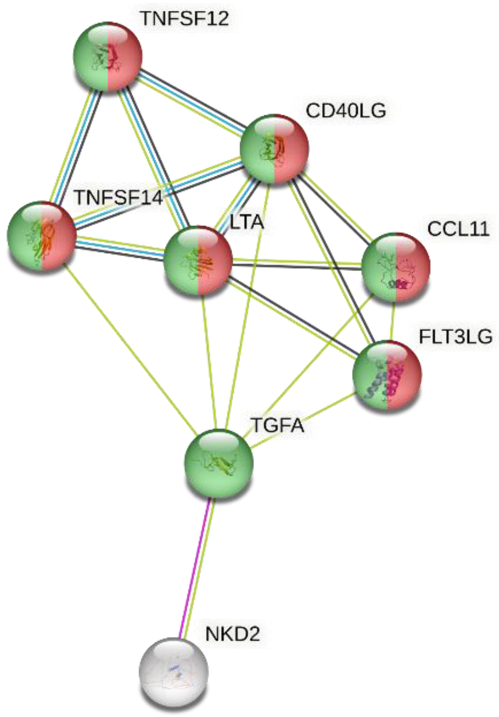
The STRING network analysis, which identified functionally enriched molecular functions among the input proteins (max 5 interactors) showed two enriched GO Molecular Function terms: “cytokine activity” (GO:0005125) indicating significant involvement of several nodes in cytokine-mediated signaling and “receptor ligand activity “(GO:0048018), representing proteins with potential to act as ligands for receptor-mediated processes (Figure 4).
4. Discussion
NAFLD [17] is becoming globally a highly prevalent disease in the general population [38], especially in those patients affected by type 2 diabetes mellitus and obesity [39]. Of relevance, NAFLD is also the most frequent pediatric liver disease [40,41]. Although no specific drug is currently available for NAFLD, this disease may be amenable to preventive, therapeutic interventions through the adherence to international guidelines recommending a lifestyle-based approach, relying on healthy diets [42]. This can modulate hepatic steatosis and counteract liver damage, preventing and delaying the evolution of NAFLD to cirrhosis and cancer [43]. In this context, phytochemical and natural compounds are undergoing an in-depth investigation [44]. Carotenoid are of major interests [45] and multidisciplinary evidences strongly indicate that lycopene [10], a potent and largely available dietary antioxidant/anti-inflammatory nutrient, may be a potential candidate for the management of NAFLD as it has been proven to be efficacious in the case of alcoholic liver hepatitis [46].
On the other hand, evidence correlating lycopene intake, either as a single carotene or as lycopene-containing foods, has so far failed to provide conclusive evidence of its protective efficacy on NAFLD [47]. This limitation is likely to be multifactorial, as assignable to the wide range of individual variability in the metabolism of lycopene into its bioavailable cis configuration [18] and by the need to resort to high consumption of lycopene-containing foods lacking defined nutrients profiles [48] and associated with high calories uptake. It should be underlined that lycopene with a daily requirement of 0,5 mg/kg and a plasma elimination half-life of 5 days [49,50], taken in the range of 5 to 7 mg/day [51] is mainly deriving from lycopene E 160d red food coloring agent intake [50].
To overcome these limitations, an improved formulation of this functional food has been reported using whole tomato fruits in view of their lipid metabolism modulating activity [28,52,53]. This WTFS, produced by calibrated heating of the berry and spray drying [28] is biofortified with olive wastewater micronutrients and by the presence of Fru-His Amadori’s chelators [54], displays an overall superior composition compared to available tomato commodities. WTFS is also endowed with the ability to interfere with metabolic pathways mediating oxidative stress and inflammation, as demonstrated by in vitro [33], animal [55] and human conditions of known susceptibility to tomato micronutrients benefits [29].
Within the limits of using a transformed hepatocyte cell line [56], the information gathered in this study may be informative to explore strategies to contrast liver lipid storage. Indeed, although the progression of NAFLD is the result of a stepwise engagement of other parenchymal, i.e .Kuppfer cells [57], stellate cells [58], and non-parenchymal, i.e. immune cells [59], the drivers of this progressive disease stem from the triglycerides accumulation [60] and lipogenesis [61] in hepatocytes overrunning their dismissal capacity through fatty acid oxidation and/or higher production rates of very-low-density lipoprotein particles [62].
Although the ability of WTFS single components to downmodulate the lipidomic asset of HepG2 cells cannot be fully appreciated, a converging ability of lycopene and other WTFS components tocopherol, tyrosol, hydroxytyrosol, oleopeurina, in lowering tryglycerides through also inhibition of STAT-3 phosphorylation [63], and AhR receptors activation [64] can be hypothesized [33].
Considering that WTFS has been shown to interfere with a several cell signaling, i.e. TRK receptor activation, nuclear factor-kappa B (NF-κB), mitogen-activated protein kinases (MAPK), upregulating the recruiting of inflammatory cells and inhibiting JAK/STAT kinases modulating inflammatory genes [33], the proteomic analysis identified three major targets of WTFS in HepG2 cells, which, by variably activating different signaling, may interfere with multiple metabolic pathways, thus modulating mechanisms fostering the progression of NAFLD. Indeed, a) TGF-α, by engaging the EGF receptor, triggers multiple downstream signaling, i.e. RTK, phosphatidylinositol-3 kinase (PI3K), ERK, and mTOR, which are relevant to liver regeneration [65], thus being a target of WTFS complex of micronutrients. In addition, TGF-α has been identified as an independent indicator of significant liver fibrosis [66]; b) TWEAK, a mitogen for liver progenitor cells [67], while undetectable in normal liver, it is significantly upregulated in patients with fatty liver than in other liver diseases, thus offering a potential therapeutic target [68], also in view that lowering its signaling in vivo may decrease levels of inflammation [36,69] and unbalanced signaling may lead to altered tissue remodeling [70].
Modelling of ligand and receptor interactions at multi cellular level [71] and integrative single-cell and spatial transcriptomic analyses have identified TNFRSF12A [72], as a relevant actor in supporting fibrinogenesis in liver pathology and a potential therapeutic target [73]; c) activation of FLT3LG, which through binding to the receptor Flt3/CD135, causes dimerization and autophosphorylation of the receptor with activation of iPI3K/Akt/mTOR, JAK/STAT, and RAS/RAF/Erk pathways [74], concurs to fibrosis [75] through epithelial mesenchymal transition [76]. In this regard, WTFS contains a complex of anti fibrogenic nutrients (lycopene, quercetin, narigenin, verbascoside) which can modulate epithelial-mesenchymal transition [77]. Our STRING network analysis revealed significant functional enrichment in key molecular functions among the input proteins, notably “cytokine activity” and “receptor ligand activity”. The enrichment of cytokine activity, as evidenced by the clustering of multiple nodes, suggests a prominent role for cytokine-mediated signaling pathways in the biological context under investigation. This aligns with the known involvement of cytokines in modulating inflammatory responses and cellular communication, particularly in pathological states such as immune activation or tissue remodeling. Moreover, the identification of receptor-ligand activity underscores the functional relevance of proteins capable of engaging receptor-mediated mechanisms, which are critical for transducing extracellular signals into specific cellular responses. Together, these enriched terms point toward a functional network characterized by intercellular communication and signal transduction, offering mechanistic insights into the observed phenotypic effects and highlighting potential targets for further experimental validation.
A constant dietary supplementation with WTFS containing a complex of highly bioactive nutrients with share biological activities with lycopene may have in vivo healthy effects that go beyond those produced on hepatocytes since it can modulate high-density lipoprotein [78], multiple signaling relevant to progression of NAFLD because upregulated in variety of cell types contributing to inflammation, angiogenesis, and fibrosis. It should be recalled that WTFS has been shown in fact to interfere with signaling of a number of cytokine and chemokines [55] and to block STAT-3 activation involved in NAFLD progression [79]. Furthermore, the WTFS content of highly bioavailable cis lycopene may be advantageous in patients with NAFLD whose liver has impaired ability to generate lycopene active metabolites (i.e. apo-lycopenals and apo-lycopenones) [10].
Although interventional studies will assess by what extent WTFS or similar supplements may ameliorate NAFLD [24], this functional food free of tomato culinary-associated side effects [30] is an attractive candidate to complement the Mediterranean diet, highly recommended for the prevention/treatment of NAFLD [80] but now recognized as hard to follow [81], thus hampering its wide compliance by large population fractions [82]. Those include patients with glucose intolerance [83], in whom NAFLD is often co-existing [84] but refrain from consuming high-calorie tomato-seasoned dishes, the main source of adequate amounts of bioavailable carotenoids. Since tomatoes are largely present in the international cuisine, the possibility of the culinary use of WTFS or similar products capable of fortifying meals may provide a lifetime efficacious strategy of delivering whole tomato improved nutrients.
Supplementary Materials
The following supporting information can be downloaded at the website of this paper posted on Preprints.org.
Author Contributions
P.G.N.: investigation, methodology, validation, writing—original draft, review and editing; L.I.: writing—original draft, review and editing; M.P.: review and editing; A.S.: review and editing; E.G.: methodology, data curation, investigation, supervision, review and editing; C.B.: data curation, formal analysis, and validation.
Funding
This research was partially supported by the Federico Calabresi Foundation, Rome, Italy.
Institutional Review Board Statement
Not applicable.
Informed Consent Statement
Not applicable.
Data Availability Statement
Authors agree to make data and materials supporting the results or analyses presented in their paper available upon reasonable request.
Conflicts of Interest
P.G.N. is principal research investigator of Janus Pharma Srl., Rome, Italy. M.P. is co-inventor of Euro patent 3 052 113 B1. The other authors declare no conflicts of interest.
Abbreviations
The following abbreviations are used in this manuscript:
| CTRL | control |
| DMSO | dimethyl sulfoxide |
| NAFLD | non-alcoholic fatty liver disease |
| WTFS | whole tomato-based food supplement |
References
- Harrison, S.A.; Torgerson, S.; Hayashi, P.; Ward, J.; Schenker, S. Vitamin E and vitamin C treatment improves fibrosis in patients with nonalcoholic steatohepatitis. Am. J. Gastroenterol. 2003, 98, 2485–2490. [Google Scholar] [CrossRef]
- Kugelmas, M.; Hill, D.B.; Vivian, B.; Marsano, L.; McClain, C.J. Cytokines and NASH: A pilot study of the effects of lifestyle modification and vitamin E. Hepatology 2003, 38, 413–419. [Google Scholar] [CrossRef] [PubMed]
- Tessari, P.; Coracina, A.; Cosma, A.; Tiengo, A. Hepatic lipid metabolism and non-alcoholic fatty liver disease. Nutr. Metab. Cardiovasc. Dis. 2009, 19, 291–302. [Google Scholar] [CrossRef] [PubMed]
- Thorup, A.C.; Kristensen, H.L.; Kidmose, U.; Lambert, M.N.T.; Christensen, L.P.; Fretté, X.; Clausen, M.R.; Hansen, S.M.; Jeppesen, P.B. Strong and Bitter Vegetables from Traditional Cultivars and Cropping Methods Improve the Health Status of Type 2 Diabetics: A Randomized Control Trial. Nutrients 2021, 13, 1813. [Google Scholar] [CrossRef] [PubMed]
- Liu, Y.; Wang, D.; Zhang, D.; Lv, Y.; Wei, Y.; Wu, W.; Zhou, F.; Tang, M.; Mao, T.; Li, M.; et al. Inhibitory effect of blueberry polyphenolic compounds on oleic acid-induced hepatic steatosis in vitro. J. Agric. Food Chem. 2011, 59, 12254–12263. [Google Scholar] [CrossRef]
- Tilg, H.; Moschen, A.R. Evolution of inflammation in nonalcoholic fatty liver disease: The multiple parallel hits hypothesis. Hepatology 2010, 52, 1836–1846. [Google Scholar] [CrossRef]
- Pouwels, S.; Sakran, N.; Graham, Y.; Leal, A.; Pintar, T.; Yang, W.; Kassir, R.; Singhal, R.; Mahawar, K. ; Ramnarain, D Non-alcoholic fatty liver disease (NAFLD): a review of pathophysiology, clinical management and effects of weight loss. BMC Endocr. Disord. 2022, 22, 63. [Google Scholar] [CrossRef]
- Wang, L.L.; Zhang, P.H.; Yan, H.H. Functional foods and dietary supplements in the management of non-alcoholic fatty liver disease: A systematic review and meta-analysis. Front. Nutr. 2023, 10, 1014010. [Google Scholar] [CrossRef]
- Subhash, K.; Bose, C.; Agrawal, B.K. Effect of short-term supplementation of tomatoes on antioxidant enzymes and lipid peroxidation in type-II diabetes. Indian J. Clin. Biochem. 2007, 22, 95–98. [Google Scholar] [CrossRef]
- Shafe, M.O.; Gumede, N.M.; Nyakudya, T.T.; Chivandi, E. Lycopene: A Potent Antioxidant with Multiple Health Benefits. J. Nutr. Metab. 2024, 6252426. [Google Scholar] [CrossRef]
- Wang, J.; Geng, T.; Zou, Q.; Yang, N.; Zhao, W.; Li, Y.; Tan, X.; Yuan, T.; Liu, X.; Zhigang. Liu. Lycopene prevents lipid accumulation in hepatocytes by stimulating PPARα and improving mitochondrial function. J. Funct. Foods 2020, 103857. [CrossRef]
- Mein, J.R.; Lian, F.; Wang, X.D. Biological activity of lycopene metabolites: implications for cancer prevention. Nutr Rev. 2008, 66, 667–683. [Google Scholar] [CrossRef]
- Sultan Alvi, S.; Ansari, I.A.; Khan, I.; Iqbal, J.; Khan, M.S. Potential role of lycopene in targeting proprotein convertase subtilisin/kexin type-9 to combat hypercholesterolemia. Free Radic. Biol. Med. 2017, 108, 394–403. [Google Scholar] [CrossRef]
- Schmitz, H. H.; Poor, C. L.; Wellman, R. B.; Erdman, J. W., Jr. Concentrations of selected carotenoids and vitamin A in human liver, kidney and lung tissue. J. Nutr. 1991, 121, 1613–1621. [Google Scholar] [CrossRef]
- Wang, X.D. Lycopene metabolism and its biological significance. Am. J. Clin. Nutr. 2012, 96, 1214S– 1222S. [CrossRef]
- Elvira-Torales, L.I.; García-Alonso, J.; Periago-Castón, M.J. Nutritional Importance of Carotenoids and Their Effect on Liver Health: A Review. Antioxidants (Basel). 2019, 8, 229. [Google Scholar] [CrossRef] [PubMed]
- Makri, E.; Goulas, A.; Polyzos, S.A. Epidemiology, pathogenesis, diagnosis and emerging treatment of nonalcoholic fatty liver disease. Arch. Med. Res. 2021, 52, 25–37. [Google Scholar] [CrossRef] [PubMed]
- Bohn, T.; Desmarchelier, C.; Dragsted, L.O.; Nielsen, C.S.; Stahl, W.; Rühl, R.; Keijer, J.; Borel, P. Host-related factors explaining interindividual variability of carotenoid bioavailability and tissue concentrations in humans. Mol. Nutr. Food Res. 2017, 61, 1600685. [Google Scholar] [CrossRef] [PubMed]
- Marquez, C.S.; Reis Lima, M.J.; Oliveira, J:, Teixeira-Lemos, E. Tomato lycopene: functional proprieties and health benefits. Int. J. Agric. Eng. 2015, 9, 1089–1099. [CrossRef]
- Johnson, E.J.; Qin, J.; Krinsky, N.I.; Russell, R.M. Ingestion by men of a combined dose of beta-carotene and lycopene does not affect the absorption of beta-carotene but improves that of lycopene. J Nutr. 1997, 127, 1833–1837. [Google Scholar] [CrossRef]
- Tamanna, N.; Mahmood, N. Food processing and Maillard reaction products: effect on human health and nutrition. Int. J. Food Sci. 2015, 2015, 526762. [Google Scholar] [CrossRef]
- Rowles, J.L. 3r.; Erdman, J.W. Jr. Carotenoids and their role in cancer prevention. Biochim. Biophys. Acta Mol. Cell. Biol. Lipids 2020, 1865, 158613. [Google Scholar]
- Linnewiel-Hermoni, K.; Khanin, M.; Danilenko, M.; Zango, G.; Amosi, Y.; Levy, J.; Sharoni, Y. The anti-cancer effects of carotenoids and other phytonutrients resides in their combined activity. Arch. Biochem. Biophys. 2015, 572, 28–35. [Google Scholar] [CrossRef]
- Landrier, J.F.; Breniere, T.; Sani, L.; Desmarchelier, C.; Mounien, L.; Borel, P. Effect of tomato, tomato-derived products and lycopene on metabolic inflammation: from epidemiological data to molecular mechanisms. Nutr. Res. Rev. 2023, 18, 1–17. [Google Scholar] [CrossRef]
- Mohri, S.; Takahashi, H.; Sakai, M.; Takahashi, S.; Waki, N.; Aizawa, K.; Suganuma, H.; Ara, T.; Matsumura, Y.; Shibata, D.; et al. Wide-range screening of anti-inflammatory compounds in tomato using LC-MS and elucidating the mechanism of their functions. PLoS One 2018, 13, e0191203. [Google Scholar] [CrossRef]
- Mazidi, M.; Katsiki, N.; George, E.S.; Banach, M. Tomato and lycopene consumption is inversely associated with total and cause-specific mortality: a population-based cohort study, on behalf of the International Lipid Expert Panel (ILEP). Br. J. Nutr. 2020, 124, 1303–10. [Google Scholar] [CrossRef] [PubMed]
- Fogliano, V.; Iacobelli, S.; Piantelli, M. Euro Patent 3 052 113 B1, Italian Health Ministry (registration n. 68843, 2018–2019). Available online: https://worldwide.espacenet.com/patent/search/family/049226079/publication/EP3052113A1?q=3052113 (accessed on July 8, 2025).
- Gholami, F.; Antonio, J.; Evans, C.; Cheraghi, K.; Rahmani, L.; Amirnezhad, F. Tomato powder is more effective than lycopene to alleviate exercise-induced lipid peroxidation in well-trained male athletes: randomized, double-blinded cross-over study. J. Int. Soc. Sports Nutr. 2021, 18, 17. [Google Scholar] [CrossRef] [PubMed]
- Natali, P.G.; Piantelli, M.; Minacori, M.; Eufemi, M.; Imberti, L. Improving whole tomato transformation for prostate health: benign prostate hypertrophy as an exploratory model. Int. J. Mol. Sci. 2023, 24, 5795. [Google Scholar] [CrossRef] [PubMed]
- Natali, P.G.; Piantelli, M.; Sottini, A.; Eufemi, M.; Banfi, C.; Imberti, L. A step forward in enhancing the health-promoting properties of whole tomato as a functional food to lower the impact of non-communicable diseases. Front. Nutr. 2025, 12, 1519905. [Google Scholar] [CrossRef]
- Aden, D.P.; Fogel, A.; Plotkin, S.; Damjanov, I.; Knowles, B.B. Controlled synthesis of HBsAg in a differentiated human liver carcinoma-derived cell line. Nature 1979, 282, 615–616. [Google Scholar] [CrossRef]
- Banfi, C.; Mussoni, L.; Risé, P.; Cattaneo, M.G.; Vicentini, L.; Battaini, F.; Galli, C.; Tremoli, E. Very low density lipoprotein-mediated signal transduction and plasminogen activator inhibitor type 1 in cultured HepG2 cells. Circ Res. 1999, 85, 208–217. [Google Scholar] [CrossRef]
- Rubini, E.; Minacori, M.; Paglia, G.; Macone, A.; Chichiarelli, S.; Altieri, F.; Eufemi, M. Tomato and olive bioactive compounds: A natural shield against the cellular effects induced by β-hexachlorocyclohexane-Activated Signaling Pathways. Molecules. 2021, 26, 7135. [Google Scholar] [CrossRef]
- Andus, T.; Bauer, J.; Gerok, W. Effects of cytokines on the liver. Hepatology 1991, 13, 364–375. [Google Scholar] [CrossRef]
- Kudlow, J.E.; Bjorge, J.D. TGF-alpha in normal physiology. Semin. Cancer Biol. 1990, 1, 293–302. [Google Scholar]
- Winkles, J.A. The TWEAK-Fn14 cytokine-receptor axis: discovery, biology and therapeutic targeting. Nat. Rev. Drug Discov. 2008, 7, 411–425. [Google Scholar] [CrossRef] [PubMed]
- Fausto, N. Liver regeneration and repair: Hepatocytes, progenitor cells, and stem cells. Hepatology 2004; 39, 1477-1487. [CrossRef]
- Younossi, Z.M.; Koenig, A.B.; Abdelatif, D.; Fazel, Y.; Henry, L.; Wymer, M. Global epidemiology of nonalcoholic fatty liver disease-meta-analytic assessment of prevalence, incidence, and outcomes. Hepatology 2016, 64, 73–84. [Google Scholar] [CrossRef] [PubMed]
- Henry, L.; Paik, J.; Younossi, Z.M. Review article: the epidemiologic burden of non-alcoholic fatty liver disease across the world. Alimen. Pharmacol. Ther. 2022, 56, 942–956. [Google Scholar] [CrossRef]
- Goldner, D.; Lavine, J.E. Nonalcoholic Fatty Liver Disease in Children: Unique Considerations and Challenges. Gastroenterology 2020, 158, 1967–1983.e1. [Google Scholar] [CrossRef] [PubMed]
- Nobili. V.; Alisi, A.; Valenti, L.; Miele, L.; Feldstein, A.E.; Alkhouri, N. NAFLD in children: new genes, new diagnostic modalities and new drugs. Nat. Rev. Gastroenterol. Hepatol. 2019, 16, 517–530. [Google Scholar] [CrossRef]
- Haigh, L.; Kirk, C.; El Gendy, K.; Gallacher, J.; Errington, L.; Mathers, J.C. Anstee, Q.M. The effectiveness and acceptability of Mediterranean diet and calorie restriction in non-alcoholic fatty liver disease (NAFLD): A systematic review and meta-analysis. Clin. Nutr. 2022, 41, 1913–1931. [Google Scholar] [CrossRef]
- Hydes, T.J.; Ravi, S. , Loomba, R.; Gray, M.E. Evidence-based clinical advice for nutrition and dietary weight loss strategies for the management of NAFLD and NASH. Clin. Mol. Hepatol. 2020, 26, 383–400. [Google Scholar] [CrossRef]
- Pezzino, S.; Sofia, M.; Mazzone, C.; Litrico, G.; Greco, L.P.; Gallo, L.; La Greca, G.; Latteri, S. Innovative treatments for obesity and NAFLD: A bibliometric study on antioxidants, herbs, phytochemicals, and natural compounds. Heliyon 2024, 10, e35498. [Google Scholar] [CrossRef]
- Christensen, K.; Lawler, T.; Mares, J. Dietary carotenoids and Non-Alcoholic Fatty Liver Disease among US adults, NHANES 2003⁻2014. Nutrients 2019, 11, 1101. [Google Scholar] [CrossRef]
- Stice, C.P.; Xia, H.; Wang, X.D. Tomato lycopene prevention of alcoholic fatty liver disease and hepatocellular carcinoma development. Chronic Dis. Transl. Med. 2018, 4, 211–224. [Google Scholar] [CrossRef] [PubMed]
- Donghia, R.; Campanella, A.; Bonfiglio, C.; Cuccaro, F.; Tatoli, R.; Giannelli, G. Protective Role of Lycopene in Subjects with Liver Disease: NUTRIHEP Study. Nutrients 2024, 16, 562. [Google Scholar] [CrossRef] [PubMed]
- Paetau, I.; Khachik, F.; Brown, E.D.; Beecher, G.R.; Kramer, T.R.; Chittams, J.; Clevidence, B.A. Chronic ingestion of lycopene-rich tomato juice or lycopene supplements significantly increases plasma concentrations of lycopene and related tomato carotenoids in humans. Am. J. Clin. Nutr. 1998, 68, 1187–1195. [Google Scholar] [CrossRef] [PubMed]
- Ross, A.B.; Vuong, T.; Ruckle, J.; Synal, H.A.; Schulze-König, T.; Wertz, K.; Rümbeli, R.; Liberman, R.G.; Skipper, P.L.; Tannenbaum, S.R.; et al. Lycopene bioavailability and metabolism in humans: an accelerator mass spectrometry study. Am. J. Clin. Nutr. 2011, 93, 1263–1273. [Google Scholar] [CrossRef]
- EFSA Panel on Nutrition, Novel Foods and Food Allergens (NDA); Turck, D; Bohn, T. ; Cámara, M.; Castenmiller, J.; De Henauw, S.; Jos, Á.; Maciuk, A.; Mangelsdorf, I.; McArdle, H.J.; et al. Safety of yellow tomato extract as a novel food pursuant to Regulation (EU) 2015/2283. EFSA J. 2025, 23, e9373. [Google Scholar] [CrossRef]
- Petyaev, I.M. Lycopene Deficiency in Ageing and Cardiovascular Disease. Oxidative Med. Cell. Longev. 2016, 2016, 321805. [Google Scholar] [CrossRef]
- Hirose, A.; Terauchi, M.; Tamura, M.; Akiyoshi, M.; Owa, Y.; Kato, K.; Kubota, T. Tomato juice intake increases resting energy expenditure and improves hypertriglyceridemia in middle-aged women: an open-label, single-arm study. Nutr. J. 2015, 14, 34. [Google Scholar] [CrossRef]
- Nishimura, M.; Tominaga, N.; Ishikawa-Takano, Y.; Maeda-Yamamoto, M.; Nishihira, J. Effect of 12-Week Daily Intake of the High-Lycopene Tomato (Solanum Lycopersicum), A Variety Named “PR-7”, on Lipid Metabolism: A Randomized, Double-Blind, Placebo-Controlled, Parallel-Group Study. Nutrients 2019, 11, 1177. [Google Scholar] [CrossRef]
- Zhou, R.; Zhu, X.; Xie, T.; Li, W.; Xie, D.; Zhang, G.; Xiao, Y.; Zhang, L. Amadori compounds (N-(1-Deoxy-D-fructos-1-yl)-amino acid): The natural transition metal ions (Cu2+, Fe2+, Zn2+) chelators formed during food processing. LWT 2024, 191, 115600. [Google Scholar] [CrossRef]
- Pannellini, T.; Iezzi, M.; Liberatore, M.; Sabatini, F.; Iacobelli, S.; Rossi, C.; Alberti, S.; Di Ilio, C.; Vitaglione, P.; Fogliano, V.; et al. A dietary tomato supplement prevents prostate cancer in TRAMP mice. Cancer Prev. Res. 2010, 3, 1284–1291. [Google Scholar] [CrossRef]
- Arzumanian, V.A.; Kiseleva, O.I.; Poverennaya, E.V. The Curious Case of the HepG2 Cell Line: 40 Years of Expertise. Int. J. Mol. Sci. 2021, 22, 13135. [Google Scholar] [CrossRef]
- Park, S-J. ; Garcia Diaz, J.; Um, E.; Hahn, Y.S. Major roles of kupffer cells and macrophages in NAFLD development. Front. Endocrinol. 2023, 14, 1150118. [Google Scholar]
- Wiering, L.; Subramanian, P.; Hammerich. L. Hepatic Stellate Cells: Dictating Outcome in Nonalcoholic Fatty Liver Disease. Cell. Mol. Gastroenterol. Hepatol. 2023, 15, 1277–1292. [Google Scholar] [CrossRef]
- Moayedfard, Z.; Sani, F.; Alizadeh, A.; Bagheri Lankarani, K.; Zarei, M.; Azarpira, N. The role of the immune system in the pathogenesis of NAFLD and potential therapeutic impacts of mesenchymal stem cell-derived extracellular vesicles. Stem Cell Res. Ther. 2022, 13, 242. [Google Scholar] [CrossRef] [PubMed]
- Kawano, Y.; Cohen, D.E. Mechanisms of hepatic triglyceride accumulation in non-alcoholic fatty liver disease. J. Gastroenterol. 2013, 48, 434–441. [Google Scholar] [CrossRef] [PubMed]
- Lambert, J.E.; Ramos-Roman, M.A.; Browning, J.D.; Parks, E.J. Increased de novo lipogenesis is a distinct characteristic of individuals with nonalcoholic fatty liver disease. Gastroenterology 2014, 146, 726–735. [Google Scholar] [CrossRef] [PubMed]
- Alves-Bezerra, M.; Cohen, DE. Triglyceride Metabolism in the Liver. Compr. Physiol. 2017, 8, 1–8. [Google Scholar] [CrossRef]
- Belloni, L.; Di Cocco, S.; Guerrieri, F.; Nunn, A.D.G.; Piconese, S.; Salerno, D.; Testoni, B.; Pulito, C.; Mori, F.; Pallocca, M.; et al. Targeting a phospho-STAT3-miRNAs pathway improves vesicular hepatic steatosis in an in vitro and in vivo model. Sci. Rep. 2018, 8, 13638. [Google Scholar] [CrossRef]
- Lee, J.H.; Wada, T.; Febbraio, M.; He, J.; Matsubara, T.; Lee, M.J.; Gonzalez, F.J.; Xie, W. A novel role for the dioxin receptor in fatty acid metabolism and hepatic steatosis. Gastroenterology 2010, 139, 653–663. [Google Scholar] [CrossRef]
- Kimura, M.; Moteki, H.; Ogihara, M. Role of hepatocyte growth regulators in liver regeneration. Cells 2023, 12, 208. [Google Scholar] [CrossRef]
- Deng, Y.Q.; Zhao, H.; Ma, A.L.; Zhou, J.Y.; Xie, S.B.; Zhang, X.Q.; Zhang, D.Z.; Xie. Q.; Zhang, G.; Shang, J. et al. Selected cytokines serve as potential biomarkers for predicting liver inflammation and fibrosis in chronic hepatitis b patients with normal to mildly elevated aminotransferases. Medicine (Baltimore) 2015, 94, e2003. [Google Scholar] [CrossRef]
- Tirnitz-Parker, J.E.; Viebahn, C.S.; Jakubowski, A.; Klopcic, B.; Olynyk, J.; Yeoh, G.; Knight, B. Tumor necrosis factor–like weak inducer of apoptosis is a mitogen for liver progenitor cells. Hepatology 2010, 52, 291–302. [Google Scholar] [CrossRef] [PubMed]
- Suppli, M.P.; Rigbolt, K.T.G.; Veidal, S.S.; Heebøll, S.; Eriksen, P.L.; Demant, M.; Bagger, J.I.; Nielsen, J.C.; Oró, D.; Thrane, S.W. Hepatic transcriptome signatures in patients with varying degrees of nonalcoholic fatty liver disease compared with healthy normal-weight individuals. Am. J. Physiol. Gastrointest. Liver Physiol. 2019, 316, G462–G472. [Google Scholar] [CrossRef] [PubMed]
- Kong, Y.; Yang, Y.; Wu, S.; Li, W. TWEAK promotes inflammatory response in liver fibrosis. J. Biochem. Mol. Toxicol. 2023, 37, e23483. [Google Scholar] [CrossRef] [PubMed]
- Wilhelm, A.; Shepherd, E.L.; Amatucci, A.; Munir, M.; Reynolds, G.; Humphreys, E.; Resheq, Y.; Adams, D.H.; Hübscher, S.; Burkly, L.C. Interaction of TWEAK with Fn14 leads to the progression of fibrotic liver disease by directly modulating hepatic stellate cell proliferation. J. Pathol. 2016, 239, 109–121. [Google Scholar] [CrossRef]
- Ramachandran, P.; Dobie, R.; Wilson-Kanamori, J.R.; Dora, E.F.; Henderson, B.E.P.; Luu, N.T.; Portman, J.R.; Matchett, K.P.; Brice, M.; Marwick, J.A.; et al. Resolving the fibrotic niche of human liver cirrhosis at single-cell level. Nature 2019, 575, 512–518. [Google Scholar] [CrossRef]
- Xiao, M.H.; Ma, D.; Wu, S.; Huang, Z.; Liang, P.; Chen, H.; Zhong, Z.; Li, W.; Wang, F.; Tang, Y.; et al. Integrative single-cell and spatial transcriptomic analyses identify a pathogenic cholangiocyte niche and TNFRSF12A as therapeutic target for biliary atresia. Hepatology 2025, 81, 1146–1163. [Google Scholar] [CrossRef]
- Huang, T.; Zheng, D.; Song, Y.; Pan, H.; Qiu, G.; Xiang, Y.; Wang, Z.; Wang, F. Demonstration of the impact of COVID-19 on metabolic associated fatty liver disease by bioinformatics and system biology approach. Medicine (Baltimore) 2023, 102, e34570. [Google Scholar] [CrossRef]
- Voronov, I.; Manolson, M.F. Editorial: Flt3 ligand-friend or foe? J. Leukoc. Biol. 2016, 99, 401–403. [Google Scholar] [CrossRef]
- Rahman, A.H. , Aloman, C. Dendritic cells and liver fibrosis. Biochim. Biophys. Acta. 2013, 1832, 998–1004. [Google Scholar] [CrossRef]
- Lovisa, S. Epithelial-to Mesenchymal Transition in Fibrosis: Concepts and Targeting Strategies. Front. Pharmacol. 2021, 12, 737570. [Google Scholar] [CrossRef]
- Daniello, V.; De Leo, V.; Lasalvia, M.; Hossain, M.N.; Carbone, A.; Catucci, L.; Zefferino, R.; Ingrosso, C.; Conese, M.; Di Gioia, S. Solanum lycopersicum (Tomato)-derived nanovesicles accelerate wound healing by eliciting the migration of keratinocytes and fibroblasts. Int. J. Mol. Sci. 2024, 25, 2452. [Google Scholar] [CrossRef] [PubMed]
- McEneny, J.; Henry, S.-L.; Woodside, J.; Moir, S.; Rudd, A.; Vaughan, N.; Thies. F. Lycopene-rich diets modulate HDL functionality and associated inflammatory markers without aecting lipoprotein size and distribution in moderately overweight, disease-free, middle-aged adults: A randomized controlled trial. Front. Nutr. 2022, 9, 954593. [Google Scholar] [CrossRef] [PubMed]
- Jiao, J.; Sanchez, J.I.; Saldarriaga, O.A.; Solis, L.M.; Tweardy, D.J.; Maru, D.M.; Stevenson, H. L.; Beretta, L. Spatial molecular and cellular determinants of STAT3 activation in liver fibrosis progression in non-alcoholic fatty liver disease. JHEP Rep. 2022, 5, 100628. [Google Scholar] [CrossRef] [PubMed]
- Pugliese, N.; Plaz Torres, M.C.; Petta, S.; Valenti, L.; Giannini, E.G.; Aghemo, A. Is there an ‘ideal’ diet for patients with NAFLD? Eur. J. Clin. Invest. 2022, 52, e13659. [Google Scholar] [CrossRef]
- Kiani, A.K.; Medori, M.C.; Bonetti, G.; Aquilanti, B.; Velluti, V.; Matera, G.; Iaconelli, A.; Stuppia, L.; Connelly, S.T.; Herbst, K.L.; et al. Modern vision of the Mediterranean diet. J. Prev. Med. Hyg. 2022, 63, E36–E43. [Google Scholar]
- Caparello, G.; Galluccio, A.; Giordano, C.; Lofaro, D.; Barone, I.; Morelli, C.; Sisci, D.; Catalano, S.; Andò, S.; Bonofiglio, D. Adherence to the Mediterranean diet pattern among university staff: a cross-sectional web-based epidemiological study in Southern Italy. Int. J. Food Sci. Nutr. 2020, 71, 581–592. [Google Scholar] [CrossRef]
- Leh, H.E.; Lee, L.K. Lycopene: a potent antioxidant for the amelioration of type II diabetes mellitus. Molecules 2022, 27, 2335. [Google Scholar] [CrossRef]
- European Association for the Study of the Liver (EASL); European Association for the Study of Diabetes (EASD); European Association for the Study of Obesity (EASO). EASL-EASD-EASO Clinical Practice Guidelines for the management of non-alcoholic fatty liver disease. J. Hepatol. 2016, 64, 1388–1402. [Google Scholar] [CrossRef]
Figure 1.
The bubble plot reports various lipid classes examined through targeted lipidomic analysis in positive ion mode. A notable reduction in the relative levels of cholesterol esters (CE), diacylglycerols (DG), lysophosphatidylcholine (LysoPC), lysophosphatidylethanolamine (LysoPE), and triacylglycerols (TG) was observed in cells treated with WTFS. A significant decrease was recorded in the levels of all TG and CE in the treated cells. The size of the bubbles corresponds to the significance of the fold changes (WTFS vs. CTRL), reported as 1 minus the p-values. The other lipid classes analyzed include glucosylceramides (GCer), phosphatidylcholines (PC), phosphatidylethanolamine (PE), and sphingomyelins (SM).
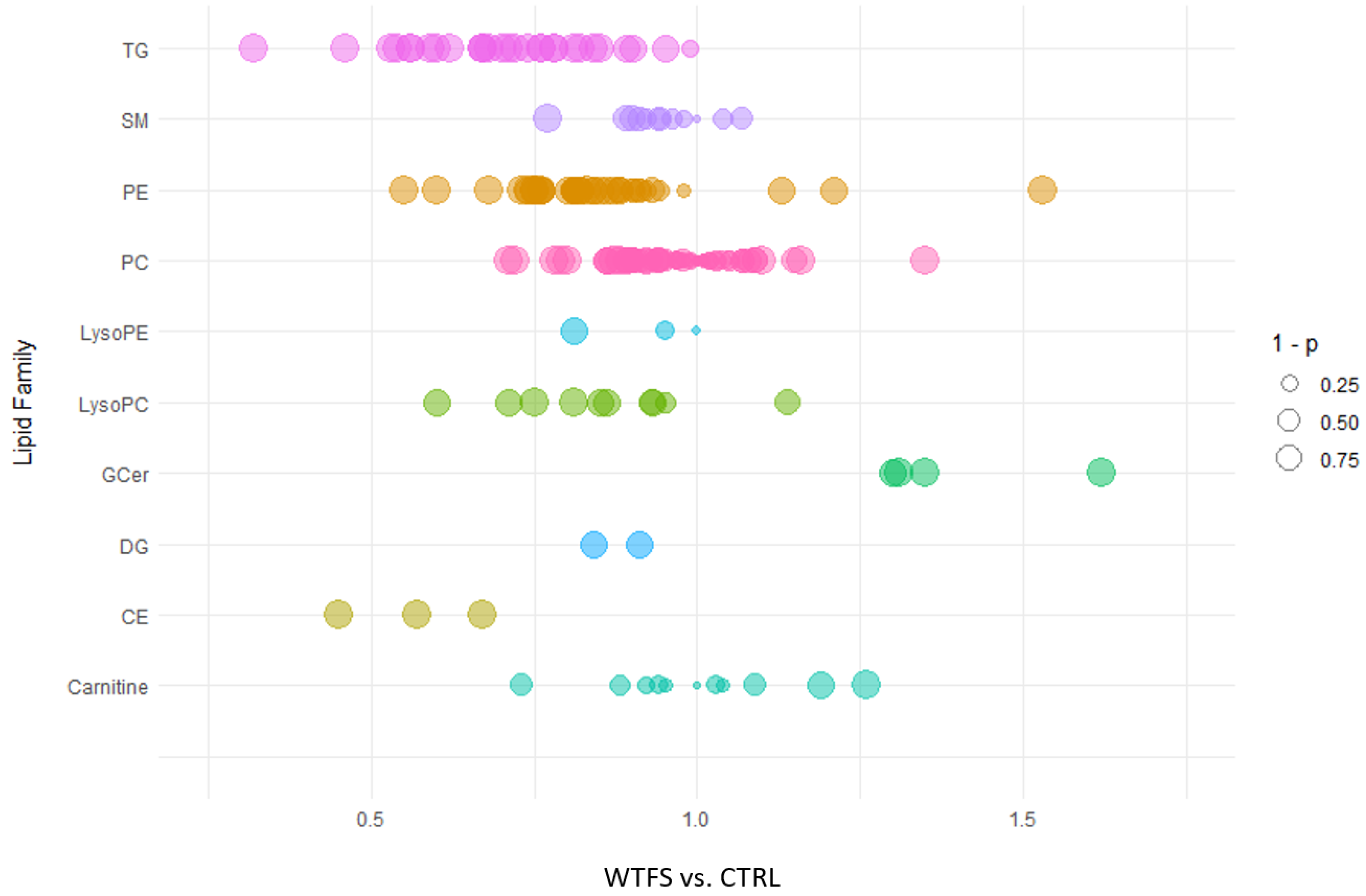
Figure 2.
The bubble plot represents different lipid classes measured by targeted lipidomic analysis in negative ion mode. The size of the bubbles corresponds to the significance of the fold changes (WTFS vs. CTRL), reported as 1 minus the p-values. The lipid classes analyzed include bile acids (BA), cardiolipins (CL), lysophosphatidic acids (LPA), lysophosphatidylinositol acids (LPI), phosphatidic acids (PA), phosphatidylinositol acids (PI), phosphatidylglycerol acids (PG), and sulfatides (Sul-d).
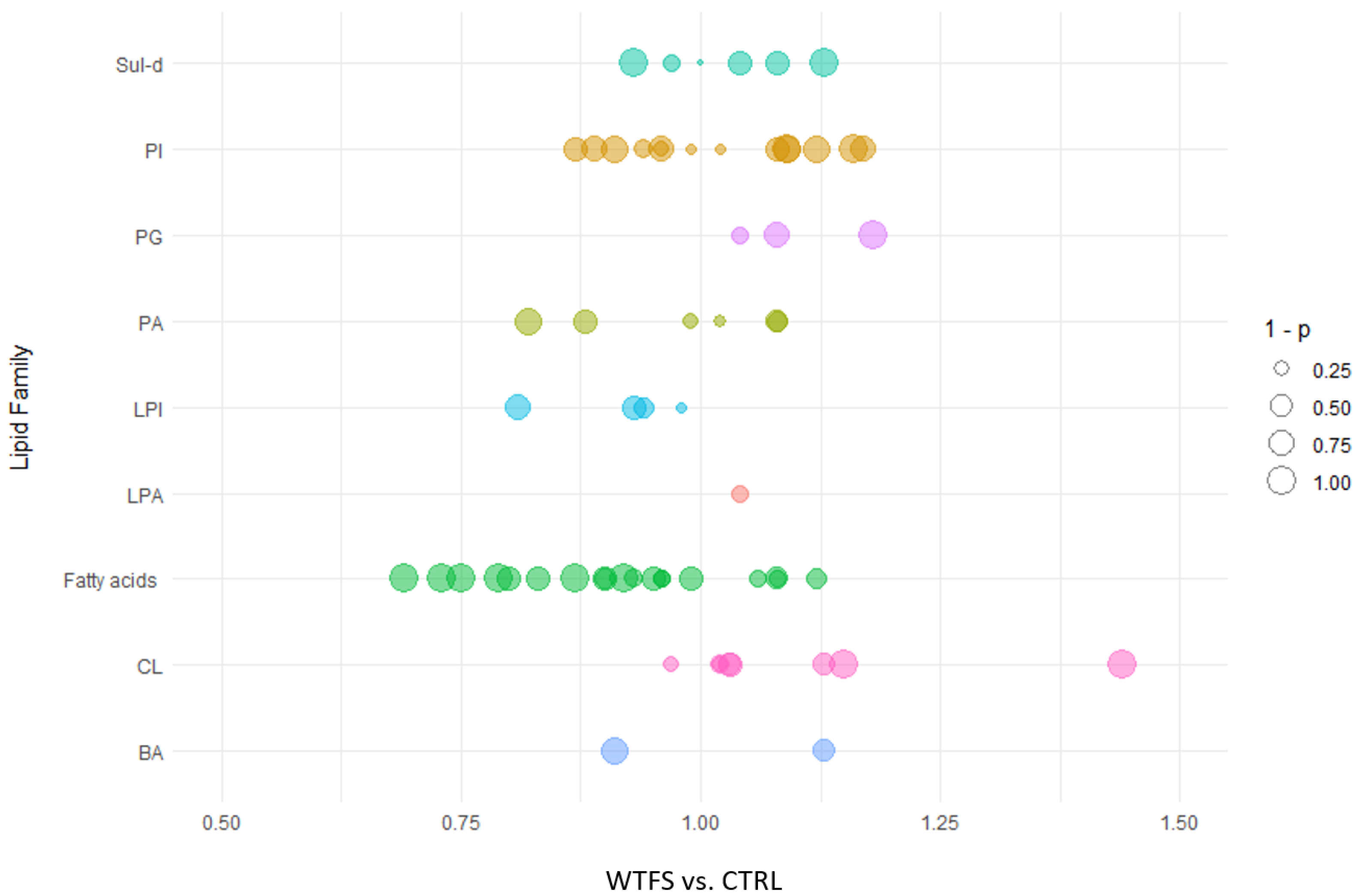
Figure 3.
Targeted proteomic analysis was conducted on HepG2 cells treated with WTFS, utilizing the Olink Target 48 cytokine assay.
Figure 3.
Targeted proteomic analysis was conducted on HepG2 cells treated with WTFS, utilizing the Olink Target 48 cytokine assay.
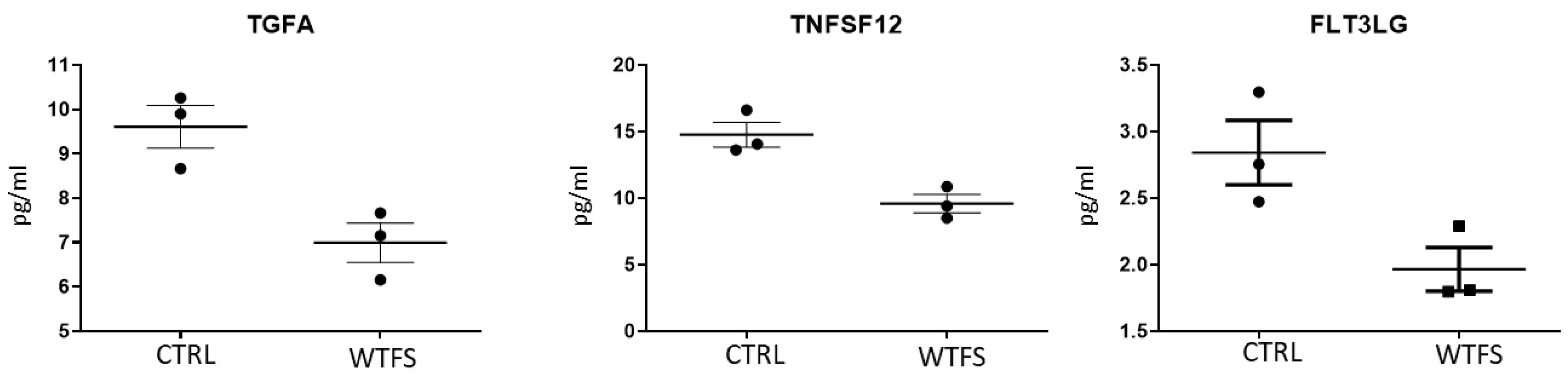
Figure 4.
Protein-protein interaction network generated using STRING v12.0. Nodes represent proteins, while edges indicate predicted functional associations. Enriched GO Molecular Function terms are annotated with color highlights: red indicates “cytokine activity” and green indicates “receptor ligand activity”. The analysis was performed with a confidence threshold of 0.4 and a maximum of 5 interactors to maintain interpretability. Only significantly enriched categories are shown.
Disclaimer/Publisher’s Note: The statements, opinions and data contained in all publications are solely those of the individual author(s) and contributor(s) and not of MDPI and/or the editor(s). MDPI and/or the editor(s) disclaim responsibility for any injury to people or property resulting from any ideas, methods, instructions or products referred to in the content. |
© 2025 by the authors. Licensee MDPI, Basel, Switzerland. This article is an open access article distributed under the terms and conditions of the Creative Commons Attribution (CC BY) license (http://creativecommons.org/licenses/by/4.0/).
Copyright: This open access article is published under a Creative Commons CC BY 4.0 license, which permit the free download, distribution, and reuse, provided that the author and preprint are cited in any reuse.


