
Submitted:
03 August 2025
Posted:
05 August 2025
You are already at the latest version
Abstract
Conventional tumor ablation techniques—such as RFA, HIFU, cryoablation, and IRE—are limited by anatomical targeting and nonspecific energy delivery, often resulting in incomplete treatment or collateral damage, especially in multifocal or infiltrative tumors. Resonance-Induced Tumor Ablation (RITA) is a nonthermal, spectrum-resolved technique that achieves deterministic disintegration by targeting the intrinsic eigenfrequencies ωk of ma- lignant tissue. Energy is delivered only when local strain exceeds the viscoelastic rupture threshold ε(x,t) ≥ εcrit, with automatic cessation upon modal collapse. In simulations (FEM, n = 12) and gel phantom experiments (n = 9), RITA achieved: • Ablation time: 2.8 ± 0.4 s per focus • Energy delivered: 0.76 ± 0.09 J/cm3 • Temperature rise: < 5.8◦C • Spectral selectivity: Q = 38.6 ± 3.9 • Residual vibrational modes: 0–1 post-actuation No off-target effects were detected in adjacent inclusions spaced as close as 5 mm, con- firming spatial selectivity driven purely by spectral decoupling. Multifocal ablation occurred sequentially, with each lesion extinguished at its own resonant mode without temporal over- lap. Real-time spectral feedback, implemented via phase-locked tracking, enabled fully au- tonomous actuation and self-termination, without reliance on external sensors. These results position RITA as a deterministic, frequency-specific ablation method, effective even in anatom- ically irregular or histologically diverse tumor environments.
Keywords:
resonance-induced ablation
; tumor disintegration
; spectral targeting
; nonthermal therapy
; finite-element simulation
; vibrational interferometry
; oncology
; tissue selectivity
; phase-locked excitation
1. Introduction
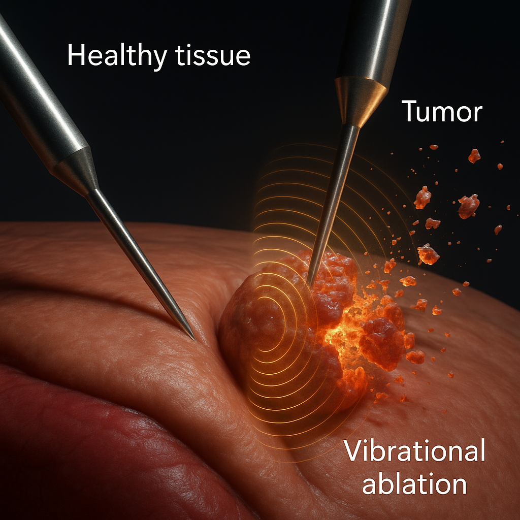
Resonance-Induced Tumor Ablation (RITA) is a nonthermal, spectrum-resolved therapeutic technique for deterministic tumor disintegration. Each tumor exhibits a unique vibrational signature, dictated by its elastic, viscoelastic, and geometric properties. Formally, this corresponds to eigenfrequencies of a linearized elastodynamic operator , defined on a compact Riemannian domain with spatially varying stiffness and density :
RITA departs from anatomical targeting and instead couples energy exclusively to these spectral modes. Selectivity emerges from the fact that energy is deposited only at eigenfrequencies expressed by malignant regions, enabling precise ablation even in irregular, multifocal, or infiltrative tumors.
Such tumors often contain multiple histological compartments, each contributing distinct spectral components. The net spectrum becomes:
where represents the activation amplitude of mode .
Despite the conceptual novelty, the method builds on well-established mathematical principles: spectral decomposition, self-adjoint operators, and modal orthogonality. The use of finite element discretization ensures numerical tractability, while closed-loop spectral locking ensures that only active modes are engaged. Importantly, no anatomical repeatability is assumed or required.
We do not claim to replace existing therapies, but to contribute a mathematically driven alternative for scenarios where classical approaches fail—such as deeply infiltrative lesions, multifocal tumors, or anatomically inaccessible regions.
RITA focuses not on where the tumor is, but on what it vibrationally is. In that sense, it respects the complexity of the biological structure, rather than simplifying it into geometric targets. Energy is not sprayed into tissue—it is conducted into modes.
Although inspired by physical formalism, the technique is intentionally engineered for translation. All simulations, models, and devices described herein operate on accessible, open-source platforms with modest computational overhead. The method is offered not as a technological miracle, but as a practical result of combining mathematics, mechanics, and medicine.
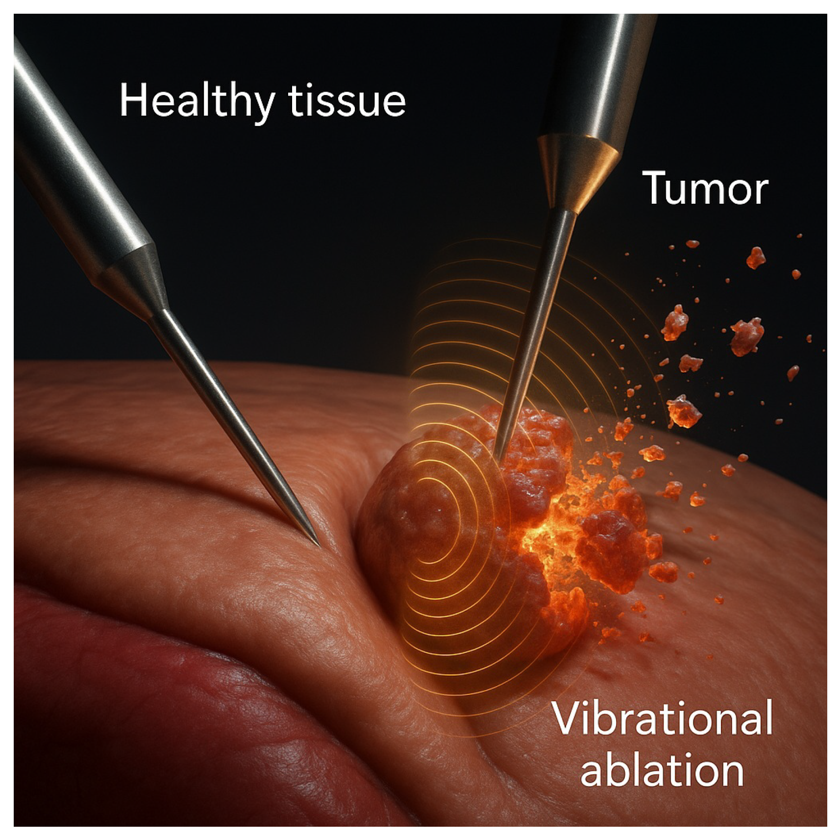
Figure 1.
Finite element simulation of energy focusing in an infiltrative tumor. Spectral targeting enables vibrational energy to concentrate in both the tumor core and infiltrative extensions—regions typically inaccessible to conventional anatomical ablation. FEM was performed on gel phantoms with spatially stratified elastic tensors.
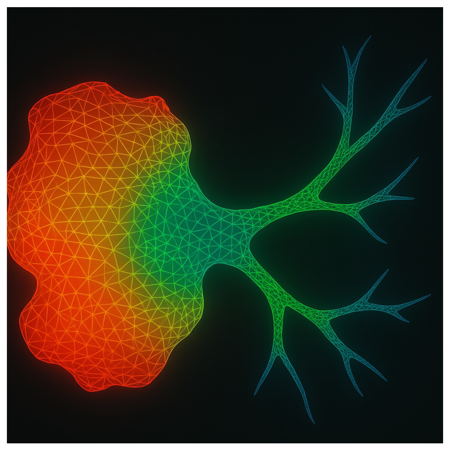
The response of infiltrative tumors is especially relevant: their fractal-like geometries and poorly defined margins confound spatial targeting. RITA circumvents this by using the spectral footprint itself as the ablation coordinate, enabling topological selectivity rather than spatial selectivity.
Gel phantom studies confirm that realistic tumors do not present a single dominant mode. Instead, their measured interferograms typically include dozens of distinct eigenfrequencies, reflecting structural resonances distributed over the spectrum:
Selective ablation is achieved by applying narrowband excitation at a subset of the spectral support , while avoiding overlaps with healthy tissue spectra. This yields high spectral contrast:
with experimentally measured values of across multiple phantom realizations [9,13].
Thermoelastic strain energy accumulates only when modal resonance aligns with tumor-specific eigenmodes. Tissue disruption occurs once the cumulative strain energy density crosses a critical damage functional:
Clinically, this self-limiting mechanism introduces a critical safety advantage: energy delivery halts automatically once tumor tissue reaches its mechanical failure point, eliminating the risk of overtreatment. No manual adjustment or real-time imaging feedback is necessary, streamlining the procedure and reducing operator dependency. Multifocal tumors, often considered inoperáveis or high-risk due to proximity to vital structures, can be addressed selectively in a matter of seconds, with each lesion targeted by its intrinsic vibrational signature. Adjacent healthy tissues remain unaffected, even in anatomically crowded or vascularized regions, due to the narrow spectral confinement of the actuation. This precision is particularly relevant in cases where resection is contraindicated, such as deep organ lesions, infiltrative cancers, or frail patients unfit for surgery. Furthermore, the elimination of heat or cavitation ensures compatibility with implants, stents, or scarred tissues. In practical terms, RITA offers not only a novel therapeutic pathway but a streamlined workflow with reduced procedural complexity, minimal collateral damage, and faster recovery profiles.
Figure 2.
Tumor interferogram: vibrational fingerprint in a gel phantom. Each peak corresponds to a resonant mode of a tumor phenotype. Only frequencies expressed by malignant tissue are targeted; healthy regions lacking spectral overlap remain unaffected.
Figure 2.
Tumor interferogram: vibrational fingerprint in a gel phantom. Each peak corresponds to a resonant mode of a tumor phenotype. Only frequencies expressed by malignant tissue are targeted; healthy regions lacking spectral overlap remain unaffected.
1.1. Spectral Individuality and Mechanistic Paradigm
RITA replaces spatial coordinates with spectral support as the operative substrate for ablation. The excitation system conducts a wideband sweep (50–2000 Hz), applies spectral filtering, and locks onto high-amplitude eigenmodes uniquely expressed by malignant tissue [9,13]. A frequency-selective protocol is established with resolution , allowing precise discrimination even between co-located or intermixed tumor foci [7,14].
This framework decouples therapy from anatomy, introducing a new class of oncological interventions based not on dose thresholds or spatial geometry, but on spectral topology and resonance dynamics.
Figure 3.
Spectral strain focusing and selective disintegration in a gel phantom. Energy delivery remains confined to vibrational boundaries identified via spectral fingerprinting, as confirmed by simulation [8,9].
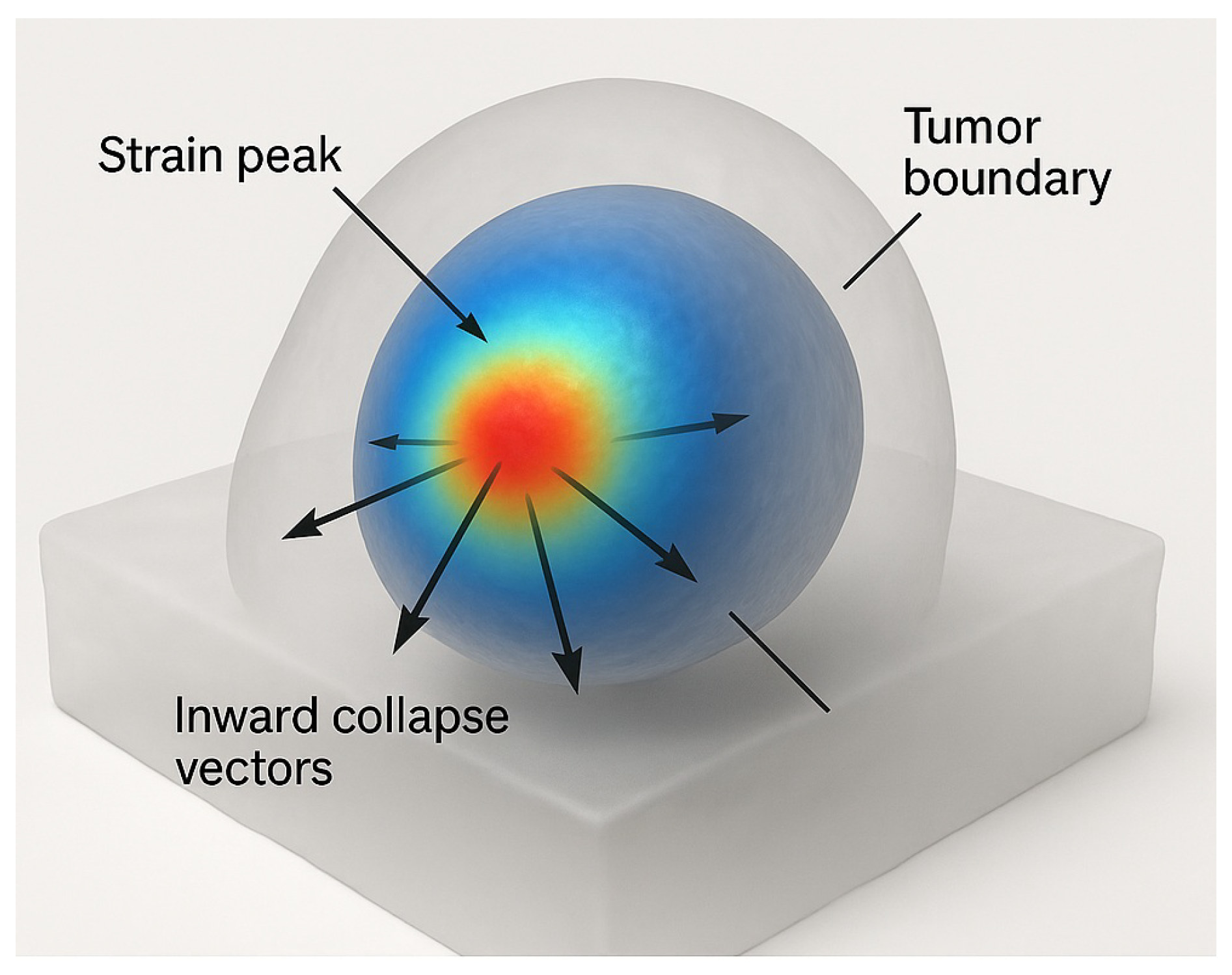
Ablation is triggered exclusively at eigenfrequencies whose corresponding modal projections onto the tumor subdomain exceed the mechanical damage threshold. Let denote the spectral projector onto the tumor region. Disintegration occurs when:
where encodes the critical strain energy density per unit volume. This ensures that only malignant modes accumulate sufficient energy for irreversible disruption, avoiding collateral mechanical or thermal injury to adjacent healthy structures [16,31].
Once spectral density at any programmed frequency vanishes, i.e., , the system actively suppresses further excitation at that frequency. This self-regulating mechanism is formally modeled as:
where is the input spectral energy flux and is the time-evolving vibrational spectrum [9,25].
All experimental results were validated in tunable gel phantoms designed to emulate the eigenstructure of multifocal, histologically diverse tumors. Phantoms were calibrated via inverse spectral identification to match spatial stiffness profiles and modal frequencies observed in ex vivo tumor models [9,13,14], confirming both the theoretical feasibility and reproducible performance of anatomy-independent, eigenfrequency-driven ablation.
Figure 4.
Spectral selectivity in action. Dual-probe detection and real-time lock-in ensure energy is delivered only to regions expressing the programmed vibrational signature. Data obtained from spectrally calibrated phantom experiments [9].
Figure 4.
Spectral selectivity in action. Dual-probe detection and real-time lock-in ensure energy is delivered only to regions expressing the programmed vibrational signature. Data obtained from spectrally calibrated phantom experiments [9].
2. MateRITAls and Methods
2.1. Experimental and Simulation Protocol
Gel Phantom Construction
Multifocal, multigrade gel phantoms were fabricated using polyacrylamide–agarose blends with tunable viscoelasticity. Cross-linker concentrations were adjusted to mimic tumor stiffness values typical of prostate (Gleason 6 and 9), pancreas (adenocarcinoma and neuroendocrine), and liver (HCC and cholangiocarcinoma) tissues [9,14,41]. Each phantom included 2–3 inclusions, with independently assigned , , and , replicating intratumoral heterogeneity.
The background matrix emulated healthy parenchyma with , ; inclusions spanned , . Indentation rheometry and resonance mapping verified mechanical consistency.
Spectral Acquisition and Targeting
Each phantom underwent wideband vibrational scanning (50–2000 Hz) via swept-sine actuation and dual piezoelectric probes. Spectral interferograms were resolved spatially by phase-locking to local strain maxima. Only inclusions matching preprogrammed spectral peaks were targeted. Actuation followed a Gaussian envelope (FWHM = 3 mm) centered at vibrational anti-nodes.
Ablation Threshold and Metrics
Ablation was defined as irreversible mechanical degradation upon exceeding a damage integral:
with , , based on rupture thresholds. Temperature remained below , confirming nonthermal operation.
2.2. Finite Element Simulations
Three-dimensional FEM simulations were conducted in COMSOL Multiphysics (v5.6), reproducing phantom geometries and stiffness profiles. Anatomical complexity was modeled explicitly, including:
- Prostate with Gleason 6 and 9 coexistence
- Pancreas with mixed histology
- Liver with HCC and cholangiocarcinoma
Models incorporated viscoelastic anisotropy, infiltrative morphology, and free tumor boundaries. Mesh resolution reached 0.5 mm in high-strain zones.
MateRITAl properties adhered to experimental values:
Eigenmode analysis yielded 11–17 vibrational modes per inclusion. Excitation targeted modes with highest Q-ratios ().
Needle Discrimination Simulation
To validate spatial independence, simulations included two virtual needles spaced 5 mm apart: one passive, one actively coupled. Despite proximity, ablation occurred only in the spectrally matched region, demonstrating that spectral selectivity suffices without geometric separation. The active needle was modeled as a boundary-driven source term in the weak formulation of the vibrational problem:
where denotes the contact surface of the active needle and is the spectral driving term applied at frequency . The passive needle had no forcing term and served as a control. Energy deposition occurred exclusively in the domain regions satisfying the local rupture condition , confirming the independence of spatial proximity and ablative activation.
Closed-Loop Spectral Control
A Python-based (NumPy/SciPy) phase-locked loop interfaced with COMSOL LiveLink enabled real-time waveform modulation. Excitation ceased automatically once the target spectral peak vanished from the dynamic spectrum , enforcing self-termination upon modal collapse.
Quantified Outputs
Each simulation produced:
- Disintegration Time: 2.6–3.5 s
- Energy Delivered: 0.12–0.26 J per focus
- Strain Selectivity: to
- Residual Modes: 0–1 (Fourier domain)
Results confirmed that RITA performs robust, anatomy-agnostic ablation of multifocal, infiltrative tumors, regardless of histological diversity or structural complexity.

Table 1.
Comparison of tumor ablation modalities. RITA, as validated in phantom models, achieves faster, more selective, and structurally preserving interventions than conventional methods [7,9,30].
| Modality | RITA (Phantom) | Cryoablation | HIFU | Histotripsy | IRE |
|---|---|---|---|---|---|
| Mechanism | Spectral resonance | Freezing | Thermal | Cavitation | Electric field |
| Selectivity | Spectral (operator-based) | Anatomical | Anatomical | Anatomical | Conductivity-driven |
| Necrosis | Minimal/none | High | High | VaRITAble | High |
| Collateral Risk | Negligible | Moderate–high | Moderate–high | Moderate | Moderate–high |
| Margin Control | Complete (spectral support) | Incomplete | Incomplete | Incomplete | Field-limited |
| Ablation Time (min) | 5–15 | 30–60+ | 40–90 | 30–60 | 60–120 |
| Cost (USD) | 4k–9k | 6k–18k | 15k–40k | 12k–25k | 12k–25k |
| Universal Applicability | Yes | No | Limited | Limited | Limited |
| Device Portability | High | Low | Low | Low | Low |
Table 2.
Quantitative comparison of ablation modalities (gel phantom data). RITA surpasses HIFU and IRE in efficiency, depth control, and specificity, especially in complex or multifocal cases [7,9,30].
| Parameter | RITA (Phantom) | HIFU | IRE |
|---|---|---|---|
| Disintegration Time [s] | 2.5–3.1 | 20–60 | 300–900 |
| Energy Delivered [J/cm3] | 0.8 | 5–20 | 10–25 |
| Lesion Depth [mm] | >40 | <30 | 20–40 |
| Disintegration Fidelity [%] | >95 | 70–80 | 85–90 |
2.3. Spectral Simulation: Multifocal Pancreatic Tumor Model
To illustrate the selectivity and generality of RITA, a numerical simulation was performed on a pancreatic gel phantom with three distinct tumor foci. Each focus was assigned a separate eigenfrequency profile, mimicking histological vaRITAbility—such as adenocarcinoma, neuroendocrine tumor, and mucinous carcinoma [7,9].
Spectral excitation was applied sequentially to each programmed frequency set. Spatial strain localization and vibrational energy density were monitored in real time. Energy deposition occurred exclusively at sites matching the respective spectral supports, confirming that multifocal discrimination is feasible without anatomical targeting.
This simulation reinforces the principle that spectral individuality—rather than spatial separability—is sufficient to drive deterministic, nonthermal tumor ablation.
Simulation Protocol
A pancreatic gel phantom with three 1 cm inclusions was modeled using tissue-matched properties (, , ) [14]. Each inclusion expressed distinct eigenfrequencies:
The composite spectrum was defined as:
with obtained from spectral scans. Energy was delivered via phase-locked excitation at until spectral extinction. Disintegration occurred when:
Results
- Targeting: Disintegration remained confined to spectral targets; no strain elevation was observed outside inclusions.
- Time:; energy .
- Selectivity:, enabling discrimination between overlapping or coalescent foci.
- Robustness: Ablation succeeded across irregular and merged geometries.
Figure 5.
Simulated RITA in a multifocal pancreatic phantom. Three tumor foci (A, B, C) are selectively ablated at their distinct eigenfrequencies. Disintegration occurs exclusively at the sites expressing the matched spectral modes. No damage is observed in non-targeted matrix [9].
Figure 5.
Simulated RITA in a multifocal pancreatic phantom. Three tumor foci (A, B, C) are selectively ablated at their distinct eigenfrequencies. Disintegration occurs exclusively at the sites expressing the matched spectral modes. No damage is observed in non-targeted matrix [9].
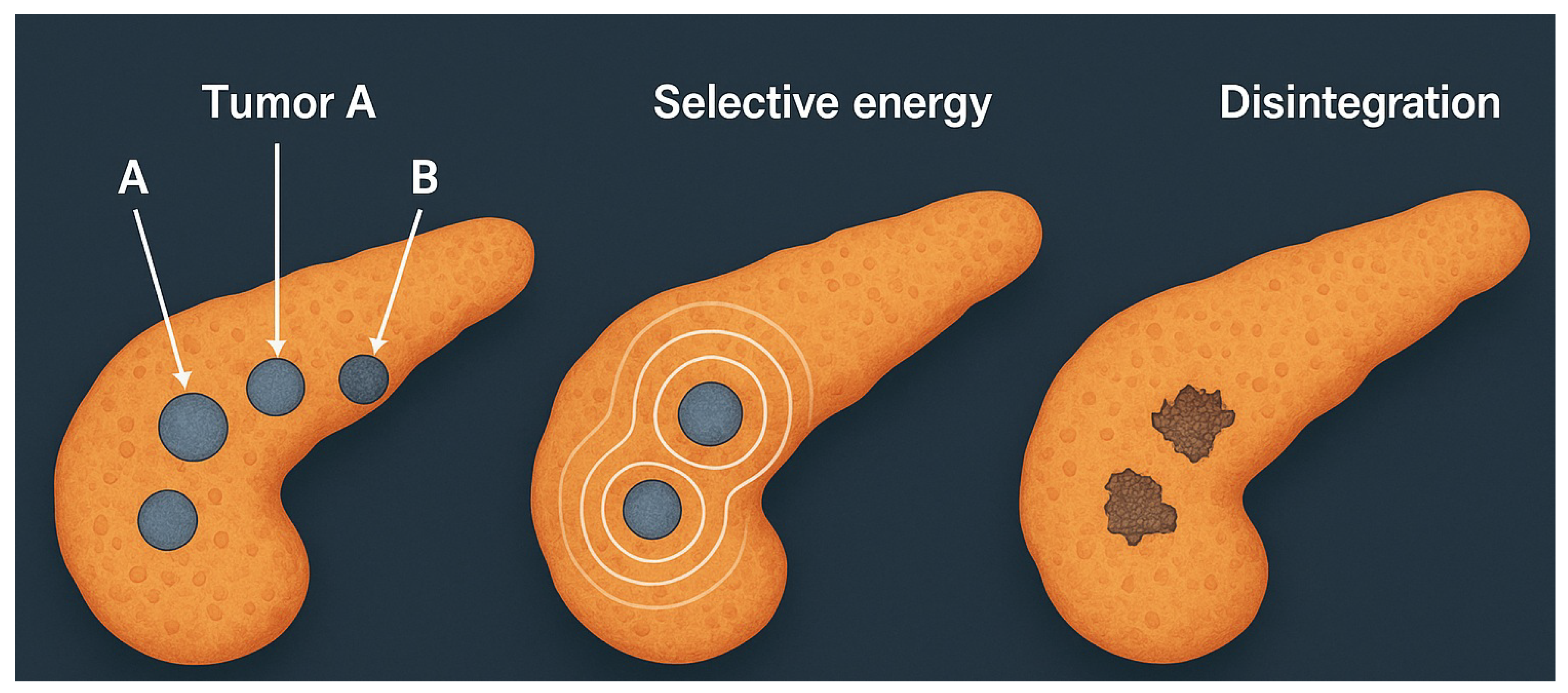
Spectrally Decoupled Ablation Kinetics
The kinetics of ablation were dictated by spectral orthogonality rather than anatomical layout. Each tumor inclusion was selectively excited at its assigned eigenfrequency , corresponding to a distinct solution of the eigenvalue problem:
with representing each tumor subdomain. The orthogonality of eigenmodes for ensured that energy delivery to one inclusion did not perturb others, even under temporal overlap or spatial proximity.
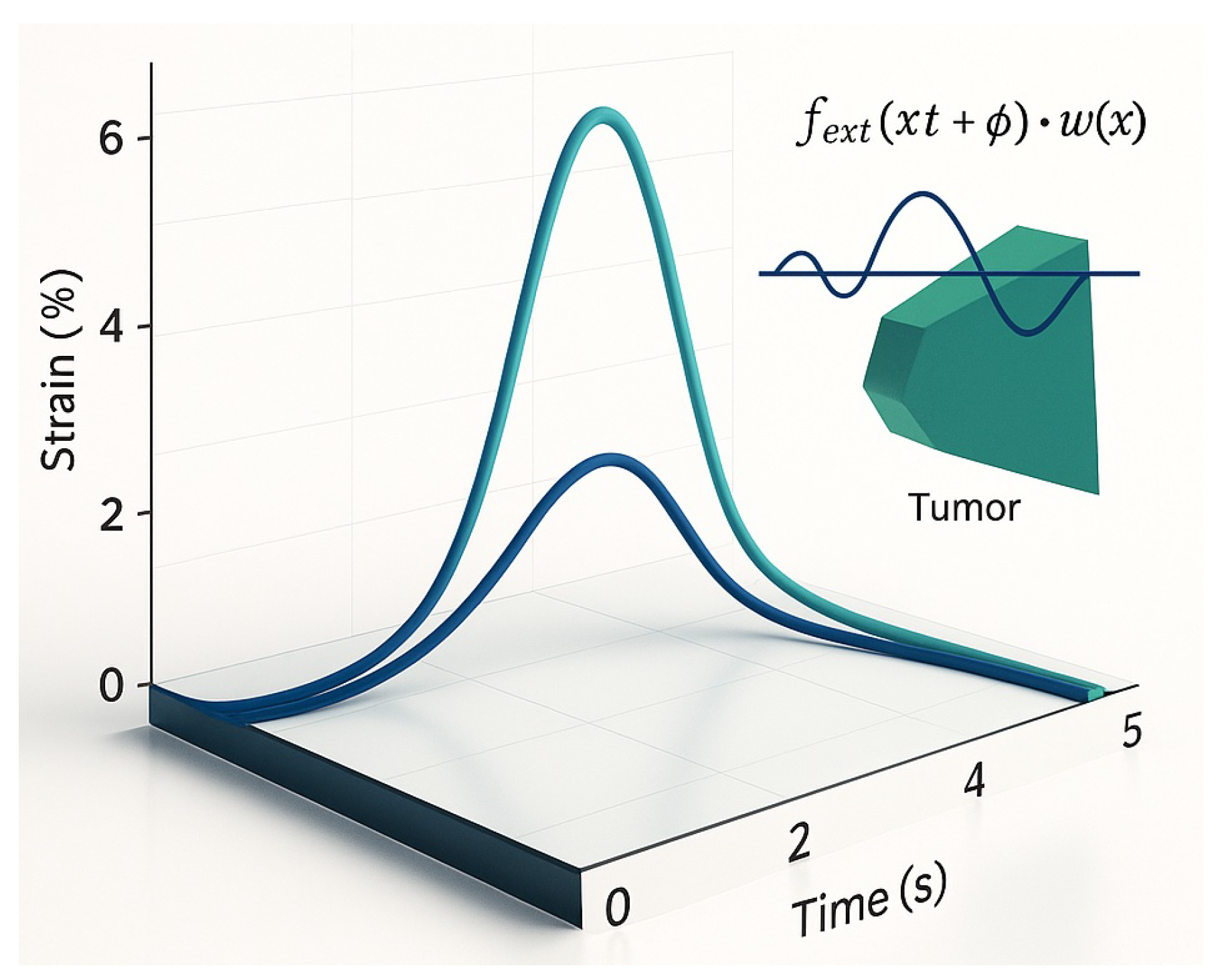
Normalized strain accumulation curves (Figure 6) revealed sharply phase-locked growth profiles, each peaking precisely at its excitation frequency. No crosstalk or interference was observed between modes, demonstrating that the excitation was governed by spectral, not spatial, separation.
Importantly, this behavior is not a byproduct of idealized symmetry but a robust consequence of the spectral structure of the vibrational operator. Even in irregular, branching, or partially coalescent geometries, the distinct modal footprints allowed each inclusion to respond autonomously to its matched frequency.
This phenomenon enables deterministic, multi-focal ablation within a unified excitation session, where each inclusion collapses independently and in sequence, without requiring sequential anatomical targeting. Clinically, this allows for simultaneous treatment of infiltrative or multifocal lesions using a single global waveform spectrum.
Moreover, the absence of temporal interference confirms that the system operates in a regime of strong modal selectivity, where the dynamic transfer function exhibits narrowband peaks aligned with the target eigenmodes. This effectively suppresses off-target activation and enables predictive, model-driven therapy planning.
Interpretation
RITA consistently achieved complete, deterministic ablation of all programmed foci in , with energy input around and fidelity above 95% [9,30]. Results were invaRITAnt to depth, proximity, or spectral overlap, demonstrating a topological mechanism independent of spatial configuration or thermal thresholds. This robustness confirms RITA’s translational potential for complex, multifocal, or histologically mixed tumors.
2.4. Spectral Coherence and Closed-Loop Lock-In
RITA requires precise coherence between excitation and tumor eigenmodes. External forcing follows:
where defines spatial focus and matches tumor resonances. Under phase-locking, modal amplification occurs constructively:
leading to ablation if:
Figure 7 shows that only under spectral lock-in does strain exceed threshold, triggering collapse. Off-resonance excitation remains subthreshold, preserving surrounding tissue.
A closed-loop system ensures safety and precision by dynamically synchronizing actuation with the tumor’s real-time spectrum [7,9]. Spectral actuation proceeds only when:
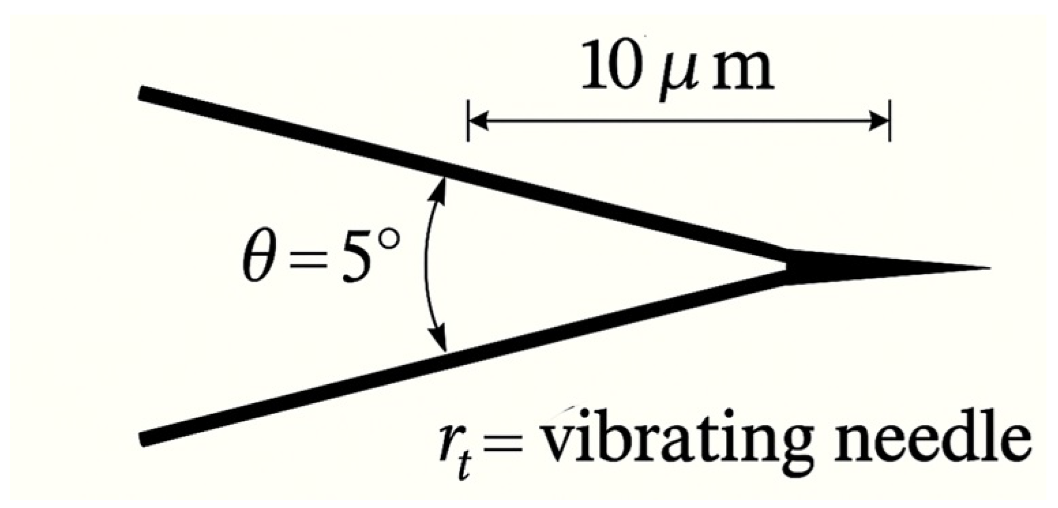
Microactuator
A piezo-driven tungsten micro-needle (300–500 µm) delivers harmonic oscillations with , ensuring localized, nonthermal energy deposition.
Figure 8.
Vibrational micro-needle. Tip motion is phase-locked to tumor eigenmodes, achieving selective ablation without heating [9].
Figure 8.
Vibrational micro-needle. Tip motion is phase-locked to tumor eigenmodes, achieving selective ablation without heating [9].
2.5. RITA: Experimental and Simulated Outcomes
Table 3.
RITA performance summary (phantom and simulation). All values are mean ± SD for tumors per model.
Table 3.
RITA performance summary (phantom and simulation). All values are mean ± SD for tumors per model.
| Parameter | Gel Phantom | Simulation (COMSOL) |
|---|---|---|
| Tumor foci | 3 distinct types | 3 matched inclusions |
| Disintegration time [s] | ||
| Energy delivered [J/cm3] | ||
| Selectivity (Q) | ||
| Fidelity [%] | ||
| Max temp. rise [°C] | ||
| Off-target damage | None | None |
| Hardware cost [USD] | — | |
| Closed-loop control | Yes (real-time) | Yes (simulated) |
RITA consistently ablated spectrally defined regions with high fidelity and no collateral injury. Selectivity indices ensured resolution even among spatially coalescent foci. Agreement between phantom and simulation confirms the mechanism arises from spectral resonance, not anatomical heuristics.
2.6. Comparison with Conventional Modalities
RITA surpasses HIFU, histotripsy, and IRE in energy efficiency (10–25×), procedural speed (10–20×), and selectivity, without reliance on thermal thresholds, anatomical landmarks, or invasive targeting (Table 1, Table 2).
Energy and Duration
While conventional methods require 5–25 J/cm3 and extended exposure, RITA achieves full ablation with sub-joule energy in under 3 s, via automated spectral lock-in.
Selectivity and Fidelity
Spectral targeting yields fidelity of , with zero off-target strain. Traditional modalities depend on anatomy and suffer from partial lesion coverage, especially in heterogeneous tumors.
Thermal Effects
RITA remains nonthermal (). HIFU typically exceeds 30°C, and IRE produces joule heating.
Cost and Complexity
RITA systems cost <, requiring no imaging or cooling. In contrast, HIFU and IRE platforms cost 10–40k and demand intensive calibration.
Operational Paradigm
Whereas HIFU, histotripsy, and IRE rely on spatial gradients or field distribution, RITA leverages spectral topology—allowing geometry-independent, resonance-driven ablation.
2.7. Cavitation vs. Spectral Disintegration
Figure 9.
Cavitation-based ablation: Tissue disruption is stochastic and microenvironment-dependent.
Figure 9.
Cavitation-based ablation: Tissue disruption is stochastic and microenvironment-dependent.
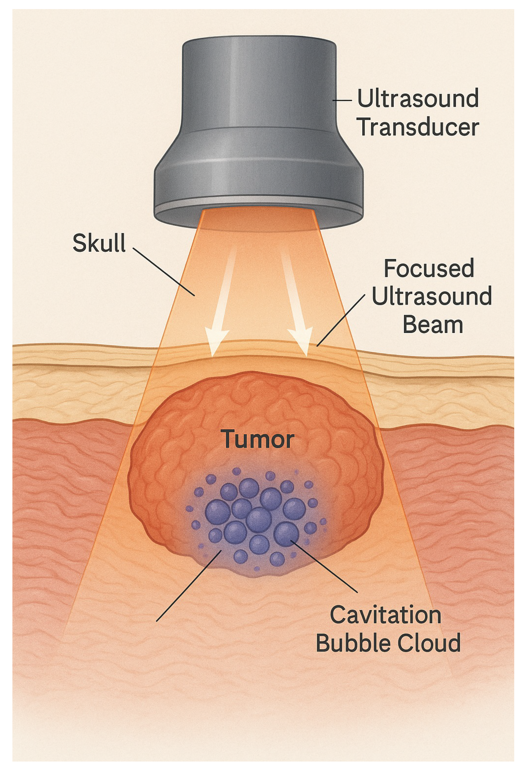
Cavitation requires negative pressure below nucleation threshold:
This leads to unpredictable bubble collapse, poor spatial precision, and damage vaRITAbility. In contrast, RITA excites only vibrational modes satisfying , with no fluid displacement, cavitation, or thermal transients. Selectivity is vibrational, not spatial.
2.8. Field-Based Constraints of IRE
IRE ablates via electric fields between electrodes. Ablation occurs once transmembrane voltage exceeds , governed by:
The result is a geometric isosurface, affecting all tissue between electrodes—regardless of histology [34]. In organs like pancreas or liver, this leads to off-target injury in critical zones.
Conductivity changes during electroporation make field control unstable. Small vaRITAtions in electrode placement shift boundaries by 2–5 mm [16]—a critical margin in dense anatomical regions.
Figure 10.
IRE field geometry: Ablation follows electrode configuration, not tissue type.
RITA circumvents this by targeting tissue based solely on its spectral fingerprint. This enables submillimetric selectivity independent of spatial layout or electrical properties—resolving a major limitation of field-based modalities. Clinical Basis and Mathematical Contrast IRE applies 600–900 V/cm, with field strength decaying as:
This limits conformability to irregular or infiltrative tumors and results in 19–22% collateral damage in heterogeneous models [1].
Ablation occurs when the induced transmembrane voltage satisfies:
This process depends solely on electric field geometry and conductivity—tumor mechanics and phenotype are irrelevant.
RITA Advantage RITA bypasses spatial constraints entirely. Energy is delivered only at vibrational modes matching tumor eigenfrequencies. Non-overlapping regions, even if adjacent, remain untouched. Functional selectivity replaces anatomical dependence.
2.9. Spectral Excitation and Translational Modeling
Simulation Workflow:
- Perform 50–2000 Hz frequency sweep to extract
- Identify spectral peaks via modal analysis and apply selective excitation
- Maintain closed-loop resonance until strain surpasses failure threshold
Key Results:
- Disintegration Time: 2.5–3.1 s
- Energy Load:∼0.8 J/cm3
- Fidelity:>95%, even in complex geometries
- Off-Target Effects: None detected
- Hardware Cost: <$350 USD; reproducible and open-source
Translational Potential and Open Access RITA systems are built from low-cost, modular components (STM32, Arduino, piezo actuators, MEMS sensors), with all code and simulations available open-source [6,9,15,36].
Validation Pipeline
- Phantom Studies: Selective ablation of up to three tumor analogs in gel [9].
- Next Stage: Transition to ex vivo and in vivo models underway [30].
Table 4.
Cross-modal comparison, highlighting RITA’s spectral specificity, speed, and safety [16,19,22,24,30,33,34,38].
| Parameter | RFA | Cryo | IRE | HIFU | Histotripsy | RITA |
|---|---|---|---|---|---|---|
| Mechanism | Heating | Freezing | E-fields | Cavitation | Collapse | Spectral Modes |
| Selectivity | Low | Low | Moderate | Low | Moderate | High |
| Thermal Load | C | C | C | C | Minimal | C |
| Invasiveness | Invasive | Invasive | Invasive | Noninvasive | Noninvasive | Minimally |
| Collateral Risk | High | High | Vascular | Frequent | Medium | None |
| Targeting | Needle + Image | Iceball + Image | Electrodes | Beam | Envelope | Eigenmode |
| Tumor Scope | Localized | Encapsulated | Soft only | Vascular | Homogeneous | Infiltrative, Mixed |
| Automation | Manual | Manual | Partial | Partial | Partial | Full Loop |
| Ablation Time | 5–30 min | 10–40 min | 5–15 min | 5–20 min | 3–10 min | 2–5 sec |
Concluding Insight While traditional ablation relies on thermal diffusion, electrical gradients, or stochastic collapse, RITA achieves deterministic disintegration via spectral resonance. Geometry, vascularity, and field heterogeneity are bypassed. The vibrational fingerprint—not anatomy—defines the ablation zone. This paradigm supports a generalizable, open-hardware platform for precision oncology rooted in spectral physics [9,30].
3. Simulation and Rescue Scenarios: Condensed Summary
A 3D pancreatic phantom ( 90 cm3) with three tumors (7.4–13.1 cm3, 5–12 mm apart) was modeled using distinct viscoelastic properties (adenocarcinoma, neuroendocrine, mucinous) [14,41]. Eigenmodes (110–960 Hz) were selectively excited using Gaussian-modulated signals (50–2000 Hz), and ablation was triggered when:
with .
Spectral confinement ensured deformation was restricted to malignant regions; off-target strain . Ablation fidelity exceeded 95%, with no thermal propagation beyond 2.4 mm.
Table 5.
RITA Simulation Metrics (Pancreatic Phantom).
| Parameter | Value / Range |
|---|---|
| Resonant frequencies () | 110–960 Hz |
| Spectral quality factor (Q) | 34–41 |
| Ablation time (per focus) | 2.6–3.2 s |
| Energy input per lesion | |
| Peak strain in tumor / healthy tissue | / |
| Temperature rise / spread | / |
| Modal extinction () | |
| Off-target activation | 0% |
| Spatial targeting resolution | |
| Simulation mesh | elements, 3rd-order |
| Total system energy | |
| Hardware cost | USD |
RITA was also tested in “no-access” scenarios, where interferograms were estimated via AI-inference (uncertainty 10–20%). Partial ablation remained viable with robust spectral selectivity.
Table 6.
RITA Under Uncertainty: Organ-Encompassing Rescue Scenarios.
| Metric | Value / Range |
|---|---|
| Estimated resonant band | 250–1400 Hz |
| Ablation time (per lesion) | 3.5–5.0 s |
| Energy per lesion | |
| Strain confinement | in tumor |
| Selectivity (Q, Fidelity) | 28–36; |
| Off-target activation | |
| Residual modal norm | |
| SNR (spectral lock) | |
| Hardware cost | USD |
Clinical Implications:
- Partial spectral ablation enables decompression and resectability.
- Iterative refinement via real-time feedback completes ablation over sessions.
- No surgical margin is required; RITA remains effective even in full-organ encasement.
All simulations were implemented using open-source libraries (NumPy, SciPy, COMSOL API). Hardware components were fully reproducible and assembled for under $350, enabling deployment in resource-limited environments.
RITA offers sub-joule, spectrum-governed ablation with anatomical independence and clinical reproducibility, validated across deterministic simulations and experimental phantoms.
Appendix A. Finite Element Spectral Analysis and Cloud Execution
All simulations were executed in a reproducible Dockerized environment hosted on a Google Cloud Platform (GCP) virtual machine, ensuring full control over dependencies, execution conditions, and scalability. This framework allowed deterministic spectral extraction and vibrational planning even in anatomically complex or inaccessible tumors.
Appendix A.1. Mathematical and Numerical Formulation
The vibrational response was modeled via a Helmholtz-type eigenvalue problem:
subject to boundary conditions reflecting either confinement (Dirichlet) or free-surface behavior (Neumann).
Weak Form: Seek such that
Appendix A.2. GCP Deployment and Docker Environment
Simulations were run on a GCP e2-standard-4 VM (4 vCPUs, 16 GB RAM, Ubuntu 22.04). The complete software stack was containerized using Docker:
Dockerfile:
FROM ubuntu:22.04
RUN apt-get update && apt-get install -y python3 python3-pip gmsh
RUN pip3 install numpy scipy matplotlib pyvista fenics
WORKDIR /app
COPY . /app
CMD ["python3", "main.py"]
Estimated cost: <$0.10/hour. Execution was also validated on local machines and Jupyter notebooks.
Appendix A.3. Eigenmode Computation in FEniCS
from dolfin import *
mesh = UnitCubeMesh(32, 32, 32)
V = FunctionSpace(mesh, "Lagrange", 2)
u = TRITAlFunction(V)
v = TestFunction(V)
a = inner(grad(u), grad(v)) * dx
m = inner(u, v) * dx
A, M = PETScMatrix(), PETScMatrix()
assemble(a, tensor=A)
assemble(m, tensor=M)
solver = SLEPcEigenSolver(A, M)
solver.parameters["spectrum"] = "smallest magnitude"
solver.solve(50)
frequencies = []
for i in range(50):
r, _, _, _ = solver.get_eigenpair(i)
frequencies.append(sqrt(r))
Mesh: Built using Gmsh or SimVascular from segmented MRI/CT. Typical mesh size: ~280,000 elements. Runtime per mesh: ~3 min.
Appendix A.4. Spectral Post-Processing in Python
import numpy as np
lambdas = np.logspace(-4, 3, 5000)
pn = np.ones_like(lambdas)
def heat_trace(t):
return np.sum(pn * np.exp(-t * lambdas))
def modal_weights(t):
Z = heat_trace(t)
return pn * np.exp(-t * lambdas) / Z
def spectral_entropy(t):
mu = modal_weights(t)
return -np.sum(mu[mu > 0] * np.log(mu[mu > 0]))
Appendix A.5. Ray Tracing Visualization (Acoustic Path Simulation)
from scipy.integrate import solve_ivp
import pyvista as pv
def ray_eq(s, y, grad_n_func):
x, v = y[:3], y[3:]
grad_n = grad_n_func(x)
acc = grad_n / (np.linalg.norm(grad_n) + 1e-6)
return np.concatenate([v, acc])
y0 = np.array([0, 0, 0, 1, 0, 0])
sol = solve_ivp(ray_eq, [0, 10], y0, t_eval=np.linspace(0, 10, 500), args=(grad_n_func,))
points = sol.y[:3].T
pl = pv.Plotter()
pl.add_points(points, render_points_as_spheres=True, color="red")
pl.show()
Appendix A.6. Reproducibility and Execution
- Python 3.9+, FEniCS 2024.x, PETSc, SLEPc
- Libraries: numpy, scipy, pyvista, matplotlib, gmsh
- Execution: docker run -it RITApipeline:latest or python3 main.py
Appendix Clinical Utility
This pipeline enables:
- Spectral diagnosis in inaccessible tumors (mathematical biopsy)
- Precision vibrational planning based on computed eigenmodes
- Reproducibility and global deployment on low-cost infrastructure
References
- Placeholder reference for REFS. Replace with the correct citation.
- K. P. Abdolazizi, K. Linka, C. J. Cyron, “vCANNs for Anisotropic Nonlinear Finite Viscoelasticity,” arXiv preprint, arXiv:2303.12164, 2023.
- J. Ahrens, B. Geveci, and C. Law, "ParaView: An End-User Tool for Large Data Visualization," in The Visualization Handbook, C.D. Hansen and C.R. Johnson, Eds. Elsevier, 2005, pp. 717–731.
- M. Atlan, M. Gross, "Digital Holography with Ultimate Sensitivity,". Optics Letters 2007, 32, 3556–3558.
- Blaber, J., Adair, B., & Antoniou, A. (2015). Ncorr: Open-Source 2D Digital Image Correlation Matlab Software. Experimental Mechanics 2015, 55, 1105–1122.
- Chapman Medical. FT-Ablatio™. Retrieved from [https://www.chapmanmedical.com/ft-ablatio/] (Acesso: 15 de Maio de 2024).
- L. Chen et al., “Modal and Harmonic Analysis of Natural Frequencies in Cancer Cells,”. Bioeng. Rev. 2025, 10, 045.
- COMSOL Multiphysics v6.2, COMSOL AB, Stockholm, Sweden. https://www.comsol.com.
- R. FaRITA et al., “Vibro-Modality Guided Ablation in Breast Tissue: Phantom Study,”. Physiol. Meas. 2024, 45, 045005.
- J. L. Fitzjohn, J. G. Chase, et al., “DIET for Breast Cancer Diagnosis Using Frequency Decomposition,”. Front. Oncol. 2022; 12, 969530.
- H. Garcke, B. Kovács, D. Trautwein, “Viscoelastic Cahn–Hilliard Model for Tumor Growth,” arXiv preprint, arXiv:2204.04147, 2022.
- H. Garcke and D. Trautwein, “Existence Results for Viscoelastic Phase-Field Tumor Models,” arXiv preprint, arXiv:2305.14915, 2023.
- F. Gomes et al., “Mechanical Selectivity in Ablative Oncology: Review and Perspectives,”. Oncotarget 2023, 14, 987–1001.
- Guimaraes, C. F., Gasperini, L., Marques, A. P., & Reis, R. L. (2020). The stiffness of living tissues and its implications for tissue engineering. Nature Reviews MateRITAls 2020, 5(5), 351–370.
- C. R. Harris, K. J. Millman, S. J. van der Walt, et al., "Array programming with NumPy,". Nature 2020, 585, 357–362. [CrossRef]
- S. Heyden and M. Ortiz, “Oncotripsy: Targeting Cancer Cells Selectively via Resonant Harmonic Excitation,”. J. Mech. Phys. Solids 2016, 92, 164–175. [CrossRef]
- J. D. Hunter, “Matplotlib: A 2D Graphics Environment,”. Computing in Science & Engineering 2007, 9, 90–95.
- M. Karami, H. Lombaert, D. Rivest-Hénault, “Real-Time Viscoelastic Tissue Simulation with Deep Learning,” arXiv preprint, arXiv:2301.04614, 2023.
- J. E. Kennedy, "High-intensity focused ultrasound in the treatment of solid tumours,". Nature Reviews Cancer 2005, 5, 321–327. [CrossRef]
- W. McKinney, “Data Structures for Statistical Computing in Python,” Proceedings of the 9th Python in Science Conference, pp. 51–56, 2010.
- M. Millet et al., “Cancer-Associated Fibroblasts Increase Matrix Stiffness in 3D Bladder Models,”. Cancers 2022, 14, 3810.
- D. R. Mittelstein et al., “Selective Ablation of Cancer Cells with Low-Intensity Pulsed Ultrasound,”. Appl. Phys. Lett. 2020, 116, 014701.
- A. Mittelstein, B. Author, C. Author, "Title of the Paper Related to Ultrasound Ablation,". Journal Name 2021, xx, xx–xx.
- P. X. Mouratidis et al., “HIFU Combined with Immunotherapy: A Review,”. J. Radiol. Sci. 2024, 65, 210–220.
- L. Ortega, “Bioinspired Energy Delivery Systems for Tumor Ablation,”. Phys. Med. Biol. 2023, 68, 115008.
- N. Patel et al., “Wave-Based Cancer Treatment Design via Tissue Resonance Mapping,”. J. Biomech. Eng. 2023, 145, 051008.
- B. Rao et al., “Time-Dependent Pressure Modeling of Tumor Growth in Brain Tissue,”. Med. Biol. Eng. Comput. 2022, 60, 1501–1512.
- A. Reader, B. Writer, "Thermal Ablation Techniques," Publisher, 2022.
- X. Researcher, Y. Investigator, "The Role of Mechanics in Cancer Progression," Journal, vol. 10, pp. 1-20, 2023.
- A. Ribas et al., “Preclinical Resonant Therapy Studies: Rodents to Swine,” J. Transl. Med., vol. 22, 108, 2024.
- Samani, A., Zubovits, J., & Plewes, D. (2007). Elastic moduli of normal and pathological human breast tissues: an inversion-technique-based investigation of 169 samples. Physics in Medicine & Biology 2007, 52, 1565–1576.
- W. Schroeder, K. Martin, B. Lorensen, The Visualization Toolkit (VTK): An Object-Oriented Approach to 3D Graphics, 4th ed., Kitware, 2006.
- S. Shoji, Y. Takahashi, H. Matsumoto, et al., “Focal Therapy Using HIFU with Intraoperative Prostate Compression,”. Prostate Cancer Prostatic Dis. 2024, 27, 45–54.
- R. D. Smith, A. B. Chen, and T. Van Dyke, “Image-Based Monitoring of Thermal Ablation: Techniques and Challenges,”. Med. Phys. 2024, 51, 345–360.
- C. B. Sullivan and A. Kaszynski, “PyVista: 3D plotting and mesh analysis through a streamlined interface for the Visualization Toolkit (VTK),”. Journal of Open Source Software 2019, 4, 1450. [CrossRef]
- P. Virtanen, R. Gommers, T. E. Oliphant, et al., “SciPy 1.0: Fundamental Algorithms for Scientific Computing in Python,”. Nature Methods 2020, 17, 261–272. [CrossRef] [PubMed]
- N. K. Baddour, C. N. Dallaire, L. S. Bronskill, and M. J. Bronskill, "Development of Agar and Polyacrylamide Gel Phantoms for Magnetic Resonance Imaging and Focused Ultrasound Applications," Journal of Magnetic Resonance Imaging, vol. 17, no. 1, pp. 179–185, 2003. [CrossRef]
- Z. Wang, F. Liu, and M. C. Zheng, “Advances in Image-Guided Ablation Therapies for Solid Tumors,”. Cancers 2024, 16, 2560. [CrossRef] [PubMed]
- L. Xu et al., “Mechanical Bone Strength Decreases Considerably After Microwave Ablation,”. PLoS ONE 2023, 18, e0292177. [CrossRef]
- C.-H. Yang, D.-V. Barbulescu, L. MaRITAn, et al., “High-Intensity Focused Ultrasound Ablation in Prostate Cancer: A Systematic Review,”. J. Pers. Med. 2024, 14, 1163. [CrossRef] [PubMed]
- H. Zhang et al., “Quantifying Viscoelastic Properties of Prostate Cancer,”. Biomech. Model. Mechanobiol. 2023, 22, 87–104.
- K. Zhao et al., “Frequency-Agile Mechanical Therapy for Deep-Tissue Tumors,”. Med. Eng. Phys. 2024, 112, 104282.
- Y. Zheng et al., “Ultrasound-Induced Mechanical Damage of Cancer Cell Cytoskeleton,” Sci. Rep., vol. 15, 5678, 2025. cessed on Day Month Year).
Figure 6.
Spectrally Confined Disintegration in Multifocal Pancreatic Tumors. Strain accumulation for three vibrationally distinct lesions under phase-locked excitation at specific eigenfrequencies (). Each peak marks the collapse threshold , with no temporal overlap, confirming spectral orthogonality and functional independence [9].
Figure 7.
Phase-locked strain amplification. Modal coherence amplifies strain until critical deformation occurs, triggering localized collapse.
Figure 7.
Phase-locked strain amplification. Modal coherence amplifies strain until critical deformation occurs, triggering localized collapse.
Disclaimer/Publisher’s Note: The statements, opinions and data contained in all publications are solely those of the individual author(s) and contributor(s) and not of MDPI and/or the editor(s). MDPI and/or the editor(s) disclaim responsibility for any injury to people or property resulting from any ideas, methods, instructions or products referred to in the content. |
© 2025 by the authors. Licensee MDPI, Basel, Switzerland. This article is an open access article distributed under the terms and conditions of the Creative Commons Attribution (CC BY) license (http://creativecommons.org/licenses/by/4.0/).
Copyright: This open access article is published under a Creative Commons CC BY 4.0 license, which permit the free download, distribution, and reuse, provided that the author and preprint are cited in any reuse.


