
Submitted:
11 July 2025
Posted:
11 July 2025
You are already at the latest version
Abstract
Background: The lipid components of the skin barrier have the strongest structure when arranged in an orthorhombic packing. This structure can be influenced by the external supply of lipophilic ingredients. While the benefits of ceramide supplementation are well-documented, the effects of the cosmetic formulation's oil-based ingredients have been less explored. Methods: The packing structures of commonly used oil and wax ingredients in cosmetics were analyzed using FT-IR. These components were then combined to formulate a cosmetic composition with an orthorhombic packing structure. The strength of the skin barrier was assessed by measuring transepidermal water loss (TEWL), and the lipid packing of the porcine skin was analyzed using FT-IR. Results: Combining oil, wax and emulsifiers resulted in a cosmetic composition with an orthorhombic packing structure. In contrast, other combinations resulted in a hexagonal packing structure. The orthorhombic formulations maintained the structure and function of the porcine skin lipid barrier without disruption Conclusion: This study confirmed that an orthorhombic packing structure could be achieved without using the three main components of the skin barrier, such as ceramides, by reconstituting the formulation with ingredients that inherently have an orthorhombic structure. This formulation effectively maintains the integrity of healthy skin barriers.
Keywords:
orthorhombic packing
; skin lipid barrier
; cosmetic formulation
; FT-IR analysis
1. Introduction
The skin barrier plays a crucial role in maintaining homeostasis by regulating water loss and protecting against external irritants [1]. The outermost layer of the skin, the stratum corneum, is composed of a highly organized lipid matrix primarily consisting of ceramides, free fatty acids, and cholesterol [2,3]. These intercellular lipids exist in distinct structural arrangements, such as orthorhombic and hexagonal packing [4], which directly influence barrier integrity and function [4]. The stability of the lipid organization is critical for maintaining the protective function of the skin, and disruptions in this structure can lead to increased transepidermal water loss (TEWL) and vulnerability to external stressors [5].
Understanding lipid packing arrangements and their impact on barrier function is essential for developing effective cosmetic and dermatological formulations. Fourier Transform Infrared Spectroscopy (FT-IR) has been widely utilized to analyze lipid structural organization within the skin [6,7]. FT-IR spectroscopy allows for the identification of specific vibrational modes associated with hydrocarbon chain stretching, scissoring, making it a powerful tool for assessing the impact of external agents on lipid integrity (Figure 1). In particular, the symmetric CH2 stretching peak at approximately 2848 cm⁻¹ and the scissoring bands at 1471 cm⁻¹ and 1463 cm⁻¹ serve as key markers for distinguishing orthorhombic and hexagonal lipid arrangements [8]. Changes in these spectral features provide insights into how oils and emulsions influence the lipid organization of the skin.
Despite the extensive research on ceramides [9,10,11] and other bioactive components in skin barrier function [12], studies on the impact of oil-based ingredients in cosmetic formulations remain limited [13]. Additionally, research analyzing the packing structures of oil-based components is lacking. The molecular structure of oils is expected to play a crucial role in their effects on the skin barrier. Components such as linoleic acid have also been found to weaken lipid packing [8]. Oils can be broadly classified into hydrocarbons, esters [14], and triglycerides based on their chemical composition and molecular configuration. Hydrocarbon oils, such as mineral oil and squalane [15], are composed solely of carbon and hydrogen and tend to exhibit stable, non-polar characteristics. Ester-based oils [16], which contain ester bonds linking fatty acid chains, include ingredients like isopropyl myristate and isononyl isononanoate, offering varied polarity and skin absorption properties. Triglycerides, which consist of three fatty acid chains attached to a glycerol backbone, include natural oils such as coconut oil and shea butter, providing enhanced emollient properties and lipid replenishment [17]. Their packing state and combination are also expected to affect the structure of skin lipids.
This study aims to investigate the lipid packing states of various cosmetic oils and emulsions to determine their potential to maintain or disrupt the skin barrier. By elucidating the relationship between lipid structural organization and barrier function, this research provides valuable insights into the formulation of skin-friendly cosmetic products that can effectively mimic or support the natural lipid architecture of the skin.
2. Materials and Methods
Materials
Porcine skin (1 mm thickness, APURES, Korea) were purchased. ART-FT-IR (Jasco 4200 [JASCO, Japan] and Corneometer [Howskin, Korea]) were used. Waxes were supplied by Shell MDS(Malaysia). Montanov68 (Seppic, France), n-paraffin 70; Sarawax 500(MDS, Malaysia), Isononyl Isononanoate (ININ; BASF, Germany), beeswax (Dain, Korea), SDS (Sigma, USA), squalane (Kishimoto, Japan) were purchased from their respective suppliers.
Sample Treatment for Analyzing the Impact on Skin Barrier
PBS was used in the receiver chamber of the Franz cell. In the donor chamber, 100 µL of diluted SDS, oils and formulations were applied and incubated at 32°C for 16 hours. After incubation, the skin was washed with 0.01% SDS solution and distilled water. The outermost layer was then removed using tape stripping before measurement.
ATR-FT-IR Measurement
ATR-FT-IR measurements were performed at five different points using an ATR-FT-IR spectrometer (Jasco 4200, Jasco, Japan). For the scissoring band, a second derivative graph [19] was generated for the range of 1460–1480 cm⁻¹. The local heights of two peaks (1471/1463 cm⁻¹) were calculated, and their relative ratio was quantified. For the symmetric stretch, the absorbance peak points were analyzed.
Statistical Processing
All samples were measured with five replicates, followed by the mean and standard deviation. Only the significant difference between the two groups was analyzed, and **p < 0.05 was indicated after the statistical verification of the student’s t-test (EXCEL, USA).
3. Results
Analysis of Skin Barrier Function and Lipid Packing Structure in Porcine Skin Following SDS Treatment
Porcine skin was treated with 5% SDS or PBS for 16 hours, followed by surface washing, and the skin barrier function was analyzed using a TEWL device. In the control group treated with PBS, the transepidermal water loss (TEWL) was measured at 14.90 ± 2.18 g/m²/h, whereas the SDS 5%-treated group exhibited significantly increased TEWL at 31.75 ± 3.40 g/m²/h, confirming skin barrier disruption (Figure 2E).
To further investigate the lipid packing state, Fourier Transform Infrared Spectroscopy (FT-IR) analysis was performed. The symmetric stretching peak of hydrocarbon chains in intercellular lipids, which appears at 2849.31 cm⁻¹ in the orthorhombic structure, shifted to 2851.56 ± 0.56 cm⁻¹ following SDS 5% treatment, indicating structural alterations in the orthorhombic phase (Figure 2A,B). Additionally, analysis of the scissoring band revealed that the control group treated with PBS exhibited characteristic orthorhombic peaks at 1471 cm⁻¹ and 1463 cm⁻¹. In contrast, the SDS 5%-treated samples showed the disappearance of the 1463 cm⁻¹ band, further confirming the disruption of the orthorhombic structure (Figure 2C).
Furthermore, when the SDS concentration was increased or the treatment duration was extended, the characteristic split bands of the orthorhombic phase disappeared, and a single peak at 1466 cm⁻¹ corresponding to the hexagonal phase was observed (data not shown). In this study, the relative intensity of the scissoring bands in the orthorhombic structure was compared using the local band ratio of 1463/1471 as a score. The baseline orthorhombic structure of porcine skin exhibited a value of 0.39 ± 0.04, whereas in the SDS 5%-treated group, the 1463 cm⁻¹ band disappeared, resulting in a ratio of 0 (Figure 2D). This analysis was further applied to compare the structural organization and packing characteristics of oil-based ingredients used in cosmetic formulations and to assess their impact on the lipid structure of porcine skin.
Analysis of Lipid Packing Structure in Cosmetic Oils and Formulations Using FT-IR
To investigate the lipid packing state of commonly used oils in cosmetics, different types of oils were analyzed using ATR-FT-IR. The scissoring band was measured to determine the structural organization of these oils. Squalane, a single-chain hydrocarbon, exhibited characteristic orthorhombic peaks at 1471 cm⁻¹ and 1463 cm⁻¹, (Figure 3A) indicating a tightly packed hydrocarbon structure. In contrast, ININ, an ester-based oil with a branched structure, displayed a peak near 1466 cm⁻¹ (Figure 3B), characteristic of a hexagonal packing arrangement. The simple linear structure of squalane allows efficient hydrocarbon packing, resulting in an orthorhombic phase. Conversely, the ester bond in ININ creates a branched molecular structure, making tight packing more challenging and leading to a hexagonal phase. A similar trend was observed for ester oils such as IPM (data not shown). Further analysis was conducted on normal paraffin, a solid single-chain hydrocarbon with a melting point of approximately 70°C. It exhibited an orthorhombic structure, as indicated by its peaks at 1471 cm⁻¹ and 1463 cm⁻¹ (Figure 3C). The comparable intensities of these two bands suggest that the orthorhombic score of normal paraffin is higher than that of porcine skin. Additionally, beeswax, which consists of ester-bonded hydrocarbon structures, also displayed peaks at 1471 cm⁻¹ and 1463 cm⁻¹, confirming its orthorhombic packing arrangement (Figure 3D). However, compared to ester oils, the longer hydrocarbon chains in beeswax likely result in a more tightly packed structure. Nevertheless, normal paraffin, with its simple linear structure, exhibited even stronger packing interactions than beeswax. To evaluate the final structural organization of these oils in an oil-in-water (O/W) formulation, emulsions were prepared using Montanov 68 as an emulsifier (water 76%; oil 16%, wax 4%, emulsifier 4%). The structural analysis revealed that formulations composed of squalane and normal paraffin maintained an orthorhombic packing arrangement (Figure 3E), whereas those containing ININ and beeswax exhibited a hexagonal phase (Figure 3F). This study demonstrates that even without the presence of ceramides, free fatty acids, and cholesterol—key components of the skin lipid barrier—an orthorhombic structure can still be formed. This suggests that formulations containing simple, unbranched hydrocarbon-based oils can effectively mimic the packing characteristics of naturally occurring lipid structures.
Impact of Oil Packing Structures on Skin Barrier Function and Lipid Structures
To investigate the effects of oil components with different structures on the skin barrier, squalane and ININ were selected for analysis. These substances were applied to porcine skin for 16 hours, followed by a washing process, and their impact on skin barrier function was evaluated. The results showed that in the PBS-treated control group, transepidermal water loss (TEWL) was measured at 11.21 ± 3.39 g/m²/h (Figure 4D). In contrast, squalane treatment led to a decrease in TEWL to 5.58 ± 1.36 g/m²/h (Figure 4D), indicating an enhancement in barrier function. However, treatment with ININ resulted in an increased TEWL of 19.44 ± 4.88 g/m²/h (Figure 4D), suggesting a deterioration of barrier function. To determine whether these effects were associated with changes in lipid packing structure, Fourier-transform infrared (FT-IR) spectroscopy was conducted. The PBS-treated control group exhibited characteristic bands at 1471 cm⁻¹ and 1463 cm⁻¹ (Figure 4A). In the squalane-treated group, the 1463 cm⁻¹ band was intensified (Figure 4B), suggesting reinforcement of the orthorhombic packing structure. In contrast, the ININ-treated group showed the disappearance of the characteristic peaks at 1471 cm⁻¹ and 1463 cm⁻¹, with a shift towards a band near 1466 cm⁻¹ (Figure 4C), indicative of a transition to hexagonal packing. These findings suggest that externally applied oil components can influence the lipid packing structure of the skin barrier. Moreover, maintaining an orthorhombic lipid packing structure appears to be advantageous for preserving barrier integrity.
Effects of Emulsion Formulations on Skin Barrier Function: A Comparative Analysis of Orthorhombic and Hexagonal Lipid Packing Structures
Most cosmetic formulations are designed as oil-in-water (O/W) emulsions rather than using oil alone. To achieve this, emulsifiers are added, and in addition to liquid oil components, solid waxes are also incorporated. This study examined the impact of lipid packing structures on skin barrier function, not only in the presence of oil-based components alone but also in the final emulsion formulation, particularly when an orthorhombic packing structure was formed. In the control group, the transepidermal water loss (TEWL) was measured at 8.42 ± 1.70 g/m²/h (Figure 5C). The formulation exhibiting orthorhombic packing showed a TEWL of 8.23 ± 2.21 g/m²/h (Figure 5C), indicating a similar level of barrier function to the control. In contrast, the formulation with a hexagonal lipid packing structure resulted in a significantly increased TEWL of 13.28 ± 4.26 g/m²/h (Figure 5C), suggesting compromised barrier integrity. Furthermore, lipid packing structure analysis revealed that skin treated with the hexagonal packing formulation showed the disappearance of the 1463 cm⁻¹ peak (Figure 5A), indicating a substantial alteration of the orthorhombic structure. Meanwhile, the formulation with orthorhombic packing also exhibited a slight decrease in the 1463/1471 local height ratio (Figure 5B), though it largely maintained the lipid barrier structure. These findings suggest that even in O/W emulsions, an orthorhombic lipid packing structure is more beneficial for preserving skin barrier integrity compared to a hexagonal packing structure.
4. Discussion
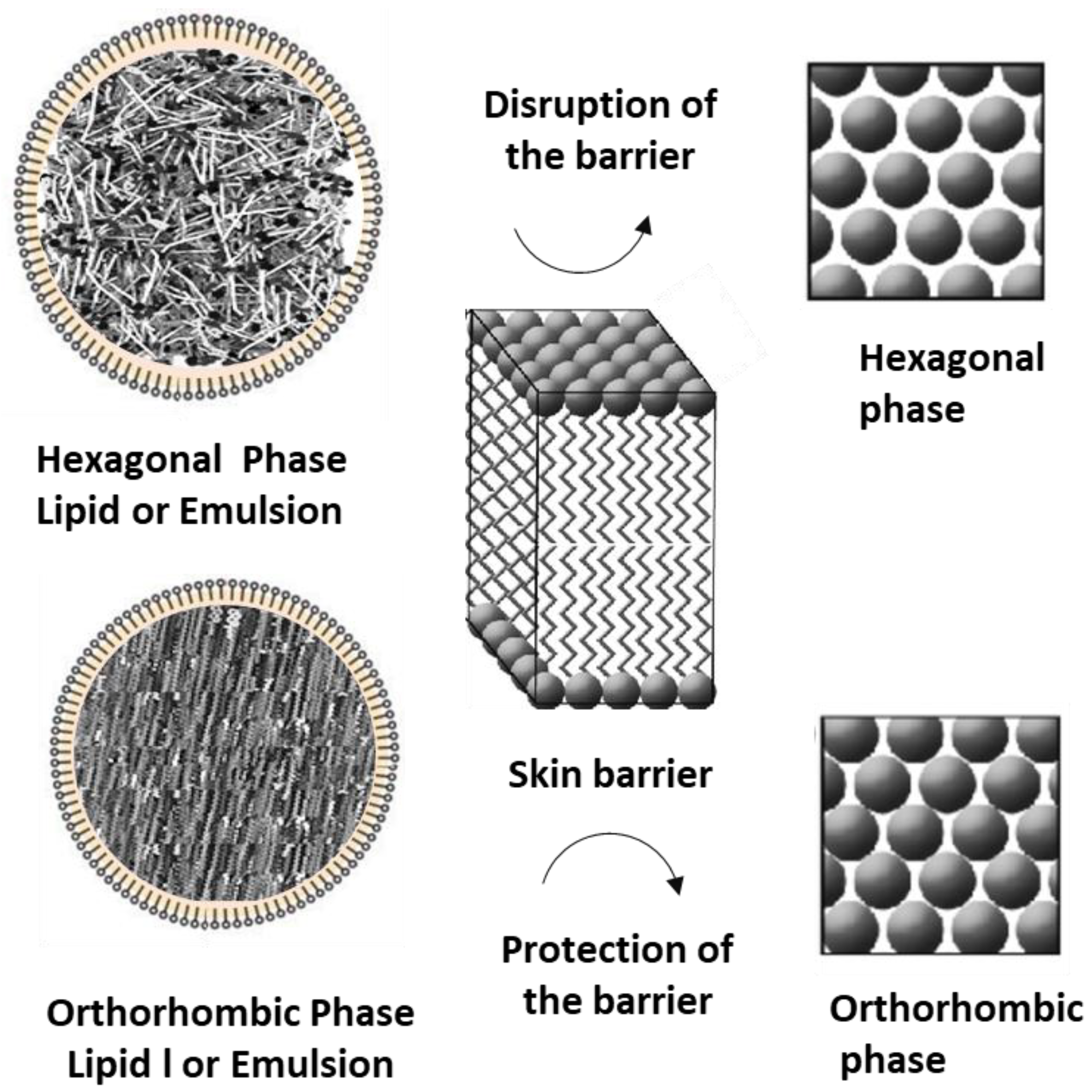
This study investigated the effects of lipid components in cosmetic formulations, which constitute a significant portion of formulations and are expected to influence the lipid barrier when applied to the skin. Since an orthorhombic lipid structure provides the most stable barrier, we analyzed the packing structures of individual lipid components and examined their impact on skin barrier integrity. Squalane and normal paraffin, both of which have simple linear structures [20], exhibited orthorhombic packing, whereas ester oils with ester bonds forming branched structures did not. However, despite having ester bonds, beeswax, which has a longer molecular structure, formed an orthorhombic packing structure. In general, waxes that exist as solid-state materials demonstrated a more pronounced orthorhombic structure compared to squalane. The peak interval between the second derivatives of scissoring was larger [6], and the local height was also greater. Interestingly, porcine skin exhibited a weaker orthorhombic structure than waxes. For intercellular lipids composed of ceramides, fatty acids, and cholesterol, an appropriate balance of these components is necessary to maintain an orthorhombic packing structure [21]. The study confirmed that disruptive agents such as SDS (sodium dodecyl sulfate) could disturb this structure. However, simple oils like squalane did not compromise the barrier function; rather, they enhanced it. In contrast, hexagonal-structured ININ disrupted the skin barrier. Unlike SDS, which disturbs the lamellar structure due to its strong hydrophilic charge, hexagonal-structured ININ, which features looser lipid packing, also influenced the skin’s lipid organization. Furthermore, when formulating emulsions using a combination of oils and waxes with simple molecular structures, orthorhombic-structured formulations were successfully developed (Figure 6). However, these formulations exhibited a weaker orthorhombic structure compared to the native skin barrier. Despite this, no significant impact on the skin barrier function was observed based on TEWL measurements. On the other hand, formulations with a hexagonal packing structure had a detrimental effect on both skin barrier function and lipid organization. Future research should explore whether stronger orthorhombic structures can be designed through different lipid combinations and whether such formulations could enhance barrier function or aid in the recovery of a damaged skin barrier. Additionally, further studies should investigate whether formulations with non-orthorhombic lipid components can still establish an orthorhombic structure, as seen in natural skin lipid organization. This study holds significant implications as it confirms that lipid packing structure can directly influence skin barrier function.
Author Contributions
Conceptualization, Methodology, Supervision; S.-H.L. Validation, Investigation, Data curation; Y.Y.
Acknowledgments
This thesis was supported by the Dongduk Women's University Grant.
Conflicts of Interest
The authors state no conflict of interest to declare.
References
- Bouwstra, J. A.; Ponec, M. The Skin Barrier in Healthy and Diseased State. Biochimica et Biophysica Acta (BBA) - Biomembranes 2006, 1758, 2080–2095. [Google Scholar] [CrossRef] [PubMed]
- Coderch, L.; Lopez, O.; de la Maza, A.; Parra, J. L. Ceramides and Skin Function. Am J Clin Dermatol 2003, 4, 107–129. [Google Scholar] [CrossRef] [PubMed]
- Vietri Rudan, M.; Watt, F. M. Mammalian Epidermis: A Compendium of Lipid Functionality. Frontiers in Physiology. 2022. [CrossRef] [PubMed]
- Damien, F. Boncheva, M. The Extent of Orthorhombic Lipid Phases in the Stratum Corneum Determines the Barrier Efficiency of Human Skin In Vivo. J. Investig. Dermatol. 2010, 130, 611–614. [Google Scholar] [CrossRef]
- Alsamad, F.; Stamatas, G. N. Directional Assessment of the Skin Barrier Function in Vivo. Skin Research and Technology 2023, 29, e13346. [Google Scholar] [CrossRef]
- Pensack, R. D.; Michniak, B. B.; Moore, D. J.; Mendelsohn, R. Infrared Kinetic/Structural Studies of Barrier Reformation in Intact Stratum Corneum Following Thermal Perturbation. Appl Spectrosc 2006, 60, 1399–1404. [Google Scholar] [CrossRef]
- Lee, S. H.; Jun, S. H.; Yeom, J.; Park, S. G.; Lee, C. K.; Kang, N. G. Optical Clearing Agent Reduces Scattering of Light by the Stratum Corneum and Modulates the Physical Properties of Coenocytes via Hydration. Skin Research and Technology 2018, 24, 371–378. [Google Scholar] [CrossRef]
- Boncheva, M.; Damien, F.; Normand, V. Molecular Organization of the Lipid Matrix in Intact Stratum Corneum Using ATR-FTIR Spectroscopy. Biochimica et Biophysica Acta (BBA) - Biomembranes 2008, 1778, 1344–1355. [Google Scholar] [CrossRef]
- Berkers, T.; Visscher, D.; Gooris, G. S.; Bouwstra, J. A. Topically Applied Ceramides Interact with the Stratum Corneum Lipid Matrix in Compromised Ex Vivo Skin. Pharm Res 2018, 35, 1–13. [Google Scholar] [CrossRef]
- Kucharekova, M.; Schalkwijk, J.; Van De Kerkhof, P. C. M.; Van De Valk, P. G. M. Effect of a Lipid-Rich Emollient Containing Ceramide 3 in Experimentally Induced Skin Barrier Dysfunction. Contact Dermatitis 2002, 46, 331–338. [Google Scholar] [CrossRef]
- Kim, D.-H.; Park, W. R.; Kim, J. H.; Cho, E. C.; An, E. J.; Kim, J.-W.; Oh, S.-G. Fabrication of Pseudo-Ceramide-Based Lipid Microparticles for Recovery of Skin Barrier Function. Colloids Surf B Biointerfaces 2012, 94, 236–241. [Google Scholar] [CrossRef] [PubMed]
- Hashizume, E.; Nakano, T.; Kamimura, A.; Morishita, K. Topical Effects of N-Acetyl-L-Hydroxyproline on Ceramide Synthesis and Alleviation of Pruritus. Clin Cosmet Investig Dermatol 2013, 6, 43–49. [Google Scholar] [CrossRef] [PubMed]
- Nisbet, S.; Mahalingam, H.; Gfeller, C. F.; Biggs, E.; Lucas, S.; Thompson, M.; Cargill, M. R.; Moore, D.; Bielfeldt, S. Cosmetic Benefit of a Biomimetic Lamellar Cream Formulation on Barrier Function or the Appearance of Fine Lines and Wrinkles in Randomized Proof-of-Concept Clinical Studies. Int J Cosmet Sci 2019, 41, 1–11. [Google Scholar] [CrossRef]
- Demski, K.; Ding, B.-J.; Wang, H.-L.; Tran, T. N. T.; Durrett, T. P.; Lager, I.; Löfstedt, C.; Hofvander, P. Manufacturing Specialized Wax Esters in Plants. Metab Eng 2022, 72, 391–402. [Google Scholar] [CrossRef]
- Soni, V. K.; Sharma, R. K. Palladium-Nanoparticles-Intercalated Montmorillonite Clay: A Green Catalyst for the Solvent-Free Chemoselective Hydrogenation of Squalene. ChemCatChem 2016, 8, 1763–1768. [Google Scholar] [CrossRef]
- Rodrigues, J. D. A.; Cardoso, F. D. P.; Lachter, E. R.; Estevão, L. R. M.; Lima, E.; Nascimento, R. S. V. Correlating Chemical Structure and Physical Properties of Vegetable Oil Esters. J Am Oil Chem Soc 2006, 83, 353–357. [Google Scholar] [CrossRef]
- Jadhav, H. B.; Annapure, U. S. Triglycerides of Medium-Chain Fatty Acids: A Concise Review. J Food Sci Technol 2023, 60, 2143–2152. [Google Scholar] [CrossRef]
- Pilgram, G. S. K.; Vissers, D. C. J.; Van Der Meulen, H.; Pavel, S.; Lavrijsen, S. P. M.; Bouwstra, J. A.; Koerten, H. K. Aberrant Lipid Organization in Stratum Corneum of Patients with Atopic Dermatitis and Lamellar Ichthyosis. Journal of Investigative Dermatology 2001, 117, 710–717. [Google Scholar] [CrossRef]
- Rieppo, L.; Saarakkala, S.; Närhi, T.; Helminen, H. J.; Jurvelin, J. S.; Rieppo, J. Application of Second Derivative Spectroscopy for Increasing Molecular Specificity of Fourier Transform Infrared Spectroscopic Imaging of Articular Cartilage. Osteoarthritis Cartilage 2012, 20, 451–459. [Google Scholar] [CrossRef]
- Rawlings, A. V.; Lombard, K. J. A Review on the Extensive Skin Benefits of Mineral Oil. Int J Cosmet Sci 2012, 34, 511–518. [Google Scholar] [CrossRef]
- Kim, S.; Lee, S.-H. A Study on the Enhancement of Barrier Function and Improvement of Lipid Packing Structure in a 3D Skin Model by Ginsenoside Rg3. Journal of the Society of Cosmetic Scientists of Korea 2023, 49, 323–330. [Google Scholar] [CrossRef]
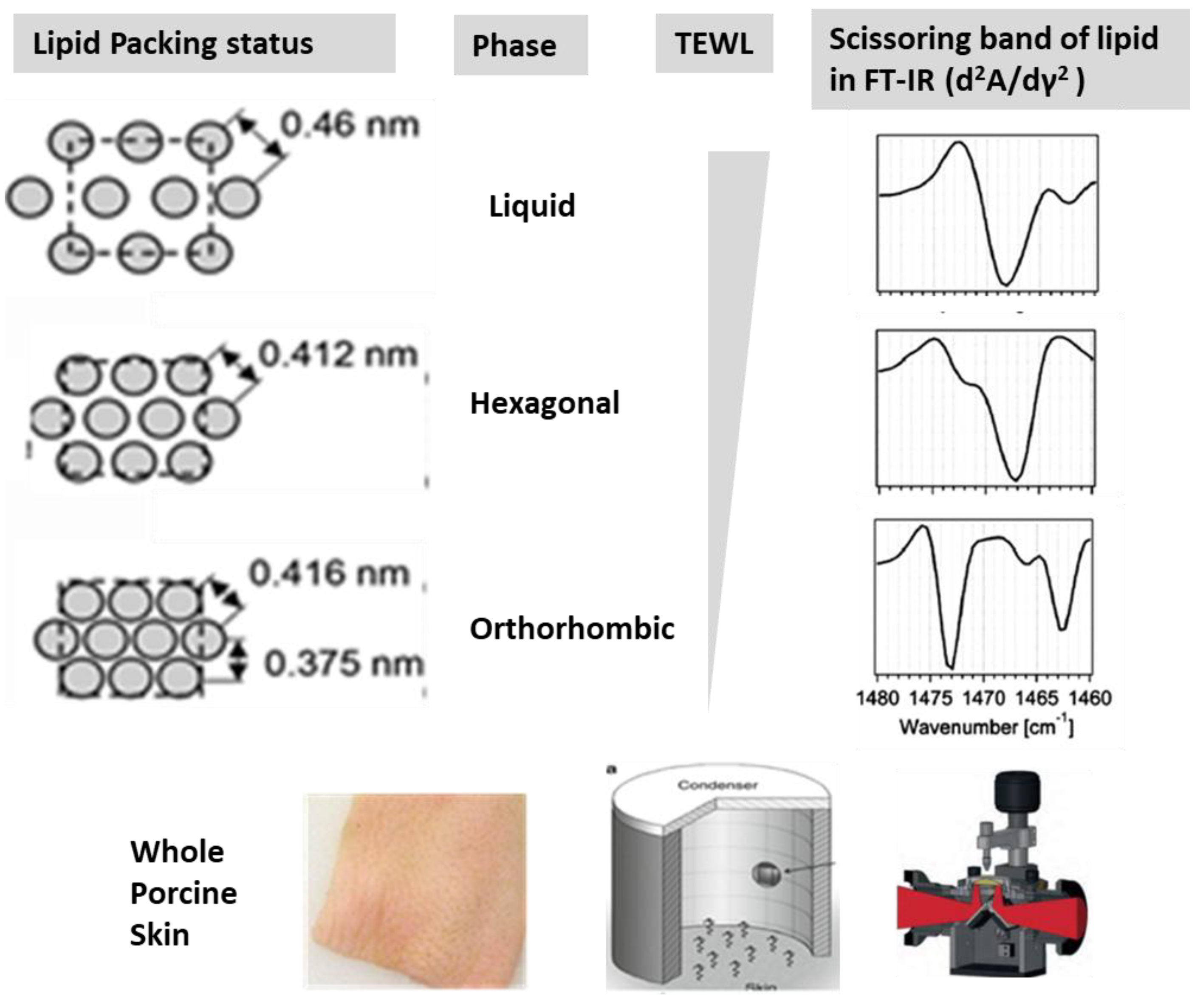
Figure 2.
Relationship Between Lipid Packing State and Barrier Function in Porcine Skin Damage Analysis Induced by SDS. The analysis was conducted on porcine skin. (A) represents the second derivative curve of the symmetric stretch, (B) indicates the peak wavenumber, (C) shows the second derivative curve of the scissoring band, (D) represents the height ratio, and (E) corresponds to TEWL.
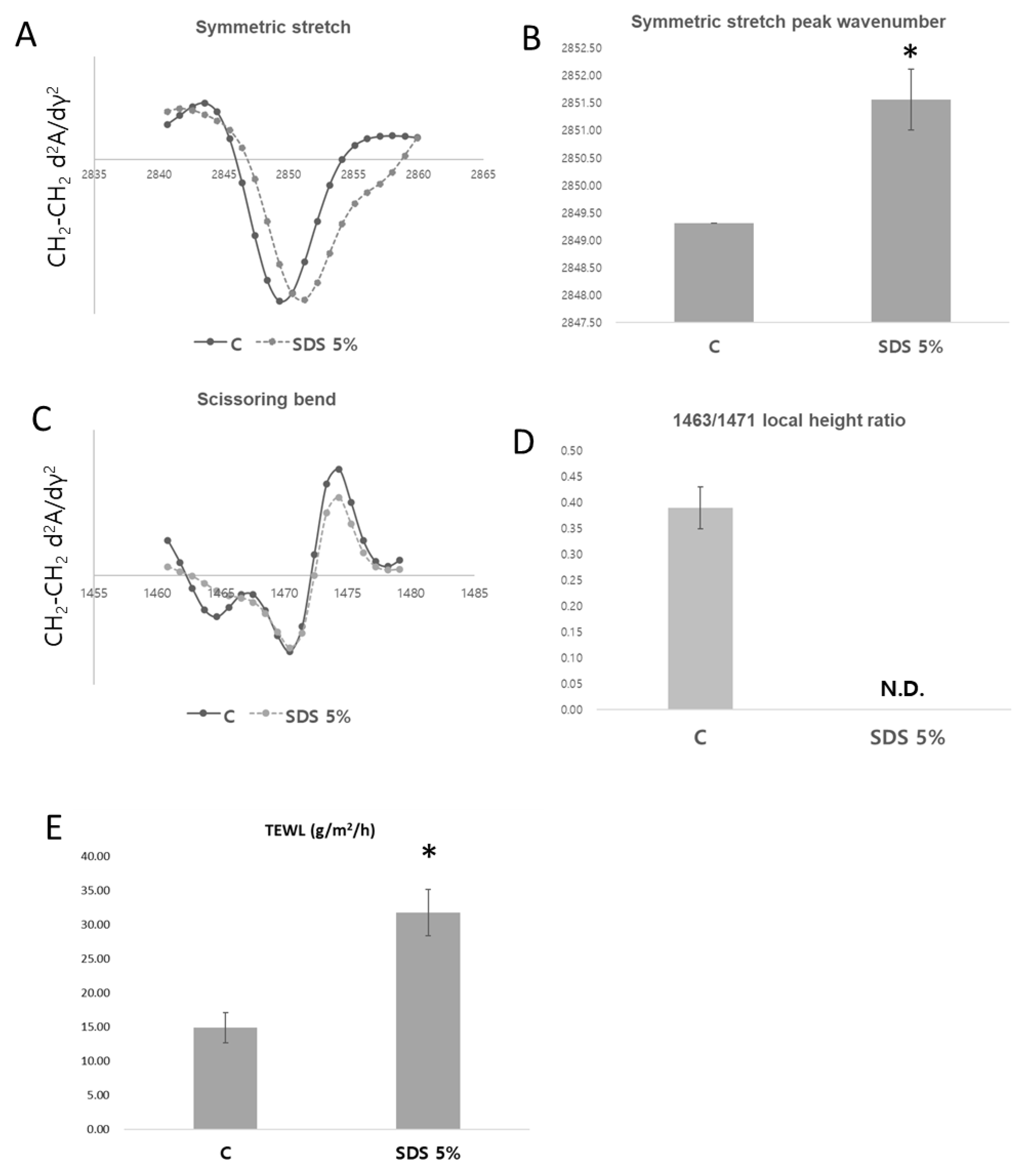
Figure 3.
Construction of Two Distinct Packing Structure Formulations Using a Combination of Lipid Components. The second derivative curve of the representative scissoring band was presented for (A) Squalane, (B) ININ, (C) n-Paraffin, (D) Beeswax, (E) Formulation 1, and (F) Formulation 2, where (A) to (D) represent raw materials.
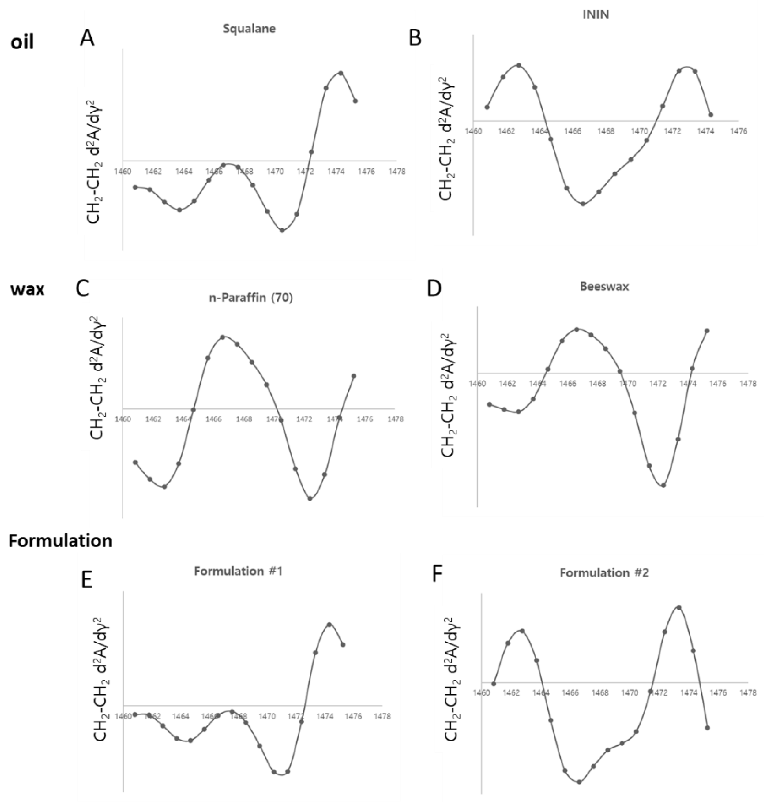
Figure 4.
Structural and Functional Changes in the Skin Barrier Induced by Application of Oils with Different Packing Structures. (A), (B), and (C) show the second derivative curves of the scissoring band for porcine skin after treatment with PBS (A), Squalane(B), and ININ (C), respectively. (D) presents the TEWL data corresponding to these conditions.
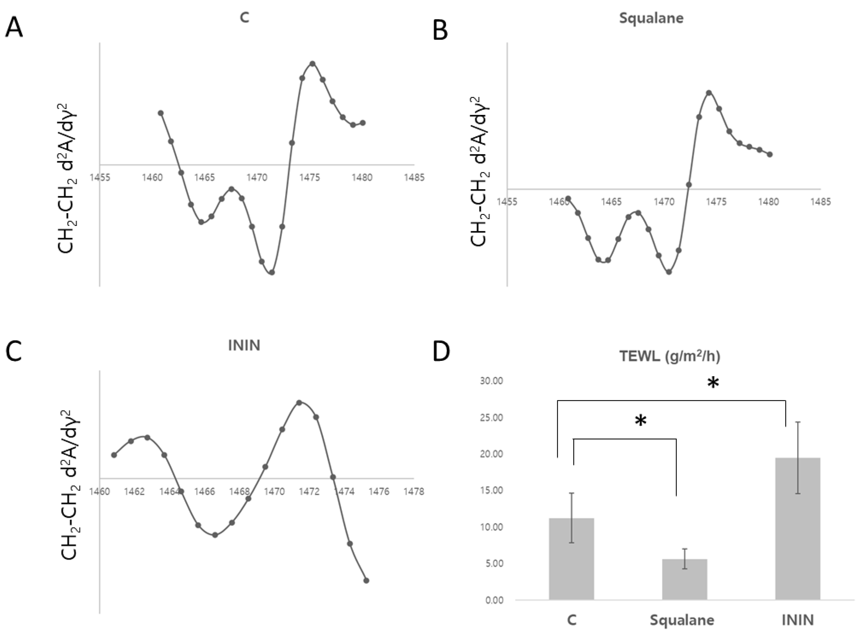
Figure 5.
Structural and Functional Changes in the Skin Barrier of Pigskin Induced by the Application of Formulations with Different Packing Structure. (A) represents the second derivative curves of the scissoring band for porcine skin after treatment with PBS, hexagonal formulation, and orthorhombic formulation. (B) indicates the local height ratio. (C) presents the TEWL analysis results corresponding to these conditions.
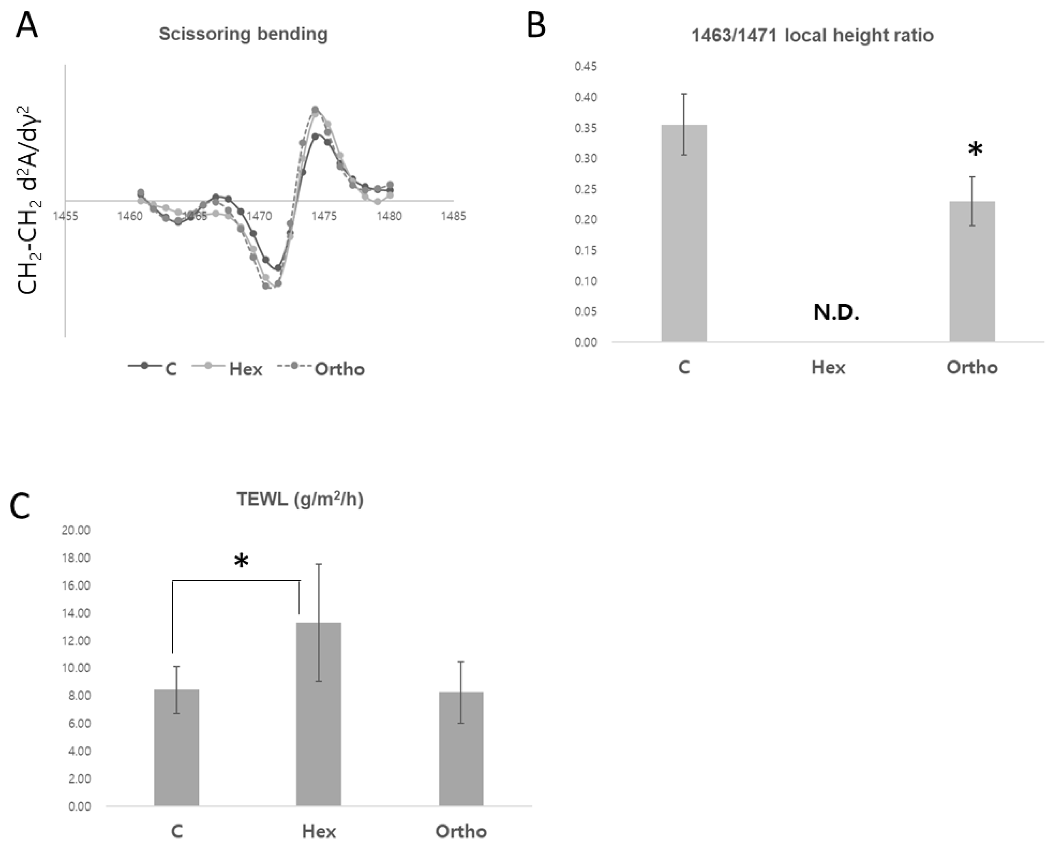
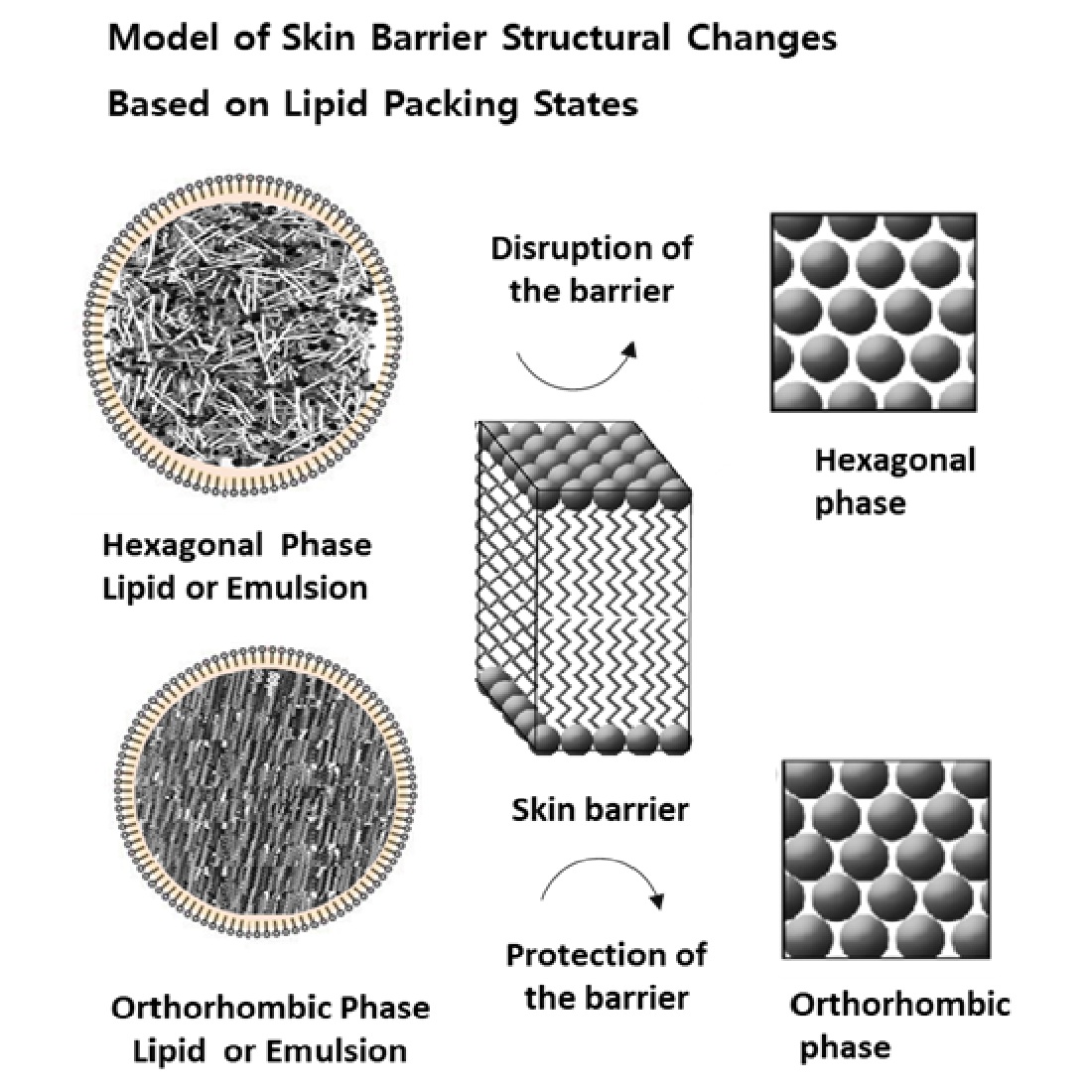
Figure 6.
A Model of Skin Barrier Structural Changes Based on Lipid Packing States.
Disclaimer/Publisher’s Note: The statements, opinions and data contained in all publications are solely those of the individual author(s) and contributor(s) and not of MDPI and/or the editor(s). MDPI and/or the editor(s) disclaim responsibility for any injury to people or property resulting from any ideas, methods, instructions or products referred to in the content. |
© 2025 by the authors. Licensee MDPI, Basel, Switzerland. This article is an open access article distributed under the terms and conditions of the Creative Commons Attribution (CC BY) license (http://creativecommons.org/licenses/by/4.0/).
Copyright: This open access article is published under a Creative Commons CC BY 4.0 license, which permit the free download, distribution, and reuse, provided that the author and preprint are cited in any reuse.


