Submitted:
09 July 2025
Posted:
10 July 2025
You are already at the latest version
Abstract
Vervain (Verbena officinalis L., Verbenaceae family) is a perennial plant which grows widely in Europe. Its raw material is rich in iridoids, phenolic acids, phenylpropanoid glycosides, flavonoids, and terpenoids. Verbena has traditionally been used in folk medicine to support nervous system health, but there is a lack of scientific data about it. The aim of this study was to explore and characterise the chemical profile and neurotropic effects of V. officinalis dry extracts and its amino acid-based preparations. We determined a total of eight main phenolic compounds and 17 amino acids in the V. officinalis dry extracts. To evaluate the neurotropic effects of the Verbena extracts, the following behavioural pharmacology tests were used: Open Field Test, Elevated Plus Maze, Black-and-White Box Test and Tail Suspension Test. The dry aqueous-ethanolic extract (extractant 70% ethanol) demonstrated strong anxiolytic and antidepressant effects, while its dry modified extracts with valine and arginine consistently exhibited pronounced sedative activity across all studies. For example, the Tail Suspension Test demonstrated that the total immobility time in animals receiving the dry aqueous-ethanolic extract was the lowest, being 1.22-fold (p < 0.05) lower than in intact animals and 2.25-fold (p < 0.05) lower than in the animals treated with the reference drug preparation (“Sedaphyton”). A novel aqueous-based gel formulation feasible for semi-solid extrusion (SSE) 3D printing was designed. This printing gel enables the fabrication of new oral dosage forms for V. officinalis dry extracts.
Keywords:
verbena officinalis L.
; herb extract
; phenolic compounds
; amino acids
; neurotropic activity
; 3D printing
1. Introduction
Vervain or common verbena (Verbena officinalis L., Verbenaceae) is a perennial plant that grows widely in Europe. The plant also spreads to North America, Asia and North Africa [1].
V. officinalis L. is rich in iridoids, phenolic acids, phenylpropanoid glycosides, flavonoids, and terpenoids [2]. The most characteristic iridoids of this plant raw material are verbenaline and hastatoside. The iridoid glycosides, such as 3,4-dihydroverbenaline, aucubin, 7-hydroxydehydrohastatoside, and the secoiridoids such as verbenoside A, verbeofflin I and verbenoside B, have also been identified [3,4,5]. The most prevalent phenylpropanoid glycosides of the plant include verbascoside, isoverbascoside and eukovoside, while isomers of leukoseptoside and cistanoside are also present [6]. Regarding fatty acids, the most abundant compounds are α-linolenic acid, palmitic acid, linoleic acid, and oleic acid. The phenolic acids identified in the plant include gallic acid, syringic acid, ferulic acid, cinnamic acid, and protocatechuic acid, along with quinic, chlorogenic, rosmarinic acids, and dicaffeoylquinic acid derivatives [2,7]. Among the flavonoids found in V. officinalis, notable compounds include apigenin, luteolin, 5,7,4’-trihydroxy-8-methoxyflavone, scutellarein, and its glucoside derivatives (scutellarein 7-glucoside, scutellarein-7-diglucuronide, scutellarein-7-glucuronide). Additionally, the plant contains pedalitin, pedalitin-6-galactoside, quercetin, kaempferol, isorhamnetin, diosmetin, and rutin [2,8,9,10].
The essential oil composition of V. officinalis varies greatly depending on the geographical location and the specific plant part used in distillation. The plant contains mono-, di-, tri-, and sesquiterpenoids [11,12]. Among monoterpenoids, the key compounds include citral, limonene, eucalyptol, menthol, α-pinene, β-pinene, sabinene, and β-phellandrene. Carnosol and rosmanol are identified as the diterpenoids of the plant [13,14]. The most abundant sesquiterpenoids are caryophyllene oxide, α-curcumin, β-caryophyllene, hexahydrofarne-sylacetone, and spathulenol. The dominant triterpenoids include squalene, ursolic acid, barbinervic acid, and oleanoic acid [2,15]. Regarding sterols, the plant yields β-sitosterol, γ-sitosterol, daucosterol, stigmasterol, campesterol, and androst-5,15-dien-3-ol-acetate from its aerial parts [8,13,14,15].
Since 2008, a specific monograph on Verbena officinalis L. has been included in the European Pharmacopoeia (Ph.Eur.), and according to this monograph, the present plant must contain a minimum of 1.5% of verbenaline [16]. In addition to the abovementioned monograph, Ph.Eur. presents also another monograph for lemon verbena leaf (Verbenae citriodorae folium, Verbena citriodora (Palau) Cav.), also known as Aloysia citriodora Palau, with synonyms Aloysia triphylla (L’Her.) Kuntze, Verbena triphylla L’Her., and Lippia citriodora Kunth. The raw material is standardized based on its phenylethanoid content, specifically the tyrosol derivative acteoside (verbascoside), which must comprise a minimum of 2.5% of dry weight, expressed as ferulic acid. The essential oil content should be at least 3.0 mL/kg in the whole drug and 2.0 mL/kg in the fragmented drug (dry weight) [16].
Verbena has traditionally been used in folk medicine to support nervous system health and aid conditions, such as stress, anxiety, depression and insomnia. Moreover, it has been utilised for headache relief, premenstrual tension, and the treatment of cramps, jaundice and asthma [17,18]. Externally, verbena herb infusion serves as a gargle for tonsillitis and stomatitis, and as a lotion for skin conditions. It is also incorporated in numerous herbal formulations, such as Sinupret® (Bionorika, SE), which is a complex plant-based preparation known for its effectiveness in managing acute viral rhinosinusitis in children, thus helping to accelerate symptom relief [19].
The antioxidant, anti-inflammatory and hepatoprotective effects of V. officinalis L. have also been validated in separate studies [3,6,20,21]. Besides its antioxidant properties, common vervain leaf extracts have presented antimicrobial and antifungal activity. Moreover, the caffeoyl derivatives of such leaf extracts have shown a strong antifungal effect against P. expansum and R. stolonifera [22].
The tumor growth inhibitory properties of common vervain aqueous extract have been evaluated in vitro on rat and human colon adenocarcinoma cell lines. These studies revealed that polysaccharides from V. officinalis effectively suppressed colorectal cancer cell invasion and metastasis [23]. Moreover, two newly identified phenylethanoid glycosides exhibited cytotoxic activity comparable to vinblastine sulfate, a standard chemotherapy agent [24]. Methanolic extracts of semi-purified fractions demonstrated tumor growth inhibition across multiple melanoma cell lines [25]. Further animal studies confirmed its potential in hepatocellular carcinoma treatment. The administration of aqueous extract led to a 38.78% tumor size reduction compared to controls [26].
Gharachorloo and co-workers studied the antioxidant and antibacterial properties of the essential oil isolated from V. officinalis L. against S. aureus and E. coli [27]. The authors reported that E. coli exhibited greater susceptibility to yarrow essential oil compared to S. aureus, despite the fact that gram-negative bacteria generally display lower sensitivity to essential oils. The authors also found that increasing the essential oil concentration correlated with enhanced antioxidant activity [27]. According to a more recent study, silver nanoparticles synthesized from common vervain leaf extract demonstrated antibacterial effects against both gram-positive and gram-negative bacteria [28]. The potential of common vervain in managing hyperlipidemia has also been explored. Crude plant extract was shown to decrease total cholesterol, triglycerides, low-density lipoproteins (LDL), and very low-density lipoproteins (VLDL) levels in vivo, and these results were comparable to the results obtained with atorvastatin as a reference drug [29]. It has been suggested that V. officinalis influences lipid metabolism, and quercetin, luteolin and kaempferol were identified as key bioactive compounds beneficial for atherosclerosis treatment [8]. The impact of common vervain aqueous extract on physical stress was further investigated in an animal study, and the present study revealed antioxidant modifications in red blood cell membranes and a significant protective effect against physical stress. Therefore, due to these beneficial properties, common vervain extract was suggested as a promising candidate for inclusion in sports supplements aimed at accelerating post-exercise recovery [30]. In addition, the biologically active compounds of V. officinalis have been demonstrated to have promising effects on depression, anxiety and insomnia [31,32,33].
Modifying biologically active substances found in plant extracts can significantly enhance their therapeutic potential. One widely adopted strategy involves the conjugation of extract constituents with amino acids [34,35,36]. For example, linking valine to acyclovir led to the formation of valacyclovir, a compound that substantially improves acyclovir’s systemic bioavailability, thereby increasing patient convenience and treatment efficacy [37]. It has also been shown that arginine enhances the absorption and stability of perindopril while simultaneously mitigating its adverse effects [38]. The incorporation of amino acids into an extract of Leonurus cardiaca L. resulted in novel preparations exhibiting stronger anxiolytic properties [39]. Moreover, combining arginine with extracts of highbush blueberry (Vaccinium corymbosum L., Ericaceae) [40] and cranberry leaves (Vaccinium macrocarpon Aiton, Ericaceae) [36] led to the development of new active substances with potential hypoglycemic and lipid-lowering activities. These cases underline the potential of modifying V. officinalis herb extracts to generate new biologically active phytosubstances.
Herbal medicinal products are generally known for their favourable safety profiles. However, galenic formulations—such as tinctures, teas, decoctions, and liquid extracts—often encounter limitations related to insufficient standardization and reduced patient adherence. A novel innovative approach to overcome these challenges is the application of pharmaceutical 3D printing technologies [41,42,43], which allow the development of advanced oral dosage forms aimed at improving both the therapeutic efficiency and patient compliance of phytomedicines. Considering the broad range of V. officinalis extracts’ therapeutic applications, further studies are warranted to explore its phytochemical composition, pharmacodynamic properties, and to determine the most suitable pharmaceutical delivery systems for its extracts. The development of 3D-printed preparations containing V. officinalis extracts could offer a more precise, effective, and patient-oriented approach to the use of herbal medicines in modern clinical and pharmaceutical settings.
The objective of this study was to explore and characterize the chemical profile and neurotropic effects of V. officinalis dry extract and its amino acid-based preparations. Additionally, we designed a novel aqueous gel formulation feasible for semi-solid extrusion (SSE) 3D printing, thus enabling the fabrication of new printed oral dosage forms loaded with V. officinalis dry extract.
2. Results
The V. officinalis extracts were yellow-brown-greenish powders with a characteristic smell. The yields of the dry extracts V1 (extractant 70% ethanol) and V2 (extractant water) were 13.62% and 18.44% respectively.
2.1. Phytochemical Research
The phenolic compounds and amino acids in the V. officinalis extracts were studied by LC-MS (Table 1). A total of eight main phenolic compounds and 17 amino acids were determined.
2.2. Pharmacological Research
The studied V. officinalis extracts (dissolved in water) were administered to animals orally as a single dose at 50 mg/kg. The control group animals received the “Sedaphyton” drug at the same 50 mg/kg dose. Intact animals were given an equivalent volume of drinking water. For 3-4 hours before administration, the animals were deprived of food but had free access to water. The study began 1 hour after the administration of extracts. The experimental results were processed using standard statistical methods including the arithmetic mean, standard deviation, Student’s t-test, and significance coefficient.
The “Open Field” test was based on assessing the natural defensive and exploratory behavioral reactions of rodents in a new open space [44,45,46]. The results of the study on the neurotropic activity of the V. officinalis extracts are presented in Table 2.
The Elevated Plus Maze test is another model for assessing situational anxiety, and this test allows the evaluation of the anxiolytic, psychostimulant, and sedative effects of pharmacological agents [44,46]. This test is designed to study rodent behaviour under conditions of variable stressogenicity, where they can freely choose comfortable environments. It helps to assess anxiety levels, including exploration of dark compartments, natural fear of open spaces, and height-related anxiety. The anxiolytic effect of a substance is evaluated based on the time spent in the dark and illuminated arms of the maze. Increased time spent in illuminated arms, along with more instances of peeking out and hanging from open sections, is classified as an anxiolytic effect. Overall motor activity reduction may indicate a sedative effect, while increased activity without changes in time spent in open arms suggests a psychostimulant or nonspecific motor influence. The test duration was 5 minutes, during which behavioural characteristics of mice were visually recorded. Such behavioural characteristics include the time spent in the open arm of the maze (exploratory activity), the time spent in the closed arm of the maze, number of peeks from the closed arm, number of downward glances from the ends of open arms (risk assessment), and number of crossings over the central platform. The results of the study on the effects of the Verbena extracts on mouse behaviour are presented in Table 3.
The anxiolytic effect of the Verbena herb extracts on exploratory reactions of mice under stress conditions was studied using a “Black-and-White Box” test [44,45,47]. The animals were first placed in the dark compartment of the apparatus for 2 minutes to adapt to the dark environment. After this adaptation period, the experiment began, and for 3 minutes, the following parameters were recorded: time spent in the dark compartment (burrow reflex), number of peeks and exits into the illuminated compartment, and the time spent in the illuminated compartment. It is known that anxiolytics increase the number of exits and the time spent in the illuminated compartment, whereas control animals prefer to remain in the dark compartment. The results of the study are presented in Table 4.
The Tail Suspension Test was used for the initial assessment of the antidepressant activity of the Verbena extracts [44,48,49]. This test models the animal behaviour based on despair. The Tail Suspension Test provides a quick and reliable assessment of antidepressant behavior in mice. For 6 minutes, the behavior of experimental animals was observed, and the immobility time (motionless hanging) was recorded. The results of the study are presented in Table 5.
2.3. Novel 3D-Printed oral Dosage Forms for the Verbena Officinalis Extract
The PEO printing gel loaded with the dry V. officinalis extract (V1) appeared as a greenish-brown viscous semisolid with a distinct odour. The gel was quite homogeneous, as proven by a microscopic analysis (Figure 1). The viscosity of gel assessed at a rotational speed of 0.03 RPM and a shear rate of 0.060 1/s (22 ± 2 °C), was 209900 ± 26540 cP. The standard square- and round-shaped 3D-printed scaffolds were prepared by using an aqueous PEO gels loaded with the V. officinalis extract for SSE 3D printing (Figure 2). The printing quality of the preparations was studied by measuring the surface area (396.75 ± 60.08 mm2), Spractical/Stheoretical ratio (1.22), and the average mass of the printed scaffolds (lattices 214.4 ± 21.4 mg and discs 175.0 ± 1.0 mg).
3. Discussion
Table 1 shows the content of phenolic compounds and amino acids found in the Verbena officinalis extracts. The main identified phenolic compounds in the dry V. officinalis extracts were isovanillin, quercetin and protocatechuic acid. According to the literature, gallic and ferulic acids, quercetin, and rutin have effects on the central nervous system (CNS) [44,45,46,47,48]. Gallic acid presents neuroprotective effects by reducing oxidative stress and inflammation in the brain. Gallic acid may also improve cognitive function and protect neurons from degeneration [50]. Ferulic acid has antioxidant and anti-inflammatory properties, and it could play a role in the prevention of neurodegenerative disorders like Alzheimer’s disease [51]. Rutin and quercetin are flavonoids that may enhance cerebral circulation, reduce inflammation, and promote neuronal protection [52,53,54]. p-Coumaric acid modulates hypothalamic AMPK activity and enhances leptin signalling, thus improving glucose homeostasis and reducing food intake [55]. Protocatechuic acid has been reported to enhance cognitive function, reduce neuronal apoptosis, and improve synaptic plasticity by regulating dopamine turnover, by inhibiting oxidative stress and inflammation, and by modulating BDNF and MAPK signalling [56,57]. Syringic acid acts as a neuroprotective agent in the models of Alzheimer’s disease, Parkinson’s disease and neurotrauma [58]. With isovanillin, however, there is only limited direct evidence on CNS effects. Structurally, it is related to vanillin, which has mild neuroactive properties. To date, the effects of isovanillin on CNS are not well established in the state-of-the-art literature.
Among the 17 identified amino acids (Table 1), the following eight are essential: leucine, valine, phenylalanine, lysine, histidine, isoleucine, threonine and tryptophan. The dominant amino acids identified were asparagine, glutamine, α-alanine, arginine, threonine and valine. Leucine, valine, phenylalanine and lysine have effects on the CNS. Leucine is involved in neurotransmitter synthesis and brain metabolism regulation. It activates the mTOR signalling pathway, thus influencing neuron growth and survival [59]. Valine is essential for neurotransmitter synthesis and supporting cognitive function. The recent studies confirm its role in brain metabolism and show potential impact on neurodegenerative diseases [60]. Phenylalanine is a precursor to dopamine, norepinephrine, and adrenaline, and it influences mood, motivation and cognitive function [61]. Lysine was shown to affect serotonin levels, and thus help to reduce stress and anxiety [60]. These amino acids play vital roles in CNS function, and their effects are actively studied in the context of neurodegenerative diseases and cognitive disorders.
The results of the “Open Field” test (Table 2) showed that the animals administered V1-Val and V1-Arg extracts exhibited a 1.49-fold (p < 0.05) and 1.11-fold reduction in locomotor activity compared to intact animals. In contrast, the animals receiving V1, V1-Gly, V1-Phe and V1-Lys extracts showed a slight increase in locomotor activity, but the difference was not statistically significant compared to intact animals. After the administration of V2 extract, an increase in locomotor activity was observed. The locomotor activity was 1.25 to 1.28 times higher (p < 0.05) compared to intact animals and a comparison group, respectively. The indicators of exploratory behaviour (number of vertical stands) were slightly higher with the animals administered V1 and V2 extracts compared to intact animals. The administration of V1-Gly, V1-Phe and V1-Lys extracts had only a minimal impact on this indicator. The number of hole explorations was lower in all experimental groups compared to the intact animal group. The exploratory behaviour indicators in the animals receiving V1-Val and V1-Arg extracts were lower than in the comparison group treated with “Sedaphyton”. The number of vertical stands decreased by 1.17-fold and 1.80-fold (p < 0.05), respectively. Perhaps surprisingly, the number of hole explorations remained at the same level as was found with a comparison group. The Verbena extracts did not significantly affect the emotional response indicators of mice in the “Open Field” test. Overall, the effects of V1-Val and V1-Arg extracts on the neurotropic activities in mice were slightly (1.02 to 1.21 times) higher than those observed in the comparison group receiving ‘Sedaphyton’. This was found when analysing the relative units of all neurotropic activities. Considering all types of activities in the “Open Field” test, it is evident that the administration of V1, V2 and V1-Gly extracts in mice resulted in a mild anxiolytic effect, and this effect exceeded the corresponding effect with intact animals by 19.4%, 18.4% and 10.8%, respectively. The total activity indicators of V1-Phe and V1-Lys extracts were comparable to those of intact animals, while the V1-Val and V1-Arg extracts showed similar effects to the reference drug “Sedaphyton”. This suggests a sedative effect of the modified Verbena extracts after oral administration.
The results of the Elevated Plus Maze test (Table 3) showed that the administration of V1, V1-Phe, and V1-Lys extracts led to significant changes in the emotional and behavioural responses of experimental animals in the test compared to the intact animals and those receiving the comparison drug “Sedaphyton”. Regarding exploratory activity, the time spent in the open arm of the maze increased by 2.60-fold (p < 0.05), by 2.46-fold (p < 0.05) and by 1.95-fold (p < 0.05) after the administration of V1, V1-Phe and V1-Lys extracts, respectively (compared to intact animals). Compared to the comparison group, the increase was even 3.40-fold (p < 0.05), 3.21-fold (p < 0.05), and 2.55-fold (p < 0.05), respectively. The number of downward glances from the ends of open arms (risk assessment) significantly increased by 2.17-fold (p < 0.05) after the administration of V1, 2.42-fold (p < 0.05) after the administration of V1-Phe, and 2.35-fold (p < 0.05) after the administration of V1-Lys compared to intact animals. In addition, the animals receiving V1, V1-Phe, and V1-Lys extracts showed an increase in crossings over the central platform, while the number of peeks from dark arms remained unchanged compared to intact animals. After the administration of V2, V1-Gly, and V1-Arg extracts, the time spent in open arms, the number of downward glances, and the number of crossings over the central platform did not differ from those observed with the intact animals. Conversely, the oral administration of V1-Val extract in mice appeared to induce a sedative effect. The time spent in the open arm of the maze significantly decreased by 2.54-fold (p < 0.05) compared to the intact animals and 1.95-fold (p < 0.05) compared to the comparison group. A significant reduction in the number of downward glances was observed with the mice receiving V1-Val extract. The decrease in the number of downward glances was 4.67-fold (p < 0.05) compared to the intact animals and 3.00-fold (p < 0.05) compared to the “Sedaphyton”-treated animals. Moreover, the number of peeks from the closed arm and crossings over the central platform decreased by 3.93-fold (p < 0.05) and 1.82-fold compared to the intact animals. Therefore, our results suggest the presence of an anxiolytic effect in mice after the oral administration of V1, V1-Phe, and V1-Lys extracts (Table 3). This was evidenced by increased exploratory and locomotor activity in mice. It is evident that V1, V1-Phe, and V1-Lys extracts exert an activating effect on the CNS by reducing fear perception and enhancing adaptation speed to new conditions. The results for V1-Val suggest the presence of a sedative effect in mice.
All three V. officinalis extracts showed anxiolytic activity in mice, and the most pronounced activity was observed with V1 extract (Table 4). After the administration of V1 extract, the mice showed an increase in time spent in the illuminated compartment. This increase in time was 1.28-fold (p < 0.05) higher compared to intact animals and 1.68-fold (p < 0.05) compared to the animals receiving “Sedaphyton”. High anxiolytic activity was also observed with the V1-Gly and V1-Phe extracts. After the administration of these two extracts, the time spent in the illuminated compartment increased by 1.31-fold (p < 0.05) for V1-Gly and by 1.35-fold (p < 0.05) for V1-Phe compared to intact animals. Compared to “Sedaphyton”-treated animals, the time spent in the illuminated compartment increased by 1.73-fold (p < 0.05) and 1.77-fold (p < 0.05) after the administration of V1-Gly and V1-Phe, respectively. Following the administration of V1-Arg, a decrease in exploratory behaviour was observed in mice, and the time spent in the illuminated compartment decreased by 1.46-fold (p < 0.05) compared to intact animals. With the mice, the time spent in the illuminated compartment was at the same level as was found with the ‘Sedaphyton’-treated group. These findings suggest a predominantly sedative effect of V1-Arg extract. After the oral administration of V1-Lys and V1-Val extracts to the mice, no statistically significant anxiolytic effect was observed compared to intact animals. We used the values for the time spent in the illuminated compartment and the number of exits from the dark section to calculate the average time of a single stay in the illuminated compartment (Table 4). The highest value for the average time of a single stay in the illuminated compartment was found after the administration of V1 extract to the mice (47.98 ± 12.85 s), while the lowest value was observed with the mice after the administration of V1-Arg extract (11.66 ± 2.66 s). Therefore, it is evident that the V1, V1-Gly and V1-Phe extracts exhibit anxiolytic effects, while V1-Arg extract presents a sedative effect.
The Tail Suspension Test (Table 5) showed that the total immobility time in mice receiving V1 extract was the lowest (84.67 ± 5.38 s), and this value was 1.22-fold (p < 0.05) lower than the value observed with intact animals and 2.25-fold (p < 0.05) lower than the value recorded with the animals treated with “Sedaphyton”. Following the administration of V1-Phe and V1-Lys extracts, the total immobility time of mice decreased by 1.96-fold (p < 0.05) and 1.91-fold (p < 0.05) compared to the “Sedaphyton”-treated group. However, the difference in the total immobility time was not statistically significant compared to the value obtained with the intact mice. After the administration of V2, V1-Gly, V1-Val, and V1-Arg extracts, an increase in total immobility time with mice was 1.21-fold (p < 0.05), 1.20-fold (p < 0.05), 1.71-fold (p < 0.05) and 1.29-fold (p < 0.05), respectively (compared to intact mice). The values for a total immobility time, however, were lower than those observed with a comparison group. Therefore, the present Tail Suspension Test results (Table 5) indicate that the V1, V1-Phe, and V1-Lys extracts present antidepressant activity in mice. The V2, V1-Gly, and V1-Arg extracts did not show antidepressant effects, but these three extracts demonstrated a mild sedative activity. However, the sedative activity in mice was significantly lower than that found in the mice treated with “Sedaphyton”. The V1-Val extract, however, exhibited a sedative activity in mice comparable to “Sedaphyton”. In conclusion, the V1 extract showed strong anxiolytic and antidepressant effects in mice, while the V1-Val extract consistently exhibited pronounced sedative activity across all studies.
The pharmacological studies here revealed that the Verbena extracts (unmodified and modified with amino acids) exhibit diverse biological activity (i.e., the anxiolytic, antidepressant, locomotor, and sedative effects). The non-modified extracts may influence the GABAergic, serotonergic, and dopaminergic systems, which explains their anxiolytic and antidepressant effects [62,63].
It is evident that the amino acid-modified extracts enhance the inhibitory processes in the CNS. This is primarily due to valine, which possesses neurotrophic and inhibitory properties affecting the CNS. Valine can reinforce inhibitory processes either indirectly through metabolism or directly via GABA receptor interactions. As a result, these compounds exhibit sedative effects, manifested as reduced locomotor activity, prolonged time spent in dark compartments, and fewer transitions into illuminated areas. Moreover, valine improves drug absorption and facilitates passage through the blood-brain barrier (BBB) via LAT1 (Large Amino Acid Transporter 1), thus enabling the release of active compounds in the brain. However, valine may also inhibit serotonin synthesis through competitive transport of tryptophan via LAT1, which in turn enhances neurotropic activity and alters neurotransmitter balance, thus leading to reduced serotonin levels [64,65,66,67].
Arginine, as a modifying amino acid, can significantly affect drug bioavailability and BBB penetration via improving the solubility and absorption of the drug substance. The verbena extract with arginine utilises CAT-1 (Cationic Amino Acid Transporter 1) in the endothelial cells of BBB to facilitate brain entry. Since CAT-1 regulates brain arginine levels, which serves as a precursor for nitric oxide (NO), it influences neurotransmission, neuroinflammation, and cerebral blood flow. NO can modulate neuronal activity, thus leading to sedation. The mice receiving the V1-Arg avoided open areas and exhibited reduced activity, thus indicating sedative effects. This explains the weaker anxiolytic action and stronger sedative effect of the arginine-modified extract [68,69,70].
We propose that by modifying the Verbena extract with specific amino acids, one can alter its pharmacological profile and shift its activity from anxiolytic to sedative effects, depending on the neurochemical pathways activated or inhibited.
The aqueous PEO-based gel containing 1.0 g of the V. officinalis extract (V1) per 10 ml of gel, was successfully used for SSE 3D printing. The 3D-printed lattices and round disc-shaped preparations exhibited consistent shape and dimensions (Figure 2). The applicability and compatibility of PEO as a carrier polymer with V. officinalis extract in the gel-based printing inks were verified. In our previous studies, we used surface-active agents to improve the 3D-printed formulations of German chamomile [35], Solidago canadensis [71] and eucalyptus extracts [43,72]. In these studies, it was shown in the pilot in-vitro disintegration tests that the SSE 3D-printed PEO discs loaded with the plant-based extract lost their structural integrity and fully disintegrated within 20-25 minutes in purified water at room temperature (22 ± 2 °C). Since all samples were completely disintegrated within 30 minutes, such 3D-printed preparations showed potential as immediate-release oral delivery systems [73,74].
For further development, it would be important to establish the standardisation techniques for these 3D-printed herbal dosage forms. Moreover, both preclinical and clinical trials should be undertaken to verify the safety and efficacy of the preparations. The pharmaceutical, chemical, and microbiological quality of these preparations should be validated through the successful completion of all development stages. Currently, the regulatory framework and market authorisation guidelines for 3D-printed medicinal products, however, remain unclear and under-developed. This in turn makes an obstacle to the adoption of V. officinalis extract-based printed formulations in the wider use.
4. Materials and Methods
4.1. Materials
The V. officinalis herb was collected in 2023 from the Kubja Ürditalu farm, Estonia. To prepare the dry extracts, 300 g of V. officinalis herb was macerated in 1500 ml of 70% aqueous ethanol at room temperature for 24 hours. The liquid extract was then separated, and the extraction process was repeated once more using another 1500 ml of 70% ethanol solution. The two liquid extracts were combined, settled for two days, and filtered. From the combined extract, five 200 ml portions were taken, with each portion receiving a different amino acid: glycine (OstroVit, Zambrov, Poland) 0.3705 g, phenylalanine (OstroVit, Zambrov, Poland) 0.8153 g, L-lysine (FITS, Tallinn, Estonia) 0.7212 g, valine (Acros Organics, Geel, Belgium) 0.5780 g, and L-arginine (FITS, Tallinn, Estonia) 0.8596 g. The amino acid solutions and the remaining liquid extract were infused for 24 hours, followed by evaporation using a Buchi B-300 rotary vacuum evaporator (Buchi AG, Flawil, Switzerland) to obtain soft extracts. These were then freeze-dried (lyophilized) using a SCANVAC COOLSAFE 55-4 Pro freeze dryer (LaboGene ApS, Lillerød, Denmark). The final dry extracts were designated as V1, V1-Gly, V1-Phe, V1-Lys, V1-Val and V1-Arg, respectively.
For preparing the aqueous extract V2, 100.0 g of dried V. officinalis herb was used as an infusion with 1250.0 ml of water, heating up to 100 °C within 15 minutes and macerated within 24 hours. After that, the aqueous extract was separated from the plant material through paper filtration and converted into a dry extract (V2) through lyophilization by using a SCANVAC COOLSAFE 55-4 Pro freeze dryer (LaboGene ApS, Lillerød, Denmark).
4.2. Assay of Main Phytochemicals by Spectrophotometry
The determination of key phenolic compounds (hydroxycinnamic acids, flavonoids, and total phenolic substances) in the dry extract of V. officinalis herb and its amino acid preparations was performed using a Shimadzu UV-1800 spectrophotometer (Shimadzu Corporation, Kyoto, Japan) according to the European Pharmacopoeia protocols. The quantification of hydroxycinnamic acids was conducted using a reaction with sodium molybdate and sodium nitrite. Chlorogenic acid was used as an equivalence standard [16,71]. The flavonoid content was determined based on a reaction with aluminium chloride, and the absorbance was measured at the wavelength of 417 nm [16,71]. Rutin was used as a reference compound. Total phenolic compounds were analyzed at the wavelength of 270 nm by using gallic acid as the standard [75,76]. To ensure statistical accuracy, each experiment was conducted in triplicate.
4.3. Analysis of Phenolic Compounds by LC-MS/MS
The qualitative and quantitative assessment of phenolic compounds in the V. officinalis extracts was carried out with a triple quadrupole LC-MS/MS system using an Agilent 1290 Infinity chromatography system (Agilent Technologies, Santa Clara, California, USA). The temperature of the column (Acquity UPLC BEH C18 column (2.1 x 100 mm, 1.7 µm) from Waters Corp., Milford, Massachusetts, USA) was kept at 45 °C. The mobile phase flow rate was 0.3 ml/min with solvent A (0.1% aqueous formic acid) and solvent B (methanol). The gradient-based elution was performed as follows: (1) from 0 to 4 min, the concentration of solvent B was maintained at 5%, (2) from 4 to 7.5 min, the concentration of solvent B was increased to 21% and kept at 21% till 8 min, (3) from 8 to 11 min, the concentration of solvent B was increased to 25% and then kept 25% till 13 min, (4) from 13 to 15 min, the concentration of solvent B was increased to 95%, followed by (5) a column wash step with 95% solvent B from 15 to 20 min and re-equilibration to the initial conditions (5% solvent B) from 21 to 30 min. An Agilent 6495 triple quadrupole tandem mass spectrometer (Agilent Technologies, Santa Clara, California, USA) was used for detection. All phenolic compounds were detected using heated electrospray ionisation (ESI, Agilent JetStream, Agilent Technologies, Santa Clara, California, USA, in negative ionisation mode. The data was recorded using a dynamic MRM scan type. The following ESI-MS parameters were used: capillary voltage -3 kV, the temperature of sheath gas (N2) 350 °C and flow rate 12 l/min. The identification of phenolics was carried out by comparing the retention times and MS/MS spectral data with standards. Samples and standards were prepared in a solvent with 20% methanol, 80% 0.005 M aqueous HCl [77,78].
4.4. Assay of Amino Acids by LC-MS/MS
The amino acids assay in the V. officinalis extracts was carried out using an Agilent 1290 Infinity LC system equipped with an Agilent 6460 triple quadrupole mass spectrometer. Agilent (Agilent Technologies, Santa Clara, California, USA) Eclipse plus C18 column (50 mm × 2.1 mm, 1.8 µm) was used at 45 °C. The mobile phase consisted of 0.1% aqueous formic acid (eluent A) and acetonitrile (eluent B). The flow rate was 0.4 mL/min. The gradient-based elution was started with a 10% eluent B from 0 to 1 min. From 1 to 1.5 min, the concentration of eluent B was increased to 15%, and kept 15% till 4.5 min. From 4.5 to 6.5 min, the concentration of eluent B was increased to 35%, and kept 35% till 12 min. From 12 to 13 min, the concentration of eluent B was increased to 100% and maintained at 100% till 16 min. From 16 to 17 min, the gradient was returned to the initial conditions and kept till 23 min for column equilibration. The mass spectrometer was operated in positive electrospray ionisation (ESI, Agilent JetStream) mode. The data was recorded using a dynamic MRM scan type. The following ESI-MS parameters were used: capillary voltage +3.5 kV, sheath gas flow was 12 l/min and temperature 400 °C. The identification of amino acids was carried out by comparing the retention times and MS/MS spectral data with standards. Amino acids were analysed following the derivatisation with dethylethoxymethylene malonate (DEEMM) as follows: to 100 µl of sample (or standard) solution, 7 µl of DEEM and 518 µl of 0.1 M HCl in 30% methanol and 875 µl of borate buffer 0.75 M (pH 9.8) were added. Solutions were mixed for 1 min and injected into the LC-MS/MS [79,80].
4.5. Pharmacological Research
The study of neurotropic activity of the V. officinalis extracts was conducted in accordance with the methodological guidelines of the State Expert Centre of the Ministry of Health of Ukraine [44] at the clinical-biological experimental base of Ivano-Frankivsk National Medical University (IFNMU).
All animal experiments were conducted in strict accordance with the National General Ethical Principles of Animal Research (Ukraine, 2001). These Ethical Principles are aligned with the European Convention for the Protection of Vertebrate Animals Used for Experimental and Other Scientific Purposes (Strasbourg, 1986) [81,82,83]. Additionally, the study adhered to ethical, moral, and legal standards aimed at ensuring the humane treatment of experimental animals in scientific and educational contexts, as outlined in Protocol No. 151/25 of the Ethics Committee of IFNMU, approved on April 10, 2025.
The study of neurotropic activity of the V. officinalis extracts was conducted with white outbred sexually mature mice of both sexes (weighing 20-24 g, bred in the vivarium of IFNMU. The animals were standardized based on physiological and biochemical parameters and maintained in accordance with sanitary-hygienic norms. The animals received a standard diet while ensuring humane treatment.
The mice were housed under standard sanitary conditions at 20-24 °C, relative humidity of 50-55%, and a natural light cycle (“day-night”). They were kept in plastic cages with a balanced diet in compliance with current regulations. The study was conducted following the guidelines of “Preclinical Studies of Medicinal Products” [44].
To evaluate the neurotropic effects of the Verbena extracts, the following behavioral pharmacology tests were used: Open Field Test, Elevated Plus Maze, and Black-and-White Box Test [44,45,46].
A Tail Suspension Test was used to investigate antidepressant activity. The present test models despair-based behavior [46,47,48]. Each test was conducted on a separate day.
The animals were divided into nine groups (six mice per group): (1) Intact animals (control group); (2) Comparison group—the mice receiving “Sedaphyton” (Fitopharm, Ukraine); (3) The mice receiving Verbena extract (solvent: 70% ethanol) (V1); (4) The mice receiving Verbena extract (solvent: purified water) (V2); (5) The mice receiving Verbena extract with glycine (V1-Gly); (6) The mice receiving Verbena extract with phenylalanine (V1-Phe); (7) The mice receiving Verbena extract with lysine (V1-Lys); (8) The mice receiving Verbena extract with valine (V1-Val); (9) The mice receiving Verbena extract with arginine (V1-Arg).
4.6. Three-Dimensional (3D) Printing of V. Officinalis Extracts
A polyethylene oxide (PEO) gel was prepared for SSE 3D printing by adding PEO (MW ~900,000, Sigma-Aldrich, USA) in purified water at a 12% (w/w) concentration. The preparation involved dispersing 1.2 g of PEO in 10 ml of purified water and allowing to hydrate at room temperature for 13-15 hours [43,84]. Tween 80 (Laborat GMBH, Berlin, Germany) was mixed in the gel to improve gel stability, to maintain homogeneity, and to enhance the release of V. officinalis extract from the printed scaffolds [43,72]. The final printing gel formulation consisted of V. officinalis extract 1.0 g and Tween 80 0.5 g as a surfactant. The gel viscosity was measured at 22 ± 2 °C using a Physica MCR 101 rheometer (Anton Paar, Graz, Austria). For 3D printing, a bench-top Hyrel 3D printer (System 30 M, Hyrel 3D, Norcross, GA, USA) was used. The printing operation was controlled via Repetrel software, Rev3.083_K (Hyrel 3D, Norcross, GA, USA). The SSE 3D printing head speed was set at 0.5 mm/s. The SSE 3D printer was equipped with a blunt needle (Gauge 21G), and no heating was applied to the syringe or printing platform. Two types of 3D-printed constructs were prepared and evaluated: (1) a square-shaped 4 × 4 grid lattice (30 × 30 × 0.5 mm) and (2) a round scaffold with a diameter of 20 mm.
The 3D models were generated using Autodesk 3ds Max Design 2017 (Autodesk Inc., San Francisco, CA, USA) and FreeCAD (version 0.19, released in 2021) [85]. The lattices consisted of six printed layers, while the round scaffolds had five layers. The printed preparations were left to air dry on the printing platform at a room temperature (22 ± 2 °C) before removal. To evaluate 3D printability, the weight and surface area of the printed preparations were measured. The theoretical surface area of the lattice structure was 324 mm2, and this value was compared with the actual surface area of the experimentally printed lattices [43,84]. The images of the printed preparations were processed using ImageJ software (National Institute of Health, Bethesda, MD, USA, version 1.51k). The weight of the preparations was determined with an analytical balance (Scaltec SBC 33, Scaltec, Göttingen, Germany).
4.7. Statistical Analysis
All experimental data were analyzed using variational statistical methods, calculating arithmetic mean values and standard deviations. Student’s t-test (p ≤ 0.05) was applied for statistical validation, with computations performed using Microsoft Excel 2007 (Microsoft Corporation, Redmond, WA, USA), following the guidelines of the State Pharmacopoeia of Ukraine [86,87].
5. Conclusions
The chemical profile and neurotropic effects of Verbena officinalis dry extract and its amino acid-based preparations were investigated. A total of seven main phenolic compounds and 13 amino acids were found in the Verbena officinalis dry extracts. The dry aqueous-ethanolic extract (extractant 70% ethanol) demonstrated strong anxiolytic and antidepressant effects, while the dry extract modified with valine and arginine consistently exhibited pronounced sedative activity across all studies. The novel aqueous gel formulation loaded with the V. officinalis dry extract was successfully designed and used for SSE 3D printing applications. The SSE 3D printing enables the fabrication of new oral dosage forms for Verbena officinalis dry extracts.
Author Contributions
Conceptualization, O.K., K.H., A.G, I.K., J.H. and A.R.; methodology, O.K., A.G., I.K., J.H. and A.R.; software, G.D., U.W.M., and Y.H.; validation, U.W.M., L.G., and I.K.; formal analysis, G.D., U.W.M., Y.H., L.G., and O.K.; investigation, G.D., U.W.M., Y.H., L.G., and O.K.; resources, K.H., A.G., J.H. and A.R.; data curation, O.K., K.H., L.G., A.G, I.K., J.H. and A.R.; writing—original draft preparation, O.K., G.D., L.G., A.G., I.K., J.H., and A.R.; writing—review and editing, O.K., K.H., A.R., I.K., J.H., and A.R.; visualization, G.D., U.W.M., Y.H. and O.K.; supervision, K.H., A.G., J.H., and A.R.; project administration, K.H., A.G., J.H., and A.R.; funding acquisition, K.H., A.G., J.H., and A.R. All authors have read and agreed to the published version of the manuscript.
Funding
This work was supported by the Estonian Research Council grant (PRG1903).
Institutional Review Board Statement
The study was conducted in accordance with the Declaration of Helsinki, and approved by the Ethics Committee of Ivano-Frankivsk National Medical University (the Protocol of No. 151/25 dated April 10, 2025).
Data Availability Statement
The data supporting the results of this study can be obtained from the corresponding authors upon reasonable request.
Conflicts of Interest
The authors declare no conflicts of interest.
References
- Vélez-Gavilán, J. Verbena Officinalis (Vervain) 2020, 56184.
- Kubica, P.; Szopa, A.; Dominiak, J.; Luczkiewicz, M.; Ekiert, H. Verbena Officinalis (Common Vervain)—A Review on the Investigations of This Medicinally Important Plant Species. Planta Med 2020, 86, 1241–1257. [Google Scholar] [CrossRef]
- Rehecho, S.; Hidalgo, O.; García-Iñiguez De Cirano, M.; Navarro, I.; Astiasarán, I.; Ansorena, D.; Cavero, R.Y.; Calvo, M.I. Chemical Composition, Mineral Content and Antioxidant Activity of Verbena Officinalis L. LWT—Food Science and Technology 2011, 44, 875–882. [CrossRef]
- Shu, J.; Chou, G.; Wang, Z. Two New Iridoids from Verbena Officinalis L. Molecules 2014, 19, 10473–10479. [Google Scholar] [CrossRef] [PubMed]
- Xu, W.; Xin, F.; Sha, Y.; Fang, J.; Li, Y.-S. Two New Secoiridoid Glycosides from Verbena Officinalis. Journal of Asian Natural Products Research 2010, 12, 649–653. [Google Scholar] [CrossRef] [PubMed]
- Kubica, P.; Szopa, A.; Kokotkiewicz, A.; Miceli, N.; Taviano, M.F.; Maugeri, A.; Cirmi, S.; Synowiec, A.; Gniewosz, M.; Elansary, H.O.; et al. Production of Verbascoside, Isoverbascoside and Phenolic Acids in Callus, Suspension, and Bioreactor Cultures of Verbena Officinalis and Biological Properties of Biomass Extracts. Molecules 2020, 25, 5609. [Google Scholar] [CrossRef]
- El-Wakil, E.S.; El-Shazly, M.A.M.; El-Ashkar, A.M.; Aboushousha, T.; Ghareeb, M.A. Chemical Profiling of Verbena Officinalis and Assessment of Its Anti-Cryptosporidial Activity in Experimentally Infected Immunocompromised Mice. Arabian Journal of Chemistry 2022, 15, 103945. [Google Scholar] [CrossRef]
- Chen, Y.; Gan, Y.; Yu, J.; Ye, X.; Yu, W. Key Ingredients in Verbena Officinalis and Determination of Their Anti-Atherosclerotic Effect Using a Computer-Aided Drug Design Approach. Front Plant Sci 2023, 14, 1154266. [Google Scholar] [CrossRef] [PubMed]
- Gibitz-Eisath, N.; Eichberger, M.; Gruber, R.; Sturm, S.; Stuppner, H. Development and Validation of a Rapid Ultra-High Performance Liquid Chromatography Diode Array Detector Method for Verbena Officinalis L. Journal of Pharmaceutical and Biomedical Analysis 2018, 160, 160–167. [Google Scholar] [CrossRef]
- Falleh, H.; Hafsi, C.; Mohsni, I.; Ksouri, R. Évaluation de Différents Procédés d’extraction Des Composés Phénoliques d’une Plante Médicinale: Verbena Officinalis. Biologie Aujourd’hui 2021, 215, 133–142. [Google Scholar] [CrossRef]
- Raal, A.; Dolgošev, G.; Ilina, T.; Kovalyova, A.; Lepiku, M.; Grytsyk, A.; Koshovyi, O. The Essential Oil Composition in Commercial Samples of Verbena Officinalis L. Herb from Different Origins. Crops 2025, 5, 16. [Google Scholar] [CrossRef]
- De Martino, L.; D’Arena, G.; Minervini, M.M.; Deaglio, S.; Fusco, B.M.; Cascavilla, N.; De Feo, V. Verbena Officinalis Essential Oil and Its Component Citral as Apoptotic-Inducing Agent in Chronic Lymphocytic Leukemia. Int J Immunopathol Pharmacol 2009, 22, 1097–1104. [Google Scholar] [CrossRef]
- Mohini, K.; Mohini, U.; Amrita, K.; Aishwarya, Z.; Disha, S.; Padmaja, K. Verbena Officinalis (Verbenaceae): Pharmacology, Toxicology and Role in Female Health. IJAM 2022, 13, 296–304. [Google Scholar] [CrossRef]
- Posatska, N.M.; Grytsyk, A.R.; Struk, O.A. RESEARCH OF STEROID AND VOLATILE COMPOUNDS IN VERBENA OFFICINALIS L. HERB. Scientific and practical journal 2024, 120–125. [Google Scholar] [CrossRef]
- Zhang, Y.; Jin, H.; Qin, J.; Fu, J.; Cheng, X.; Zhang, W. Chemical Constituents from Verbena Officinalis. Chem Nat Compd 2011, 47, 319–320. [Google Scholar] [CrossRef]
- European Pharmacopoeia; 11th ed.; Council of Europe: Strasbourg, 2022.
- Chevallier, A. Encyclopedia of Herbal Medicine; Second edition.; DK Publishing Inc., 2000.
- Popova, A.; Mihaylova, D.; Spasov, A. Plant-Based Remedies with Reference to Respiratory Diseases—A Review. TOBIOTJ 2021, 15, 46–58. [Google Scholar] [CrossRef]
- Popovich, V.I.; Beketova, H.V. Results of a Randomised Controlled Study on the Efficacy of a Combination of Saline Irrigation and Sinupret Syrup Phytopreparation in the Treatment of Acute Viral Rhinosinusitis in Children Aged 6 to 11 Years. Clin Phytosci 2018, 4, 21. [Google Scholar] [CrossRef]
- Dziurka, M.; Kubica, P.; Kwiecień, I.; Biesaga-Kościelniak, J.; Ekiert, H.; Abdelmohsen, S.A.M.; Al-Harbi, F.F.; El-Ansary, D.O.; Elansary, H.O.; Szopa, A. In Vitro Cultures of Some Medicinal Plant Species (Cistus × Incanus, Verbena Officinalis, Scutellaria Lateriflora, and Scutellaria Baicalensis) as a Rich Potential Source of Antioxidants—Evaluation by CUPRAC and QUENCHER-CUPRAC Assays. Plants 2021, 10, 454. [Google Scholar] [CrossRef] [PubMed]
- Hrytsyk, A.R.; Posatska, N.M.; Klymenko, А.O. OBTAINING AND STUDY OF PROPERTIES OF EXTRACTS VERBENA OFFICINALIS. Pharmaceutical review. 2016, 39–44. [Google Scholar] [CrossRef]
- Casanova, E.; García-Mina, J.M.; Calvo, M.I. Antioxidant and Antifungal Activity of Verbena Officinalis L. Leaves. Plant Foods Hum Nutr 2008, 63, 93–97. [Google Scholar] [CrossRef]
- Jin, C.; Liu, X.; Ma, D.; Hua, X.; Jin, N. Optimization of Polysaccharides Extracted from Verbena Officinalis L and Their Inhibitory Effects on Invasion and Metastasis of Colorectal Cancer Cells. Trop. J. Pharm Res 2017, 16, 2387–2394. [Google Scholar] [CrossRef]
- Encalada, M.A.; Rehecho, S.; Ansorena, D.; Astiasarán, I.; Cavero, R.Y.; Calvo, M.I. Antiproliferative Effect of Phenylethanoid Glycosides from Verbena Officinalis L. on Colon Cancer Cell Lines. LWT—Food Science and Technology 2015, 63, 1016–1022. [Google Scholar] [CrossRef]
- Nisar, R.; Adhikary, S.; Ahmad, S.; Alam, M.A. In Vitro Antimelanoma Properties of Verbena Officinalis Fractions. Molecules 2022, 27, 6329. [Google Scholar] [CrossRef] [PubMed]
- Kou, W.-Z.; Yang, J.; Yang, Q.-H.; Wang, Y.; Wang, Z.-F.; Xu, S.-L.; Liu, J. Study on In-Vivo Anti-Tumor Activity of Verbena Officinalis Extract. Afr. J. Trad. Compl. Alt. Med. 2013, 10, 512–517. [Google Scholar] [CrossRef] [PubMed]
- Gharachorloo, M.; Amouheidari, M. Chemical Composition, Antibacterial and Antioxidant Activities of the Essential Oil Isolated from Verbena Officinalis. Journal of Food Biosciences and Technology 2016, 6, 33–40. [Google Scholar]
- Sanchooli, N.; Saeidi, S.; Barani, H.K.; Sanchooli, E. In Vitro Antibacterial Effects of Silver Nanoparticles Synthesized Using Verbena Officinalis Leaf Extract on Yersinia Ruckeri, Vibrio Cholera and Listeria Monocytogenes. Iran J Microbiol 2018, 10, 400–408. [Google Scholar]
- Ashfaq, A.; Khan, A.; Minhas, A.M.; Aqeel, T.; Assiri, A.M.; Bukhari, I.A. Anti-Hyperlipidemic Effects of Caralluma Edulis (Asclepiadaceae) and Verbena Officinalis (Verbenaceae) Whole Plants against High-Fat Diet-Induced Hyperlipidemia in Mice. Trop. J. Pharm Res 2017, 16, 2417–2423. [Google Scholar] [CrossRef]
- Rodrigues Oliveira, S.M.; Dias, E.; Girol, A.P.; Silva, H.; Pereira, M.D.L. Exercise Training and Verbena Officinalis L. Affect Pre-Clinical and Histological Parameters. Plants 2022, 11, 3115. [Google Scholar] [CrossRef]
- Bekara, A.; Amazouz, A.; Benyamina Douma, T. Evaluating the Antidepressant Effect of Verbena Officinalis L. (Vervain) Aqueous Extract in Adult Rats. Basic Clin. Neurosci. J. 2020, 91–98. [CrossRef]
- Jawaid, T.; Imam, S.A.; Kamal, M. Antidepressant Activity of Methanolic Extract of Verbena Officinalis Linn. Plant in Mice. Asian Journal of Pharmaceutical and Clinical Research 2015, 8, 308–310. [Google Scholar]
- Rashidian, A.; Kazemi, F.; Mehrzadi, S.; Dehpour, A.R.; Mehr, S.E.; Rezayat, S.M. Anticonvulsant Effects of Aerial Parts of Verbena Officinalis Extract in Mice: Involvement of Benzodiazepine and Opioid Receptors. J Evid Based Complementary Altern Med 2017, 22, 632–636. [Google Scholar] [CrossRef]
- Kravchenko, G.; Krasilnikova, O.; Raal, A.; Mazen, M.; Chaika, N.; Kireyev, I.; Grytsyk, A.; Koshovyi, O. Arctostaphylos Uva-Ursi L. Leaves Extract and Its Modified Cysteine Preparation for the Management of Insulin Resistance: Chemical Analysis and Bioactivity. Nat. Prod. Bioprospect. 2022, 12, 30. [Google Scholar] [CrossRef]
- Koshovyi, O.; Sepp, J.; Jakštas, V.; Žvikas, V.; Kireyev, I.; Karpun, Y.; Odyntsova, V.; Heinämäki, J.; Raal, A. German Chamomile (Matricaria Chamomilla L.) Flower Extract, Its Amino Acid Preparations and 3D-Printed Dosage Forms: Phytochemical, Pharmacological, Technological, and Molecular Docking Study. IJMS 2024, 25, 8292. [Google Scholar] [CrossRef]
- Koshovyi, O.; Vlasova, I.; Jakštas, V.; Vilkickytė, G.; Žvikas, V.; Hrytsyk, R.; Grytsyk, L.; Raal, A. American Cranberry (Oxycoccus Macrocarpus (Ait.) Pursh) Leaves Extract and Its Amino-Acids Preparation: The Phytochemical and Pharmacological Study. Plants 2023, 12, 2010. [Google Scholar] [CrossRef]
- MacDougall, C. Pharmacokinetics of Valaciclovir. Journal of Antimicrobial Chemotherapy 2004, 53, 899–901. [Google Scholar] [CrossRef]
- Karpov, Yu.A. Perindopril Arginine: A New ACE Inhibitor Salt Increases Therapeutic Potential. Cardiovascular therapy and prevention 2008, 7, 64–72. [Google Scholar]
- Koshovyi, O.; Raal, A.; Kireyev, I.; Tryshchuk, N.; Ilina, T.; Romanenko, Y.; Kovalenko, S.M.; Bunyatyan, N. Phytochemical and Psychotropic Research of Motherwort (Leonurus Cardiaca L.) Modified Dry Extracts. Plants 2021, 10, 230. [Google Scholar] [CrossRef]
- Koshovyi, O.; Granica, S.; Piwowarski, J.P.; Stremoukhov, O.; Kostenko, Y.; Kravchenko, G.; Krasilnikova, O.; Zagayko, A. Highbush Blueberry (Vaccinium Corymbosum L.) Leaves Extract and Its Modified Arginine Preparation for the Management of Metabolic Syndrome—Chemical Analysis and Bioactivity in Rat Model. Nutrients 2021, 13, 2870. [Google Scholar] [CrossRef] [PubMed]
- Azad, M.A.; Olawuni, D.; Kimbell, G.; Badruddoza, A.Z.M.; Hossain, Md.S.; Sultana, T. Polymers for Extrusion-Based 3D Printing of Pharmaceuticals: A Holistic Materials–Process Perspective. Pharmaceutics 2020, 12, 124. [Google Scholar] [CrossRef]
- El Aita, I.; Rahman, J.; Breitkreutz, J.; Quodbach, J. 3D-Printing with Precise Layer-Wise Dose Adjustments for Paediatric Use via Pressure-Assisted Microsyringe Printing. European Journal of Pharmaceutics and Biopharmaceutics 2020, 157, 59–65. [Google Scholar] [CrossRef] [PubMed]
- Koshovyi, O.; Heinämäki, J.; Raal, A.; Laidmäe, I.; Topelius, N.S.; Komisarenko, M.; Komissarenko, A. Pharmaceutical 3D-Printing of Nanoemulsified Eucalypt Extracts and Their Antimicrobial Activity. European Journal of Pharmaceutical Sciences 2023, 187, 106487. [Google Scholar] [CrossRef]
- Stefanov, O.V. Preclinical Studies of Drugs; Avicenna: Kyiv, Ukraine, 2001. [Google Scholar]
- Prokopchuk, О.G.; Aleksandrova, O.I.; Kravchenko, I.A. Study of the Dynamics of Anticonvulsant and Anxiolytic Action after Oral Administration of Tin (II) Chloride. Rep. of Vinnytsia Nation. Med. Univ. 2019, 23, 204–208. [Google Scholar] [CrossRef]
- Tovchiga, O.V.; Shtrygol’, S.J.; Balia, A.A. The Influence of Goutweed (Aegopodium Podagraria l.) Extract and Tincture on the Behavioural Reactions Of Mice against the Background of Caffeine-Sodium Benzoate. Klìn. farm. 2018, 22, 29–37. [Google Scholar] [CrossRef]
- Singh, B.; Jalwal, P.; Dahiya, J.; Khokhara, S. Research Methods for Animal Studies of the Anxiolytic Drugs. The Pharma Innovation Journal 2016, 5, 19–22. [Google Scholar]
- Cryan, J.F.; Mombereau, C.; Vassout, A. The Tail Suspension Test as a Model for Assessing Antidepressant Activity: Review of Pharmacological and Genetic Studies in Mice. Neuroscience & Biobehavioral Reviews 2005, 29, 571–625. [Google Scholar] [CrossRef]
- Steru, L.; Chermat, R.; Thierry, B.; Simon, P. The Tail Suspension Test: A New Method for Screening Antidepressants in Mice. Psychopharmacology 1985, 85, 367–370. [Google Scholar] [CrossRef] [PubMed]
- Gojak-Salimović, S.; Alijagić, N.; Ramić, S. Investigation of Antioxidant Activity of Gallic, Protocatechuic and Vanilic Acid Using the Briggs-Rauscher Reaction as Tool. Glas. hem. tehnol. Bosne Herceg. 2023, 7–12. [Google Scholar] [CrossRef]
- Da Silva, A.P.G.; Sganzerla, W.G.; John, O.D.; Marchiosi, R. A Comprehensive Review of the Classification, Sources, Biosynthesis, and Biological Properties of Hydroxybenzoic and Hydroxycinnamic Acids. Phytochem Rev 2025, 24, 1061–1090. [Google Scholar] [CrossRef]
- Enogieru, A.B.; Haylett, W.; Hiss, D.C.; Bardien, S.; Ekpo, O.E. Rutin as a Potent Antioxidant: Implications for Neurodegenerative Disorders. Oxidative Medicine and Cellular Longevity 2018, 2018, 6241017. [Google Scholar] [CrossRef]
- Ganeshpurkar, A.; Saluja, A.K. The Pharmacological Potential of Rutin. Saudi Pharmaceutical Journal 2017, 25, 149–164. [Google Scholar] [CrossRef]
- Al-Dhabi, N.A.; Valan Arasu, M.; Park, C.H.; Park, S.U. An Up-to-Date Review of Rutin and Its Biological and Pharmacological Activities. EXCLI Journal; 14:Doc59; ISSN 1611-2156 2015. [CrossRef]
- Nguyen, L.V.; Nguyen, K.D.A.; Ma, C.-T.; Nguyen, Q.-T.; Nguyen, H.T.H.; Yang, D.-J.; Tran, T.L.; Kim, K.W.; Doan, K.V. P-Coumaric Acid Enhances Hypothalamic Leptin Signaling and Glucose Homeostasis in Mice via Differential Effects on AMPK Activation. IJMS 2021, 22, 1431. [Google Scholar] [CrossRef]
- Liang, S.; Zhao, Z.; Liu, L.; Zhang, Y.; Liu, X. Research Progress on the Mechanisms of Protocatechuic Acid in the Treatment of Cognitive Impairment. Molecules 2024, 29, 4724. [Google Scholar] [CrossRef]
- Yin, X.; Zhang, X.; Lv, C.; Li, C.; Yu, Y.; Wang, X.; Han, F. Protocatechuic Acid Ameliorates Neurocognitive Functions Impairment Induced by Chronic Intermittent Hypoxia. Sci Rep 2015, 5, 14507. [Google Scholar] [CrossRef]
- Ogut, E.; Armagan, K.; Gül, Z. The Role of Syringic Acid as a Neuroprotective Agent for Neurodegenerative Disorders and Future Expectations. Metab Brain Dis 2022, 37, 859–880. [Google Scholar] [CrossRef]
- Sperringer, J.E.; Addington, A.; Hutson, S.M. Branched-Chain Amino Acids and Brain Metabolism. Neurochem Res 2017, 42, 1697–1709. [Google Scholar] [CrossRef]
- Fu, Y.; Wang, Y.; Ren, H.; Guo, X.; Han, L. Branched-Chain Amino Acids and the Risks of Dementia, Alzheimer’s Disease, and Parkinson’s Disease. Front. Aging Neurosci. 2024, 16, 1369493. [Google Scholar] [CrossRef]
- Holeček, M. Branched-Chain Amino Acids in Health and Disease: Metabolism, Alterations in Blood Plasma, and as Supplements. Nutr Metab (Lond) 2018, 15, 33. [Google Scholar] [CrossRef]
- Abouyaala, O.; Bougrine, S.; Brikat, S.; Brouzi, M.Y.E.; Elhessni, A.; Mesfioui, A.; Ouahidi, M.L. The Anxiolytic, Anti-Depressive, and Antioxidative Effects of Lemon Verbena in Rat Rendered Diabetic by Streptozotocin Injection. Neurosci Behav Physi 2025, 55, 31–42. [Google Scholar] [CrossRef]
- Khan, A.W.; Khan, A.; Ahmed, T. Anticonvulsant, Anxiolytic, and Sedative Activities of Verbena Officinalis. Front. Pharmacol. 2016, 7. [Google Scholar] [CrossRef] [PubMed]
- Cowen, P.J.; Williamson, D.J.; McTavish, S.F.B. Effect of Valine on 5-HT Neurotransmission and Mood. In Recent Advances in Tryptophan Research; Filippini, G.A., Costa, C.V.L., Bertazzo, A., Eds.; Advances in Experimental Medicine and Biology; Springer US: Boston, MA, 1996; Vol. 398, pp. 67–71 ISBN 978-1-4613-8026-9.
- Singh, N.; Ecker, G.F. Insights into the Structure, Function, and Ligand Discovery of the Large Neutral Amino Acid Transporter 1, LAT1. IJMS 2018, 19, 1278. [Google Scholar] [CrossRef] [PubMed]
- Feng, J.; Cai, X.; Zhao, J.; Yan, Z. Serotonin Receptors Modulate GABAA Receptor Channels through Activation of Anchored Protein Kinase C in Prefrontal Cortical Neurons. J. Neurosci. 2001, 21, 6502–6511. [Google Scholar] [CrossRef] [PubMed]
- Rautio, J.; Gynther, M.; Laine, K. LAT1-Mediated Prodrug Uptake: A Way to Breach the Blood–Brain Barrier? Therapeutic Delivery 2013, 4, 281–284. [Google Scholar] [CrossRef]
- Li, C.; Huang, W.; Harris, M.B.; Goolsby, J.M.; Venema, R.C. Interaction of the Endothelial Nitric Oxide Synthase with the CAT-1 Arginine Transporter Enhances NO Release by a Mechanism Not Involving Arginine Transport. Biochemical Journal 2005, 386, 567–574. [Google Scholar] [CrossRef]
- Banjarnahor, S.; König, J.; Maas, R. Screening of Commonly Prescribed Drugs for Effects on the CAT1-Mediated Transport of l-Arginine and Arginine Derivatives. Amino Acids 2022, 54, 1101–1108. [Google Scholar] [CrossRef] [PubMed]
- Shima, Y.; Maeda, T.; Aizawa, S.; Tsuboi, I.; Kobayashi, D.; Kato, R.; Tamai, I. L-Arginine Import via Cationic Amino Acid Transporter CAT1 Is Essential for Both Differentiation and Proliferation of Erythrocytes. Blood 2006, 107, 1352–1356. [Google Scholar] [CrossRef] [PubMed]
- Koshovyi, O.; Hrytsyk, Y.; Perekhoda, L.; Suleiman, M.; Jakštas, V.; Žvikas, V.; Grytsyk, L.; Yurchyshyn, O.; Heinämäki, J.; Raal, A. Solidago Canadensis L. Herb Extract, Its Amino Acids Preparations and 3D-Printed Dosage Forms: Phytochemical, Technological, Molecular Docking and Pharmacological Research. Pharmaceutics 2025, 17, 407. [Google Scholar] [CrossRef]
- Koshovyi, O.; Heinämäki, J.; Laidmäe, I.; Topelius, N.S.; Grytsyk, A.; Raal, A. Semi-Solid Extrusion 3D-Printing of Eucalypt Extract-Loaded Polyethylene Oxide Gels Intended for Pharmaceutical Applications. Annals of 3D Printed Medicine 2023, 12, 100123. [Google Scholar] [CrossRef]
- Kute, V.G.; Patil, R.S.; Kute, V.G.; Kaluse, P.D. Immediate-Release Dosage Form; Focus on Disintegrants Use as a Promising Excipient. J. Drug Delivery Ther. 2023, 13, 170–180. [Google Scholar] [CrossRef]
- Mohalkar, R.; Poul, B.; Patil, S.S.; Hetkar, M.A.; Chavan, S.D. A Review on Immediate Release Drug Delivery Systems. PharmaTutor 2014, 2, 95–109. [Google Scholar]
- Kovaleva, A.M.; Georgievskyi, G.V.; Kovalev, V.M.; et al. Development of the Method of Standardization of the New Medicinal Product Piflamin. Pharmacom 2002, 92–97. [Google Scholar]
- Krivoruchko, E.; Markin, A.; Samoilova, V.A.; Ilina, T.; Koshovyi, O. Research in the Chemical Composition of the Bark of Sorbus Aucuparia. Ceska a Slovenska Farmacie 2018, 67, 113–115. [Google Scholar] [CrossRef]
- Riguene, H.; Moussaoui, Y.; Salem, R.B.; Rigane, G. Response Surface Methodology as a Predictive Tool and UPLC-HRMS Analysis of Phenolic Rich Extract from Verbena Officinalis L. Using Microwave-Assisted Extraction: Part I. Chemistry Africa 2023, 6, 2857–2869. [Google Scholar] [CrossRef]
- Polumackanycz, M.; Petropoulos, S.A.; Añibarro-Ortega, M.; Pinela, J.; Barros, L.; Plenis, A.; Viapiana, A. Chemical Composition and Antioxidant Properties of Common and Lemon Verbena. Antioxidants 2022, 11, 2247. [Google Scholar] [CrossRef]
- Zapata Flores, E.D.J.; Herodes, K.; Leito, I. Comparison of the Ionisation Mode in the Determination of Free Amino Acids in Beers by Liquid Chromatography Tandem Mass Spectrometry. Journal of Chromatography A 2022, 1677, 463320. [Google Scholar] [CrossRef] [PubMed]
- Maciel, L.S.; Marengo, A.; Rubiolo, P.; Leito, I.; Herodes, K. Derivatization-Targeted Analysis of Amino Compounds in Plant Extracts in Neutral Loss Acquisition Mode by Liquid Chromatography-Tandem Mass Spectrometry. Journal of Chromatography A 2021, 1656, 462555. [Google Scholar] [CrossRef] [PubMed]
- Law of Ukraine No. 3447-IV of 21.02.2006 “On the Protection of Animals from Cruelty” (as amended and supplemented). 2006.
- On Approval of the Procedure for Preclinical Study of Medicinal Products and Examination of Materials of Preclinical Study of Medicinal Products; 2009.
- European Convention for the Protection of Vertebrate Animals Used for Experimental and Other Scientific Purposes 1999.
- Viidik, L.; Seera, D.; Antikainen, O.; Kogermann, K.; Heinämäki, J.; Laidmäe, I. 3D-Printability of Aqueous Poly(Ethylene Oxide) Gels. European Polymer Journal 2019, 120, 109206. [Google Scholar] [CrossRef]
- Riegel, J.; Mayer, W.; Havre, Y.V. FreeCAD 2001.
- State Pharmacopoeia of Ukraine; 2nd ed.; Ukrainian Scientific Pharmacopoeial Center of Drugs Quality: Kharkiv, Ukraine, 2015.
- Lapach, S.N.; Chubenko, A.V.; Babich, P.N. Statistical Methods in Biomedical Research Using Excel; MORION: Kyiv, 2000.
Figure 1.
Optical light microscopy images of the PEO gels loaded with the Verbena officinalis extract. Magnification 40×, 100×, 200× and 400×.
Figure 1.
Optical light microscopy images of the PEO gels loaded with the Verbena officinalis extract. Magnification 40×, 100×, 200× and 400×.
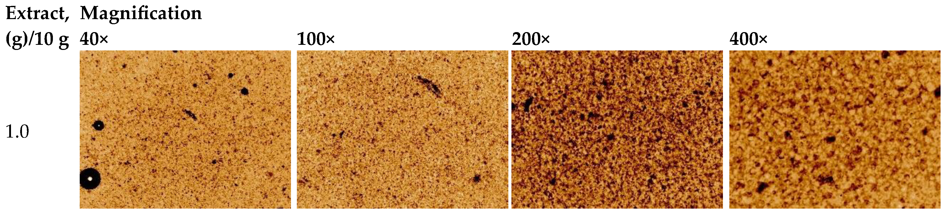
Figure 2.
Photographs of the 3D-printed scaffolds (lattices and discs) loaded with the Verbena officinalis extract.
Figure 2.
Photographs of the 3D-printed scaffolds (lattices and discs) loaded with the Verbena officinalis extract.
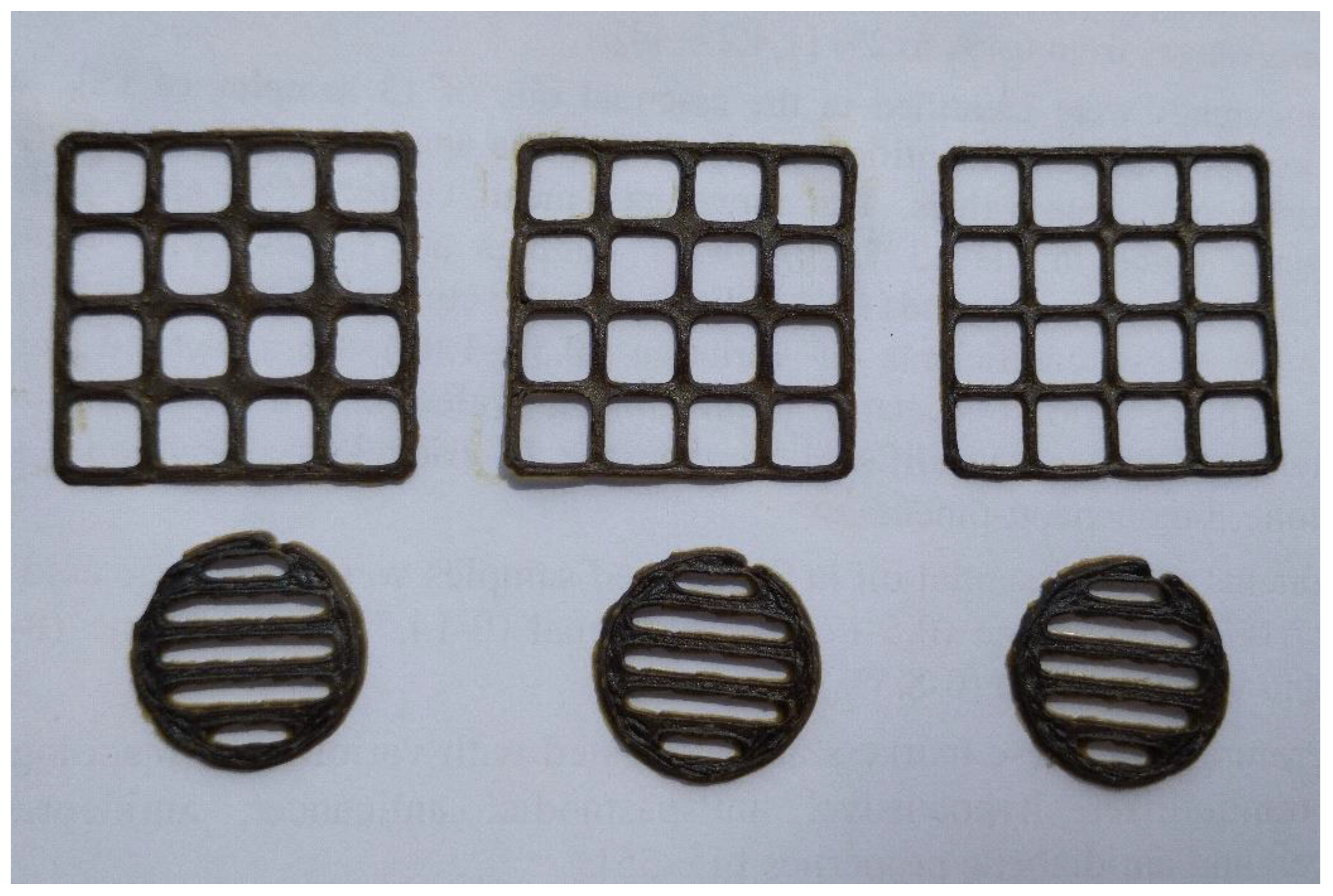

Table 1.
Content of phenolic compounds and amino acids in the Verbena officinalis extracts.
| Compound | Content in the dry extract, mg/kg | ||||||
|---|---|---|---|---|---|---|---|
| V1 (extractant 70% ethanol) |
V2 (extractant water) |
V1-Gly | V1-Phe | V1-Lys | V1-Val | V1-Arg | |
| Phenolic compounds | |||||||
| p-Coumaric acid | 14 ± 3 | 25 ± 5 | 8 ± 1 | 13 ± 1 | 15 ± 2 | 15 ± 1 | 12 ± 5 |
| Quercetin | 285 ± 10 | 137 ± 6 | 137 ± 8 | 231 ± 11 | 240 ± 3 | 217 ± 5 | 208 ± 8 |
| Gallic acid | 11 ± 3 | 102 ±14 | 13 ±1 | 16 ±2 | 13 ±1 | 13 ±1 | 11 ±1 |
| Protocatechuic acid | 79 ± 11 | 116 ± 27 | 91 ± 14 | 168 ± 12 | 78 ± 11 | 82 ± 11 | 76 ± 8 |
| Syringic acid | 54 ± 3 | 101 ± 6 | 51 ± 7 | 62 ± 15 | 70 ± 8 | 66 ± 11 | 60 ± 7 |
| Isovanillin | 5405 ± 1432 | 6414 ± 1514 | 983 ± 281 | 5435 ± 847 | 824 ± 70 | 3401 ± 533 | 2519 ± 354 |
| Ferulic acid | 33 ± 1 | 53 ± 5 | 21 ± 3 | 36 ± 3 | 35 ± 1 | 31 ± 2 | 27 ± 2 |
| Rutin | 14 ± 2 | 7 ± 1 | 6 ± 0 | 16 ± 1 | 14 ± 0 | 11 ± 1 | 10 ± 1 |
| Amino acids | |||||||
| Histidine | 27 ± 4 | 52 ± 4 | 17 ± 3 | 33 ± 7 | 31 ± 9 | 27 ± 7 | 23 ± 6 |
| Arginine | 187 ± 7 | 289 ± 8 | 154 ± 6 | 216 ± 14 | 195 ± 9 | 175 ± 13 | 134100 ± 2600 |
| Asparagine | 1253 ± 56 | 2238 ± 77 | 1114 ± 51 | 1533 ± 24 | 1265 ± 93 | 1141 ± 41 | 1196 ± 46 |
| Glutamine | 4995 ± 533 | 666 ± 37 | 3958 ± 124 | 5839 ± 27 | 5574 ± 1219 | 4501 ± 324 | 4612 ± 333 |
| Serine | 329 ± 22 | 421 ± 14 | 317 ± 10 | 411 ± 10 | 356 ± 34 | 326 ± 11 | 310 ± 9 |
| Aspartic acid | 345 ± 36 | 359 ± 36 | 295 ± 6 | 331 ± 29 | 306 ± 50 | 292 ± 22 | 286 ± 16 |
| Glycine | 46 ± 5 | 90 ± 5 | 58500 ± 1900 | 489 ± 8 | 57 ± 8 | 72 ± 4 | 54 ± 2 |
| Threonine | 121 ± 3 | 181 ± 3 | 128 ± 4 | 172 ± 1 | 132 ± 8 | 120 ± 3 | 118 ± 2 |
| β-Alanine | 29 ± 6 | 43 ± 7 | 34 ± 2 | 50 ± 2 | 33 ± 7 | 28 ± 5 | 37 ± 3 |
| α-Alanine | 721 ± 30 | 915 ±45 | 763 ± 9 | 919 ± 12 | 789 ± 65 | 950 ± 24 | 722 ± 13 |
| Tyrosine | 71 ± 4 | 113 ± 1 | 70 ± 3 | 167 ± 4 | 73 ± 1 | 114 ± 4 | 75 ± 7 |
| Valine | 97 ± 22 | 305 ± 47 | 196 ±21 | 333 ±50 | 135 ±32 | 85900 ±3000 | 102 ± 32 |
| Tryptophan | 56 ± 9 | 74 ± 9 | 50 ± 4 | 78 ± 9 | 56 ± 6 | 53 ± 6 | 46 ± 8 |
| Phenylalanine | 48 ± 11 | 106 ± 7 | 93 ± 6 | 183500 ± 5600 | 57 ± 11 | 123 ± 8 | 55 ± 8 |
| Isoleucine | 93 ± 28 | 182 ± 26 | 124 ± 9 | 379 ± 28 | 94 ± 20 | 249 ± 23 | 108 ± 25 |
| Leucine | 73 ±6 | 182 ±4 | 97 ±4 | 999 ±34 | 78 ±3 | 814 ±40 | 86 ± 6 |
| Lysine | 26 ±12 | 67 ±13 | 27 ± 3 | 50 ± 7 | 68000 ± 4200 | 53 ± 10 | 29 ± 8 |
| Spectrophotometry, % | |||||||
| Hydroxycinnamic acids (chlorogenic acid equivalents, spectrophotometry), % | 5.79 ± 0.39 | 3.95 ± 0.31 | 5.51 ± 0.73 | 5.15 ± 0.47 | 4.46 ± 0.43 | 4.34 ± 0.67 | 3.78 ± 0.75 |
| Flavonoids (rutin equivalents, spectrophotometry), % | 2.91 ± 0.12 | 2.00 ± 0.09 | 2.48 ± 0.05 | 2.39 ± 0.06 | 2.27 ± 0.06 | 2.24 ± 0.08 | 2.07 ± 0.04 |
| Total phenolic compounds (gallic acid equivalents, spectrophotometry), % | 7.77 ± 0.28 | 5.75 ± 0.25 | 6.76 ± 0.38 | 6.26 ± 0.10 | 5.93 ± 0.18 | 5.85 ± 0.18 | 5.56 ± 0.16 |
Notes: V1—the dry V. officinalis extract, obtained with 70% aqueous ethanol and its amino acids preparations with glycine (V1-Gly), phenylalanine (V1-Phe), lysine (V1-Lys), valine (V1-Val) and arginine (V1-Arg); V2—the dry V. officinalis extract, obtained with water.
Table 2.
Results of the study of the neurotropic activity of the Verbena officinalis extracts in the Open Field Test.
Table 2.
Results of the study of the neurotropic activity of the Verbena officinalis extracts in the Open Field Test.
| Investigated indicator | Intact animals | Comparison group (Sedaphyton) | V1 (extractant 70% ethanol) |
V2 (extractant water) |
V1-Gly | V1-Phe | V1-Lys | V1-Val | V1-Arg |
|---|---|---|---|---|---|---|---|---|---|
| Number of crossed squares | 75.33±5.68 | 73.17±5.85 | 79.50±4.55 | 94.17±6.64 */** | 80.50±8.24 | 83.50±3.77 | 81.50±5.96 | 50.33±5.27*/** | 67.83±7.46 |
| Number of vertical stands | 10.50±1.77 | 7.83±0.91 | 12.17±1.25** | 11.17±1.05** | 7.33±0.67* | 10.67±1.02** | 9.00±0.58 | 6.67±1.09* | 4.33±0.49*/** |
| Number of hole explorations | 14.17±1.08 | 2.83±0.48* | 12.50±0.76** | 12.17±0.98** | 11.00±1.39** | 8.17±0.48*/** | 9.83±0.95*/** | 2.17±0.60* | 3.17±0.60* |
| Conditional units of exploratory research activity | 3 | 1.91 | 3.10 | 3.17 | 2.54 | 2.70 | 2.58 | 1.45 | 1.53 |
| Boluses | 1.33±0.33 | 1.50±0.81 | 1.50±0.22 | 2.33±0.42 | 2.67±0.56 | 1.33±0.21 | 1.00±0.26 | 1.67±0.21 | 1.17±0.60 |
| Grooming | 0.67±0.21 | 0.33±0.33 | 1.17±0.31 | 0.67±0.33 | 0.67±0.21 | 0.83±0.17 | 1.00±0.26 | 0.5±0.22 | 0.33±0.21 |
| Conditional units of emotional reaction indicators | 2 | 1.62 | 2.87 | 2.75 | 3 | 2.25 | 2.25 | 2 | 1.37 |
| Conditional units of all activities | 5 | 3.54 | 5.97 | 5.92 | 5.54 | 4.95 | 4.83 | 3.45 | 2.91 |
Note: *—significance of deviations relative to intact animal data (p<0.05).
Table 3.
Behavior of animals in the Elevated Plus Maze test after the administration of the Verbena officinalis extracts.
Table 3.
Behavior of animals in the Elevated Plus Maze test after the administration of the Verbena officinalis extracts.
| Group of animals | Time spent in the open arm of the maze, s | Time spent in the closed arm of the maze, s. | Number of peeks from the closed arm | Number of crossings over the central platform | Number of downward glances from the ends of open arms |
|---|---|---|---|---|---|
| Intact animals | 24.17±3.95 | 273.17±5.40 | 10.50±1.80 | 3.33±1.2 | 4.67±0.61 |
| Comparison group (Sedaphyton) | 18.50±1.84 | 279.00±8.54 | 3.33±0.42* | 1.00±0.37* | 3.00±0.63* |
| V1 (extractant 70% ethanol) |
54.67±2.80*/** | 235.67±5.52*/** | 8.50±0.43** | 8.50±1.06*/** | 10.17±1.05*/** |
| V2 (extractant water) |
24.00±2.53 | 270.17±3.84 | 10.83±1.35** | 6.33±0.92*/** | 5.33±0.42** |
| V1-Gly | 29.33±2.75** | 262.50±6.57 | 3.17±0.40* | 3.83±0.06** | 4.33±0.42 |
| V1-Phe | 59.50±4.21*/** | 238.00±5.85*/** | 10.33±0.49** | 8.50±1.06*/** | 11.33±1.20*/** |
| V1-Lys | 47.17±3.32*/** | 250.50±8.50*/** | 8.00±0.58*/** | 5.50±1.52** | 11.00±1.41*/** |
| V1-Val | 9.50±1.43*/** | 287.17±1.96* | 2.67±0.42*/** | 1.83±0.48* | 1.00±0.37*/** |
| V1-Arg | 23.50±2.01 | 275.00±5.94 | 4.67±1.02*/** | 3.00±0.68** | 3.83±1.25 |
Note: *—significance of deviations relative to intact animal data (p<0.05), **—significance of deviations relative to comparison group data (p<0.05).
Table 4.
Anxiolytic activity of the Verbena officinalis extracts in the “Black-and-White Box” test.
| Group of animals | Time spent in the dark compartment, s | Number of peeks from the dark compartment | Time spent in the illuminated compartment, s | Number of exits into the illuminated compartment | Average time of a single stay in the illuminated compartment, s |
|---|---|---|---|---|---|
| Intact animals | 101.00±4.93 | 5.83±1.17 | 78.33±4.89 | 4.83±0.31 | 16.73±1.87 |
| Comparison group (Sedaphyton) | 119.00±5.16* | 2.17±0.70* | 59.67±4.67* | 3.33±0.71* | 25.68±9.18 |
| V1 (extractant 70% ethanol) |
24.67±5.28*/** | 2.00±1.06* | 100.33±5.28*/** | 3.17±0.91 | 47.98±12.85** |
| V2 (extractant water) |
98.33±6.82** | 7.00±1.15** | 80.17±6.84** | 5.83±0.54 | 14.14±1.48 |
| V1-Gly | 75.67±6.58*/** | 2.33±0.61* | 103.00±6.61*/** | 7.33±0.71*/** | 14.60±1.33 |
| V1-Phe | 73.67±5.49*/** | 2.17±1.22 | 105.5±5.60*/** | 6.33±0.61** | 17.43±1.82 |
| V1-Lys | 97.67±10.55 | 3.83±1.11 | 82.00±10.56 | 5.33±0.42** | 15.60±1.97 |
| V1-Val | 104.33±9.27 | 3.17±1.08 | 75.33±9.28 | 4.83±0.87 | 20.23±6.57 |
| V1-Arg | 126.67±6.80* | 2.67±0.92 | 53.83±7.06* | 5.50±1.12 | 11.66±2.66 |
Note: *—significance of deviations relative to intact animal data (p<0.05).
Table 5.
Antidepressant activity of the Verbena officinalis extracts in the Tail Suspension Test.
| Group of animals | Total immobility time, s |
|---|---|
| Intact animals | 103.67±5.00 |
| Comparison group (Sedaphyton) | 190.67±12.44* |
| V1 (extractant 70% ethanol) | 84.67±5.38*/** |
| V2 (extractant water) | 125.00±3.01*/** |
| V1-Gly | 124.83±7.96** |
| V1-Phe | 97.50±5.76** |
| V1-Lys | 99.67±6.25** |
| V1-Val | 177.50±8.11* |
| V1-Arg | 133.67±7.29*/** |
Note: *—significance of deviations relative to intact animal data (p<0.05).
Disclaimer/Publisher’s Note: The statements, opinions and data contained in all publications are solely those of the individual author(s) and contributor(s) and not of MDPI and/or the editor(s). MDPI and/or the editor(s) disclaim responsibility for any injury to people or property resulting from any ideas, methods, instructions or products referred to in the content. |
© 2025 by the authors. Licensee MDPI, Basel, Switzerland. This article is an open access article distributed under the terms and conditions of the Creative Commons Attribution (CC BY) license (http://creativecommons.org/licenses/by/4.0/).
Copyright: This open access article is published under a Creative Commons CC BY 4.0 license, which permit the free download, distribution, and reuse, provided that the author and preprint are cited in any reuse.


