
Submitted:
08 July 2025
Posted:
09 July 2025
You are already at the latest version
Abstract
Context: Medicinal plants have long been a cornerstone of traditional medicine and modern pharmacology, offering a rich source of biologically active compounds with diverse therapeutic applicationsObjective: Detection and identification of amino acid composition of Iraqi Ocimum basilicum (basil) leaves and evaluation the cytotoxic effects of the plant leaf extract on human colorectal cancer cells (HRT-18).Methods: Leaves of Ocimum basilicum were collected from Tikrit in November 2024. After drying and powdering, the plant material goes through cold methanol extraction. Initial phytochemical screening was conducted to identify the presence of alkaloids, flavonoids, coumarins, and terpenoids. Amino acids analysis was completed by amino acid analyzer with fluorescence detection. Cytotoxic effect was evaluated via the MTT assay on HRT-18 cell lines exposed to various concentrations of the plant extract. Morphological changes were further tested using dual Propidium Iodide/Acridine Orange assay fluorescent staining.Results: Seventeen amino acids were detected in the plant extract. The extract showed dose-dependent cytotoxic effects on HRT-18 cells, with significant reduction in cell viability at concentrations of more than 25 µg/mL. Morphological alterations of membrane blebbing and cell shrinkage were observed suggesting apoptotic activity. The IC₅₀ value confirmed strong cytotoxic potential.Conclusion: The extract of Ocimum basilicum leaf cultivated in Iraq shows a rich amino acid profile and significant cytotoxic activity against colorectal cancer cells that highlights its potential effect as a natural source of anticancer compounds and permit further analysis into its active constituents and the mechanisms of action.
Keywords:
amino acids
; cytotoxicity
; HRT-18 cell line
; leaf extract
1. Introduction
Medicinal plants have long been a cornerstone of traditional medicine and modern pharmacology, offering a rich source of biologically active compounds with diverse therapeutic applications. Among these, Ocimum basilicum Linn., a member of the Lamiaceae family, is widely cultivated in Asia, the Middle East, and the Mediterranean for culinary, aromatic, and medicinal uses [1]. “Sweet basil” is the popular name of Ocimum basilicum that is used in both Ayurvedic and Unani medicine system [2]. There were more than 150 species of the genus Ocimum present. Basil is the main crop of essential oil that is cultivated commercially in many countries [3]. Its popularity is due to its rich and spicy, mildly peppery flavor with a trace amounts of clove and mint and has been widely used as a food ingredient for flavoring enhancement, meat products and baked foods [4].
The plant is used for its stomachic, antipyretic and alexipharmic effects. It also has emmenagogue and diuretic properties. An infusion of the plant extract is considered to be anthelminthic, antiemetic, diaphoretic and anti diarrheaic in Annam. Diuretic, anti-dysenteric and aphrodisiac effects have also been attributed to this plant seeds. The plant juice exhibits stimulant, carminative and antibacterial effects; its essential oil has antifungal, antibacterial and insecticidal properties [5]. The plant flowers have diuretic, demulcent and stimulant properties. These flowers are also considered to have anti-spasmodic, carminative and digestive stimulant effects [6].
High concentration of vitamins, minerals and oils are present in the green plant leaves [7]. The phytochemical screening reports of O.basilicum showed the presence of the following I it: proteins, amino acids, glycoside, mucilage, gums, tannins, triterpenoids steroids, phenolic compound, saponins, sterols, flavones and flavonoids. A total of 29 compounds representing 98.0–99.7% of the oils are identified in this plant which are contributed to its pharmacological potential [8,9]. However, limited studies have explored the amino acid composition of basil, particularly in local varieties grown in Iraq. Amino acids are crucial components of plant metabolism and human nutrition; they play central roles in protein synthesis, neurotransmission, metabolic regulation, and immune function [10]. Profiling amino acids in medicinal plants like O. basilicum can provide insights into their nutritional value and enhance their use in dietary supplements and therapeutic formulations [11].
In recent years, increasing attention has been paid to the anticancer properties of medicinal plants. Plant products are promising alternative or adjunct to conventional chemotherapy owing to their potential for high efficacy and lower toxicity [12]. One of the most common malignancies worldwide is the colorectal cancer, and the study of plant-derived compounds with selective cytotoxic effects is of critical importance [13]. The HRT-18 cell line that is derived from human colorectal adenocarcinoma can serve as a reliable in vitro model for screening cytotoxicity and evaluating apoptosis-inducing activity [14].
This study was carried out to consider two key features of the Iraqi Ocimum basilicum plant: (1) to determine its amino acid profile using a high-performance amino acid analyzer, and (2) to evaluate cytotoxic effects of the plant on HRT-18 colorectal cancer cells by using the MTT assay and Propidium Iodide/Acridine Orange assay fluorescent staining (AO/PI) fluorescent staining.
2. Material and Methods
2.1. Plant Material
Ocimum basilicum leaves were collected from Tikrit in November 2024. Leaves were washed thoroughly, dried under shade, and ground in a mechanical grinder to a fine powder.
2.2. Experimental Work
2.2.1. Extraction Method (Cold Method)
A Hundred grams of the powdered plant material was soaked in 1500ml methanol, with occasional shaking, at room temperature. After 3 days, the methanol-soluble materials were filtered off. The filtrate was evaporated to dryness under a vacuum using a rotary evaporator. A dark greenish residue was obtained. The residue evaporated to dryness and take to the laboratory for detection of amino acid.
2.2.2. Preliminary Phytochemical Examination of Crude Extracts
Phytochemical analysis for the screening and identification of bioactive chemical constituents in the medicinal plants under study was carried out on crude extracts, and fractions as well as powder specimens using the standard procedures as described [15,16].
Alkaloid test: approximately 0.5 to 0.6 g of each of plant extract and fractions were mixed in 8 ml of 1% HCl, warmed, and filtered. Two ml of the filtrate was treated separately with both reagents (Mayer’s and Dragendorff‘s), after which it was observed whether the alkaloids were present or absent in the turbidity or precipitate formation.
Coumarins test: 0.5 g of each of plant extract and fractions were mixed in a test tube. The mouth of the tube was covered with filter paper treated with 1 N NaOH solution. The test tube was placed for a few minutes in a boiling water, and then the filter paper was removed and examined under the UV light for yellow fluorescence indicating the presence of coumarins.
Terpenoids test (Salkowski test): 5 ml of each of plant extract and fractions were mixed in 2 ml of chloroform followed by the careful addition of 3 ml concentrated (H2SO4). A layer of reddish-brown coloration was formed at the interface thus indicating a positive result for the presence of terpenoids.
Flavonoids test: 0.5 g of each of plant extract and fractions were shaken with petroleum ether to remove the fatty materials (lipid layer). The defatted residue was dissolved in 20 ml of 80% ethanol and filtered. The filtrate was used for the following tests:
(a) Three ml of the filtrate was mixed with 4 ml of 1% aluminum chloride in methanol in a test tube, and the color was observed. The formation of yellow color indicated the presence of flavonoids.
(b) Three ml of the filtrate was mixed with 4 ml of 1% potassium hydroxide in a test tube, and the color was observed. A dark yellow color indicated the presence of flavonoids.
2.3. Amino Acids Determination
One ml was taken from the extracted sample and 200 microliters of orthophthalein aldehyde (5%) was added to it and the sample was shaken for two minutes after which 100 microliters of the last mixture were taken and injected into the amino acid analyzer [17].
The test was conducted in the laboratories of the Scientific Research Authority / Environment and Water Research Center using the amino acid analyzer (Korean made). The method provided by the scientist (Scriver CR, 2001) was used, where the carrier phase consisting of (methanol: acetonitrile: 5% formic acid) was used in proportions (20: 60: 20) at a flow rate of (1 ml / minute). A separation column (C18–NH2 (250mm * 4.6 mm) was used to separate the amino acids, while a fluorescence detector was used to detect the amino acids at wavelengths (Ex = 445 nm, Em = 465 nm). The (clarity 2015) program was used to analyze the amino acids [18].
2.4. Cytotoxicity Assay
2.4.1. Cell Lines Used
HCT-8 [HRT-18] cells were isolated from the large intestine of a 67-year-old, male, adenocarcinoma patient. HCT-8 [HRT-18] is used for cancer and toxicology research.
2.4.2. MTT Cytotoxicity Assay
Cell Culture Conditions
The cell lines were cultured in cultured in MEM (US Biological, USA) supplemented with 10% (v/v) fetal bovine serum (FBS) (Capricorn-Scientific, Germany), and 100 IU penicillin, and 100µg streptomycin (Capricorn-Scientific, Germany) and incubated in a humidified atmosphere at 37 °C. Exponentially growing cells were used for experiments [19].
MTT Procedure
Cells were seeded at a density of 10000 cells in a 96-well microplate (NEST Biotech, China) and incubated at 37 °C for 72 h until monolayer confluence was achieved. Cytotoxicity was investigated through 3-(4,5-dimethylthiazol-2-yl)-2,5-diphenyltetrazolium bromide (MTT) assay (Elabscience, China). The cells were exposed to a range of concentrations (1000, 500, 250, 125, 62, 31 ug). After 72h of infection, MTT dye solution 28 µL of (2 mg/ml) was added to each well. The incubation continued for three hours. A total of 100 μl of DMSO was added to each well and incubated for 15 min. The optical density was measured at 492 nm using a microplate reader [19]. Cytotoxicity % was calculated using the following equation:
Cytotoxicity % = (OD Control − OD sample)/OD Control × 100,
were OD control being the mean optical density of untreated wells, and OD Sample is the optical density of treated wells [20].
2.5. DNA Damage
Apoptosis Estimation (Propidium Iodide/Acridine Orange Assay)
The apoptotic attentions in cell lines (infected and control) were measured using (AO/PI). 5000 cells/well were seeded in plate, next infected with (Ocimum Basilicum) for 24 hours in a 37 °C incubator. The tested wells received exactly 50µl of the AO/PI stain mixture (at room temperature) for 30 seconds. After then, the stain was removed. The images were taken using a Leica fluorescent microscope [21]. Fluorescent intensity measured by fluorescent microscopy and via using image j software.
Statistical analysis:
The obtained data were statically analyzed using an unpaired t-test and Tukey’s ANOVA multiple comparisons test with GraphPad Prism 8. The values were presented as the mean ± SD of triplicate measurements [20].
3. Results
3.1. Amino Acid Detection
The chromatographic analysis clearly (Figure 1) demonstrates multiple peaks corresponding to different amino acids present in the extract. The sharp and well-separated peaks indicate good resolution and efficient separation by the amino acid analyzer. The presence of numerous peaks suggests a diverse amino acid profile, which may contribute to the biological activity of the plant. While, Table 1 illustrates the specific retention times for the 17 amino acids identified in the sample. These times were consistent with standard references, confirming the identity of the compounds. The reproducibility of retention times across multiple runs confirms the reliability and sensitivity of the analytical method. Therefore, Ocimum basilicum leaves contained 17 amino acid detected by amino acid analyzer.
3.2. Cytotoxic Effects Evaluation
Figure 2A shows HRT-18 cells treated with Ocimum basilicum extract with noticeable morphological changes are observed, such as: cell shrinkage, membrane blebbing, detachment from the surface and reduced overall cell density. These features are typical signs of cytotoxicity and apoptosis. In contrast, Figure 2B shows untreated cells that appear: well-spread, with normal polygonal shape, forming a dense and uniform monolayer and with no signs of damage. These visual differences support the conclusion that the basil extract induces cell death in cancer cells.
Figure 3 indicates the inhibitory curve which shows the cytotoxic effect of different concentrations of Ocimum basilicum extract on HRT-18 cancer cells. As the concentration of the extract increases, there is a clear increase in the percentage of cell death, indicating a dose-dependent inhibitory effect. Higher doses of the extract can cause greater cytotoxicity, suggesting that the extract contains active compounds capable of killing cancer cells.
Figure 4 represents the dose-response curve used to determine the IC50 value of Ocimum basilicum on cancer cells. The x-axis represents the concentration of the test compound (in µg), and the y-axis represents the percentage of cell inhibition. The IC50 value, represented by the Log of drug concentration of the test Ocimum basilicum that inhibits 50% of cell viability.
As shown in Figure 5 the post hoc Tukey’s test demonstrates a significant, dose-dependent cytotoxic effect of Ocimum basilicum extract on HRT-18 cells. Higher concentrations (particularly 100 µg/mL) show statistically significant reductions in cell viability compared to lower concentrations and the control group (p<0.0001). Minimal or non-significant differences at lower doses suggest a threshold concentration is needed to elicit a strong cytotoxic response. These results support the extract’s potential as an anticancer agent at higher doses.
3.3. DNA Damage Analysis
Figure 6 illustrates a clear difference in fluorescence intensity between treated and control HRT-18 cells, indicating changes in cell viability. In the treated group, fluorescence from dead cells is significantly higher than viable cells (p<0.01), while the control group shows a predominance of viable cells with no significant difference between viability and death signals. This suggests that treatment with Ocimum basilicum extract induces cytotoxicity in HRT-18 cells, leading to increased cell death.
Figure 7 compares HRT-18 colorectal cancer cells before and after treatment with Ocimum basilicum extract. Figure 7A, the untreated (control) cells show normal morphology, with a flat, polygonal shape, intact membranes, and a dense, healthy monolayer covering the surface. While, Figure 7B, the treated cells display clear signs of cytotoxicity, including Cell shrinkage, membrane blebbing, loss of adherence, reduced cell density and rounded or fragmented cells, indicating possible apoptosis. These visual differences support the extract’s antiproliferative and cytotoxic effects on cancer cells.
4. Discussion
The amino acid analysis of Ocimum basilicum leaves reveals a rich and diverse profile of both essential and non-essential amino acids. A total of 17 amino acids were identified, each with varying concentrations, indicating the nutritional and potentially therapeutic value of this plant. Among the detected amino acids, Phenylalanine exhibited the highest concentration (38.00 g/100 g), followed by Alanine (29.08 g/100 g), Histidine (28.90 g/100 g), and Serine (27.88 g/100 g). The high levels of these amino acids are significant meanwhile phenylalanine is an essential amino acid that is involved in protein biosynthesis and to be neurotransmitters precursor like dopamine and norepinephrine [22]. Alanine exhibits a vital role in glucose metabolism and production of energy, especially under stress conditions [23]. Other essential amino acids like Valine (35.65 g/100 g), Leucine (30.65 g/100 g), Isoleucine (23.54 g/100 g), and Methionine (19.08 g/100 g) were also present in considerable amounts. These branched-chain amino acids are principally important for immune function, muscle repair, and neurotransmitter synthesis, making Ocimum basilicum potentially beneficial for physical endurance and metabolic health.
The presence of Aspartic acid (17.45 g/100 g) and Glutamic acid (27.44 g/100 g) further supports the plant’s nutritional value. Glutamic acid is a key neurotransmitter and has a role in memory and learning, while aspartic acid is involved in the citric acid cycle and DNA synthesis [24]. Interestingly, Arginine (26.14 g/100 g) and Cysteine (24.15 g/100 g) were also detected in plant extract. Cysteine contributes to antioxidant defense through glutathione synthesis [25], while arginine supports nitric oxide production, aiding in cardiovascular health and immune modulation [26]. The broad spectrum and high amino acids concentration detected in this analysis support the traditional uses of Ocimum basilicum in herbal medicine. The obtained results are consistent with other studies that highlight the nutritional and pharmacological properties of basil species (Ocimum sanctum and Ocimum gratissimum) for their amino acid content [27,28].
The cytotoxicity assessment of Ocimum basilicum extract on HRT-18 colorectal cancer cells reveals significant dose-dependent antiproliferative activity. Figure 2A demonstrates clear morphological changes in treated cells, including shrinkage, membrane blebbing, and detachment, all of which are hallmark indicators of apoptosis and cell stress. In contrast, Figure 2B shows the untreated control group maintaining normal morphology, indicating that the observed effects are indeed induced by the basil extract. Figure 3 illustrates a dose-dependent inhibitory curve, confirming that increasing concentrations of O. basilicum extract result in elevated cytotoxic effects. This supports the hypothesis that the plant contains bioactive compounds—such as flavonoids, terpenoids, and phenolics—that interfere with cancer cell metabolism or induce programmed cell death, as supported by literature indicating the anticancer potential of plant-derived compounds [29,30].
The IC50 curve in Figure 4 further quantifies the extract’s potency, showing that the concentration required to inhibit 50% of the cell population is within a practical therapeutic range. This reinforces the potential use of O. basilicum in cancer treatment formulations, especially when standardized for high-yielding cytotoxic compounds. The post hoc Tukey’s test shown in Figure 5 provides strong statistical evidence for the cytotoxic effect, particularly at 100 µg/mL, which showed highly significant reductions in cell viability (p<0.0001). The lack of significant difference at lower doses (e.g., 3.125 µg/mL) suggests a threshold concentration is needed to initiate noticeable cellular damage. These results align with findings from previous studies, which reported similar dose-dependent cytotoxicity of Ocimum species on various cancer cell lines [31,32].
The assessment of DNA damage in HRT-18 colorectal cancer cells following treatment with Ocimum basilicum extract reveals significant cytotoxic and antiproliferative effects. As illustrated in Figure 6, fluorescence intensity analysis clearly distinguishes between treated and untreated cells. The treated group exhibits markedly increased fluorescence associated with cell death, while the control group maintains high levels of viable cell fluorescence with minimal death signals. The significant difference in viability (p<0.01) confirms that the basil extract induces cytotoxicity, likely through mechanisms involving DNA damage and apoptosis. Figure 7 provides visual confirmation of this effect. Untreated cells (Figure 7A) maintain typical epithelial morphology—polygonal, well-adhered, and forming a confluent monolayer. In contrast, treated cells (Figure 7B) display multiple morphological signs of cytotoxicity, including cell shrinkage, membrane blebbing, and detachment from the culture surface. These features are consistent with apoptotic cell death, supporting the fluorescence data and previous cytotoxicity assays.
These findings align with earlier reports that plant-derived compounds, including flavonoids and essential oils found in O. basilicum, can cause oxidative stress and DNA fragmentation in cancer cells, leading to apoptosis [33,34].. The observed cell damage and morphological alterations suggest that the extract’s active constituents may disrupt cellular homeostasis and promote DNA damage pathways [35].
5. Conclusions
Ocimum basilicum leaf extract exhibits a rich amino acid profile and significant cytotoxic activity against HRT-18 colorectal cancer cells. The extract induced dose-dependent morphological changes, reduced cell viability, and caused DNA damage indicative of apoptosis. These findings highlight its potential as a natural source of anticancer compounds and warrant further investigation into its active constituents and mechanisms of action.
References
- Javanmardi, J.; Khalighi, A.; Kashi, A.; Bais, H.P.; Vivanco, J.M. Chemical Characterization of Basil (Ocimum basilicum L.) Found in Local Accessions and Used in Traditional Medicines in Iran. J. Agric. Food Chem. 2002, 50, 5878–5883. [Google Scholar] [CrossRef]
- Muralidharan A, Dhananjayan R. Cardiac stimulant activity of Ocimum basilicum Linn. extracts. Indian J Pharmacol 2004, 36, 163–166. [Google Scholar]
- Hussain, A.I.; Anwar, F.; Sherazi, S.T.H.; Przybylski, R. Chemical composition, antioxidant and antimicrobial activities of basil (Ocimum basilicum) essential oils depends on seasonal variations. Food Chem. 2008, 108, 986–995. [Google Scholar] [CrossRef]
- Chang, X.; Alderson, P.G.; Wright, C.J. Variation in the Essential Oils in Different Leaves of Basil (Ocimum basilicum L.) at Day Time. Open Hortic. J. 2009, 2, 13–16. [Google Scholar] [CrossRef]
- Jayaweera DMA. Medicinal Plants, (Indigenous and Exotic) Used in Ceylon. Part III. Colombo: The National Science Foundation of Sri Lanka; 1981. 101-3.
- Bhatti, H.A.; Tehseen, Y.; Maryam, K.; Uroos, M.; Siddiqui, B.S.; Hameed, A.; Iqbal, J. Identification of new potent inhibitor of aldose reductase from Ocimum basilicum. Bioorganic Chem. 2017, 75, 62–70. [Google Scholar] [CrossRef]
- El-Dakar, A.Y.; Shalaby, S.M.; Nemetallah, B.R.; Saleh, N.E.; Sakr, E.M.; Toutou, M.M. Possibility of using basil (Ocimum basilicum) supplementation in Gilthead sea bream (Sparus aurata) diet. Egypt. J. Aquat. Res. 2015, 41, 203–210. [Google Scholar] [CrossRef]
- Dong, L.; He, J.; Luo, L.; Wang, K. Targeting the Interplay of Autophagy and ROS for Cancer Therapy: An Updated Overview on Phytochemicals. Pharmaceuticals 2023, 16, 92. [Google Scholar] [CrossRef]
- Devika, T.; Shashi, V. Pharmacognostical and phytochemical investigation of Tulsi plants available in Western Bareilly region. Global Journal of Pharmacology 2016, 9, 60–63. [Google Scholar]
- Lopez MJ, Mohiuddin SS. Biochemistry, Essential Amino Acids. [Updated 2024 Apr 30]. In: StatPearls [Internet]. S: Treasure Island (FL), 2025.
- Calderón Bravo, H.; Vera Céspedes, N.; Zura-Bravo, L.; Muñoz, L.A. Basil Seeds as a Novel Food, Source of Nutrients and Functional Ingredients with Beneficial Properties: A Review. Foods 2021, 10, 1467. [Google Scholar] [CrossRef]
- Cragg, G.M.; Pezzuto, J.M. Natural Products as a Vital Source for the Discovery of Cancer Chemotherapeutic and Chemopreventive Agents. Med Princ. Pr. 2015, 25, 41–59. [Google Scholar] [CrossRef]
- Sung, H.; Ferlay, J.; Siegel, R.L.; Laversanne, M.; Soerjomataram, I.; Jemal, A.; Bray, F. Global Cancer Statistics 2020: GLOBOCAN Estimates of Incidence and Mortality Worldwide for 36 Cancers in 185 Countries. CA: a cancer journal for clinicians 2021, 71, 209–249. [Google Scholar] [CrossRef] [PubMed]
- Murakami, T.; Otsubo, S.; Namitome, R.; Shiota, M.; Inokuchi, J.; Takeuchi, A.; Kashiwagi, E.; Tatsugami, K.; Eto, M. Clinical factors affecting perioperative outcomes in robot-assisted radical prostatectomy. Mol. Clin. Oncol. 2018, 9, 575–581. [Google Scholar] [CrossRef] [PubMed]
- Parekh, J.; Chanda, S. In vitro antimicrobial activity and phytochemical analysis of some Indian medicinal plants. Turkish Journal of Biology 2007, 31, 53–58. [Google Scholar]
- Tiwari, P.; Kumar, B.; Kaur, M.; Kaur, G.; Kaur, H. Phytochemical screening and extraction: A review. Internationale Pharmaceutica Sciencia 2011, 1, 98–106. [Google Scholar]
- Dahl-Lassen, R.; van Hecke, J.; Jørgensen, H.; Bukh, C.; Andersen, B.; Schjoerring, J.K. High-throughput analysis of amino acids in plant materials by single quadrupole mass spectrometry. Plant Methods 2018, 14, 1–9. [Google Scholar] [CrossRef]
- Scriver CR, Beaudet AL, Valle D, Sly WS, Childs B, Kinzler KW, Vogelstein B, eds. The Metabolic and Molecular Bases of Inherited Disease. 8th ed. New York, NY: McGraw-Hill, Inc; 2001. 1665–2105.
- Salman MI, Emran MA, Al-Shammari AM. Spheroid-formation 3D engineering model assay for in vitro assessment and expansion of cancer cells. In AIP Conference Proceedings 2021 Nov 11 (Vol. 2372, No. 1). AIP Publishing. [CrossRef]
- Al-Shammari, A.M.; Salman, M.I. Antimetastatic and antitumor activities of oncolytic NDV AMHA1 in a 3D culture model of breast cancer. Front. Mol. Biosci. 2024, 11, 1331369. [Google Scholar] [CrossRef]
- Salman, M.I.; Al-Shammari, A.M.; Emran, M.A. 3-Dimensional coculture of breast cancer cell lines with adipose tissue–Derived stem cells reveals the efficiency of oncolytic Newcastle disease virus infection via labeling technology. Front. Mol. Biosci. 2022, 9, 754100. [Google Scholar] [CrossRef] [PubMed]
- Wu, G. Functional amino acids in nutrition and health. Amino Acids 2013, 45, 407–411. [Google Scholar] [CrossRef] [PubMed]
- Holeček, M. Origin and Roles of Alanine and Glutamine in Gluconeogenesis in the Liver, Kidneys, and Small Intestine under Physiological and Pathological Conditions. Int. J. Mol. Sci. 2024, 25, 7037. [Google Scholar] [CrossRef]
- Dingledine R, McBain CJ. Glutamate and Aspartate Are the Major Excitatory Transmitters in the Brain. In: Siegel GJ, Agranoff BW, Albers RW, et al., editors. Basic Neurochemistry: Molecular, Cellular and Medical Aspects. 6th edition. Philadelphia: Lippincott-Raven; 1999.
- Chiang, F.-F.; Chao, T.-H.; Huang, S.-C.; Cheng, C.-H.; Tseng, Y.-Y.; Huang, Y.-C. Cysteine Regulates Oxidative Stress and Glutathione-Related Antioxidative Capacity before and after Colorectal Tumor Resection. Int. J. Mol. Sci. 2022, 23, 9581. [Google Scholar] [CrossRef]
- Kurhaluk, N.; Tkaczenko, H. L-Arginine and Nitric Oxide in Vascular Regulation—Experimental Findings in the Context of Blood Donation. Nutrients 2025, 17, 665. [Google Scholar] [CrossRef]
- Bakhtiar, Z.; Mirjalili, M.H.; Hassandokht, M. Nutritional composition of sweet basil (Ocimum basilicum L.) agro-ecotypic populations: Prospecting and selecting for use in food products. Food Humanit. 2024, 3. [Google Scholar] [CrossRef]
- Enegide, C.; C, O.C. Ocimum Species: Ethnomedicinal Uses, Phytochemistry and Pharmacological Importance. Int. J. Curr. Res. Physiol. Pharmacol. (IJCRPP) 2021, 1–12. [Google Scholar] [CrossRef]
- Kooti, W.; Farokhipour, M.; Asadzadeh, Z.; Ashtary-Larky, D.; Asadi-Samani, M. The role of medicinal plants in the treatment of diabetes: a systematic review. Electron. Physician 2016, 8, 1832–1842. [Google Scholar] [CrossRef]
- Sivak, K.V.; Stosman, K.I.; Lesiovskaya, E.E. Bioactive compounds of medicinal plants with anti-herpes effect (part 1). Rastitelnye resursy 2024, 60, 3–20. [Google Scholar] [CrossRef]
- Pattanayak, P.; Behera, P.; Das, D.; Panda, S.K. Ocimum sanctum Linn. A reservoir plant for therapeutic applications: An overview. Pharmacogn. Rev. 2010, 4, 95–105. [Google Scholar] [CrossRef]
- D’archivio, M.; Santangelo, C.; Scazzocchio, B.; Varì, R.; Filesi, C.; Masella, R.; Giovannini, C. Modulatory Effects of Polyphenols on Apoptosis Induction: Relevance for Cancer Prevention. Int. J. Mol. Sci. 2008, 9, 213–228. [Google Scholar] [CrossRef]
- Fitsiou, E.; Pappa, A. Anticancer Activity of Essential Oils and Other Extracts from Aromatic Plants Grown in Greece. Antioxidants 2019, 8, 290. [Google Scholar] [CrossRef]
- Almutairi, B.O.; Alsayadi, A.I.; Abutaha, N.; Al-Mekhlafi, F.A.; Wadaan, M.A.; Ali, A. [Retracted] Evaluation of the Anticancer Potential of Morus nigra and Ocimum basilicum Mixture against Different Cancer Cell Lines: An In Vitro Evaluation. BioMed Res. Int. 2023, 2023. [Google Scholar] [CrossRef]
- Miller, M.A.; Zachary, J.F. Mechanisms and Morphology of Cellular Injury, Adaptation, and Death 11 For a glossary of abbreviations and terms used in this chapter see E-Glossary 1-1. Pathol. Basis Vet. Dis. 2017, 2–43.e19. [Google Scholar] [CrossRef]
Figure 1.
Chromatogram of Ocimum basilicum leaves extract.
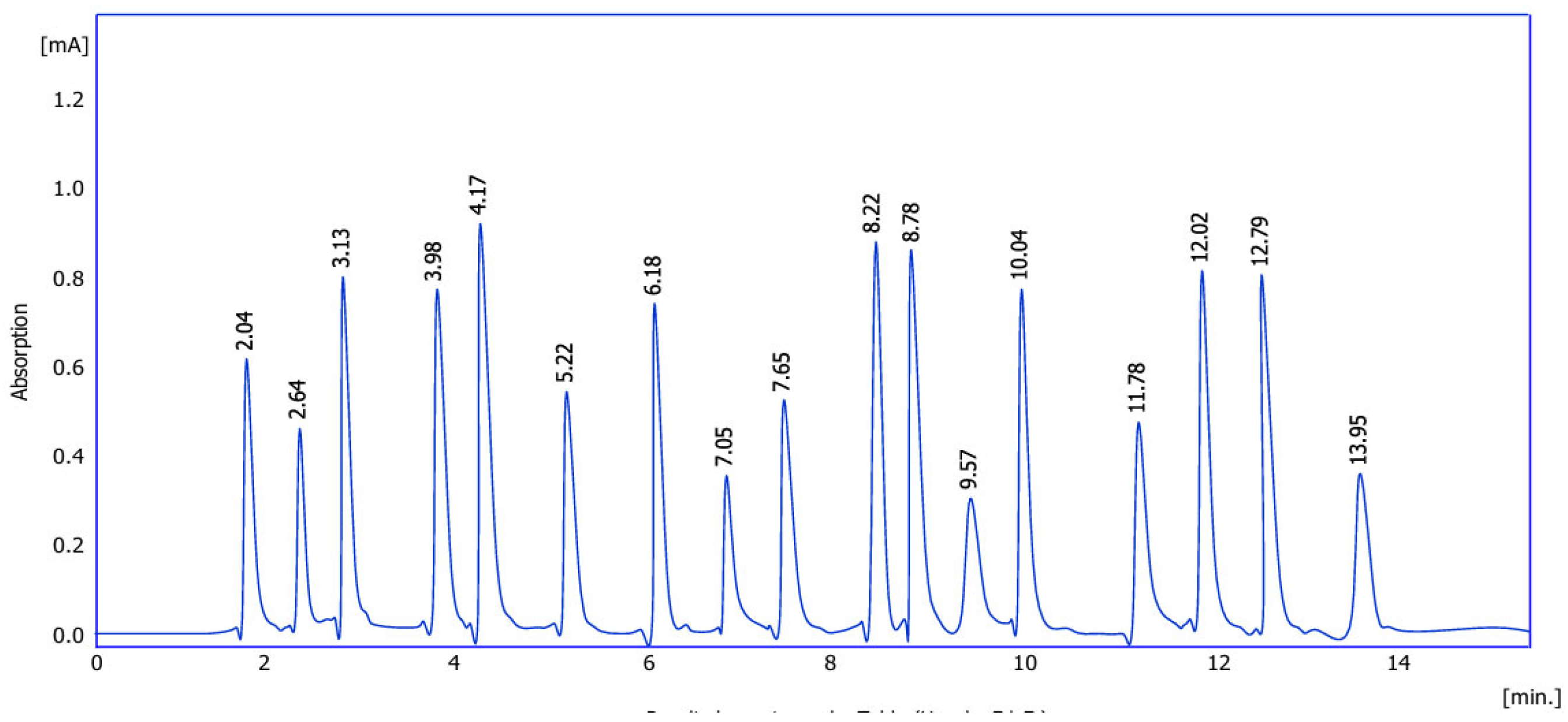
Figure 2.
Effect of Ocimum basilicum Leaves on HRT-18 Cells. A- Represent treated HRT-18 Cells. B- Represent untreated HRT-18.
Figure 2.
Effect of Ocimum basilicum Leaves on HRT-18 Cells. A- Represent treated HRT-18 Cells. B- Represent untreated HRT-18.
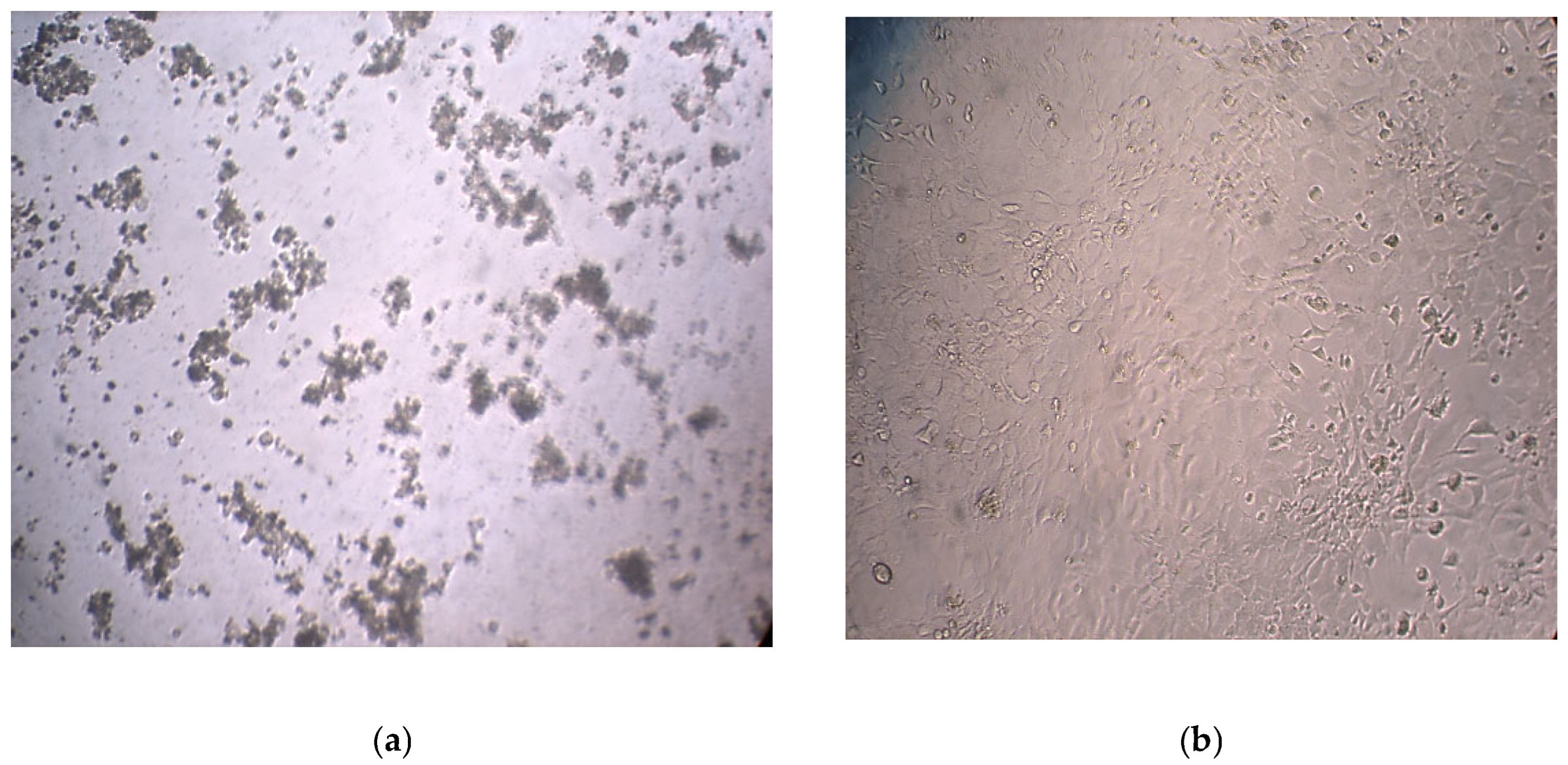
Figure 3.
Inhibitory curve showing the effect of Ocimum basilicum on cell viability. The x-axis represents the concentration of the extract (µg/mL), while the y-axis shows the percentage of cell death (cytotoxicity).
Figure 3.
Inhibitory curve showing the effect of Ocimum basilicum on cell viability. The x-axis represents the concentration of the extract (µg/mL), while the y-axis shows the percentage of cell death (cytotoxicity).
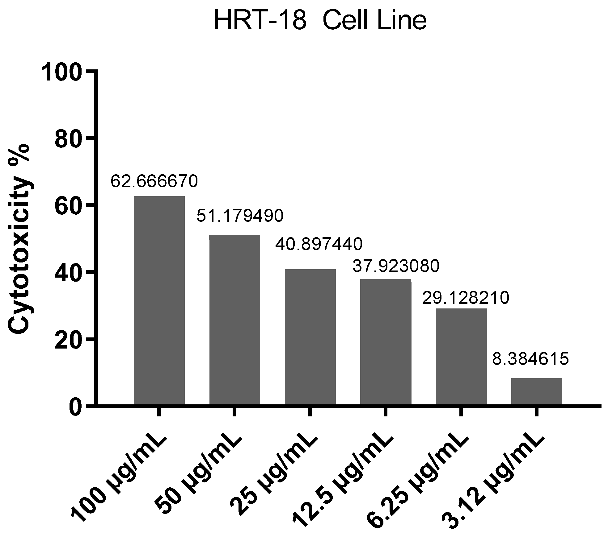
Figure 4.
IC50 showing the effect of Ocimum basilicum on cell viability.
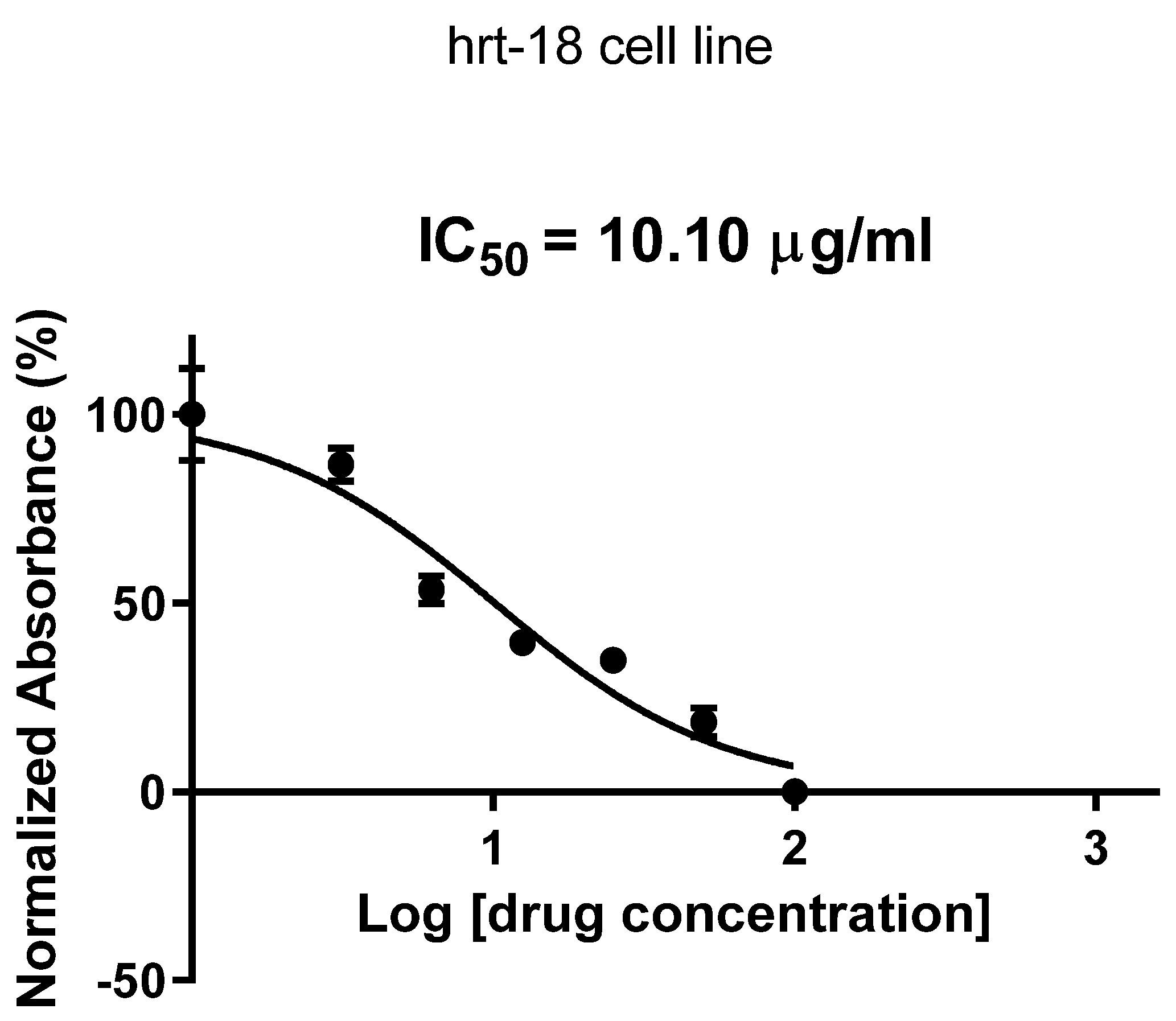
Figure 5.
Optical density (OD) reading curve over time for cells treated with Ocimum basilicum. The x-axis represents concentrations in ug/ml, and the y-axis represents the OD value at 492 nm. Each data point represents the mean ± standard deviation of triplicates.
Figure 5.
Optical density (OD) reading curve over time for cells treated with Ocimum basilicum. The x-axis represents concentrations in ug/ml, and the y-axis represents the OD value at 492 nm. Each data point represents the mean ± standard deviation of triplicates.
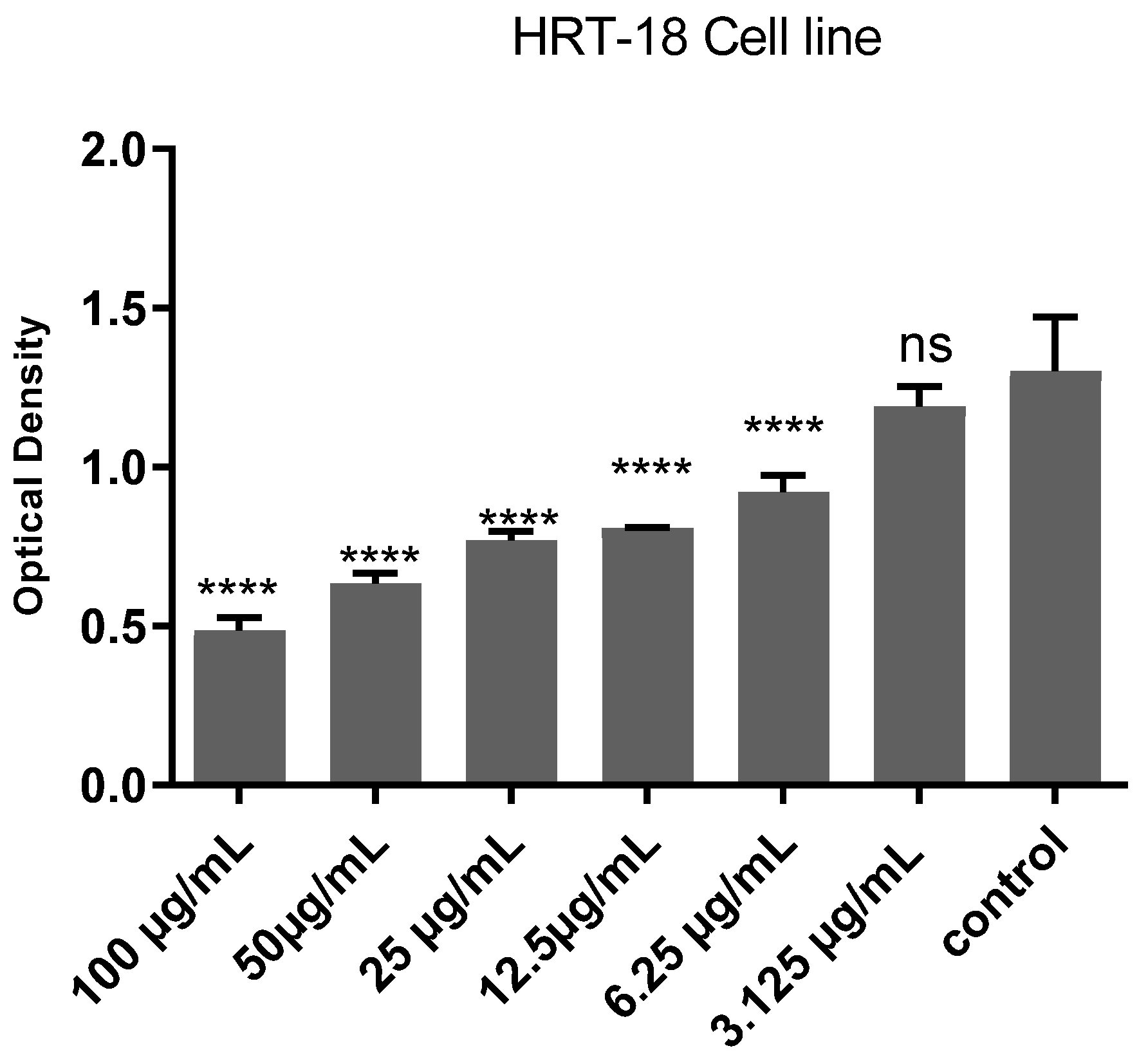
Figure 6.
Effect of Ocimum basilicum extract on viability and death of HRT-18 cells assessed by fluorescence intensity.
Figure 6.
Effect of Ocimum basilicum extract on viability and death of HRT-18 cells assessed by fluorescence intensity.
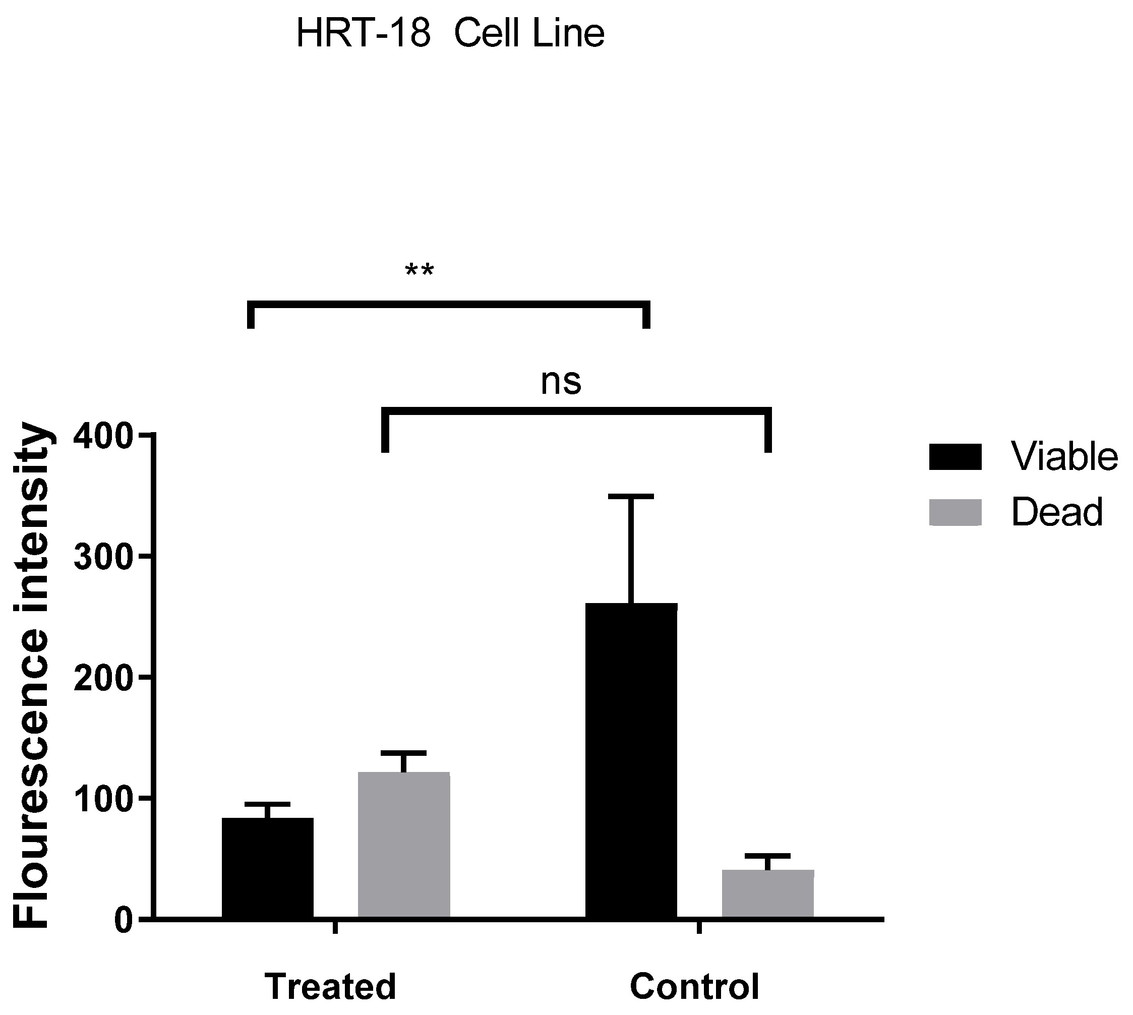
Figure 7.
Effect of Ocimum basilicum Leaves on HRT-18 Cells. A- Represent untreated HRT-18 Cells (control). B- Represent treated HRT-18.
Figure 7.
Effect of Ocimum basilicum Leaves on HRT-18 Cells. A- Represent untreated HRT-18 Cells (control). B- Represent treated HRT-18.
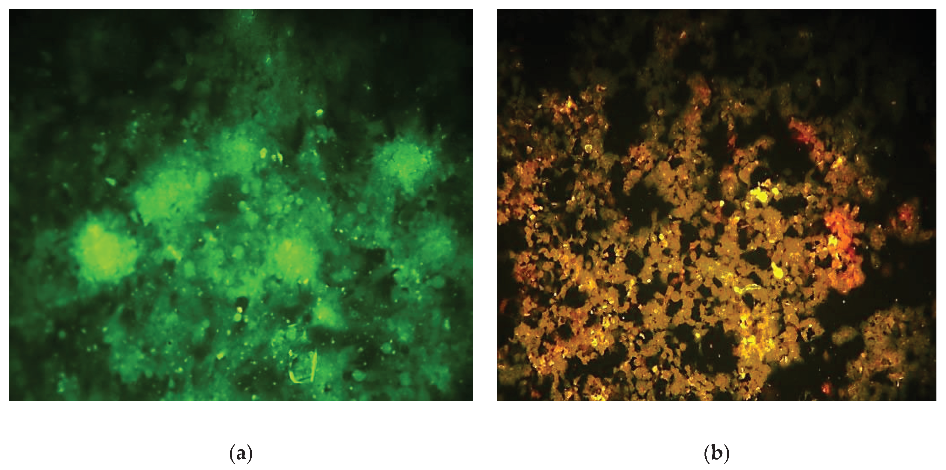

Table 1.
Retention times of amino acid detect of Ocimum basilicum Leaves.
| No | Reten. Time [min] | Area [mAU.s] | Height [mAU] | Amount (g/100 g) | Calculation | Peak type | Compound Name |
| 1 | 2.04 | 4562.9 | 604.7 | 17.45 | Calibration carve | Order | Aspartic acid |
| 2 | 2.64 | 4152.6 | 643.6 | 26.09 | Calibration carve | Order | Glycine |
| 3 | 3.13 | 6325.9 | 794.9 | 24.12 | Calibration carve | Order | Lysine |
| 4 | 3.98 | 5854.0 | 789.8 | 27.88 | Calibration carve | Order | Serine |
| 5 | 4.17 | 6214.5 | 835.7 | 23.65 | Calibration carve | Order | Threonine |
| 6 | 5.22 | 5062.6 | 594.4 | 23.54 | Calibration carve | Order | Isoleucine |
| 7 | 6.18 | 6541.8 | 781.1 | 29.08 | Calibration carve | Order | Alanine |
| 8 | 7.05 | 4562.6 | 386.5 | 35.65 | Calibration carve | Order | Valine |
| 9 | 7.65 | 4369.0 | 549.6 | 18.99 | Calibration carve | Order | Tyrosine |
| 10 | 8.22 | 7125.8 | 827.4 | 26.14 | Calibration carve | Order | Arginine |
| 11 | 8.78 | 10325.6 | 812.7 | 24.15 | Calibration carve | Order | Cysteine |
| 12 | 9.57 | 6521.4 | 284.8 | 19.08 | Calibration carve | Order | Methionine |
| 13 | 10.04 | 10568.9 | 741.9 | 21.65 | Calibration carve | Order | Proline |
| 14 | 11.78 | 8542.6 | 418.5 | 28.9 | Calibration carve | Order | Histidine |
| 15 | 12.02 | 6985.8 | 719.4 | 30.65 | Calibration carve | Order | Lucien |
| 16 | 12.79 | 11256.6 | 712.1 | 27.44 | Calibration carve | Order | Glutamic acid |
| 17 | 13.95 | 3565.0 | 362.3 | 38.0 | Calibration carve | Order | Phenylalanine |
Disclaimer/Publisher’s Note: The statements, opinions and data contained in all publications are solely those of the individual author(s) and contributor(s) and not of MDPI and/or the editor(s). MDPI and/or the editor(s) disclaim responsibility for any injury to people or property resulting from any ideas, methods, instructions or products referred to in the content. |
© 2025 by the authors. Licensee MDPI, Basel, Switzerland. This article is an open access article distributed under the terms and conditions of the Creative Commons Attribution (CC BY) license (http://creativecommons.org/licenses/by/4.0/).
Copyright: This open access article is published under a Creative Commons CC BY 4.0 license, which permit the free download, distribution, and reuse, provided that the author and preprint are cited in any reuse.


