
Submitted:
01 July 2025
Posted:
03 July 2025
You are already at the latest version
Abstract
Dengue virus (DENV) continues to pose a major global health burden, yet therapeutic options remain limited due to the virus's capacity for immune evasion, serotype variability, and persistence. While exosomes have been implicated as vehicles for viral dissemination and immune evasion, the cellular mechanisms underlying their generation during DENV infection remain poorly defined. Here, we identify the endoplasmic reticulum (ER)-associated host protein Reticulon 3 (RTN3), particularly its short isoform RTN3S, as a critical regulator of replication-competent viral cargo loading during infectious exosome biogenesis in DENV infection. Using hepatic and monocytic cell models, we revealed that RTN3S expression is induced upon infection and that RTN3S directly associates with DENV replication complexes, facilitating the packaging of replication-competent viral RNA and host proteins into infectious exosomes. Loss of RTN3 function via CRISPR-Cas9 markedly attenuated exosome production and reduced the transfer of infectious viral components to recipient naïve cells. Mutational analyses of RTN3S further revealed that both its N-terminal amphipathic and C-terminal domains are essential for exosomal loading of viral material. Single-cell RNA-sequencing of peripheral blood mononuclear cells (PBMCs) from DENV-infected individuals confirmed RTN3 upregulation in monocytes, particularly in those displaying intermediate/classical phenotypes, and revealed a transcriptional signature linking RTN3 to ER stress, vesicle trafficking, and impaired antiviral responses. These findings uncover a previously unrecognized RTN3-centered mechanism by which DENV hijacks the host exosomal machinery to propagate infection and potentially escape immune surveillance. Thus, our findings demonstrate a novel function for RTN3 in orchestrating the biogenesis of infectious exosomes, providing mechanistic insight and identifying a new therapeutic axis for combating flavivirus infections through host-directed approaches.
Keywords:
reticulon 3
; dengue
; flavivirus
; infectious exosomes
; endoplasmic
; multivesicular
; trafficking
; monocytes
; immunoevasion
; pathogenesis
; antivirals
Introduction
Dengue virus (DENV) infection has emerged as an urgent global health threat, with its incidence escalating at an alarming rate and its geographic footprint expanding steadily. Once confined largely to tropical regions, this infection now threatens populations across more than 125 countries[1,2]. The World Health Organization (WHO) reports an eightfold increase in annual DENV cases over the past two decades, estimating 100-400 million infections each year and placing about half of the world’s population at risk[3]. Multiple converging factors are suggested to drive this surge. These include rapid urbanization and global population growth that have created dense human-mosquito contact zones. At the same time, increased international travel and trade have helped ferry Aedes mosquito vectors and DENV across borders[4]. Moreover, warmer climate changes characterized by altered rainfall and humidity are extending the habitat range of Aedes aegypti and Aedes albopictus mosquitoes, opening the door for DENV transmission in previously unaffected areas, including parts of the United States, Canada, and Europe[4,5]. The result is a burgeoning global DENV infection burden that can potentially strain public health systems and require new strategies to curb its spread. Yet, no specific antiviral therapy or broadly effective vaccine exists to counter DENV[6], making it imperative to deepen our understanding of Dengue pathogenesis as a foundation for novel interventions.
Dengue’s clinical spectrum ranges from mild to life-threatening hemorrhagic fever[7], largely shaped by the complex interplay between the virus and the host immune system. A hallmark of DENV pathogenesis is its proclivity to infect and replicate within immune cells that would ordinarily coordinate antiviral defenses. Monocytes, macrophages, dendritic cells (DCs), and even B and T lymphocytes are all targets for DENV[6,8]. By seeding itself in these cells, DENV actively subverts immune responses: the infection of DCs and monocyte-lineage cells impairs their antigen presentation and cytokine production, dysregulating antiviral functions and facilitating viral dissemination[9,10]. In essence, the virus turns the body’s defenders into Trojan horses. Concurrently, DENV has evolved mechanisms to evade innate immune detection and antiviral signaling. Its nonstructural proteins (like NS2A, NS4A, NS4B, and NS5) blunt the type-I interferon response, a key early antiviral defense[11,12], by targeting critical signaling molecules. For example, DENV can prevent phosphorylation of STAT1 (via NS2A/NS4A/NS4B) and even induce degradation of STAT2 (via NS5)[13,14,15], thereby antagonizing interferon pathways and allowing the virus to replicate unabated in host cells. This immune evasion is further compounded in secondary infections by antibody-dependent enhancement (ADE)[16], wherein non-neutralizing antibodies from a prior DENV exposure facilitate increased infection of Fc-receptor-bearing cells, often exacerbating disease severity. Together, the ability of DENV to cripple innate antiviral signaling and exploit the host’s immune cells underlies the uncontrolled viral replication and hyperinflammatory cascades seen in severe Dengue cases.
Amid these virus-host dynamics, an intriguing role has emerged for extracellular vesicles, particularly exosomes, in DENV pathogenesis[17,18,19]. Exosomes are nanoscale vesicles released by cells, capable of ferrying proteins, RNA, and other biomolecules between cells. During DENV infection, mosquito vectors and human host cells secrete exosomes laden with viral material. Remarkably, exosomes from DENV-infected cells have been found to contain the full-length viral genome and viral proteins[20], rendering them infectious to new target cells. These virus-packed exosomes can shuttle DENV between cells covertly, effectively forming a hidden transmission route that shields the virus from neutralizing antibodies and immune surveillance[21]. By altering the cargo and even the size of exosomes (DENV-induced exosomes tend to be larger, presumably to accommodate the entire genome), the virus ensures its successful transfer and persistence in the host. The immunomodulatory effects of these vesicles are also under intense scrutiny: exosomal cargo from DENV-infected cells (such as specific microRNAs, cytokines, and even soluble NS1 protein) can modulate recipient cells’ immune responses[22], skewing them in ways that may promote virus survival or contribute to vascular leak and other pathogenic outcomes[22,23].
Considering DENV’s reliance on host cell machinery for both replication and stealth, host factors that mediate these processes have attracted growing interest. One such factor is Reticulon 3 (RTN3), an endoplasmic reticulum (ER)-associated protein that has recently been implicated in the life cycles of several flaviviruses[24]. RTN3 belongs to a family of ER membrane-shaping proteins and is widely expressed in human tissues. Intriguingly, RTN3 appears to be hijacked by flaviviruses to facilitate the formation of viral replication organelles[24]. A recent study demonstrated that RTN3 (specifically the RTN3.1A isoform) is required for efficient replication of West Nile virus, Zika virus, and DENV, likely through a direct or indirect interaction with the viral NS4A protein[24]. NS4A is a DENV nonstructural protein known to remodel ER membranes and create vesicle packets where viral RNA replication occurs[24,25]. RTN3, as an ER membrane protein, may serve as a cofactor for NS4A’s membrane-bending activities[25]. Consistently, the absence of RTN3 was shown to trigger the degradation of viral NS4A and disrupt the assembly of DENV replication complexes, resulting in impaired production of new viral particles. Beyond supporting replication, RTN3 has also been linked to the exosome-mediated phase of viral infection. Our recent studies with the hepatitis C virus (HCV) - another positive-strand RNA virus in the Flavivirus family - revealed that RTN3 is upregulated during infection and is incorporated into exosomes carrying infectious viral RNA[26]. Knocking down RTN3 in HCV-infected cells significantly reduced the release of infectious virus-containing exosomes, whereas RTN3 overexpression enhanced it. These findings led us to conclude that RTN3 acts as a key regulator of viral exosome loading and release, effectively helping to smuggle viral genomes out of the cell under the radar of the immune system[26]. By analogy, in the context of Dengue, RTN3 might play a dual role: (i) assisting DENV replication by stabilizing critical viral proteins and remodeling membranes, and (ii) facilitating the packaging of DENV RNA into exosomal vesicles for cell-to-cell transmission. This emerging picture places RTN3 at the intersection of two central facets of the DENV life cycle - intracellular replication and intercellular spread - making it a particularly compelling subject for further research.
In all, existing evidence indicates that RTN3 plays a critical role in DENV pathogenesis; however, its precise function in the generation and/ or loading of infectious exosomes remains unresearched. To date, no study has definitively shown whether or how RTN3 contributes to the assembly of these infectious DENV exosomes, revealing a pressing gap in our understanding. Our current research seeks to clarify if and how RTN3 might influence or modulate infectious exosome generation, potentially unveiling new antiviral strategies.
Materials and Methods
Huh7 Cell Culture, Infection, and Co-Culture Experiments
Huh7 hepatoma cells were cultured in Dulbecco’s Modified Eagle Medium (DMEM; ThermoFisher Scientific, Cleveland, OH, USA) supplemented with 10% exosome-depleted FBS. For infection, 2.5 × 10⁵ cells were seeded in 6-well plates and infected with DENV-2 (DENV-2, New Guinea C strain, ATCC VR-1584) at MOI 0.1. After 1 hour of incubation at 37°C with 5% CO2, unbound virions were removed, and cells were maintained for 72 hours (h). Supernatants were collected and centrifuged at 500 × g for 10 minutes to remove cell debris. These were used for downstream plaque assay and exosome isolation. Cells were harvested for RNA and protein extraction. For co-culture experiments, 2.5 × 10⁵ Huh7 cells were seeded and treated with titrated infectious exosomes at MOI 0.1, incubated for 72h, and used for downstream RNA, protein extraction, Western blotting, and qPCR analyses.
THP-1 Cell Culture, Infection, and Flow Cytometry Analysis
The THP-1 human monocytic leukemia cell line was cultured in RPMI1640 medium (ThermoFisher Scientific, Cleveland, OH, USA) supplemented with 10% exosome-depleted fetal bovine serum (FBS, Gibco, USA), 1% penicillin/streptomycin (P/S), and 0.5 nM 2-mercaptoethanol. For infection experiments, 1 × 10⁶ cells were seeded in 6-well plates and infected with DENV-2 at a multiplicity of infection (MOI) of 5. Cells were incubated for 24-, 48-, and 72-hours post-infection. Supernatants were harvested and clarified by centrifugation at 500 × g for 10 minutes to remove debris, followed by storage at -80°C for exosome extraction and NanoFCM analysis, or at -150°C for viral titration. Cells were harvested for downstream RNA and protein extraction. Flow cytometry was performed to assess viability and infection efficiency. Cells were stained with Fixable Viability Stain 7-AAD (BD Biosciences Cat# 555815) and antibodies against CD14 (BD Horizon Cat # 563419), CD16 (Biolegend Cat # 302012), and intracellular NS3 (GeneTex Cat # GTX124252). Flowcytometry data were analyzed using FlowJo software v10.7.1.
Exosome Isolation and Characterization
Conditioned media from THP-1 and Huh7 cultures were centrifuged at 500 × g for 10 minutes, followed by a second centrifugation at 2,000 × g for 10 minutes to remove cell debris. Supernatants were processed using immunomagnetic positive selection with the EasySep Human Pan-Extracellular Vesicle Positive Selection Kit (Stemcell Technologies, Catalog #18000), which isolates exosomes based on surface markers. First, extracellular vesicles (EVs) were isolated using size exclusion chromatography (SEC) with a 2 mL EV SEC Column (Stemcell Technologies, Catalog #100-0415), collecting 500μL fractions. Fractions were analyzed via NanoFCM (nFCM Inc., China), and protein concentration was assessed by bicinchoninic acid (BCA) assay. Exosome-rich fractions (typically fraction 13) were resuspended in 100μL PBS with 0.2% Bovine Serum Albumin (Life Technologies, Carlsbad, CA, USA) for storage. Exosome samples were RNase-treated to eliminate free viral RNA. Exosomes were used for infectivity titration, Western blotting, and protein profiling.
Plaque Assay
Viral titers of infectious exosomes were determined by plaque assay on Vero cells seeded at 2.5 × 10⁵ cells/well in 24-well plates. Monolayers were infected with 10-fold serial dilutions of exosome preparations in 0.2 mL volumes, adsorbed for 1 hour at 37 °C with gentle rocking every 15 minutes. A 1 mL overlay of MEM-CMC was applied, and cells were incubated at 37°C with 5% CO2 for 5 days. Plates were fixed with 10% formaldehyde, stained with crystal violet, and plaques counted to calculate PFU/mL.
Exosome Protein Extraction and Cargo Profiling
Exosomes (200-300 µL) were lysed in cytoplasmic extraction buffer (10 mM HEPES, pH 7.9, 10 mM KCl, 0.1 mM EDTA, 0.1 mM EGTA, 1 mM DTT, and protease inhibitor cocktail). Samples were incubated on ice for 15 minutes using RIPA lysis buffer supplemented with protease inhibitor cocktail (cOmplete, Mini Protease Inhibitor Cocktail REF. # 11836153001) and incubated an additional 30 minutes on ice. Lysates were vortexed, incubated further on ice, and centrifuged at 14,000 × g for 20 minutes at 4°C. Supernatants containing extracted proteins were collected for analysis.
Western Blotting
Proteins extracted from cells and exosomes were quantified by BCA assay, resolved by SDS-PAGE using 8% or 12% gels, and transferred to PVDF membranes. RTN3 long and short isoforms were detected using ProteinTech 12055-2-AP and BETHYL A302-860A as well as NS3(GeneTex GTX124252). Exosomal markers CD63 (Santacruz Cat. # Sc-365604) and HSP70 (Abcam Cat. # Ab181606) confirmed EV identity; calnexin (Abcam Cat. # Ab181606) was used to exclude ER contamination.
Overexpression Plasmid Transfection
Huh7 cells were transfected using FUGENE 4K (Promega, Madison, WI, USA) with plasmids encoding RTN3S full-length or truncation mutants (ΔN11, ΔN45, ΔC36) in the Flag-CMV-2 backbone (gift from Prof. Mitsuo Tagaya). Transfection was followed by infection with DENV. Supernatants were collected and centrifuged to remove debris. Total particle and exosome counts were assessed by NanoFCM.
RNA Immunoprecipitation (RIP) and Co-Immunoprecipitation Analysis
Infected Huh7 cells samples were fixed at room temperature with 4% formaldehyde buffered saline for 10 minutes to initiate covalent crosslinking of RNA-protein complexes. Crosslinking was quenched by adding 1/10 volume of 1.25 M glycine (MULTICELL Cat. # 800-045-eg) to reach a final concentration of 125 mM, followed by incubation at room temperature for 5 minutes. Cells were then washed twice with ice-cold PBS, scraped into cold PBS, transferred to tubes, and pelleted by centrifugation at 500 × g for 5 minutes at 4 °C. All subsequent steps were performed on ice or at 4 °C to preserve RNA–protein interactions. The cell pellet was resuspended in 500µl of lysis buffer (50 mM Tris-HCl pH 8.0 (0.060 g), 150 mM NaCl (0.088 g), 0.5% sodium deoxycholate (0.050 g), 0.1% SDS (0.010 g), 1% NP-40 (v/v; 100 µL), 1 mM EDTA (0.0037 g) with 1x protease and RNase inhibitor cocktail. Total cellular proteins were pre-cleared with protein G beads. 100μg of total protein was incubated with anti-dsRNA (GenScript Cat. # A02181-40) and RTN3 (ProteinchTech Cat. #12055-2-AP) antibodies. Immunoprecipitation was performed overnight at 4˚C using 1 in 100 dilution of primary antibody and normal rabbit/mouse IgG (Santa Cruz cat #sc-3877 and sc-69786) non-specific antibody serving as IP control. A mixture of Protein A/G PLUS-Agarose beads (Santa Cruz cat. #sc-2003) was added, and the incubation was continued for an additional 60 minutes. The samples were washed with SDS ChIP lysis buffer supplemented with protease inhibitor and RNase inhibitor. The immunoprecipitants (protein-RNA complexes) were either used for Western blot analysis or RNA purification using the RNeasy kit.
Quantitative Real-Time PCR Assay for DENV Genomic RNA and Host Gene Transcripts
Cell pellets and purified exosome fractions were lysed in TRIzol reagent (Invitrogen, Carlsbad, CA), and total RNA was recovered using the RNeasy Micro Kit (Qiagen, cat. # 74004) according to the supplier’s instructions. One microgram of RNA from each sample was converted to cDNA with the iScript™ Reverse Transcription Supermix (Bio-Rad, Hercules, CA). The resulting cDNA served as a template for SYBR Green-based quantitative PCR (qPCR) on a Bio-Rad CFX96 instrument. Primer pairs and cycling parameters were identical to those reported previously[26], and the full primer list is provided in Table 1. The assay simultaneously quantified DENV genomic RNA and the mRNA levels of selected host genes of interest. Relative expression was calculated with the 2^−ΔΔCt method, normalizing each target to 18S rRNA as an internal control, as outlined in earlier studies[26].
Single-Cell Transcriptomic Analysis
We retrieved publicly available single-cell RNA sequencing (ScRNA-seq) data from peripheral blood mononuclear cells (PBMCs) of healthy individuals (C), Dengue patients (D), Dengue warning signs (DWS), and severe Dengue patients (SD). These datasets were obtained from the Gene Expression Omnibus (GEO) database, which is owned and operated by the National Center for Biotechnology Information (NCBI), a part of the National Library of Medicine (NLM) at the National Institutes of Health (NIH) in the United States. The GEO database is a public archive and resource for gene expression data. Specifically, we used datasets with accession code GSM6833297. A total of 4 healthy controls, 8 Dengue, 4 DWS, and 8 SD patients were included. Analyses were performed using Seurat (v5.1.0). Cells expressing 200-5,000 genes, <40,000 counts, and <5% mitochondrial transcripts were retained (n = 154,220). Data was normalized (LogNormalize), top 2,000 variable genes identified, and PCA was applied (top 20 PCs). UMAP and clustering (resolution = 0.4) followed. Cell types were annotated using SingleR and the Human Primary Cell Atlas. For monocyte re-clustering (n = 27,169), standard preprocessing was repeated. Differential gene expression between groups was determined using the Wilcoxon test (p < 0.05, avg_logFC threshold applied).
Data Availability
Upon acceptance and publication of this manuscript, all relevant data files generated using GraphPad Prism version 10.2.3 will be made publicly available. The data will be accessible through a dedicated FigShare repository and on GitHub. Detailed access information, including repository links, will be provided at the time of publication.
Ethics Statement
In this research, we performed a secondary analysis using a publicly available dataset. Therefore, the institutional review board (IRB) and ethical approval were not required, as the participant data had already been anonymized and made publicly accessible. Consequently, there were no additional ethical concerns regarding participant confidentiality or consent for this study.
Results
DENV Infection Upregulates RTN3 Isoforms and Exosome Secretion
Flaviviruses can remodel the endoplasmic reticulum (ER) into replication organelles and exploit endosomal trafficking to release infectious exosomes carrying viral genomes. This vesicular route can aid in subverting extracellular immunity and enables receptor-independent viral spread. Recently, we showed that the ER-shaping protein Reticulon-3 (RTN3), particularly its short isoform RTN3S, can modulate flavivirus replication complexes and drive exosomal export of infectious hepatitis C virus RNA. Whether the Dengue virus similarly recruits RTN3S or other RTN3 variants to regulate infectious exosome biogenesis remains unknown. Dengue virus (DENV) infection markedly altered the expression of Reticulon 3 (RTN3) isoforms and the release of extracellular vesicles. In human Huh7 cells, DENV infection induced a pronounced increase in the short isoform of RTN3 (RTN3S) protein, with a concurrent moderate change in the long isoform (RTN3L) (Figure 1A). Western blots showed that RTN3S (~25 kDa) levels were low or barely detectable in uninfected cells but became strongly upregulated upon infection, whereas RTN3L (~100 kDa) exhibited a modest increase. The viral NS3 protein (68 kDa) was readily detected only in infected cells, confirming active DENV replication (Figure 1A). Consistent with protein data, RT-qPCR (Figure 1B) indicated that DENV infection drove a significant rise in RTN3 mRNA levels. In infected Huh7 cultures, RTN3 transcripts (not distinguishing isoforms) were elevated ~3-4-fold relative to uninfected controls (p<0.001), while DENV genomic RNA was abundant only in infected cells (undetectable in controls). These results demonstrate that Dengue infection induces RTN3 expression, especially the RTN3S isoform, at both mRNA and protein levels.
Concomitant with RTN3 induction, DENV infection stimulated the biogenesis and secretion of exosomes. Nano flow cytometry (NanoFCM) revealed that DENV-infected Huh7 cells released roughly twice as many small extracellular vesicles (exosomes) as uninfected cells (1.5×10^9 vs 0.7×10^9 particles per mL, p<0.0001). The modal diameter of secreted vesicles also shifted from ~74 nm in controls to ~88 nm upon infection, suggesting slightly larger vesicles in the infected condition (Figure 1C). These size and concentration increases are in line with previous reports that viral infection can augment exosome output and alter vesicle morphology. To confirm the identity and purity of the isolated vesicles, exosomal marker proteins and a negative control marker were examined. Exosome fractions from both uninfected and DENV-infected cells were enriched in HSP70 and CD63 (classical exosome markers), whereas calnexin (an endoplasmic reticulum protein) was undetectable in these fractions (Figure 1D). The absence of calnexin alongside the presence of HSP70/CD63 confirmed that the preparations consisted of bona fide exosomes without other cellular contamination. Notably, NS3 viral protein was found associated with exosomal fractions from infected cultures but not from uninfected controls (Figure 1E), indicating that DENV components are specifically incorporated into secreted exosomes.
We next asked whether exosomes derived from DENV-infected cells could transfer viral material to naive recipient cells. Naïve Huh7 cells were incubated with purified exosomes from DENV-infected donor cells, and viral uptake was assessed in comparison to direct virus infection. Remarkably, cells receiving DENV-derived exosomes showed substantial levels of DENV RNA after 72h, comparable to those in free DENV-infected cells (Figure 1F, left). DENV RNA remained undetectable in cells given exosomes from uninfected donors (negative control). The exosome-treated cells also contained DENV NS3 protein at 72h post-treatment (Figure 1E), demonstrating that viral proteins were delivered or produced in recipient cells. Although the level of viral RNA in exosome-treated cells was slightly lower than in directly infected cells, the difference was not statistically significant (p>0.05), suggesting that exosomal transfer can deliver a considerable viral RNA payload. Intriguingly, exposure to DENV exosomes also recapitulated the effect of live virus on RTN3 expression in target cells. Cells treated with DENV-containing exosomes upregulated RTN3S mRNA to a similar extent (~3-fold increase) as cells infected with DENV itself (Figure 1F, right). This induction of RTN3S by viral exosomes was significant (p<0.01 vs untreated control) and not significantly different from the RTN3S increase triggered by direct infection.
RTN3 Is Required for Virus-Induced Exosome Release and Viral Cargo Loading
To determine the functional role of RTN3 in DENV infection and exosome production, we next employed RTN3 loss-of-function approaches. Huh7 cells were subjected to RTN3 knockdown (KD) via siRNA before infection, and the impact on viral replication and exosome release was assessed (Figure 2). Efficient RTN3 silencing was confirmed by a ~50% reduction in basal RTN3S transcript levels compared to control cells (p<0.01). Notably, whereas DENV infection normally caused a robust ~3-4-fold upregulation of RTN3 mRNA, this induction was completely abrogated in RTN3-KD cells (Figure 2B). RTN3-KD infected cells showed only minimal RTN3 transcript levels (down to ~10% of infected controls, p<0.001), indicating sustained knockdown despite the infection-triggered response. These data confirm that the experimental knockdown effectively overrides the virus-induced RTN3 upregulation. Strikingly, RTN3 knockdown led to a significant impairment of DENV replication and exosomal virus export. Western blot analysis revealed that DENV NS3 and NS4B proteins were markedly reduced in RTN3-deficient cells compared to RTN3-competent infected cells (Figure 2A). In control infected cells, NS3 and NS4B accumulated to high levels, whereas in RTN3-KD infected cells, their expression was visibly diminished (NS3 band intensity reduced by ~40–50%). This suggests that RTN3 is important for optimal viral replication or protein stability. Importantly, the absence of RTN3 blunted the dramatic increase in exosome secretion normally driven by DENV. NanoFCM quantification showed that while infected control cells released an average of ~1×10^10 particles/mL (a ~5-fold increase over uninfected baseline, p<0.0001), infected RTN3-KD cells produced only ~1.5×10^9 particles/mL; a level only modestly above uninfected controls and about 65% lower than wild-type infected cells (Figure 2C). The difference in exosome yield between RTN3-KD and control infected cultures was highly significant (p<0.001), indicating that RTN3 is required for the full magnitude of DENV-induced exosome biogenesis.
In addition to quantity, RTN3 knockdown also affected the incorporation of viral components into exosomes. Exosomal fractions isolated from RTN3-deficient infected cells contained substantially less DENV NS3 protein than exosomes from RTN3-intact infected cells. By Western blot, NS3 was readily detected in exosomes derived from DENV-infected control cells, whereas exosomes from DENV-infected RTN3-KD cells showed only faint NS3 signals (Figure 2D). This suggests that RTN3 facilitates the loading of viral proteins (and likely viral RNA) into exosomal vesicles.
RTN3S Overexpression and Truncated Mutants Differentially Affect Exosomal Viral Packaging and Infectivity
Having established that endogenous RTN3 is important for viral exosomal release, we next investigated whether augmenting or altering RTN3 can modulate this process. We overexpressed the short RTN3 isoform (RTN3S) and several deletion mutants in DENV-infected Huh7 cells to pinpoint domains required for viral cargo packaging into exosomes (Figure 3). The constructs included full-length RTN3S with a FLAG tag (RTN3S-FL), two N-terminal truncation mutants (ΔN11 and ΔN45, lacking the first 11 or 45 amino acids, respectively), and a C-terminal truncation mutant (ΔC36, lacking the last 36 amino acids). All plasmid constructs were expressed to comparable levels, as confirmed by anti-FLAG immunoblots (Figure 3A). Full-length RTN3S-FL migrated at the expected size (~25kDa), while the N- and C-terminal deletions showed slightly reduced molecular weight bands (consistent with their truncated sizes). DENV NS3 protein was present in all infected cultures, but its abundance differed depending on the RTN3 construct. Notably, cells overexpressing full-length RTN3S supported higher NS3 levels than empty-vector controls, whereas expression of the ΔC36 mutant correlated with a reduction in NS3 accumulation (Figure 3A). These trends suggested that an intact RTN3S might enhance viral replication or stability of viral proteins, while disrupting the C-terminus could be detrimental to the virus.
We then quantified the impact of RTN3 constructs on exosome secretion. Overexpression of wild-type RTN3S dramatically boosted the release of exosomes from infected cells. Huh7 cells transfected with RTN3S-FL and infected with DENV secreted an approximately 5-7-fold greater number of exosome particles than infected cells carrying an empty vector (p<0.0001) (Figure 3B). This hyper-secretion phenotype was partially reduced by small N-terminal deletions: the ΔN45 mutant still increased exosome output (~3-4-fold over control, p<0.01) but not to the level of full-length RTN3S, while the larger N-terminal deletion ΔN11 had a more attenuated effect (~2-fold increase, p<0.01). Strikingly, the C-terminal truncation mutant ΔC36 failed to promote exosome biogenesis; in fact, exosome release in ΔC36-expressing cells was at or below the level of control infected cells (and significantly lower than that seen with full-length RTN3S, p<0.001). These results indicate that the C-terminus of RTN3 is essential for its pro-exosomal function during DENV infection, whereas the extreme N-terminus plays a contributory but less critical role.
To determine how distinct RTN3S domains influence the incorporation of viral material into exosomes, we examined exosome-associated NS3 protein levels following overexpression of full-length RTN3S (RTN3S-FL) and domain-deletion mutants (ΔN11, ΔN45, ΔC36) in DENV-infected Huh7 cells. Western blot analysis of purified exosomes subsequently co-cultured with naïve Huh7 cells revealed that exosomes derived from RTN3S-FL–expressing cells contained the highest levels of DENV NS3 protein, consistent with enhanced loading of viral cargo (Figure 3C). In contrast, exosomes from cells expressing the ΔC36 mutant exhibited negligible NS3 content, suggesting a severe defect in viral protein packaging. Exosomes from ΔN45- and ΔN11-expressing cells showed moderately reduced NS3 levels compared to RTN3S-FL, with ΔN45 exosomes retaining slightly higher NS3 than ΔN11, supporting the role of the N-terminal region in stabilizing viral incorporation. These findings demonstrate that the RTN3S C-terminal domain is essential for efficient viral protein packaging and transmission through exosomes, likely by mediating interactions with the DENV replication complex or exosomal sorting machinery. Together, the gain- and loss-of-function analyses indicate that full-length RTN3S facilitates the generation of infectious DENV-containing exosomes, whereas truncation of either the N- or C-terminal domains disrupts this process, with C-terminal deletion exhibiting the most profound impairment in both cargo loading and infectivity.
RTN3 Associates with DENV Replication Complexes and Viral RNA
To elucidate the mechanism by which RTN3 facilitates viral packaging, we examined physical interactions between RTN3, viral proteins, and viral RNA in infected cells. Co-immunoprecipitation (co-IP) and RNA immunoprecipitation (RIP) experiments revealed that RTN3 is intimately associated with the DENV replication complex (Figure 4). Using an anti-RTN3 antibody, we immunoprecipitated RTN3 from lysates of DENV-infected Huh7 cells and probed for co-precipitating viral components. Western blot analysis showed that viral NS3 protein specifically co-immunoprecipitated with RTN3 in infected cells (Figure 4B). NS3 was readily detected in the RTN3 IP fraction from DENV-infected samples, whereas an isotype-matched IgG control IP pulled down no NS3. This indicates that RTN3 and NS3 form a complex or are part of the same membrane-bound assemblies during infection. In uninfected cells, RTN3 IP did not bring down NS3 (which is absent), confirming the specificity of the interaction under infection conditions (Figure 4B). Likewise, RTN3 itself was enriched in the RTN3-IP (and absent in IgG control), as expected. Interestingly, immunoprecipitation with a pan-dsRNA antibody (J2), which binds double-stranded RNA replicative intermediates, also pulled down RTN3 from infected cell lysates (Figure 4B). The J2 IP captured abundant NS3 (consistent with NS3 being a component of the viral replicative organelles that contain dsRNA) and it also co-precipitated RTN3S (and to a lesser extent RTN3L). The presence of RTN3 in the dsRNA-bound fraction suggests that RTN3 is physically associated with sites of viral RNA replication, likely the virus-induced membrane vesicles harboring dsRNA. These findings are consistent with RTN3 being an endoplasmic reticulum (ER)-localized membrane-bending protein that the virus co-opts as part of its replication organelle or exosomal budding complex.
We directly probed whether RTN3 binds viral RNA by performing RIP followed by RT-qPCR for DENV RNA. Cell lysates were subjected to immunoprecipitation with anti-RTN3 or control IgG, and the pulled-down RNA was quantified. In DENV-infected samples, RTN3 immunoprecipitation brought down a significant amount of DENV genomic RNA, whereas the IgG control IP yielded none (Figure 4A). On average, RTN3-bound RNA contained DENV sequences at levels ~80–100% of those obtained by immunoprecipitating with the anti-dsRNA J2 antibody (the latter serves as a positive capture of replicating viral RNA). There was no detectable DENV RNA in RTN3 IP from uninfected cells. Thus, RTN3 specifically associates with DENV RNA during infection, likely through its interaction with the replication complex. This result mirrors findings in other flaviviruses (e.g., HCV) where RTN3 was shown to bind viral replicative RNA. Together, the co-IP and RIP data demonstrate that RTN3 is a component of the DENV replication machinery: it interacts with the NS3 protein and viral dsRNA, positioning it ideally to mediate the envelopment of replication complexes into budding exosomal vesicles. By tethering viral RNA/protein complexes at the ER membranes that give rise to multivesicular bodies, RTN3 appears to directly facilitate the selective loading of DENV genomes into exosomes for extracellular export.
Monocyte Infection Triggers RTN3-Linked Exosome Pathways and Immune Activation
To extend our findings to immune cells relevant in Dengue pathogenesis, we examined DENV infection in a human monocytic cell context. THP-1 monocytes were infected with DENV and monitored over 72 hours (h) for RTN3 expression, exosome release, and innate immune responses (Figure 5). Kinetic analysis revealed a delayed but substantial activation of the RTN3-exosome pathway in these cells. Western blots showed that RTN3S protein was minimally expressed in resting THP-1 cells, but it became strongly induced by 72 h post-infection, concomitant with a relative decrease in the RTN3L isoform (Figure 5A). By 48-72h of infection, the RTN3S band intensity increased markedly compared to uninfected controls, suggesting a shift toward the short isoform as infection progressed. This timing corresponded with the accumulation of viral NS3-positive cells in the culture. Flow cytometric analysis indicated that only a small fraction (~1-2%) of THP-1 cells were NS3-positive at 24h, increasing to ~4% at 48h and ~13% by 72h (Figure 5C). Thus, as the infection spread to more monocytes over time, RTN3S upregulation became pronounced in the cell population. RT-qPCR confirmed a gradual rise in DENV RNA levels in THP-1 cultures over 3 days (Figure 5B), though the increase was not exponential (consistent with limited infectivity of THP-1 and the low percentage of cells infected). By 72h, viral RNA was ~ less than 1-fold higher than at 24h, paralleling the modest expansion of NS3+ cells.
Despite the relatively slow spread of infection, DENV elicited a significant enhancement of exosome secretion from monocytes at all time points. Even as early as 24 h, infected THP-1 cultures released more exosomes than mock-infected controls (Figure 5D). The exosome count in infected samples was ~1.5×10^9 particles/mL at 24 h, compared to ~5×10^8 in controls (≈33% increase, p<0.001). At 48 h and 72h, infected cells continued to secrete elevated exosome levels (~1.5 and 2×10^9 particles/mL, respectively) relative to controls (~6 ×10^8 – ~ 1.5×10^9; p<0.01 for 48 h, p<0.05 for 72h). Thus, DENV prompts monocytes to augment exosome release, though the fold-change (~1.2-1.3×) was more subtle than that observed in the highly permissive Huh7 cell system. This may reflect the partial infection of the THP-1 population or intrinsic differences in how myeloid cells regulate vesicle secretion. Nonetheless, the trend of virus-induced exosome biogenesis revealed in monocytes aligns with our observations in epithelial cells.
DENV-infected monocytes also showed transcriptional upregulation of key regulators of the endosomal-exosomal pathway. Notably, mRNA levels of CHMP2A, a core component of the ESCRT-III complex essential for multivesicular body formation, rose significantly in infected THP-1 cells (Figure 5E). By 48h post-infection, CHMP2A transcripts were ~5-fold higher than in uninfected cells (p<0.0001). Similarly, at 72h post-infection, the CHMP2A transcript levels were significantly higher than the controls (p<0.001) but relatively low in comparison to the 48h time point. This robust induction of CHMP2A suggests that DENV may stimulate the ESCRT machinery to facilitate budding of virus-containing exosomes. Similarly, RAB7A, a small GTPase governing late endosome trafficking and maturation, was upregulated during infection (Figure 5F). RAB7A expression peaked at 48 h post-infection (~6-fold increase vs. control, p<0.001) and remained ~2.5-fold elevated at 72h (p<0.01). The transient peak at 48h might indicate an early cellular response to virus entry or an autophagic interaction, as Rab7A is known to modulate endolysosomal dynamics and exosome release. By 72h, as infection became established, Rab7A levels slightly tapered but stayed above control levels. We also observed a significant induction of HSPA5 (Grp78/BiP), an ER chaperone and unfolded protein response marker, in infected monocytes (Figure 5G). HSPA5 transcripts increased ~6-fold by 24 h (p<0.0001) and remained elevated at 48 h without a significant change versus 24, before declining towards 72h. This indicates activation of ER stress pathways during DENV infection, possibly related to viral protein accumulation in the ER and engagement of RTN3’s ER-associated functions. Together, the upregulation of CHMP2A, RAB7A, and HSPA5 points to a concerted host cell response in monocytes involving enhanced vesicle biogenesis and protein handling, likely to cope with and exploit the production of virus-laden exosomes.
In addition to modulating vesicle pathways, DENV-infected monocytes underwent phenotypic changes and mounted an innate immune response. We noted that DENV induced a shift in monocyte subset markers over time. The proportion of CD14+ cells (monocyte marker) in culture decreased steadily with infection, dropping from ~95% in controls to ~90% at 24 h and ~ 75% by 72h (p<0.0001 at 72h vs control; Figure 5H). Conversely, the percentage of CD16+ monocytes (FCGR3A+, corresponding to intermediate monocytes) rose significantly in infected samples, especially at 48h post infection (p<0.0001 vs control; Figure 5I). This suggests that DENV infection skews monocytes toward a CD14+ CD16+ phenotype, reminiscent of the pro-inflammatory, intermediate monocyte subset. Such a monocyte subset is known to expand in Dengue patients, and our in vitro data recapitulate that expansion, likely as a result of infection and autocrine activation. Indeed, CD16+ monocytes are known to produce higher levels of inflammatory cytokines during DENV infection, which correlates with our observed cytokine profile.
By 48h post-infection, THP-1 cells exhibited robust induction of cytokine and chemokine genes associated with antiviral and inflammatory responses. CXCL10 (IP-10), a chemokine induced by interferon and a known marker of Dengue severity, was strikingly upregulated in infected monocytes (Figure 5J). CXCL10 mRNA increased ~6-fold at 24 h (p<0.05) and continued to rise to ~100-fold above control by 48h (p<0.001). This chemokine likely reflects an antiviral state, as IP-10 is typically produced in response to type I IFNs and can recruit immune cells. IFNB (IFN-β) itself was also induced, albeit with delayed kinetics (Figure 5K). Again, the levels of IFN-β transcripts were ~5-fold higher than baseline (p<0.001) at 48h, which correlates with the increased CXCL10 levels for this particular time point. This suggests that a subset of monocytes eventually triggered a type I interferon response, possibly once a threshold of viral RNA or immune recognition was reached at 48h. The combination of high CXCL10 and IFN-β indicates an antiviral and pro-inflammatory milieu developing in the infected monocyte culture, which could drive the observed shift toward CD16+ activated monocytes. Taken together, results from the monocyte model (Figure 5) highlight that DENV infection activates RTN3-linked exosomal pathways (with induction of RTN3S, CHMP2A, RAB7A) and simultaneously provokes innate immune responses (ER stress, IFN-β, IP-10), culminating in an activated monocyte phenotype. These changes mirror known features of Dengue immunopathogenesis, underscoring the relevance of RTN3-mediated exosomal release in the context of immune cells.
Single-Cell Transcriptomics Links RTN3-High FCGR3A+ Monocytes with Exosome-Related Pathways and Clinical Severity in Dengue Virus Infection
High-resolution, single-cell transcriptomics offers an unparalleled lens through which to dissect the cellular heterogeneity and pathway rewiring that underlie clinical deterioration in dengue virus (DENV) infection. By resolving individual immune cells in patient blood, this approach permits direct linkage of gene-expression programs to disease severity. To evaluate the RTN3-centred, vesicle-trafficking mechanism during DENV infection, we leveraged a publicly available PBMC scRNA-seq dataset spanning healthy controls, uncomplicated Dengue, Dengue with warning signs (Dengue WS), and severe Dengue. We evaluated whether the RTN3-exosome axis is mobilized in vivo and whether its activation tracks with clinical outcome. Unsupervised UMAP projection of the complete immune compartment resolved major leukocyte lineages, with monocytes (clusters 1, 10) segregated from T, B and NK cells (Figure 6A). Re-clustering of the monocyte fraction uncovered two transcriptionally distinct groups: cluster 1 was dominated by control cells, whereas cluster 2 was enriched for Dengue samples, especially Dengue WS and severe Dengue (Figure 6B). Quantification revealed a reduction in the monocytes pool in Dengue patients compared to healthy individuals, with a slight increase observed in Dengue WS and a mild decrease in severe Dengue cases. (Figure 6C). Marker co-embedding showed a shift from CD14high classical monocytes towards FCGR3A [CD16+ non-classical and intermediate monocytes in dengue (Figure 6D)], mirroring our THP-1 differentiation results. Violin plots confirmed a significant elevation of total RTN3 transcripts in Dengue monocytes versus controls (t-test, p < 0.001), with a severity-graded rise from mild to severe illness (Figure 6E). Spatial co-projection demonstrated maximal RTN3 expression within FCGR3A+ cells (Figure 6F), designating the non-classical subset as a principal RTN3 reservoir in vivo.
Consistent with an activated vesicle-trafficking, ESCRT-III component CHMP2A, late endosomal GTPase RAB7A, and ER-stress chaperone HSPA5, all required for infectious exosome production, showed progressive up-regulation across the clinical severity gradient, with peak induction in Dengue WS and severe Dengue (Figure 6G). UMAP overlays corroborated the co-localization of these transcripts with RTN3-high monocyte islands (Figure 6H).
A supporting dot plot highlighted concomitant enrichment of interferon-stimulated genes (ISG15, CXCL10) in Dengue WS monocytes, whereas HLA-A/B up-regulation was prominent in uncomplicated Dengue but attenuated in severe disease (Supporting Figure). This suggests that RTN3-high monocytes initially amplify antiviral and inflammatory cues, yet may down-modulate antigen presentation as patients enter critical phases.
Collectively, these single-cell data (Figure 6 and Supporting Figure) reinforce the in-vitro landscape portrayed in our schematic model (Figure 7) and corroborate that RTN3 is robustly up-regulated in DENV-infected Huh7 cells. They show that (i) RTN3S is selectively up-regulated in FCGR3A+ monocytes and Huh7 cells during Dengue infection, (ii) its expression intensifies in parallel with clinical severity, and (iii) RTN3S-high cells co-express a coordinated vesicle-trafficking signature, including RAB7A, VPS4A, and other MVB regulators, that echoes the ER-to-MVB route depicted. By directly clasping replicative dsDENV RNA within curved ER invaginations and escorting this ribonucleoprotein cargo into multivesicular bodies for exosomal release, the RTN3S-RNA axis supplies a streamlined, immune-evasive conduit for cell-to-cell viral spread. Its strong correlation with worsening patient status, therefore, suggests RTN3S as a critical host regulator of exosome-mediated dissemination and immunopathology in Dengue.
Discussion
Exosomes are increasingly recognized as critical mediators of viral pathogenesis, functioning as non-canonical vehicles for the cell-to-cell spread of RNA viruses. In hepatitis C virus (HCV) infection, for example, serum-derived exosomes were shown to carry replication-competent HCV RNA and assemble a complex of viral RNA with host factors (Ago2, miR-122, HSP90) that could infect naïve hepatocytes in a receptor-independent manner[27]. These findings underscore that exosomes can subvert classical entry pathways to disseminate infection. By analogy, recent works in flaviviruses (including Dengue virus [DENV]) and coronaviruses (SARS-CoV-2) have revealed that infected cells release EVs containing viral RNAs and proteins, which can promote transmission and modulate immunity. However, until now, the cellular mechanisms that drive the packaging of viral components into infectious exosomes have remained poorly defined. Here, we identify Reticulon-3 (RTN3), an ER-membrane curvature protein, as a key regulator of infectious exosome biogenesis in DENV infection, thereby filling a major gap in our understanding of how infectious vesicles are generated during RNA virus infection.
Our study shows that DENV infection robustly upregulates RTN3 (particularly the short isoform, RTN3S) in host cells and that RTN3 localizes to sites of viral replication and budding. We found that RTN3 co-immunoprecipitated with DENV non-structural protein 3 (NS3) and double-stranded viral RNA, suggesting a direct interaction with the viral replication complex (analogous to RTN3’s known binding of HCV RNA). Functionally, CRISPR-mediated RTN3 knockdown profoundly reduced the release of exosomes and almost abolished the export of DENV NS3 and viral RNA into extracellular vesicles. Conversely, overexpressing RTN3S strongly increased exosome secretion and the infectivity of DENV-containing exosomes. Using a series of RTN3S deletion mutants, we mapped the effect on the carboxy-terminal amphipathic helices of the protein: deletion of the C-terminal 36 amino acids abrogated the ability of RTN3 to enhance exosome release or to load infectious cargo, whereas N-terminal deletions had lesser effects. These gain- and loss-of-function studies definitively establish RTN3 as an ER-shaping factor whose membrane-bending activity is co-opted by DENV to bud vesicles containing viral genomes. Importantly, these findings mirror and extend prior work in HCV[26]. showed that RTN3 KD decreased the number of infectious HCV-bearing exosomes while RTN3 overexpression increased them. Thus, RTN3 emerges as a novel selective cargo-sorting scaffold that directs viral RNA and proteins into secreted exosomes. In mechanistic terms, RTN3’s C-terminal helices likely induce membrane curvature and recruit the nascent viral RNP complex into intraluminal vesicles of multivesicular bodies, coupling ER morphological remodeling to EV biogenesis. This mode of action is distinct from, yet complementary to, classical vesicle-sorting pathways[28,29,30].
RTN3’s role can be compared with other known ER- or EV-related host factors. The double-stranded RNA-binding protein Staufen1 (STAU1) is a cytosolic regulator that has been shown to bind viral RNAs and promote HCV and influenza replication[31]. Like RTN3, STAU1 can interact with viral genomic RNA and replication complexes, but STAU1 functions primarily in RNA transport and translational regulation, not membrane shaping. In DENV, STAU1 might theoretically help escort viral RNA, but it would not directly induce vesicle formation as RTN3 does. The ESCRT-III component CHMP2A has been reported to bind Dengue virus RNA and participate in the budding of virus particles and vesicles.[32] However, CHMP2A’s role in EV formation is broad and not specific to RNA cargo selection[33,34]. In contrast, RTN3 appears to be an RNA-sensing factor that specifically enriches viral replication products into vesicles. Similarly, small GTPases like RAB7A regulate endosomal fate: normally, RAB7A drives late endosomes toward lysosomal degradation and thereby limits exosome secretion[35,36,37]. By contrast, RTN3 overexpression effectively increases vesicle output even in the face of high RAB7A, indicating that RTN3 can override endolysosomal routing to favor secretion.
Another ER chaperone, HSPA5 (BiP/GRP78), is induced by DENV infection and is critical for the proper folding of viral proteins[38]. HSPA5 was identified in recent high-throughput screens as a proviral factor in flaviviruses. We observed that DENV infection of monocytes upregulated HSPA5, along with other ER-stress markers, and these factors were prominent in exosome-associated protein networks. However, unlike HSPA5’s role as a general chaperone and stress sensor, RTN3 provides a structural scaffold. TMEM41B (an ER transmembrane protein important for lipid mobilization and autophagy) is another proviral host factor for DENV, as part of the replication membrane platform. Whereas TMEM41B facilitates membrane lipid flux, RTN3 specifically sculpts the curved ER membranes into vesicular buds[39]. DNAJC3 (P58^IPK), an ER luminal co-chaperone involved in the unfolded protein response, is also implicated in viral infections, but its function is again chaperone-like rather than membrane-bending[40]. In summary, RTN3 is unique among these factors: it is an ER-membrane remodeler that directly connects viral replication sites to the extracellular vesicle biogenesis pathway.
Our results also highlight how RTN3-driven EV production tangibly alters immune responses. In DENV-infected monocyte cultures, we observed not only a surge in exosome release but also a phenotypic shift in monocyte subsets. DENV infection induced expansion of CD16+ intermediate monocytes (CD14+CD16+), both in vitro and as reflected in human Dengue patient single-cell data. This population was identified previously in Dengue patients and is associated with stimulating plasmablast and antibody responses. Our flow-cytometry data showed an increased CD16 on THP-1 cells mostly at 48 hours of infection, and our scRNA-seq analysis revealed that patient monocytes in the Dengue cohort co-express FCGR3A (CD16) with RTN3. Transcriptionally, infected monocytes upregulated several exosome/ER-related genes: CHMP2A and RAB7A were induced, and HSPA5 remained elevated. These changes suggest a coordinated antiviral stress program: CHMP2A and RAB7A increases may reflect enhanced endomembrane turnover, while HSPA5 indicates ER stress from viral protein load. Concomitantly, infected monocytes exhibited elevated CXCL10 and IFN-β, consistent with an interferon-driven antiviral state. Thus, DENV not only hijacks RTN3 to make infectious exosomes, but the resulting vesicle traffic and ER remodeling feed back into immune activation pathways. It is plausible that infectious exosomes (laden with viral RNA/NS proteins) contribute to innate sensing in bystander cells, or conversely, could temper immune detection by cloaking viral RNA within host membranes.
These findings have significant implications for DENV pathogenesis and potential therapies. By packaging viral genomes into exosomes, DENV may amplify infection foci and subvert neutralizing antibodies, much as was seen with HCV. Targeting RTN3 or its interactors could therefore block a parallel transmission route. For example, we note that HSP90 inhibitors impaired exosome-mediated HCV spread[26]; given RTN3 complexes with HSP90-bound RNA[26], similar strategies might limit DENV EV infectivity. More broadly, viruses that exploit exosomes (including flaviviruses like Zika and even SARS-CoV-2) may share components of this pathway. Indeed, SARS-CoV-2-infected cells release exosomes containing viral RNA and proteins, potentially promoting spread while evading immune recognition. Our discovery of RTN3 as an EV regulator suggests new therapeutic angles: modulation of ER curvature proteins or inhibition of specific cargo-loading domains could attenuate exosome-mediated infection. Notably, RTN3 also influences autophagy and ER-phagy, hinting that autophagy-modulating drugs (e.g., V-ATPase inhibitors) might indirectly affect EV release as we observed in HCV models.
In conclusion, this work advances the field by revealing a direct mechanistic link between the ER membrane-shaping machinery and the genesis of infectious viral exosomes. By identifying RTN3 and its functional domains as critical for packaging DENV RNA into EVs, we address a long-standing knowledge gap in extracellular vesicle biology during viral infection. Our integrated analysis, from molecular virology to single-cell immunophenotyping, highlights how RTN3-driven vesicles reshape the host response and viral spread. These insights open new research directions, such as exploring RTN3 inhibitors or studying ER curvature proteins in other virus systems, intending to disrupt EV-mediated pathogenesis and enhance antiviral strategies.
Supplementary Materials
The following supporting information can be downloaded at the website of this paper posted on Preprints.org, Supporting Figure. Monocyte expression of antigen presentation and interferon-stimulated genes by Dengue severity. Single cell transcriptomics dot plot showing expression of HLA-A, HLA-B (MHC class I), ISG15, and CXCL10 in monocytes from healthy controls and Dengue patients (DF, DFWS, SD). Dot size represents the percentage of monocytes expressing each gene; color intensity indicates average expression. HLA-A and HLA-B (top row) are similarly expressed across all groups. In contrast, ISG15 and CXCL10 (bottom row) are highly upregulated in Dengue patient monocytes, particularly in severe Dengue (larger, darker dots), reflecting an interferon-driven response signature.
Funding
This research was made possible through the support of the Natural Sciences and Engineering Research Council of Canada (NSERC), under the Discovery Grant (RGPIN-2021-03548) and the Discovery Launch Supplement (DGECR-2021-00398) awarded to Terence Ndonyi Bukong.
Contributions
Conceptualization, R.B. and T.N.B.; methodology, R.B. C.N.A. and T.N.B.; formal analysis, R.B., C.N.A., S.S., T.N., J.v.G., S.T.I., P.L., and T.N.B.; investigation, R.B., C.N.A. and B.T.N.; resources, T.N.B.; data curation, R.B. C.N.A. and B.T.N.; writing-original draft preparation, R.B. and T.N.B.; writing -review and editing, R.B., C.N.A., S.S.; T.N.; J.v.G., S.T.I., P.L., and T.N.B.; supervision, R.B. and T.N.B.; project administration, T.N.B; funding acquisition, T.N.B. All authors have read and agreed to the published version of the manuscript.
Ethics Declaration
This study involved secondary analysis of publicly available datasets. As the participant data was already anonymized and publicly accessible, institutional review board (IRB) approval and ethical clearance were not required. Thus, there were no additional ethical concerns related to participant confidentiality or consent in this research.
Acknowledgements
We sincerely thank the patients and their families who donated samples as part of this study for their unwavering cooperation and bravery throughout this study, particularly those who tragically lost their lives to Dengue Virus infection. Their selfless contributions were instrumental to our research.
Competing Interests
The authors declare that there are no financial or commercial relationships that could be viewed as potential conflicts of interest with this research.
References
- Venkatesan, P., Global upsurge in dengue in 2024. Lancet Infect Dis 2024, 24, (10), e620. [CrossRef]
- eClinicalMedicine, Dengue as a growing global health concern. EClinicalMedicine 2024, 77, 102975.
- (WHO), W. H. O., Dengue and severe dengue. In 2025.
- Bhatt, S.; Gething, P. W.; Brady, O. J.; Messina, J. P.; Farlow, A. W.; Moyes, C. L.; Drake, J. M.; Brownstein, J. S.; Hoen, A. G.; Sankoh, O.; Myers, M. F.; George, D. B.; Jaenisch, T.; Wint, G. R.; Simmons, C. P.; Scott, T. W.; Farrar, J. J.; Hay, S. I., The global distribution and burden of dengue. Nature 2013, 496, (7446), 504-7. [CrossRef]
- Reinhold, J. M.; Lazzari, C. R.; Lahondere, C., Effects of the Environmental Temperature on Aedes aegypti and Aedes albopictus Mosquitoes: A Review. Insects 2018, 9, (4). [CrossRef]
- Khanam, A.; Gutierrez-Barbosa, H.; Lyke, K. E.; Chua, J. V., Immune-Mediated Pathogenesis in Dengue Virus Infection. Viruses 2022, 14, (11). [CrossRef]
- Benda, R.; Hala, S.; Dostalova, J.; Vojtiskova-Tuckova, E.; Weidenhoffer, Z.; Rehn, F., [Preventive effect of 1-aminoadamantane of domestic origin on experimental asiatic influenza]. Cesk Epidemiol Mikrobiol Imunol 1970, 19, (2), 72-9.
- Fernandes-Santos, C.; Azeredo, E. L., Innate Immune Response to Dengue Virus: Toll-like Receptors and Antiviral Response. Viruses 2022, 14, (5).
- Lee, M. F.; Voon, G. Z.; Lim, H. X.; Chua, M. L.; Poh, C. L., Innate and adaptive immune evasion by dengue virus. Front Cell Infect Microbiol 2022, 12, 1004608. [CrossRef]
- Schmid, M. A.; Diamond, M. S.; Harris, E., Dendritic cells in dengue virus infection: targets of virus replication and mediators of immunity. Front Immunol 2014, 5, 647. [CrossRef]
- Castillo Ramirez, J. A.; Urcuqui-Inchima, S., Dengue Virus Control of Type I IFN Responses: A History of Manipulation and Control. J Interferon Cytokine Res 2015, 35, (6), 421-30. [CrossRef]
- Thurmond, S.; Wang, B.; Song, J.; Hai, R., Suppression of Type I Interferon Signaling by Flavivirus NS5. Viruses 2018, 10, (12). [CrossRef]
- Tian, J.; Xu, Z.; Smith, J. S.; Hofherr, S. E.; Barry, M. A.; Byrnes, A. P., Adenovirus activates complement by distinctly different mechanisms in vitro and in vivo: indirect complement activation by virions in vivo. J Virol 2009, 83, (11), 5648-58. [CrossRef]
- Green, A. M.; Beatty, P. R.; Hadjilaou, A.; Harris, E., Innate immunity to dengue virus infection and subversion of antiviral responses. J Mol Biol 2014, 426, (6), 1148-60. [CrossRef]
- Kao, Y. T.; Lai, M. M. C.; Yu, C. Y., How Dengue Virus Circumvents Innate Immunity. Front Immunol 2018, 9, 2860. [CrossRef]
- Teo, A.; Tan, H. D.; Loy, T.; Chia, P. Y.; Chua, C. L. L., Understanding antibody-dependent enhancement in dengue: Are afucosylated IgG1s a concern? PLoS Pathog 2023, 19, (3), e1011223. [CrossRef]
- Latanova, A.; Karpov, V.; Starodubova, E., Extracellular Vesicles in Flaviviridae Pathogenesis: Their Roles in Viral Transmission, Immune Evasion, and Inflammation. Int J Mol Sci 2024, 25, (4).
- Zhou, W.; Woodson, M.; Neupane, B.; Bai, F.; Sherman, M. B.; Choi, K. H.; Neelakanta, G.; Sultana, H., Exosomes serve as novel modes of tick-borne flavivirus transmission from arthropod to human cells and facilitates dissemination of viral RNA and proteins to the vertebrate neuronal cells. PLoS Pathog 2018, 14, (1), e1006764. [CrossRef]
- Sultana, H.; Ahmed, W.; Neelakanta, G., GW4869 inhibitor affects vector competence and tick-borne flavivirus acquisition and transmission by blocking exosome secretion. iScience 2024, 27, (8), 110391. [CrossRef]
- Vora, A.; Zhou, W.; Londono-Renteria, B.; Woodson, M.; Sherman, M. B.; Colpitts, T. M.; Neelakanta, G.; Sultana, H., Arthropod EVs mediate dengue virus transmission through interaction with a tetraspanin domain containing glycoprotein Tsp29Fb. Proc Natl Acad Sci U S A 2018, 115, (28), E6604-E6613. [CrossRef]
- Martinez-Rojas, P. P.; Monroy-Martinez, V.; Ruiz-Ordaz, B. H., Role of extracellular vesicles in the pathogenesis of mosquito-borne flaviviruses that impact public health. J Biomed Sci 2025, 32, (1), 4. [CrossRef]
- Mishra, R.; Lahon, A.; Banerjea, A. C., Dengue Virus Degrades USP33-ATF3 Axis via Extracellular Vesicles to Activate Human Microglial Cells. J Immunol 2020, 205, (7), 1787-1798. [CrossRef]
- Mishra, R.; Lata, S.; Ali, A.; Banerjea, A. C., Dengue haemorrhagic fever: a job done via exosomes? Emerg Microbes Infect 2019, 8, (1), 1626-1635.
- Aktepe, T. E.; Liebscher, S.; Prier, J. E.; Simmons, C. P.; Mackenzie, J. M., The Host Protein Reticulon 3.1A Is Utilized by Flaviviruses to Facilitate Membrane Remodelling. Cell Rep 2017, 21, (6), 1639-1654. [CrossRef]
- Ci, Y.; Shi, L., Compartmentalized replication organelle of flavivirus at the ER and the factors involved. Cell Mol Life Sci 2021, 78, (11), 4939-4954. [CrossRef]
- Li, J.; Abosmaha, E.; Coffin, C. S.; Labonte, P.; Bukong, T. N., Reticulon-3 modulates the incorporation of replication competent hepatitis C virus molecules for release inside infectious exosomes. PLoS One 2020, 15, (9), e0239153. [CrossRef]
- Bukong, T. N.; Momen-Heravi, F.; Kodys, K.; Bala, S.; Szabo, G., Exosomes from hepatitis C infected patients transmit HCV infection and contain replication competent viral RNA in complex with Ago2-miR122-HSP90. PLoS Pathog 2014, 10, (10), e1004424. [CrossRef]
- Kratzel, A.; Thiel, V., RTN3 and RTN4: Architects of SARS-CoV-2 replication organelles. J Cell Biol 2023, 222, (7). [CrossRef]
- Wilson, A.; McCormick, C., Reticulophagy and viral infection. Autophagy 2025, 21, (1), 3-20.
- Park, J.; Kim, J.; Park, H.; Kim, T.; Lee, S., ESCRT-III: a versatile membrane remodeling machinery and its implications in cellular processes and diseases. Anim Cells Syst (Seoul) 2024, 28, (1), 367-380. [CrossRef]
- Dixit, U.; Pandey, A. K.; Mishra, P.; Sengupta, A.; Pandey, V. N., Staufen1 promotes HCV replication by inhibiting protein kinase R and transporting viral RNA to the site of translation and replication in the cells. Nucleic Acids Res 2016, 44, (11), 5271-87. [CrossRef]
- Tabata, K.; Arimoto, M.; Arakawa, M.; Nara, A.; Saito, K.; Omori, H.; Arai, A.; Ishikawa, T.; Konishi, E.; Suzuki, R.; Matsuura, Y.; Morita, E., Unique Requirement for ESCRT Factors in Flavivirus Particle Formation on the Endoplasmic Reticulum. Cell Rep 2016, 16, (9), 2339-47. [CrossRef]
- McKnight, K. L.; Xie, L.; Gonzalez-Lopez, O.; Rivera-Serrano, E. E.; Chen, X.; Lemon, S. M., Protein composition of the hepatitis A virus quasi-envelope. Proc Natl Acad Sci U S A 2017, 114, (25), 6587-6592. [CrossRef]
- Effantin, G.; Dordor, A.; Sandrin, V.; Martinelli, N.; Sundquist, W. I.; Schoehn, G.; Weissenhorn, W., ESCRT-III CHMP2A and CHMP3 form variable helical polymers in vitro and act synergistically during HIV-1 budding. Cell Microbiol 2013, 15, (2), 213-26. [CrossRef]
- Duhaini, M.; Fares, P.; Hafezi, L.; El-Zein, H.; Kondapalli, K. C., Sodium proton exchanger NHE9 pHine-tunes exosome production by impairing Rab7 activity. J Biol Chem 2025, 301, (3), 108264. [CrossRef]
- Liang, W.; Sagar, S.; Ravindran, R.; Najor, R. H.; Quiles, J. M.; Chi, L.; Diao, R. Y.; Woodall, B. P.; Leon, L. J.; Zumaya, E.; Duran, J.; Cauvi, D. M.; De Maio, A.; Adler, E. D.; Gustafsson, A. B., Mitochondria are secreted in extracellular vesicles when lysosomal function is impaired. Nat Commun 2023, 14, (1), 5031. [CrossRef]
- Song, P.; Trajkovic, K.; Tsunemi, T.; Krainc, D., Parkin Modulates Endosomal Organization and Function of the Endo-Lysosomal Pathway. J Neurosci 2016, 36, (8), 2425-37. [CrossRef]
- Wati, S.; Soo, M. L.; Zilm, P.; Li, P.; Paton, A. W.; Burrell, C. J.; Beard, M.; Carr, J. M., Dengue virus infection induces upregulation of GRP78, which acts to chaperone viral antigen production. J Virol 2009, 83, (24), 12871-80. [CrossRef]
- Schneider, W. M.; Hoffmann, H. H., Flavivirus-host interactions: an expanding network of proviral and antiviral factors. Curr Opin Virol 2022, 52, 71-77. [CrossRef]
- Kohli, E.; Causse, S.; Baverel, V.; Dubrez, L.; Borges-Bonan, N.; Demidov, O.; Garrido, C., Endoplasmic Reticulum Chaperones in Viral Infection: Therapeutic Perspectives. Microbiol Mol Biol Rev 2021, 85, (4), e0003521. [CrossRef]
Figure 1.
Dengue virus infection upregulates RTN3 isoforms and promotes exosome release in Huh7 cells. (A) Western blot analysis of Huh7 cell lysates without (Ctrl) or with Dengue virus (DENV) infection. Membranes were probed for RTN3 long (RTN3L, ~100 kDa) and short (RTN3S, ~25 kDa) isoforms, Dengue NS3 (~68 kDa), and β-Actin (~42 kDa) as a loading control. (B) RT-qPCR quantification of RTN3L and RTN3S mRNA in Huh7 cells ± DENV, normalized to 18S rRNA. The label ND denotes ‘not detected’. (C) NanoFlow Cytometry analysis (NanoFCM) of purified exosomes from Huh7 culture supernatants. Left: size-distribution histograms (30-150 nm) for exosomes from mock or DENV-infected cells. Right: bar graph of exosome concentration (particles/mL). (D) Western blots of whole-cell lysate (Huh7) and purified exosome fractions (Huh Exo, DENV Exo). Exosomal markers HSP70 and CD63 were assessed, whereas the ER protein Calnexin was probed to ascertain the purity of the exosome preparation. (E) Huh7 cells infected with DENV were either untreated or co-treated with purified DENV-derived exosomes (+Exo). Cell lysates were western blotted then probed for RTN3L/S, DENV NS3, and β-Actin. (F) RT-qPCR quantification of DENV genomic RNA and RTN3S mRNA in Huh7 cells treated with DENV exosomes (+Exo) or with active DENV infection, normalized to 18S rRNA. Data in panels (B), (C), and (F) represent mean ± SEM of ≥3 independent experiments; statistical significance was determined by Student’s t-test (*p<0.05, **p<0.01, ***p<0.001, ****p<0.0001; ns = not significant).
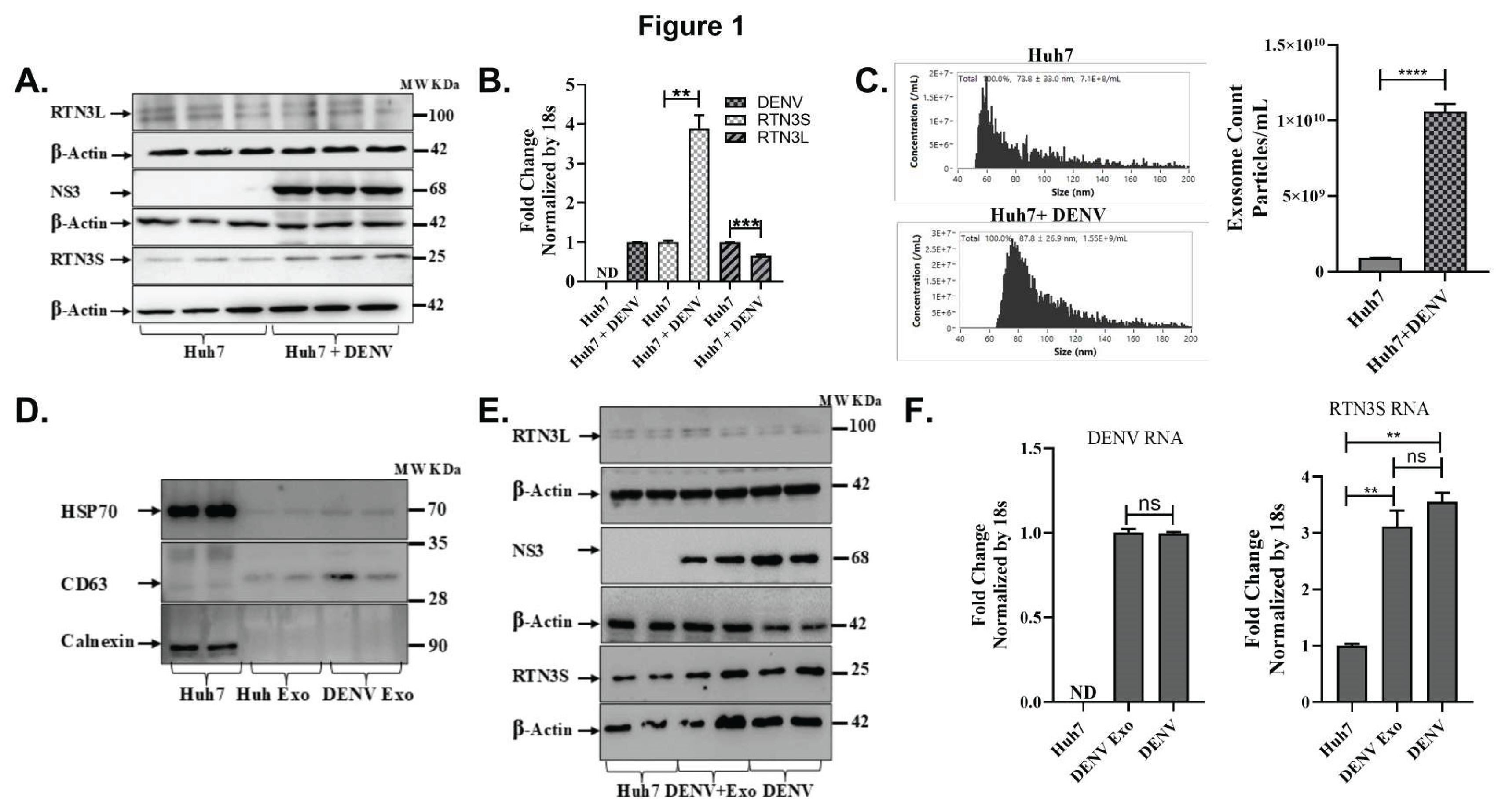
Figure 2.
RTN3 knockdown abrogates RTN3S expression and inhibits Dengue-induced exosome release in Huh7 cells. (A) Western blot of Huh7 cell lysates after transfection with control siRNA (Ctrl) or RTN3-targeting siRNA (RTN3-KD) followed by DENV infection. Blots were probed for DENV NS3 (~68 kDa), NS4B (~27 kDa), RTN3S (~25 kDa, arrow), and β-Actin (~42 kDa). (B) RT-qPCR of RTN3 mRNA in Huh7 cells (Ctrl or RTN3-KD, ± DENV), normalized to 18S rRNA. (C) NanoFCM analysis of exosome release for each condition. Control and RTN3-KD cells without infection release few exosomes (ns). (D) Western blot of exosome preparations from Ctrl (mock), DENV, and DENV+RTN3-KD conditions. Probing for DENV NS3 shows that exosomes from infected cells contain NS3, and this viral cargo is strongly diminished when RTN3 is knocked down. β-Actin is shown as a control. Data (B,C) are mean ± SEM of ≥3 experiments; significance by ANOVA or t-test (*p<0.05, **p<0.01, ***p<0.001, ****p<0.0001; ns = not significant).
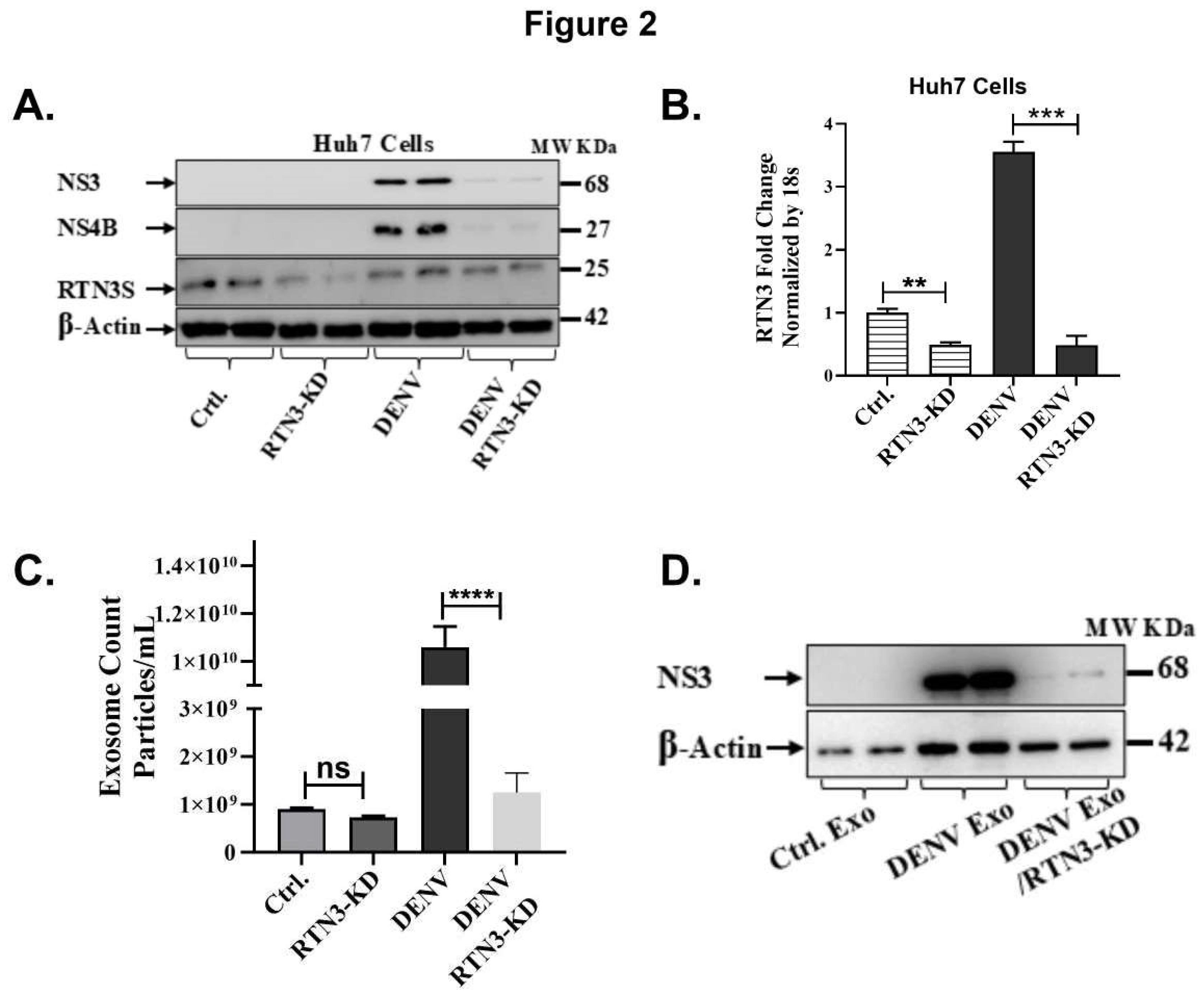
Figure 3.
The C-terminal domain of RTN3S is required for exosome production and viral transfer. (A) Western blot of Huh7 cells infected with DENV and transfected with empty vector (EV), full-length FLAG-RTN3S (FL), or deletion mutants (ΔN11, ΔN45 remove N-terminal regions; ΔC36 removes C-terminal tail). Membranes were probed with anti-FLAG to detect RTN3S constructs (all ~20- 37 kDa) and anti-DENV NS3 (~68 kDa); β-Actin (~42 kDa) is a loading control. (B) NanoFCM quantification of exosome release under each condition. The bar graph shows exosome concentration (particles/mL) for EV, RTN3S-FL, ΔN11, ΔN45, and ΔC36. Data are mean ± SEM (n=3). (C) Huh7 cells were treated with exosomes from donor cells expressing each construct (lanes: no-exosome control, Exo-ΔC36, Exo-ΔN11, Exo-ΔN45, Exo-RTN3S-FL). Cell lysates were blotted for DENV NS3 (~68 kDa) and β-Actin.
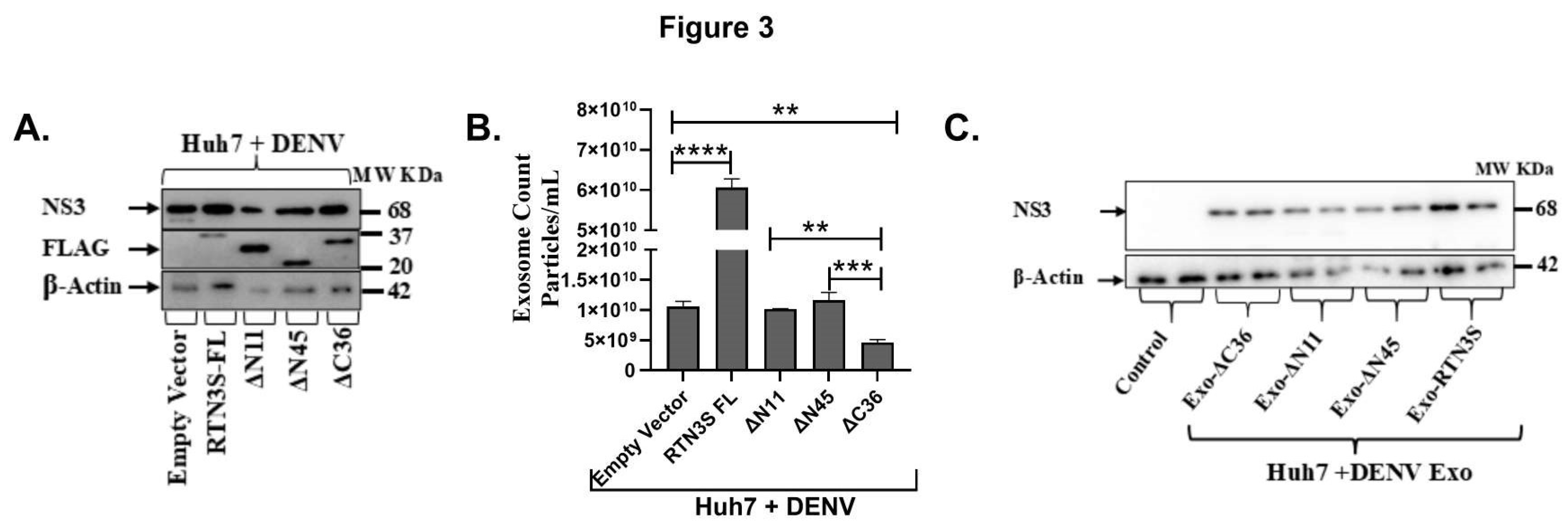
Figure 4.
RTN3 associates with Dengue viral double-stranded RNA and NS3 protein in infected Huh7 cells. (A) RT-qPCR detection of DENV genomic RNA in immunoprecipitated material from Huh7 cells 72 h after DENV infection. Cell lysates were immunoprecipitated with anti-RTN3 or anti-dsRNA (J2) antibodies; non-specific IgG was used as a negative control. Bars show relative enrichment of viral RNA in each IP (normalized to input). (B) Western blot of the IP eluates from Huh7+DENV lysates. Lanes: IgG IP (Huh7+DENV), RTN3 IP (Huh7+DENV), and anti-dsRNA (J2) IP (Huh7+DENV), with input lysate as control. Blots were probed for RTN3L (~100 kDa), DENV NS3 (~68 kDa), and RTN3S (~25 kDa).
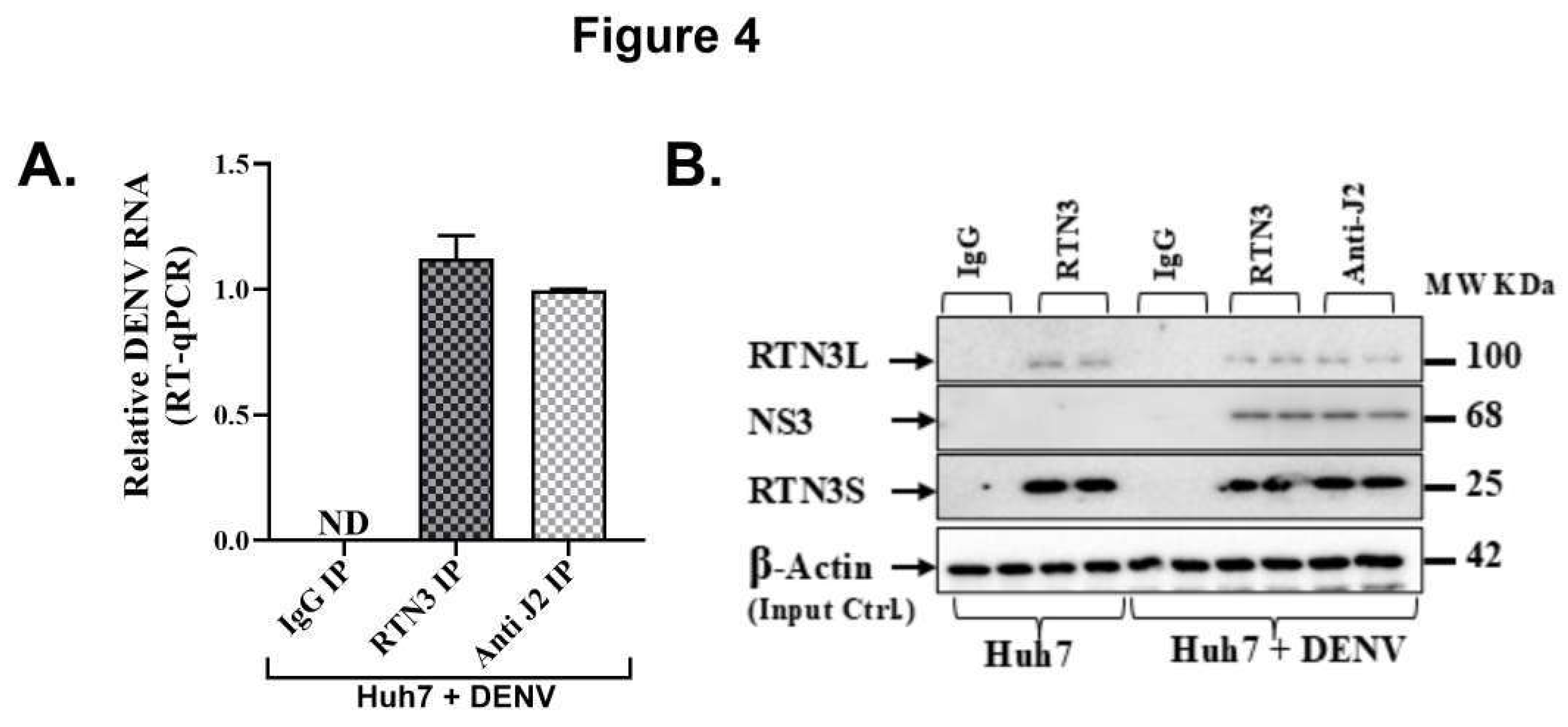
Figure 5.
Dengue virus induces RTN3 expression, exosome release, and innate immune responses in THP-1 monocytes. (A) Western blot of THP-1 cells uninfected (Ctrl) or infected with DENV for 24, 48, or 72 hours. Blots were probed for RTN3L (~100 kDa) and RTN3S (~25 kDa), with β-Actin (~42 kDa) as a loading control. (B) RT-qPCR of DENV genomic RNA in THP-1 cells (normalized to 18S) at each time point. (C) Flow cytometry of THP-1 cells for DENV NS3. (D) NanoFCM evaluation of exosomes in THP-1 culture supernatants with and without DENV infection. (E-G) RT-qPCR of THP-1 mRNA (normalized to 18S) for exosome biogenesis, trafficking genes and chaperone protein: (E) CHMP2A, (F) RAB7A, and (G) HSPA5. (H-I) Flow cytometry quantification of monocyte markers. Bars show the percentage of cells expressing CD14 or CD16 in mock vs DENV samples. (J-K) RT-qPCR of cytokine mRNAs: CXCL10 and IFN-β (normalized to 18S). Data (A–I) are mean ± SEM of triplicates; significance by ANOVA or t-test (ns = not significant, *p<0.05, **p<0.01, ***p<0.001, ****p<0.0001).
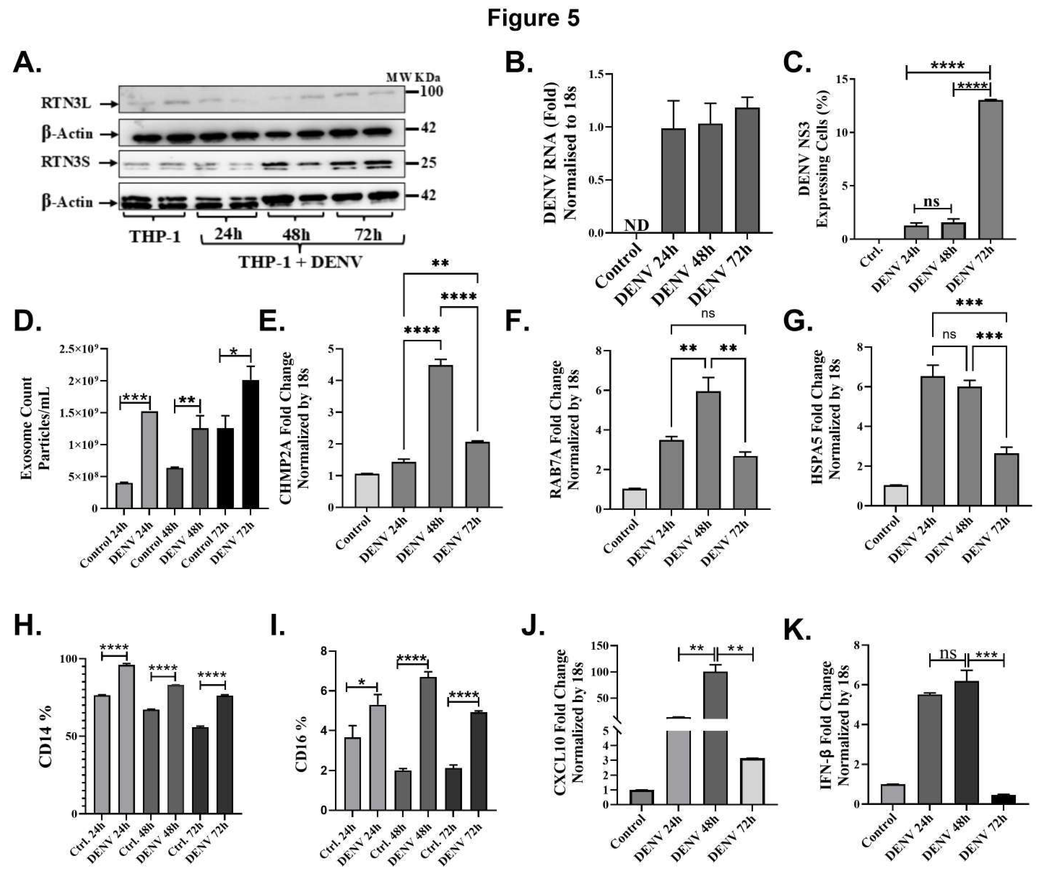
Figure 6.
Single-cell transcriptomics of blood cells reveal monocyte subsets and RTN3-related signatures in Dengue patients. (A) UMAP projection of PBMCs from healthy controls and Dengue patients, colored by major immune cell types (monocytes, T cells, B cells, NK cells, etc.). Monocytes form a distinct cluster (red). (B) UMAP highlighting monocyte subclusters: cluster 1 (blue) contains mainly healthy-donor cells, whereas cluster 2 (orange) is dominated by Dengue patient cells (including DFWS and severe cases). (C) Bar graph of the percentage of monocytes (CD14+ cells) among total PBMCs in each group (healthy control, Dengue fever (DF), Dengue with warning signs (DFWS), and severe Dengue (SD)). (D) Monocyte UMAP colored by expression of CD14 (red) and FCGR3A/CD16 (green) in healthy vs Dengue samples. Classical monocytes (CD14^hi, red) and non-classical monocytes (FCGR3A^hi, green) are indicated. (E) Violin plots of RTN3 mRNA expression in monocytes from each group. Dengue patient monocytes, especially from severe cases, show higher RTN3 expression than controls. (F) Feature scatter plots on the monocyte UMAP showing FCGR3A (red) and RTN3 (green) expression for each condition; yellow indicates co-expression. (G) Dot plot summarizing monocyte expression of HSPA5, RAB7A, and CHMP2A. Dot size corresponds to the proportion of cells expressing the gene; color indicates average expression. Dengue patient monocytes (DFWS, SD) display larger and darker dots for these genes, indicating upregulation. (H) UMAP feature plots of RTN3, CHMP2A, RAB7A, and HSPA5 in monocyte clusters across conditions, confirming stronger expression (yellow) of all four genes in Dengue patient monocytes.
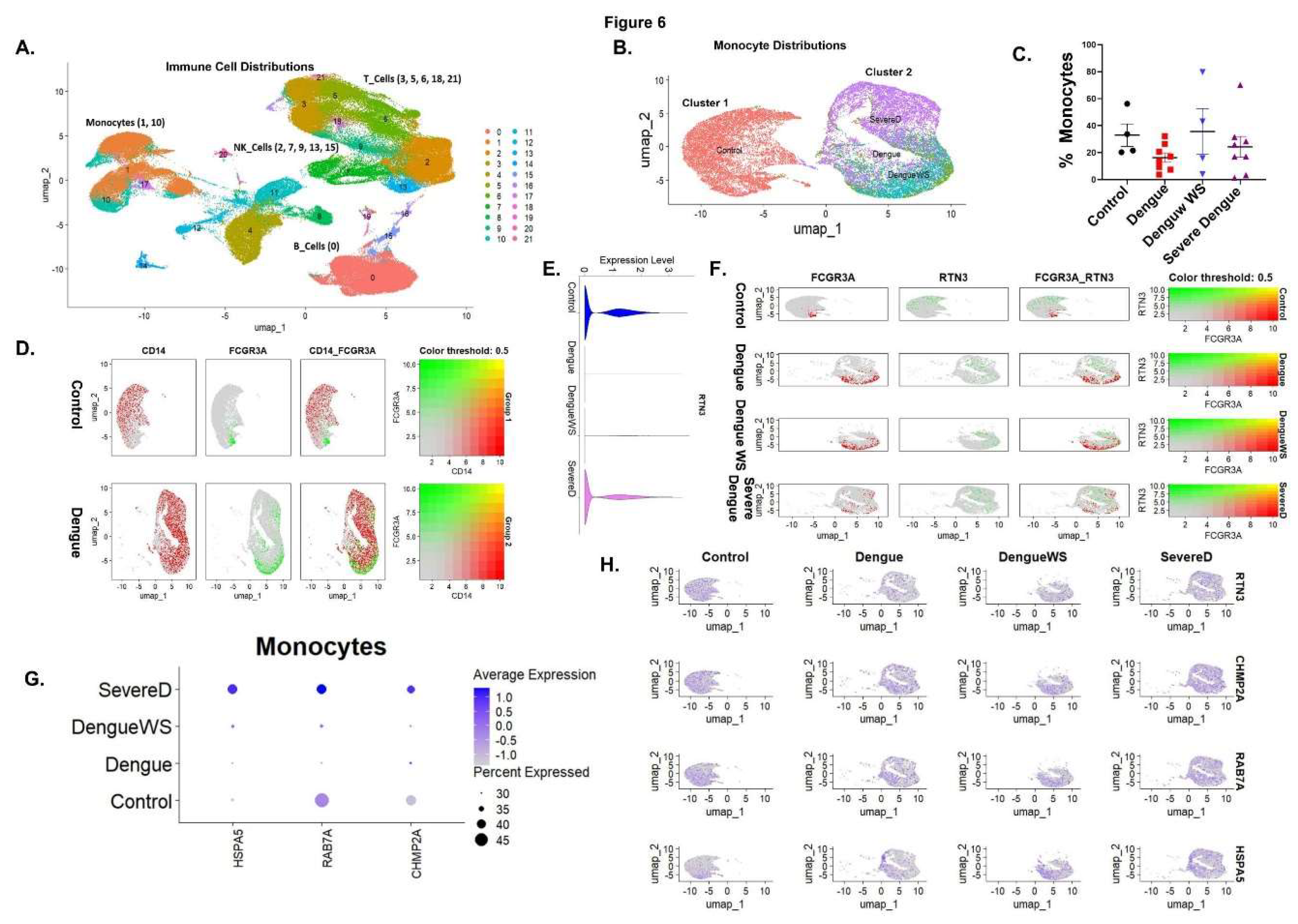
Figure 7.
Dengue virus manipulates the endoplasmic reticulum (ER) landscape through the short Reticulon-3 isoform (RTN3S) to generate infectious exosomes. Following DENV entry, positive-strand genomic RNA is translated on rough ER membranes. RTN3S (green hair-pin elements) accumulates at the peri-nuclear ER and sculpts it into bulb-like invaginations toward the ER lumen. These curved subdomains recruit ribosomes (navy spheres), newly synthesized viral polyprotein (dashed red coils), and non-structural replication factors (lavender icons), forming sealed single-membrane vesicle pockets that entrap double-stranded replicative DENV RNA (magenta helices). (A) Magnified inset in the graphic highlights how RTN3S helices directly clasp dsDENV RNA, creating a selective ribonucleoprotein scaffold that favours viral, over cellular, cargo. (B) Nascent RTN3S-dsDENV RNA vesicles bud from the ER and converge with early endosomes to seed multivesicular bodies (MVBs). Inside the maturing MVB, they become intraluminal vesicles destined to leave the cell as exosomes. A bold “cargo in/out” arrow at the MVB boundary mirrors RTN3S’s dual role: it both guides viral material into the endosomal lumen and licenses its export once the MVB fuses with the plasma membrane. The released exosomes (burgundy triangles) are depicted dispersing from the cell, each carrying replication-competent DENV RNA, non-structural proteins, and occasional immature virions, an immune-evasive inoculum primed for cell-to-cell spread. Modulating RTN3S levels alters every step in this pathway. RTN3S over-expression (upper left) intensifies ER curvature, increases the number of replication organelles, and drives a surge in cargo-rich exosome release. Conversely, RTN3S depletion (lower left) yields elongated, poorly scissioned replication tubules, drastically reduces vesicle trafficking to MVBs, and suppresses exosome output. These opposing phenotypes, annotated directly on the schematic, underscore RTN3S as a gatekeeper that links flaviviral RNA replication to selective exosome biogenesis and extracellular dissemination.
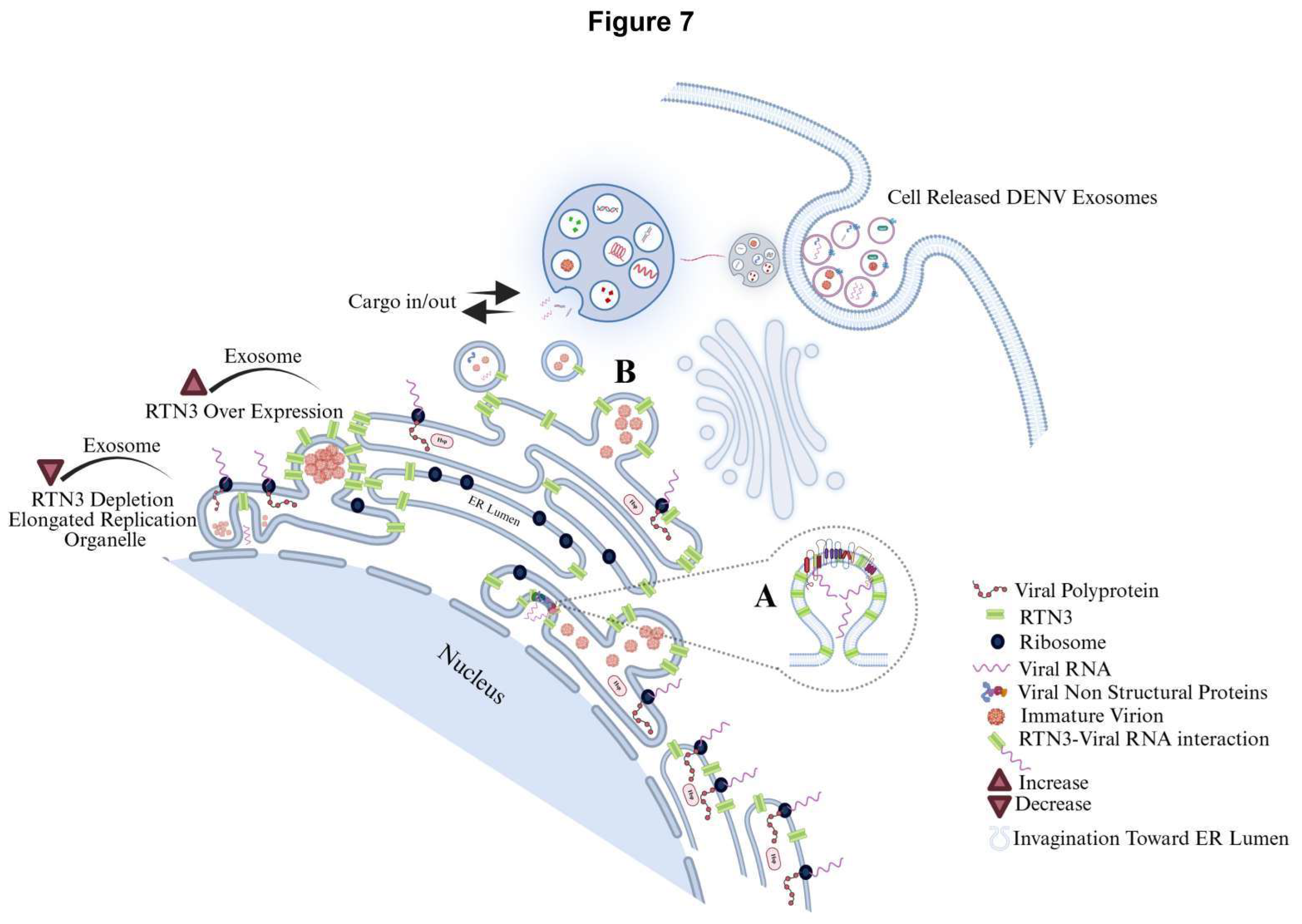

Table 1.
Sequences of Oligonucleotides.
| Primer | Sequence 5’ to 3’ |
|---|---|
| RTN3S |
F: GGAGAGATGTGAAGAAGACTGCC R: AGATCCTGAAGCTGATGGTGA |
| RTN3L |
F: GTAGGGAGGCTAAAACTGCA R: CTCCTGAAACTTTGGATGGAGA |
| DENV |
F: TTATCAGTTCAAAATCCAATGTTGGT R: AGGAGGAAGCTGGGTTGACA |
| CHMP2A |
F: GAAGACGCCAGAGGAGCTACTGC R: GCTTGGCCATCTTCTTAATGTCTGC |
| RAB7A |
F: GTCGGGAAGACATCACTCA R: CTAGCCTGTCATCCACCAT |
| HSPA5 |
F: GGGAGGTGTCATGACCAAAC R: GCAGGAGGAATTCCAGTCAG |
| CXCL10 |
F: GTGGATGTTCTGACCCTGCT R: GGAGGATGGCAGTGGAAGTC |
| IFNB |
F: TGGGAGGCTTGAATACTGCCTCAA R: TCCTTGGCCTTCAGGTAATGCAGA |
| 18S |
F: GTAACCCGTTGAACCCCATT R: CCATCCAATCGGTAGTAGCG |
| gRNA cloning verification |
F: AGCTGCAGCTCTTCGTC R: TCGGCGGCCACTCAGTC |
| gRNA sequence targeting RTN3S | CTCGGCTCCGAAGGACGACG |
Disclaimer/Publisher’s Note: The statements, opinions and data contained in all publications are solely those of the individual author(s) and contributor(s) and not of MDPI and/or the editor(s). MDPI and/or the editor(s) disclaim responsibility for any injury to people or property resulting from any ideas, methods, instructions or products referred to in the content. |
© 2025 by the authors. Licensee MDPI, Basel, Switzerland. This article is an open access article distributed under the terms and conditions of the Creative Commons Attribution (CC BY) license (https://creativecommons.org/licenses/by/4.0/).
Copyright: This open access article is published under a Creative Commons CC BY 4.0 license, which permit the free download, distribution, and reuse, provided that the author and preprint are cited in any reuse.


