
Submitted:
24 June 2025
Posted:
25 June 2025
You are already at the latest version
Abstract
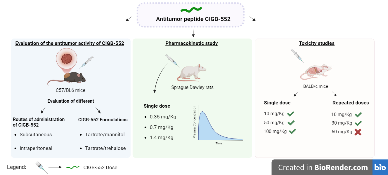
Background/Objectives: The CIGB-552 peptide is a novel therapeutic alternative for the treatment of cancer. In this study, we aimed to determine the optimal formulation and route of administration for CIGB-552. Other objectives were to characterize the peptide's pharmacokinetic profile in rats and conduct toxicity studies at different dosage regimens. Methods: The antitumor activity of CIGB-552 was evaluated by different routes of administration (intraperitoneal, subcutaneous) and also with different peptide formulations (Tartrate/mannitol, Tartrate/trehalose) using a TC-1 tumor model in C57BL/6 mice. The pharmacokinetic profile of the peptide was also characterized after subcutaneous administration in Sprague-Dawley rats using PK Solver software. In addition, the safety of the peptide was evaluated following single-dose and repeated-dose administration schedules in healthy BALB/c mice. Results: In the tumor model, subcutaneous administration of CIGB-552 and its formulation with tartrate/trehalose resulted in a significant reduction in tumor volume compared to untreated groups. CIGB-552 presented a typical extravascular administration profile with rapid absorption, along with a rapid tissue distribution phase and rapid blood clearance. The peptide's half-life was 2.5 h, and peak plasma concentration (Cmax) was reached within approximately 15 min. Furthermore, CIGB-552 administration in the evaluated regimens was safe, with toxicity and lethal outcomes only at the 60 mg/kg dose. Conclusions: The peptide’s safety profile in repeated dosing, combined with evidence of no systemic accumulation, supports the development of new regimens involving higher and more frequent doses to enhance antitumor efficacy in clinical studies.
Keywords:
antitumor peptide
; cancer
; CIGB-552
; pharmacokinetics
; toxicity
1. Introduction
The use of therapeutic peptides is a novel and promising approach for cancer treatment. Peptides have several advantages over proteins or antibodies: they have little complexity in synthesis, high specificity and selectivity for the biological target, low immunogenicity, good biocompatibility, and low toxicity [1,2].
CIGB-552 is a peptide from the second generation that demonstrates antitumor properties. It was created at the Center for Genetic Engineering and Biotechnology in Havana through the screening of a mutant alanine peptide library, which was based on the 32-51 amino acid segment of the Limulus anti-LPS factor (LALF) [3]. This peptide elevates the amount of the copper metabolism domain-containing protein 1 (COMMD1) within the cytoplasm of cells. The buildup of COMMD1 inside the cell suppresses the activity of nuclear factor kappa B (NF-κB), a key regulator involved in cell growth and the formation of new blood vessels [4]. Additionally, COMMD1 inhibits hypoxia-inducible factor 1 (HIF-1) by facilitating its ubiquitination and subsequent degradation via the proteasome. HIF-1 plays a crucial role in helping cells survive low-oxygen environments, such as those commonly present in tumors [5].
The optimization of the CIGB-552 peptide and the characterization of its pharmacokinetic and toxicological profiles are critical for enhancing its antitumor activity and securing regulatory approval for use in clinical studies [6]. Therefore, the objectives of this work were: To evaluate and determine the optimal administration route and formulation of CIGB-552 in a TC-1 tumor model in C57BL/6 mice, to characterize the pharmacokinetic profile of the peptide in Sprague Dawley rats, and to evaluate the toxicity of CIGB-552 administered in single-dose and repeated-dose regimens in BALB/c mice.
2. Results and Discussion
2.1. Evaluation of Different Routes of Administration of the CIGB-552 Peptide in the TC-1 Tumor Model
The antitumor peptide CIGB-552 has demonstrated efficacy in preclinical studies, including in vitro and in vivo models of lung and colorectal cancer [7,8]. However, therapeutic peptides face challenges in in vivo applications due to low bioavailability, primarily caused by proteolytic degradation and rapid systemic clearance [9]. The proper selection of the administration route is critical to improving bioavailability, safety, and therapeutic outcomes. To address these challenges, we evaluated the antitumor effect of CIGB-552 via i.p. and s.c. routes. The oral route was excluded to avoid gastrointestinal degradation and first-pass metabolism [10]. Using the TC-1 cell line in C57BL/6 mice, five treatment groups were assessed. Only Group I (0.5 mg/kg s.c.) and Group III (1 mg/kg s.c.) showed significant tumor growth reduction compared to the PSS-treated control (p < 0.05) (Figure 1a). All treatment regimens significantly improved survival versus the PSS control (p < 0.01) (Figure 1b). Notably, the subcutaneous route (Groups I and III) exhibited the strongest antitumor activity and survival benefits.
In this study, we demonstrated that subcutaneous administration of the CIGB-552 peptide at 0.5 mg/kg achieved the best outcomes in terms of antitumor activity and survival. These results are likely attributed to the higher bioavailability of the subcutaneous route compared to the intraperitoneal route, which is associated with rapid degradation and variable absorption [10].
2.2. Evaluation of Different Routes of Administration of the CIGB-552 Peptide in the TC-1 Tumor Model
CIGB-552 incorporates structural modifications, including N-terminal acylation and replacement of natural proline and leucine residues with their D-amino acid counterparts, to enhance stability by increasing resistance to metabolic degradation [7]. To further improve stability, we developed lyophilized formulations of CIGB-552 with different stabilizers and pH regulators. In this study, we evaluated the antitumor activity of formulations containing tartrate/mannitol or tartrate/trehalose in a TC-1 tumor model. All peptide-treated groups exhibited significant tumor growth reduction compared to the PSS-treated control (p < 0.01; Figure 2a). Notably, the tartrate/trehalose formulation showed superior survival outcomes, with three animals surviving at the trial’s conclusion, outperforming other formulations (p < 0.01; Figure 2b). Based on these findings, we selected the tartrate/trehalose combination as the optimal formulation for CIGB-552.
2.3. Pharmacokinetics of the CIGB-552 Peptide
The first pharmacokinetic study of the CIGB-552 peptide was conducted in BALB/c mice [7]. The pharmacokinetic (PK) profile was determined after a single subcutaneous (s.c.) administration of 0.2 mg/kg (5 µg of ¹³¹I- CIGB-552). This route was selected to avoid first-pass hepatic metabolism and to align with clinical practicality, offering patients greater convenience, safety, and autonomy compared to intravenous administration. The 0.2 mg/kg dose was chosen based on prior studies demonstrating tumor growth inhibition in a syngeneic CT-26 colon cancer model [7]. The PK profile of CIGB-552 was further evaluated in the phase I clinical trial “ARGOS” (RPCEC00000196, Cuban Public Registry of Clinical Trials) [11] which evaluated two level doses (0.06 mg/kg and 0.1 mg/kg). Remarkably, the 0.06 mg/kg human dose corresponds to threefold the 0.2 mg/kg murine dose when adjusted for interspecies scaling [7,11]. In both studies, CIGB-552 exhibited a PK profile typical of extravascular administration, characterized by: rapid absorption (Cmax reached within 15 min), sharp distribution phase and fast clearance mediated by proteolytic degradation and elimination via kidneys/liver, as shown in a radiolabeled biodistribution study (¹³¹I-CIGB-552) in BALB/c mice [7,12]. However, prior studies failed to establish a dose-dependent relationship for PK parameters [7,11]. To address this, we evaluated CIGB-552 in Sprague-Dawley rats at three doses (0.35, 0.7, and 1.4 mg/kg), equivalent to human doses of 0.04 mg/kg, 0.06 mg/kg, and 0.1 mg/kg via body surface area normalization [13].
Plasma concentrations of CIGB-552 at 0 h, 0.25 h, 0.5 h, 1 h, 2 h and 5 h were determined using a sandwich ELISA previously validated by our working group for the quantification of the peptide in rat plasma (Table 1).
Figure 3 shows the plasma concentration vs. time graphs obtained for each group evaluated and the pharmacokinetic parameters of the peptide were determined using the NCA method; the results are shown in Table 2.
The plasma concentration-time curve of CIGB-552 in rats showed a typical extravascular administration profile. The curve presented an initial phase of rapid absorption, where Cmax ranged from 5.03 to 16.86 ng/ml achieved within 15 min post-administration, likely due to the peptide’s low molecular weight and structural permeability. This absorption phase was followed by a sharp drop in plasma concentration that could be attributed to protease degradation, tissue distribution (high Vz/F values), and rapid renal/hepatic clearance [12]. As expected for peptides and in agreement with previous results, CIGB-552 showed a relatively short half-life, between 1.76 and 4.62 h, and a rapid clearance, between 8.69 and 9.52 L/h, after subcutaneous administration [14]. These results suggest that the peptide does not accumulate between subsequent administrations.
The AUC0–t (Figure 4a) and Cmax (Figure 4b) showed significant dose-dependent increases, contrasting with earlier data from Phase I Clinical Trial [11]. The AUC0–t accounted for more than 50% of AUC0–∞, suggesting the need for frequent sampling within the first hour post-administration. These findings underscore the importance of dose optimization to maintain therapeutic blood levels while balancing efficacy and safety.
2.4. Acute Toxicity of the CIGB-552 Peptide in BALB/c Mice
Demonstrating the safety of the CIGB-552 in in vivo models is critical for regulatory approval in humans [15]. In this study, we evaluated acute toxicity of CIGB-552 to determine its maximum tolerated dose (MTD) and identify potential toxic effects after administering high doses in BALB/c mice: 10 mg/kg, 50 mg/kg, and 100 mg/kg. Daily monitoring revealed no systemic toxicity. Animals exhibited normal eye color/appearance, mucous membranes, and behavior, with appropriate responses to stimuli. Localized alopecia at the injection site was observed in three female mice from the 100 mg/kg group, potentially linked to high-dose administration. No significant differences in body weight (Figure 5) or food consumption (Supplemental Information (SI), Figure S1) were observed across groups. Body weight trends correlated with food intake. Macroscopic examination of organs (liver, spleen, kidneys, thymus, brain, lungs, stomach, colon, ovaries, skin, thyroid, pancreas, heart and subcutaneous tissue) revealed normal morphology with no anatomical abnormalities.
Microscopic analysis of organs (Table 3) revealed histological changes in mesenteric lymph nodes and spleen, and liver across all treatment groups. These findings occurred in all groups (including controls), suggesting these changes are unrelated to CIGB-552 administration. Antigenic stimulation was evident in the mesenteric lymph nodes, marked by the formation of germinal centers in the cortical region. Germinal centers were observed in the periarteriolar lymphoid sheath, a process driven by B-cell activation in response to antigenic stimuli [16]. Similar to lymph node findings, these changes appeared in all study groups, indicating they are independent of the test substance.
In this trial, no treatment-related toxic effects were observed following single-dose administration of CIGB-552, even at the highest tested doses. This finding provides evidence of the peptide’s safety profile, supporting its safe use in humans with a favorable risk-benefit ratio.
2.5. Toxicity of the CIGB-552 Peptide Administered in Repeated Doses in BALB/c Mice
Previous studies characterized the pharmacokinetic and tissue biodistribution profiles of s.c. administered CIGB-552, demonstrating no accumulation of the peptide in blood or healthy tissues [7,11]. The rapid systemic clearance of CIGB-552 supports the feasibility of more frequent dosing regimens. To assess safety under repeated dosing, we performed a repeated dose toxicity study in BALB/c mice using doses of 10 mg/kg, 30 mg/kg and 60 mg/kg of the peptide. In this study were evaluated: adverse/toxic effects, residual peptide effects on target organs, physiological/metabolic tolerance and reversibility of potential adverse effects in the satellite group. Body weight (Figure 6) and food consumption (SI Figure S2) showed no significant differences between groups. Body weight increased proportionally with food intake, indicating normal metabolic activity.
Clinical observations revealed no treatment-related abnormalities: the animals exhibited normal coat, skin coloration, eye appearance, and mucous membranes, with typical behavior and appropriate responses to stimuli throughout the study. No signs of toxicity were observed except in the highest dose group (60 mg/kg, Group V), where adverse events and mortality occurred. Two deaths were recorded on day 10 and three additional deaths occurred on day 11.
In addition, hematological parameters (neutrophils, lymphocytes, monocytes, basophils, eosinophils; SI Tables S1–S2), lymphocyte subpopulations (CD4+/CD8+ T cells; SI Figure S3), blood biochemistry (glucose, albumin, total proteins, cholesterol, creatinine, alanine aminotransferase (ALT), aspartate aminotransferase (AST), alkaline phosphatase, total bilirubin; SI Tables S3–S4), and relative organ weights (SI Table S5–S6) showed no significant differences between treatment groups and controls.
Macroscopic observations of all organs were conducted during necropsy. Necropsies were performed on days 16 and 17 for Groups I-V, while the Satellite group, assessing delayed toxicity, was euthanized on day 31. Tissue samples were collected from the following organs/tissues: endocrine: adrenal glands, thymus, pituitary gland, thyroid/parathyroid glands; cardiorespiratory: heart, lungs (including bronchi/bronchioles), trachea, aorta; digestive: liver, spleen, pancreas, stomach, small/large intestine, esophagus, tongue; urogenital: kidneys, ureters, urinary bladder, uterus (cervix, oviducts), ovaries, testes, prostate, seminal vesicles, vagina; nervous: brain, spinal cord, peripheral nerves, eyes/optic nerve and others: lymph nodes, mammary glands, salivary glands, skeletal muscle, bone marrow, larynx, skeletal muscle, and the subcutaneous injection site. No macroscopic abnormalities indicative of treatment-related toxicity was observed in any organs or tissues, confirming their gross anatomical integrity.
Histopathological examination (Table 4) revealed a high incidence of germinal centers in the spleen and mesenteric lymph nodes across all study groups. This finding aligns with normal physiological processes in humans and laboratory animals, reflecting a typical humoral immune response to antigenic stimuli [16]. Additionally, the observation of megakaryocytes in the splenic red pulp and focal clusters of immature cells with hyperchromatic nuclei in the liver parenchyma across all groups indicates extramedullary hematopoiesis in these organs. This phenomenon, well-documented in rodents, represents a physiological adaptation to moderate stress during experimental procedures [17]. Notably, extramedullary hematopoiesis is not exclusive to pathological states; it has been reported under normal conditions in rodents, hamsters, dogs, and humans [17]. Thus, these findings are unrelated to the experimental treatment and likely reflect baseline or stress-induced biological responses.
At the injection site, a mixed inflammatory cell infiltrate composed of neutrophils and mononuclear cells was observed in the subcutaneous tissue. This response was evident in Groups III, IV, and V, with 4, 3, and 7 affected animals in these groups, respectively, and is therefore attributed to the CIGB-552 formulation. In contrast, Groups I, II, and VI showed no histological changes at the injection site. Positively, the absence of pathological changes in the Satellite Group (VI) evaluated after a recovery period suggests the inflammatory process was reversible.
Based on the findings of this repeated-dose toxicity study, subcutaneous administration of CIGB-552 at 60 mg/kg demonstrated toxicity, whereas 30 mg/kg exhibited an adequate safety margin. Consequently, the 30 mg/kg dosing regimen could be recommended for clinical use. These results establish 30 mg/kg as the MTD for repeated administration of CIGB-552.
3. Materials and Methods
3.1. Synthesis of CIGB-552 Peptide
The CIGB-552 peptide was synthesized as described by Vallespi et al. (2017). Briefly, CIGB552 was obtained by solid state synthesis using the Fmoc strategy, and purified by RP-HPLC inverse (purity > 95%) under an acetonitrile/trifluoroacetic acid gradient. The peptide was characterized by electrospray mass spectrometry (Micromass, UK) and NMR spectroscopy [18].
3.2. Animals
Female C57/BL6 mice 8-10 weeks old and weighing 18-20 g, male Sprague Dawley rats 4-8 weeks of age and with a body weight between 250-300 g, and BALB/c mice both sexes, 8-10 weeks old and weighing 18-20 g were supplied by the National Center for Animal Production (CENPALAB). All animal studies were conducted after obtaining the permission from the Internal Committee for the Care and Use of Laboratory Animals (CICUAL) in accordance to the guidelines provided by Institute of Laboratory Animal Resources (ILAR) and the National regulations on the use of laboratory animals. The toxicology studies were carried out according to the ICH guidelines for nonclinical safety studies [19].
3.3. Evaluation of Different Routes of Administration of the CIGB-552 Peptide in the TC-1 Tumor Model in C57/BL6 Mice
Fifty female C57BL/6 mice were randomly divided into five groups of ten animals each. Mice were injected with 5×10⁴ TC-1 cells in the right flank, following established protocols for this animal model [20]. Treatment began when tumors reached 70-90 mm³, administered at the indicated doses. Groups I and II received CIGB-552 at 0.5 mg/kg, while groups III and IV received 1 mg/kg. Groups I and III were administered the peptide subcutaneously (s.c.), and groups II and IV received it intraperitoneally (i.p.). CIGB-552 was administered on days 11, 14, and 17 post-tumor engraftments. Each dose was delivered in 0.2 mL of 0.9% physiological saline solution (PSS), injected s.c. into the left lateral flank. Group V served as the untreated (placebo) control and received 0.2 mL of PSS alone. Body weight was recorded weekly from the start of the experiment. Tumor dimensions were measured twice weekly using calipers, and volume was calculated using the formula: volume = length × (width)2 / 2. Survival was monitored daily throughout the study. Mice were monitored daily to look for humane endpoints such as weight loss, loss of mobility, tumor volume > 2000 mm3, etc. When humane endpoint was reached mice were sacrificed after anesthesia by cervical dislocation.
3.4. Evaluation of Different Formulations of the CIGB-552 Peptide in the TC-1 Tumor Model in C57/BL6 Mice
A total of 30 female C57BL/6 mice were randomly divided into three groups of 10 animals each. Each animal received an injection of 5×10⁴ TC-1 murine tumor cells into the right flank, following established protocols for this model [20]. Treatment began when tumors reached 70-90 mm³, with doses administered as specified. Groups I and II were treated with CIGB-552 peptide (0.5 mg/kg) administered s.c. in 50 mM of sodium tartrate, mannitol at 2% and 50 mM of sodium tartrate, trehalose at 2% respectively as excipients. Group III served as the placebo and received 0.2 mL of tartrate/trehalose solution diluted in PSS (without peptide) via the same route. The peptide was administered on days 21, 24, 28, and 31 post-tumor engraftments injected s.c. into the left lateral flank. Monitoring was carried out as described in the section 2.3.
3.5. ELISA Protocol for the Quantification of the CIGB-552 Peptide
The quantification of the CIGB-552 peptide was carried out using an ELISA developed by Gómez et al. (2025). Costar 3590 (USA) 96-well polystyrene plates were coated with 100 µL/well of anti-CIGB-552 polyclonal antibody (pAb) [21] diluted in coating buffer (0.1 M sodium carbonate and 0.1 M sodium hydrogen carbonate, pH 9.6) at 10 µg/mL and incubated for 1 h at 37°C in a humidified chamber. Plates were washed three times with 200 µL/well of washing solution (0.1% Tween 20 in water for injection). Blocking was performed with 200 µL/well of blocking buffer (1% BSA, 2.5% sheep serum, and 0.05% Tween 20 in PBS) for 1 h at 37°C in a humidified chamber. After discarding the blocking buffer by inversion, the standard curve, positive and negative controls, and validation samples were added in duplicate. Plates were incubated with samples for 1 h at 37°C, followed by three washes with 200 µL/well washing solution. Next, 100 µL/well of anti-CIGB-552 monoclonal antibody (mAb) [22] (5 µg/mL in PBS with 0.05% Tween 20) was added and incubated for 1 h at 37°C. After repeating the wash steps, 100 µL/well of horseradish peroxidase (HRP)-conjugated anti-mouse Fc secondary antibody (Sigma-Aldrich), diluted 1:25,000 in PBS with 0.05% Tween 20, was added and incubated for 30 min at 37°C. Plates were washed four times, and 100 µL/well of TMB substrate solution was added. After 10 min of incubation in the dark, the reaction was stopped with 50 µL/well of 2 M H2SO4, and absorbance was measured at 450 nm using a Varioskan Flash microplate reader (Thermo Scientific, Vantaa, Finland) [21]. The ELISA was previously validated for use with rat plasma in accordance with the ICH M10 guideline “Bioanalytical Method Validation and Study Sample Analysis” [23].
3.6. Experimental Design of the Pharmacokinetic Study of CIGB-552 in Sprague Dawley Rats
Male Sprague-Dawley rats were used in the pharmacokinetic study. Three groups of three rats each were randomly assigned to receive subcutaneous doses of 0.35 mg/kg, 0.7 mg/kg, or 1.4 mg/kg of the CIGB-552 peptide formulated in 50 mM of sodium tartrate, trehalose at 2%, respectively. Blood samples were collected at 0 min, 0.25 h, 0.5 h, 1 h, 2 h, and 5 h post-administration. Plasma was isolated by centrifugation (3500 rpm for 20 min at 4°C) using Vacutainer K2E tubes and stored at -80°C until analysis. Plasma peptide concentrations were quantified using a validated sandwich ELISA. Pharmacokinetic parameters were analyzed using the PK Solver software [24].
3.7. Acute Toxicity of the CIGB-552 Peptide in BALB/c Mice
Fifty BALB/c mice (25 females and 25 males) were randomly divided into five groups of 10 animals each (5 females and 5 males per group). Treatments were administered as a single subcutaneous dose as follows: Group I: PSS, Group II tartrate/trehalose solution as placebo. Groups III, IV, and V were treated with formulated tartrate/trehalose CIGB-552 at doses of 10 mg/kg (50x the therapeutic dose), 50 mg/kg (250x the therapeutic dose) and 100 mg/kg (500x the therapeutic dose) respectively. Clinical monitoring of the animals was carried out daily, including mortality, clinical signs (skin, fur, eyes, mucous membranes), and systemic alterations (respiratory, circulatory, central/autonomic nervous systems, somatomotor activity, behavior). Body weight and food intake were measured on days 0, 7, and 14 post-administrations. Fifteen days post-administration, animals were euthanized, and organs were subjected to gross and histopathological examinations.
3.8. Toxicity of the CIGB-552 Peptide Administered in Repeated Doses in BALB/c Mice
120 BALB/c mice (60 females and 60 males) were randomly divided into six groups of twenty animals each (10 females and 10 males). Treatments were administered s.c. every other day for seven total doses, as follows: Group I: Untreated control, Group II: Placebo control (tartrate/trehalose), Groups III, IV, V: CIGB-552 formulated at 10 mg/kg (50x the therapeutic dose), 30 mg/kg (150x the therapeutic dose), and 60 mg/kg (300x the therapeutic dose), respectively and Group VI (Satellite group): CIGB-552 at 60 mg/kg to assess delayed toxicity. Clinical monitoring of the animals was carried out. After the study termination, 1 mL of blood was collected per animal for serum and plasma isolation. Animals were euthanized, and organs were subjected to gross and histopathological examinations. The Satellite group (Group VI) was euthanized 15 days after the final dose. Relative organ weights (relative weight [%] = organ weight * 100 / body weight), hematology (neutrophils, lymphocytes, monocytes, basophils, eosinophils), blood chemistry (glucose, albumin, total protein, cholesterol, creatinine, aspartate aminotransferase, alanine aminotransferase, alkaline phosphatase, total bilirubin), and lymphocyte subpopulations (CD4 and CD8) were evaluated. All kits used in this assay were purchased from HUMAN Diagnostics Worldwide (Germany).
3.9. Statistical Analysis
All statistical analysis was done using the software GraphPad Prism 8. Tumor volumes between experimental groups in each in vivo assay were compared using two-way ANOVA with Tukey’s multiple comparisons test. Animal survival between experimental groups in each in vivo assay were compared using the log-rank (Mantel-Cox) test. Differences were considered statistically significant when *p < 0.05, **p < 0.01, ***p < 0.001, ****p < 0.0001.
4. Conclusions
Subcutaneous administration of CIGB-552 at 0.5 mg/kg demonstrated superior antitumor efficacy compared to intraperitoneal administration. The tartrate/trehalose formulation of CIGB-552 significantly enhanced survival outcomes in tumor-bearing mice, establishing it as the optimal formulation for clinical translation. CIGB-552 exhibits rapid absorption (within 15 min) and fast clearance (half-life: 1.76-4.62 h), supporting frequent dosing regimens. No systemic toxicity was observed at doses up to 100 mg/kg (500× therapeutic dose), except localized alopecia at the injection site. The MTD for repeated administration was established at 30 mg/kg, with 60 mg/kg inducing mortality. The observed immune activation and extramedullary hematopoiesis were attributed to physiological responses and not related with the product. Inflammatory infiltrates at the injection site resolved in the satellite group, confirming the reversibility of local adverse effects. These findings have practical clinical impacts standing CIGB-552 as a promising candidate for oncology clinical trials, with a robust preclinical safety profile and evidence of dose-dependent efficacy.
Supplementary Materials
The following supporting information can be downloaded at: Preprints.org, Figure S1: Food consumption of BALB/c mice during the acute toxicity study of CIGB-552; Figure S2: Food consumption of BALB/c mice during the toxicity study of CIGB-552 administered in repeated doses; Figure S3: CD4 and CD8 lymphocyte population in each experimental group of the CIGB-552 toxicity study; Table S1: Hematological parameters of female mice by experimental group; Table S2: Hematological parameters of male mice by experimental group; Table S3: Blood biochemistry of female mice; Table S4: Blood biochemistry of male mice; Table S5: Average relative organ weight (%) of female mice sacrificed at the end of treatments; Table S6: Average relative organ weight (%) of male mice sacrificed at the end of treatments.
Author Contributions
Conceptualization, J.R.F. and N.A.G.; methodology, N.A.G., L.A. and J.C.; formal analysis, N.A.G., L.A., J.C. and J.R.F.; investigation, N.A.G., L.M.M., H.E.G., A.Y.E., B.O., L.A., J.C. and J.R.F.; writing—original draft preparation, N.A.G. and J.R.F.; writing—review and editing, N.A.G. and J.R.F.; supervision, J.R.F.; project administration, J.R.F.; funding acquisition, J.R.F. All authors have read and agreed to the published version of the manuscript.
Funding
This work was supported by the Ministerio de Ciencia, Tecnología y Medio Ambiente (CITMA, Project No. PN385LH007-013)
Institutional Review Board Statement
The procedures and care of the animals were carried out according to the guidelines established by the Program for the Use and Management of Laboratory Animals for Experimental Purposes of the Center for Genetic Engineering and Biotechnology (CIGB, Havana, Cuba). The CIGB Animal Care and Use Committee approved all experiments. Animal Protocol Approval Code: 5SIE0802010; 8SIE1605012; CICUAL/CIGB/23005; EP/TX/03.12; EP/TX/06.12. This animal study complies with all the international requirements and is according the National Institutes of Health guide for care and use of laboratory animals.
Informed Consent Statement
Not applicable.
Data Availability Statement
The datasets generated and analyzed during the present study are available upon reasonable request from the corresponding author.
Acknowledgments
We would like to thank all the colleagues from the vivarium of the Center for Genetic Engineering and Biotechnology who were part of this study.
Conflicts of Interest
The authors declare no conflicts of interest.
Abbreviations
The following abbreviations are used in this manuscript:
| AUC0-∞ | Area under plasma concentration-time curve from time zero to infinity |
| AUC0-t | Area under plasma concentration-time curve from time zero to the last measurable time point |
| BSA | Bovine serum albumin |
| Cl/F | Total plasma clearance |
| Cmax | Maximum observed plasma concentration |
| COMMD1 | MURR1 domain-containing protein 1 of copper metabolism |
| ELISA | Enzyme-linked immunosorbent assay |
| HIF-1 | Hypoxia-induced factor 1 |
| HRP | Horseradish peroxidase |
| LALF | Limulus anti-LPS factor |
| MRT | Mean residence time |
| MTD | Maximum tolerable dose |
| NF-κB | Nuclear factor kappa light chain enhancer of activated B cells |
| RP-HPLC | Reversed-phase high-performance liquid chromatography |
| t1/2 | Terminal half-life |
| Tmax | Time to peak |
| Vz/F | Volume of distribution at steady state |
| λz | Elimination rate constant |
References
- Blanco-Míguez, Aitor et al. “From amino acid sequence to bioactivity: The biomedical potential of antitumor peptides.” Protein science: a publication of the Protein Society vol. 25,6 (2016): 1084-95. [CrossRef]
- Marqus, Susan et al. “Evaluation of the use of therapeutic peptides for cancer treatment.” Journal of biomedical science vol. 24,1 21. 21 Mar. 2017. [CrossRef]
- Vallespi, Maribel G et al. “Identification of a novel antitumor peptide based on the screening of an Ala-library derived from the LALF (32-51) region.” Journal of peptide science: an official publication of the European Peptide Society vol. 16,1 (2010): 40-7. [CrossRef]
- Dolcet, Xavier et al. “NF-kB in development and progression of human cancer.” Virchows Archiv: an international journal of pathology vol. 446,5 (2005): 475-82. [CrossRef]
- van de Sluis, Bart et al. “COMMD1 disrupts HIF-1alpha/beta dimerization and inhibits human tumor cell invasion.” The Journal of clinical investigation vol. 120,6 (2010): 2119-30. [CrossRef]
- Khan, Abad et al. “Editorial: The practical implication of clinical pharmacokinetics in drug development, pharmaceutical analysis, and clinical research.” Frontiers in pharmacology vol. 14 1252030. 25 Jul. 2023. [CrossRef]
- Vallespi, Maribel G et al. “Antitumor efficacy, pharmacokinetic and biodistribution studies of the anticancer peptide CIGB-552 in mouse models.” Journal of peptide science: an official publication of the European Peptide Society vol. 20,11 (2014): 850-9. [CrossRef]
- Gomez Rodriguez, Yolanda et al. “Synergic effect of anticancer peptide CIGB-552 and Cisplatin in lung cancer models.” Molecular biology reports vol. 49,4 (2022): 3197-3212. [CrossRef]
- Diao, Lei, and Bernd Meibohm. “Pharmacokinetics and pharmacokinetic-pharmacodynamic correlations of therapeutic peptides.” Clinical pharmacokinetics vol. 52,10 (2013): 855-68. [CrossRef]
- Ibraheem, D et al. “Administration strategies for proteins and peptides.” International journal of pharmaceutics vol. 477,1-2 (2014): 578-89. [CrossRef]
- Vallespi, Maribel G et al. “A first-in-class, first-in-human, phase I trial of CIGB-552, a synthetic peptide targeting COMMD1 to inhibit the oncogenic activity of NF-κB in patients with advanced solid tumors.” International journal of cancer vol. 149,6 (2021): 1313-1321. [CrossRef]
- Koenitz, Laura et al. “Pharmacokinetic differences between subcutaneous injection and intradermal microneedle delivery of protein therapeutics.” European journal of pharmaceutics and biopharmaceutics: official journal of Arbeitsgemeinschaft fur Pharmazeutische Verfahrenstechnik e.V vol. 204 (2024): 114517. [CrossRef]
- Reagan-Shaw, Shannon et al. “Dose translation from animal to human studies revisited.” FASEB journal: official publication of the Federation of American Societies for Experimental Biology vol. 22,3 (2008): 659-61. [CrossRef]
- Cabrales, Ania et al. “Pharmacokinetic study of Growth Hormone-Releasing Peptide 6 (GHRP-6) in nine male healthy volunteers.” European journal of pharmaceutical sciences: official journal of the European Federation for Pharmaceutical Sciences vol. 48,1-2 (2013): 40-6. [CrossRef]
- Pognan, Francois et al. “The evolving role of investigative toxicology in the pharmaceutical industry.” Nature reviews. Drug discovery vol. 22,4 (2023): 317-335. [CrossRef]
- Ruberte, Jesús et al. “Bridging mouse and human anatomies; a knowledge-based approach to comparative anatomy for disease model phenotyping.” Mammalian genome: official journal of the International Mammalian Genome Society vol. 34,3 (2023): 389-407. [CrossRef]
- Greaves P. Histopathology of Preclinical Toxicity Studies. Interpretation and relevance in Grug Safety Evaluation. Second Edition. ELSEVIER. 2000.
- Vallespi, Maribel G et al. “The first report of cases of pet dogs with naturally occurring cancer treated with the antitumor peptide CIGB-552.” Research in veterinary science vol. 114 (2017): 502-510. [CrossRef]
- CPMP/ICH/286/95 (2009) No-clinical safety studies for the conduct of Human clinical trials for pharmaceuticals. ICH M3 (M). https://www.ich.org/page/multidisciplinary-guidelines.
- Tanaka, Tomoyuki et al. “Treatment of lung cancer using clinically relevant oral doses of the cyclooxygenase-2 inhibitor rofecoxib: potential value as adjuvant therapy after surgery.” Annals of surgery vol. 241,1 (2005): 168-78. [CrossRef]
- Gómez Hernández, Nivaldo Angel et al. “A sandwich ELISA for the quantification of the anticancer peptide CIGB-552 in human plasma.” Analytical biochemistry vol. 698 (2025): 115725. [CrossRef]
- Gómez Y, González M, Hernández D, Aragón H, Vallespi MG, et al. (2018) Monoclonal Antibodies for the CIGB-552 Antitumor Synthetic Peptide Quantification. J Vet Sci Ani Husb 6(5): 503.
- European Medicines Agency. ICH guideline M10 on bioanalytical method validation and study sample analysis, 2022. Available: https://www.ema.europa.eu/en/documents/scientific-guideline/ich-guideline-m10-bioanalytical-method-validation-step-5_en.pdf.
- Zhang, Yong et al. “PKSolver: An add-in program for pharmacokinetic and pharmacodynamic data analysis in Microsoft Excel.” Computer methods and programs in biomedicine vol. 99,3 (2010): 306-14. [CrossRef]
Figure 1.
In vivo evaluation of CIGB-552 administered by different routes of administration. (a) Mean tumor volume of treated and control mice, 21 to 33 days after TC-1 cell implantation. (b) Kaplan-Meier survival curves of control and treated animals from implantation to 50 days post implantation. Data are presented as mean ± SD, n = 10, *, statistical significance as calculated with two-way ANOVA with Tukey’s multiple comparisons test *p < 0.05.
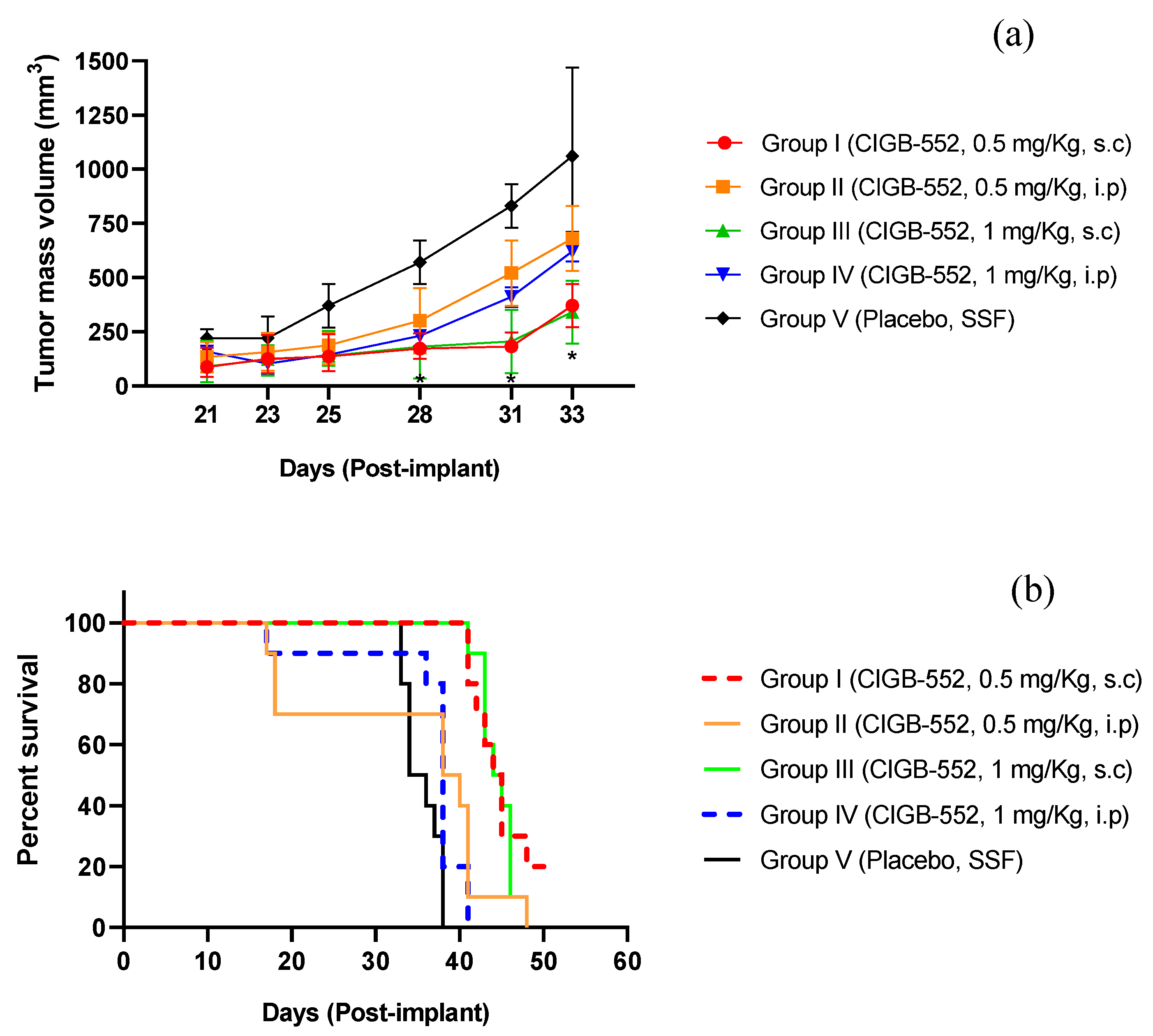
Figure 2.
In vivo evaluation of different formulations of CIGB-552. (a) Mean tumor volume of treated and control mice 28 to 38 days after TC-1 cell implantation. (b) Kaplan-Meier survival curves of control and treated animals from implantation to 52 days post implantation. Data are presented as mean ± SD, n = 10, *, statistical significance as calculated with two-way ANOVA with Tukey’s multiple comparisons test *p < 0.05, **p < 0.01.
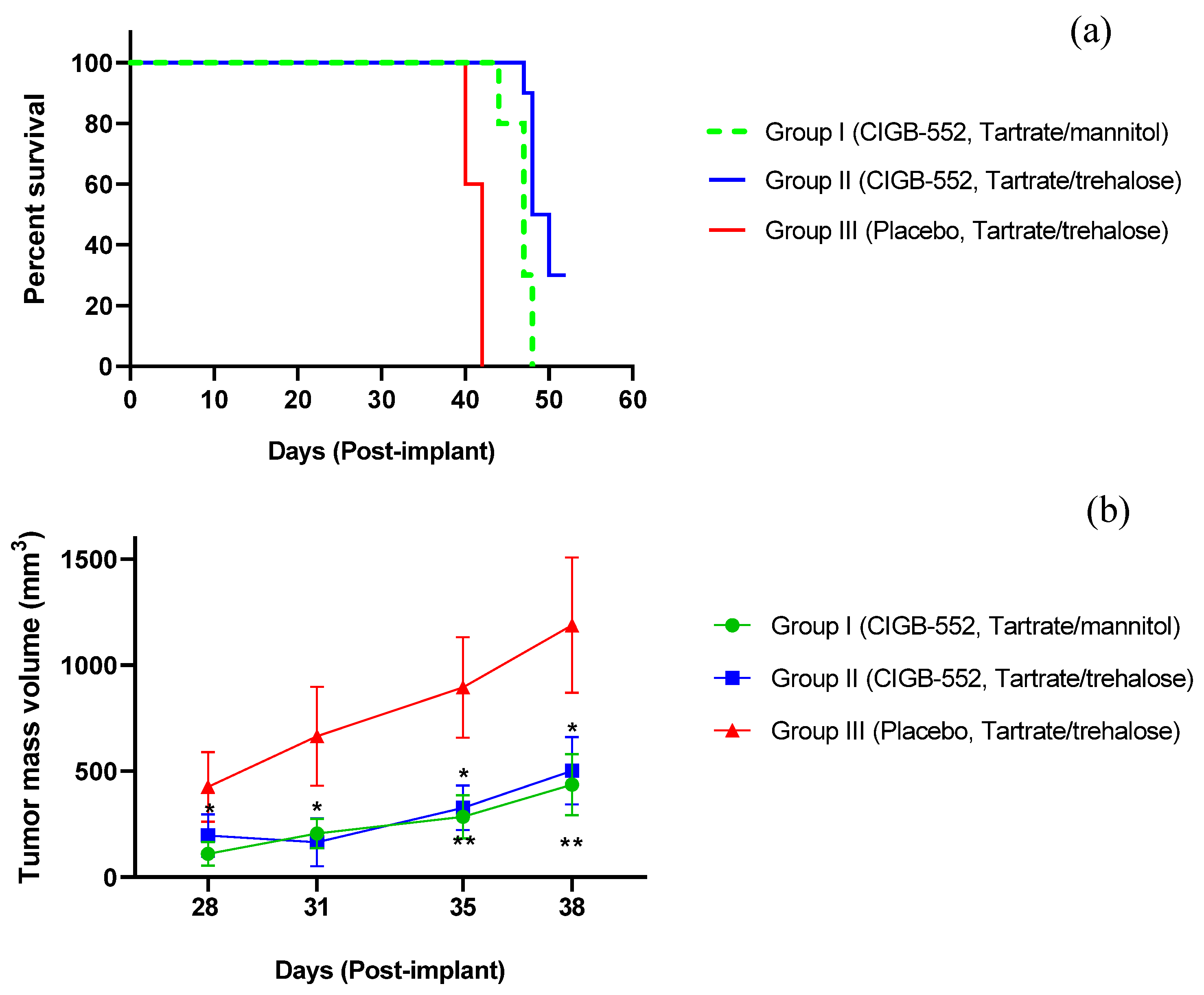
Figure 3.
Pharmacokinetic profile of CIGB-552 in Sprague Dawley rats. Plasma concentration vs time curves for each of the evaluated doses of the peptide (0.35 mg/Kg, 0.7 mg/Kg and 1.4 mg/Kg). Data are presented as mean ± SD, n = 3.
Figure 3.
Pharmacokinetic profile of CIGB-552 in Sprague Dawley rats. Plasma concentration vs time curves for each of the evaluated doses of the peptide (0.35 mg/Kg, 0.7 mg/Kg and 1.4 mg/Kg). Data are presented as mean ± SD, n = 3.
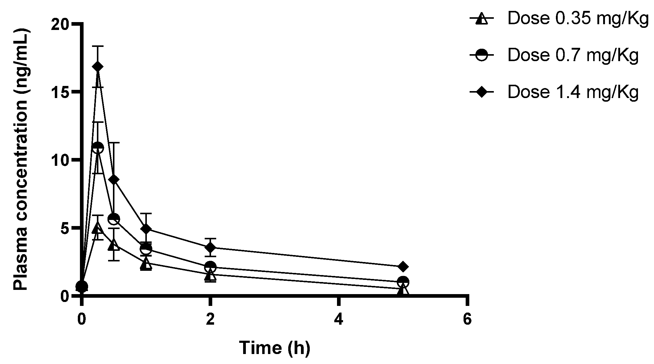
Figure 4.
Pharmacokinetic parameters AUC0-t and Cmax obtained at each dose of CIGB-552 evaluated. (a) AUC0-t in µg/L*h. (b) Cmax in ng/mL. Data are presented as mean ± SD, n = 3. *, statistical significance as calculated with two-way ANOVA with Tukey’s multiple comparisons test *p < 0.05, **p < 0.01, ***p < 0.001, ****p < 0.0001.
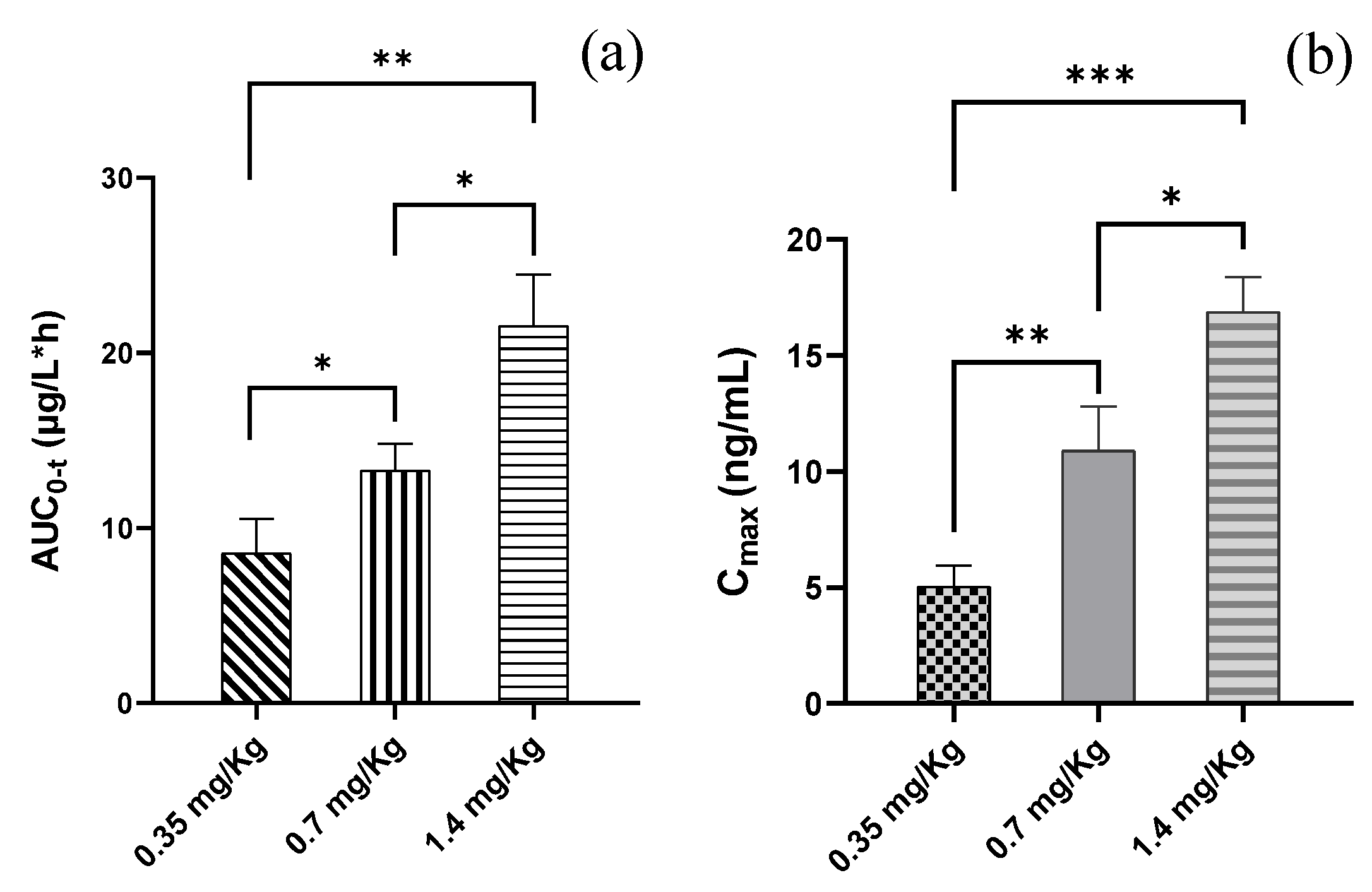
Figure 5.
Body weight of BALB/c mice during the acute toxicity study of CIGB-552. (a) Body weight of females (g). (b) Body weight of males (g). The results are expressed as the mean of the body weights of animals of each sex, n = 5.
Figure 5.
Body weight of BALB/c mice during the acute toxicity study of CIGB-552. (a) Body weight of females (g). (b) Body weight of males (g). The results are expressed as the mean of the body weights of animals of each sex, n = 5.
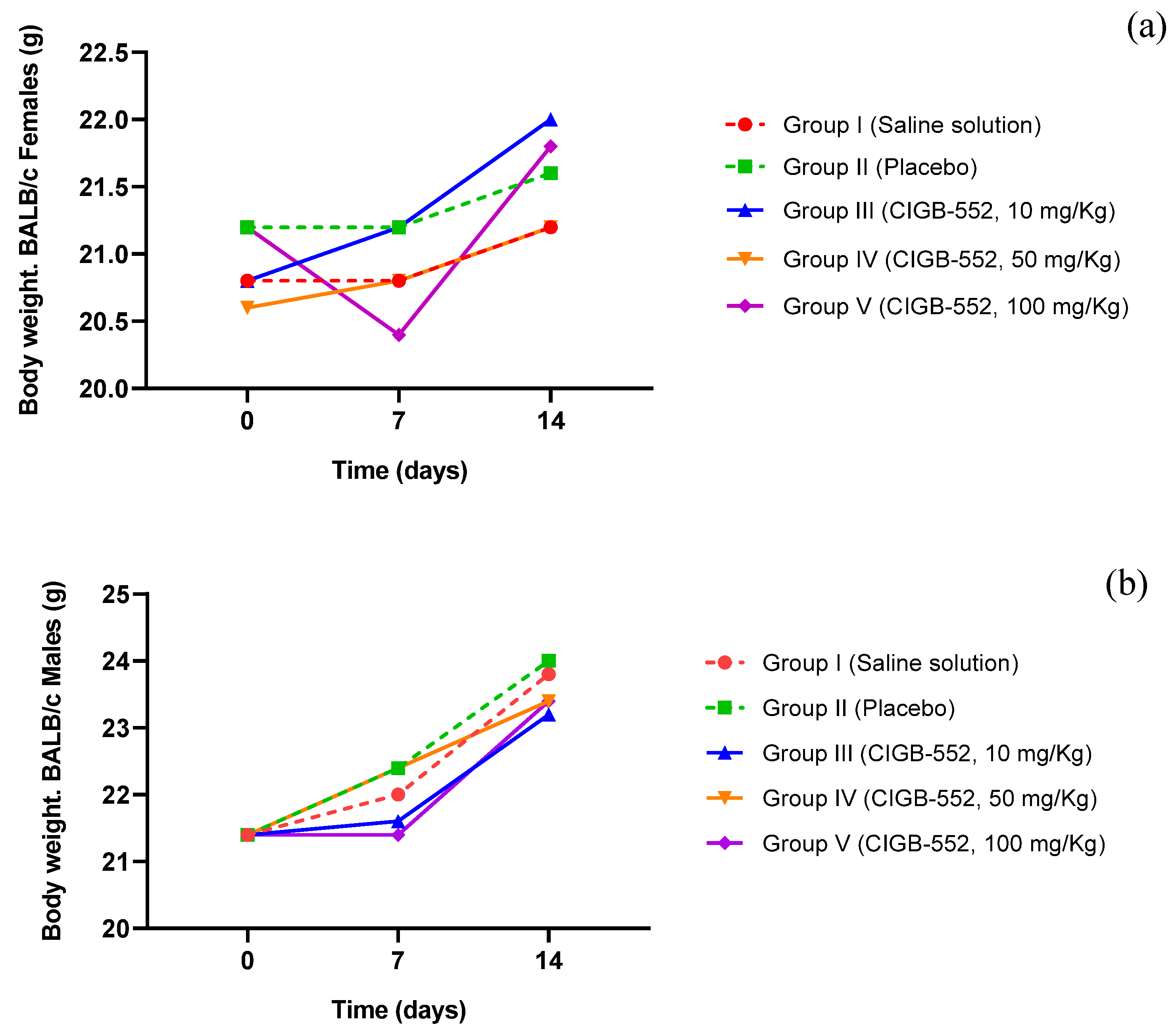
Figure 6.
Body weight of BALB/c mice during the toxicity study of CIGB-552 administered in repeated doses. (a) Body weight of females (g). (b) Body weight of males (g). The results are expressed as the mean of the body weights of animals of each sex, n = 10.
Figure 6.
Body weight of BALB/c mice during the toxicity study of CIGB-552 administered in repeated doses. (a) Body weight of females (g). (b) Body weight of males (g). The results are expressed as the mean of the body weights of animals of each sex, n = 10.

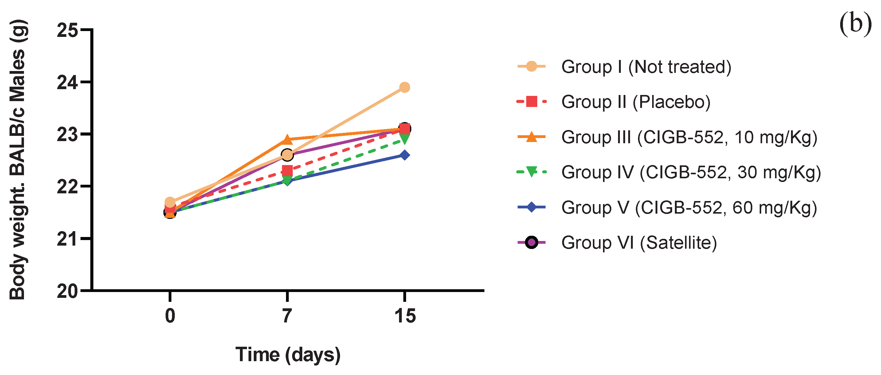

Table 1.
Plasma concentration values (ng/mL) for each sampling time, corresponding to each of the evaluated dose groups of the CIGB-552 peptide.
Table 1.
Plasma concentration values (ng/mL) for each sampling time, corresponding to each of the evaluated dose groups of the CIGB-552 peptide.
| Time (h) | Plasma concentration of CIGB-552 (ng/mL) | ||
|---|---|---|---|
| Group I (0.35 mg/Kg) |
Group II (0.7 mg/Kg) |
Group III (1.4 mg/Kg) |
|
| 0 | 0.81 ± 0.32 | 0.69 ± 0.04 | 0.64 ± 0.03 |
| 0.25 | 5.03 ± 0.90 | 10.88 ± 1.89 | 16.85 ± 1.51 |
| 0.5 | 3.79 ± 1.19 | 5.64 ± 0.4 | 8.55 ± 2.70 |
| 1 | 2.42 ± 0.52 | 3.46 ± 0.49 | 4.93 ± 1.13 |
| 2 | 1.58 ± 0.55 | 2.11 ± 0.30 | 3.56 ± 0.65 |
| 5 | 0.51 ± 0.09 | 1.02 ± 0.14 | 2.15 ± 0.29 |
Data are presented as mean ± SD, n = 3.
Table 2.
Pharmacokinetic parameters of CIGB-552 in Sprague Dawley rats and determined from the NCA method with the PK Solver tool built into Microsoft Excel on Windows 10.
Table 2.
Pharmacokinetic parameters of CIGB-552 in Sprague Dawley rats and determined from the NCA method with the PK Solver tool built into Microsoft Excel on Windows 10.
| PK parameters | Group I (0.35 mg/Kg) |
Group II (0.7 mg/Kg) |
Group III (1.4 mg/Kg) |
|---|---|---|---|
| ʎz(1/h) | 0.41 ± 0.09 | 0.34 ± 0.06 | 0.20 ± 0.10 |
| t1/2 (h) | 1.76 ± 0.36 | 2.26 ± 0.44 | 4.62 ± 3.20 |
| Tmax (h) | 0.25 ± 0.00 | 0.25 ± 0.00 | 0.25 ± 0.00 |
| Cmax (µg/L) | 5.03 ± 0.91 | 10.86 ± 1.90 | 16.86 ± 1.51 |
| AUC0-t (µg/L*h) | 8.56 ± 1.98 | 11.91 ± 1.53 | 21.56 ± 2.91 |
| AUC0-∞ (µg/L*h) | 9.90 ± 1.81 | 16.98 ± 1.92 | 36.80 ± 9.75 |
| MRT (h) | 2.41 ± 0.50 | 2.90 ± 0.51 | 6.09 ± 4.48 |
| Vz/F (L) | 22.50 ± 8.58 | 30.88 ± 6.87 | 56.65 ± 23.61 |
| Cl/F (L/h) | 8.69 ± 1.72 | 9.23 ± 1.07 | 9.52 ± 2.21 |
Cmax, maximum observed plasma concentration; Tmax, time to peak; t1/2, terminal half-life; λz, elimination rate constant; AUC0-t, area under plasma concentration-time curve from time zero to the last measurable time point; AUC0-∞, area under plasma concentration-time curve from time zero to infinity; MRT, mean residence time; Vz/F, volume of distribution at steady state; Cl/F, total plasma clearance. Data are expressed as mean ± SD, n = 3.
Table 3.
Frequency of appearance of microscopic findings by organ observed and by treatment group in the acute toxicity study of CIGB-552.
Table 3.
Frequency of appearance of microscopic findings by organ observed and by treatment group in the acute toxicity study of CIGB-552.
| Groups | Liver | Spleen | GLM | |||
| HE | CG | HE | CG | |||
| I(Saline solution) | 5/10 | 10/10 | 10/10 | 10/10 | ||
| II (Placebo) | 1/10 | 8/10 | 9/10 | 10/10 | ||
| III(CIGB-552, 10 mg/Kg) | 4/10 | 8/10 | 9/10 | 10/10 | ||
| IV(CIGB-552, 50 mg/Kg) | 6/10 | 10/10 | 10/10 | 10/10 | ||
| V(CIGB-552, 100 mg/Kg) | 4/10 | 8/10 | 9/10 | 10/10 | ||
HE, extramedullary hematopoiesis, GLM, mesenteric lymph node; CG, germinal center.
Table 4.
Frequency of appearance of microscopic findings by organ observed and by treatment group in the CIGB-552 repeat-dose toxicity study.
Table 4.
Frequency of appearance of microscopic findings by organ observed and by treatment group in the CIGB-552 repeat-dose toxicity study.
| Groups | Liver | Spleen | GLM | Applicationsite | ||||
| NF | HE | CG | HE | PALS | CG | II | ||
| I (Not treated) | 0/20 | 14/20 | 16/20 | 20/20 | 16/20 | 19/20 | 0/20 | |
| II (Placebo) | 1/20 | 14/20 | 18/20 | 20/20 | 18/20 | 20/20 | 0/20 | |
| III (CIGB-552, 10 mg/Kg) | 0/20 | 15/20 | 15/20 | 20/20 | 15/20 | 20/20 | 4/20 | |
| IV (CIGB-552, 30 mg/Kg) | 0/20 | 16/20 | 19/20 | 20/20 | 19/20 | 20/20 | 3/20 | |
| V (CIGB-552, 60 mg/Kg) | 1/20 | 10/20 | 17/20 | 20/20 | 17/20 | 20/20 | 7/20 | |
| VI (CIGB-552, 60 mg/Kg) | 0/15 | 10/15 | 15/15 | 15/15 | 15/15 | 15/15 | 0/15 | |
GLM, mesenteric lymph node; NF, focal necrosis; CG, germinal center; HE, extramedullary hematopoiesis; PALS, periarteriolar lymphoid sheath; II, inflammatory infiltrate.
Disclaimer/Publisher’s Note: The statements, opinions and data contained in all publications are solely those of the individual author(s) and contributor(s) and not of MDPI and/or the editor(s). MDPI and/or the editor(s) disclaim responsibility for any injury to people or property resulting from any ideas, methods, instructions or products referred to in the content. |
© 2025 by the authors. Licensee MDPI, Basel, Switzerland. This article is an open access article distributed under the terms and conditions of the Creative Commons Attribution (CC BY) license (http://creativecommons.org/licenses/by/4.0/).
Copyright: This open access article is published under a Creative Commons CC BY 4.0 license, which permit the free download, distribution, and reuse, provided that the author and preprint are cited in any reuse.


