
Submitted:
01 July 2025
Posted:
01 July 2025
You are already at the latest version
Abstract
Extracellular vesicles (EVs) are emerging as versatile mediators of intercellular communication and promising tools for drug discovery and targeted therapies. These lipid bilayer-bound nanovesicles facilitate the transfer of functional proteins, RNAs, lipids, and other biomolecules between cells, thereby influencing various physiological and pathological processes. This review outlines the molecular mechanisms governing EV biogenesis and cargo sorting, emphasizing the role of regulators, such as ubiquitin-like 3 (UBL3), in modulating protein packaging. We explored the critical involvement of EVs in various disease microenvironments, including cancer progression, neurodegeneration, and immune modulation. Their ability to cross biological barriers and deliver bio-active cargo renders them highly attractive for precise drug delivery systems, especially in neurological and oncological disorders. Moreover, this review highlights advances in engineering EVs for delivering RNA therapeutics, CRISPR-Cas systems, and targeted small molecules. The utility of EVs as diagnostic tools in liquid biopsies and their integration into personalized medicine and companion diagnostics were also discussed. Patient-derived EVs offer dynamic insights into disease state and enable real-time treatment stratification. Despite their potential, challenges such as scalable isolation, cargo heterogeneity, and regulatory ambiguity remain significant hurdles. We also reported novel pharmacological approaches targeting EV biogenesis, secretion, and uptake pathways, and considered UBL3 as a promising drug target for EV cargo modulation. Future directions include the standardization of EV analytics, scalable biomanufacturing, and classification of EV-based therapeutics under evolving regulatory frameworks. This review emphasizes the multifaceted roles of EVs and their transformative potential as therapeutic platforms and biomarker reservoirs in next-generation precision medicine.
Keywords:
Extracellular vesicles
; ubiquitin-like 3
; disease microenvironment
; precision medicine
; next-generation drug delivery
1. Introduction
Extracellular vesicles (EVs) are lipid bilayer-enclosed nanostructures secreted by almost all cell types and classified into subtypes—mainly exosomes (30–150 nm, endosomal origin), microvesicles (100–1000 nm, plasma membrane origin), and apoptotic bodies—based on their biogenesis and size. [1,2,3]. These vesicles carry molecular cargo such as proteins, lipids, DNA, mRNA, miRNAs, and other non-coding RNAs that influence intercellular communication [4,5,6]. EVs are present in nearly all biological fluids, including the blood, urine, saliva, and cerebrospinal fluid, and function as mediators of both physiological and pathological processes [2,7,8]. Under homeostatic conditions, EVs contribute to tissue regeneration, immune modulation, and neural development [9,10]. However, in pathological settings, EVs have been implicated in cancer metastasis, neurodegeneration, cardiovascular disease, and immune dysregulation [11,12]. Their dual role as disease biomarkers and delivery vehicles highlights their relevance in diagnostics and therapeutics, particularly in precision medicine [13,14,15].
One of the most attractive features of EVs is their natural ability to cross biological barriers, such as the blood-brain barrier, making them suitable candidates for delivering therapeutic agents to the central nervous system (CNS) [16,17]. Their surface molecules, including integrins, tetraspanins, and other receptors, confer targeting specificity, allowing selective delivery to recipient cells and tissues [18,19,20].
In pharmacology and drug discovery, EVs are gaining traction as next-generation therapeutic platforms owing to their biocompatibility, low immunogenicity, and intrinsic targeting capacity [21,22]. These properties have inspired numerous preclinical and clinical investigations exploring EVs in cancer, neurodegenerative diseases, inflammation, and regenerative medicine [23,24,25].
In addition to their native roles, EVs are now recognized as dynamic players in the regulation of the extracellular environment and are capable of modulating host-pathogen interactions, intercellular metabolism, and even systemic physiological states. For example, microbial-derived EVs have been shown to influence tumor progression and immune responses by mimicking host-derived vesicles, revealing a complex interplay between the microbiota and host EV signaling pathways [2,26]. Furthermore, the lipid composition of EVs, especially sphingomyelins and cholesterol, contributes to their structural stability and the modulation of recipient cell signaling cascades. These lipid-rich membranes enable vesicles to fuse with target cells or interact with surface receptors, triggering downstream effects that may vary by tissue type and disease context [6,27]. Recent investigations into EV-associated enzymes and ion channels have also uncovered novel mechanisms by which EVs regulate metabolic reprogramming and stress adaptation in target cells [28]. This functional plasticity makes EVs highly attractive therapeutic platforms with adaptable properties. Importantly, leveraging omics-based analyses, such as proteomics, lipidomics, and transcriptomics, has enabled a deeper understanding of EV heterogeneity and specialization, laying the groundwork for precision-designed vesicles tailored to specific therapeutic goals [29,30].
Recent studies have demonstrated the engineering of EVs to encapsulate various therapeutic agents, including small molecules, siRNAs, miRNAs, proteins, and CRISPR/Cas9 components [31,32,33,34]. For example, exosomes engineered to express brain-targeting ligands (e.g., Lamp2b-RVG peptide) have shown enhanced central nervous system delivery via systemic administration [16]. Additionally, surface-functionalized EVs equipped with targeting peptides, antibodies, or aptamers exhibit significantly improved biodistribution and cellular uptake [35,36,37].
The translational potential of EVs has been demonstrated in various diseases. In oncology, EVs loaded with chemotherapeutics or siRNAs targeting oncogenes, such as KRAS, have shown antitumor efficacy [11]. In stroke and neurodegeneration, BDNF- or catalase-loaded exosomes reduce brain damage and improve functional recovery [38]. Notably, our recent study developed label-free imaging of EVs in breast cancer, highlighting their potential applications in targeted breast cancer therapies [39]. Another study conducted in 2023 reported the neuroprotective effects of EVs in neurodegenerative disease models [14]. Furthermore, neurological disorders like Parkinson’s and Alzheimer’s diseases have been shown to benefit from EV-based therapies, given their ability to modulate neuroinflammation and synaptic function [40,41]. A recent integrative molecular study uncovered how EVs mediate drug transport and metabolic reprogramming, further supporting their therapeutic utility [42,43].
Despite their promise, technical challenges remain, including scalable and standardized isolation, purity control, in vivo tracking, and storage stability [44,45]. The adoption of international guidelines such as MISEV2018 and MISEV2023 has provided a much-needed framework for EV characterization and nomenclature [46]. The integration of multi-omics technologies, bioinformatics, and nanotechnology is essential for overcoming the current barriers and unlocking the full clinical potential of EVs [46,47,48].
2. Molecular Mechanisms of EV Biogenesis and Cargo Sorting
The biogenesis and cargo loading of EVs are controlled by coordinated molecular pathways that determine their structures and functions. EVs are broadly categorized as exosomes, formed via inward budding of late endosomes into multivesicular bodies (MVBs), and microvesicles, generated by outward budding from the plasma membrane [4,7,49].
Exosome formation is primarily governed by the Endosomal Sorting Complex Required for Transport (ESCRT) machinery, which includes ESCRT-0 to -III and accessory proteins such as ALIX and TSG101 [50,51,52,53]. These components enable membrane budding and cargo selection. ESCRT-independent mechanisms also contribute to EV biogenesis, involving tetraspanins (CD9, CD63, and CD81), ceramides, and lipid rafts, which influence both membrane curvature and selective cargo sorting [54,55,56].
Rab GTPases, such as Rab27a/b, Rab11, and Rab35, regulate MVB trafficking, docking, and fusion with the plasma membrane, thereby modulating EV secretion [57,58,59]. For instance, Rab27a positions MVBs in the membrane, whereas Rab11 mediates the recycling pathways [60,61].
Among the emerging regulators, UBL3 (Ubiquitin-like protein 3) plays a non-canonical role in post-translational modifications and cargo sorting. UBL3 localizes to the plasma membrane and directs S-prenylation-dependent protein modifications, facilitating their selective packaging into EVs [39,62,63]. It influences the secretion of immune-regulatory and tumor-related proteins, and UBL3 deficiency disrupts EV composition in pathological contexts [39,62]. Thus, UBL3 is a promising target for the production of engineered EVs. Recent insights suggest that other post-translational modifications, such as SUMOylation and neddylation, may also contribute to selective cargo loading into EVs. These modifications influence the interaction of target proteins with sorting machinery and membrane domains, thus determining their inclusion or exclusion from vesicles [64,65]. Moreover, RNA-binding proteins, such as YBX1 and hnRNPA2B1, have been implicated in sorting miRNAs into exosomes by recognizing specific sequence motifs or structures. The phosphorylation state of these RNA-binding proteins may further modulate their activity and specificity [66]. Additionally, the lipid environment of multivesicular bodies, particularly the role of phosphatidylserine and cholesterol-rich microdomains, has emerged as a determinant of cargo affinity and membrane curvature. Together, these complex regulatory layers offer multiple intervention points for modulating EV content for therapeutic purposes, especially in diseases with aberrant intercellular communication [67,68].
The cargo content of EVs is dynamically modulated by their cellular state. Stress conditions such as hypoxia, inflammation, or oxidative stress alter EV composition, enriching them with proteins like HIF-1α, VEGF, or pro-inflammatory miRNAs [69,70,71]. Immune activation, for example, triggers the release of EVs carrying checkpoint proteins and miR-155, whereas neuronal activity affects EV cargo during synaptic signaling and injury responses [72,73].
Together, these tightly regulated processes ensure that EVs carry highly specific molecular signatures, enabling precise intercellular communication and presenting opportunities for the therapeutic customization of drug delivery systems. The processes of EV biogenesis and selective cargo loading are visually summarized in Figure 1 [74].
3. EV-Mediated Intercellular Communication in Disease Microenvironments
EVs are critical modulators of the disease microenvironment, contributing to cancer progression, neurodegeneration, and immune modulation through the transfer of bioactive molecules, such as proteins, lipids, mRNAs, and non-coding RNAs [75,76,77]. Their ability to deliver specific cargo into recipient cells enables EVs to shape the behavior of neighboring or distant cells, supporting pathological processes such as tumor metastasis, inflammation, and the propagation of toxic proteins [39,62].
In the tumor microenvironment (TME), EVs mediate bidirectional communication between cancer cells and stromal, endothelial, and immune cells [78]. Tumor-derived EVs (TDEs) carry oncogenic proteins (e.g., EGFRvIII), immunosuppressive molecules (e.g., PD-L1), and pro-angiogenic factors (e.g., VEGF), promoting tumor growth, immune escape, and vascular remodeling [79,80,81]. TDEs also reprogram fibroblasts and recruit tumor-associated macrophages (TAMs), further amplifying their metastatic potential [82,83]. For example, exosomal integrins (α6β4 and αvβ5) have been implicated in organotropic metastasis by preconditioning distant tissues [84].
In neurodegenerative diseases, EVs act as vectors for the cell-to-cell transmission of pathogenic proteins [14,85]. Studies have shown that EVs transport misfolded tau, amyloid-β (Aβ), α-synuclein, and TDP-43, facilitating their propagation in Alzheimer’s disease (AD), Parkinson’s disease (PD), and amyotrophic lateral sclerosis (ALS) [86,87,88,89]. This prion-like spread contributes to disease progression and neural network dysfunctions. Moreover, neuron- and astrocyte-derived EVs influence microglial activation, contributing to neuroinflammation during the early stages of neurodegeneration [90,91].
Extracellular vesicles (EVs) play a dual role in the immune system. On one hand, they mediate antigen presentation and T-cell activation; on the other hand, cancer-derived EVs often suppress immunity by delivering PD-L1, FasL, or miRNAs that target immune checkpoints [92,93]. Furthermore, EVs secreted by dendritic cells and T cells carry MHC-peptide complexes, costimulatory molecules, and miRNAs that modulate the activity of effector and regulatory cells, thereby influencing inflammation and tolerance [94,95]. An overview of EV functions across different disease microenvironments is shown in Figure 2 [74].
Emerging evidence indicates that EVs also contribute to metabolic rewiring within the disease microenvironment. Tumor-derived EVs have been shown to transfer metabolic enzymes and regulatory RNAs that alter the glycolytic or oxidative phosphorylation pathways in recipient stromal or immune cells, thereby supporting cancer cell survival under hypoxic and nutrient-deprived conditions [96,97]. In parallel, EVs released under inflammatory stress can induce metabolic shifts in macrophages, polarizing them toward a pro-tumorigenic M2 phenotype [98]. Similarly, in neurodegenerative disorders, astrocyte-derived EVs containing lactate dehydrogenase and other metabolic enzymes influence the neuronal energy balance and redox homeostasis [99]. These metabolic modulations sustain disease progression and affect therapeutic responses, especially in the context of resistance to targeted therapies. As such, EV-mediated metabolic crosstalk represents a promising yet underexplored dimension of intercellular communication that may provide novel targets for intervention in cancer and neurological diseases [100,101].
Together, EVs act as intercellular shuttles that remodel the disease microenvironment by enhancing malignancy, spreading pathogenic proteins, and modulating the immune response. Understanding these EV-mediated mechanisms opens new avenues for novel therapeutic interventions and biomarker discovery in complex diseases [102].
4. Pharmacological Targeting of EV Pathways
The pharmacological modulation of extracellular vesicle (EV) pathways is a promising strategy that can inhibit pathological EV activity and enhance therapeutic EV function. Targeting the key steps in EV biogenesis, release, uptake, and cargo packaging opens new avenues for drug development and disease intervention.

Table 1.
Caption.
| Agent/Strategy | Target/Mechanism | Effect on EVs | Application Context | Citation |
| GW4869 | Inhibits neutral sphingomyelinase | Reduces exosome release | Cancer, inflammation | [103] |
| Imipramine | Disrupts endolysosomal trafficking | Blocks exosome secretion | Oncology, neurodegeneration | [104] |
| Monensin | Alters Golgi pH, enhances EV secretion | Promotes EV release | Therapeutic EV production | [105] |
| Forskolin | Activates adenylyl cyclase/cAMP pathway | Increases EV release | Neuroregeneration | [106] |
| Dynasore | Inhibits dynamin-dependent endocytosis | Blocks EV uptake | Prevents EV-mediated signal spread | [107] |
| Trichostatin A | HDAC inhibitor, alters gene expression | Modifies EV cargo | Immunomodulatory EVs | [108] |
| Fenretinide | Inhibits dihydroceramide desaturase (DES1) | Disrupts ceramide-dependent EV release | Antitumor EV suppression | [109] |
| AMPK activators | Modulate cellular metabolism | Downregulate Rab27-mediated secretion | Oncogenic EV suppression | [110] |
| UBL3 modulation | Alters S-prenylation of surface proteins | Reprograms cargo packaging | EV engineering for precision therapy | [62,111] |
Several small-molecule inhibitors that suppress EV release have been identified. GW4869, a neutral sphingomyelinase inhibitor, is widely used to block ceramide-mediated exosome biogenesis and has been shown to reduce EV-mediated inflammation and tumor progression [112,113]. Similarly, imipramine, an FDA-approved tricyclic antidepressant, inhibits exosome secretion via endolysosomal disruption [114,115]. In contrast, certain compounds such as monensin and forskolin have been reported to enhance EV release, which could be leveraged for therapeutic EV production [116,117]. A summary of the key pharmacological agents and strategies targeting EV pathways is presented in Table 1 for comparison.
The uptake of EVs by recipient cells can be pharmacologically regulated. Molecules such as dynasore and chlorpromazine inhibit clathrin- or caveolin-mediated endocytosis, thereby offering tools to prevent unwanted EV-mediated signal propagation [102,118].
Beyond trafficking, emerging methods enable the manipulation of EV content. Engineered donor cells or post-isolation techniques like electroporation, sonication, and chemical transfection allow for the loading of therapeutic cargos such as siRNAs, CRISPR-Cas systems, or chemotherapeutics [115,119].
Among the molecular targets, UBL3 has recently gained attention for its role in S-prenylation-dependent EV cargo loading. UBL3 facilitates the selective inclusion of immune and disease-related proteins into small EVs and represents a druggable pathway for cargo-level modulation [39,120]. Altering UBL3 activity may allow researchers to reprogram EV content for cancer immunotherapy or neuroinflammation modulation [39,62,121].
Together, these strategies emphasize the therapeutic potential of EV pathway modulation, positioning pharmacological EV targeting as the next-generation modality in precision medicine.
In addition to small-molecule inhibitors and engineering approaches, recent studies have explored the targeting of metabolic and epigenetic regulators that influence EV biogenesis and composition. For example, the inhibition of histone deacetylases (HDACs) with agents like trichostatin-A has been shown to modulate EV cargo by altering gene expression profiles in donor cells, enhancing the release of immunomodulatory proteins and RNAs [108,122]. Similarly, the metabolic state of a cell, governed by enzymes such as AMP-activated protein kinase (AMPK), can affect the quality and quantity of secreted EVs, which may have implications for cancer or inflammation-associated EV signaling. Pharmacological activation of AMPK has been found to downregulate Rab27-dependent EV secretion pathways, representing a novel method to suppress oncogenic EV spread [123,124].
Furthermore, the sphingolipid biosynthesis pathway has garnered attention as a druggable axis for EV modulation. Inhibitors of dihydroceramide desaturase (DES1), such as fenretinide, have demonstrated the ability to disrupt exosome release and reduce tumorigenic EV signaling [125,126]. These findings support a broader strategy that integrates metabolic reprogramming with EV-targeted therapy. Additionally, emerging nanoparticle-drug conjugates are being designed to bind EV surface markers (e.g., CD63, CD81), enabling selective neutralization or uptake blockade in vivo [127,128].
Collectively, these pharmacological interventions, including lipid metabolism, epigenetic regulation, and surface targeting, provide a multifaceted approach for manipulating the EV pathway. Such strategies may synergize with conventional treatments and offer precision-tuned therapeutic avenues for diseases where EVs play a central pathological role.
5. EVs as Drug Delivery Vehicles and Biomarker Reservoirs
EVs have emerged as powerful platforms for therapeutic delivery and diagnostic applications owing to their inherent biocompatibility, low immunogenicity, and natural targeting ability [129]. Their ability to transport a variety of bioactive molecules, including RNAs, proteins, and small-molecule drugs, makes them particularly promising for drug delivery and non-invasive biomarker discovery [130].
Engineered EVs have been extensively explored for the delivery of therapeutic RNAs (e.g., siRNA, miRNA, and mRNA), proteins, and genome-editing tools such as CRISPR/Cas9. Loading strategies include donor cell transfection, electroporation, sonication, and extrusion [131,132,133,134]. For example, Alvarez-Erviti et al. (2011) successfully delivered siRNA across the blood-brain barrier using exosomes engineered with the Lamp2b-RVG fusion peptide [16]. Similarly, MSC-derived EVs have been used to deliver anti-inflammatory miRNAs and neuroprotective factors in models of stroke, spinal cord injury, and myocardial infarction [135].
Surface modification techniques, such as ligand display, aptamer conjugation, and peptide anchoring, can further enhance targeted delivery. Ligands such as GE11 (for EGFR) or RGD (for integrins) have been conjugated to EV surfaces to improve tissue-specific accumulation [136,137]. These strategies significantly increase therapeutic efficacy while minimizing off-target effects and systemic toxicity.
In parallel, EVs are increasingly being recognized as rich reservoirs of diagnostic biomarkers, particularly in liquid biopsy platforms. Circulating EVs in the blood, urine, and CSF contain disease-specific proteins, lipids, and RNAs that reflect the physiological or pathological state of the originating cells [138,139,140]. In cancer, EV-derived miRNAs (e.g., miR-21, miR-1246), proteins (e.g., EpCAM, CD63), and DNA fragments have shown strong diagnostic and prognostic potential across various tumor types [141,142]. Similarly, EVs in neurodegenerative diseases carry α-synuclein, tau, or Aβ species, which can distinguish between disease stages and subtypes [143].
Together, these properties position EVs as multifunctional agents in drug delivery and clinical diagnostics. Their use in ongoing clinical trials further highlights their potential as the next-generation precision tools.
In recent years, researchers have also explored hybrid systems that combine EVs with synthetic nanoparticles or biomaterials to improve drug payload, release kinetics, and targeting specificity. For instance, EVs fused with liposomes or polymeric nanocarriers can synergize the benefits of natural and synthetic vectors, thereby enhancing delivery efficiency and immune evasion [144,145]. Additionally, bioengineered EVs expressing targeting ligands on their surface, such as antibodies or nanobodies, have shown promise in navigating complex tissue environments, including solid tumors and inflamed organs [146]. Another advancement includes the development of stimuli-responsive EVs that release their cargo under specific physiological triggers such as pH, redox state, or enzymatic activity, enabling precise spatiotemporal drug release [147].
Parallel to their delivery potential, EVs are being profiled for longitudinal disease monitoring and early relapse detection. The integration of EV-based multi-omics (proteomics, transcriptomics, and metabolomics) is now feasible using high-throughput platforms, allowing for comprehensive biomarker panels with enhanced sensitivity and specificity [148,149]. This multiplexed profiling capability makes EVs particularly suitable for diseases with dynamic progression such as cancer, neurodegenerative disorders, and chronic inflammatory conditions. Moreover, coupling EV analysis with machine learning algorithms can stratify patients more accurately and predict therapeutic responses in real-time [150,151].
Collectively, these emerging technologies continue to expand the landscape of EV research, solidifying their role not just as passive carriers but also as intelligent, programmable platforms for integrated drug delivery and diagnostics in precision medicine.
6. EVs in Personalized Medicine and Companion Diagnostics
The paradigm shift toward personalized medicine has underscored the need for dynamic, minimally invasive biomarkers to guide real-time therapeutic decisions. Owing to their stability in circulation, molecular richness, and tissue specificity, EVs have emerged as promising tools in this area. EVs derived from patient biofluids (e.g., blood, urine, cerebrospinal fluid) reflect the molecular landscape of the parent cell, and thus offer a window into disease progression, therapeutic resistance, and treatment response [152,153].
6.1. Patient-Derived EVs in Cancer Monitoring
Tumor-derived EVs (TDEVs) carry oncogenic proteins, RNA, DNA fragments, and lipids that mirror the mutational status and signaling activity of the tumor. These vesicles have been detected in various cancer types, including lung, breast, prostate, and colorectal cancers, offering diagnostic and prognostic utility [154,155,156]. For instance, EGFR mutations and ALK rearrangements, which are critical for therapeutic selection in non-small cell lung cancer, have been identified in circulating EVs, enabling real-time monitoring of tumor evolution [157,158].
Importantly, EVs are less prone to degradation than circulating free nucleic acids and can be isolated longitudinally, allowing clinicians to track tumor heterogeneity and the emergence of drug resistance without repeated tissue biopsies [159,160]. This has profound implications for detecting minimal residual disease and recurrence earlier than imaging-based modalities.
6.2. EVs in Targeted Therapy Matching and Disease Profiling
EVs provide an opportunity to profile actionable mutations, RNA expression signatures, or proteomic alterations non-invasively, thereby supporting companion diagnostics. For example, exosomal PD-L1 expression has been correlated with immune evasion and treatment resistance in melanoma and lung cancer, thereby guiding the use of checkpoint inhibitors [161,162]. Similarly, exosomal KRAS mutations have been used to stratify colorectal cancer patients for anti-EGFR therapy [163,164].
As EVs can be sampled frequently, they enable real-time molecular profiling of disease states, helping personalize targeted therapies based on dynamic tumor biology. This is especially valuable in cases where traditional biopsies are infeasible due to tumor inaccessibility or patient frailty [165,166].
6.3. EVs in Precision Immunotherapy and Patient Stratification
EVs provide immunotherapeutic strategies that significantly benefit from EV-based diagnostics. EVs secreted by immune or tumor cells carry immune checkpoint molecules (e.g., PD-L1, CTLA-4), cytokines, and antigenic peptides that modulate immune responses [167,168]. Measuring these biomarkers in patient-derived EVs can help to predict immunotherapy responsiveness and stratify patients accordingly.
Recent studies have demonstrated that T-cell–derived EVs enriched with CD28 or LAG3 can reflect T-cell exhaustion and immunotherapy resistance [169,170]. In addition, EV profiling has shown promise in distinguishing “hot” (inflamed) versus “cold” (non-inflamed) tumors, which are key determinants of checkpoint blockade efficacy [171].
Taken together, EVs offer a unique combination of molecular fidelity, sampling convenience, and clinical relevance. As platforms for companion diagnostics, they hold transformative potential in personalized therapy regimens, minimizing toxicity, and improving clinical outcomes in oncology and immunotherapy.
7. Challenges and Future Perspectives in EV-Based Drug Discovery
Despite their promise as drug carriers and diagnostic tools, extracellular vesicles (EVs) face several technical and translational challenges that must be overcome for their successful clinical implementation. These include issues with isolation and purification, cargo heterogeneity, dosing standardization, and an uncertain regulatory framework.
One of the primary challenges is the lack of standardized isolation protocols. Current methods, such as ultracentrifugation, size-exclusion chromatography, and precipitation, vary widely in efficiency and purity [172]. This inconsistency affects reproducibility and downstream functional analyses. Moreover, the heterogeneous nature of EV populations, even within the same biofluid, complicates their characterization and therapeutic efficacy [173]. New microfluidic platforms and affinity-based purification systems offer improved selectivity but are not yet scalable or cost-effective for clinical applications [174].
Another hurdle is the regulatory and translational gap. The lack of global consensus on EV classification, potency assays, and quality control has hindered the development of good manufacturing practice (GMP)-compliant EV therapies [174].
However, dosing strategies and biodistribution profiling pose significant challenges. Quantifying EVs remains difficult due to overlapping size ranges with other nanoparticles and variability in protein-to-vesicle ratios [177,178]. Additionally, the long-term effects of EV administration and immune clearance mechanisms are not yet fully understood, which raises safety concerns regarding repeated dosing.
Personalized EV-based therapeutics are expected to gain attention in the future. Patient-derived or engineered EVs tailored to individual genetic or proteomic profiles could revolutionize precision medicine [29,179]. In this context, UBL3 has emerged as a promising druggable regulator for EV cargo sorting. By modulating S-prenylation of surface proteins, UBL3 controls the selective inclusion of immune and disease-associated factors in small EVs [63]. Targeting UBL3 and its downstream pathways may allow for the precise reprogramming of EV content, particularly in cancer and neuroinflammatory conditions [39].
To further facilitate clinical translation, robust and harmonized analytics for EV pharmacokinetics and dynamics must be developed. Traditional pharmacokinetic metrics, such as half-life, clearance, and bioavailability, remain challenging to define for EVs because of their endogenous nature and diverse cargo profiles. Advanced imaging techniques and EV-specific labeling strategies, such as super-resolution microscopy or click chemistry-based tagging, are beginning to shed light on their in vivo behavior [180,181]. However, scalable quantitative tools are required. Another pressing challenge is the scalability of EV production. Current manufacturing platforms, including ultracentrifugation and tangential flow filtration, lack the necessary throughput and reproducibility for the clinical-grade EV therapeutics. Emerging bioreactor systems and cell-free synthesis approaches offer promise for industrial-scale EV generation but are still in their infancy [182]. Additionally, ethical and safety considerations must be addressed, particularly for engineered EVs carrying potent gene-editing tools or immune modulators. Regulatory authorities will need to classify EVs appropriately, whether as biologics, drug delivery systems, or cell therapy derivatives, to influence preclinical testing and approval routes [183]. Cross-disciplinary collaboration among bioengineers, clinicians, and regulatory bodies will be key to resolving these issues and realizing the full potential of EV-based therapies [184,185]. Additionally, inter-individual variability in EV profiles poses challenges for therapeutic standardization. Factors such as age, sex, comorbidities, and circadian rhythm can influence EV composition and efficacy, complicating reproducibility across patient populations [186].
In conclusion, although significant hurdles remain, continued advancements in EV engineering, standardization, and molecular targeting (including UBL3) are expected to accelerate the clinical translation of EV-based therapeutics in the near future.
Author Contributions
Conceptualization- M.M.H; writing—original draft preparation- M.A.M; Review and editing- M.AM, S.M.S, A.S.M.W and M.M.H. All authors have read and agreed to the published version of this manuscript.
Funding
This study received no external funding.
Institutional Review Board Statement
Not applicable.
Informed Consent Statement
Not applicable.
Data Availability Statement
Not applicable.
Conflicts of Interest
The authors declare no conflicts of interest.
Abbreviations
The following abbreviations are used in this manuscript.
| AD | Alzheimer’s Disease |
| ALS | Amyotrophic Lateral Sclerosis |
| Aβ | Amyloid beta |
| BBB | Blood-Brain Barrier |
| CNS | Central Nervous System |
| CM | Conditioned Medium |
| CRC | Colorectal Cancer |
| DC | Dendritic Cell |
| DNA | Deoxyribonucleic Acid |
| EV | Extracellular Vesicle |
| FDA | Food and Drug Administration |
| GSC | Glioma Stem Cell |
| GTPase | Guanosine Triphosphatase |
| HNSCC | Head and Neck Squamous Cell Carcinoma |
| HSP | Heat Shock Protein |
| ILV | Intraluminal Vesicle |
| ISEV | International Society for Extracellular Vesicles |
| KO | Knockout |
| LAMP | Lysosomal-Associated Membrane Protein |
| miRNA | MicroRNA |
| miR | microRNA (generic notation) |
| MISEV | Minimal Information for Studies of Extracellular Vesicles |
| MSC | Mesenchymal Stem Cell |
| NSCLC | Non-Small Cell Lung Cancer |
| PD | Parkinson’s Disease |
| PD-L1 | Programmed Death-Ligand 1 |
| RNA | Ribonucleic Acid |
| siRNA | Small Interfering RNA |
| sEV | Small Extracellular Vesicle |
| TME | Tumor Microenvironment |
| TNBC | Triple-Negative Breast Cancer |
| UBL3 | Ubiquitin-Like Protein 3 |
| WT | Wild-Type |
References
- Moghassemi, S.; Dadashzadeh, A.; Sousa, M.J.; Vlieghe, H.; Yang, J.; León-Félix, C.M.; Amorim, C.A. Extracellular Vesicles in Nanomedicine and Regenerative Medicine: A Review over the Last Decade. Bioact Mater 2024, 36, 126–156. [Google Scholar] [CrossRef] [PubMed]
- Chronopoulos, A.; Kalluri, R. Emerging Role of Bacterial Extracellular Vesicles in Cancer. Oncogene 2020 39:46 2020, 39, 6951–6960. [Google Scholar] [CrossRef] [PubMed]
- El Andaloussi, S.; Mäger, I.; Breakefield, X.O.; Wood, M.J.A. Extracellular Vesicles: Biology and Emerging Therapeutic Opportunities. Nature Reviews Drug Discovery 2013 12:5 2013, 12, 347–357. [Google Scholar] [CrossRef]
- Van Niel, G.; D’Angelo, G.; Raposo, G. Shedding Light on the Cell Biology of Extracellular Vesicles. Nat Rev Mol Cell Biol 2018, 19, 213–228. [Google Scholar] [CrossRef]
- Anand, S.; Samuel, M.; Kumar, S.; Mathivanan, S. Ticket to a Bubble Ride: Cargo Sorting into Exosomes and Extracellular Vesicles. Biochim Biophys Acta Proteins Proteom 2019, 1867. [Google Scholar] [CrossRef] [PubMed]
- Yáñez-Mó, M.; Siljander, P.R.M.; Andreu, Z.; Zavec, A.B.; Borràs, F.E.; Buzas, E.I.; Buzas, K.; Casal, E.; Cappello, F.; Carvalho, J.; et al. Biological Properties of Extracellular Vesicles and Their Physiological Functions. J Extracell Vesicles 2015, 4, 27066. [Google Scholar] [CrossRef]
- Colombo, M.; Raposo, G.; Théry, C. Biogenesis, Secretion, and Intercellular Interactions of Exosomes and Other Extracellular Vesicles. Annu Rev Cell Dev Biol 2014, 30, 255–289. [Google Scholar] [CrossRef]
- Raposo, G.; Stoorvogel, W. Extracellular Vesicles: Exosomes, Microvesicles, and Friends. Journal of Cell Biology 2013, 200, 373–383. [Google Scholar] [CrossRef]
- Pascucci, L.; Coccè, V.; Bonomi, A.; Ami, D.; Ceccarelli, P.; Ciusani, E.; Viganò, L.; Locatelli, A.; Sisto, F.; Doglia, S.M.; et al. Paclitaxel Is Incorporated by Mesenchymal Stromal Cells and Released in Exosomes That Inhibit in Vitro Tumor Growth: A New Approach for Drug Delivery. Journal of Controlled Release 2014, 192, 262–270. [Google Scholar] [CrossRef]
- Roefs, M.T.; Sluijter, J.P.G.; Vader, P. Extracellular Vesicle-Associated Proteins in Tissue Repair. Trends Cell Biol 2020, 30, 990–1013. [Google Scholar] [CrossRef]
- Kamerkar, S.; Lebleu, V.S.; Sugimoto, H.; Yang, S.; Ruivo, C.F.; Melo, S.A.; Lee, J.J.; Kalluri, R. Exosomes Facilitate Therapeutic Targeting of Oncogenic KRAS in Pancreatic Cancer. Nature 2017, 546, 498–503. [Google Scholar] [CrossRef] [PubMed]
- Skog, J.; Würdinger, T.; van Rijn, S.; Meijer, D.H.; Gainche, L.; Curry, W.T.; Carter, B.S.; Krichevsky, A.M.; Breakefield, X.O. Glioblastoma Microvesicles Transport RNA and Proteins That Promote Tumour Growth and Provide Diagnostic Biomarkers. Nat Cell Biol 2008, 10, 1470–1476. [Google Scholar] [CrossRef] [PubMed]
- Doyle, L.M.; Wang, M.Z. Overview of Extracellular Vesicles, Their Origin, Composition, Purpose, and Methods for Exosome Isolation and Analysis. Cells 2019, 8. [Google Scholar] [CrossRef] [PubMed]
- Chen, B.; Hasan, M.M.; Zhang, H.; Zhai, Q.; Waliullah, A.S.M.; Ping, Y.; Zhang, C.; Oyama, S.; Mimi, M.A.; Tomochika, Y.; et al. UBL3 Interacts with Alpha-Synuclein in Cells and the Interaction Is Downregulated by the EGFR Pathway Inhibitor Osimertinib. Biomedicines 2023, 11. [Google Scholar] [CrossRef] [PubMed]
- Oyama, S.; Zhang, H.; Ferdous, R.; Tomochika, Y.; Chen, B.; Jiang, S.; Islam, M.S.; Hasan, M.M.; Zhai, Q.; Waliullah, A.S.M.; et al. UBL3 Interacts with PolyQ-Expanded Huntingtin Fragments and Modifies Their Intracellular Sorting. Neurol Int 2024, 16, 1175–1188. [Google Scholar] [CrossRef]
- Alvarez-Erviti, L.; Seow, Y.; Yin, H.; Betts, C.; Lakhal, S.; Wood, M.J.A. Delivery of SiRNA to the Mouse Brain by Systemic Injection of Targeted Exosomes. Nature Biotechnology 2011 29:4 2011, 29, 341–345. [Google Scholar] [CrossRef]
- Haney, M.J.; Klyachko, N.L.; Zhao, Y.; Gupta, R.; Plotnikova, E.G.; He, Z.; Patel, T.; Piroyan, A.; Sokolsky, M.; Kabanov, A. V.; et al. Exosomes as Drug Delivery Vehicles for Parkinson’s Disease Therapy. Journal of Controlled Release 2015, 207, 18–30. [Google Scholar] [CrossRef]
- Kwok, Z.H.; Wang, C.; Jin, Y. Extracellular Vesicle Transportation and Uptake by Recipient Cells: A Critical Process to Regulate Human Diseases. Processes (Basel) 2021, 9, 273. [Google Scholar] [CrossRef]
- French, K.C.; Antonyak, M.A.; Cerione, R.A. Extracellular Vesicle Docking at the Cellular Port: Extracellular Vesicle Binding and Uptake. Semin Cell Dev Biol 2017, 67, 48. [Google Scholar] [CrossRef]
- Hoshino, A.; Costa-Silva, B.; Shen, T.L.; Rodrigues, G.; Hashimoto, A.; Tesic Mark, M.; Molina, H.; Kohsaka, S.; Di Giannatale, A.; Ceder, S.; et al. Tumour Exosome Integrins Determine Organotropic Metastasis. Nature 2015, 527, 329–335. [Google Scholar] [CrossRef]
- Vader, P.; Mol, E.A.; Pasterkamp, G.; Schiffelers, R.M. Extracellular Vesicles for Drug Delivery. Adv Drug Deliv Rev 2016, 106, 148–156. [Google Scholar] [CrossRef] [PubMed]
- Du, S.; Guan, Y.; Xie, A.; Yan, Z.; Gao, S.; Li, W.; Rao, L.; Chen, X.; Chen, T. Extracellular Vesicles: A Rising Star for Therapeutics and Drug Delivery. J Nanobiotechnology 2023, 21, 231. [Google Scholar] [CrossRef] [PubMed]
- Wang, L.; Zhang, X.; Yang, Z.; Wang, B.; Gong, H.; Zhang, K.; Lin, Y.; Sun, M. Extracellular Vesicles: Biological Mechanisms and Emerging Therapeutic Opportunities in Neurodegenerative Diseases. Transl Neurodegener 2024, 13, 60. [Google Scholar] [CrossRef]
- Yan, J.; Kahyo, T.; Zhang, H.; Ping, Y.; Zhang, C.; Jiang, S.; Ji, Q.; Ferdous, R.; Islam, M.S.; Oyama, S.; et al. Alpha-Synuclein Interaction with UBL3 Is Upregulated by Microsomal Glutathione S-Transferase 3, Leading to Increased Extracellular Transport of the Alpha-Synuclein under Oxidative Stress. Int J Mol Sci 2024, 25, 7353. [Google Scholar] [CrossRef]
- Klyachko, N.L.; Arzt, C.J.; Li, S.M.; Gololobova, O.A.; Batrakova, E. V. Extracellular Vesicle-Based Therapeutics: Preclinical and Clinical Investigations. Pharmaceutics 2020, 12, 1171. [Google Scholar] [CrossRef]
- Marar, C.; Starich, B.; Wirtz, D. Extracellular Vesicles in Immunomodulation and Tumor Progression. Nature Immunology 2021 22:5 2021, 22, 560–570. [Google Scholar] [CrossRef] [PubMed]
- Fyfe, J.; Casari, I.; Manfredi, M.; Falasca, M. Role of Lipid Signalling in Extracellular Vesicles-Mediated Cell-to-Cell Communication. Cytokine Growth Factor Rev 2023, 73, 20–26. [Google Scholar] [CrossRef]
- Crewe, C. Energetic Stress-Induced Metabolic Regulation by Extracellular Vesicles. Compr Physiol 2023, 13, 5051–5068. [Google Scholar] [CrossRef]
- Beetler, D.J.; Di Florio, D.N.; Bruno, K.A.; Ikezu, T.; March, K.L.; Cooper, L.T.; Wolfram, J.; Fairweather, D.L. Extracellular Vesicles as Personalized Medicine. Mol Aspects Med 2023, 91. [Google Scholar] [CrossRef]
- Fu, E.; Pan, K.; Li, Z. Engineering Extracellular Vesicles for Targeted Therapeutics in Cardiovascular Disease. Front Cardiovasc Med 2024, 11, 1503830. [Google Scholar] [CrossRef]
- Aureliano, M.; Maihemuti, M.; Afsana Mimi, M.; Sohag, S.M.; Hasan, M.M. Single-Cell Transcriptomics in Spinal Cord Studies: Progress and Perspectives. BioChem 2025, Vol. 5, Page 16 2025, 5, 16. [Google Scholar] [CrossRef]
- Barile, L.; Vassalli, G. Exosomes: Therapy Delivery Tools and Biomarkers of Diseases. Pharmacol Ther 2017, 174, 63–78. [Google Scholar] [CrossRef] [PubMed]
- O’Brien, K.; Breyne, K.; Ughetto, S.; Laurent, L.C.; Breakefield, X.O. RNA Delivery by Extracellular Vesicles in Mammalian Cells and Its Applications. Nature Reviews Molecular Cell Biology 2020 21:10 2020, 21, 585–606. [Google Scholar] [CrossRef] [PubMed]
- Sluijter, J.P.G.; Davidson, S.M.; Boulanger, C.M.; Buzás, E.I.; De Kleijn, D.P.V.; Engel, F.B.; Giricz, Z.; Hausenloy, D.J.; Kishore, R.; Lecour, S.; et al. Extracellular Vesicles in Diagnostics and Therapy of the Ischaemic Heart: Position Paper from the Working Group on Cellular Biology of the Heart of the European Society of Cardiology. Cardiovasc Res 2018, 114, 19–34. [Google Scholar] [CrossRef]
- Gong, Z.; Cheng, C.; Sun, C.; Cheng, X. Harnessing Engineered Extracellular Vesicles for Enhanced Therapeutic Efficacy: Advancements in Cancer Immunotherapy. Journal of Experimental & Clinical Cancer Research 2025 44:1 2025, 44, 1–32. [Google Scholar] [CrossRef]
- Murphy, D.E.; de Jong, O.G.; Brouwer, M.; Wood, M.J.; Lavieu, G.; Schiffelers, R.M.; Vader, P. Extracellular Vesicle-Based Therapeutics: Natural versus Engineered Targeting and Trafficking. Exp Mol Med 2019, 51, 1–12. [Google Scholar] [CrossRef]
- Pham, T.C.; Jayasinghe, M.K.; Pham, T.T.; Yang, Y.; Wei, L.; Usman, W.M.; Chen, H.; Pirisinu, M.; Gong, J.; Kim, S.; et al. Covalent Conjugation of Extracellular Vesicles with Peptides and Nanobodies for Targeted Therapeutic Delivery. J Extracell Vesicles 2021, 10, e12057. [Google Scholar] [CrossRef]
- Doeppner, T.R.; Herz, J.; Görgens, A.; Schlechter, J.; Ludwig, A.-K.; Radtke, S.; de Miroschedji, K.; Horn, P.A.; Giebel, B.; Hermann, D.M. Extracellular Vesicles Improve Post-Stroke Neuroregeneration and Prevent Postischemic Immunosuppression. Stem Cells Transl Med 2015, 4, 1131–1143. [Google Scholar] [CrossRef]
- Mimi, M.A.; Hasan, M.M.; Takanashi, Y.; Waliullah, A.S.M.; Mamun, M. Al; Chi, Z.; Kahyo, T.; Aramaki, S.; Takatsuka, D.; Koizumi, K.; et al. UBL3 Overexpression Enhances EV-Mediated Achilles Protein Secretion in Conditioned Media of MDA-MB-231 Cells. Biochem Biophys Res Commun 2024, 738. [Google Scholar] [CrossRef]
- Han, J.; Zhang, X.; Kang, L.; Guan, J. Extracellular Vesicles as Therapeutic Modulators of Neuroinflammation in Alzheimer’s Disease: A Focus on Signaling Mechanisms. J Neuroinflammation 2025, 22, 120. [Google Scholar] [CrossRef]
- Cabrera-Pastor, A. Extracellular Vesicles as Mediators of Neuroinflammation in Intercellular and Inter-Organ Crosstalk. Int J Mol Sci 2024, 25, 7041. [Google Scholar] [CrossRef] [PubMed]
- Kumar, M.A.; Baba, S.K.; Sadida, H.Q.; Marzooqi, S. Al; Jerobin, J.; Altemani, F.H.; Algehainy, N.; Alanazi, M.A.; Abou-Samra, A.B.; Kumar, R.; et al. Extracellular Vesicles as Tools and Targets in Therapy for Diseases. Signal Transduction and Targeted Therapy 2024 9:1 2024, 9, 1–41. [Google Scholar] [CrossRef] [PubMed]
- Tang, Y.; Liu, X.; Sun, M.; Xiong, S.; Xiao, N.; Li, J.; He, X.; Xie, J. Recent Progress in Extracellular Vesicle-Based Carriers for Targeted Drug Delivery in Cancer Therapy. Pharmaceutics 2023, 15, 1902. [Google Scholar] [CrossRef]
- Sódar, B.W.; Kittel, Á.; Pálóczi, K.; Vukman, K. V.; Osteikoetxea, X.; Szabó-Taylor, K.; Németh, A.; Sperlágh, B.; Baranyai, T.; Giricz, Z.; et al. Low-Density Lipoprotein Mimics Blood Plasma-Derived Exosomes and Microvesicles during Isolation and Detection. Sci Rep 2016, 6, 1–12. [Google Scholar] [CrossRef]
- Lener, T.; Gimona, M.; Aigner, L.; Börger, V.; Buzas, E.; Camussi, G.; Chaput, N.; Chatterjee, D.; Court, F.A.; del Portillo, H.A.; et al. Applying Extracellular Vesicles Based Therapeutics in Clinical Trials - An ISEV Position Paper. J Extracell Vesicles 2015, 4. [Google Scholar] [CrossRef] [PubMed]
- Zhang, Y.; Lan, M.; Chen, Y. Minimal Information for Studies of Extracellular Vesicles (MISEV): Ten-Year Evolution (2014–2023). Pharmaceutics 2024, 16, 1394. [Google Scholar] [CrossRef]
- Liu, J.; Ren, L.; Li, S.; Li, W.; Zheng, X.; Yang, Y.; Fu, W.; Yi, J.; Wang, J.; Du, G. The Biology, Function, and Applications of Exosomes in Cancer. Acta Pharm Sin B 2021, 11, 2783–2797. [Google Scholar] [CrossRef]
- Zhang, Y.; Yang, H.; An, X.; Wang, Z.; Yang, X.; Yu, M.; Zhang, R.; Sun, Z.; Wang, Q. Controlled Synthesis of Ag2Te@Ag2S Core–Shell Quantum Dots with Enhanced and Tunable Fluorescence in the Second Near-Infrared Window. Small 2020, 16, 2001003. [Google Scholar] [CrossRef]
- Zhao, Y.; Li, X.; Zhang, W.; Yu, L.; Wang, Y.; Deng, Z.; Liu, M.; Mo, S.; Wang, R.; Zhao, J.; et al. Trends in the Biological Functions and Medical Applications of Extracellular Vesicles and Analogues. Acta Pharm Sin B 2021, 11, 2114–2135. [Google Scholar] [CrossRef]
- Lee, Y.J.; Shin, K.J.; Chae, Y.C. Regulation of Cargo Selection in Exosome Biogenesis and Its Biomedical Applications in Cancer. Experimental & Molecular Medicine 2024 56:4 2024, 56, 877–889. [Google Scholar] [CrossRef]
- Ju, Y.; Bai, H.; Ren, L.; Zhang, L. The Role of Exosome and the ESCRT Pathway on Enveloped Virus Infection. Int J Mol Sci 2021, 22, 9060. [Google Scholar] [CrossRef] [PubMed]
- Tanaka, N.; Kyuuma, M.; Sugamura, K. Endosomal Sorting Complex Required for Transport Proteins in Cancer Pathogenesis, Vesicular Transport, and Non-endosomal Functions. Cancer Sci 2008, 99, 1293. [Google Scholar] [CrossRef]
- Gurung, S.; Perocheau, D.; Touramanidou, L.; Baruteau, J. The Exosome Journey: From Biogenesis to Uptake and Intracellular Signalling. Cell Communication and Signaling 2021 19:1 2021, 19, 1–19. [Google Scholar] [CrossRef] [PubMed]
- Van Deun, J.; Mestdagh, P.; Sormunen, R.; Cocquyt, V.; Vermaelen, K.; Vandesompele, J.; Bracke, M.; De Wever, O.; Hendrix, A. The Impact of Disparate Isolation Methods for Extracellular Vesicles on Downstream RNA Profiling. J Extracell Vesicles 2014, 3. [Google Scholar] [CrossRef]
- Andreu, Z.; Yáñez-Mó, M. Tetraspanins in Extracellular Vesicle Formation and Function. Front Immunol 2014, 5, 109543. [Google Scholar] [CrossRef] [PubMed]
- Trajkovic, K.; Hsu, C.; Chiantia, S.; Rajendran, L.; Wenzel, D.; Wieland, F.; Schwille, P.; Brügger, B.; Simons, M. Ceramide Triggers Budding of Exosome Vesicles into Multivesicular Endosomes. Science (1979) 2008, 319, 1244–1247. [Google Scholar] [CrossRef]
- Homma, Y.; Hiragi, S.; Fukuda, M. Rab Family of Small GTPases: An Updated View on Their Regulation and Functions. FEBS J 2020, 288, 36. [Google Scholar] [CrossRef]
- Villarroya-Beltri, C.; Baixauli, F.; Gutiérrez-Vázquez, C.; Sánchez-Madrid, F.; Mittelbrunn, M. SORTING IT OUT: REGULATION OF EXOSOME LOADING. Semin Cancer Biol 2014, 28, 3. [Google Scholar] [CrossRef]
- Ostrowski, M.; Carmo, N.B.; Krumeich, S.; Fanget, I.; Raposo, G.; Savina, A.; Moita, C.F.; Schauer, K.; Hume, A.N.; Freitas, R.P.; et al. Rab27a and Rab27b Control Different Steps of the Exosome Secretion Pathway. Nat Cell Biol 2010, 12, 19–30. [Google Scholar] [CrossRef]
- Han, Q.F.; Li, W.J.; Hu, K.S.; Gao, J.; Zhai, W.L.; Yang, J.H.; Zhang, S.J. Exosome Biogenesis: Machinery, Regulation, and Therapeutic Implications in Cancer. Mol Cancer 2022, 21, 207. [Google Scholar] [CrossRef]
- Alenquer, M.; Amorim, M.J. Exosome Biogenesis, Regulation, and Function in Viral Infection. Viruses 2015, 7, 5066. [Google Scholar] [CrossRef] [PubMed]
- Takanashi, Y.; Kahyo, T.; Kamamoto, S.; Zhang, H.; Chen, B.; Ping, Y.; Mizuno, K.; Kawase, A.; Koizumi, K.; Satou, M.; et al. Ubiquitin-like 3 as a New Protein-Sorting Factor for Small Extracellular Vesicles. Cell Struct Funct 2022, 47, 1–18. [Google Scholar] [CrossRef] [PubMed]
- Ageta, H.; Ageta-Ishihara, N.; Hitachi, K.; Karayel, O.; Onouchi, T.; Yamaguchi, H.; Kahyo, T.; Hatanaka, K.; Ikegami, K.; Yoshioka, Y.; et al. UBL3 Modification Influences Protein Sorting to Small Extracellular Vesicles. Nature Communications 2018 9:1 2018, 9, 1–12. [Google Scholar] [CrossRef]
- Carnino, J.M.; Ni, K.; Jin, Y. Post-Translational Modification Regulates Formation and Cargo-Loading of Extracellular Vesicles. Front Immunol 2020, 11. [Google Scholar] [CrossRef] [PubMed]
- Atukorala, I.; Mathivanan, S. The Role of Post-Translational Modifications in Targeting Protein Cargo to Extracellular Vesicles. Subcell Biochem 2021, 97, 45–60. [Google Scholar] [CrossRef]
- Ma, L.; Singh, J.; Schekman, R. Two RNA-Binding Proteins Mediate the Sorting of MiR223 from Mitochondria into Exosomes. Elife 2023, 12. [Google Scholar] [CrossRef]
- Mir, B.; Goettsch, C. Extracellular Vesicles as Delivery Vehicles of Specific Cellular Cargo. Cells 2020, 9, 1–19. [Google Scholar] [CrossRef]
- Kumar, M.A.; Baba, S.K.; Sadida, H.Q.; Marzooqi, S. Al; Jerobin, J.; Altemani, F.H.; Algehainy, N.; Alanazi, M.A.; Abou-Samra, A.B.; Kumar, R.; et al. Extracellular Vesicles as Tools and Targets in Therapy for Diseases. Signal Transduction and Targeted Therapy 2024 9:1 2024, 9, 1–41. [Google Scholar] [CrossRef]
- Menck, K.; Sönmezer, C.; Worst, T.S.; Schulz, M.; Dihazi, G.H.; Streit, F.; Erdmann, G.; Kling, S.; Boutros, M.; Binder, C.; et al. Neutral Sphingomyelinases Control Extracellular Vesicles Budding from the Plasma Membrane. J Extracell Vesicles 2017, 6, 1378056. [Google Scholar] [CrossRef]
- Keerthikumar, S.; Gangoda, L.; Liem, M.; Fonseka, P.; Atukorala, I.; Ozcitti, C.; Mechler, A.; Adda, C.G.; Ang, C.S.; Mathivanan, S. Proteogenomic Analysis Reveals Exosomes Are More Oncogenic than Ectosomes. Oncotarget 2015, 6, 15375–15396. [Google Scholar] [CrossRef]
- King, H.W.; Michael, M.Z.; Gleadle, J.M. Hypoxic Enhancement of Exosome Release by Breast Cancer Cells. BMC Cancer 2012, 12, 1–10. [Google Scholar] [CrossRef] [PubMed]
- Mittelbrunn, M.; Gutiérrez-Vázquez, C.; Villarroya-Beltri, C.; González, S.; Sánchez-Cabo, F.; González, M.Á.; Bernad, A.; Sánchez-Madrid, F. Unidirectional Transfer of MicroRNA-Loaded Exosomes from T Cells to Antigen-Presenting Cells. Nature Communications 2011 2:1 2011, 2, 1–10. [Google Scholar] [CrossRef] [PubMed]
- Budnik, V.; Ruiz-Cañada, C.; Wendler, F. Extracellular Vesicles Round off Communication in the Nervous System. Nature Reviews Neuroscience 2016 17:3 2016, 17, 160–172. [Google Scholar] [CrossRef] [PubMed]
- Liu, Y.J.; Wang, C. A Review of the Regulatory Mechanisms of Extracellular Vesicles-Mediated Intercellular Communication. Cell Communication and Signaling 2023 21:1 2023, 21, 1–12. [Google Scholar] [CrossRef]
- Prieto-Vila, M.; Yoshioka, Y.; Ochiya, T. Biological Functions Driven by MRNAs Carried by Extracellular Vesicles in Cancer. Front Cell Dev Biol 2021, 9. [Google Scholar] [CrossRef]
- Bao, Q.; Huang, Q.; Chen, Y.; Wang, Q.; Sang, R.; Wang, L.; Xie, Y.; Chen, W. Tumor-Derived Extracellular Vesicles Regulate Cancer Progression in the Tumor Microenvironment. Front Mol Biosci 2022, 8, 796385. [Google Scholar] [CrossRef]
- Jurj, A.; Zanoaga, O.; Braicu, C.; Lazar, V.; Tomuleasa, C.; Irimie, A.; Berindan-neagoe, I. A Comprehensive Picture of Extracellular Vesicles and Their Contents. Molecular Transfer to Cancer Cells. Cancers (Basel) 2020, 12. [Google Scholar] [CrossRef]
- Bao, Q.; Huang, Q.; Chen, Y.; Wang, Q.; Sang, R.; Wang, L.; Xie, Y.; Chen, W. Tumor-Derived Extracellular Vesicles Regulate Cancer Progression in the Tumor Microenvironment. Front Mol Biosci 2022, 8, 796385. [Google Scholar] [CrossRef]
- Choi, D.; Montermini, L.; Kim, D.K.; Meehan, B.; Roth, F.P.; Rak, J. The Impact of Oncogenic EGFRvIII on the Proteome of Extracellular Vesicles Released from Glioblastoma Cells. Mol Cell Proteomics 2018, 17, 1948. [Google Scholar] [CrossRef]
- Lawler, S.E.; Nowicki, M.O.; Ricklefs, F.L.; Chiocca, E.A. Immune Escape Mediated by Exosomal PD-L1 in Cancer. Adv Biosyst 2020, 4. [Google Scholar] [CrossRef]
- Raimondo, S.; Pucci, M.; Alessandro, R.; Fontana, S. Extracellular Vesicles and Tumor-Immune Escape: Biological Functions and Clinical Perspectives. Int J Mol Sci 2020, 21, 2286. [Google Scholar] [CrossRef]
- Belgiovine, C.; Digifico, E.; Anfray, C.; Ummarino, A.; Andón, F.T. Targeting Tumor-Associated Macrophages in Anti-Cancer Therapies: Convincing the Traitors to Do the Right Thing. J Clin Med 2020, 9, 1–24. [Google Scholar] [CrossRef]
- Mir, M.A.; Mehraj, U. Double-Crosser of the Immune System: Macrophages in Tumor Progression and Metastasis. Current Immunology Reviews (Discontinued) 2019, 15, 172–184. [Google Scholar] [CrossRef]
- Hoshino, A.; Costa-Silva, B.; Shen, T.L.; Rodrigues, G.; Hashimoto, A.; Tesic Mark, M.; Molina, H.; Kohsaka, S.; Di Giannatale, A.; Ceder, S.; et al. Tumour Exosome Integrins Determine Organotropic Metastasis. Nature 2015, 527, 329–335. [Google Scholar] [CrossRef] [PubMed]
- Takeuchi, T. Pathogenic and Protective Roles of Extracellular Vesicles in Neurodegenerative Diseases. J Biochem 2021, 169, 181–186. [Google Scholar] [CrossRef]
- Lee, J.Y.; Kim, H.S. Extracellular Vesicles in Neurodegenerative Diseases: A Double-Edged Sword. Tissue Engineering and Regenerative Medicine 2017 14:6 2017, 14, 667–678. [Google Scholar] [CrossRef] [PubMed]
- Yuan, Q.; Li, X. dong; Zhang, S. miao; Wang, H. wei; Wang, Y. liang Extracellular Vesicles in Neurodegenerative Diseases: Insights and New Perspectives. Genes Dis 2021, 8, 124–132. [Google Scholar] [CrossRef]
- Hill, A.F. Extracellular Vesicles and Neurodegenerative Diseases. Journal of Neuroscience 2019, 39, 9269–9273. [Google Scholar] [CrossRef]
- Raghav, A.; Singh, M.; Jeong, G.B.; Giri, R.; Agarwal, S.; Kala, S.; Gautam, K.A. Extracellular Vesicles in Neurodegenerative Diseases: A Systematic Review. Front Mol Neurosci 2022, 15, 1061076. [Google Scholar] [CrossRef] [PubMed]
- Oyarce, K.; Cepeda, M.Y.; Lagos, R.; Garrido, C.; Vega-Letter, A.M.; Garcia-Robles, M.; Luz-Crawford, P.; Elizondo-Vega, R. Neuroprotective and Neurotoxic Effects of Glial-Derived Exosomes. Front Cell Neurosci 2022, 16. [Google Scholar] [CrossRef]
- Basso, M.; Bonetto, V. Extracellular Vesicles and a Novel Form of Communication in the Brain. Front Neurosci 2016, 10. [Google Scholar] [CrossRef] [PubMed]
- Poggio, M.; Hu, T.; Pai, C.C.; Chu, B.; Belair, C.D.; Chang, A.; Montabana, E.; Lang, U.E.; Fu, Q.; Fong, L.; et al. Suppression of Exosomal PD-L1 Induces Systemic Anti-Tumor Immunity and Memory. Cell 2019, 177, 414–427.e13. [Google Scholar] [CrossRef]
- Czernek, L.; Düchler, M. Functions of Cancer-Derived Extracellular Vesicles in Immunosuppression. Arch Immunol Ther Exp (Warsz) 2017, 65, 311–323. [Google Scholar] [CrossRef] [PubMed]
- Robbins, P.D.; Morelli, A.E. Regulation of Immune Responses by Extracellular Vesicles. Nature Reviews Immunology 2014 14:3 2014, 14, 195–208. [Google Scholar] [CrossRef] [PubMed]
- Hodge, A.L.; Baxter, A.A.; Poon, I.K.H. Gift Bags from the Sentinel Cells of the Immune System: The Diverse Role of Dendritic Cell-Derived Extracellular Vesicles. J Leukoc Biol 2022, 111, 903–920. [Google Scholar] [CrossRef]
- Fridman, E.S.; Ginini, L.; Gil, Z. The Role of Extracellular Vesicles in Metabolic Reprogramming of the Tumor Microenvironment. Cells 2022, 11. [Google Scholar] [CrossRef]
- Encarnação, C.C.; Faria, G.M.; Franco, V.A.; Botelho, L.G.X.; Moraes, J.A.; Renovato-Martins, M. Interconnections within the Tumor Microenvironment: Extracellular Vesicles as Critical Players of Metabolic Reprogramming in Tumor Cells. J Cancer Metastasis Treat 2024;10:28. 2024, 10, N/A-N/A. [Google Scholar] [CrossRef]
- Wang, L.; Wang, W.; Hu, D.; Liang, Y.; Liu, Z.; Zhong, T.; Wang, X. Tumor-Derived Extracellular Vesicles Regulate Macrophage Polarization: Role and Therapeutic Perspectives. Front Immunol 2024, 15. [Google Scholar] [CrossRef]
- Zhu, Y.; Wang, F.; Xia, Y.; Wang, L.; Lin, H.; Zhong, T.; Wang, X. Research Progress on Astrocyte-Derived Extracellular Vesicles in the Pathogenesis and Treatment of Neurodegenerative Diseases. Rev Neurosci 2024, 35. [Google Scholar] [CrossRef]
- Patras, L.; Banciu, M. Intercellular Crosstalk Via Extracellular Vesicles in Tumor Milieu as Emerging Therapies for Cancer Progression. Curr Pharm Des 2019, 25, 1980–2006. [Google Scholar] [CrossRef]
- Hu, S.; Hu, Y.; Yan, W. Extracellular Vesicle-Mediated Interorgan Communication in Metabolic Diseases. Trends in Endocrinology and Metabolism 2023, 34, 571–582. [Google Scholar] [CrossRef] [PubMed]
- Mulcahy, L.A.; Pink, R.C.; Raul, D.; Carter, F.; David, D.; Carter, R.F. Routes and Mechanisms of Extracellular Vesicle Uptake. J Extracell Vesicles 2014, 3, 24641. [Google Scholar] [CrossRef] [PubMed]
- Hu, W.; Ru, Z.; Xiao, W.; Xiong, Z.; Wang, C.; Yuan, C.; Zhang, X.; Yang, H. Adipose Tissue Browning in Cancer-Associated Cachexia Can Be Attenuated by Inhibition of Exosome Generation. Biochem Biophys Res Commun 2018, 506, 122–129. [Google Scholar] [CrossRef]
- Kumar, A.; Kumar, P.; Sharma, M.; Kim, S.; Singh, S.; Kridel, S.J.; Deep, G. Role of Extracellular Vesicles Secretion in Paclitaxel Resistance of Prostate Cancer Cells. Cancer Drug Resistance 2022, 5, 612. [Google Scholar] [CrossRef]
- He, Y.; Wang, K.; Lu, Y.; Sun, B.; Sun, J.; Liang, W. Monensin Enhanced Generation of Extracellular Vesicles as Transfersomes for Promoting Tumor Penetration of Pyropheophorbide-a from Fusogenic Liposome. Nano Lett 2022, 22, 1415–1424. [Google Scholar] [CrossRef] [PubMed]
- Erwin, N.; Serafim, M.F.; He, M. Enhancing the Cellular Production of Extracellular Vesicles for Developing Therapeutic Applications. Pharm Res 2022, 40, 833. [Google Scholar] [CrossRef]
- Tu, C.; Du, Z.; Zhang, H.; Feng, Y.; Qi, Y.; Zheng, Y.; Liu, J.; Wang, J. Endocytic Pathway Inhibition Attenuates Extracellular Vesicle-Induced Reduction of Chemosensitivity to Bortezomib in Multiple Myeloma Cells. Theranostics 2021, 11, 2364. [Google Scholar] [CrossRef]
- Man, K.; Brunet, M.Y.; Fernandez-Rhodes, M.; Williams, S.; Heaney, L.M.; Gethings, L.A.; Federici, A.; Davies, O.G.; Hoey, D.; Cox, S.C. Epigenetic Reprogramming Enhances the Therapeutic Efficacy of Osteoblast-Derived Extracellular Vesicles to Promote Human Bone Marrow Stem Cell Osteogenic Differentiation. J Extracell Vesicles 2021, 10. [Google Scholar] [CrossRef]
- WO2021046550A1 - Extracellular Vesicle-Fenretinide Compositions, Extracellular Vesicle-c-Kit Inhibitor Compositions, Methods of Making and Uses Thereof - Google Patents. Available online: https://patents.google.com/patent/WO2021046550A1/en (accessed on 1 July 2025).
- Arkwright, R.; Deshmukh, R.; Adapa, N.; Stevens, R.; Zonder, E.; Zhang, Z.; Farshi, P.; Ahmed, R.; El-Banna, H.; Chan, T.-H.; et al. Lessons from Nature: Sources and Strategies for Developing AMPK Activators for Cancer Chemotherapeutics. Anticancer Agents Med Chem 2015, 15, 657–671. [Google Scholar] [CrossRef]
- Zhang, H.; Chen, B.; Waliullah, A.S.M.; Aramaki, S.; Ping, Y.; Takanashi, Y.; Zhang, C.; Zhai, Q.; Yan, J.; Oyama, S.; et al. A New Potential Therapeutic Target for Cancer in Ubiquitin-Like Proteins—UBL3. Int J Mol Sci 2023, 24. [Google Scholar] [CrossRef]
- Essandoh, K.; Yang, L.; Wang, X.; Huang, W.; Qin, D.; Hao, J.; Wang, Y.; Zingarelli, B.; Peng, T.; Fan, G.C. Blockade of Exosome Generation with GW4869 Dampens the Sepsis-Induced Inflammation and Cardiac Dysfunction. Biochim Biophys Acta 2015, 1852, 2362. [Google Scholar] [CrossRef] [PubMed]
- Menck, K.; Sönmezer, C.; Worst, T.S.; Schulz, M.; Dihazi, G.H.; Streit, F.; Erdmann, G.; Kling, S.; Boutros, M.; Binder, C.; et al. Neutral Sphingomyelinases Control Extracellular Vesicles Budding from the Plasma Membrane. J Extracell Vesicles 2017, 6. [Google Scholar] [CrossRef]
- Catalano, M.; O’Driscoll, L. Inhibiting Extracellular Vesicles Formation and Release: A Review of EV Inhibitors. J Extracell Vesicles 2019, 9, 1703244. [Google Scholar] [CrossRef]
- O’Brien, K.; Breyne, K.; Ughetto, S.; Laurent, L.C.; Breakefield, X.O. RNA Delivery by Extracellular Vesicles in Mammalian Cells and Its Applications. Nature Reviews Molecular Cell Biology 2020 21:10 2020, 21, 585–606. [Google Scholar] [CrossRef]
- He, Y.; Wang, K.; Lu, Y.; Sun, B.; Sun, J.; Liang, W. Monensin Enhanced Generation of Extracellular Vesicles as Transfersomes for Promoting Tumor Penetration of Pyropheophorbide-a from Fusogenic Liposome. Nano Lett 2022, 22, 1415–1424. [Google Scholar] [CrossRef]
- Hao, Y.; Song, H.; Zhou, Z.; Chen, X.; Li, H.; Zhang, Y.; Wang, J.; Ren, X.; Wang, X. Promotion or Inhibition of Extracellular Vesicle Release: Emerging Therapeutic Opportunities. Journal of Controlled Release 2021, 340, 136–148. [Google Scholar] [CrossRef] [PubMed]
- Jackson Cullison, S.R.; Flemming, J.P.; Karagoz, K.; Wermuth, P.J.; Mahoney, M.G. Mechanisms of Extracellular Vesicle Uptake and Implications for the Design of Cancer Therapeutics. Journal of extracellular biology 2024, 3, e70017. [Google Scholar] [CrossRef]
- Lener, T.; Gimona, M.; Aigner, L.; Börger, V.; Buzas, E.; Camussi, G.; Chaput, N.; Chatterjee, D.; Court, F.A.; del Portillo, H.A.; et al. Applying Extracellular Vesicles Based Therapeutics in Clinical Trials - An ISEV Position Paper. J Extracell Vesicles 2015, 4. [Google Scholar] [CrossRef]
- Ageta, H.; Ageta-Ishihara, N.; Hitachi, K.; Karayel, O.; Onouchi, T.; Yamaguchi, H.; Kahyo, T.; Hatanaka, K.; Ikegami, K.; Yoshioka, Y.; et al. UBL3 Modification Influences Protein Sorting to Small Extracellular Vesicles. Nat Commun 2018, 9. [Google Scholar] [CrossRef]
- Zhang, H.; Chen, B.; Waliullah, A.S.M.; Aramaki, S.; Ping, Y.; Takanashi, Y.; Zhang, C.; Zhai, Q.; Yan, J.; Oyama, S.; et al. A New Potential Therapeutic Target for Cancer in Ubiquitin-Like Proteins—UBL3. International Journal of Molecular Sciences 2023, Vol. 24, Page 1231 2023, 24, 1231. [Google Scholar] [CrossRef]
- Dixson, A.C.; Dawson, T.R.; Di Vizio, D.; Weaver, A.M. Context-Specific Regulation of Extracellular Vesicle Biogenesis and Cargo Selection. Nature Reviews Molecular Cell Biology 2023 24:7 2023, 24, 454–476. [Google Scholar] [CrossRef]
- Fonseca, P.; Vardaki, I.; Occhionero, A.; Panaretakis, T. Metabolic and Signaling Functions of Cancer Cell-Derived Extracellular Vesicles. Int Rev Cell Mol Biol 2016, 326, 175–199. [Google Scholar] [CrossRef]
- Beaumont, J.E.J.; Barbeau, L.M.O.; Ju, J.; Savelkouls, K.G.; Bouwman, F.G.; Zonneveld, M.I.; Bronckaers, A.; Kampen, K.R.; Keulers, T.G.H.; Rouschop, K.M.A. Cancer EV Stimulate Endothelial Glycolysis to Fuel Protein Synthesis via MTOR and AMPKα Activation. J Extracell Vesicles 2024, 13. [Google Scholar] [CrossRef]
- Catalano, M.; O’Driscoll, L. Inhibiting Extracellular Vesicles Formation and Release: A Review of EV Inhibitors. J Extracell Vesicles 2020, 9. [Google Scholar] [CrossRef]
- Siddique, M.M.; Bikman, B.T.; Wang, L.; Ying, L.; Reinhardt, E.; Shui, G.; Wenk, M.R.; Summers, S.A. Ablation of Dihydroceramide Desaturase Confers Resistance to Etoposide-Induced Apoptosis In Vitro. PLoS One 2012, 7. [Google Scholar] [CrossRef]
- Jiang, H.; Kumarasamy, R.V.; Pei, J.J.; Raju, K.; Kanniappan, G.V.; Palanisamy, C.P.; Mironescu, I.D. Integrating Engineered Nanomaterials with Extracellular Vesicles: Advancing Targeted Drug Delivery and Biomedical Applications. Frontiers in Nanotechnology 2024, 6, 1513683. [Google Scholar] [CrossRef]
- Armstrong, J.P.K.; Holme, M.N.; Stevens, M.M. Re-Engineering Extracellular Vesicles as Smart Nanoscale Therapeutics. ACS Nano 2017, 11, 69–83. [Google Scholar] [CrossRef]
- Stremersch, S.; De Smedt, S.C.; Raemdonck, K. Therapeutic and Diagnostic Applications of Extracellular Vesicles. Journal of Controlled Release 2016, 244, 167–183. [Google Scholar] [CrossRef]
- Du, S.; Guan, Y.; Xie, A.; Yan, Z.; Gao, S.; Li, W.; Rao, L.; Chen, X.; Chen, T. Extracellular Vesicles: A Rising Star for Therapeutics and Drug Delivery. Journal of Nanobiotechnology 2023 21:1 2023, 21, 1–51. [Google Scholar] [CrossRef]
- Han, Y.; Jones, T.W.; Dutta, S.; Zhu, Y.; Wang, X.; Narayanan, S.P.; Fagan, S.C.; Zhang, D. Overview and Update on Methods for Cargo Loading into Extracellular Vesicles. Processes 2021, 9, 1–19. [Google Scholar] [CrossRef]
- Lu, Y.; Godbout, K.; Lamothe, G.; Tremblay, J.P. CRISPR-Cas9 Delivery Strategies with Engineered Extracellular Vesicles. Mol Ther Nucleic Acids 2023, 34. [Google Scholar] [CrossRef]
- Sutaria, D.S.; Badawi, M.; Phelps, M.A.; Schmittgen, T.D. Achieving the Promise of Therapeutic Extracellular Vesicles: The Devil Is in Details of Therapeutic Loading. Pharm Res 2017, 34, 1053–1066. [Google Scholar] [CrossRef]
- Dooley, K.; McConnell, R.E.; Xu, K.; Lewis, N.D.; Haupt, S.; Youniss, M.R.; Martin, S.; Sia, C.L.; McCoy, C.; Moniz, R.J.; et al. A Versatile Platform for Generating Engineered Extracellular Vesicles with Defined Therapeutic Properties. Molecular Therapy 2021, 29, 1729–1743. [Google Scholar] [CrossRef]
- Wang, L.; Wang, D.; Ye, Z.; Xu, J. Engineering Extracellular Vesicles as Delivery Systems in Therapeutic Applications. Advanced Science 2023, 10, 2300552. [Google Scholar] [CrossRef]
- Li, Z.; Zhao, R.; Wu, X.; Sun, Y.; Yao, M.; Li, J.; Xu, Y.; Gu, J. Identification and Characterization of a Novel Peptide Ligand of Epidermal Growth Factor Receptor for Targeted Delivery of Therapeutics. The FASEB Journal 2005, 19, 1978–1985. [Google Scholar] [CrossRef]
- Walker, S.; Busatto, S.; Pham, A.; Tian, M.; Suh, A.; Carson, K.; Quintero, A.; Lafrence, M.; Malik, H.; Santana, M.X.; et al. Extracellular Vesicle-Based Drug Delivery Systems for Cancer Treatment. Theranostics 2019, 9, 8001. [Google Scholar] [CrossRef]
- Liu, J.; Chen, Y.; Pei, F.; Zeng, C.; Yao, Y.; Liao, W.; Zhao, Z. Extracellular Vesicles in Liquid Biopsies: Potential for Disease Diagnosis. Biomed Res Int 2021, 2021, 6611244. [Google Scholar] [CrossRef]
- Revenfeld, A.L.S.; Bæk, R.; Nielsen, M.H.; Stensballe, A.; Varming, K.; Jørgensen, M. Diagnostic and Prognostic Potential of Extracellular Vesicles in Peripheral Blood. Clin Ther 2014, 36, 830–846. [Google Scholar] [CrossRef]
- Boukouris, S.; Mathivanan, S. Exosomes in Bodily Fluids Are a Highly Stable Resource of Disease Biomarkers. Proteomics Clin Appl 2015, 9, 358–367. [Google Scholar] [CrossRef]
- Yoshioka, Y.; Kosaka, N.; Konishi, Y.; Ohta, H.; Okamoto, H.; Sonoda, H.; Nonaka, R.; Yamamoto, H.; Ishii, H.; Mori, M.; et al. Ultra-Sensitive Liquid Biopsy of Circulating Extracellular Vesicles Using ExoScreen. Nature Communications 2014 5:1 2014, 5, 1–8. [Google Scholar] [CrossRef] [PubMed]
- Holtzman, J.; Lee, H. Emerging Role of Extracellular Vesicles in the Respiratory System. Exp Mol Med 2020, 52, 887–895. [Google Scholar] [CrossRef]
- Thompson, A.G.; Gray, E.; Heman-Ackah, S.M.; Mäger, I.; Talbot, K.; El Andaloussi, S.; Wood, M.J.; Turner, M.R. Extracellular Vesicles in Neurodegenerative Disease — Pathogenesis to Biomarkers. Nature Reviews Neurology 2016 12:6 2016, 12, 346–357. [Google Scholar] [CrossRef]
- Rodríguez, D.A.; Vader, P. Extracellular Vesicle-Based Hybrid Systems for Advanced Drug Delivery. Pharmaceutics 2022, 14. [Google Scholar] [CrossRef] [PubMed]
- Ducrot, C.; Loiseau, S.; Wong, C.; Madec, E.; Volatron, J.; Piffoux, M. Hybrid Extracellular Vesicles for Drug Delivery. Cancer Lett 2023, 558. [Google Scholar] [CrossRef]
- Frolova, L.; Li, I.T.S. Targeting Capabilities of Native and Bioengineered Extracellular Vesicles for Drug Delivery. Bioengineering 2022, Vol. 9, Page 496 2022, 9, 496. [Google Scholar] [CrossRef]
- Sheng, Y.; Hu, J.; Shi, J.; Lee, L.J. Stimuli-Responsive Carriers for Controlled Intracellular Drug Release. Curr Med Chem 2019, 26, 2377–2388. [Google Scholar] [CrossRef] [PubMed]
- Vasconcelos, M.H.; Caires, H.R.; Ābols, A.; Xavier, C.P.R.; Linē, A. Extracellular Vesicles as a Novel Source of Biomarkers in Liquid Biopsies for Monitoring Cancer Progression and Drug Resistance. Drug Resistance Updates 2019, 47. [Google Scholar] [CrossRef] [PubMed]
- Rayamajhi, S.; Sipes, J.; Tetlow, A.L.; Saha, S.; Bansal, A.; Godwin, A.K. Extracellular Vesicles as Liquid Biopsy Biomarkers across the Cancer Journey: From Early Detection to Recurrence. Clin Chem 2024, 70, 206–219. [Google Scholar] [CrossRef]
- Paproski, R.J.; Pink, D.; Sosnowski, D.L.; Vasquez, C.; Lewis, J.D. Building Predictive Disease Models Using Extracellular Vesicle Microscale Flow Cytometry and Machine Learning. Mol Oncol 2023, 17, 407–421. [Google Scholar] [CrossRef]
- Jiang, C.; Fu, Y.; Liu, G.; Shu, B.; Davis, J.; Tofaris, G.K. Multiplexed Profiling of Extracellular Vesicles for Biomarker Development. Nanomicro Lett 2022, 14. [Google Scholar] [CrossRef]
- Wang, S.E. Extracellular Vesicles in Cancer Therapy. Semin Cancer Biol 2022, 86, 296. [Google Scholar] [CrossRef] [PubMed]
- Vasconcelos, M.H.; Caires, H.R.; Ābols, A.; Xavier, C.P.R.; Linē, A. Extracellular Vesicles as a Novel Source of Biomarkers in Liquid Biopsies for Monitoring Cancer Progression and Drug Resistance. Drug Resistance Updates 2019, 47. [Google Scholar] [CrossRef]
- Saleem, T.; Sumrin, A.; Bilal, M.; Bashir, H.; Khawar, M.B. Tumor-Derived Extracellular Vesicles: Potential Tool for Cancer Diagnosis, Prognosis, and Therapy. Saudi J Biol Sci 2022, 29, 2063. [Google Scholar] [CrossRef]
- Welsh, J.A.; Goberdhan, D.C.I.; O’Driscoll, L.; Buzas, E.I.; Blenkiron, C.; Bussolati, B.; Cai, H.; Di Vizio, D.; Driedonks, T.A.P.; Erdbrügger, U.; et al. Minimal Information for Studies of Extracellular Vesicles (MISEV2023): From Basic to Advanced Approaches. J Extracell Vesicles 2024, 13. [Google Scholar] [CrossRef]
- Yoshioka, Y.; Kosaka, N.; Konishi, Y.; Ohta, H.; Okamoto, H.; Sonoda, H.; Nonaka, R.; Yamamoto, H.; Ishii, H.; Mori, M.; et al. Ultra-Sensitive Liquid Biopsy of Circulating Extracellular Vesicles Using ExoScreen. Nature Communications 2014 5:1 2014, 5, 1–8. [Google Scholar] [CrossRef]
- Sánchez-Herrero, E.; Campos-Silva, C.; Cáceres-Martell, Y.; Robado De Lope, L.; Sanz-Moreno, S.; Serna-Blasco, R.; Rodríguez-Festa, A.; Ares Trotta, D.; Martín-Acosta, P.; Patiño, C.; et al. ALK-Fusion Transcripts Can Be Detected in Extracellular Vesicles (EVs) from Nonsmall Cell Lung Cancer Cell Lines and Patient Plasma: Toward EV-Based Noninvasive Testing. Clin Chem 2022, 68, 668–679. [Google Scholar] [CrossRef] [PubMed]
- Purcell, E.; Owen, S.; Prantzalos, E.; Radomski, A.; Carman, N.; Lo, T.W.; Zeinali, M.; Subramanian, C.; Ramnath, N.; Nagrath, S. Epidermal Growth Factor Receptor Mutations Carried in Extracellular Vesicle-Derived Cargo Mirror Disease Status in Metastatic Non-Small Cell Lung Cancer. Front Cell Dev Biol 2021, 9, 724389. [Google Scholar] [CrossRef] [PubMed]
- Casanova-Salas, I.; Aguilar, D.; Cordoba-Terreros, S.; Agundez, L.; Brandariz, J.; Herranz, N.; Mas, A.; Gonzalez, M.; Morales-Barrera, R.; Sierra, A.; et al. Circulating Tumor Extracellular Vesicles to Monitor Metastatic Prostate Cancer Genomics and Transcriptomic Evolution. Cancer Cell 2024, 42, 1301–1312.e7. [Google Scholar] [CrossRef]
- Ciani, Y.; Nardella, C.; Demichelis, F. Casting a Wider Net: The Clinical Potential of EV Transcriptomics in Multi-Analyte Liquid Biopsy. Cancer Cell 2024, 42, 1160–1162. [Google Scholar] [CrossRef]
- de Miguel-Perez, D.; Russo, A.; Arrieta, O.; Ak, M.; Barron, F.; Gunasekaran, M.; Mamindla, P.; Lara-Mejia, L.; Peterson, C.B.; Er, M.E.; et al. Extracellular Vesicle PD-L1 Dynamics Predict Durable Response to Immune-Checkpoint Inhibitors and Survival in Patients with Non-Small Cell Lung Cancer. Journal of Experimental and Clinical Cancer Research 2022, 41, 1–14. [Google Scholar] [CrossRef]
- Poggio, M.; Hu, T.; Pai, C.C.; Chu, B.; Belair, C.D.; Chang, A.; Montabana, E.; Lang, U.E.; Fu, Q.; Fong, L.; et al. Suppression of Exosomal PD-L1 Induces Systemic Anti-Tumor Immunity and Memory. Cell 2019, 177, 414–427.e13. [Google Scholar] [CrossRef] [PubMed]
- Lucchetti, D.; Zurlo, I.V.; Colella, F.; Ricciardi-Tenore, C.; Di Salvatore, M.; Tortora, G.; De Maria, R.; Giuliante, F.; Cassano, A.; Basso, M.; et al. Mutational Status of Plasma Exosomal KRAS Predicts Outcome in Patients with Metastatic Colorectal Cancer. Sci Rep 2021, 11, 1–14. [Google Scholar] [CrossRef]
- Melo, S.A.; Luecke, L.B.; Kahlert, C.; Fernandez, A.F.; Gammon, S.T.; Kaye, J.; LeBleu, V.S.; Mittendorf, E.A.; Weitz, J.; Rahbari, N.; et al. Glypican-1 Identifies Cancer Exosomes and Detects Early Pancreatic Cancer. Nature 2015 523:7559 2015, 523, 177–182. [Google Scholar] [CrossRef] [PubMed]
- Goričar, K.; Dolžan, V.; Lenassi, M. Extracellular Vesicles: A Novel Tool Facilitating Personalized Medicine and Pharmacogenomics in Oncology. Front Pharmacol 2021, 12, 671298. [Google Scholar] [CrossRef] [PubMed]
- Beetler, D.J.; Di Florio, D.N.; Bruno, K.A.; Ikezu, T.; March, K.L.; Cooper, L.T.; Wolfram, J.; Fairweather, D.L. Extracellular Vesicles as Personalized Medicine. Mol Aspects Med 2023, 91. [Google Scholar] [CrossRef]
- Jiramonai, L.; Liang, X.J.; Zhu, M. Extracellular Vesicle-Based Strategies for Tumor Immunotherapy. Pharmaceutics 2025, Vol. 17, Page 257 2025, 17, 257. [Google Scholar] [CrossRef]
- Kuang, L.; Wu, L.; Li, Y. Extracellular Vesicles in Tumor Immunity: Mechanisms and Novel Insights. Molecular Cancer 2025 24:1 2025, 24, 1–50. [Google Scholar] [CrossRef]
- Li, J.; Wang, J.; Chen, Z. Emerging Role of Exosomes in Cancer Therapy: Progress and Challenges. Molecular Cancer 2025 24:1 2025, 24, 1–34. [Google Scholar] [CrossRef]
- Ahn, M.; Mun, J.-G.; Han, Y.; Ho Seo, J.; Spada, S.; Cortese, K.; Aloi, N.; Area della Ricerca di Palermo, C.; Anna Piro, I. Cancer Cell-Derived Extracellular Vesicles: A Potential Target for Overcoming Tumor Immunotherapy Resistance and Immune Evasion Strategies. Front Immunol 2025, 16, 1601266. [Google Scholar] [CrossRef]
- Wu, B.; Zhang, B.; Li, B.; Wu, H.; Jiang, M. Cold and Hot Tumors: From Molecular Mechanisms to Targeted Therapy. Signal Transduction and Targeted Therapy 2024 9:1 2024, 9, 1–65. [Google Scholar] [CrossRef]
- Li, P.; Kaslan, M.; Lee, S.H.; Yao, J.; Gao, Z. Progress in Exosome Isolation Techniques. Theranostics 2017, 7, 789–804. [Google Scholar] [CrossRef]
- Welsh, J.A.; Goberdhan, D.C.I.; O’Driscoll, L.; Buzas, E.I.; Blenkiron, C.; Bussolati, B.; Cai, H.; Di Vizio, D.; Driedonks, T.A.P.; Erdbrügger, U.; et al. Minimal Information for Studies of Extracellular Vesicles (MISEV2023): From Basic to Advanced Approaches. J Extracell Vesicles 2024, 13. [Google Scholar] [CrossRef] [PubMed]
- Contreras-Naranjo, J.C.; Wu, H.J.; Ugaz, V.M. Microfluidics for Exosome Isolation and Analysis: Enabling Liquid Biopsy for Personalized Medicine. Lab Chip 2017, 17, 3558. [Google Scholar] [CrossRef] [PubMed]
- Riazifar, M.; Mohammadi, M.R.; Pone, E.J.; Yeri, A.; Lasser, C.; Segaliny, A.I.; McIntyre, L.L.; Shelke, G.V.; Hutchins, E.; Hamamoto, A.; et al. Stem Cell-Derived Exosomes as Nanotherapeutics for Autoimmune and Neurodegenerative Disorders. ACS Nano 2019, 13, 6670–6688. [Google Scholar] [CrossRef]
- Buschmann, D.; Mussack, V.; Byrd, J.B. Separation, Characterization, and Standardization of Extracellular Vesicles for Drug Delivery Applications. Adv Drug Deliv Rev 2021, 174, 348–368. [Google Scholar] [CrossRef] [PubMed]
- Mateescu, B.; Kowal, E.J.K.; van Balkom, B.W.M.; Bartel, S.; Bhattacharyya, S.N.; Buzás, E.I.; Buck, A.H.; de Candia, P.; Chow, F.W.N.; Das, S.; et al. Obstacles and Opportunities in the Functional Analysis of Extracellular Vesicle RNA - An ISEV Position Paper. J Extracell Vesicles 2017, 6. [Google Scholar] [CrossRef]
- Gupta, D.; Zickler, A.M.; El Andaloussi, S. Dosing Extracellular Vesicles. Adv Drug Deliv Rev 2021, 178, 113961. [Google Scholar] [CrossRef]
- Armstrong, J.P.K.; Stevens, M.M. Strategic Design of Extracellular Vesicle Drug Delivery Systems. Adv Drug Deliv Rev 2018, 130, 12–16. [Google Scholar] [CrossRef]
- Liu, J.; Nordin, J.Z.; McLachlan, A.J.; Chrzanowski, W. Extracellular Vesicles as the Next-Generation Modulators of Pharmacokinetics and Pharmacodynamics of Medications and Their Potential as Adjuvant Therapeutics. Clin Transl Med 2024, 14, e70002. [Google Scholar] [CrossRef]
- Takakura, Y.; Matsumoto, A.; Takahashi, Y. Therapeutic Application of Small Extracellular Vesicles (SEVs): Pharmaceutical and Pharmacokinetic Challenges. Biol Pharm Bull 2020, 43, 576–583. [Google Scholar] [CrossRef]
- Estes, S.; Konstantinov, K.; Young, J.D. Manufactured Extracellular Vesicles as Human Therapeutics: Challenges, Advances, and Opportunities. Curr Opin Biotechnol 2022, 77. [Google Scholar] [CrossRef] [PubMed]
- Villata, S.; Canta, M.; Cauda, V. Evs and Bioengineering: From Cellular Products to Engineered Nanomachines. Int J Mol Sci 2020, 21, 1–32. [Google Scholar] [CrossRef] [PubMed]
- Claridge, B.; Lozano, J.; Poh, Q.H.; Greening, D.W. Development of Extracellular Vesicle Therapeutics: Challenges, Considerations, and Opportunities. Front Cell Dev Biol 2021, 9. [Google Scholar] [CrossRef] [PubMed]
- de Almeida Fuzeta, M.; Gonçalves, P.P.; Fernandes-Platzgummer, A.; Cabral, J.M.S.; Bernardes, N.; da Silva, C.L. From Promise to Reality: Bioengineering Strategies to Enhance the Therapeutic Potential of Extracellular Vesicles. Bioengineering 2022, 9. [Google Scholar] [CrossRef]
- Nelson, B.C.; Maragh, S.; Ghiran, I.C.; Jones, J.C.; Derose, P.C.; Elsheikh, E.; Vreeland, W.N.; Wang, L. Measurement and Standardization Challenges for Extracellular Vesicle Therapeutic Delivery Vectors. Nanomedicine 2020, 15, 2149–2170. [Google Scholar] [CrossRef]
Figure 1.
Molecular mechanisms of EV biogenesis and cargo sorting. EVs are generated through multiple pathways, including ESCRT-dependent and ESCRT-independent mechanisms. Rab GTPases and UBL3 also regulate EV trafficking and protein loading. Adapted and modified with permission from Anand et al., Cell Communication and Signaling (2023) [74].
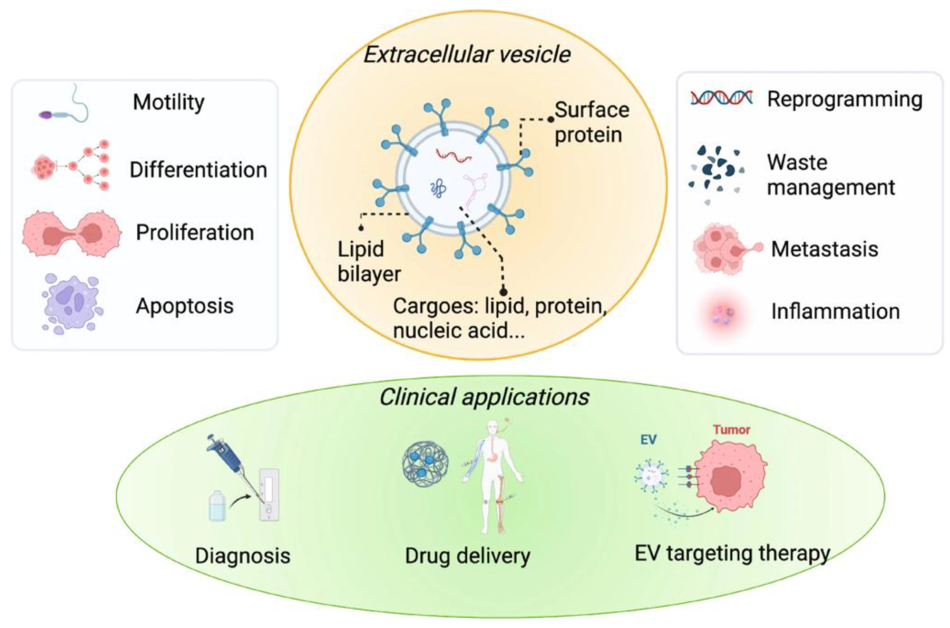
Figure 2.
Schematic illustration of extracellular vesicle functions in disease microenvironments, including cancer, neurodegeneration, and immune modulation. EVs mediate intercellular communication by transferring bioactive molecules, thereby influencing disease progression and immune response. Adapted and modified with permission from Anand et al., Cell Communication and Signaling (2023) [74].
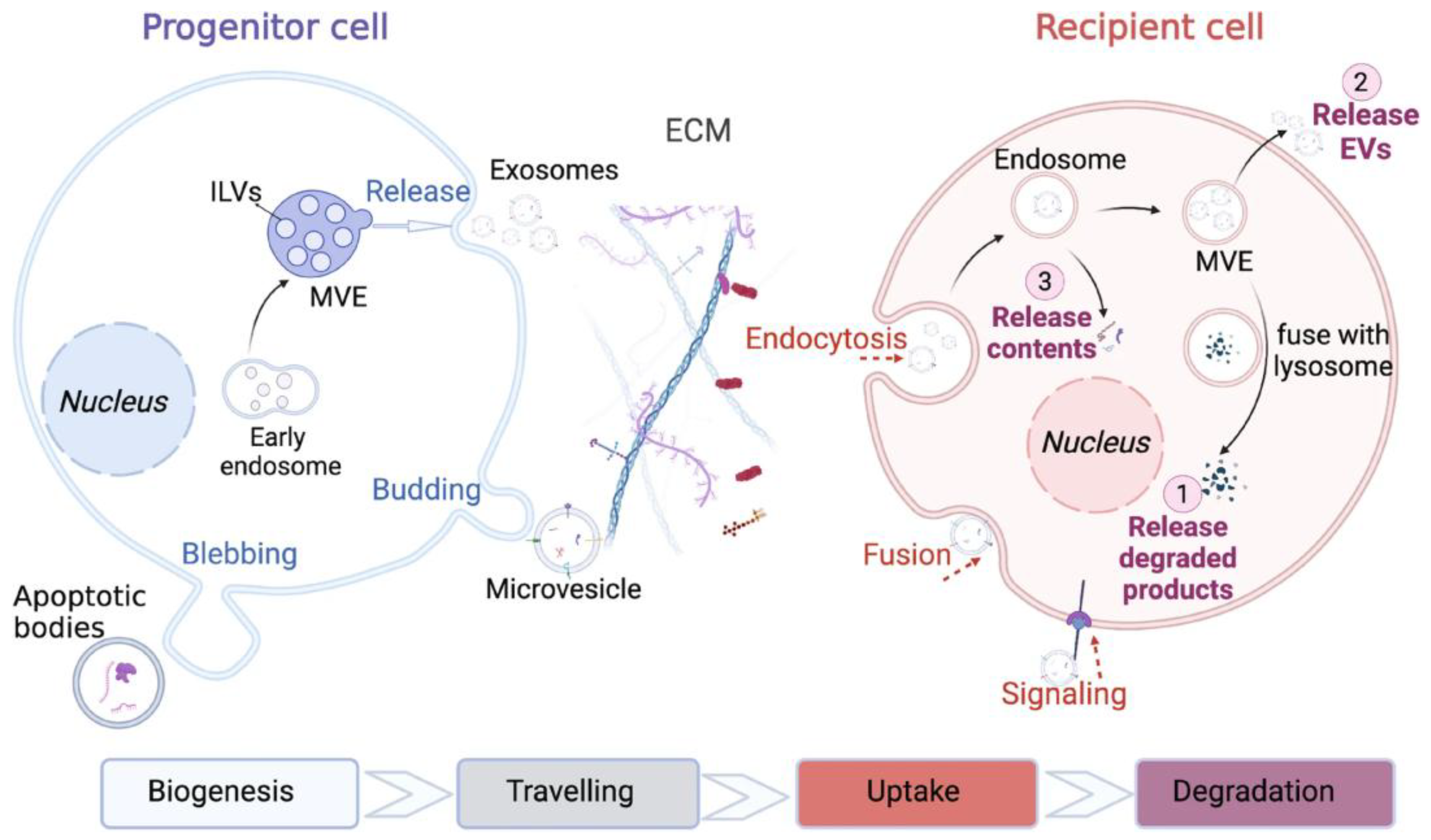
Disclaimer/Publisher’s Note: The statements, opinions and data contained in all publications are solely those of the individual author(s) and contributor(s) and not of MDPI and/or the editor(s). MDPI and/or the editor(s) disclaim responsibility for any injury to people or property resulting from any ideas, methods, instructions or products referred to in the content. |
© 2025 by the authors. Licensee MDPI, Basel, Switzerland. This article is an open access article distributed under the terms and conditions of the Creative Commons Attribution (CC BY) license (http://creativecommons.org/licenses/by/4.0/).
Copyright: This open access article is published under a Creative Commons CC BY 4.0 license, which permit the free download, distribution, and reuse, provided that the author and preprint are cited in any reuse.


