
Submitted:
05 June 2025
Posted:
06 June 2025
You are already at the latest version
Abstract
Brazil has a high abundance of monkey species, including Mico melanurus. In general, primates are vulnerable to parasites owing to their behavior. We aimed to identify the occurrence of gastrointestinal parasites in M. melanurus in the Brazilian Cerrado and to perform morphometric and morphological analyses of these helminths. Helminths of 21 M. melanurus were collected from Cuiabá in the Brazilian Cerrado. The animals were found dead within the urban perimeter, necropsied, and tested for parasites. Parasites found in the gastrointestinal tract were fixed in 70% ethanol for morphological identification. We identified Trypanoxyuris spp. in two samples (mean intensity of 63 and mean abundance of 6.3) and Primasubulura jacchi in the other 18 samples (mean intensity of 34.22 and mean abundance of 30.8). One sample was deteriorated; therefore, it was not necropsied. P. jacchi infections in same cases are asymptomatic or have gastrointestinal signs such as diarrhea. Trypanoxyuris spp. are commonly found in the primates of Mato Grosso. Only these two parasites were identified, provided by a small parasite richness, and were associated with the impact of habitat fragmentation in an anthropized area, indicating an imbalance in the environment in which they lived.
Keywords:
parasitic diseases
; Primasubulura
; primates
; Trypanoxyuris
1. Introduction
Brazil has one of the world's highest primate diversities, with 116 known species occurring in the country, corresponding to 48.4% of all primate diversity in the world [1]. According to the IUCN [2], approximately one-quarter of the mammals on the planet are threatened by extinction, with primates and ungulates being at the greatest risk. Primates present in Neotropical regions are predominantly arboreal and represent 40% of frugivorous vertebrate biomass in tropical forests. Thus, they constitute an essential group for maintaining plant structure and forest regeneration [3].
Primates are divided into two suborders, Strepsirrhini (which includes lemurs, galagos, and lorisids) and Haplorrhini (which includes tarsiers, monkeys, and humans). The Haplorrhini suborder is further divided into two infraorders: Tarsiiformes, which includes only tarsiers, and Simiiformes, which includes monkeys and humans. Monkeys are classified as New World monkeys, which are found only in the Americas, and Old World monkeys, which are found in Asia, Africa, and Europe. The New World monkey group includes the families Callitrichidae (marmosets and tamarins), Cebidae ( capuchin monkeys and squirrel monkeys), Nyctipithecidae (night monkeys), Pitheciidae (sauás, guigós, parauaçus, and uacaris), and Atelidae (howler monkeys or guari-bas, spider monkeys, woolly monkeys, and muriquis). Brazil has the greatest diversity of primates in the world, which is favored by its vast territorial extension and the fact that it is the largest country in the intertropical zone (92% of its territory is located in this range). Recent revisions of the Order Primates revealed that Brazil supports 129 primate species, with 150 terminal taxa belonging to 22 distinct genera. Thus, Brazil is home to more than a fifth of the world's known primates and 71% of the neotropical taxa. About 57% of the total taxa already recorded are endemic to the country; that is, they exclusively inhabit the Brazilian territory [4,5,6,7]
Mico melanurus, which belong to the Mico (Callitrichidae), presents pigmented ears and auricular surfaces, the absence of ear tufts, white or yellowish-white spots on the thighs, and a completely black tail, with color variations observed among different individuals [8]. Considerable similarities have been reported among specimens from Cáceres, Palmeiras, Rio Aricá, Santo Antônio do Leverger, Chapada dos Guimarães, Aripuanã (all areas from Brazil) and Água Dulce (Chaco, Paraguay). These specimens exhibited brown backs and thoracic limbs, whereas specimens from Corumbá and Rio Paraguai were visibly lighter.
The average group size of M. melanurus ranges from 4 to 15 individuals, but in Bolivia, this average is lower (2.5 +/- 2.1) [9,10]. Van Roosmalen [11] reported that this species has a wide geographic distribution, extending from the headwaters of the Madeira, Mamoré and Guaporé Rivers, to the Aripuanã and Juruena Rivers. Previously, M. melanurus was found only in Bolivia, Paraguay, and in the Mato Grosso and Mato Grosso do Sul states of Brazil [12]. However, Rímoli and Milagres [13] concluded that it is also found in Rondônia and the Amazon Forest.
Tamarin population has decreased by approximately 20 to 30% in the last three generations due to threats such as fires, rural settlements, urban expansion, agriculture, and livestock. Therefore, the species has been categorized as near threatened (NT), as it approaches the limits of the A4c criterion [13]. This implies that the population reduction data are distributed over a period of three generations or 10 years, with some of this decline occurring in the past and some of it projected into the future (up to a maximum of 100 years). The causes of this decline may be ongoing, poorly understood, or irreversible, and are linked to the decline in the area of occupancy, extent of occurrence, and/or habitat quality [14]. The high consumption of invertebrates indicates an important role of the species M. melanurus in the population control of these prey species. The diet of these primates includes fruits (one of the main sources of consumption for callitrichids), tree exudates (offered throughout the year without much seasonal interference), and seeds [15,16].
According to Ludlage and Mansfield [17], gastrointestinal diseases are the most common clinical manifestations in captive callitrichids. In addition, the prevalence of inflammatory bowel disease has been reported to be high (ranging from 28% to 60%), presenting with diarrhea, weight loss, enteritis, muscle atrophy, alopecia, and failure to thrive and may also be associated with hypoproteinemia, anemia, and elevated liver enzyme levels. Bacterial, viral, and parasitic pathogens cause gastrointestinal diseases in tamarins and marmosets, which may be the primary cause of clinical signs or may be related to other factors such as stress, diet, and inflammation [18]. However, data on the prevalence and diversity of intestinal parasites in Brazilian primate species, including callitrichid species, are scarce [19].
Primates are especially vulnerable to parasitic infections because of their social behavior, which facilitates the transmission of these pathogens. Additionally, several primate species are omnivorous and consume invertebrates, increasing the chances of trophic transmission. There are several parasites in non-human primates, some of which have adapted to their hosts, causing few health problems in these animals [19]. A total of 50 species of helminth parasites have been identified in 46 species of wild primates in Brazil, including Mico species. Most of the helminths identified include nematodes, representing 60% of the total. Acanthocephala accounted for 20%, Trematoda 12%, and Cestoda 8% of the helminths identified. Most Brazilian primate species (61%) have no records of helminth infections, indicating that the helminth fauna found in wild primates in Brazil is underestimated. Hence, additional studies are warranted to complement our understanding of helminthological biodiversity, as well as to provide a better estimate of parasitic infection [20].
A frequently reported nematode species in primates is Primasubulura jacchi, whose adult worms mainly inhabit the small intestine and cecum of the host, causing ulcerative lesions in the intestinal mucosa and diarrhea [21]. Melo [22] observed that when C. penicillata is maintained on a diet without insects, P. jacchi was gradually lost, indicating that insects are necessary for maintaining the life cycle of worms.
Barbosa et al. [23] investigated the presence of gastrointestinal parasites in captive non-human primates in the center of the state of Rio de Janeiro and obtained more than 1000 fecal samples from the individuals belong to the genera Mico, Callithrix, Saguinus and Leontopithecus. Among the samples analyzed, there was a prevalence of 56.7% of parasites in general, with the majority being protozoa and, to a lesser extent, helminths. In the samples from the genus Mico, nematode larvae were found in only one host.
It is important to study parasitic diseases and parasites that affect tamarins, including their identification and occurrence, because, in addition to impacting health and, consequently, affecting the conservation of these species, some of these parasites also exhibit zoonotic potential. Parasitic diseases in wild animals can trigger significant risks to humans and domestic animals, and monitoring these diseases is extremely necessary [24]. Here, we aimed to identify the occurrence of gastrointestinal parasites in M. melanurus in the Brazilian Cerrado and to perform morphometric and morphological analyses of the identified helminths.
2. Materials and Methods
This study was approved by the Animal Use Ethics Committee of the Federal University of Jataí (protocol no. 011/2022), according to Brazilian Federal Law No. 11,794/08, and was conducted on vertebrate animals. The animal collection license was granted by the Instituto Chico Mendes de Conservação da Biodiversidade (ICMBio) under registration No. 84201-3.
The sampling site was the municipality of Cuiabá located at 15°35′45″S and 56°05′49″W (Figure 1), within the Brazilian Cerrado. This municipality supports more than 650,000 inhabitants and has several green areas within the urban perimeter that wild animals and include parks, squares and banks of streams and rivers. Helminth samples were collected from 21 specimens of M. melanurus from the municipality of Cuiabá. We sampled the animals that were found dead within the urban perimeter. The animals likely came from small wooded areas; the cause of death was varied and ranged from attacks by other animals, malnutrition, diseases, and road accidents in 2021 and 2022. Helminth samples were stored in 70% alcohol, and morphological, morphometric, and taxonomic analyses were performed.
For morphometric and morphological analyses, the gastrointestinal parasites were hydrated and clarified in a lactophenol or 90% glicerol solution [25]. Temporary slides were prepared and analyzed under a bright field microscope (Olympus CX41) at 40x and 100x of magnification, and relevant anatomical structures, were recorded with the aid of a camera (Moticam 3.0®) attached to the microscope. Identification and taxonomic classification of the parasites were performed using taxonomic keys for nematodes [26,27]. The occurrence, intensity of parasitism, and population abundance were calculated as described by Bush et al. [28].
3. Results
Of the 21 samples obtained, one contained degenerated individual, and it was not possible to identify the genus and species; therefore, this sample was excluded from the data analysis. We identified Trypanoxyuris spp. in two samples (only females). In the remaining 18 samples, we recovered male and female P. jacchi. The morphological details of the helminths are shown in Figure 2. The specimens were deposited in the Helminthological Collection of the Federal University of Jataí according to the codes provided in Table 1. In addition to the species and genus reported, the number of individuals and hosts of each sample (Table 1).
The mean intensity of parasitism by P. jacchi was 34.22, whereas that of Trypanoxyuris spp. was 63. The mean abundance of P. jacchi was 30.8 and of Trypanoxyuris spp. was 6.3. Table 2 and Table 3 show the morphometric characterization of the parasites identified P. jacchi and Trypanoxyuris spp., respectively, along with the corresponding values reported in other studies for comparison.
4. Discussion
Parasitic diseases are the most common conditions in non-human primates, whether in captivity or in wild [31,32]. Therefore, information about the parasites present in free-living tamarins and their characteristics, in addition to the average abundance and intensity of parasitism, is of great importance. It can help improve our understanding their impact on the health of the host and the environment in which they live, especially in anthropized areas with restricted natural habitats, such as in the municipality of Cuiabá. Information about parasitism in Mico species remains scarce [23,33]. In the present study, we focused on identifying gastrointestinal parasites in as many M. melanurus individuals as possible. We selected this species because it is native to Cerrado and has been identified as vulnerable. Moreover, its habitat has been extremely fragmented owing to human action, resulting in the species often being restricted to small areas of forest within urban environments.
Primasubulura jacchi are nematodes belonging to the order Ascaridida and the family Subluridae, whose infestations are frequently reported in primates and are considered the most prevalent in this group [21]. In the present study, we observed a high occurrence of these nematodes, which can be partly explained by the feeding habits of M. melanurus; it consumes large quantities of invertebrates, which may be necessary to maintain the worm's life cycle [15,22]. Thus, this species has an indirect cycle, as arthropods, including cicadas, grasshoppers, and cockroaches can serve as intermediate hosts for helminths [33,34]. The occurrence of 90% of the tamarins in this study was similar to that reported in other primates; for example, in Callithrix, an occurrence of approximately 80% has been reported [35]. In some cases of parasitism by P. jacchi, diarrhea and ulcerative lesions in the intestinal mucosa may occur [21]; however, asymptomatic cases have been described [35], suggesting variations in clinical conditions. Symptomatic variation may be based on ]] the intensity of parasitism; however, the data presented in the literature to date are insufficient to confirm this.
Given that parasitism by P. jacchi can be asymptomatic, this may favor the maintenance of the parasite within the hosts and its consequent spread. Asymptomatic carriers can become reservoirs of the disease [36], which would also justify its high prevalence in these small primates. In the parasite samples included in the study, the number of individuals varied greatly, ranging from 1 to 132, with a mean intensity of 34.22 individuals per positive sample.
The prevalence of Trypanoxyuris spp. was 10%, and its identification was of utmost importance because nematodes of the Oxyuridae family trigger small lesions in the intestinal mucosa of their hosts, which can diffuse and be accompanied by an inflammatory response during severe infestations. Its life cycle is direct, and infection occurs through the ingestion of eggs present in the perianal skin deposited by adult females. In addition, retroinfection may occur when newly hatched larvae enter the host directly through the rectum [37,38].
Only female individuals were found in the Trypanoxyuris samples, which may be because male individuals are very small in size and therefore may not be found during sample collection and can be easily destroyed if the content is not analyzed quickly, thus favoring the collection of only female individuals in the collection of samples from the cecum of the hosts [26,27]. The number of individuals varied from 11 to 115, providing a mean intensity of 63 individuals per sample, with an extremely low mean abundance (6.3 helminths per sampled animal) due to the low number of positive animals. The occurrence of Trypanoxyuris minutus was recorded in two Alouatta guariba clamitans individuals in the state of Minas Gerais, which after necropsy, exhibited a mean intensity of 6,753 ± 490.73 parasites in the cecum, and a total of 6,650 parasites were reported in the rest of the large intestine [30]. These intensities were extremely discrepant in relation to our results and may be due to differences in host susceptibility and/or because they are parasites of different species of the Trypanoxyuris genus. The occurrence of Trypanoxyuris in M. melanurus has already been reported [39], who identified Trypanoxyuris callithricis in primates of the genera Sapajus and Mico, including four individuals of M. melanurus from three different locations (Chapada dos Guimarães, Cuiabá, and Poconé), suggesting that this genus of parasites is commonly found in other locations in Mato Grosso. Since T. callithricis is commonly observed infecting Callithrix jacchus and other species of the same primate genus [26], in addition to possibly having been adapted to parasitize primate infracommunities in the Amazon and Cerrado biomes [39], it may be the same species as in our study; however, because of the absence of males, the species cannot be confirmed solely by morphological and morphometric analyses of females. This is due to the fact that females of the Trypanoxyuris genus are similar even in different species, requiring structures of males, such as gubernaculum and spicules, which have individual characteristics, allowing the distinction of species.
The mean abundance of parasites is a quantitative parameter of epidemiological importance that helps understand the parasite-host relationship. It is considered multifactorial in nature and its variation may occur due to biotic and abiotic factors [28,40,41]. The most recommended way to study patterns related to parasite abundance is to use databases that include hosts from the same location, thus minimizing the impact of geographic variations and allowing the selection of common biotic characteristics [42]. The data used in this study fit this requirement, favoring a more reliable assessment of parasite abundance. Our results showed that P. jacchi presented a higher abundance value than that obtained from the genus Trypanoxyuris, indicating that this species has a greater capacity for dispersing parasites. The genus Trypanoxyuris has been described as non-pathogenic and common in primates [43], but there have been reports of death in spider and owl monkeys that were severely infested by this nematode [44,45]. In marmosets, T. callithricis causes intense parasitism in its hosts, triggering severe malnutrition and pulmonary edema as a consequence of hypoproteinemia [46]. It has been suggested that this genus causes a more serious condition that can lead to the death of its host, thus reducing its dispersion rate. Furthermore, individuals of this genus present a direct cycle, that is, the absence of intermediate hosts involved in the development of the parasite, unlike P. jacchi, which possibly has this type of host that favors its dissemination [22,38]. The mean intensity was higher for the genus Trypanoxyuris compared to the value obtained for the species P. jacchi, thus suggesting that this genus triggers more serious infestations for the tamarins [28,38].
Co-infections are considered temporally dynamic; therefore, it is more likely that two parasites infect the same host sequentially rather than simultaneously, because, in general, they are considered competitive for limited host resources. In addition, host exposure to different parasites varies over time; that is, there is spatiotemporal variation in the interactions between parasites and hosts in the wild, which may explain why all hosts evaluated in the present study presented with a single helminth type [47,48].
Most human and domestic animal pathogens in Europe are sensitive to climate, and often to more than one climatic factor, such as temperature, precipitation, and humidity. With global warming, rising temperatures are expected to accelerate the development of environmental parasitic stages (oocysts, eggs, and larvae), increase the abundance of poikilothermic intermediate hosts, and accelerate parasite maturation [49,50]. This climatic influence may have favored the greater abundance of P. jacchi in the primates in our sample, which may have used invertebrates to disseminate. Additionally, the region in which they live has a naturally predominantly semi-humid tropical climate [51].
Natural habitats have disappeared or become extremely modified, reduced, and fragmented, thereby influencing the relationship between wild animals, domestic animals, and humans, and favors the emergence of diseases [52]. The set of environmental changes triggered by anthropogenic pressure allows for increased contact between pathogenic species and new hosts, leading to an increase in infectious diseases and zoonoses [53]. Despite all the factors that favor the transmission of diseases between these small primates, tamarins, and humans, no species of zoonotic gastrointestinal parasites were identified in the present study; however, the restricted number of helminth species (only two) indicated that habitat fragmentation interferes with relationships within the biome, which suggests increased pressure on native groups, whether hosts or parasites, In addition to the disappearance of subordinate species, adaptations may arise in relationships that manage to survive in a fragmented environment. Corroborating our low parasite richness, we expected to report the occurrence of acanthocephalans and/or Spirurida nematodes, which have already been described in several primate species in the literature [20]. Furthermore, these parasites have an indirect life cycle, with vertebrates as definitive hosts and invertebrates as intermediate hosts [54,55]. Thus, M. melanurus, by including invertebrates in its diet, would functions as a definitive host by ingesting infected intermediate hosts. Although the diet favored the occurrence of acanthocephalans and Spirurida nematodes, they were not identified in the samples of this primate species in our study.
5. Conclusions
It can be concluded that in the sampled individuals of M. melanurus, the mean abundance of P. jacchi was higher than that of the genus Trypanoxyuris, which can be attributed to both the biological characteristics of the parasites and the pathogenesis triggered in the host. Regarding the mean intensity, the results were the opposite; the value was higher for Trypanoxyuris sp. than for P. jacchi, suggesting that the parasite causes more severe conditions in its host. Additionally, only these two parasites were identified, provided by a small parasite richness, and were associated with the impact of habitat fragmentation in an anthropized area, indicating an imbalance in the environment in which they lived.
Author Contributions
Conceptualization, L.C.Z, R.C.P. and D.G.S.R.; methodology, L.C.Z., L.F.S., Z.M.A.S., N.F.U., V.L.B.S. and I.S.M.; validation, K.C.S., Í.A.B., E.M.C., R.C.P. and D.G.S.R.; formal analysis, H.T.F., K.C.S., E.M.C. R.C.P. and D.G.S.R.; investigation, L.C.Z., V.L.B.S., E.M.C., R.C.P. and D.G.S.R.; resources, E.M.C, R.C.P. and D.G.S.R.; data curation, E.M.C, R.C.P. and D.G.S.R.; writing—original draft preparation, L.C.Z., L.F.S., Z.M.A.S., N.F.U., V.L.B.S. and I.S.M.; writing—review and editing, H.T.F., K.C.S., Í.A.B., E.M.C. R.C.P. and D.G.S.R.; visualization, H.T.F., K.C.S., Í.A.B., E.M.C. R.C.P. and D.G.S.R..; supervision, K.C.S., E.M.C. and R.C.P.; project administration, D.G.S.R.; funding acquisition, E.M.C, R.C.P. and D.G.S.R.
Funding
This research was funded by Fundação de Amparo à Pesquisa do Estado de Goiás (FAPEG), grant number 202310267001408; Conselho Nacional de Desenvolvimento Científico e Tecnológico (CNPq), research grant for R.C.P and D.G.S.R., and scholarship for N.F.U.; and Coordenação de Aperfeiçoamento de Pessoal de Nível Superior (CAPES), scholarship for L.F.S., Z.M.A.S, V.L.B.S and I.S.M.
Institutional Review Board Statement
This study was approved by the Animal Use Ethics Committee of the Federal University of Jataí (protocol No. 011/2022), according to Brazilian Federal Law No. 11,794/08 attributed to research with vertebrate animals. The animal collection license was granted by Instituto Chico Mendes de Conservação da Biodiversidade (ICMBio) under registration No. 84201-3.
Data Availability Statement
The original contributions presented in this study are included in the article/supplementary material. Further inquiries can be directed to the corresponding author(s).
Acknowledgments
To Fundação de Amparo à Pesquisa do Estado de Goiás (FAPEG), Conselho Nacional de Desenvolvimento Científico e Tecnológico (CNPq), and Coordenação de Aperfeiçoamento de Pessoal de Nível Superior (CAPES) for financial support.
Conflicts of Interest
The authors declare no conflicts of interest.
References
- Reis, N.R.; Peracchi, A.L.; Batista, C.B.; Rosa, G.L.M. Primatas Brasileiros: guia de campo, 1st ed.; Technical Books: Rio de Janeiro, Brazil, 2015; p. 328. [Google Scholar]
- IUCN. Primates in peril, 2012. Available online: http://www.iucnredlist.org/news/primates-in-peril (accessed on 23 March 2023).
- Culot, L.; Mann, D.J.; Munoz, F.J.J.L.; Huynen, M.C.; Heymann, E.W. Tamarins and dung beetles: an efficient diplochorous dispersal system for Forest regeneration. Biotropica 2010, 43, 84–92. [Google Scholar] [CrossRef]
- Jerusalinsky, L.; Melo, F.R. Conservação de Primatas no Brasil: perspectivas e desafios. In La primatología en Latinoamérica 2 – A primatologia na America Latina 2 Tomo II Costa Rica-Venezuela, 2nd ed.; Urbani, B., Kowalewski, M., Cunha, R.G.T., Torre, S., Cortés-Ortiz, L., Eds.; Instituto Venezolano de Investigaciones Científicas (IVIC): Caracas, Venezuela, 2018; pp. 161–186. [Google Scholar]
- Wilson, D.E.; Reeder, D.M. Mammal Species of the World: A Taxonomic and Geographic Reference, 3rd ed.; Johns Hopkins University Press: Baltimore, United States of America, 2005; pp. 111–184. [Google Scholar] [CrossRef]
- Committee on Well-Being of Nonhuman Primates Institute for Laboratory Animal Research Commission on Life Sciences National Research Council. The Psychological Well-Being of Nonhuman Primates, 1st ed.; National Academy Press: Washington, United States of America, 1998; pp. 68–80. [Google Scholar]
- Rylands, A.B.; Doris, S.F. Marmosets and Tamarins: Systematics, Behaviour, and Ecology, 1st ed.; Oxford Science Publication: Oxford, England, 1993; pp. 262–272. [Google Scholar] [CrossRef]
- Vivo, M. Taxonomia de Callithrix Erxleben, 1777 (Callitrichidae, Primates), 1st ed.; Fundação Biodiversitas: Belo Horizonte, Brasil, 1991. [Google Scholar]
- Pyritz, L.; Buntge, A.; Herzog, S.; Kessler, M. Effects of habitat structure and fragmentation on diversity and abundance of primates in tropical decíduos forests in Bolívia. Int. J. Primatol. 2010, 31, 796–812. [Google Scholar] [CrossRef]
- Milagres, A.P.; Rímoli, J.; Santos, M.C.; Wallace, R.B.; Rumiz, D.I.; Mollinedo, J.; Rylands, A.B. Mico melanurus (amended version of 2020 assessment). The IUCN Red List of Threatened Species 2021, e.T136294A192400781. [Google Scholar] [CrossRef]
- Van Roosmalen, M.G.M.; Van Roosmalen, T.; Mittermeier, R.A.; Rylands, A.B. Two new species of marmoset, genus Callithrix Erxleben, 1777 (Callithrichidae, Primates) from the Tapajós/Madeira interfluvium, South Central Amazonia, Brazil. Neotrop. Primates 2000, 8, 2–18. [Google Scholar] [CrossRef]
- Noronha, M.A.; Spironello, W.R.; Ferreira, D.C. New occurrence records for Mico melanurus (Primates, Callitrichidae). Neotrop. Primates 2008, 15, 26–28. [Google Scholar] [CrossRef]
- Rímoli, J.; Milagres, A.P. Mamíferos—Mico melanurus—Sagui Marrom. Avaliação do Risco de Extinção de Mico melanurus (É. Geofrroy Em Humboldt, 1812) No Brasil. Processo de Avaliação do Risco de Extinção Da Fauna Brasileira. ICMBio, 2015.
- Instituto Chico Mendes de Conservação da Biodiversidade (ICMBio). Livro Vermelho da Fauna Brasileira Ameaçada de Extinção, Vol. I, 1st ed.; ICMBio/MMA492: Brasilia, Brazil, 2018; p. 492. [Google Scholar]
- Sauer, A.C.L.; Barroso, W.A.; Santos, U.F.; Portela, J.L.; Machado, A.F.; Camera, B.F.; Canale, G.R. Efeito da sazonalidade sobre o padrão comportamental de um grupo de saguis-do-rabo-preto (Mico melanurus) em um fragmento florestal urbano. In A Primatologia no Brasil, 1st ed.; Silva, V.L., Ferreira, R.G., Oliveira, M.A., Eds.; Sociedade Brasileira de Primatologia: Recife, Brazil, 2017; Volume 14, pp. 266–276. [Google Scholar]
- Martins, I.G. Padrão de atividade do sagui Callithrix jacchus numa área de Caatinga. Master's dissertation, Universidade Federal do Rio Grande do Norte, Natal, 2007.
- Ludlage, E.; Mansfield, K. Clinical care and diseases of the common marmoset (Callithrix jacchus). Comp. Med. 2003, 53, 369–382. [Google Scholar] [PubMed]
- Fitz, C.; Goodroe, A.; Wierenga, L.; Mejia, A.; Simmons, H. Clinical Management of Gastrointestinal Disease in the Common Marmoset (Callithrix jacchus). ILAR J. 2020, 61, 199–217. [Google Scholar] [CrossRef]
- Catenacci, L.S.; Oliveira, J.B.S.; Vleeschouwer, K.M.; Carvalho Oliveira, L.; Deem, S.L.; Sousa Júnior, S.C.D.; Santos, K.R.D. Gastrointestinal parasites of Leontopithecus chrysomelas in the Atlantic Forest, Brazil. Rev. Bras. Parasitol. Vet. 2022, 31. [Google Scholar] [CrossRef]
- Corrêa, P.; Bueno, C.; Soares, R.; Vieira, F.M.; Muniz-Pereira, L.C. Checklist of helminth parasites of wild primates from Brazil. Rev. Mex. Biodivers. 2016, 87, 908–918. [Google Scholar] [CrossRef]
- Resende, D.M.; Pereira, L.H.; Melo, A.L.; Tafuri, W.L.; Moreira, N.B.; Oliveira, C.L. Parasitism by Primasubulura jacchi (Marcel, 1857) Inglis, 1958 and Trichospirura leptostoma Smith and Chitwood, 1967 in Callithrix penicillata marmosets, trapped in wild environment and maintained in captivity. Mem. Inst. Oswaldo Cruz 1994, 89, 123–125. [Google Scholar] [CrossRef]
- de Melo, A.L. Helminth parasites of Callithrix geoffroyi. Lab. Primate News 2004, 43, 7–9. [Google Scholar]
- Barbosa, A.S.; Pissinatti, A.; Dib, L.V.; Siqueira, M.P.; Cardozo, M.L.; Fonseca, A.B.M.; Amendoeira, M.R.R. Balantidium coli and other gastrointestinal parasites in captives non-human primates of Rio de Janeiro, Brazil. J. Med. Primatol. 2015, 44, 18–26. [Google Scholar] [CrossRef] [PubMed]
- Galecki, R.; Sokol, R.; Koziatek, S. Parasites of wild animals as a potential source of hazard to humans. Annals of Parasitology 2015, 61. [Google Scholar]
- Hoffman, R.P. Diagnóstico de parasitismo veterinário, 1st ed.; Sulina: Porto Alegre, Brazil, 1987; p. 156. [Google Scholar]
- Vicente, J.J.; Rodrigues, H.O.; Gomes, D.C.; Pinto, R.M. Nematóides do Brasil. Parte V: nematóides de mamíferos. Rev. Brasil. Zoo. 1997, 14, 1–452. [Google Scholar] [CrossRef]
- Anderson, R.C.; Chabaud, A.G.; Willmott, S. Keys to the nematode parasites of vertebrates: archival volume, 1st ed.; Cab international: Oxfordshire, England, 2009. [Google Scholar]
- Bush, A.O.; Lafferty, K.D.; Lotz, J.M.; Shostak, A.W. Parasitology meets ecology on its own terms: Margolis et al. revisited. J. Parasitol. 1997, 83, 575–583. [Google Scholar] [CrossRef]
- Rocha, B.M. Taxonomia de nematóides parasitos de primatas neotropicais, Callithrix penicillata (Geoffroy, 1812)(Primata: Callitrichidae), Alouatta guariba (Humboldt, 1812) (Primata: Atelidae) e Sapajus apella (Linnaeus, 1758) grooves, 2005 (Primata: Cebidae), do estado de Minas Gerais, 2014. Master 's dissertation, Universidade Federal de Juiz de Fora, Juiz de Fora, 2014.
- Souza, D.D.P.; Magalhães, C.M.F.R.; Vieira, F.M.; Souzalima, S. Ocorrência de Trypanoxyuris (Trypanoxyuris) minutus (Schneider, 1866) (Nematoda, Oxyuridae) em Alouatta guariba clamitans Cabrera, 1940 (Primates, Atelidae) em Minas Gerais, Brasil. Rev. Bras. Parasitol. Vet. 2010, 19, 124–126. [Google Scholar] [CrossRef]
- Verona, C.E.D.S. Parasitos em sagui-de-tufo-branco (Callithrix jacchus) no Rio de Janeiro. Doctorate’s dissertation, Escola Nacional de Saúde Pública Sergio Arouca, Rio de Janeiro, 2008.
- Strait, K.; Elese, J.G.; Eberhard, M.L. Parasitic diseases of nonhuman primates. In Nonhuman primates in biomedical research, 2nd ed.; Abee, C.R., Mansfield, K., Tardif, S.D., Morris, T., Eds.; Elsevier: Canada, 2012; pp. 197–297. [Google Scholar]
- Cândido, S.L.; Pereira, N.A.; Fonseca, M.J.O.R.; Pacheco, R.C.; Morgado, T.O.; Colodel, E.M.; Nakazato, L.; Dutra, V.; Vieira, T.S.W.J.; Aguiar, D.M. Molecular detection and genetic characterization of Ehrlichia canis and Ehrlichia sp. in neotropical primates from Brazil. Ticks Tick Borne Dis. 2023, 14, 102179. [Google Scholar] [CrossRef]
- Alonso, C.; Langguth, A. Ecologia e comportamento de Callithrix jacchus (Primates: Callitrichidae) numa ilha de floresta atlântica. Rev. Nordestina Biol. 1989, 6, 105–137. [Google Scholar]
- Tavela, A.D.O.; Fuzessy, L.F.; Silva, V.H.D.; Silva, F.F.R.; Junior, M.C.; Silva, I.D.O.; Souza, V.B. Helmintos de saguis (Callithrix sp.) híbridos de vida livre vivendo em ambientes com alta atividade humana. Rev. Bras. Parasitol. Vet. 2013, 22, 391–397. [Google Scholar] [CrossRef]
- Brasil. Ministério da Saúde. Secretaria de Atenção à Saúde. Protocolos clínicos e diretrizes terapêuticas. Ministério da Saúde: Brasília 2010; 3.
- Urquhart, G.M.; Armour, J.; Duncan, J.L.; Dunn, A.M.; Jennings, F.W. Parasitologia veterinária, 2nd ed.; Guanabara Koogan: Rio de Janeiro, Brazil, 1998; p. 273. [Google Scholar]
- Felt, S.A.; White, C.E. Evaluation of a timed and repeated perianal tape test for the detection of pinworms (Trypanoxyuris microon) in owl monkeys (Aotus nancymae). J. Med. Primatol. 2005, 34, 209–214. [Google Scholar] [CrossRef]
- Ramos, D.G.S.; Santos, A.R.G.L.O.; Freitas, L.C.; Correa, S.H.R.; Kempe, G.V.; Morgado, T.O.; Aguiar, D.M.; Wof, R.W.; Rossi, R.V.; Pacheco, R.C. Endoparasites of wild animals from three biomes in the State of Mato Grosso, Brazil. Arq. Bras. Med. 2016, 68, 571–578. [Google Scholar] [CrossRef]
- Poulin, R. Are there general laws in parasite ecology? Parasitol. 2007, 134, 763–76. [Google Scholar] [CrossRef]
- Vignon, M.; Sasal, P. Multiscale determinants of parasite abundance: a quantitative hierarchical approach for coral reef fishes. Int. J. Parasitol. 2010, 40, 443–451. [Google Scholar] [CrossRef] [PubMed]
- Amarante, C.F.; Tassinari, W.S.; Luque, J.L.; Pereira, M.J.S. Factors associated with parasite aggregation levels in fishes from Brazil. Rev. Bras. Parasitol. Vet. 2015, 24, 174–182. [Google Scholar] [CrossRef]
- Tantaleán, M.; Gozalo, A. Parasites of the Aotus monkey. In Aotus: the owl monkey, 1st ed.; Baer, J.F., Weller, R.E., Kakoma, I., Eds.; Academic Press: San Diego, United States of America, 1994; pp. 353–374. [Google Scholar]
- Fiennes, R.N. Pathology of simian primates Part II: infectious and parasitic diseases, 1st ed.; S. Karger: London, England, 1972; p. 770. [Google Scholar]
- Sánchez, N.; Galvez, H.; Montoya, E.; Gozalo, A. Mortalidad en crías de Aotus sp. (Primates: Cebidae) en cautiverio: una limitante para estudios biomédicos con modelos animales. Rev. Peru. Med. Exp. Salud Publica 2006, 23, 221–224. [Google Scholar]
- Dias, R.F.F. Doenças de primatas não humanos de vida livre e em cativeiro no nordeste do Brasil. Master 's dissertation, Universidade Federal da Paraíba, Paraíba, 2021.
- Jousimo, J.; Tack, A.J.; Ovaskainen, O.; Mononen, T.; Susi, H.; Tollenaere, C.; Laine, A.L. Disease ecology. Ecological and evolutionary effects of fragmentation on infectious disease dynamics. Science 2014, 13. [Google Scholar] [CrossRef]
- Karvonen, A.; Jokela, J.; Laine, A.L. Importance of Sequence and Timing in Parasite Coinfections. Trends Parasitol. 2019, 35, 109–118. [Google Scholar] [CrossRef]
- Mcintyre, K.M.; Setzkorn, C.; Hepworth, P.J.; Morand, S.; Morse, A.P.; Babylis, M. Systematic Assessment of the Climate Sensitivity of Important Human and Domestic Animals Pathogens in Europe. Sci. Rep. 2017, 7, 7134. [Google Scholar] [CrossRef]
- Poglayen, G.; Gelati, A.; Scala, A.; Naitana, S.; Musalla, V.; Nocerino, M.; Cringoli, G.; Regalbono, A.F.D.; Habluetzel, A. Do natural catastrophic events and exceptional climatic conditions also affect parasites? Parasitol. 2023, 150. [Google Scholar] [CrossRef]
- IBGE – INSTITUTO BRASILEIRO DE GEOGRAFIA E ESTATÍSTICA. Available online: https://www.ibge.gov.br/estatisticas/sociais/saude/22827-censo-demografico-2022.html (accessed on 1 December 2024).
- Alho, C.J.R. Importância da biodiversidade para a saúde humana: uma perspectiva ecológica. Estud. Av. 2012, 26, 151–166. [Google Scholar] [CrossRef]
- Câmara, V.M.; Tambellina, A.T.; Castro, H.A.; Waissmann, W. Saúde Ambiental e Saúde do Trabalhador: epidemiologia das relações entre a produção, o ambiente e a saúde. In Epidemiologia e Saúde, 6th ed.; MEDSI: Rio de Janeiro, Brazil, 2003; pp. 469–497. [Google Scholar]
- Seip, D.R.; Bunnell, F.L. Foraging behaviour and food habits of Stone's sheep. Can. J. Zoo. 1985, 49, 1638–1646. [Google Scholar] [CrossRef]
- Amin, O.M. Classification of the Acanthocephala. Folia Parasitol. 2013, 60, 273–305. [Google Scholar] [CrossRef] [PubMed]
Figure 1.
Location of the municipality of Cuiabá, Brazil, in central South America.
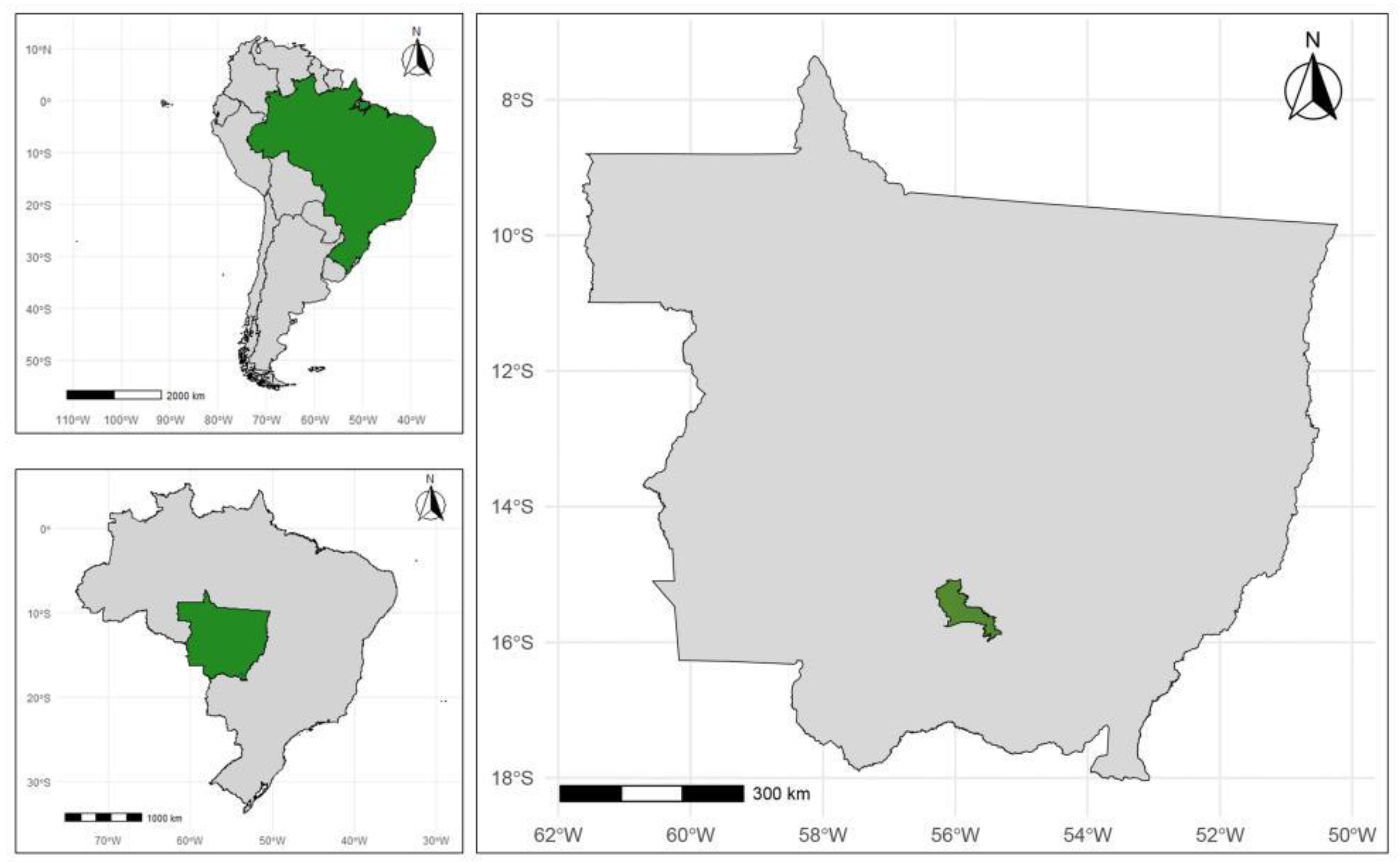
Figure 2.
Bright-field light microscopy photomicrographs of helminth samples obtained from Mico melanurus individuals from Cuiabá, Brazil (Cerrado biome). A) Anterior portion of Primasubulura jacchi; white arrow marks the buccal opening and buccal capsule; B) Anterior portion of P. jacchi; me=muscular esophagus, and b=esophageal bulb; C) Posterior end of female P. jacchi; a=anus; D) Posterior end of male P. jacchi; s=spicule, ps=precloacal sucker; E) Anterior portion of female Trypanoxyuris spp.; ca=cervical alae, and b=esophageal bulb; F) Median portion of female Trypanoxyuris spp. showing a uterus full of eggs.
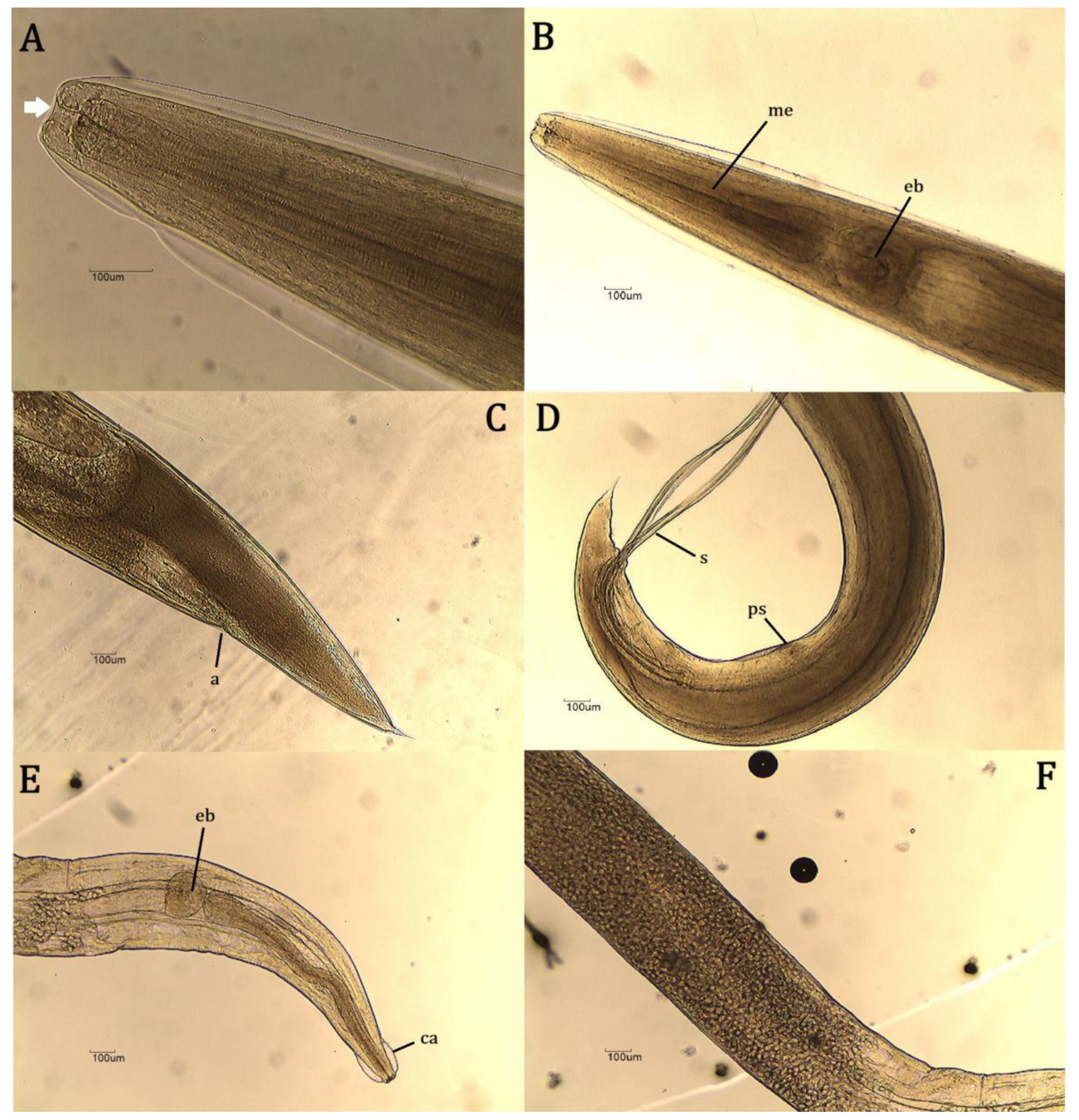

Table 1.
Occurrence of nematodes in Mico melanurus from the municipality of Cuiabá, in the Brazilian Cerrado, along with their deposit codes in the Helminthological Collection of the Federal University of Jataí (CHUFJ).
Table 1.
Occurrence of nematodes in Mico melanurus from the municipality of Cuiabá, in the Brazilian Cerrado, along with their deposit codes in the Helminthological Collection of the Federal University of Jataí (CHUFJ).
| Sample number | Helminth species/genus | Number of individuals |
|---|---|---|
| CHUFJ - 0016 | Primasubulura jacchi | 1 |
| CHUFJ - 0017 | P. jacchi | 2 |
| CHUFJ - 0018 | P. jacchi | 5 |
| CHUFJ - 0019 | Trypanoxyuris spp. | 115 |
| CHUFJ - 0020 | P. jacchi | 9 |
| CHUFJ - 0021 | P. jacchi | 49 |
| CHUFJ - 0022 | P. jacchi | 7 |
| CHUFJ - 0023 | P. jacchi | 2 |
| CHUFJ - 0024 | P. jacchi | 15 |
| CHUFJ - 0025 | P. jacchi | 66 |
| CHUFJ - 0026 | Trypanoxyuris spp. | 11 |
| CHUFJ - 0027 | P. jacchi | 25 |
| CHUFJ - 0028 | P. jacchi | 1 |
| CHUFJ - 0029 | P. jacchi | 5 |
| CHUFJ - 0030 | P. jacchi | 94 |
| CHUFJ - 0031 | P. jacchi | 66 |
| CHUFJ - 0032 | P. jacchi | 38 |
| CHUFJ - 0033 | P. jacchi | 2 |
| CHUFJ - 0034 | P. jacchi | 132 |
| CHUFJ - 0035 | P. jacchi | 97 |
Table 2.
Morphometric characterization of females and males of Primasubulura jacchi with mean ± standard deviation (SD), in micrometers.
Table 2.
Morphometric characterization of females and males of Primasubulura jacchi with mean ± standard deviation (SD), in micrometers.
| Measurements (micrometers) | Present Study | Rocha (2014) [29] | ||
|---|---|---|---|---|
| Males | Females | Males | Females | |
| Length | 14.100 ± 1.200 | 23.000 ± 6.000 | 12.800 ± 2.644 | 13.235 ± 7.291 |
| Width | 477 ± 111 | 658 ± 163 | 542 ± 180 | 750 ± 264 |
| Buccal capsule length | 50 ± 12 | 54 ± 9,5 | 34,5 ± 9,5 | 45 ± 10 |
| Buccal capsule width | 37 ± 7,8 | 47 ± 18 | 24 ± 9 | 23 ± 13 |
| Esophagus length | 1.238 ± 121 | 1.345 ± 221 | 991,5 ± 54,5 | 1.140 ± 119 |
| Bulb length | 267 ± 32 | 267 ± 32 | 248,5 ± 31 | 272,5 ± 71 |
| Bulb width | 225 ± 40 | 270 ± 63 | 245 ± 42 | 243,5 ± 74 |
| Spike length | 1.892 ± 249 | - | 1.542 ± 288 | - |
| Gubernaculum length | 195 ± 22,5 | - | 198 ± 15 | - |
| Distance from cloaca to posterior portion | 290,5 ± 19 | - | - | - |
| Pre-cloacal sucker length | 65 ± 22 | - | 214 ± 32 | - |
| Cloaca to suction cup distance | 584 ± 227,5 | - | - | - |
| Nerve ring length | - | 42 ± 18 | - | - |
| Nerve ring width | - | 161 ± 68 | - | - |
| Distance from anus to posterior portion | - | 902 ± 187 | - | - |
| Egg length | - | 66 ± 13 | - | 62 ± 8,5 |
| Egg width | - | 50 ± 11 | - | 52 ± 9 |
* (-) corresponds to a missing measurement. Measurement of the length and width of the nerve ring (N = 2) and other measurements (N=10).
Table 3.
Morphometric characterization of females of Trypanoxyuris spp.; with mean ± standard deviation (SD), in micrometers.
Table 3.
Morphometric characterization of females of Trypanoxyuris spp.; with mean ± standard deviation (SD), in micrometers.
| Measurements (micrometers) | Our Study | Rocha (2014) [29] | Souza et al. (2010) [30] |
|---|---|---|---|
| Length | 7.500 ± 1.000 | 6.650 ± 911 | 6.650 ± 690 |
| Width | 351 ± 87 | 311 ± 40 | 307 ± 45 |
| Esophagus length | 984 ± 119 | 1.600 ± 80 | 1.600 ± 50 |
| Bulb length | 136 ± 17 | - | - |
| Bulb width | 122 ± 17 | - | - |
| Nerve ring length | 44 ± 19,8 | - | - |
| Nerve ring width | 115 ± 7,8 | - | - |
| Distance from anus to posterior portion | 1.159 ± 629 | 1.470 ± 82 | 1.470 ± 90 |
| Egg length | 42,5 ± 6,2 | 47 ± 2,8 | 47 ± 2,8 |
| Egg width | 23,5 ± 1,8 | 23,5 ± 1 | 23,5 ± 0,8 |
| Vulva to the anterior portion | 1.538 ± 262 | 2.600 ± 270 | 2.600 ± 230 |
* (-) corresponds to a measurement not performed. Measurement of the length and width of the nerve ring (N = 2) and other measurements (N=10).
Disclaimer/Publisher’s Note: The statements, opinions and data contained in all publications are solely those of the individual author(s) and contributor(s) and not of MDPI and/or the editor(s). MDPI and/or the editor(s) disclaim responsibility for any injury to people or property resulting from any ideas, methods, instructions or products referred to in the content. |
© 2025 by the authors. Licensee MDPI, Basel, Switzerland. This article is an open access article distributed under the terms and conditions of the Creative Commons Attribution (CC BY) license (http://creativecommons.org/licenses/by/4.0/).
Copyright: This open access article is published under a Creative Commons CC BY 4.0 license, which permit the free download, distribution, and reuse, provided that the author and preprint are cited in any reuse.


