Submitted:
29 May 2025
Posted:
29 May 2025
You are already at the latest version
Abstract
Encouraging discoveries and excellent advances in the fight against cancer have led to innovative therapies such as photothermal therapy (PTT), photodynamic therapy (PDT), drug targeting (DT), gene therapy (GT), immunotherapy (IT) and therapies which combine these treatments with conventional chemotherapy (CT). Anyway, 2,041,910 new cancer cases and 618,120 cancer deaths have been estimated in the United States for the year 2025. The low survival rate (< 50%) and poor prognosis of several cancers, despite aggres-sive treatments, are due to therapy-induced secondary tumorigenesis and the emergence of drug resistance. Moreover, serious adverse effects and/or great pain, usually arise dur-ing treatments and/or in survived individuals, thus lower the overall effectiveness of these cures. Although prevention is of paramount importance, novel anticancer approaches are urgently needed to address these issues. In the field of anticancer nanomedicine, carbon nanotubes (CNTs) could be of exceptional help, due to their intrinsic unprecedented opti-cal, thermal, electronic, mechanic features, easy functionalization and large surface al-lowing excellent drug loading to serve as drug carriers, as well to engineer multifunctional platforms associable with diverse treatments for both anticancer therapy and diagnosis. The present review debates the most relevant advancements about the adjuvant role which CNTs could have in cancer diagnosis and therapy, when associated with PTT, PDT, DT, GT, CT and IT. Numerous sensing strategies utilizing CNTs-based electrochem-ical, colorimetric, plasmonic biosensors, as well as immunosensors for cancer diagnosis have been disserted in detail, never forgetting the still not fully clarified toxicological as-pects existing by their use in biomedicine. The unsolved challenges to be faced to make possible the translation of CNTs-based material in clinic have been discussed, to push scientists to focus on the development of advanced synthetic methods and purification work-up procedures, thus achieving more perfect CNTs for their safer real-life clinical use.
Keywords:
carbon nanotubes (CNTs)
; CNT-improved anticancer therapies
; CNT-improved cancer diagnosis
; photothermal therapy (PTT)
; photodynamic therapy (PDT)
; gene therapy (GT)
; immunotherapy (IT)
1. Introduction: Carbon Nanotubes in Cancer Therapy
Cancer is considered the nastiest disorder of the past decades, whose rate of mortality is at worrying levels [1]. It represents the major societal, public health, and economic problem in our century. According to International Agency for Research on Cancer (IARC) and GLOBOCAN, which estimated the incidence and mortality worldwide for 36 cancers in 185 countries in 2022, cancer caused almost 16.8-22.8% of deaths deriving from noncommunicable diseases (NCDs) [2], where NCDs are intended as diseases that are not transmissible directly from one person to another. NCDs killed at least 43 million people in 2021, equal to 75% of non-pandemic-related deaths globally (World Health Organization (WHO), Noncommunicable diseases, available online at https://www.who.int/news-room/fact-sheets/detail/noncommunicable-diseases. Accessed on 26 May 2025). Specifically, NCDs include Parkinson’s disease, autoimmune diseases, strokes, heart diseases, diabetes, chronic kidney disease, osteoarthritis, osteoporosis, Alzheimer’s disease, cataracts, and cancer among others (Non-communicable disease – Wikipedia, available online at https://en.wikipedia.org/wiki/Non-communicable_disease. Accessed on 26 May 2025). Three out of ten global premature deaths from NCDs are caused by cancer and the 30% of individuals prematurely dead were aged 30–69 years [3]. Globally, 20 million new cases of cancer, including nonmelanoma skin cancers (NMSCs) have been observed in the year 2022, which were responsible of 9.7 million deaths [3]. Lung cancer was the most frequently diagnosed cancer in 2022, followed by cancers of the female breast (11.6%), colorectum (9.6%), prostate (7.3%), and stomach (4.9%). Lung cancer was also the leading cause of cancer death, with an estimated 1.8 million deaths (18.7%), followed by colorectal (9.3%), liver (7.8%), female breast (6.9%), and stomach (6.8%) cancers [2]. Only in the United States, 2,041,910 new cancer cases and 618,120 cancer deaths have been estimated for this year (2025). Demographics-based predictions indicate that the number of new cases of cancer will reach 35 million worldwide by 2050. Cancer represents a notable obstacle to life expectancy, causing substantial societal and macroeconomic costs, which can depend on cancer types, geography, and gender [4]. This detrimental condition is often characterised by an uncontrolled development of cancer cells without inhibition, which invasively spread to other organs and tissues in whole the body [5]. As a direct consequence, the physiological functions of normal cells are impaired and the health and quality of life of the affected individuals are totally compromised [6,7]. Investments in prevention, mainly regarding the tentative to limit/reduce the key risk factors for cancer, such as smoking, overweight, obesity and infection, could avert millions of future cancer diagnoses and save many lives worldwide, leading to a massive economic and societal improvement in countries over the approaching years [2]. Meanwhile, the current state of cancer therapy has greatly improved owing to the rapid development of treatment modalities, such as surgery, chemotherapy, radiotherapy, endocrine therapy, immunotherapy, phototherapy, and gene therapy. Unfortunately, these promising therapeutic strategies have still to face numerous obstacles that limit their widespread use, particularly for the treatment of metastatic cancer and cancer cells which have acquired resistance [8,9]. On this scenario, consisting of a severe and lethal disease currently difficult to treat and a limited or poorly efficient arsenal to limit its global damage, new strategies to counteract cancer are urgently needed. In the recent year bio nanoparticles (NPs) including single, centre/shell, and multi-material composite nanomaterials, in terms of inorganic/organic, inorganic/inorganic, organic/organic, and organic/inorganic composites [10,11,12,13,14,15,16,17,18,19] have become a cutting-edge cancer treatment option [20,21,22,23]. Among nanomaterials, carbon nanotubes (CNTs), discovered in 1991 by Ijima [24], belong to the fullerene (carbon allotropes and C60) and graphene family. In their pristine form, they are made of sp2-bonded carbon atoms, which confer them a structure encompassing planar graphite sheets, which roll up forming tubes with a diameter of a few nanometres and a variable length, decidedly greater in size (microns) than diameters [25]. As reported, CNTs comprise multiwalled carbon nanotubes (MWCNTs), which contain numerous concentric tubes sharing a common axis (Figure 1a), and single-walled carbon nanotubes (SWCNTs), which are made of a single graphene sheet ‘rolled’ into a tube (Figure 1b) [25,26]. Also, two models of MWCNTs are reported including the Russian Doll model, in which sheets of graphite are arranged in concentric SWCNTs cylinders (Figure 1a, left), and the Parchment model, in which a single sheet of graphite is rolled in around itself, resembling a scroll of parchment or a rolled up newspaper (Figure 1a, right).
Depending on their length, internal diameter, chirality or rotational conformations different CNTs can exist [26]. CNTs are characterized by great surface area, mechanical resistance, thermal conductivity, photoluminescence, transparency, constructional durability and remarkable electrical conductivity[29]. Nowadays, CNTs have a wide-ranging of possible application and are increasingly inserted in several nanocomposites, such as thin-film transistors, transparent conducting electrodes, photovoltaics, supercapacitors, printed electronics, e-readers, flexible displays, conductive and/or waterproof paper, catalyst supports, nano-porous filters, coatings. CNTs are also considered in the I/R optics industry, and in the food industry for nanomaterial-improved food packaging [5,25,26]. Also, CNTs find several applications in nanomedicine as biosensors, DNA based sensors, piezoelectric and gas sensor [30], and are applied for nanomaterials-improved tissue engineering and energy storage medical treatments[25]. CNTs have great potential in nanomedicine for disease diagnosis and are capable to transport various biomolecules, such as proteins, DNA, RNA, immune-active compounds, and lectins, thus being promising new drug delivery systems and gene therapy [31,32,33]. Additionally, cationic CNTs have demonstrated interesting antibacterial and antifungal activity [25,30]. SWNTs have influenced a significant amount of activity in both research and industry across the world. Furthermore, the increasing application of SWNTs have stimulated considerable investment in ameliorating manufacturing methods, characterization and purification techniques, to obtain more precise CNTs without defects and impurities, thus reducing their toxicity and improve the development of safer applications. The main recognized drawback of CNTs consists of their scarce solubility in most solvents, which limits their dispersibility in water and their use in nanocomposites manufacture [29]. To address this issue, several surface modifications have been carried out to increase their hydrophilicity and decrease toxicity[29,34,35]. In addition to advanced synthetic methods, highly efficient purification procedures and post-synthesis chemical modifications, preventive behavioural conducts have already been developed to limit CNTs’ toxic effects[25]. Anyway, more and incessant studies should be implemented to make their large-scale production safer and to allow a no-risk extensive utilization. Despite CNTs can be considered the most relevant and valuable nanomaterial ever used until now, the knowledge of their nanotoxicology is still limited. Nowadays, CNTs are receiving substantial interest in cancer therapy as theragnostic [36], photodynamic therapy (PDT) sensitiser [37], and are very promising materials for uses in photothermal treatment (PTT), due to their significant absorption in near-infrared (NIR) areas [5]. CNTs are advantageous for cancer diagnosis and imaging because they can convert laser energy into acoustic signals and display strong resonant Raman scattering and photoluminescence within the NIR range [38]. Many authors have reported that CNTs can enter different kind of cells due to their needle-like structure, which promotes efficient tumour penetration [39]. CNTs can also easily reach the different constituents of tumour microenvironment (TME), thus irreversibly disturb the living conditions of tumour cells, and allow the arrival of therapeutic compounds to various intracellular destinations, including nuclei, mitochondria, cytoplasm, for synergistic anticancer effects[5]. CNTs are evolving in many biomedical devices, including genetic engineered ones, and are applied in imaging, and bio sensing to treat and diagnose various cancers [40,41,42]. Due to their exclusive structural characteristics, CNTs demonstrated great hydrophobicity in water, which confer them intrinsic cytotoxic properties to tumour cells [43]. Functionalization and/modification of pristine CNTs with various chemical groups or biomolecules, not only has demonstrated to ameliorate their hydrophilicity, solubility, dispersibility and to reduce toxicity to normal cells, but has shown to be crucial in bettering their effectiveness for widespread and safer use in cancer therapy[44]. Anyway, other several factors, including their chemical composition, target cells and environmental reactions, can affect the CNTs’ biological effects[45].
Table S1, in Supplementary Materials, reports some structural properties demonstrated by several modified and/or activated CNTs, compared with those of unmodified ones (first row).
In this paper, we have debated the most relevant advancements about the adjuvant role which CNTs could have in cancer diagnosis and therapy, when associated with photothermal therapy (PTT), photodynamic therapy (PDT), drug targeting (DT), gene therapy (GT), chemotherapy (CT) and immunotherapy (IT). Numerous sensing strategies utilizing CNTs-based electrochemical, colorimetric, plasmonic biosensors, as well as immunosensors for cancer diagnosis have been disserted in detail, never forgetting the still not fully clarified toxicological aspects existing by their use in biomedicine. The unsolved challenges to be faced to make possible the translation of CNTs-based material in clinic anticancer practice, have been discussed, revealing the existence of only few clinical trials and no clinical use. The main scope of this review was to provide scientists with a huge, updated information on the high potential of CNTs if applied in cancer therapy, to push them to focus on the development of advanced synthetic methods and purification work-up procedures, thus achieving more perfect CNTs for their safer real-life clinical use.
2. Current Synthetic Methods to Obtain CNTs
Table S2 (Supplementary Materials) includes the main conventional methods extensively explained in our previous works [25,26,46]. Other less used methods to produce CNTs have been extensively described in Alfei et al.l 2022 [26] and listed in Alfei et al.l 2025[25,46]. Collectively, all available methods described in Table S2 (Supplementary Materials), use precursor gases such as CH4 and C2H2, which derive from coal and petroleum raw materials and Fe, Co and Ni NPs as catalysts. Therefore, the major limitations of CVD processes consist in the use of not renewable carbon source and in the large emission of waste gases such as toluene[47], thus worsening energy shortages and environmental pollution[48]. Aiming at developing new environmentally friendly, safe, inexpensive methods to produce CNTs, renewable raw materials as precursors to prepare CNTs, such as biomass, seem to be a promising option to the use of unrenewable gases[46]. Table S3 (Supplementary Materials) collects several examples of CNTs prepared using biomass as an eco-sustainable source of carbon [46]. Operating conditions, types of catalysts, methods as well as the CNTs types obtainable and their physical characteristics have been also reported in Table S3 [46].
3. Carbon Nanotubes as Delivery Systems for Anticancer Drugs
CNTs have been extensively experimented as nano-vectors for the transport and delivery of various agents, including contrast media and drugs. Drug-based, nucleotide-based, and plasmid-based CNTs complexes have been developed and have been tested in vitro and in vivo, against several typologies of cancer. Also, being intrinsically cytotoxic mainly for their needle-like structure, which allows their easy penetration of cells membranes, CNTs associated with existing cytotoxic agents in drugs/carrier complexes, were essayed to assess possible synergistic effects. Hybrid nanocomposites based on CNTs have demonstrated great potential for realizing cancer combination treatments, when associated with different current available techniques, including photothermal therapy (PTT) and sonodynamic therapy (SDT). CNTs are studied for both early detection of tumorigenic cells and for treating enhanced metastatic cancers. Cirillo et al., realized the pH dependent selective delivery of MTX to H1299 cancer cells, thus lowering the damage to healthy MRC-5 cells [41], while MWCNTs loaded with Pt-NPs and polybenzimidazole (PBI) caused cycle arrest in breast cancer stem cells (CSCs), thus diminishing drug resistance and impeding DNA repair [49]. CNTs embedded with Span, PEG, folic acid (FA) and paclitaxel were capable to easy penetrate breast cancer cells, thus inhibiting their development, inducing tumor cells death [50]. Behazadpour, et al., reported that the administration of 250 mg/mL of MWCNTs loaded with poly pyrrole to C540 Male Balb/c mice reduced tumor cells viability under 9%, under multi-step ultrasonic irradiation. 8.9% viable cells presenting 75% necrosis and 50% tumor volume reduction (TVR) were observed after 10 days of SDT[51]. 6.4 times higher accumulation of CREKA peptide in tumor tissues were observed when the peptide was administered to mice embedded in MWCNTs, with xenograft eradication after 4 cycles of illumination [52]. Also, higher toxicity to tumor cells were observed when PEG-O-CNTs, O-CNTs, as well as pure CNTs were administered to both tumor cells and female mice, under continuous-wave NIR laser diode (808 nm) for 10 min, with higher TVR in animal group administered with PEG-O-CNTs [53].Table 1 schematically summarizes the above-mentioned papers where various CNTs-based nanomaterials have been used as drug carriers in vitro and in vivo to treat several forms of cancer in association with PTT and SDT.
Cancer diseases are usually treated using chemotherapy associated with surgery and radiation, with good achievements in terms of tumor mass reduction and its expansion limitation, but nonspecific drug release and toxic side effects to normal cells can promote drug resistance and restrain the therapeutic window[55]. Moreover, other limitations are the susceptibility of drugs towards enzyme degradation and denaturation, which can alter their in vivo efficacy (Figure 2).
To limit such issues and improve therapeutic efficacy of chemotherapeutics, while reducing their toxicity towards normal cells and enhancing the patient’s compliance with chemotherapy, new techniques for specific drug targeting have emerged[56,57]. Specifically, nanotechnology provided several nanomaterial-based delivery systems (DSs), which demonstrated high potential to solve most of these issues. Nanomaterial-based DSs can accumulate near tumor sites, thanks to enhanced permeation and retention (EPR) effects, due to more penetrable blood vessels and the missing lymphatic drainage in tumor tissue[58]. When in vivo administered, drugs enveloped into nanomaterials result protected from the degradative environment, while the nanocarriers could be capable to provide drugs release in a controlled and protracted mode [59]. Among nanomaterials, CNTs possess unparalleled properties, such as large surface area and high aspect ratio, thus allowing high loading capacity and being ideal candidates for efficient drug delivery[60]. Also, CNTs showed the capability to realize pH-dependent sustained drug release, enhanced cellular internalization, as well as demonstrated high stability and possibility of post-synthesis modification to further enhance their original properties [60]. Table 2 summarizes the most relevant functionalizing moieties, which have been used to functionalize/modify pristine CNTs and the applications of obtained CNTs derivatives in cancer therapy.
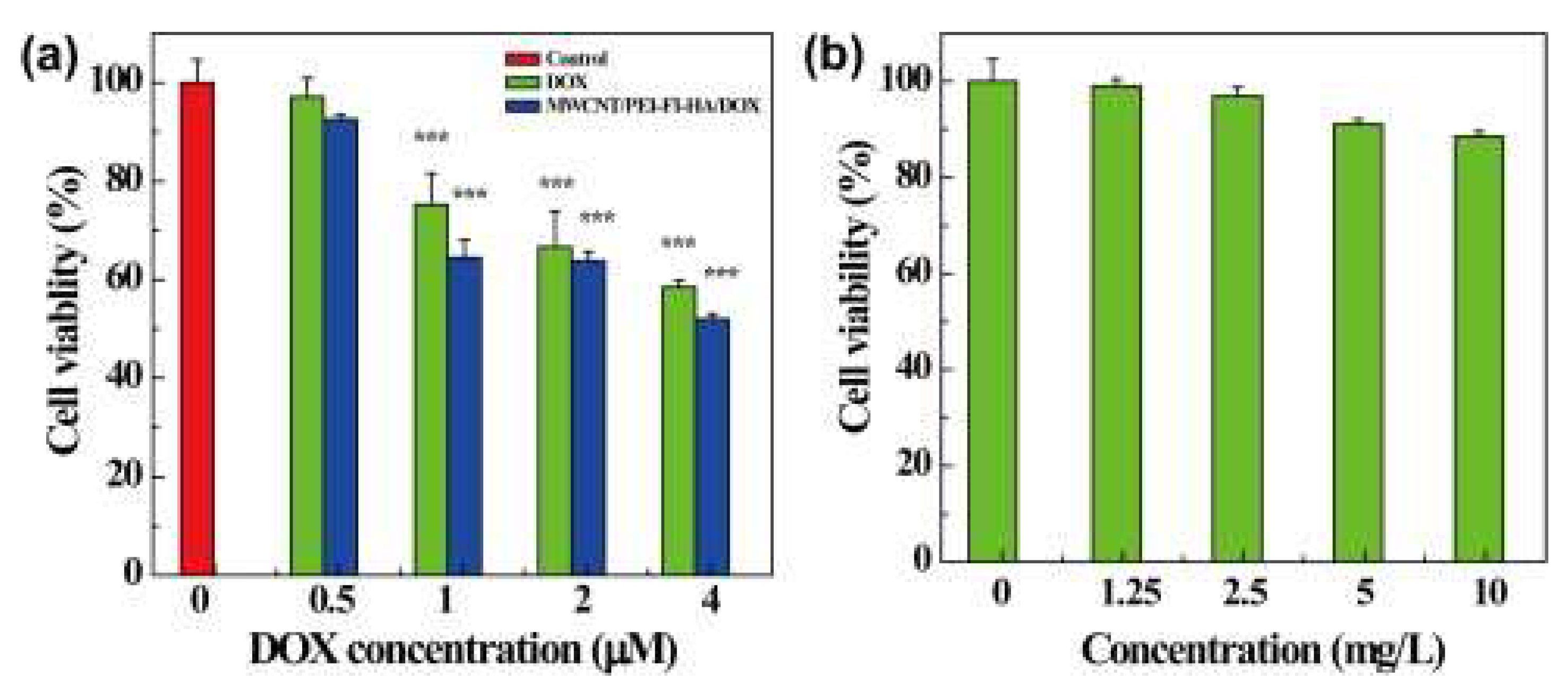
Yang et al. observed that SWCNT@PEG@PEI@COOH DSs displayed higher anti-cancer effects against MCF-7 cells and better drug delivery under acidic conditions, than CNT@COOH and CNT@PEG. Flow cytometry and fluorescence experiments evidenced improved internalization of SWCNT@PEG@PEI@COOH DSs and enhanced tumor cell death via apoptotic mechanisms, due to their high dispersibility and greater affinity towards cancer cells [70]. Similarly, Liu et al. prepared PEG-functionalized SWCNTs loaded with DOX, which demonstrated a loading capacity of ~400% by weight, and a controlled release rate of cargo [71]. SWCNTs generally exhibited a higher drug loading capacity than MWCNTs[72], while short SWCNTs and MWCNTs showed higher and faster loading capacity than long CNTs[73]. Furthermore, aromatic-peptides showed high binding affinity to SWCNTs, due to their interaction with the π electrons of their surface[74]. A foremost advantage of using CNTs-based nanocomposites to develop cancer drug DSs (CDDSs) consists of their susceptibility to release drugs specifically in the acidic environment existing in tumor tissues, thus allowing selective passive targeting of the tumor site, with reduction of side effects to normal cells. Ph-sensitive release of anticancer drugs from CNTs CDDSs increased drug residence time in circulation and selectivity, decreased administration frequency, with possible major compliance of cancer-affected patients to therapy, while preserved the optimum drug concentration[75]. In this regard, Cao et al. demonstrated that PEI and hyaluronic acid (HA) modified MWCNTs designed for the targeted delivery of DOX to cancer cells overexpressing CD44 receptors, showed a drug loading capacity of 72% and a higher release rate in acidic pH (5.8 pH in cancer) than in physiological conditions (pH 7.4 in normal cells)[76]. Therefore, PEI@MWCNT@HA@DOX nanocomposite showed good biocompatibility in the tested concentration range, while exercised substantial cytotoxic effects to cancer cells (Figure 4).
Figure 4.
(A) MTT viability assay of HeLa cells treated with free DOX and MWCNT/PEI–FI–HA/DOX complexes at the DOX concentrations of 0–4 µM for 24 h, and (B) DOX-free MWCNT/PEI–FI–HA at corresponding DOX concentrations of the complexes between 1.25 and 10 mg/L. Reprinted with permission from Carbohydrate Research, Copyright 2015, Elsevier [76]. License number 6018200526333 released on 29 April 2025 by Elsevier and Copyright Clearance Centre.
Similar results were reported by Gu et al., for SWCNTs modified with benzoic acid via hydrazine bonding (HBA) loaded with DOX when tested on HepG2 cells[77]. The release to cancer cells occurred at the pH 5.5 of tumor microenvironment (TME). Additionally, after 60 h of incubation, SWCNT@HBA@DOX complex demonstrated a higher release of DOX than SWCNT@DOX composite (73% drug release vs only 50%). Also, higher cytotoxic effects were observed for SWCNT@HBA@DOX than for SWCNT@ DOX complex, due to enhanced cellular internalization. Since cancer cells require high amounts of folic acid for DNA synthesis and rapid proliferation, they overexpress folate receptors. Conjugation of DDSs with folic acid (FA) is a common approach for targeting cancer cells[78]. Lu et al. functionalized a magnetic nanocomposite composed of MWCNTs and Fe2O3 nanoparticles (IONPs) with poly (acrylic acid) (PAA) by free radical polymerization (FRP) and the obtained nanocomposite was further conjugated to FA and loaded with DOX[79]. When utilized on U87 human glioblastoma cells, the nanocomposite exhibited a dual-targeting effect via both magnetic field and ligand-receptor interaction[79]. The nanocomposite demonstrated higher efficiency than DOX alone, due to easier hydrogen bonding interactions and π–π stacking, while exhibited enhanced cytotoxic effects[79]. The higher effectiveness of DOX when delivered by the CNTs-based DDS derived also by its efficient internalization inside the cells and transport to the nucleus, which permitted the intracellular release of DOX[79]. The following Table 3 and Table 4 collect various reported experiments which used CNTs for the target delivery of different anticancer drugs and genetic material. Specifically, Table 3 summarizes several in vitro and in vivo relevant experiments on the delivery of anti-tumor drugs and/or nucleic acids using modified CNTs-based nanocarriers, while Table 4 collects results about the cytotoxicity to different tumors and biocompatibility results concerning several modified CNTs-based anticancer DDSs.
To effectively manage and precisely remove malignant tumours, a true understanding of cancer features and the interchanges between tumour cells and their surrounding microenvironment is necessary. As extensively demonstrated in the previous sections, many anticancer strategies focus on the target delivery of chemotherapeutics to tumour cells, to enhance their selectivity and anticancer efficiency, while reducing toxic effects to normal cells. The emergence of cancer nanomedicine has allowed huge advancements in the treatment of cancer and in the targeted cancer therapy and several types of nanomaterial-based drug delivery systems have been developed for anticancer target delivery. Among nanomaterials, carbon nanomaterials including CNTs, and mainly functionalized CNTs, have demonstrated to be versatile multifunctional platforms very promising for innovative cancer treatment (Figure 5).
Figure 5.
Advantages of CNT functionalization and multiple applications of functionalized CNTs in cancer treatment by different therapeutic approaches. PAT = photo-acoustic tomography; PTT = photothermal therapy; PDT = photodynamic therapy; ROS = reactive oxygen species; TME = tumour microenvironment. The image has been reproduced from an open access article distributed under the terms of the Creative Commons Attribution License CC BY 4.0 licence (https://creativecommons.org/licenses/by/4.0/, accessed on 26 May 2025). See also http://ivyspring.com/terms for full terms and conditions, accessed on 25 May 2025 [119].
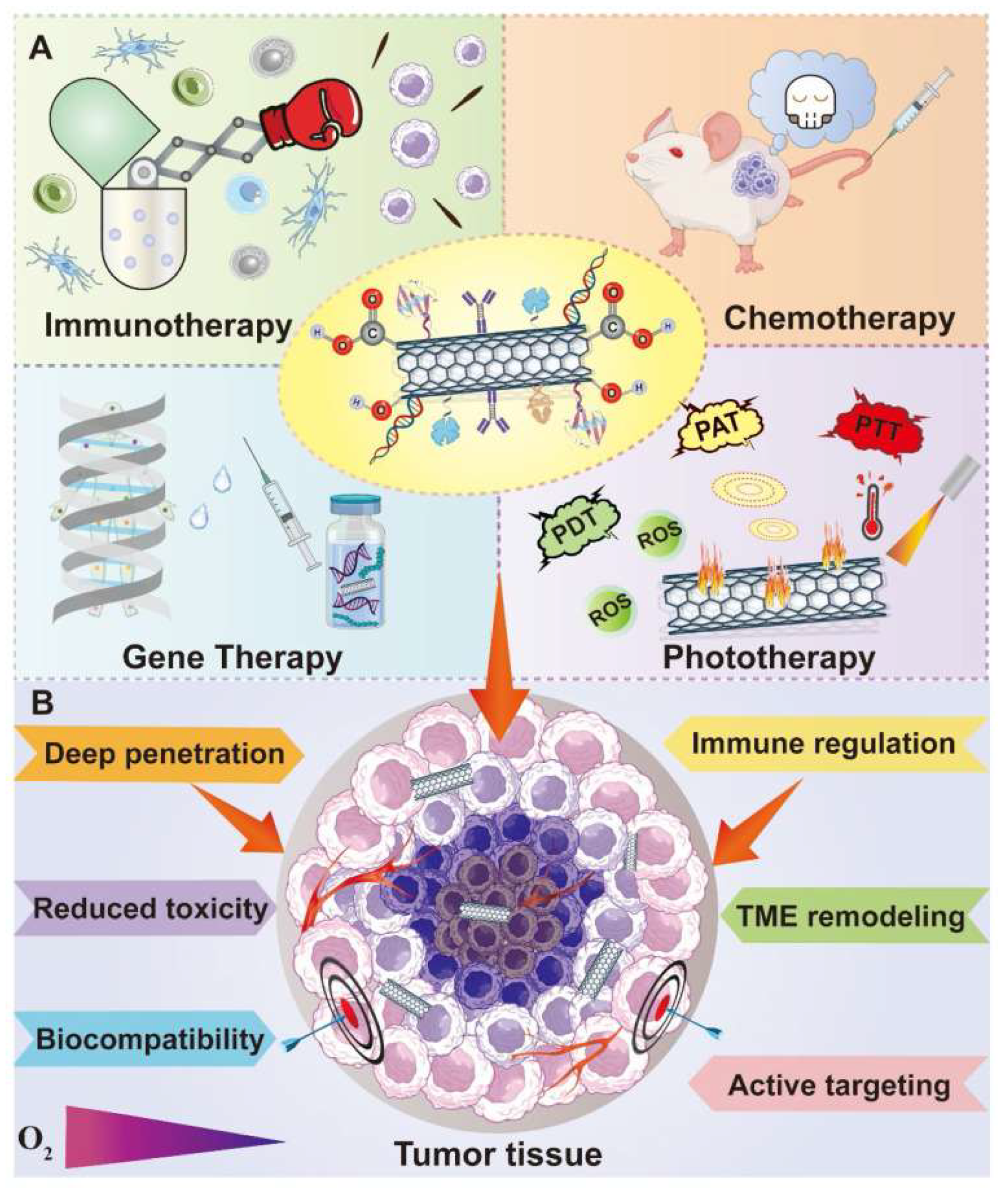
As schematized in Figure 5, CNTs have been successfully experimented in association with many treatment modalities like chemotherapy, gene therapy, phototherapy and immunotherapy, and resulted efficient tools to develop CNTs-based nanocarriers, capable to deliver various anticancer agents to the intracellular sites of interest in cancer cells, such as nucleus, mitochondria, cytoplasm and other organelles, thus realizing direct tumoricidal effects [8,120]. A promising current approach in cancer therapy regards targeting the components present in the tumour microenvironment (TME) where tumour cells live, thus detrimentally impacting on cells survival by impairing their living environment [121,122]. TME is a very complicated system stuffed with various types of cells and full of extracellular matrix (ECM), characterized by altered vasculature, higher acidity and interstitial pressure, abundant glutathione (GSH) levels, poor blood perfusion, abnormal metabolism and hypoxia [119]. Collectively, TME is essential for tumour establishment and progression and represents the obstacle that limits the efficacy of numerous cancer treatment approaches [123]. Anyway, despite the immunosuppressive properties of TME promoting the tumour cells resistance to immunotherapy and despite it produces a lot of impediments to effective cancer treatment, TME can be also exploited as therapeutic target, where abnormalities can be exploited for the development of new selective anticancer strategies. In this context, many innovative anticancer approaches, such as ECM modulation, angiogenesis and cancer stem cells (CSCs) inhibition, immunoregulation, and TME-responsive controlled drug delivery, aiming at remodelling and impairing TME, have been widely studied (Figure 5). CSCs inhibition strategy has been experimented by Faraj et al., in the attempt to address the depressing failures of many currently adopted treatments in the cure of breast cancer. They designed multimodal nanoplatforms based on SWCNTs to achieve noninvasive imaging and specific targeting towards breast CSCs[124]. To this end, SWCNTs were modified with PEG and, since CD44 is a surface marker of breast CSCs, PEGylated SWCNTs were conjugated with CD44 antibodies, thus realizing active targetability toward breast CSCs. Magnetic resonance imaging (MRI), single photon emission computed tomography (SPECT) and NIR fluorescence techniques evidenced an enhanced selective tumor targeting phenomenon in MDA-MB-231 tumor-bearing mice. Moreover, it was observed that PEG@SWCNTs@anti CD44 distributed specifically in the tumor sites where CD44 receptors are abundant, thus further confirming an elevated targetability to CSCs. As previously reported, the immunosuppressive power of TME, often prevents the immune system from effective tumor eradication. Aiming at addressing this problem, Hassan et al. employed MWCNTs as DDSs for the co-delivering of immunoadjuvants such as CpG, anti-CD40 Ig, and OVA antigen, for enhanced immunotherapy. The covalent conjunction of OVA and CpG remarkably elevated the responses of OVA-specific T cells both in vitro and in C57BL/6 mice, while the subsequent loading of anti-CD40 Ig amplified the antitumor immune reactions. Using MWCNTs the co-loading ability of the three ingredients was improved translating into a significant inhibition of tumor growth and metastasis in OVA-expressing B16F10 melanoma model. Also, Tang et al. engineered a co-delivery platform based on PEI-functionalized MWCNTs to inhibit angiogenesis in lung cancer TME for its treatment[117]. Integrin ανβ3 is strictly correlated with angiogenesis, thus representing a therapeutic target for anticancer therapy. RGD peptides are capable to bind with integrin ανβ3, thus being a targeting ligand for anticancer drug delivery[125]. Fu and co-workers connected iRGD peptide and candesartan, an angiotensin receptor blocker, to PEI-modified MWCNTs, and assembled the achieved nanocomposite with plasmid angiotensin II type 2 receptor (pAT2) through electrostatic interaction. A multifunctional DDS was achieved, which demonstrated to be capable of successfully delivering candesartan and pAT2 into tumor cells, thus significantly inhibiting tumor growth and neovascularization in A549 lung cancer model. Table 5 and 6 report several examples of CNTs based DDSs which demonstrated capability to deliver the transported material to specific intracellular sites and TME constituents.
- 3.2.2.
- Carbon Nanotubes-Based Drug Delivery Systems for the Tumor Microenvironment (TME)-Responsive Release of Chemotherapeutics
The enhanced permeability and retention (EPR) effect is a concept, [143,144]establishing that small molecules, typically liposomes, nanoparticles, and macromolecular drugs tend to accumulate in tumour tissue much more than they do in normal tissues [145,146]. The conventional drug delivery systems (DDSs) work mainly exploiting the EPR effect and receptor-mediated endocytosis. Unfortunately, they have habitually to afford many obstacles, due to the biological intricacy of the tumour microenvironment (TME), where cancer cells reside [147]. Anyway, the acid and hypoxic characteristics of TME and the high concentrations of glutathione (GSH) and H2O2 allow to conceive TME-responsive drug release tactics. Activated by several stimuli deriving by the conditions existing in TME, the drugs transported by these appositely engineered DDSs, can be released in a controlled or sustained mode, thus realizing various therapeutic effects and specifically penetrating deeply in the tumour tissue. Moreover, the reaction occurring between drugs-loaded DDSs and these sensitive factors can lessen tumour hypoxia and acidity, thus creating an environment hostile to tumour cells and more suitable for better therapeutic results. In this context, Yang et al. ideated oxidized MWCNTs gifted with large inner diameter, which allowed them to entrap inside cisplatin and to load doxorubicin (DOX) on the surface[148]. To hamper the early release of cisplatin, polyethylene glycol (PEG) and folic acid (FA) were also employed. With this approach, the carried cargos were endowed with a pH-sensitive release profile, and was released under pH = 6.5, which is just the weak acidic condition existing in TME. When tested on MCF-7 breast cancer cells, the nanocomposite demonstrated a more marked cytotoxicity in the pH = 6.5 condition than in pH = 7.4, confirming its best capacity to specifically kill tumour cells in acidic TME. Wang et al. engineered MWCNTs-based nanocomposites to be employed in magnetic resonance imaging (MRI)-guided and TME-responsive phototherapy[149]. A uniform MnO2 sheet merged with Ce6 photosensitizer were modified with MWCNTs achieving a nano-system for enhanced phototherapy. MnO2 was used because it was capable to rapidly deplete GSH, through redox reaction of Mn4+ into Mn2+ ions and to decompose H2O2 present in the TME, to give 1O2. By this approach, the photothermal effect engendered by MWCNTs was encouraged, tumour hypoxia was downed, and Ce6-mediated PDT was eased. ROS-mediated cell death increased via chemo-dynamic therapy and Ce6 release was sped up. Overall, this multifaceted MWCNTs-based platform could represent a promising strategy to achieve synergistic cancer diagnosis and therapy. Also, Qin et al. developed a CNTs-based nanoplatform encapsulated into a certain thermos-/pH-sensitive nanogel, thus obtaining a DDS capable of near infrared (NIR)-triggered, TME-responsive drug release[150]. Upon loading with DOX, this nano-system showed a quicker release rate of DOX at 40 C than at 25 C, and at pH = 5.0 than pH = 7.4, which indicated combinational outcomes, due to CNTs-mediated photothermal effects under NIR irradiation and TME-responsive drug release. The following Table 7 collects the above-mentioned case studies and several other ones.
4. Carbon Nanotubes Application in Anticancer Phototherapy
4.1. Anticancer Photothermal Therapy
As an unconventional anticancer therapeutic technique, photothermal therapy (PTT) made part of a family of minimally invasive strategies, that are based on the use of photosensitizers. Specifically, PTT kills cancer cells thermically, by creating local heat using an optical absorption mediator, that is a photosensitizer, capable of adsorbing electromagnetic energy (EME) and transforming it into heat[151]. By generating a condition of hyperthermia, tumor eradication is significantly improved by boosting immune activation and causing immunity towards metastatic cancer cells for long time[152]. Unfortunately, conventional photosensitizers are affected by several drawbacks, including undesired adverse effects on skin, limited target to cancer cells, and scarce therapeutic effects in hypoxic TME. In this regard, CNTs represent excellent next-generation photosensitizer agents for a more effective PTT. They possess better photophysical properties and the ability to target cancer cells and accumulate in the tumor site. They are gifted with a broad electromagnetic absorbance spectrum and the capability to convert near-infrared I and II windows, which match the optical transmission window of biological tissues[153]. Moreover, the use of CNT-based tumor-targeting conjugates, in combination with PTT treatment, can result in more precise and efficient tumor elimination. Using well dispersible PEG-wrapped CNTs containing 80% (w/w) PEG on the CNTs surface, Sobhani et al. demonstrated the effectiveness of PTT treatments against HeLa and HepG2 cells[53]. Moreover, when PEG@CNTs were evaluated against melanoma, using a tumor-bearing mice model exposed to a continuous-wave near-infrared laser diode for 10 min, a more significant reduction in tumor size was observed in mice receiving also PEG@CNTs associated with laser irradiation, than in mice receiving only the laser radiation. Similarly, Zhu et al., avoiding the use of surfactants, prepared biocompatible, normal cell-friendly, and not toxic hybrid complexes, encompassing MWCNTs and gold nano-stars (MWCNTs@Au@NSs) [154]. MWCNTs@Au@NSs were experimented associated to PTT, on B16F10 mouse melanoma cells, observing that under 808 nm radiation, a photothermal effect 12.4% and 2.4 times higher than NSs and Au@NSs, respectively was produced, which caused enhanced cancer cells death. Also, by merging PTT with immune-stimulation by means of annexin A5 (ANXA5)-conjugated SWCNTs and anti-CTLA-4 check point inhibitor, McKernan et al. tried to treat metastatic breast cancer cells[155]. The designed combined anticancer therapy caused a significant increase in the survival rate of mice, in the number of CD4+ helpers and of CD8+ cytotoxic T cells, while SWCNTs had no toxic effects during the experiment. Moreover, even if the targeted and controlled release of genes remains a major challenge, Zhao et al. showed the synergistic effect of SWCNTs/MWCNTs-based PTT and gene therapy in exerting anti-tumor activity[126]. SWCNTs and MWCNTs were coated with peptide lipid and sucrose laurate to form a bifunctional DDS with improved photothermal effects and temperature sensitivity, which was loaded with siRNA. The complex silenced the survivin expression, thus effectively repressing tumor growth, while exhibiting photothermal effects under NIR exposure. Peptide lipid and sucrose laurate facilitated the phase transition of lipids, thus enabling the systemic delivery of siRNA to the tumor site. SWCNTs produced by the CoMoCAT®method possess high nanotube chirality, display an absorption band at 980 nm, and photothermal activity, which was widely used for selective photo-tissue interaction[155,156]. In this regard, with the aim to target folate receptors on surface of cancer cells, Zhou et al. coupled CoMoCAT®-SWCNTs with folic acid (FA)[157]. In vitro and in vivo experiments demonstrated that the conjugate significantly reduced the photothermal destruction of normal cells, while significantly enhancing the photothermal death of tumor cells. Fibrin is a final product of coagulation response, which is highly concentrated at the vasculature injury site[158]. Fibrin existing in the tumor vessels can serve as therapeutic target for drug delivery, due to its easy accessibility, ubiquitous presence, and high expression. On these considerations, Zhang et al. engineered fibrin-targeting CREKA peptide-conjugated PEG@MWCNT nanocomposites for the PTT of cancer cells[159]. Exposure of a tumor-bearing mice model to MWCNT@PEG significantly increased the temperature of the tumor site, after 24 hours NIR radiation. It was shown that, upon illumination, IR783-labeled MWCNT@PEG accumulation in the tumor site was 6.4 times higher than in the control group[159]. MWCNT@PEG completely suppressed tumor xenografts after four illuminations cycles. Overall, MWCNT@PEG demonstrated significant tumor targeting and photothermal therapeutic effectiveness. Suo et al. proposed MWCNTs coupled to a Pgp-specific antibody (Pab) for photothermal P-Glycoprotein (Pgp)-mediated extirpation of MDR ovarian cancer cells[159]. Results demonstrated a remarkably improved internalization of the Pab–MWCNTs in 3T3-MDR1 than in 3T3 cells at different time periods. Under NIR irradiation, Pab–MWCNTs demonstrated higher dose-dependent specific photokilling in 3T3-MDR1 than in 3T3 cells[159]. Due to the higher temperatures to cancer cells reachable by plasmon phenomenon, differently modified MWCNTs demonstrated higher cytotoxicity to cancer cells. The highest temperatures for MWCNT-COO, MWCNT-COOPt, and MWCNT-Pt were 43.4°C, 45.8°C, and 46.2°C, respectively, while those of MWCNT-COO, MWCNT-COOAu, and MWCNT-Au were 44.1 °C, 46 °C, and 46.9 °C, respectively [160,161].
4.2. Anticancer Photodynamic Therapy
Another unconventional anticancer therapeutic technique, included in the family of noninvasive strategies against cancer and based on the use of photosensitizers, is the low toxic photodynamic treatment (PDT). PDT utilizes a combination of light, chemical photosensitizers and molecular oxygen, to induce cell death[56]. Briefly, upon the topical or systemic administration of a photosensitizer to tumor cells, it can be activated by light, at specific wavelength (NIR)[56]. An energy transfer cascade occurs, that induces ROS overproduction in the presence of oxygen, resulting in selective cytotoxicity against cancerous cells[162]. Racheal et al. engineered zinc phthalocyanine@spermine@SWCNTs and compared them to mono carboxy phenoxy phthalocyanine not containing zinc and zinc mono carboxy phenoxy phthalocyanine-conjugated spermine deprived of the SWCNTs. Photophysical properties and PDT efficiency towards MCF-7 breast cancer cell lines were assessed[163]. Results demonstrated that both ZnMCPPc-spermine and ZnMCPPc-spermine-SWCNT possessed photophysical characteristics superior to those of zinc-free phthalocyanine, with > 50% improvements in triplet and singlet oxygen quantum yields. In vitro cytotoxicity experiment carried out using MCF-7 cancer cells evidenced that the PDT carried out using ZnMCPPc-spermine and ZnMCPPc-spermine-SWCNT caused 97% and 95% cell viability reduction, respectively at 40 mM. Also, the same Racheal with colleague Nyokong assessed the PDT outcomes of a Zn-phthalocyanine modified with SWCNTs in association with ascorbic acid against MCF-7 tumor cells[164]. ZnMCPPc, ZnMCPPc@AA, ZnMCPPc@SWCNT, and ZnMCPPc@AA@SWCNT were evaluated for their photophysical properties (PPPs) and PDT performance (PDTP). ZnMCPPc@SWCNTs demonstrated enhanced PPPs, prolonged lifetimes and improved singlet oxygen quantum yields, with respect for ZnMCPPc alone. ZnMCPPc@SWCNT showed excellent PDTP against MCF-7 cells, leading to 77% cell viability reduction. Sundaram et al. studied the PDTP of SWCNTs-modified hyaluronic acid (HA) and chlorin e6 (Ce6) nanocomposites (NCs) on colon cancer cells[165]. HA coating significantly enhanced the dispersibility of NCs. The as-synthesized SWCNT@HA@Ce6 NCs proved higher anticancer effects on Caco-2 cells, than free Ce6. Also, they showed improved capacity to deliver photosensitizers, as well as stronger apoptotic activity in colon tumor cells, due to a high surface area and strong binding capacity [166]. Nano-bio-composites associated with PDT (both 5 and 10 J/cm2 laser irradiated) triggered both early and late-stage apoptotic death at major extent in cancer cells then in control ones. Specifically, exposure to 10 J/cm2 (41.9, 6.65) caused more early-stage apoptosis than 5 J/cm2 (36, 6.4). Similarly, survived cells were in lower percentages when exposed to 10 J/cm2 (53.4, 6.8) than to 5 J/cm2 (58.27, 5.9). Moreover, SWCNT@HA@Ce6 NCs under 10 J/cm2 laser irradiation showed higher apoptotic effects than those displayed by free Ce6 and empty SWCNTs. It was reported by Shi et al., that HA-conjugated CNTs NCs were endowed with tumor targeting capability and improved solubility [167]. CNTs were modified with hematoporphyrin monomethyl ether (HMME) PDT agent achieving CNT@HA@HMME NCs, which were capable to combine local selective PDT with exterior near-infrared PTT, thus exerting improved therapeutic efficacy and reduced toxicity to normal cells in the treatment of cancer, by a synergistic effect [167]. Overall, HMME@HA@CNTs could perform both PDT and PTT treatment simultaneously in future enhanced tumor therapy.
4.3. Enhanced Anticancer Phototherapies by CNTs-Improved Drug Delivery
Light-induced hyperthermia in PTT and PDT frequently needs high-power intensity, which may damage nontarget normal cells[168]. To address this issue, Daquan Wang designed a nanoplatform (NP) by coating cut MWCNTs (C-MWNTs) with poly-N-vinyl pyrrole (PVPy) and binding folic acid (FA)-polyethylene glycol (PEG)-SH with thiolene achieving MWCNT@PVPy@S@PEG@FA nanocomposites (NCs). NCs possessed high drug-carrier ratio, pH-sensitive release capability of doxorubicin (DOX), and broad-spectrum anticancer effects. These features depended mainly on their exceptional photothermal conversion efficiency, capacity of high loading of DOX and of its target deliver, due to the presence of PVPy shell[169]. Similarly, Oh et al. used DOX-loaded SWCNTs to exert NIR cancer PTT chemotherapy[132]. Conversely, FA and methotrexate (MTX) were linked to the surface of -COOH modified MWCNTs (COOH-MWCNT) by using ethylenediamine (ED) as coupling agent, achieving MWCNT@ED@FA and MWCNT@ED@MTX NCs, demonstrating the NIR- or IR-laser promoted highest rate of MCF-7 cancer cell death. Cell death was high also at low doses of MTX when the laser light and MTX in MWCNT@ED@MTX or the laser light and MWCNT@ED@FA worked together [170]. Thermal tumour suppression triggered by CNTs was exploited by Znang et al. to treat cancer cells by chemotherapy and PTT using an association of MWCNTs, gemcitabine and lentinan, an anticancer drug and an immune-stimulator, respectively[171]. Under 808 nm laser radiation, the proposed NC was more efficient in penetrating and suppressing cancer cells and less toxic than each system ingredient when used alone. Also, CNT-based NCs give the possibility to combine therapeutic and diagnosis activities. CNTs can be associated with other nanoparticles of different origin to produce synergistic therapeutic and diagnostic effects[172]. Karimi et al. investigated the cytotoxic effects of MWCNT@MTX and MWCNT@MTX@PEI@FA under 808 nm laser radiation, without observing noticeable differences[173]. DOX or CpG entrapped onto MWCNTs significantly increased MWCNTs water dispersibility. When melanoma-bearing mice were exposed to such modified MWCNTs under 808 nm NIR laser radiation at a dose of 1 W/cm2 for 5 min, suppression of tumour growth and improved number of lymph draining CD4+ and CD8+ T cells were observed in the spleen, with increased antitumour efficacy[142]. PTT using targeted SWCNTs associated with immune system activation using a checkpoint inhibitor was proposed as an innovative treatment for the management of metastatic breast cancer. Selective NIR photothermal ablation of the primary orthotope at an energy and power level of 175 J/cm2 and 1 W/cm2, respectively, enhanced the anti-cytotoxic T lymphocyte-dependent abs copal response, translating into an increase in survival rate (55%), 100 days after tumour vaccination. The Annexin A5 functionalised SWCNTs, SWCNTs@ANXA5 NC was administered systemically before PTT[155]. Biocompatible 3D CNT@MXene@DOX microspheres were engineered displaying nonpareil photothermal effects and photothermal stability under 650 and 808 nm NIR laser radiation. As-manufactured microspheres were endowed with a maximum drug loading capacity of 85.6% for DOX. Also, 3D CNT@MXene microspheres effectively created singlet oxygen, due to TiO2 photosensitized present on their surface. In-vitro experiments showed that 3D CNT@MXene@DOX successfully inhibited HeLa cancer cell proliferation[174].
4.4. Application of CNTs for Enhanced Anticancer Combined PTT and PDT
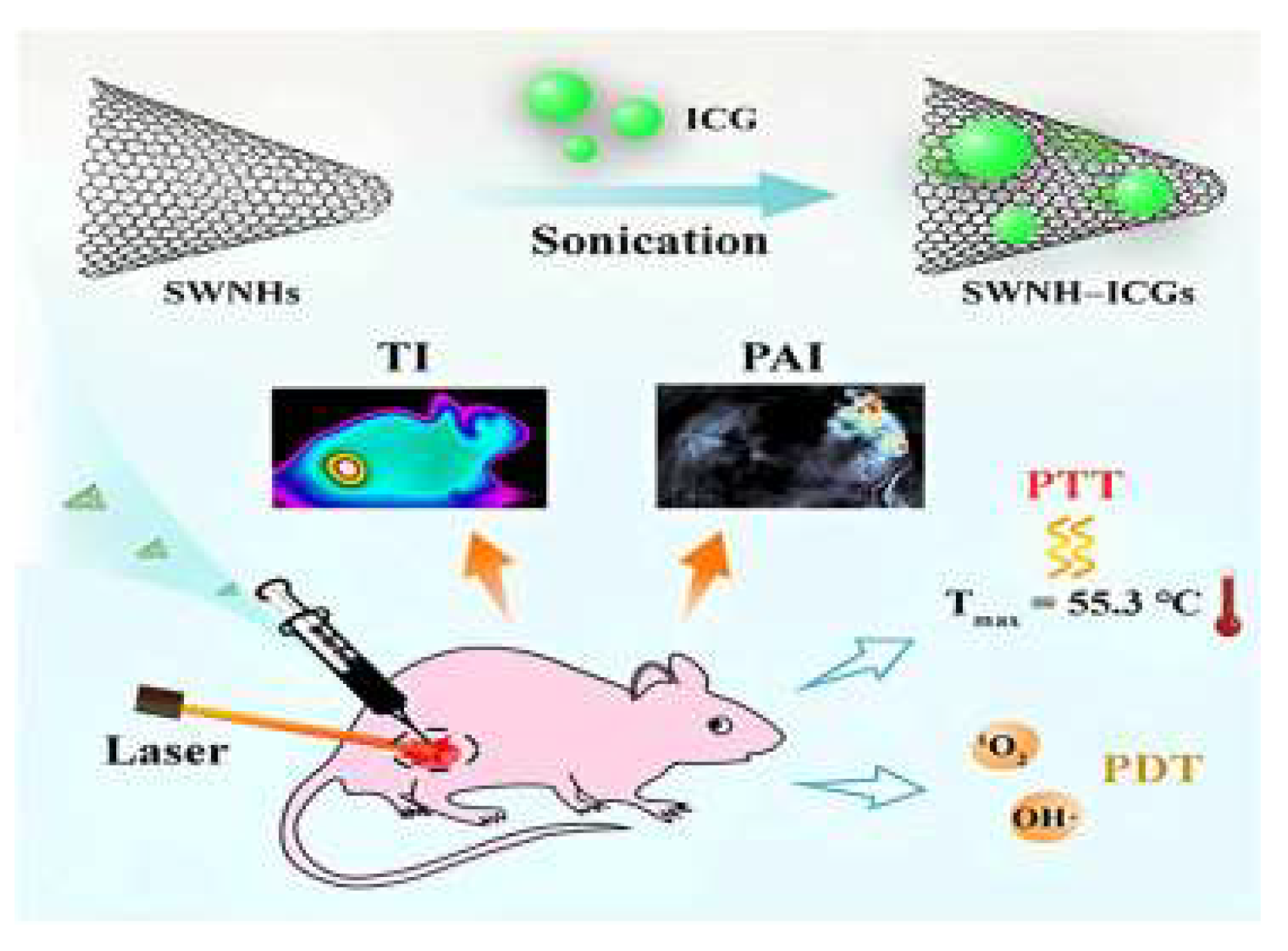
Single walled carbon nano-horns (SWCNHs) are a particular typology of CNTs, characterized by high photothermal conversion efficiency (PCE), which were studied to develop nanoplatforms suitable for different phototherapies thus enabling to engineer nanocomposites for combined PTT and PDT. In all relevant articles where PDT and PTT were experimented in combination, it was evidenced an increase of the pace at which tumours were reduced in animal models. Additionally, to further improve this impact, such combination can also be associated with another treatment, such as chemotherapy. In this context, Gao and co-authors developed a SWCNH@indocyanine green (ICG) theragnostic nano-system, which was experimented for diagnosis and combined PTT and PDT in breast tumour cells at low laser power, observing enhanced synergistic antitumour effects. Particularly, SWCNH@ICG induced ROS overproduction and hyperthermia effects, under light radiation. The mechanism which led to kill the tumour and slows its growth is shown in Figure 6[175].
Figure 6.
Thermal/photoacoustic imaging-guided PTT and PDT synergistic therapy nanoplatform developed by Gao et al. This image is published [175] and it has been reproduced under licence number 6027091475870 provided by John Wiley and Sons and Copyright Clearance Centre on 13 May 2025 (available online at Rightslink® by Copyright Clearance Center, accessed on 13 May 2025).
The same authors created a similar platform, this time containing hypericin (Hyps). Upon laser radiation at 590 and 808 nm, this nanocomposite functioned as dual agent for simultaneous PDT and PTT against 4T1 cells implanted subcutaneously in mice as tumour models. Optimum outcomes in terms of anticancer impact by induced hyperthermia and ROS overproduction were observed [176]. Yang et al., immobilized Ce6 and Gd3+ on the surface of polymer-coated SWCNHs. Authors investigated its effects when experimented in combined PDT and PTT against tumours associated to the immune system’s response [177]. High tumour targeting, enhanced tumour penetration efficiency and immune adjuvant effects were evidenced [177]. The possible synergistic effects of PDT and PTT were investigated by Yin et al., using a CNTs-based nanocomposite comprising Ce6 and MnO2 to prevent tumour hypoxia, under radiation treatments at both 660 and 808 nm to activate both therapies. Despite tumour growth was inhibited, also by each therapy used independently, the PS, PA, MnO2, and Ce6-MnO2@CNTs (CMCs) compounds produced the best outcomes, when applied in simultaneous PDT and PTT, by synergistic effects[178]. Zhang et al., in addition to combine PDT with PTT, associated PDT and PTT with CT developing a nanoplatform including both SWCNTs and carbon quantum dots (CQDs). To provide outstanding selectivity toward tumour cells, water solubility of nanoplatform was improved using PEG, it was gifted with magnetic characteristics to allow IMR using Fe3O4 NPs and DOX was loaded thus engineering SWCNTs@PEGFe3O4@CQDs@DOX@Apt nanocomposite. Despite all groups who receive radiation outperform those who receive only CT treatment, when the combination PTT, PDT and CT was used, tumour was suppressed, thus establishing its superiority for treating tumours[179]. Similarly, Wang et al. created a multifunctional nanomaterial, namely CNT@MnO2-PEG@Ce6 for PDT, PTT, and MRI therapy. It comprised CNTs, MnO2 NPs layers, PEG and Ce6 as photosensitiser (PS). When this nanoplatform was used for PDT and MRI, Ce6 and Mn2+ were rapidly released, due to the low pH and high GSH concentration of the TME, respectively. When CNT@MnO2-PEG@Ce6 was applied for realizing a PDT and PTT combined treatment, by radiating it using simultaneously two-wavelength sources at 660 and 808 nm, a synergistic PDT/PTT effect was observed. which significantly decreased the tumour-development rate[149]. Also, Marangon et al. engineered a nanocomposite for treating SKOV3 ovarian cancer cells, using a combination of PDT and PTT. It was based on MWCNTs and the m-tetra hydroxyphenyl chlorin (m-THPC) as photosensitiser. The photothermal and photodynamic cytotoxic effects of m-THPC@MWCNTs complexes were investigated at the cellular level by several methods including viability tests, analysis of apoptosis-related proteins, genomic analysis of 84 genes involved in OS etc. The combination of PDT and PTT therapy resulted in cancer cells death, by the activation of various signalling pathways and by suppressing the cell’s defence against OS[180]. Then, as previously reported, a new PDT agent, namely HMME, was adsorbed onto HA-modified CNTs (HA@CNTs), thus creating HMME@HA@CNTs nanocomposite, endowed with high aqueous solubility, neutral pH, and tumour-targeting activity. When this new nanocomposite was used in combination with PTT and PDT to treat tumour in vivo and in vitro, improved anticancer efficacy and low toxicity to normal organs was observed, due to the capacity of HMME@HA@CNTs NPs to combine local selective PDT with exterior NIR PTT, thus achieving a synergistic impact [167]. The following Table 8 and Table 9 collect some relevant case studies reporting on the application of CNTs in anticancer PTT and PDT, while Table 10 summarizes reports on enhanced anticancer phototherapy by CNTs-based drug delivery. Lastly, Table 11 collects case studies on the application of CNTs to realize nanoplatforms to perform combination of PTT, PDT and CT.
Gene therapy (GT) is a novel engineered therapeutic approach which aims at using genes to cure several diseases, including cancer, by replacing defective genome of diseased cells with healthy one [187]. In GT, the opportunely selected healthy genes are inserted into the selected cells, including cancer ones, by transfection, to repair defects in their genome or compensate for the cells’ deficiencies, due to uncontrolled mutations inexplicably occurred [187]. GT and its synergistic combination with CT are gaining strong interest in cancer treatment. Unfortunately, insufficient endosomal escape of genes/nanocarriers complexes, due to an inadequate buffer capacity of the carrier cause lysosomal early degradation [56,187]. This event causes poor transfection capability and strongly limits the therapeutic applicability in vivo of this approach. The use of CNTs-based carriers, opportunely modified to enhance their buffer capacity, can promote endosomal effects and survival of the gene complex, thus ameliorating significantly the transfection efficiency. The use of suicide genes is among the most efficacious approaches to realize efficient anticancer gene therapy. It consists of using therapeutic transgenes to express toxic products from a toxic gene or to convert a nontoxic prodrug into a toxic one, or both, to fight the effects of cancer disease. This strategy was essayed to treat several cancers, such as breast[188], liver, colon[189], prostate[190], glioma[191], and lung cancer [192] also when cancer cells have acquired chemo-resistance [193]. Additionally, this approach has demonstrated to enhance the efficacy of radiation therapy [194]. Furthermore, combination therapy usually is more efficient than monotherapy, due to its capability to elude the cell cycle arrest caused by chemical drugs. Several studies were developed based on the above-mentioned considerations. Cao et al. engineered a novel pH-responsive SWCNTs-carrier functionalized with PEI-betaine (PB) (SPB), improved with BR2 peptide and loaded with DOX and survivin siRNA (SPB@BR2@DOX@sur/siRNA), for the co-delivery of an anticancer drug and a silencing gene and realizing efficient anticancer gene therapy[114]. When administered to both HeLa and A549 cancer cells and 293T normal cells, the nanocomposite was selectively internalized into cancer cells, while it did not enter normal cells[114]. The nanocomposite caused less survivin expression and a higher apoptotic index than Lipofectamine 2000, due to release of siRNA/DOX into the A549 cell cytoplasm and nuclei, without lysosomal degradation. In comparison to SPB@BR2@siRNA or SPB@BR2@DOX separate treatments, that in association using SPB@BR2@DOX@sur/siRNA demonstrated synergistic effects, causing significant reduction in the volume of tumor both in A549 cells and in nude mice[114]. Based on the capability of iC9 suicide gene to induce apoptotic death in MCF-7 human breast cancer cells, Dargah et al. used pyridine modified MWCNTs (pyr@MWCNTs) as carriers to transfect iC9, achieving the pyr@MWCNTs@iC9 complex [195]. Upon its administration, MCF-7 cells were exterminated, and when associated with chemotherapy, it evaded cell cycle arrest. Also, gene regulation and anticancer therapy were attempted by Zhang et al. experimenting the chitosan modified fluorescent carbon nanoparticle (FCN)-based siRNA conjugate (Ch@FCN@siRNA)[196]. The core-shell nanocomposite comprised a core made of Ch@FCN and a shell of siRNA. siRNA down-regulated the key regulator of mitosis, namely polo-like kinase-1 (PlK1) expression. Notable, only a concentration of FCN 30 times lower than that of AuNPs was sufficient to transfect the same amount of siRNA. The nanocomposite, in vitro treatment of A375 and MCF-7 tumor cells, was better performant of the commercial Lipofectamine 2000 inducing 31.9% and 20.33% apoptosis, respectively. Also, its intravenous administration to mice bearing the A375 tumor cells reduced tumor volume by 11-fold compared with control groups. As reported in previous sections, Guo et al. used MWCNTs as efficient siRNA vectors. Precisely, the authors modified MWCNTs with NH3 groups, which were cationic at physiological pH, obtaining positively charged MWCNTs-NH4+ tubes, which were used for siRNA delivery against PLK-1 cancer cells in mice [95]. Upon the administration of siRNS@MWCNTs-NH4+complex lung cancer xenografts were eradicated [95]. Another study by Anderson et al. used SWCNTs for the targeted delivery of siRNA to pancreatic cancer cells for anticancer gene therapy [197]. In this contribution, the prepared SWCNTs@siRNA complex was tested in vitro on pancreatic cancer cells, observing high siRNA transfection efficiency, successful internalization of the nano complex in cancer cells and low toxicity versus normal cells[197]. The release of siRNA from the nano complex resulted in the downregulation of the target oncogene [197].
6. Carbon Nanotubes Application in Anticancer Immunotherapy
Immunotherapy is a therapeutic approach, which is finalized to improve the patient’s capability to fight several types of diseases, including cancer, by either the modification or amplification of the immune system by exploiting antigenic targets [198]. This therapeutic strategy has proven to be efficacy to enhance therapeutic effects against chronic infections and cancer. Another anticancer strategy consists of blocking regulatory systems which may interfere with the immunotherapeutic effects. In the body, dendritic cells (DCs) represent a crucial connexion between innate and adaptive immunity. They are the most potent specialized antigen presenting cells (APCs), playing a pivotal role in anti-infection and anti-tumour responses. Unfortunately, cancer cells can overpower both the immune system and the functionality of DCs, limiting the efficacy of DCs-based antitumor immunotherapy. Hence, it could be of paramount help improving the antitumor immune response by controlling DCs functionality and disabling immune tolerance. Also, cytotoxic T lymphocytes (CTLs), also known as CD8+ T and CD4+ T cells, are crucial cells of the adaptive immune system. They play a determinant role in the defence against pathogens such as viruses, bacteria, and tumours [199]. An inadequate infiltration of CD8+ T cells in the immunosuppressive TME, results in a decreased antitumor response. When CD+ T cells are even absent, the body will lack antitumor immune function. Conversely, the over-presence of CD8+ T cells can trigger excessive immune responses, leading to immune-mediated tissue damage or pathological reactions [200]. Thus, enhancing CD8+ T cell infiltration in TME, as well as promoting their correct functional activity are pivotal strategies in tumour treatment. Carbon-based nanoparticles, including CNTs could be supreme platforms for tumour detection and immunotherapy. Several research indicates that polymer modified CNTs can treat tumours by acting as immune adjuvants, to promote the maturation of dendritic cells (DCs), the CD8+ T cells infiltration in TME, and to release anti-tumour factors [201]. Furthermore, MWCNTs conjugated with peptides can promote cytokine secretion, stimulating T cell differentiation and proliferation[202].
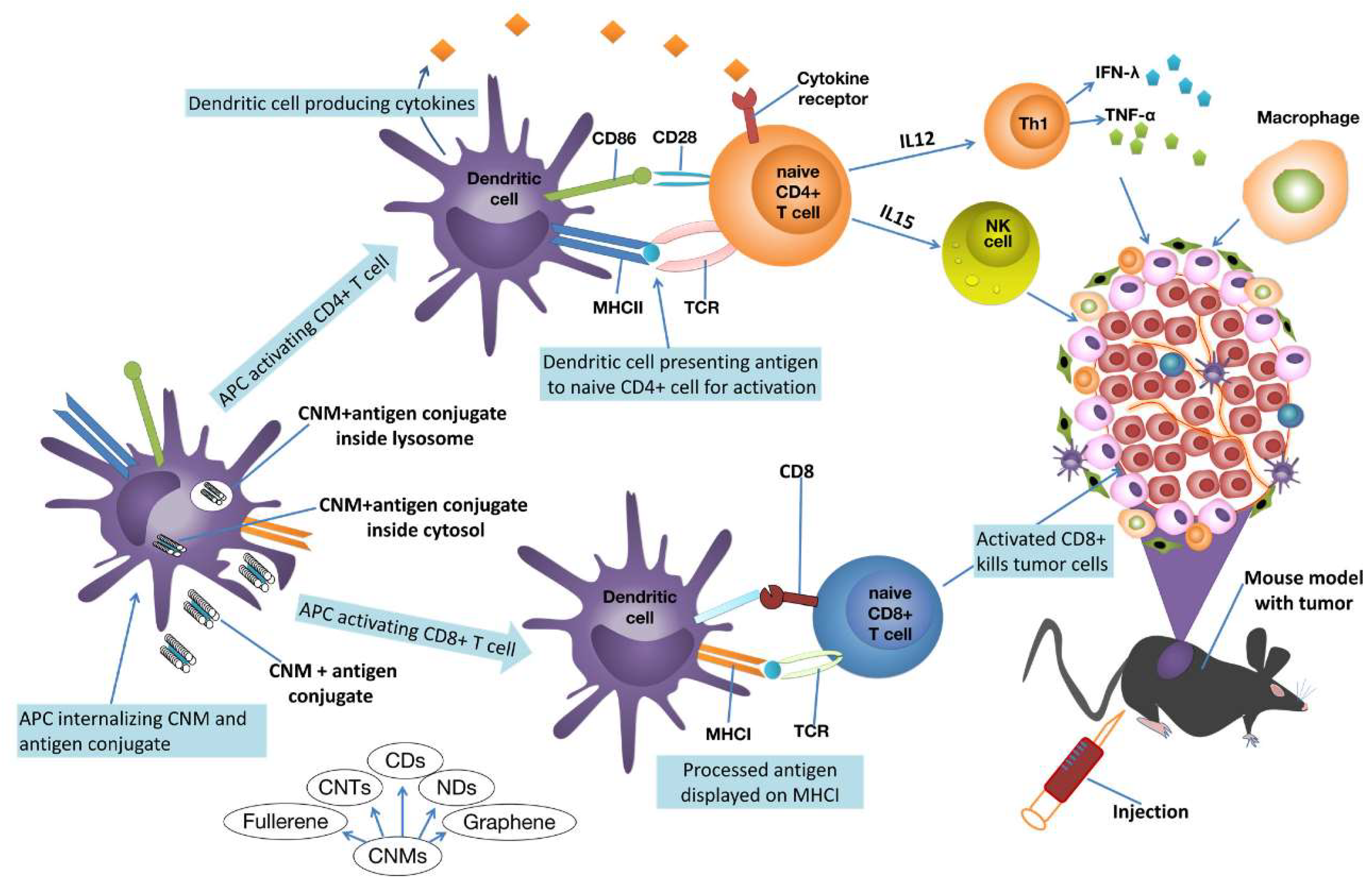
Carbon-based nanomaterials (CNMs) and CNTs can augment antitumor immunity of patients, via multiple and several mechanisms of immune system modulation. The most recognized comprises first the pickup of the CNT–antigen conjugate by DCs, which transfer the antigen peptides to naive T cells for activation. To this work, a multitude of unique receptors exists on the surface of DCs, which naturally serve as recognition sites for activating specific immune cells. The movement of antigens to specific compartments for presentation in DCs is critical. In DCs, the lysosome-dependent pathway causes the antigen breaking down into antigenic peptides (APs) inside lysosomes. APs are then loaded onto Class II major histocompatibility complex (MHC-II) molecules for presentation to CD4+ helper T cells. On the other hand, MHC-I molecules display cytosolic antigens to activate CD8+ T cells and trigger cytotoxic T lymphocyte (CTL) responses[203]. Among cytokines, TNF-α provide chemical signals to the cancer cells, causing inflammation and cell death[204]. Activated T cells and natural killer (NK) cells release IFN-γ, which activates macrophages and improves antigen presentation[205]. Other cytokines, such as IL-15 and IL-12 activate and stimulate proliferation and expansion of NK cells and other antitumor immune cells, including CD8+ T cells [206] (Figure 7).
Figure 7.
Schematic representation of the immunomodulatory effects of CNMs in cancer therapy [207]. The image has been reproduced by an open access article [207]. Reproduction is licensed under a Creative Commons Attribution 4.0 International License, which permits use, sharing, adaptation, distribution and reproduction in any medium or format. To view a copy of this licence, visit http://creativecommons.org/licenses/by/4.0/. The Creative Commons Public Domain Dedication waiver (http://creativecommons.org/publicdomain/zero/1.0/) applies to the data made available in this article, unless otherwise stated in a credit line to the data. APCs = antigen-presenting cells; DCs = dendritic cells; CNM = carbon nanomaterial; CNTs = carbon nanotubes; CDs = carbon dots; NDs = nanodots; MHC-II = histocompatibility complex II; CD4+, CD8+ = helper T cells; MHC-I = histocompatibility complex I; CTL = cytotoxic T lymphocyte NK = natural killer.
The subsequent Table 12 collects studies where CNTs were utilized to synthesize CNTs-based nanoplatforms for immune oncotherapy.
Immuno-based oncotherapy was experimented by Xia et al. in vitro and in vivo, using MWCNTs-based nano-delivery systems loaded with unmethylated CpG moieties, an oligodeoxynucleotide, and H3R6 polypeptide (MHR-CpG) for treating prostate cancer [212]. Authors observed enhanced biocompatibility, endosomal TLR9 targeting and improved immunogenicity of CpG in both the humoral and the cellular immune pathways. An increase in the expression of CD4+ T-cells, CD8+ T-cells, TNF-, and IL-6 were detected. When tested in vivo in RM-1 prostate tumor-bearing mice, the nanocomposite demonstrated to be capable of delivering the immune therapeutics to both the tumor site and to lymph nodes, thus inhibiting prostate cancer growth. Hassan et al. used antigen-bearing MWCNTs to deliver immunoadjuvants such as cytosine-phosphate-guanine oligodeoxynucleotide (CpG), anti-CD40 Ig (CD40), ovalbumin (OVA) antigen to trigger immune response against OVA-expressing cancer cells. When tested in vitro and in vivo, the MWCNTs-based nanoplatform caused a dramatically high OVA-specific T cell responses in vitro and in C57BL/6 mice. Co-loaded OVA antigen, CpG, and anti-CD40 Ig prevented the proliferation of OVA-expressing B16F10 melanoma cells in pseudo-metastatic subcutaneous or lung tumour models[129]. Furthermore, SWCNTs realized the efficient delivery of CpG in CX3CR1GFP mouse models, without toxicity to normal cells, and increased the production of proinflammatory cytokines by primary monocytes. Surprisingly, a single intracranial injection of low-dose CNT-CpG removed intracranial GL261 gliomas in half of tumour-bearing animals, via activation of NK and CD8+ cells, and protected the surviving mice from the recurrence of intracranial cancer.[213]. Oxidised MWCNTs (ox-MWCNTs) bearing COOH groups were prepared by Radzi et al. and tested for treating breast cancer in EMT6 model mice, associated with PTT. The combined therapy resulted in full cancer eradication and in a substantial increase of mice median survival rate. Additionally, MWCNTs-based nanocomposite increased the infiltration and maturation of DCs, CD8+, CD4+ T cells, macrophages and NKs in tumours treated with ox-MWCNTs–hypothermia combination therapy [214].
Wilm’s tumour protein (WT1) is a protein upregulated in many human leukaemia and cancers. Villa et al. covalently attached WT1 ligands onto soluble SWCNT architectures achieving SWCNT–peptide nanoplatforms, which were speedily absorbed by dendritic cells and macrophages in vitro. Immunization of BALB/c mice with SWCNT–peptide nanocomposite and immunological adjuvants provoked specific IgG responses versus the peptide [215].
Fadel et al. utilized SWCNT bundles in the presentation of T-cell-activating antibodies to evoke immune responses in target tumours. SWCNTs bundles delivered anti-CD3 T-cell-stimulating antibodies, with high local concentrations, resulting in powerful activation of T cells, thus demonstrating that SWCNT bundles constitute a unique model for the effective activation of lymphocytes, with implications for fundamental science and clinical immunotherapy[216].
Functionalized bundled SWCNTs (fb-SWNTs) have demonstrated to be efficient antigen-presenting substrates and were used by Fadel et al. to absorb T cell antigens and CD3 and CD28 costimulatory ligands. The as achieved nanoplatforms were used to treat splenocytes obtained from the spleens of C57BL/6 mice. The adsorption of T-cell-stimulating antibodies improved both the kinetics and amount of T cell activation, thus supporting the utilization of chemically processed nanotube bundles in clinical applications requiring the presentation of artificial antigen [217].
Later, the same authors developed a simple yet robust technique of noncovalently attaching the T cell stimulus (MHC-I) to CNT substrates, for avoiding undesired denaturation effects. They used the achieved nanocomposite to treat OT1 mice, observing increased antigen-specific T cell responses which was 3-fold higher that in control [218].
Also, Burkert et al. developed AuNPs-bearing nitrogen-doped nitrogen nanotube cups (Au-NCNCs), which demonstrated capability in entrap and target deliver of paclitaxel in the tumour site. Au-NCNCs altered the TME, reduced tumour growth rate, and counteract immunosuppressive macrophages [219].
Anyway, it is dutiful remembering that the needle-like shape of CNTs, which allows them to penetrate cellular membranes, causing damage, can also trigger inflammatory responses, that may lead to harm in both animals and humans. MWCNTs revealed high phagocytic activity towards undifferentiated HL60 cells and cytotoxic effects on differentiated HL60 cells [220].
Due to a lack of critical clinical evidence, the exact mechanisms by which CNTs can harm humans and animals remains unclear. On the other hand, many studies suggest that proper modifications can lower the hazardous effects of CNTs making them eligible for applications in the biomedical field. Scientists in different field concerning CNTs should conduct extensive research in collaboration to address concerns surrounding CNTs safety and to enhance their credibility. Looking ahead, researchers are expected to develop innovative synthetic methods or create novel composite materials to improve cancer treatment outcomes and enhance human health.
7. Carbon Nanotubes Application in Cancer Diagnosis
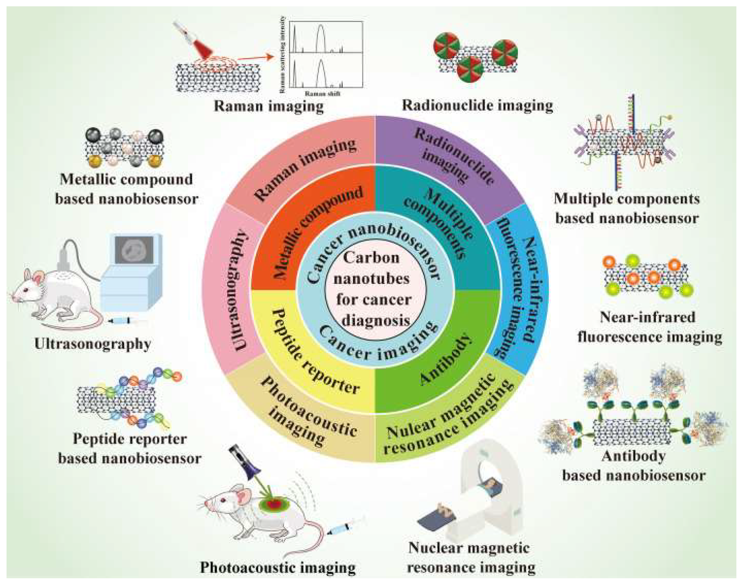
Cancer diagnosis aims at examining and detecting the etiologies and related symptoms concerning various types of cancer, using modern technologies. Due to their nonpareil properties, CNTs are increasingly attracting interest also in this field. The application of CNTs in various cancer imaging, such as Raman imaging, nuclear magnetic resonance imaging (NMRI), ultrasonography (US), photoacoustic imaging (PAI), radionuclide imaging (RNI), near-infrared fluorescence imaging (NIR-FI), as well as their use to engineer cancer nano-biosensors, as schematized in Figure 8 and summarized in Table 13, are the topics of this section.
Figure 8.
An overview of the contributions of CNTs to cancer diagnosis. The image has been reproduced by an open access article [221]. Reproduction is licensed under a Creative Commons Attribution 4.0 International License, which permits use, sharing, adaptation, distribution and reproduction in any medium or format. To view a copy of this licence, visit http://creativecommons.org/licenses/by/4.0/. The Creative Commons Public Domain Dedication waiver (http://creativecommons.org/publicdomain/zero/1.0/) applies to the data made available in this article, unless otherwise stated in a credit line to the data.
CNTs can provide multifunctional bio-probes with several unprecedented properties, including strong absorbance in NIR, good resonance Raman scattering, and high modifiability, thus representing excellent materials suitable for cancer imaging, with research and clinical prospective in cancer diagnosis.
7.1. Raman Imaging
The radial breathing model (RBM) and tangent G-module (TGM) are unique vibrational features of CNTs, which can be detected by a Raman microscope [238]. SWCNTs doped with oxygen, bearing epoxide groups and modified with PEG (o-SWNTs@PEG) were engineered by Sekiyama et al. The achieved nanocomposites were created to serve as over-thousand-nanometer (OTN)-NIR fluorescent probes, to investigate the time-dependent change in OTN-NIR fluorescence images of colon-26 cancer cells[222]. Upon administration of the probes to colon-26 cancer cells, their distribution in cells was studied using Raman microscopy, observing Raman signals on the 5th day from first administration. Since noble metals on the surface of CNTs improve their Raman signals[239], Wang et al. enriched the surface of PEG@CNTs with gold or silver, thus improving the Raman scattering (SERS) effect of pristine CNTs. The use of noble metal modified CNTs rather than that of not modified PEG@CNTs allowed to acquire the Raman images under NIR radiation in remarkably minor time [223].
7.2. Nuclear Magnetic Resonance Imaging
Nuclear magnetic resonance imaging (NMRI) is a non-invasive imaging technique, which does not use ionizing radiation, thus being not risky for the human body. It provides the original 3D cross-section images of a tissue or organ and is of paramount help in medical imaging [240]. Since CNTs can be used as T2 spin dephasing contrast agents, they are utilized to enhance nuclear magnetic resonance imaging (NMRI) in clinics [241]. Yan et al. by the non-covalent association of the NGR (asparagine-glycine-arginine) peptide with DOX and CNTs bearing NMR contrast agent, namely Gd-DTPA, prepared a new nanocomposite theragnostic, for both detecting the tumor by NMRI and anticancer therapy [224]. Zhang et al. synthesized a multimodal nanoplatform based on MWCNTs (FA@GdN@CQDs-MWNTs/DOX) using gadolinium NPs (GdN), magneto fluorescent carbon quantum dots (CQDs), and folic acid (FA) [225]. In vitro targeting NMRI experiment revealed that FA-@dN@CQDs-MWNTs worked as excellent T1 contrasting agent overstating the longitudinal proton relaxation process. As confirmation, in vivo essays showed that the NMR signal at tumor site was positively enhanced after intravenous administration of FA@GdN@CQDs-MWNTs.
7.3. Ultrasonography
Ultrasonography is a low-cost and intrinsically safe diagnostic imaging technique [242]. CNTs are nanomaterials particularly eligible for ultrasonic imaging, since during ultrasonography they are capable to produce high signals that can be detected by a contrast-enhanced ultrasound imager. Saghatchi et al. prepared multi-functionalized (mf) MWCNTs by their modification with both magnetic Fe3O4 and gold NPs (mf-MWCNT@AuNPs for simultaneous cancer imaging and therapy [226]. This nanocomposite, when applied at various concentrations, exhibited notable high contrast. Delogu et al. modified MWCNTs using azomethine ylides to improve their biocompatibility and applied the achieved nanocomposite (ox-MWNT-NH3+) in ultrasonography[227], observing a strong, long-lived and high-quality ultrasound signal, after sonication treatment.
7.4. Photoacoustic Imaging
In photoacoustic imaging (PAI), upon the irradiation of cells, tissues and/or organs by pulsed laser, the luminous energy is adsorbed, converted into ultrasonic by the thermal expansion of tissues and organs, and detected by sensors[243]. Since CNTs possess strong NIR absorption and deep tissue penetration, they could serve as ideal contrast agents for PAI, whose signal can be recorded by a photoacoustic microscopy[244]. Using PAI technology in cell imaging, Avti et al. detected, localized and quantified the content of CNTs in different tissue samples, thanks to signals clearly understandable and stable, due to the potent NIR absorbance of CNTs[228]. Wang et al. engineered a multifunctional MWCNTs-based probe encompassing the RGD peptide, a silica-coating, and Au nanorods (RGD@sGNR@MWNTs) endowed with active targeting ability, to be used for the in vivo PAI of gastric cancer[229]. After intravenous administration of RGD@sGNR@MWNTs probe, the treated mice were analyzed by an optoacoustic imaging system, evidencing that RGD@sGNR@MWNTs precisely targeted the tumor site and provided strong photoacoustic imaging effects.
7.5. Radionuclide Imaging
Radionuclide imaging (RNI) is an imaging technology which exploits radioactive isotopes such as 111In, 131I, 64Cu, and 86Y, which upon injection into the body are adsorbed by human tissues and organs, thus functioning as radiation source in vivo emitting γ-rays during their decay process [245,246]. Such rays can be detected by nuclear detection devices which provide the distribution density of the radioactive isotopes in vivo. RNI is endowed with deep tissue penetration, negligible limitation and high sensitivity[247]. The organ biodistribution of various functionalized (f)-MWCNTs in vivo was studied by Wang et al., who used MWCNTs radio-labelled with 111In to empower easy in vivo single photon emission computed tomography/computed tomography (SPECT/CT) imaging[230]. Zhao et al. modified SWCNTs with polydopamine (PDA) and PEG, achieving a nanoplatform (SWNTs@PDA-PEG), which was further labelled with 131I, for the subsequent RNI[231]. After in vivo administration, the tumor tissue distribution of 131I@SWNTs@PDA-PEG was detected by a gamma counter.
7.6. Near-Infrared Fluorescence Imaging
Near-infrared fluorescence (NIR-F) imaging, having a NIR biological wavelength window in the range of 780-1700 nm, is an appealing and fast-progressing imaging technique, extremely useful for cancer diagnosis. CNTs possess clear optical absorption and intrinsic fluorescence in the above-mentioned range, thus being promising materials for application in NIR-F imaging during cancer diagnosis, whose images are usually acquired using an in- vivo imager [248,249]. Since SWCNTs possess higher absorbing properties, stronger optical absorption, better E11 optical transitions, and less photobleaching than MWCNTs, they are more appropriate for NIR imaging than the others [250]. Ghosh et al. developed a M13-stabilized SWCNTs probe, which exhibited in vivo precise target to tumor nodules expressing secreted protein, acidic and rich in cysteines (SPARCs) [232]. Second-window NIR light (NIR-II) was used in this study, as the fluorescence source to avoid optical scattering and gain deeper tissue penetration effect during NIR-F imaging. The diagnostic result achieved using this NIR2-emitting M13@SWCNTs probe evidenced outstanding signal-to-noise performance and high specificity towards in situ ovarian tumor and tumor nodules present on the surfaces of other peritoneal organs. Welsher et al.[233] combined SWCNTs phospholipid-PEG (PL@PEG) achieving a biocompatible nanoplatform (SWCNTs@PL@PEG) with low toxicity levels and enhanced stability. Upon its administration in live mice and the application of an InGaAs camera, high-resolution intravital tumor vessel images were acquired.
7.7. CNTs in Nano Biosensors
Biosensors are studied starting from the year 1962[251], and recently, they are considered very attractive tools, mainly due to their easy application, fast response, and low cost[252]. Nano-biosensors (NBSs) are biosensors, consisting of both biological recognition elements and nanomaterials, sized 1-100 nm[253,254]. The unparallelled electrical conductivity, excellent electrocatalytic properties, high stability, slow oxidation kinetics and good modifiability of CNTs, render them a class of nanomaterial with great potential for applications as NBs in cancer diagnosis[255].
7.8. CNTs Combination with Metallic Nanoparticles
Gold and silver nanoparticles (NPs) (AuNPs and AgNPs) are used to modify CNTs, to allow the construction of NBs. Rawashdeh et al. reported on AuNPs@MWCNTs which worked well as NBs, demonstrating easy and improved detection of micro ribonucleic acid named miR-21, which is of paramount importance in the early diagnosis of pancreatic cancer [234]. AuNPs@MWCNTs had a limit of detection (LOD) as low as 3.68 femtomolar (fM) using the source measure unit (SMU).
7.9. CNTs Combination with Antibody
Nano-immunosensors (NISs) based on antibodies are a particular type of NBSs used in cancer diagnosis. Osteopontin (OPN) is a biomarker which is used to detect prostate cancer cells, and its levels are important to predict the survival time of patients bearing prostate cancer [256,257]. To overcome the issues which affect the traditional method used for the measurement of OPN (ELISA assay), Sharma et al. covalently combined OPN monoclonal antibodies onto the SWCNTs@COOH surface, achieving a NIS to determine OPN for prostate cancer diagnosis [235]. AntiOPN@SWCNTs@COOH exhibited high specificity to only OPN, broad detection range and low LOD.
7.10. CNTs Combination with Peptide Reporter
Detecting CDK1 peptide in tissues and organs could be of great help in tumors early diagnosis. Anyway, diagnostic methods using traditional BSs for the detection and quantification of activities of CDK1 poses great difficulties, due to the lack of sensitivity and specificity [258]. Aiming at overcoming these issues, functionalized MWCNTs further modified with a fluorescent peptide reporter specific to CDK1 were developed for the sensitive and fluorescence-based quantification of CDK1 activity via fluorescence imaging [236]. The as prepared NBS provided both easy detection of CDK1, reported the enzymatic activity of CDK1 and detected the CDK/cyclin activity in numerous cancer types, usually associated with poor prognosis.
7.11. CNTs Combination with Multiple Modifications
Multiple modifications on CNTs can further improve the diagnostic effects obtained using the NBSs engineered via single modification. Mahmoodi et al. developed a rGO@MWCNTs/L@Cys@AuNPs multifunctional nanocomposite and enriched its surface with a single strand DNA (ssDNA) probe, as an electrochemical DNA-biosensor to diagnose HPV-18 [237]. rGO@MWCNTs/L@Cys@AuNPs@ssDNA demonstrated high selectivity and sensitivity to HPV-18, towards both the extracted DNA from HPV-18 patients and the synthetic one. Overall, this constructed rGO@MWCNTs/L@Cys@AuNPs@ssDNA NBS represents an essential tool for the early, easy, rapid, and accurate diagnosis of HPV-18. Table 14 summarizes other relevant case studies on the application of CNTs as sensors for early diagnosis of cancer.
8. Clinical Translatability and Challenges Concerning CNTs
The clinical translatability (CT) of carbon nanomaterials (CNMs), including CNTs, is a relevant issue which must be considered, but many problems, such as their biodegradability and metabolism remain unsolved and should be addressed before their successful translation in clinical practice. CNMs are unprecedented effective tools with nonpareil properties in cancer theragnostic. Anyway, the gap between laboratory research and clinical transformation still needs long time a more in deep research. CNMs toxicity, mainly dependent on their sizes, structure and shape, surface chemistry, and on the type of cells target[276], should be taken into great consideration when they have been developed for clinical biomedical applications. Research should be focused on finding methods to prepare ideal CNMs structures without defects, impurities and low cytotoxicity mainly tuning size and designing suitable decorations. To quicken their translation in clinical practice, their stability in physiological conditions, cellular uptake, biodistribution and accumulation in different tissues and organs should be ascertained in advance. Their transformation and metabolic pathway in vivo, as well as their acute and latent toxicity should be clarified [277]. To obtain the desired theragnostic goals, the delivery efficiency of CNMs of therapeutic agents specifically at tumor site is of paramount importance. A suitable CNMs size is pivotal for realizing enhanced permeability and retention (EPR)-mediated passive tumor targeting [278]. Conversely, to achieve active targeting, a deep study of the receptors over-expressed specifically on tumor cells surface is mandatory. Once CNMs-based nanoplatforms capable of efficient therapeutics delivery are realized, the endogenous or exogenous stimuli should be fully exploited to accomplish controlled drug release. Unfortunately, many obstacles still exist hampering the achievement of these goals, such complicated designing, boring synthesis, scarce synergistic functions, poor integrated efficiency, and ambiguous biological response. Also, while the anti-tumor properties of CNMs have been extensively demonstrated [279], their antineoplastic mechanism and their tumor proliferation and metastasis regulation machinery have not been systematically clarified. In this regard, before the direct clinical application of CNMs as anticancer drugs, their metabolic pathway, biological safety and how they can be used as anticancer agents for cancer treatment to overcome the limitations of conventional chemotherapeutics should be studied and clarified. Moreover, because of hydrophobicity and structural stability, CNMs suffer of low solubility and dispersibility in aqueous media, poor miscibility and tendency to self-aggregate, which mostly limit their biological applications [280]. When CNTs are applied as drug delivery systems (DDSs), due to their stability, free CNTs survive in the body for long time after drug dissociation, thus triggering secondary damage [281]. Several are the problems which impede CNTs clinical application. They comprise the following:
- the mechanisms of action of CNTs on normal cells and tissues are not fully unveiled
- potential toxic effects can occur, due to their special structure
- the mechanisms of CNT-induced toxicity are not fully clarified
- the biodegradability of raw CNTs is too low because their hydrophobic prevents enzymes from approaching them, thus impeding enzymatic degradation
To promote CNTs translatability in clinic practice, more labors is necessary to explore the best functional molecules for the CNTs modification to improve their biocompatibility, to avoid the toxic and risk factors, to enhance their solubility and create defect sites, which offer desired binding sites for enzymes and promoting enzymatic degradation [282]. Collectively, although there is no clinical practice that have ever reported the application of CNMs as DDSs in cancer theragnostic, there are some clinical trials cases that assessed the employment of carbon nanoparticles (CNPs) in lymphatic monitoring during colorectal cancer surgeries, lymph nodes collection in advanced gastric cancer, and lymph node biopsy of papillary thyroid carcinoma, confirming the unprecedented potential and promising future of CNMs and CNTs in cancer theragnostic [283,284,285]. The following Table 15 and 16 summarize other two clinical trials carried out to evaluate the possible applications of CNTs-based nanocomposites in anticancer treatments and cancer diagnosis.
Other clinical trials regarded the application of CNTs to treat dental caries, Parkinson (available online at https://clinicaltrials.gov/study/NCT02873585, https://clinicaltrials.gov/study/NCT01246336. Accessed on 26 May 2025) and cervical pain[289]. The assessment of the clinical observational studies reported in Table 15 focused on the potential for misleading, selection of participants, measurement of exposures and outcomes, handling of missing data, and selective reporting of results. This comprehensive approach ensured a thorough examination of factors that could influence the results of the study, from the allocation of participants to the reporting of the outcomes. Each domain was rated as “low risk  ,” “high risk
  ,” or “some concerns
  ,” based on specific criteria [290,291]. This assessment processes emphasized the importance of transparency and rigor. Using the Cochrane Risk of Bias, two reviewers independently assessed the risk of bias (Table 17).
,” “high risk ,” or “some concerns ,” based on specific criteria [290,291]. This assessment processes emphasized the importance of transparency and rigor. Using the Cochrane Risk of Bias, two reviewers independently assessed the risk of bias (Table 17).Supplementary Materials
The following supporting information can be downloaded at the website of this paper posted on Preprints.org. Table S1. Structural properties of various modified/activated CNTs vs pristine CNTs (first row). Table S2. Main methods to synthesize carbon nanotubes (CNTs). Table S3. Most relevant research articles on CNT synthesis from biomass by different methods, operating parameters and main properties.
Author Contributions
Conceptualization, validation, formal analysis, investigation, data curation, writing—original draft preparation and writing—review and editing, S.A., G.A. Validation, investigation, data curation and writing—review and editing, C.R. All authors have read and agreed to the published version of the manuscript.
Funding
This research received no external funding.
Institutional Review Board Statement
Not applicable.
Informed Consent Statement
Not applicable.
Data Availability Statement
No new data were created in this review article.
Conflicts of Interest
The authors declare no conflicts of interest.
References
- Siegel, R.L.; Miller, K.D.; Sauer, A.G.; Fedewa, S.A.; Butterly, L.F.; Anderson, J.C.; Cercek, A.; Smith, R.A.; Jemal, A. Colorectal cancer statistics, 2020. CA Cancer J. Clin. 2020, 70, 145–164. [Google Scholar] [CrossRef] [PubMed]
- Bray, F.; Laversanne, M.; Sung, H.; Ferlay, J.; Siegel, R.L.; Soerjomataram, I.; Jemal, A. Global cancer statistics 2022: GLOBOCAN estimates of incidence and mortality worldwide for 36 cancers in 185 countries. CA: A Cancer J. Clin. 2024, 74, 229–263. [Google Scholar] [CrossRef] [PubMed]
- Bray, F.; Laversanne, M.; Weiderpass, E.; Soerjomataram, I. The ever-increasing importance of cancer as a leading cause of premature death worldwide. Cancer 2021, 127, 3029–3030. [Google Scholar] [CrossRef] [PubMed]
- Chen, S.; Cao, Z.; Prettner, K.; Kuhn, M.; Yang, J.; Jiao, L.; Wang, Z.; Li, W.; Geldsetzer, P.; Bärnighausen, T.; et al. Estimates and Projections of the Global Economic Cost of 29 Cancers in 204 Countries and Territories From 2020 to 2050. JAMA Oncol. 2023, 9, 465–472. [Google Scholar] [CrossRef]
- Naief, M.F.; Mohammed, S.N.; Mayouf, H.J.; Mohammed, A.M. A review of the role of carbon nanotubes for cancer treatment based on photothermal and photodynamic therapy techniques. J. Organomet. Chem. 2023, 999. [Google Scholar] [CrossRef]
- Klochkov, S.G.; Neganova, M.E.; Nikolenko, V.N.; Chen, K.; Somasundaram, S.G.; Kirkland, C.E.; Aliev, G. Implications of nanotechnology for the treatment of cancer: Recent advances. Semin. Cancer Biol. 2021, 69, 190–199. [Google Scholar] [CrossRef]
- Chen, K.; Cao, X.; Li, M.; Su, Y.; Li, H.; Xie, M.; Zhang, Z.; Gao, H.; Xu, X.; Han, Y.; et al. A TRAIL-Delivered Lipoprotein-Bioinspired Nanovector Engineering Stem Cell-Based Platform for Inhibition of Lung Metastasis of Melanoma. Theranostics 2019, 9, 2984–2998. [Google Scholar] [CrossRef]
- Wang, W.; Chen, K.; Su, Y.; Zhang, J.; Li, M.; Zhou, J. Lysosome-Independent Intracellular Drug/Gene Codelivery by Lipoprotein-Derived Nanovector for Synergistic Apoptosis-Inducing Cancer-Targeted Therapy. Biomacromolecules 2018, 19, 438–448. [Google Scholar] [CrossRef]
- Ganesh, K.; Massagué, J. Targeting metastatic cancer. Nat. Med. 2021, 27, 34–44. [Google Scholar] [CrossRef]
- Shakibaie, M.; Hajighasemi, E.; Adeli-Sardou, M.; Doostmohammadi, M.; Forootanfar, H. Antimicrobial and anti-biofilm activities of Bi subnitrate and BiNPs produced by Delftia sp. SFG against clinical isolates of Staphylococcus aureus, Pseudomonas aeruginosa, and Proteus mirabilis. IET Nanobiotechnology 2019, 13, 377–381. [Google Scholar] [CrossRef]
- Mohammed, S.N.; Al-Rawi, K.F.; Mohammed, A.M. Kinetic evaluation and study of gold-based nanoparticles and multi-walled carbon nanotubes as an alkaline phosphatase inhibitor in serum and pure form. Carbon Lett. 2023, 33, 1217–1229. [Google Scholar] [CrossRef]
- Naief, M.F.; Mohammed, A.M.; Khalaf, Y.H. Kinetic and thermodynamic study of ALP enzyme in the presence and absence MWCNTs and Pt-NPs nanocomposites. Results Chem. 2023, 5. [Google Scholar] [CrossRef]
- Naief, M.F.; Ahmed, Y.N.; Mohammed, A.M.; Mohammed, S.N. Novel preparation method of fullerene and its ability to detect H2S and NO2 gases. Results Chem. 2023, 5. [Google Scholar] [CrossRef]
- Naief, M.F.; Mohammed, S.N.; Ahmed, Y.N.; Mohammed, A.M. Synthesis and characterisation of MWCNTCOOH and investigation of its potential as gas sensor. Inorg. Chem. Commun. 2023, 157. [Google Scholar] [CrossRef]
- Latif, Z.A.A.; Mohammed, A.M.; Abbass, N.M. Synthesis and characterization of polymer nanocomposites from methyl acrylate and metal chloride and their application. Polym. Bull. 2019, 77, 5879–5898. [Google Scholar] [CrossRef]
- Abdullah, O.H.; Mohammed, A.M. GREEN SYNTHESIS OF AuNPs FROM THE LEAF EXTRACT OF PROSOPIS FARCTA FOR ANTIBACTERIAL AND ANTI-CANCER APPLICATIONS. Dig. J. Nanomater. Biostructures 2020, 15, 943–951. [Google Scholar] [CrossRef]
- Nasir, G.A.; Najm, M.A.; Hussein, A.L. The Effect of Silver Nanoparticles on Braf Gene Expression. Systematic Reviews in Pharmacy 2020, 11. [Google Scholar] [CrossRef]
- Mohammed, A.M.; Saud, W.M.; Ali, M.M. GREEN SYNTHESIS OF Fe2O3 NANOPARTICLES USING OLEA EUROPAEA LEAF EXTRACT AND THEIR ANTIBACTERIAL ACTIVITY. Dig. J. Nanomater. Biostructures 2020, 15, 175–183. [Google Scholar] [CrossRef]
- Jwied, D.H.; Nayef, U.M.; Mutlak, F.A.-H. Synthesis of C:Se nanoparticles ablated on porous silicon for sensing NO2 and NH3 gases. Optik 2021, 241, 167013. [Google Scholar] [CrossRef]
- Altaee, M.F.; Yaaqoob, L.A.; Kamona, Z.K. Evaluation of the Biological Activity of Nickel Oxide Nanoparticles as Antibacterial and Anticancer Agents. Iraqi J. Sci. 2020, 2888–2896. [Google Scholar] [CrossRef]
- Ghdeeb, N.J.; AbdulMajeed, A.M.; Mohammed, A.H. Role of Extracted Nano-metal Oxides From Factory Wastes In Medical Applications. Iraqi J. Sci. 2023, 1704–1716. [Google Scholar] [CrossRef]
- Al-Ali, A.A.; Alsalami, K.A.S.; Athbi, A.M. Cytotoxic effects of CeO2 NPs and β-Carotene and their ability to induce apoptosis in human breast normal and cancer cell lines. Iraqi J. Sci. 2022, 923–937. [Google Scholar] [CrossRef]
- Salih, A.M.A.; Abdo, G.M. Water Conservation in the Arab Region. In Water, Energy and Food Sustainability in the Middle East: The Sustainability Triangle; 2017.
- Iijima, S. Helical microtubules of graphitic carbon. Nature 1991, 354, 56–58. [Google Scholar] [CrossRef]
- Alfei, S.; Schito, G.C. Antimicrobial Nanotubes: From Synthesis and Promising Antimicrobial Upshots to Unanticipated Toxicities, Strategies to Limit Them, and Regulatory Issues. Nanomaterials 2025, 15, 633. [Google Scholar] [CrossRef]
- Alfei, S.; Schito, G.C. Nanotubes: Carbon-Based Fibers and Bacterial Nano-Conduits Both Arousing a Global Interest and Conflicting Opinions. Fibers 2022, 10, 75. [Google Scholar] [CrossRef]
- Hussain, A.; Haider, Q.; Rehman, A.; Ahmad, H.; Baili, J.; Aljahdaly, N.H.; Hassan, A. A thermal conductivity model for hybrid heat and mass transfer investigation of single and multi-wall carbon nano-tubes flow induced by a spinning body. Case Stud. Therm. Eng. 2021, 28. [Google Scholar] [CrossRef]
- Sobamowo, M.; Akanmu, J.; Adeleye, O.; Akingbade, S.; Yinusa, A. Coupled effects of magnetic field, number of walls, geometric imperfection, temperature change, and boundary conditions on nonlocal nonlinear vibration of carbon nanotubes resting on elastic foundations. Forces Mech. 2021, 3. [Google Scholar] [CrossRef]
- Abd-Elsalam, K.A. Carbon Nanomaterials: 30 Years of Research in Agroecosystems. In Carbon Nanomaterials for Agri-Food and Environmental Applications; Elsevier, 2020; pp. 1–18.
- Anzar, N.; Hasan, R.; Tyagi, M.; Yadav, N.; Narang, J. Carbon nanotube—A review on Synthesis, Properties and plethora of applications in the field of biomedical science. Sens. Int. 2020, 1, 100003. [Google Scholar] [CrossRef]
- Negri, V.; Pacheco-Torres, J.; Calle, D.; López-Larrubia, P. Carbon Nanotubes in Biomedicine. Top. Curr. Chem. 2020, 378, 15. [Google Scholar] [CrossRef]
- Lamberti, M.; Pedata, P.; Sannolo, N.; Porto, S.; De Rosa, A.; Caraglia, M. Carbon nanotubes: Properties, biomedical applications, advantages and risks in patients and occupationally-exposed workers. Int. J. Immunopathol. Pharmacol. 2015, 28, 4–13. [Google Scholar] [CrossRef]
- Kumar, S.; Rani, R.; Dilbaghi, N.; Tankeshwar, K.; Kim, K.-H. Carbon nanotubes: a novel material for multifaceted applications in human healthcare. Chem. Soc. Rev. 2016, 46, 158–196. [Google Scholar] [CrossRef] [PubMed]
- Su, Q.; Gan, L.; Liu, J.; Yang, X. Carbon dots derived from pea for specifically binding with Cryptococcus neoformans. Anal. Biochem. 2020, 589, 113476. [Google Scholar] [CrossRef] [PubMed]
- Salari, S.; Jafari, S.M. Application of nanofluids for thermal processing of food products. Trends Food Sci. Technol. 2020, 97, 100–113. [Google Scholar] [CrossRef]
- Chen, D.; Dougherty, C.A.; Zhu, K.; Hong, H. Theranostic applications of carbon nanomaterials in cancer: Focus on imaging and cargo delivery. J. Control. Release 2015, 210, 230–245. [Google Scholar] [CrossRef]
- Law, S.K.; Leung, A.W.N.; Xu, C.; Au, D.C.T. Is It Possible for Curcumin to Conjugate with a Carbon Nanotube in Photodynamic Therapy? Photodiagnosis Photodyn Ther 2022, 40. [Google Scholar] [CrossRef]
- Deshmukh, M.A.; Jeon, J.-Y.; Ha, T.-J. Carbon nanotubes: An effective platform for biomedical electronics. Biosens. Bioelectron. 2020, 150, 111919. [Google Scholar] [CrossRef]
- Costa, P.M.; Bourgognon, M.; Wang, J.T.-W.; Al-Jamal, K.T. Functionalised carbon nanotubes: From intracellular uptake and cell-related toxicity to systemic brain delivery. J. Control. Release 2016, 241, 200–219. [Google Scholar] [CrossRef]
- Vashist, A.; Kaushik, A.; Vashist, A.; Sagar, V.; Ghosal, A.; Gupta, Y.K.; Ahmad, S.; Nair, M. Advances in Carbon Nanotubes–Hydrogel Hybrids in Nanomedicine for Therapeutics. Adv. Heal. Mater. 2018, 7, e1701213–e1701213. [Google Scholar] [CrossRef]
- Cirillo, G.; Vittorio, O.; Kunhardt, D.; Valli, E.; Voli, F.; Farfalla, A.; Curcio, M.; Spizzirri, U.G.; Hampel, S. Combining Carbon Nanotubes and Chitosan for the Vectorization of Methotrexate to Lung Cancer Cells. Materials 2019, 12, 2889. [Google Scholar] [CrossRef]
- Zhang, J.; Liu, Z.; Zhou, S.; Teng, Y.; Zhang, X.; Li, J. Novel Span-PEG Multifunctional Ultrasound Contrast Agent Based on CNTs as a Magnetic Targeting Factor and a Drug Carrier. ACS Omega 2020, 5, 31525–31534. [Google Scholar] [CrossRef]
- Garriga, R.; Herrero-Continente, T.; Palos, M.; Cebolla, V.L.; Osada, J.; Muñoz, E.; Rodríguez-Yoldi, M.J. Toxicity of Carbon Nanomaterials and Their Potential Application as Drug Delivery Systems: In Vitro Studies in Caco-2 and MCF-7 Cell Lines. Nanomaterials 2020, 10, 1617. [Google Scholar] [CrossRef] [PubMed]
- Corletto, A.; Shapter, J.G. Nanoscale Patterning of Carbon Nanotubes: Techniques, Applications, and Future. Adv. Sci. 2020, 8. [Google Scholar] [CrossRef] [PubMed]
- Azizi-Lalabadi, M.; Hashemi, H.; Feng, J.; Jafari, S.M. Carbon nanomaterials against pathogens; the antimicrobial activity of carbon nanotubes, graphene/graphene oxide, fullerenes, and their nanocomposites. Adv. Colloid Interface Sci. 2020, 284, 102250. [Google Scholar] [CrossRef] [PubMed]
- Alfei, S.; Zuccari, G. Carbon-Nanotube-Based Nanocomposites in Environmental Remediation: An Overview of Typologies and Applications and an Analysis of Their Paradoxical Double-Sided Effects. J. Xenobiotics 2025, 15, 76. [Google Scholar] [CrossRef]
- Griffiths, O.G.; O'Byrne, J.P.; Torrente-Murciano, L.; Jones, M.D.; Mattia, D.; McManus, M.C. Identifying the largest environmental life cycle impacts during carbon nanotube synthesis via chemical vapour deposition. J. Clean. Prod. 2013, 42, 180–189. [Google Scholar] [CrossRef]
- Wang, Z.; Shen, D.; Wu, C.; Gu, S. State-of-the-art on the production and application of carbon nanomaterials from biomass. Green Chem. 2018, 20, 5031–5057. [Google Scholar] [CrossRef]
- Berber, M.R.; Elkhenany, H.; Hafez, I.H.; El-Badawy, A.; Essawy, M.; El-Badri, N. Efficient Tailoring of Platinum Nanoparticles Supported on Multiwalled Carbon Nanotubes for Cancer Therapy. Nanomedicine 2020, 15, 793–808. [Google Scholar] [CrossRef]
- Zhang, J.; Song, L.; Zhou, S.; Hu, M.; Jiao, Y.; Teng, Y.; Wang, Y.; Zhang, X. Enhanced ultrasound imaging and anti-tumor in vivo properties of Span–polyethylene glycol with folic acid–carbon nanotube–paclitaxel multifunctional microbubbles. RSC Adv. 2019, 9, 35345–35355. [Google Scholar] [CrossRef]
- Behzadpour, N.; Ranjbar, A.; Azarpira, N.; Sattarahmady, N. Development of a Composite of Polypyrrole-Coated Carbon Nanotubes as a Sonosensitizer for Treatment of Melanoma Cancer Under Multi-Step Ultrasound Irradiation. Ultrasound Med. Biol. 2020, 46, 2322–2334. [Google Scholar] [CrossRef]
- Zhang, B.; Wang, H.; Shen, S.; She, X.; Shi, W.; Chen, J.; Zhang, Q.; Hu, Y.; Pang, Z.; Jiang, X. Fibrin-targeting peptide CREKA-conjugated multi-walled carbon nanotubes for self-amplified photothermal therapy of tumor. Biomaterials 2016, 79, 46–55. [Google Scholar] [CrossRef]
- Sobhani, Z.; Behnam, M.A.; Emami, F.; Dehghanian, A.; Jamhiri, I. Photothermal therapy of melanoma tumor using multiwalled carbon nanotubes. Int. J. Nanomed. 2017, ume 12, 4509–4517. [Google Scholar] [CrossRef]
- Bosca, F.; Corazzari, I.; Foglietta, F.; Canaparo, R.; Durando, G.; Pastero, L.; Arpicco, S.; Dosio, F.; Zonari, D.; Cravotto, G.; et al. SWCNT–porphyrin nano-hybrids selectively activated by ultrasound: an interesting model for sonodynamic applications. RSC Adv. 2020, 10, 21736–21744. [Google Scholar] [CrossRef] [PubMed]
- Singh, R.; Kumar, S. Cancer Targeting and Diagnosis: Recent Trends with Carbon Nanotubes. Nanomaterials 2022, 12, 2283. [Google Scholar] [CrossRef] [PubMed]
- Dizaji, B.F.; Khoshbakht, S.; Farboudi, A.; Azarbaijan, M.H.; Irani, M. Far-reaching advances in the role of carbon nanotubes in cancer therapy. Life Sci. 2020, 257, 118059. [Google Scholar] [CrossRef] [PubMed]
- Son, K.H.; Hong, J.H.; Lee, J.W. Carbon nanotubes as cancer therapeutic carriers and mediators. Int. J. Nanomed. 2016, ume 11, 5163–5185. [Google Scholar] [CrossRef]
- Zhang, Y.-R.; Lin, R.; Li, H.-J.; He, W.; Du, J.-Z.; Wang, J. Strategies to improve tumor penetration of nanomedicines through nanoparticle design. WIREs Nanomed. Nanobiotechnol. 2019, 11, e1519. [Google Scholar] [CrossRef]
- Zhang, A.; Meng, K.; Liu, Y.; Pan, Y.; Qu, W.; Chen, D.; Xie, S. Absorption, distribution, metabolism, and excretion of nanocarriers in vivo and their influences. Adv. Colloid Interface Sci. 2020, 284, 102261. [Google Scholar] [CrossRef]
- Zare, H.; Ahmadi, S.; Ghasemi, A.; Ghanbari, M.; Rabiee, N.; Bagherzadeh, M.; Karimi, M.; Webster, T.J.; Hamblin, M.R.; Mostafavi, E. Carbon Nanotubes: Smart Drug/Gene Delivery Carriers. Int J Nanomedicine 2021, 16. [Google Scholar] [CrossRef]
- Taghavi, S.; Nia, A.H.; Abnous, K.; Ramezani, M. Polyethylenimine-functionalized carbon nanotubes tagged with AS1411 aptamer for combination gene and drug delivery into human gastric cancer cells. Int. J. Pharm. 2017, 516, 301–312. [Google Scholar] [CrossRef]
- Huang, Y.-P.; Lin, I.-J.; Chen, C.-C.; Hsu, Y.-C.; Chang, C.-C.; Lee, M.-J. Delivery of small interfering RNAs in human cervical cancer cells by polyethylenimine-functionalized carbon nanotubes. Nanoscale Res. Lett. 2013, 8, 267–267. [Google Scholar] [CrossRef]
- Bhirde, A.A.; Chikkaveeraiah, B.V.; Srivatsan, A.; Niu, G.; Jin, A.J.; Kapoor, A.; Wang, Z.; Patel, S.; Patel, V.; Gorbach, A.M.; et al. Targeted Therapeutic Nanotubes Influence the Viscoelasticity of Cancer Cells to Overcome Drug Resistance. ACS Nano 2014, 8, 4177–4189. [Google Scholar] [CrossRef] [PubMed]
- Leng, X.; Dong, X.; Liu, L.; Zhu, D.; Zhang, H. Transactivator of transcription (TAT) peptide–chitosan functionalized multiwalled carbon nanotubes as a potential drug delivery vehicle for cancer therapy. Int. J. Nanomed. 2015, ume 10, 3829–3841. [Google Scholar] [CrossRef]
- Cheng, Q.; Blais, M.-O.; Harris, G.M.; Jabbarzadeh, E. Correction: PLGA-Carbon Nanotube Conjugates for Intercellular Delivery of Caspase-3 into Osteosarcoma Cells. PLOS ONE 2014, 9. [Google Scholar] [CrossRef]
- Wang, C.-H.; Chiou, S.-H.; Chou, C.-P.; Chen, Y.-C.; Huang, Y.-J.; Peng, C.-A. Photothermolysis of glioblastoma stem-like cells targeted by carbon nanotubes conjugated with CD133 monoclonal antibody. Nanomedicine: Nanotechnology, Biol. Med. 2011, 7, 69–79. [Google Scholar] [CrossRef] [PubMed]
- Shao, N.; Lu, S.; Wickstrom, E.; Panchapakesan, B. Integrated molecular targeting of IGF1R and HER2 surface receptors and destruction of breast cancer cells using single wall carbon nanotubes. Nanotechnology 2007, 18. [Google Scholar] [CrossRef]
- Koh, B.; Park, S.B.; Yoon, E.; Yoo, H.M.; Lee, D.; Heo, J.-N.; Ahn, S. αVβ3-Targeted Delivery of Camptothecin-Encapsulated Carbon Nanotube-Cyclic RGD in 2D and 3D Cancer Cell Culture. J. Pharm. Sci. 2019, 108, 3704–3712. [Google Scholar] [CrossRef]
- Bhirde, A.A.; Patel, V.; Gavard, J.; Zhang, G.; Sousa, A.A.; Masedunskas, A.; Leapman, R.D.; Weigert, R.; Gutkind, J.S.; Rusling, J.F. Targeted Killing of Cancer Cellsin Vivoandin Vitrowith EGF-Directed Carbon Nanotube-Based Drug Delivery. ACS Nano 2009, 3, 307–316. [Google Scholar] [CrossRef]
- Yang, S.; Wang, Z.; Ping, Y.; Miao, Y.; Xiao, Y.; Qu, L.; Zhang, L.; Hu, Y.; Wang, J. PEG/PEI-functionalized single-walled carbon nanotubes as delivery carriers for doxorubicin: synthesis, characterization, and in vitro evaluation. Beilstein J. Nanotechnol. 2020, 11, 1728–1741. [Google Scholar] [CrossRef]
- Liu, Z.; Sun, X.; Nakayama-Ratchford, N.; Dai, H. Supramolecular Chemistry on Water-Soluble Carbon Nanotubes for Drug Loading and Delivery. ACS Nano 2007, 1, 50–56. [Google Scholar] [CrossRef]
- Rahamathulla, M.; Bhosale, R.R.; Osmani, R.A.M.; Mahima, K.C.; Johnson, A.P.; Hani, U.; Ghazwani, M.; Begum, M.Y.; Alshehri, S.; Ghoneim, M.M.; et al. Carbon Nanotubes: Current Perspectives on Diverse Applications in Targeted Drug Delivery and Therapies. Materials 2021, 14, 6707. [Google Scholar] [CrossRef]
- Mahajan, S.; Patharkar, A.; Kuche, K.; Maheshwari, R.; Deb, P.K.; Kalia, K.; Tekade, R.K. Functionalized carbon nanotubes as emerging delivery system for the treatment of cancer. Int. J. Pharm. 2018, 548, 540–558. [Google Scholar] [CrossRef] [PubMed]
- Barzegar, A.; Mansouri, A.; Azamat, J. Molecular dynamics simulation of non-covalent single-walled carbon nanotube functionalization with surfactant peptides. J. Mol. Graph. Model. 2016, 64, 75–84. [Google Scholar] [CrossRef] [PubMed]
- Hasnain, M.S.; Ahmad, S.A.; Hoda, M.N.; Rishishwar, S.; Rishishwar, P.; Nayak, A.K. Stimuli-responsive carbon nanotubes for targeted drug delivery. In Stimuli Responsive Polymeric Nanocarriers for Drug Delivery Application; Elsevier: Amsterdam, The Netherlands, 2019; pp. 321–344. [Google Scholar] [CrossRef]
- Cao, X.; Tao, L.; Wen, S.; Hou, W.; Shi, X. Hyaluronic acid-modified multiwalled carbon nanotubes for targeted delivery of doxorubicin into cancer cells. Carbohydr. Res. 2015, 405, 70–77. [Google Scholar] [CrossRef] [PubMed]
- Cheng, J.; Gu, Y.-J.; Jin, J.; Cheng, S.H.; Wong, W.-T. Development and evaluation of pH-responsive single-walled carbon nanotube-doxorubicin complexes in cancer cells. Int. J. Nanomed. 2011, 6, 2889–2898. [Google Scholar] [CrossRef]
- Fernández, M.; Javaid, F.; Chudasama, V. Advances in targeting the folate receptor in the treatment/imaging of cancers. Chem. Sci. 2018, 9, 790–810. [Google Scholar] [CrossRef]
- Lu, Y.-J.; Wei, K.-C.; Ma, C.-C.M.; Yang, S.-Y.; Chen, J.-P. Dual targeted delivery of doxorubicin to cancer cells using folate-conjugated magnetic multi-walled carbon nanotubes. Colloids Surfaces B: Biointerfaces 2012, 89, 1–9. [Google Scholar] [CrossRef]
- Feazell, R.P.; Nakayama-Ratchford, N.; Dai, H.; Lippard, S.J. Soluble Single-Walled Carbon Nanotubes as Longboat Delivery Systems for Platinum(IV) Anticancer Drug Design. J. Am. Chem. Soc. 2007, 129, 8438–8439. [Google Scholar] [CrossRef]
- Hampel, S.; Kunze, D.; Haase, D.; Krämer, K.; Rauschenbach, M.; Ritschel, M.; Leonhardt, A.; Thomas, J.; Oswald, S.; Hoffmann, V.; et al. Carbon Nanotubes Filled with a Chemotherapeutic Agent: A Nanocarrier Mediates Inhibition of Tumor Cell Growth. Nanomedicine 2008, 3, 175–182. [Google Scholar] [CrossRef]
- Wu, L.; Man, C.; Wang, H.; Lu, X.; Ma, Q.; Cai, Y.; Ma, W. PEGylated Multi-Walled Carbon Nanotubes for Encapsulation and Sustained Release of Oxaliplatin. Pharm. Res. 2012, 30, 412–423. [Google Scholar] [CrossRef]
- Liu, H.; Xu, H.; Wang, Y.; He, Z.; Li, S. Effect of intratumoral injection on the biodistribution and therapeutic potential of novel chemophor EL-modified single-walled nanotube loading doxorubicin. Drug Dev. Ind. Pharm. 2012, 38, 1031–1038. [Google Scholar] [CrossRef]
- Ali-Boucetta, H.; Al-Jamal, K.T.; McCarthy, D.; Prato, M.; Bianco, A.; Kostarelos, K. Multiwalled carbon nanotube–doxorubicin supramolecular complexes for cancer therapeutics. Chem. Commun. 2007, 459–461. [Google Scholar] [CrossRef] [PubMed]
- Li, R.; Wu, R.; Zhao, L.; Wu, M.; Yang, L.; Zou, H. P-Glycoprotein Antibody Functionalized Carbon Nanotube Overcomes the Multidrug Resistance of Human Leukemia Cells. ACS Nano 2010, 4, 1399–1408. [Google Scholar] [CrossRef] [PubMed]
- Arya, N.; Arora, A.; Vasu, K.S.; Sood, A.K.; Katti, D.S. Combination of single walled carbon nanotubes/graphene oxide with paclitaxel: a reactive oxygen species mediated synergism for treatment of lung cancer. Nanoscale 2013, 5, 2818–2829. [Google Scholar] [CrossRef] [PubMed]
- Zhang, X.; Meng, L.; Lu, Q.; Fei, Z.; Dyson, P.J. Targeted delivery and controlled release of doxorubicin to cancer cells using modified single wall carbon nanotubes. Biomaterials 2009, 30, 6041–6047. [Google Scholar] [CrossRef]
- Atyabi, F.; Sobhani, Z.; Adeli, M.; Dinarvand, R.; Ghahremani, M. Increased paclitaxel cytotoxicity against cancer cell lines using a novel functionalized carbon nanotube. Int. J. Nanomed. 2011, 6, 705–19. [Google Scholar] [CrossRef]
- Liu, Z.; Chen, K.; Davis, C.; Sherlock, S.; Cao, Q.; Chen, X.; Dai, H. Drug Delivery with Carbon Nanotubes forIn vivoCancer Treatment. Cancer Res. 2008, 68, 6652–6660. [Google Scholar] [CrossRef]
- Chen, J.; Chen, S.; Zhao, X.; Kuznetsova, L.V.; Wong, S.S.; Ojima, I. Functionalized Single-Walled Carbon Nanotubes as Rationally Designed Vehicles for Tumor-Targeted Drug Delivery. J. Am. Chem. Soc. 2008, 130, 16778–16785. [Google Scholar] [CrossRef]
- Razzazan, A.; Atyabi, F.; Kazemi, B.; Dinarvand, R. In vivo drug delivery of gemcitabine with PEGylated single-walled carbon nanotubes. Mater. Sci. Eng. C Mater Biol. Appl. 2016, 62, 614–625. [Google Scholar] [CrossRef]
- Bartholomeusz, G.; Cherukuri, P.; Kingston, J.; Cognet, L.; Lemos, R.; Leeuw, T.K.; Gumbiner-Russo, L.; Weisman, R.B.; Powis, G. In vivo therapeutic silencing of hypoxia-inducible factor 1 alpha (HIF-1α) using single-walled carbon nanotubes noncovalently coated with siRNA. Nano Res. 2009, 2, 279–291. [Google Scholar] [CrossRef]
- Varkouhi, A.K.; Foillard, S.; Lammers, T.; Schiffelers, R.M.; Doris, E.; Hennink, W.E.; Storm, G. SiRNA delivery with functionalized carbon nanotubes. Int. J. Pharm. 2011, 416, 419–425. [Google Scholar] [CrossRef]
- Wang, X.; Ren, J.; Qu, X. Targeted RNA Interference of Cyclin A2 Mediated by Functionalized Single-Walled Carbon Nanotubes Induces Proliferation Arrest and Apoptosis in Chronic Myelogenous Leukemia K562 Cells. ChemMedChem 2008, 3, 940–945. [Google Scholar] [CrossRef] [PubMed]
- Guo, C.; Al-Jamal, W.T.; Toma, F.M.; Bianco, A.; Prato, M.; Al-Jamal, K.T.; Kostarelos, K. Design of Cationic Multiwalled Carbon Nanotubes as Efficient siRNA Vectors for Lung Cancer Xenograft Eradication. Bioconjugate Chem. 2015, 26, 1370–1379. [Google Scholar] [CrossRef] [PubMed]
- Singh, P.; Samorì, C.; Toma, F.M.; Bussy, C.; Nunes, A.; Al-Jamal, K.T.; Ménard-Moyon, C.; Prato, M.; Kostarelos, K.; Bianco, A. Polyamine functionalized carbon nanotubes: Synthesis, characterization, cytotoxicity and siRNA binding. J. Mater. Chem. 2011, 21, 4850–4860. [Google Scholar] [CrossRef]
- Mohammadi, M.; Salmasi, Z.; Hashemi, M.; Mosaffa, F.; Abnous, K.; Ramezani, M. Single-walled carbon nanotubes functionalized with aptamer and piperazine–polyethylenimine derivative for targeted siRNA delivery into breast cancer cells. Int. J. Pharm. 2015, 485, 50–60. [Google Scholar] [CrossRef]
- Al-Jamal, K.T.; Toma, F.M.; Yilmazer, A.; Ali-Boucetta, H.; Nunes, A.; Herrero, M.; Tian, B.; Eddaoudi, A.; Al-Jamal, W.; Bianco, A.; et al. Enhanced cellular internalization and gene silencing with a series of cationic dendron-multiwalled carbon nanotube:siRNA complexes. FASEB J. 2010, 24, 4354–4365. [Google Scholar] [CrossRef]
- Podesta, J.E.; Al-Jamal, K.T.; Herrero, M.A.; Tian, B.; Ali-Boucetta, H.; Hegde, V.; Bianco, A.; Prato, M.; Kostarelos, K. Antitumor Activity and Prolonged Survival by Carbon-Nanotube-Mediated Therapeutic siRNA Silencing in a Human Lung Xenograft Model. Small 2009, 5, 1176–1185. [Google Scholar] [CrossRef]
- Battigelli, A.; Wang, J.T.; Russier, J.; Da Ros, T.; Kostarelos, K.; Al-Jamal, K.T.; Prato, M.; Bianco, A. Ammonium and Guanidinium Dendron–Carbon Nanotubes by Amidation and Click Chemistry and their Use for siRNA Delivery. Small 2013, 9, 3610–3619. [Google Scholar] [CrossRef]
- Wang, L.; Shi, J.; Zhang, H.; Li, H.; Gao, Y.; Wang, Z.; Wang, H.; Li, L.; Zhang, C.; Chen, C.; et al. RETRACTED: Synergistic anticancer effect of RNAi and photothermal therapy mediated by functionalized single-walled carbon nanotubes. Biomaterials 2013, 34, 262–274. [Google Scholar] [CrossRef]
- Ahmed, M.; Jiang, X.; Deng, Z.; Narain, R. Cationic Glyco-Functionalized Single-Walled Carbon Nanotubes as Efficient Gene Delivery Vehicles. Bioconjugate Chem. 2009, 20, 2017–2022. [Google Scholar] [CrossRef]
- Biris, A.; Karmakar, A.; Braton, S.M.; Dervishi, E.; Ghosh, A.; Mahmood, M.; Saeed, L.M.; Mustafa, T.; Casciano, D.; Pandya, R. Ethylenediamine functionalized-single-walled nanotube (f-SWNT)-assisted in vitro delivery of the oncogene suppressor p53 gene to breast cancer MCF-7 cells. Int. J. Nanomed. 2011, 6, 1045–1055. [Google Scholar] [CrossRef]
- Pantarotto, D.; Singh, R.; McCarthy, D.; Erhardt, M.; Briand, J.; Prato, M.; Kostarelos, K.; Bianco, A. Functionalized Carbon Nanotubes for Plasmid DNA Gene Delivery. Angew. Chem. Int. Ed. Engl. 2004, 43, 5242–5246. [Google Scholar] [CrossRef] [PubMed]
- Qin, W.; Yang, K.; Tang, H.; Tan, L.; Xie, Q.; Ma, M.; Zhang, Y.; Yao, S. Improved GFP gene transfection mediated by polyamidoamine dendrimer-functionalized multi-walled carbon nanotubes with high biocompatibility. Colloids Surfaces B: Biointerfaces 2011, 84, 206–213. [Google Scholar] [CrossRef] [PubMed]
- Yang, X.; Zhang, Z.; Liu, Z.; Ma, Y.; Yang, R.; Chen, Y. Multi-functionalized single-walled carbon nanotubes as tumor cell targeting biological transporters. J. Nanoparticle Res. 2007, 10, 815–822. [Google Scholar] [CrossRef]
- Dong, H.; Ding, L.; Yan, F.; Ji, H.; Ju, H. The use of polyethylenimine-grafted graphene nanoribbon for cellular delivery of locked nucleic acid modified molecular beacon for recognition of microRNA. Biomaterials 2011, 32, 3875–3882. [Google Scholar] [CrossRef]
- Crinelli, R.; Carloni, E.; Menotta, M.; Giacomini, E.; Bianchi, M.; Ambrosi, G.; Giorgi, L.; Magnani, M. Oxidized Ultrashort Nanotubes as Carbon Scaffolds for the Construction of Cell-Penetrating NF-κB Decoy Molecules. ACS Nano 2010, 4, 2791–2803. [Google Scholar] [CrossRef]
- Pan, B.; Cui, D.; Xu, P.; Ozkan, C.; Feng, G.; Ozkan, M.; Huang, T.; Chu, B.; Li, Q.; He, R.; et al. Synthesis and characterization of polyamidoamine dendrimer-coated multi-walled carbon nanotubes and their application in gene delivery systems. Nanotechnology 2009, 20, 125101. [Google Scholar] [CrossRef]
- Bossche, J.V.D.; Al-Jamal, W.T.; Tian, B.; Nunes, A.; Fabbro, C.; Bianco, A.; Prato, M.; Kostarelos, K. Efficient receptor-independent intracellular translocation of aptamers mediated by conjugation to carbon nanotubes. Chem. Commun. 2010, 46, 7379–7381. [Google Scholar] [CrossRef]
- Yan, Y.; Wang, R.; Hu, Y.; Sun, R.; Song, T.; Shi, X.; Yin, S. Stacking of doxorubicin on folic acid-targeted multiwalled carbon nanotubes for in vivo chemotherapy of tumors. Drug Deliv. 2018, 25, 1607–1616. [Google Scholar] [CrossRef]
- Li, J.; Du, Y.; Su, H.; Cheng, S.; Zhou, Y.; Jin, Y.; Qi, X.-R. Interfacial properties and micellization of triblock poly(ethylene glycol)-poly(ε-caprolactone)-polyethyleneimine copolymers. Acta Pharm. Sin. B 2020, 10, 1122–1133. [Google Scholar] [CrossRef]
- Sohrabi, N.; Alihosseini, A.; Pirouzfar, V.; Pedram, M.Z. Analysis of Dynamics Targeting CNT-Based Drug Delivery through Lung Cancer Cells: Design, Simulation, and Computational Approach. Membranes 2020, 10, 283. [Google Scholar] [CrossRef]
- Cao, Y.; Huang, H.-Y.; Chen, L.-Q.; Du, H.-H.; Cui, J.-H.; Zhang, L.W.; Lee, B.-J.; Cao, Q.-R. Enhanced Lysosomal Escape of pH-Responsive Polyethylenimine–Betaine Functionalized Carbon Nanotube for the Codelivery of Survivin Small Interfering RNA and Doxorubicin. ACS Appl. Mater. Interfaces 2019, 11, 9763–9776. [Google Scholar] [CrossRef] [PubMed]
- Chen, W.; Yang, S.; Wei, X.; Yang, Z.; Liu, D.; Pu, X.; He, S.; Zhang, Y. Construction of Aptamer-siRNA Chimera/PEI/5-FU/Carbon Nanotube/Collagen Membranes for the Treatment of Peritoneal Dissemination of Drug-Resistant Gastric Cancer. Adv. Heal. Mater. 2020, 9, e2001153. [Google Scholar] [CrossRef] [PubMed]
- González-Domínguez, J.; Grasa, L.; Frontiñán-Rubio, J.; Abás, E.; Domínguez-Alfaro, A.; Mesonero, J.; Criado, A.; Ansón-Casaos, A. Intrinsic and selective activity of functionalized carbon nanotube/nanocellulose platforms against colon cancer cells. Colloids Surfaces B: Biointerfaces 2022, 212, 112363. [Google Scholar] [CrossRef] [PubMed]
- Su, Y.; Hu, Y.; Li, M.; Zhou, J.; Liu, Y.; Wang, W.; Wang, Y.; Xu, X.; Yuan, Y.; Li, Y.; et al. A precision-guided MWNT mediated reawakening the sunk synergy in RAS for anti-angiogenesis lung cancer therapy. Biomaterials 2017, 139, 75–90. [Google Scholar] [CrossRef]
- Jwameer, M.R.; Salman, S.A.; Noori, F.T.M.; Sulaiman, G.M.; Jabir, M.S.; Khalil, K.A.A.; Ahmed, E.M.; Soliman, M.T.A. Antiproliferative Activity of PEG-PEI-SWCNTs against AMJ13 Breast Cancer Cells. J. Nanomater. 2023, 2023, 1–8. [Google Scholar] [CrossRef]
- Tang, L.; Li, J.; Pan, T.; Yin, Y.; Mei, Y.; Xiao, Q.; Wang, R.; Yan, Z.; Wang, W. Versatile carbon nanoplatforms for cancer treatment and diagnosis: strategies, applications and future perspectives. Theranostics 2022, 12, 2290–2321. [Google Scholar] [CrossRef]
- Harisa, G.I.; Faris, T.M. Direct Drug Targeting into Intracellular Compartments: Issues, Limitations, and Future Outlook. J. Membr. Biol. 2019, 252, 527–539. [Google Scholar] [CrossRef]
- Tang, L.; Mei, Y.; Shen, Y.; He, S.; Xiao, Q.; Yin, Y.; Xu, Y.; Shao, J.; Wang, W.; Cai, Z. Nanoparticle-Mediated Targeted Drug Delivery to Remodel Tumor Microenvironment for Cancer Therapy. Int. J. Nanomed. 2021, ume 16, 5811–5829. [Google Scholar] [CrossRef]
- Li, M.; Zhang, F.; Su, Y.; Zhou, J.; Wang, W. Nanoparticles designed to regulate tumor microenvironment for cancer therapy. Life Sci. 2018, 201, 37–44. [Google Scholar] [CrossRef]
- Kaymak, I.; Williams, K.S.; Cantor, J.R.; Jones, R.G. Immunometabolic Interplay in the Tumor Microenvironment. Cancer Cell 2020, 39, 28–37. [Google Scholar] [CrossRef]
- Al Faraj, A.; Shaik, A.S.; Al Sayed, B.; Halwani, R.; Al Jammaz, I. Specific Targeting and Noninvasive Imaging of Breast Cancer Stem Cells Using Single-Walled Carbon Nanotubes as Novel Multimodality Nanoprobes. Nanomedicine 2015, 11, 31–46. [Google Scholar] [CrossRef] [PubMed]
- Fu, S.; Zhao, Y.; Sun, J.; Yang, T.; Zhi, D.; Zhang, E.; Zhong, F.; Zhen, Y.; Zhang, S.; Zhang, S. Integrin αvβ3-targeted liposomal drug delivery system for enhanced lung cancer therapy. Colloids Surfaces B: Biointerfaces 2021, 201, 111623. [Google Scholar] [CrossRef] [PubMed]
- Zhao, Y.; Zhao, T.; Cao, Y.; Sun, J.; Zhou, Q.; Chen, H.; Guo, S.; Wang, Y.; Zhen, Y.; Liang, X.-J.; et al. Temperature-Sensitive Lipid-Coated Carbon Nanotubes for Synergistic Photothermal Therapy and Gene Therapy. ACS Nano 2021, 15, 6517–6529. [Google Scholar] [CrossRef] [PubMed]
- Lu, G.-H.; Shang, W.-T.; Deng, H.; Han, Z.-Y.; Hu, M.; Liang, X.-Y.; Fang, C.-H.; Zhu, X.-H.; Fan, Y.-F.; Tian, J. Targeting carbon nanotubes based on IGF-1R for photothermal therapy of orthotopic pancreatic cancer guided by optical imaging. Biomaterials 2019, 195, 13–22. [Google Scholar] [CrossRef]
- Wen, L.; Ding, W.; Yang, S.; Xing, D. Microwave pumped high-efficient thermoacoustic tumor therapy with single wall carbon nanotubes. Biomaterials 2016, 75, 163–173. [Google Scholar] [CrossRef]
- Hassan, H.A.; Smyth, L.; Wang, J.T.-W.; Costa, P.M.; Ratnasothy, K.; Diebold, S.S.; Lombardi, G.; Al-Jamal, K.T. Dual stimulation of antigen presenting cells using carbon nanotube-based vaccine delivery system for cancer immunotherapy. Biomaterials 2016, 104, 310–322. [Google Scholar] [CrossRef]
- Wen, Z.; Feng, Y.; Hu, Y.; Lian, L.; Huang, H.; Guo, L.; Chen, S.; Yang, Q.; Zhang, M.; Wan, L.; et al. Multiwalled carbon nanotubes co-delivering sorafenib and epidermal growth factor receptor siRNA enhanced tumor-suppressing effect on liver cancer. Aging 2021, 13, 1872–1882. [Google Scholar] [CrossRef]
- Neves, V.; Heister, E.; Costa, S.; Tîlmaciu, C.; Flahaut, E.; Soula, B.; Coley, H.M.; McFadden, J.; Silva, S.R.P. Design of double-walled carbon nanotubes for biomedical applications. Nanotechnol. 2012, 23, 365102. [Google Scholar] [CrossRef]
- Oh, Y.; Jin, J.-O.; Oh, J. Photothermal-triggered control of sub-cellular drug accumulation using doxorubicin-loaded single-walled carbon nanotubes for the effective killing of human breast cancer cells. Nanotechnology 2017, 28, 125101. [Google Scholar] [CrossRef]
- Lee, P.-C.; Chiou, Y.-C.; Wong, J.-M.; Peng, C.-L.; Shieh, M.-J. Targeting colorectal cancer cells with single-walled carbon nanotubes conjugated to anticancer agent SN-38 and EGFR antibody. Biomaterials 2013, 34, 8756–8765. [Google Scholar] [CrossRef]
- Yoong, S.L.; Wong, B.S.; Zhou, Q.L.; Chin, C.F.; Li, J.; Venkatesan, T.; Ho, H.K.; Yu, V.; Ang, W.H.; Pastorin, G. Enhanced cytotoxicity to cancer cells by mitochondria-targeting MWCNTs containing platinum(IV) prodrug of cisplatin. Biomaterials 2014, 35, 748–759. [Google Scholar] [CrossRef] [PubMed]
- Kim, S.-W.; Lee, Y.K.; Lee, J.Y.; Hong, J.H.; Khang, D. PEGylated anticancer-carbon nanotubes complex targeting mitochondria of lung cancer cells. Nanotechnol. 2017, 28, 465102. [Google Scholar] [CrossRef] [PubMed]
- Wang, M.; Ruan, L.; Zheng, T.; Wang, D.; Zhou, M.; Lu, H.; Gao, J.; Chen, J.; Hu, Y. A surface convertible nanoplatform with enhanced mitochondrial targeting for tumor photothermal therapy. Colloids Surfaces B: Biointerfaces 2020, 189, 110854. [Google Scholar] [CrossRef] [PubMed]
- Zhou, F.; Wu, S.; Yuan, Y.; Chen, W.R.; Xing, D. Mitochondria-Targeting Photoacoustic Therapy Using Single-Walled Carbon Nanotubes. Small 2012, 8, 1543–1550. [Google Scholar] [CrossRef]
- Marangon, I.; Silva, A.A.K.; Guilbert, T.; Kolosnjaj-Tabi, J.; Marchiol, C.; Natkhunarajah, S.; Chamming'S, F.; Ménard-Moyon, C.; Bianco, A.; Gennisson, J.-L.; et al. Tumor Stiffening, a Key Determinant of Tumor Progression, is Reversed by Nanomaterial-Induced Photothermal Therapy. Theranostics 2017, 7, 329–343. [Google Scholar] [CrossRef]
- Miao, Y.; Zhang, H.; Pan, Y.; Ren, J.; Ye, M.; Xia, F.; Huang, R.; Lin, Z.; Jiang, S.; Songyang, Z.; et al. Single-walled carbon nanotube: One specific inhibitor of cancer stem cells in osteosarcoma upon downregulation of the TGFβ1 signaling. Biomaterials 2017, 149, 29–40. [Google Scholar] [CrossRef]
- Das, M.; Nariya, P.; Joshi, A.; Vohra, A.; Devkar, R.; Seshadri, S.; Thakore, S. Carbon nanotube embedded cyclodextrin polymer derived injectable nanocarrier: A multiple faceted platform for stimulation of multi-drug resistance reversal. Carbohydr. Polym. 2020, 247, 116751. [Google Scholar] [CrossRef]
- Luo, X.; Wang, H.; Ji, D. Carbon nanotubes (CNT)-loaded ginsenosides Rb3 suppresses the PD-1/PD-L1 pathway in triple-negative breast cancer. Aging 2021, 13, 17177–17189. [Google Scholar] [CrossRef]
- Wang, X.; Li, B.; Jing, H.; Dong, X.; Leng, X. MWCNT-mediated combinatorial photothermal ablation and chemo-immunotherapy strategy for the treatment of melanoma. J. Mater. Chem. B 2020, 8, 4245–4258. [Google Scholar] [CrossRef]
- Nichols, J.W.; Bae, Y.H. EPR: Evidence and fallacy. J. Control. Release 2014, 190, 451–464. [Google Scholar] [CrossRef]
- Danhier, F. To exploit the tumor microenvironment: Since the EPR effect fails in the clinic, what is the future of nanomedicine? J. Control. Release 2016, 244, 108–121. [Google Scholar] [CrossRef] [PubMed]
- Vasey, P.A.; Kaye, S.B.; Morrison, R.; Twelves, C.; Wilson, P.; Duncan, R.; Thomson, A.H.; Murray, L.S.; Hilditch, T.E.; Murray, T.; et al. Phase I clinical and pharmacokinetic study of PK1 [N-(2-hydroxypropyl)methacrylamide copolymer doxorubicin]: first member of a new class of chemotherapeutic agents-drug-polymer conjugates. Cancer Research Campaign Phase I/II Committee. Clin. Cancer Res. 1999, 5, 83–94. [Google Scholar] [PubMed]
- Matsumura, Y.; Maeda, H. A new concept for macromolecular therapeutics in cancer chemotherapy: Mechanism of tumoritropic accumulation of proteins and the antitumor agent smancs. Cancer Res. 1986, 46, 6387–6392. [Google Scholar] [PubMed]
- Thomas, R.G.; Surendran, S.P.; Jeong, Y.Y. Tumor Microenvironment-Stimuli Responsive Nanoparticles for Anticancer Therapy. Front. Mol. Biosci. 2020, 7. [Google Scholar] [CrossRef]
- Yang, T.; Wu, Z.; Wang, P.; Mu, T.; Qin, H.; Zhu, Z.; Wang, J.; Sui, L. A large-inner-diameter multi-walled carbon nanotube-based dual-drug delivery system with pH-sensitive release properties. J. Mater. Sci. Mater. Med. 2017, 28, 110. [Google Scholar] [CrossRef]
- Wang, D.; Zhang, N.; Jing, X.; Zhang, Y.; Xu, Y.; Meng, L. A tumor-microenvironment fully responsive nano-platform for MRI-guided photodynamic and photothermal synergistic therapy. J. Mater. Chem. B 2020, 8, 8271–8281. [Google Scholar] [CrossRef]
- Qin, Y.; Chen, J.; Bi, Y.; Xu, X.; Zhou, H.; Gao, J.; Hu, Y.; Zhao, Y.; Chai, Z. Near-infrared light remote-controlled intracellular anti-cancer drug delivery using thermo/pH sensitive nanovehicle. Acta Biomater. 2015, 17, 201–209. [Google Scholar] [CrossRef]
- Doughty, A.C.; Hoover, A.R.; Layton, E.; Murray, C.K.; Howard, E.W.; Chen, W.R. Nanomaterial Applications in Photothermal Therapy for Cancer. Materials 2019, 12, 779. [Google Scholar] [CrossRef]
- Moy, A.J.; Tunnell, J.W. Combinatorial immunotherapy and nanoparticle mediated hyperthermia. Adv. Drug Deliv. Rev. 2017, 114, 175–183. [Google Scholar] [CrossRef]
- Sundaram, P.; Abrahamse, H. Phototherapy Combined with Carbon Nanomaterials (1D and 2D) and Their Applications in Cancer Therapy. Materials 2020, 13, 4830. [Google Scholar] [CrossRef]
- Zhu, Y.; Sun, Q.; Liu, Y.; Ma, T.; Su, L.; Liu, S.; Shi, X.; Han, D.; Liang, F. Decorating gold nanostars with multiwalled carbon nanotubes for photothermal therapy. R. Soc. Open Sci. 2018, 5, 180159. [Google Scholar] [CrossRef] [PubMed]
- McKernan, P.; Virani, N.A.; Faria, G.N.F.; Karch, C.G.; Silvy, R.P.; Resasco, D.E.; Thompson, L.F.; Harrison, R.G. Targeted Single-Walled Carbon Nanotubes for Photothermal Therapy Combined with Immune Checkpoint Inhibition for the Treatment of Metastatic Breast Cancer. Nanoscale Res. Lett. 2021, 16, 1–9. [Google Scholar] [CrossRef] [PubMed]
- Bachilo, S.M.; Balzano, L.; Herrera, J.E.; Pompeo, F.; Resasco, D.E.; Weisman, R.B. Narrow (n,m)-Distribution of Single-Walled Carbon Nanotubes Grown Using a Solid Supported Catalyst. J. Am. Chem. Soc. 2003, 125, 11186–11187. [Google Scholar] [CrossRef] [PubMed]
- Zhou, F.; Xing, D.; Ou, Z.; Wu, B.; Resasco, D.E.; Chen, W.R. Cancer photothermal therapy in the near-infrared region by using single-walled carbon nanotubes. J. Biomed. Opt. 2009, 14, 021009–021009. [Google Scholar] [CrossRef]
- Vilar, R.; Fish, R.J.; Casini, A.; Neerman-Arbez, M. Fibrin(ogen) in human disease: both friend and foe. Haematologica 2020, 105, 284–296. [Google Scholar] [CrossRef]
- Zhang, B.; Wang, H.; Shen, S.; She, X.; Shi, W.; Chen, J.; Zhang, Q.; Hu, Y.; Pang, Z.; Jiang, X. Fibrin-targeting peptide CREKA-conjugated multi-walled carbon nanotubes for self-amplified photothermal therapy of tumor. Biomaterials 2016, 79, 46–55. [Google Scholar] [CrossRef]
- Mohammed, S.N.; Mohammed, A.M.; Al-Rawi, K.F. Novel combination of multi-walled carbon nanotubes and gold nanocomposite for photothermal therapy in human breast cancer model. Steroids 2022, 186, 109091. [Google Scholar] [CrossRef]
- Naief, M.F.; Khalaf, Y.H.; Mohammed, A.M. Novel photothermal therapy using multi-walled carbon nanotubes and platinum nanocomposite for human prostate cancer PC3 cell line. J. Organomet. Chem. 2022, 975. [Google Scholar] [CrossRef]
- Doix, B.; Trempolec, N.; Riant, O.; Feron, O. Low Photosensitizer Dose and Early Radiotherapy Enhance Antitumor Immune Response of Photodynamic Therapy-Based Dendritic Cell Vaccination. Front. Oncol. 2019, 9, 811. [Google Scholar] [CrossRef]
- Ogbodu, R.O.; Limson, J.L.; Prinsloo, E.; Nyokong, T. Photophysical properties and photodynamic therapy effect of zinc phthalocyanine-spermine-single walled carbon nanotube conjugate on MCF-7 breast cancer cell line. Synth. Met. 2015, 204, 122–132. [Google Scholar] [CrossRef]
- Ogbodu, R.O.; Nyokong, T. The effect of ascorbic acid on the photophysical properties and photodynamic therapy activities of zinc phthalocyanine-single walled carbon nanotube conjugate on MCF-7 cancer cells. Spectrochim. Acta Part A: Mol. Biomol. Spectrosc. 2015, 151, 174–183. [Google Scholar] [CrossRef] [PubMed]
- Sundaram, P.; Abrahamse, H. Effective Photodynamic Therapy for Colon Cancer Cells Using Chlorin e6 Coated Hyaluronic Acid-Based Carbon Nanotubes. Int. J. Mol. Sci. 2020, 21, 4745. [Google Scholar] [CrossRef] [PubMed]
- Son, K.H.; Hong, J.H.; Lee, J.W. Carbon nanotubes as cancer therapeutic carriers and mediators. Int. J. Nanomed. 2016, ume 11, 5163–5185. [Google Scholar] [CrossRef]
- Shi, J.; Ma, R.; Wang, L.; Zhang, J.; Liu, R.; Liu, Y.; Yu, X.; Gao, J.; Li, L.; Hou, L.; et al. The application of hyaluronic acid-derivatized carbon nanotubes in hematoporphyrin monomethyl ether-based photodynamic therapy for in vivo and in vitro cancer treatment. Int. J. Nanomed. 2013, 8, 2361–73. [Google Scholar] [CrossRef] [PubMed]
- Dheyab, M.A.; Aziz, A.A.; Khaniabadi, P.M.; Jameel, M.S.; Oladzadabbasabadi, N.; Rahman, A.A.; Braim, F.S.; Mehrdel, B. Gold nanoparticles-based photothermal therapy for breast cancer. Photodiagnosis Photodyn. Ther. 2023, 42, 103312. [Google Scholar] [CrossRef]
- Wang, D.; Ren, Y.; Shao, Y.; Yu, D.; Meng, L. Facile Preparation of Doxorubicin-Loaded and Folic Acid-Conjugated Carbon Nanotubes@Poly(N-vinyl pyrrole) for Targeted Synergistic Chemo–Photothermal Cancer Treatment. Bioconjugate Chem. 2017, 28, 2815–2822. [Google Scholar] [CrossRef]
- Karimi, A.; Erfan, M.; Mortazavi, S.A.; Ghorbani-Bidkorbeh, F.; Kobarfard, F.; Shirazi, F.H. Functionalisation of carbon nanotubes by methotrexate and study of synchronous photothermal effect of carbon nanotube and anticancer drug on cancer cell death. IET Nanobiotechnology 2018, 13, 52–57. [Google Scholar] [CrossRef]
- Zhang, P.; Yi, W.; Hou, J.; Yoo, S.; Jin, W.; Yang, Q. A carbon nanotube-gemcitabine-lentinan three-component composite for chemo-photothermal synergistic therapy of cancer. Int. J. Nanomed. 2018, ume 13, 3069–3080. [Google Scholar] [CrossRef]
- Saliev, T.; Akhmetova, A.; Kulsharova, G. Multifunctional Hybrid Nanoparticles for Theranostics. Core-Shell Nanostructures for Drug Delivery and Theranostics: Challenges, Strategies and Prospects for Novel Carrier Systems 2018, 177–244. [CrossRef]
- Karimi, A.; Erfan, M.; Mortazavi, S.A.; Ghorbani-Bidkorbeh, F.; Landi, B.; Kobarfard, F.; Shirazi, F.H. The Photothermal Effect of Targeted Methotrexate-Functionalized Multi-Walled Carbon Nanotubes on MCF7 Cells. Iran J. Pharm. Res. 2019, 18, 221–236. [Google Scholar]
- Gao, W.; Zhang, W.; Yu, H.; Xing, W.; Yang, X.; Zhang, Y.; Liang, C. 3D CNT/MXene microspheres for combined photothermal/photodynamic/chemo for cancer treatment. Front. Bioeng. Biotechnol. 2022, 10, 996177. [Google Scholar] [CrossRef]
- Gao, C.; Dong, P.; Lin, Z.; Guo, X.; Jiang, B.; Ji, S.; Liang, H.; Shen, X. Near-Infrared Light Responsive Imaging-Guided Photothermal and Photodynamic Synergistic Therapy Nanoplatform Based on Carbon Nanohorns for Efficient Cancer Treatment. Chem. – A Eur. J. 2018, 24, 12827–12837. [Google Scholar] [CrossRef] [PubMed]
- Gao, C.; Jian, J.; Lin, Z.; Yu, Y.-X.; Jiang, B.-P.; Chen, H.; Shen, X.-C. Hypericin-Loaded Carbon Nanohorn Hybrid for Combined Photodynamic and Photothermal Therapy in Vivo. Langmuir 2019, 35, 8228–8237. [Google Scholar] [CrossRef] [PubMed]
- Yang, J.; Hou, M.; Sun, W.; Wu, Q.; Xu, J.; Xiong, L.; Chai, Y.; Liu, Y.; Yu, M.; Wang, H.; et al. Sequential PDT and PTT Using Dual-Modal Single-Walled Carbon Nanohorns Synergistically Promote Systemic Immune Responses against Tumor Metastasis and Relapse. Adv. Sci. 2020, 7, 2001088. [Google Scholar] [CrossRef] [PubMed]
- Yin, Z.; Chen, D.; Zou, J.; Shao, J.; Tang, H.; Xu, H.; Si, W.; Dong, X. Tumor Microenvironment Responsive Oxygen-Self-Generating Nanoplatform for Dual-Imaging Guided Photodynamic and Photothermal Therapy. ChemistrySelect 2018, 3, 4366–4373. [Google Scholar] [CrossRef]
- Zhang, M.; Wang, W.; Cui, Y.; Chu, X.; Sun, B.; Zhou, N.; Shen, J. Magnetofluorescent Fe3O4/carbon quantum dots coated single-walled carbon nanotubes as dual-modal targeted imaging and chemo/photodynamic/photothermal triple-modal therapeutic agents. Chem. Eng. J. 2018, 338, 526–538. [Google Scholar] [CrossRef]
- Marangon, I.; Ménard-Moyon, C.; Silva, A.K.; Bianco, A.; Luciani, N.; Gazeau, F. Synergic mechanisms of photothermal and photodynamic therapies mediated by photosensitizer/carbon nanotube complexes. Carbon 2016, 97, 110–123. [Google Scholar] [CrossRef]
- Behnam, M.A.; Emami, F.; Sobhani, Z.; Koohi-Hosseinabadi, O.; Dehghanian, A.R.; Zebarjad, S.M.; Moghim, M.H.; Oryan, A.; Sm, Z. Novel Combination of Silver Nanoparticles and Carbon Nanotubes for Plasmonic Photo Thermal Therapy in Melanoma Cancer Model. Adv. Pharm. Bull. 2018, 8, 49–55. [Google Scholar] [CrossRef]
- Lee, S.S.; Roche, P.J.; Giannopoulos, P.N.; Mitmaker, E.J.; Tamilia, M.; Paliouras, M.; A Trifiro, M. Prostate-specific membrane antigen–directed nanoparticle targeting for extreme nearfield ablation of prostate cancer cells. Tumor Biol. 2017, 39. [Google Scholar] [CrossRef]
- Hosseinabadi, O.K.; Behnam, M.A.; Khoradmehr, A.; Emami, F.; Sobhani, Z.; Dehghanian, A.R.; Firoozabadi, A.D.; Rahmanifar, F.; Vafaei, H.; Tamadon, A.-D.; et al. Benign prostatic hyperplasia treatment using plasmonic nanoparticles irradiated by laser in a rat model. Biomed. Pharmacother. 2020, 127, 110118. [Google Scholar] [CrossRef]
- Asrar, A.; Sobhani, Z.; Behnam, M.A. Melanoma Cancer Therapy Using PEGylated Nanoparticles and Semiconductor Laser. Adv. Pharm. Bull. 2021, 12, 524–530. [Google Scholar] [CrossRef]
- Fan, J.-P.; Xie, C.-F.; Luo, Y.-S.; Song, F.-T.; Liu, X.-Y.; Zhu, J.-H.; Zhang, X.-H. Solubilities of betulin and betulinic acid in sodium hydroxide aqueous solutions of varied mole fraction at temperatures from 283.2K to 323.2K. J. Chem. Thermodyn. 2013, 67, 1–4. [Google Scholar] [CrossRef]
- Marangon, I.; Ménard-Moyon, C.; Silva, A.K.; Bianco, A.; Luciani, N.; Gazeau, F. Synergic mechanisms of photothermal and photodynamic therapies mediated by photosensitizer/carbon nanotube complexes. Carbon 2016, 97, 110–123. [Google Scholar] [CrossRef]
- Alfei, S. Cationic Materials for Gene Therapy: A Look Back to the Birth and Development of 2,2-Bis-(hydroxymethyl)Propanoic Acid-Based Dendrimer Scaffolds. Int. J. Mol. Sci. 2023, 24, 16006. [Google Scholar] [CrossRef] [PubMed]
- Piña, M.J.; Girotti, A.; Serrano, S.; Muñoz, R.; Rodríguez-Cabello, J.C.; Arias, F.J. A double safety lock tumor-specific device for suicide gene therapy in breast cancer. Cancer Lett. 2020, 470, 43–53. [Google Scholar] [CrossRef] [PubMed]
- Cáceres, B.; Ramirez, A.; Carrillo, E.; Jimenez, G.; Griñán-Lisón, C.; López-Ruiz, E.; Jiménez-Martínez, Y.; Marchal, J.A.; Boulaiz, H. Deciphering the Mechanism of Action Involved in Enhanced Suicide Gene Colon Cancer Cell Killer Effect Mediated by Gef and Apoptin. Cancers 2019, 11, 264. [Google Scholar] [CrossRef]
- Lin, M.-C.; Wang, M.; Chou, M.-C.; Chao, C.-N.; Fang, C.-Y.; Chen, P.-L.; Chang, D.; Shen, C.-H. Gene therapy for castration-resistant prostate cancer cells using JC polyomavirus-like particles packaged with a PSA promoter driven-suicide gene. Cancer Gene Ther. 2019, 26, 208–215. [Google Scholar] [CrossRef]
- A Hossain, J.; Marchini, A.; Fehse, B.; Bjerkvig, R.; Miletic, H. Suicide gene therapy for the treatment of high-grade glioma: past lessons, present trends, and future prospects. Neuro-Oncology Adv. 2020, 2, vdaa013. [Google Scholar] [CrossRef]
- Chao, C.-N.; Lin, M.-C.; Fang, C.-Y.; Chen, P.-L.; Chang, D.; Shen, C.-H.; Wang, M. Gene Therapy for Human Lung Adenocarcinoma Using a Suicide Gene Driven by a Lung-Specific Promoter Delivered by JC Virus-Like Particles. PLOS ONE 2016, 11, e0157865. [Google Scholar] [CrossRef]
- Michaelsen, S.R.; Christensen, C.L.; Sehested, M.; Cramer, F.; Poulsen, T.T.; Patterson, A.V.; Poulsen, H.S. Single agent- and combination treatment with two targeted suicide gene therapy systems is effective in chemoresistant small cell lung cancer cells. J. Gene Med. 2012, 14, 445–458. [Google Scholar] [CrossRef]
- Shi, Q.; Sutariya, V.; Gupta, S.V.; Bhatia, D. GADD45α-targeted suicide gene therapy driven by synthetic CArG promoter E9NS sensitizes NSCLC cells to cisplatin, resveratrol, and radiation regardless of p53 status. OncoTargets Ther. 2019, ume 12, 3161–3170. [Google Scholar] [CrossRef]
- Mohseni-Dargah, M.; Akbari-Birgani, S.; Madadi, Z.; Saghatchi, F.; Kaboudin, B. Carbon Nanotube-Delivered Ic9 Suicide Gene Therapy for Killing Breast Cancer Cells In Vitro. Nanomedicine 2019, 14, 1033–1047. [Google Scholar] [CrossRef] [PubMed]
- Zhang, L.; Zheng, W.; Tang, R.; Wang, N.; Zhang, W.; Jiang, X. Gene regulation with carbon-based siRNA conjugates for cancer therapy. Biomaterials 2016, 104, 269–278. [Google Scholar] [CrossRef] [PubMed]
- Anderson, T.; Hu, R.; Yang, C.; Yoon, H.S.; Yong, K.-T. Pancreatic cancer gene therapy using an siRNA-functionalized single walled carbon nanotubes (SWNTs) nanoplex. Biomater. Sci. 2014, 2, 1244–1253. [Google Scholar] [CrossRef] [PubMed]
- Koury, J.; Lucero, M.; Cato, C.; Chang, L.; Geiger, J.; Henry, D.; Hernandez, J.; Hung, F.; Kaur, P.; Teskey, G.; et al. Immunotherapies: Exploiting the Immune System for Cancer Treatment. J. Immunol. Res. 2018, 2018, 1–16. [Google Scholar] [CrossRef]
- Zhang, N.; Bevan, M.J. CD8+ T Cells: Foot Soldiers of the Immune System. Immunity 2011, 35, 161–168. [Google Scholar] [CrossRef]
- Singer, M.; Wang, C.; Cong, L.; Marjanovic, N.D.; Kowalczyk, M.S.; Zhang, H.; Nyman, J.; Sakuishi, K.; Kurtulus, S.; Gennert, D.; et al. A Distinct Gene Module for Dysfunction Uncoupled from Activation in Tumor-Infiltrating T Cells. Cell 2016, 166, 1500–1511.e9. [Google Scholar] [CrossRef]
- Wang, C.; Xu, L.; Liang, C.; Xiang, J.; Peng, R.; Liu, Z. Immunological Responses Triggered by Photothermal Therapy with Carbon Nanotubes in Combination with Anti-CTLA-4 Therapy to Inhibit Cancer Metastasis. Adv. Mater. 2014, 26, 8154–8162. [Google Scholar] [CrossRef]
- Li, Y.; Xu, Z.; Qi, Z.; Huang, X.; Li, M.; Liu, S.; Yan, Y.; Gao, M. Application of Carbon Nanomaterials to Enhancing Tumor Immunotherapy: Current Advances and Prospects. Int. J. Nanomed. 2024, ume 19, 10899–10915. [Google Scholar] [CrossRef]
- Wang, F.; Ullah, A.; Fan, X.; Xu, Z.; Zong, R.; Wang, X.; Chen, G. Delivery of nanoparticle antigens to antigen-presenting cells: from extracellular specific targeting to intracellular responsive presentation. J. Control. Release 2021, 333, 107–128. [Google Scholar] [CrossRef]
- Jang, D.-I.; Lee, A.-H.; Shin, H.-Y.; Song, H.-R.; Park, J.-H.; Kang, T.-B.; Lee, S.-R.; Yang, S.-H. The Role of Tumor Necrosis Factor Alpha (TNF-α) in Autoimmune Disease and Current TNF-α Inhibitors in Therapeutics. Int. J. Mol. Sci. 2021, 22, 2719. [Google Scholar] [CrossRef]
- Tau, G.; Rothman, P. Biologic Functions of the IFN-γ Receptors. Allergy: European Journal of Allergy and Clinical Immunology 1999, 54.
- Paul, S.; Lal, G. The Molecular Mechanism of Natural Killer Cells Function and Its Importance in Cancer Immunotherapy. Front. Immunol. 2017, 8, 1124–1124. [Google Scholar] [CrossRef] [PubMed]
- Patrick, B.; Akhtar, T.; Kousar, R.; Huang, C.-C.; Li, X.-G. Carbon Nanomaterials: Emerging Roles in Immuno-Oncology. Int. J. Mol. Sci. 2023, 24, 6600. [Google Scholar] [CrossRef] [PubMed]
- Meng, J.; Yang, M.; Jia, F.; Kong, H.; Zhang, W.; Wang, C.; Xing, J.; Xie, S.; Xu, H. Subcutaneous injection of water-soluble multi-walled carbon nanotubes in tumor-bearing mice boosts the host immune activity. Nanotechnology 2010, 21, 145104. [Google Scholar] [CrossRef] [PubMed]
- Jin, H.; Gao, S.; Song, D.; Liu, Y.; Chen, X. Intratumorally CpG immunotherapy with carbon nanotubes inhibits local tumor growth and liver metastasis by suppressing the epithelial–mesenchymal transition of colon cancer cells. Anti-Cancer Drugs 2020, 32, 278–285. [Google Scholar] [CrossRef]
- Dutt, T.S.; Saxena, R.K. Enhanced antibody response to ovalbumin coupled to poly-dispersed acid functionalized single walled carbon nanotubes. Immunol. Lett. 2020, 217, 77–83. [Google Scholar] [CrossRef]
- Li, D.; Ahmed, M.; Khan, A.; Xu, L.; Walters, A.A.; Ballesteros, B.; Al-Jamal, K.T. Tailoring the Architecture of Cationic Polymer Brush-Modified Carbon Nanotubes for Efficient siRNA Delivery in Cancer Immunotherapy. ACS Appl. Mater. Interfaces 2021, 13, 30284–30294. [Google Scholar] [CrossRef]
- Xia, Q.; Gong, C.; Gu, F.; Wang, Z.; Hu, C.; Zhang, L.; Qiang, L.; Ding, X.; Gao, S.; Gao, Y. Functionalized Multi-Walled Carbon Nanotubes for Targeting Delivery of Immunostimulatory CpG Oligonucleotides Against Prostate Cancer. J. Biomed. Nanotechnol. 2018, 14, 1613–1626. [Google Scholar] [CrossRef]
- Zhao, D.; Alizadeh, D.; Zhang, L.; Liu, W.; Farrukh, O.; Manuel, E.; Diamond, D.J.; Badie, B. Carbon Nanotubes Enhance CpG Uptake and Potentiate Antiglioma Immunity. Clin. Cancer Res. 2011, 17, 771–782. [Google Scholar] [CrossRef]
- Radzi, M.R.M.; Johari, N.A.; Zawawi, W.F.A.W.M.; Zawawi, N.A.; Latiff, N.A.; Malek, N.A.N.N.; Wahab, A.A.; Salim, M.I.; Jemon, K. In vivo evaluation of oxidized multiwalled-carbon nanotubes-mediated hyperthermia treatment for breast cancer. Mater. Sci. Eng. C 2022, 134, 112586. [Google Scholar] [CrossRef]
- Villa, C.H.; Dao, T.; Ahearn, I.; Fehrenbacher, N.; Casey, E.; Rey, D.A.; Korontsvit, T.; Zakhaleva, V.; Batt, C.A.; Philips, M.R.; et al. Single-Walled Carbon Nanotubes Deliver Peptide Antigen into Dendritic Cells and Enhance IgG Responses to Tumor-Associated Antigens. ACS Nano 2011, 5, 5300–5311. [Google Scholar] [CrossRef]
- Fadel, T.R.; Steenblock, E.R.; Stern, E.; Li, N.; Wang, X.; Haller, G.L.; Pfefferle, L.D.; Fahmy, T.M. Enhanced Cellular Activation with Single Walled Carbon Nanotube Bundles Presenting Antibody Stimuli. Nano Lett. 2008, 8, 2070–2076. [Google Scholar] [CrossRef] [PubMed]
- Fadel, T.R.; Look, M.; Staffier, P.A.; Haller, G.L.; Pfefferle, L.D.; Fahmy, T.M. Clustering of Stimuli on Single-Walled Carbon Nanotube Bundles Enhances Cellular Activation. Langmuir 2009, 26, 5645–5654. [Google Scholar] [CrossRef] [PubMed]
- Fadel, T.R.; Li, N.; Shah, S.; Look, M.; Pfefferle, L.D.; Haller, G.L.; Justesen, S.; Wilson, C.J.; Fahmy, T.M. Adsorption of Multimeric T Cell Antigens on Carbon Nanotubes: Effect on Protein Structure and Antigen-Specific T Cell Stimulation. Small 2012, 9, 666–672. [Google Scholar] [CrossRef] [PubMed]
- Burkert, S.C.; He, X.; Shurin, G.V.; Nefedova, Y.; Kagan, V.E.; Shurin, M.R.; Star, A. Nitrogen-Doped Carbon Nanotube Cups for Cancer Therapy. ACS Appl. Nano Mater. 2021, 5, 13685–13696. [Google Scholar] [CrossRef]
- Zhang, Y.; Ali, S.F.; Dervishi, E.; Xu, Y.; Li, Z.; Casciano, D.; Biris, A.S. Cytotoxicity Effects of Graphene and Single-Wall Carbon Nanotubes in Neural Phaeochromocytoma-Derived PC12 Cells. ACS Nano 2010, 4, 3181–3186. [Google Scholar] [CrossRef]
- Tang, L.; Xiao, Q.; Mei, Y.; He, S.; Zhang, Z.; Wang, R.; Wang, W. Insights on functionalized carbon nanotubes for cancer theranostics. J. Nanobiotechnology 2021, 19, 1–28. [Google Scholar] [CrossRef]
- Sekiyama, S.; Umezawa, M.; Iizumi, Y.; Ube, T.; Okazaki, T.; Kamimura, M.; Soga, K. Delayed Increase in Near-Infrared Fluorescence in Cultured Murine Cancer Cells Labeled with Oxygen-Doped Single-Walled Carbon Nanotubes. Langmuir 2018, 35, 831–837. [Google Scholar] [CrossRef]
- Wang, X.; Wang, C.; Cheng, L.; Lee, S.-T.; Liu, Z. Noble Metal Coated Single-Walled Carbon Nanotubes for Applications in Surface Enhanced Raman Scattering Imaging and Photothermal Therapy. J. Am. Chem. Soc. 2012, 134, 7414–7422. [Google Scholar] [CrossRef]
- Yan, C.; Chen, C.; Hou, L.; Zhang, H.; Che, Y.; Qi, Y.; Zhang, X.; Cheng, J.; Zhang, Z. Single-walled carbon nanotube-loaded doxorubicin and Gd-DTPA for targeted drug delivery and magnetic resonance imaging. J. Drug Target. 2016, 25, 163–171. [Google Scholar] [CrossRef]
- Zhang, M.; Wang, W.; Cui, Y.; Zhou, N.; Shen, J. Magnetofluorescent Carbon Quantum Dot Decorated Multiwalled Carbon Nanotubes for Dual-Modal Targeted Imaging in Chemo-Photothermal Synergistic Therapy. ACS Biomater. Sci. Eng. 2017, 4, 151–162. [Google Scholar] [CrossRef]
- Saghatchi, F.; Mohseni-Dargah, M.; Akbari-Birgani, S.; Saghatchi, S.; Kaboudin, B. Cancer Therapy and Imaging Through Functionalized Carbon Nanotubes Decorated with Magnetite and Gold Nanoparticles as a Multimodal Tool. Appl. Biochem. Biotechnol. 2020, 191, 1280–1293. [Google Scholar] [CrossRef] [PubMed]
- Delogu, L.G.; Vidili, G.; Venturelli, E.; Ménard-Moyon, C.; Zoroddu, M.A.; Pilo, G.; Nicolussi, P.; Ligios, C.; Bedognetti, D.; Sgarrella, F.; et al. Functionalized multiwalled carbon nanotubes as ultrasound contrast agents. Proc. Natl. Acad. Sci. 2012, 109, 16612–16617. [Google Scholar] [CrossRef] [PubMed]
- Avti, P.K.; Hu, S.; Favazza, C.; Mikos, A.G.; Jansen, J.A.; Shroyer, K.R.; Wang, L.V.; Sitharaman, B. Detection, Mapping, and Quantification of Single Walled Carbon Nanotubes in Histological Specimens with Photoacoustic Microscopy. PLOS ONE 2012, 7, e35064. [Google Scholar] [CrossRef] [PubMed]
- Wang, C.; Bao, C.; Liang, S.; Fu, H.; Wang, K.; Deng, M.; Liao, Q.; Cui, D. RGD-conjugated silica-coated gold nanorods on the surface of carbon nanotubes for targeted photoacoustic imaging of gastric cancer. Nanoscale Res. Lett. 2014, 9, 264–264. [Google Scholar] [CrossRef]
- Wang, J.T.-W.; Fabbro, C.; Venturelli, E.; Ménard-Moyon, C.; Chaloin, O.; Da Ros, T.; Methven, L.; Nunes, A.; Sosabowski, J.K.; Mather, S.J.; et al. The relationship between the diameter of chemically-functionalized multi-walled carbon nanotubes and their organ biodistribution profiles in vivo. Biomaterials 2014, 35, 9517–9528. [Google Scholar] [CrossRef]
- Zhao, H.; Chao, Y.; Liu, J.; Huang, J.; Pan, J.; Guo, W.; Wu, J.; Sheng, M.; Yang, K.; Wang, J.; et al. Polydopamine Coated Single-Walled Carbon Nanotubes as a Versatile Platform with Radionuclide Labeling for Multimodal Tumor Imaging and Therapy. Theranostics 2016, 6, 1833–1843. [Google Scholar] [CrossRef]
- Ghosh, D.; Bagley, A.F.; Na, Y.J.; Birrer, M.J.; Bhatia, S.N.; Belcher, A.M. Deep, noninvasive imaging and surgical guidance of submillimeter tumors using targeted M13-stabilized single-walled carbon nanotubes. Proc. Natl. Acad. Sci. USA 2014, 111, 13948–13953. [Google Scholar] [CrossRef]
- Welsher, K.; Liu, Z.; Sherlock, S.P.; Robinson, J.T.; Chen, Z.; Daranciang, D.; Dai, H. A route to brightly fluorescent carbon nanotubes for near-infrared imaging in mice. Nat. Nanotechnol. 2009, 4, 773–780. [Google Scholar] [CrossRef]
- Rawashdeh, I.; Al-Fandi, M.G.; Makableh, Y.; Harahsha, T. Developing a nano-biosensor for early detection of pancreatic cancer. Sens. Rev. 2020, 41, 93–100. [Google Scholar] [CrossRef]
- Sharma, A.; Hong, S.; Singh, R.; Jang, J. Single-walled carbon nanotube based transparent immunosensor for detection of a prostate cancer biomarker osteopontin. Anal. Chim. Acta 2015, 869, 68–73. [Google Scholar] [CrossRef]
- Tîlmaciu, C.M.; Dinesh, B.; Pellerano, M.; Diot, S.; Guidetti, M.; Vollaire, J.; Bianco, A.; Ménard-Moyon, C.; Josserand, V.; Morris, M.C. Nanobiosensor Reports on CDK1 Kinase Activity in Tumor Xenografts in Mice. Small 2021, 17, 2007177. [Google Scholar] [CrossRef] [PubMed]
- Mahmoodi, P.; Rezayi, M.; Rasouli, E.; Avan, A.; Gholami, M.; Mobarhan, M.G.; Karimi, E.; Alias, Y. Early-stage cervical cancer diagnosis based on an ultra-sensitive electrochemical DNA nanobiosensor for HPV-18 detection in real samples. J. Nanobiotechnology 2020, 18, 1–12. [Google Scholar] [CrossRef] [PubMed]
- Liu, Z.; Tabakman, S.; Welsher, K.; Dai, H. Carbon nanotubes in biology and medicine: In vitro and in vivo detection, imaging and drug delivery. Nano Res. 2009, 2, 85–120. [Google Scholar] [CrossRef] [PubMed]
- Chen, Y.; Young, R.J.; Macpherson, J.V.; Wilson, N.R. Silver-decorated carbon nanotube networks as SERS substrates. J. Raman Spectrosc. 2011, 42, 1255–1262. [Google Scholar] [CrossRef]
- Dai, Y.; Su, J.; Wu, K.; Ma, W.; Wang, B.; Li, M.; Sun, P.; Shen, Q.; Wang, Q.; Fan, Q. Multifunctional Thermosensitive Liposomes Based on Natural Phase-Change Material: Near-Infrared Light-Triggered Drug Release and Multimodal Imaging-Guided Cancer Combination Therapy. ACS Appl. Mater. Interfaces 2019, 11, 10540–10553. [Google Scholar] [CrossRef]
- Ghaghada, K.B.; Starosolski, Z.A.; Bhayana, S.; Stupin, I.; Patel, C.V.; Bhavane, R.C.; Gao, H.; Bednov, A.; Yallampalli, C.; Belfort, M.; et al. Pre-clinical evaluation of a nanoparticle-based blood-pool contrast agent for MR imaging of the placenta. Placenta 2017, 57, 60–70. [Google Scholar] [CrossRef]
- Sanginario, A.; Miccoli, B.; Demarchi, D. Carbon Nanotubes as an Effective Opportunity for Cancer Diagnosis and Treatment. Biosensors 2017, 7, 9. [Google Scholar] [CrossRef]
- Ji, Z.; Ding, W.; Ye, F.; Lou, C.; Xing, D. Shape-adapting thermoacoustic imaging system based on flexible multi-element transducer. Appl. Phys. Lett. 2015, 107, 094104. [Google Scholar] [CrossRef]
- De La Zerda, A.; Zavaleta, C.; Keren, S.; Vaithilingam, S.; Bodapati, S.; Liu, Z.; Levi, J.; Smith, B.R.; Ma, T.-J.; Oralkan, O.; et al. Carbon nanotubes as photoacoustic molecular imaging agents in living mice. Nat. Nanotechnol. 2008, 3, 557–562. [Google Scholar] [CrossRef]
- Tao, Y.; Sun, Y.; Shi, K.; Pei, P.; Ge, F.; Yang, K.; Liu, T. Versatile labeling of multiple radionuclides onto a nanoscale metal–organic framework for tumor imaging and radioisotope therapy. Biomater. Sci. 2021, 9, 2947–2954. [Google Scholar] [CrossRef]
- Hernandez, R.; Grudzinski, J.J.; Aluicio-Sarduy, E.; Massey, C.F.; Pinchuk, A.N.; Bitton, A.N.; Patel, R.; Zhang, R.; Rao, A.V.; Iyer, G.; et al. 177Lu-NM600 Targeted Radionuclide Therapy Extends Survival in Syngeneic Murine Models of Triple-Negative Breast Cancer. J. Nucl. Med. 2019, 61, 1187–1194. [Google Scholar] [CrossRef] [PubMed]
- Gong, H.; Peng, R.; Liu, Z. Carbon nanotubes for biomedical imaging: The recent advances. Adv. Drug Deliv. Rev. 2013, 65, 1951–1963. [Google Scholar] [CrossRef] [PubMed]
- Mandal, A.K.; Wu, X.; Ferreira, J.S.; Kim, M.; Powell, L.R.; Kwon, H.; Groc, L.; Wang, Y.; Cognet, L. Fluorescent sp3 Defect-Tailored Carbon Nanotubes Enable NIR-II Single Particle Imaging in Live Brain Slices at Ultra-Low Excitation Doses. Sci. Rep. 2020, 10, 1–9. [Google Scholar] [CrossRef] [PubMed]
- Michalet, X.; Pinaud, F.F.; Bentolila, L.A.; Tsay, J.M.; Doose, S.; Li, J.J.; Sundaresan, G.; Wu, A.M.; Gambhir, S.S.; Weiss, S. Quantum Dots for Live Cells, in Vivo Imaging, and Diagnostics. Science 2005, 307, 538–544. [Google Scholar] [CrossRef]
- Silindir-Gunay, M.; Sarcan, E.T.; Ozer, A.Y. Near-infrared imaging of diseases: A nanocarrier approach. Drug Dev. Res. 2019, 80, 521–534. [Google Scholar] [CrossRef]
- Clark, L.C., Jr.; Lyons, C. Electrode systems for continuous monitoring in cardiovascular surgery. Ann. N. Y. Acad. Sci. 1962, 102, 29–45. [Google Scholar] [CrossRef]
- Gupta, S.; Murthy, C.; Prabha, C.R. Recent advances in carbon nanotube based electrochemical biosensors. Int. J. Biol. Macromol. 2018, 108, 687–703. [Google Scholar] [CrossRef]
- Moon, D.; Cha, Y.K.; Kim, S.-O.; Cho, S.; Ko, H.J.; Park, T.H. FET-based nanobiosensors for the detection of smell and taste. Sci. China Life Sci. 2020, 63, 1159–1167. [Google Scholar] [CrossRef]
- Bellan, L.M.; Wu, D.; Langer, R.S. Current Trends in Nanobiosensor Technology. Wiley Interdiscip Rev Nanomed Nanobiotechnol 2011, 3. [Google Scholar] [CrossRef]
- Yoosefian, M.; Etminan, N. Leucine/Pd-loaded (5,5) single-walled carbon nanotube matrix as a novel nanobiosensors for in silico detection of protein. Amino Acids 2018, 50, 653–661. [Google Scholar] [CrossRef]
- Popovics, P.; Awadallah, W.N.; Kohrt, S.E.; Case, T.C.; Miller, N.L.; Ricke, E.A.; Huang, W.; Ramirez-Solano, M.; Liu, Q.; Vezina, C.M.; et al. Prostatic osteopontin expression is associated with symptomatic benign prostatic hyperplasia. Prostate 2020, 80, 731–741. [Google Scholar] [CrossRef] [PubMed]
- Wisniewski, T.; Winiecki, J.; Makarewicz, R.; Zekanowska, E. The effect of radiotherapy and hormone therapy on osteopontin concentrations in prostate cancer patients. 2020, 25, 527–530.
- Hiraoka, D.; Hosoda, E.; Chiba, K.; Kishimoto, T. SGK phosphorylates Cdc25 and Myt1 to trigger cyclin B–Cdk1 activation at the meiotic G2/M transition. J. Cell Biol. 2019, 218, 3597–3611. [Google Scholar] [CrossRef] [PubMed]
- Li, C.; Curreli, M.; Lin, H.; Lei, B.; Ishikawa, F.N.; Datar, R.; Cote, R.J.; Thompson, M.; Zhou, C. Complementary Detection of Prostate-Specific Antigen Using In2O3 Nanowires and Carbon Nanotubes. J. Am. Chem. Soc. 2005, 127, 12484–12485. [Google Scholar] [CrossRef] [PubMed]
- Yu, X.; Munge, B.; Patel, V.; Jensen, G.; Bhirde, A.; Gong, J.D.; Kim, S.N.; Gillespie, J.; Gutkind, J.S.; Papadimitrakopoulos, F.; et al. Carbon Nanotube Amplification Strategies for Highly Sensitive Immunodetection of Cancer Biomarkers. J. Am. Chem. Soc. 2006, 128, 11199–11205. [Google Scholar] [CrossRef]
- Bi, S.; Zhou, H.; Zhang, S. Multilayers enzyme-coated carbon nanotubes as biolabel for ultrasensitive chemiluminescence immunoassay of cancer biomarker. Biosens. Bioelectron. 2009, 24, 2961–2966. [Google Scholar] [CrossRef]
- Ou, C.; Yuan, R.; Chai, Y.; Tang, M.; Chai, R.; He, X. A novel amperometric immunosensor based on layer-by-layer assembly of gold nanoparticles–multi-walled carbon nanotubes-thionine multilayer films on polyelectrolyte surface. Anal. Chim. Acta 2007, 603, 205–213. [Google Scholar] [CrossRef]
- Ou, C.; Chen, S.; Yuan, R.; Chai, Y.; Zhong, X. Layer-by-layer self-assembled multilayer films of multi-walled carbon nanotubes and platinum–Prussian blue hybrid nanoparticles for the fabrication of amperometric immunosensor. J. Electroanal. Chem. 2008, 624, 287–292. [Google Scholar] [CrossRef]
- Ding, Y.; Liu, J.; Jin, X.; Lu, H.; Shen, G.; Yu, R. Poly-l-lysine/hydroxyapatite/carbon nanotube hybrid nanocomposite applied for piezoelectric immunoassay of carbohydrateantigen 19-9. Anal. 2007, 133, 184–190. [Google Scholar] [CrossRef]
- Sánchez, S.; Roldán, M.; Pérez, S.; Fàbregas, E. Toward a Fast, Easy, and Versatile Immobilization of Biomolecules into Carbon Nanotube/Polysulfone-Based Biosensors for the Detection of hCG Hormone. Anal. Chem. 2008, 80, 6508–6514. [Google Scholar] [CrossRef]
- Li, N.; Yuan, R.; Chai, Y.; Chen, S.; An, H. Sensitive immunoassay of human chorionic gonadotrophin based on multi-walled carbon nanotube–chitosan matrix. Bioprocess Biosyst. Eng. 2008, 31, 551–558. [Google Scholar] [CrossRef]
- Kim, J.P.; Lee, B.Y.; Lee, J.; Hong, S.; Sim, S.J. Enhancement of sensitivity and specificity by surface modification of carbon nanotubes in diagnosis of prostate cancer based on carbon nanotube field effect transistors. Biosens. Bioelectron. 2009, 24, 3372–3378. [Google Scholar] [CrossRef] [PubMed]
- Chen, S.; Yuan, R.; Chai, Y.; Min, L.; Li, W.; Xu, Y. Electrochemical sensing platform based on tris(2,2′-bipyridyl)cobalt(III) and multiwall carbon nanotubes–Nafion composite for immunoassay of carcinoma antigen-125. Electrochimica Acta 2009, 54, 7242–7247. [Google Scholar] [CrossRef]
- Lin, J.; He, C.; Zhang, L.; Zhang, S. Sensitive amperometric immunosensor for α-fetoprotein based on carbon nanotube/gold nanoparticle doped chitosan film. Anal. Biochem. 2009, 384, 130–135. [Google Scholar] [CrossRef] [PubMed]
- Jin, W.; Zhang, R.; Dong, C.; Jiang, T.; Tian, Y.; Yang, Q.; Yi, W.; Hou, J. A simple MWCNTs@paper biosensor for CA19-9 detection and its long-term preservation by vacuum freeze drying. Int. J. Biol. Macromol. 2020, 144, 995–1003. [Google Scholar] [CrossRef]
- Hu, D.; Yang, L.; Deng, S.; Hao, Y.; Zhang, K.; Wang, X.; Liu, Y.; Liu, H.; Chen, Y.; Xie, M. Development of nanosensor by bioorthogonal reaction for multi-detection of the biomarkers of hepatocellular carcinoma. Sensors Actuators B: Chem. 2021, 334. [Google Scholar] [CrossRef]
- Ma, S.; Zhang, Y.; Ren, Q.; Wang, X.; Zhu, J.; Yin, F.; Li, Z.; Zhang, M. Tetrahedral DNA nanostructure based biosensor for high-performance detection of circulating tumor DNA using all-carbon nanotube transistor. Biosens. Bioelectron. 2022, 197, 113785. [Google Scholar] [CrossRef]
- Biswas, S.; Lan, Q.; Xie, Y.; Sun, X.; Wang, Y. Label-Free Electrochemical Immunosensor for Ultrasensitive Detection of Carbohydrate Antigen 125 Based on Antibody-Immobilized Biocompatible MOF-808/CNT. ACS Appl. Mater. Interfaces 2021, 13, 3295–3302. [Google Scholar] [CrossRef]
- Kil Song, C.; Oh, E.; Kang, M.S.; Shin, B.S.; Han, S.Y.; Jung, M.; Lee, E.S.; Yoon, S.-Y.; Sung, M.M.; Ng, W.B.; et al. Fluorescence-based immunosensor using three-dimensional CNT network structure for sensitive and reproducible detection of oral squamous cell carcinoma biomarker. Anal. Chim. Acta 2018, 1027, 101–108. [Google Scholar] [CrossRef]
- Liu, Y.; Zhu, F.; Dan, W.; Fu, Y.; Liu, S. Construction of carbon nanotube based nanoarchitectures for selective impedimetric detection of cancer cells in whole blood. Anal. 2014, 139, 5086–5092. [Google Scholar] [CrossRef]
- Liao, C.; Li, Y.; Tjong, S.C. Graphene Nanomaterials: Synthesis, Biocompatibility, and Cytotoxicity. Int J Mol Sci 2018, 19. [Google Scholar] [CrossRef]
- Devasena, T.; Francis, A.P.; Ramaprabhu, S. Toxicity of Graphene: An Update. In Reviews of Environmental Contamination and Toxicology; 2021; Vol. 259.
- Zhao, C.; Song, X.; Liu, Y.; Fu, Y.; Ye, L.; Wang, N.; Wang, F.; Li, L.; Mohammadniaei, M.; Zhang, M.; et al. Synthesis of graphene quantum dots and their applications in drug delivery. J. Nanobiotechnology 2020, 18, 1–32. [Google Scholar] [CrossRef] [PubMed]
- Ye, L.; Kollie, L.; Liu, X.; Guo, W.; Ying, X.; Zhu, J.; Yang, S.; Yu, M. Antitumor Activity and Potential Mechanism of Novel Fullerene Derivative Nanoparticles. Molecules 2021, 26, 3252. [Google Scholar] [CrossRef] [PubMed]
- Malhotra, N.; Audira, G.; Castillo, A.L.; Siregar, P.; Ruallo, J.M.S.; Roldan, M.J.; Chen, J.-R.; Lee, J.-S.; Ger, T.-R.; Hsiao, C.-D. An Update Report on the Biosafety and Potential Toxicity of Fullerene-Based Nanomaterials toward Aquatic Animals. Oxidative Med. Cell. Longev. 2021, 2021, 1–14. [Google Scholar] [CrossRef] [PubMed]
- Dong, J.; Ma, Q. Advances in mechanisms and signaling pathways of carbon nanotube toxicity. Nanotoxicology 2014, 9, 658–676. [Google Scholar] [CrossRef]
- Chen, M.; Qin, X.; Zeng, G. Biodegradation of Carbon Nanotubes, Graphene, and Their Derivatives. Trends Biotechnol 2017, 35, 836–846. [Google Scholar] [CrossRef]
- Li, J.; Deng, X.; Wang, L.; Liu, J.; Xu, K. Clinical application of carbon nanoparticles in lymphatic mapping during colorectal cancer surgeries: A systematic review and meta-analysis. Dig. Liver Dis. 2020, 52, 1445–1454. [Google Scholar] [CrossRef]
- Xu, X.F.; Gu, J. The application of carbon nanoparticles in the lymph node biopsy of cN0 papillary thyroid carcinoma: A randomized controlled clinical trial. Asian J. Surg. 2017, 40, 345–349. [Google Scholar] [CrossRef]
- Li, Z.; Ao, S.; Bu, Z.; Wu, A.; Wu, X.; Shan, F.; Ji, X.; Zhang, Y.; Xing, Z.; Ji, J. Clinical study of harvesting lymph nodes with carbon nanoparticles in advanced gastric cancer: a prospective randomized trial. World J. Surg. Oncol. 2016, 14, 1–8. [Google Scholar] [CrossRef]
- Qian, X.; Tucker, A.; Gidcumb, E.; Shan, J.; Yang, G.; Calderon-Colon, X.; Sultana, S.; Lu, J.; Zhou, O.; Spronk, D.; et al. High resolution stationary digital breast tomosynthesis using distributed carbon nanotube x-ray source array. Med Phys. 2012, 39, 2090–2099. [Google Scholar] [CrossRef]
- Amal, H.; Leja, M.; Funka, K.; Skapars, R.; Sivins, A.; Ancans, G.; Liepniece-Karele, I.; Kikuste, I.; Lasina, I.; Haick, H. Detection of precancerous gastric lesions and gastric cancer through exhaled breath. Gut 2015, 65, 400–407. [Google Scholar] [CrossRef]
- Xu, Z.-Q.; Broza, Y.Y.; Ionsecu, R.; Tisch, U.; Ding, L.; Liu, H.; Song, Q.; Pan, Y.-Y.; Xiong, F.-X.; Gu, K.-S.; et al. A nanomaterial-based breath test for distinguishing gastric cancer from benign gastric conditions. Br. J. Cancer 2013, 108, 941–950. [Google Scholar] [CrossRef] [PubMed]
- Bittar, R.; Alves, N.G.P.; Bertoldo, C.; Brugnera, C.; Oiticica, J. Efficacy of Carbon Microcoils in Relieving Cervicogenic Dizziness. Int. Arch. Otorhinolaryngol. 2016, 21, 4–7. [Google Scholar] [CrossRef] [PubMed]
- Sterne, J.A.C.; Hernán, M.A.; Reeves, B.C.; Savović, J.; Berkman, N.D.; Viswanathan, M.; Henry, D.; Altman, D.G.; Ansari, M.T.; Boutron, I.; et al. ROBINS-I: A tool for assessing risk of bias in non-randomised studies of interventions. BMJ 2016, 355, i4919. [Google Scholar] [CrossRef] [PubMed]
- Higgins, J.P.T.; Altman, D.G.; Gøtzsche, P.C.; Jüni, P.; Moher, D.; Oxman, A.D.; Savović, J.; Schulz, K.F.; Weeks, L.; Sterne, J.A.C.; et al. The Cochrane Collaboration’s tool for assessing risk of bias in randomised trials. BMJ 2011, 343, d5928. [Google Scholar] [CrossRef]
Figure 1.
(a) Russian Doll model (left) [27] and (b) Parchment model of a MWCNT (right) [28]. (c) Structure of a SWCNT. The left image in panel (a) has been reproduced by an article available under the Creative Commons CC-BY-NC-ND license which permits non-commercial use of the work as published, while the right image in panel (a) and that in panel (b) are by an unknown author, and are licensed under CC BY-SA 3.0 which not requires permissions [25,26].
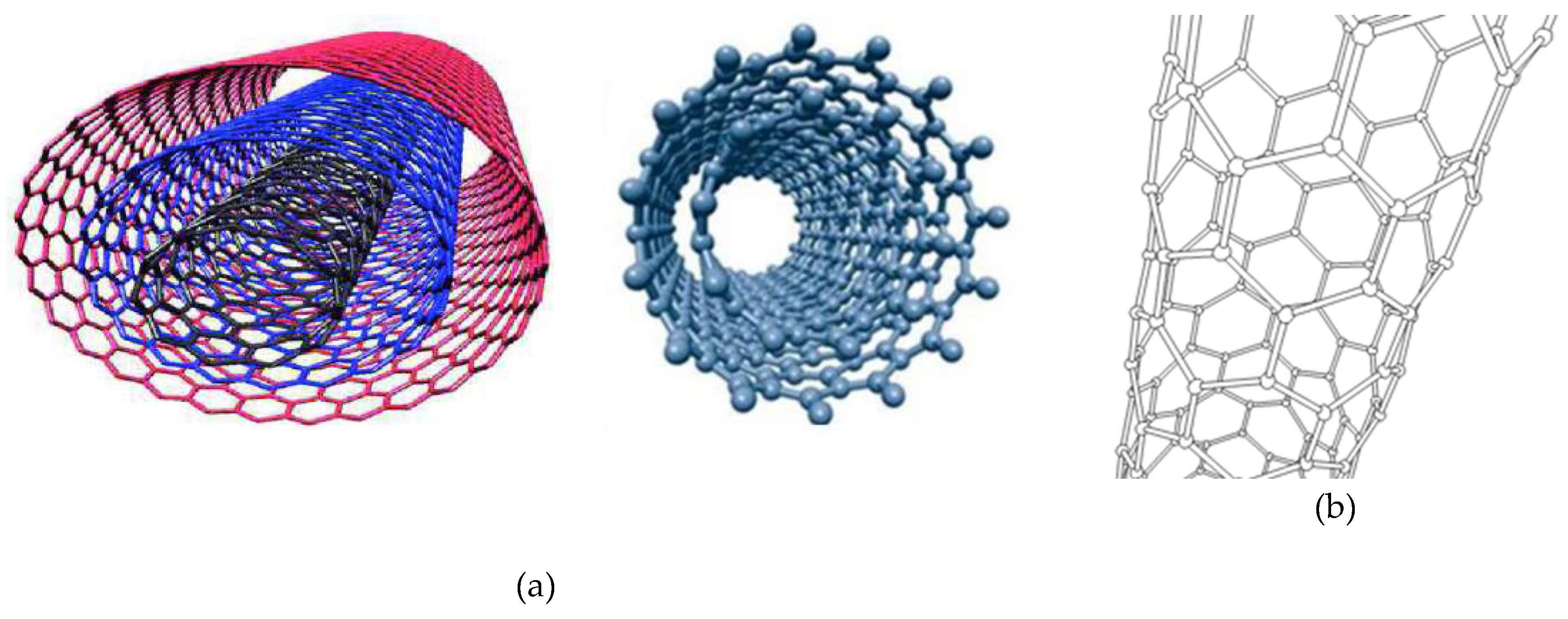
Figure 2.
Limitations and associated undesired side effects of chemotherapy for treating tumors.
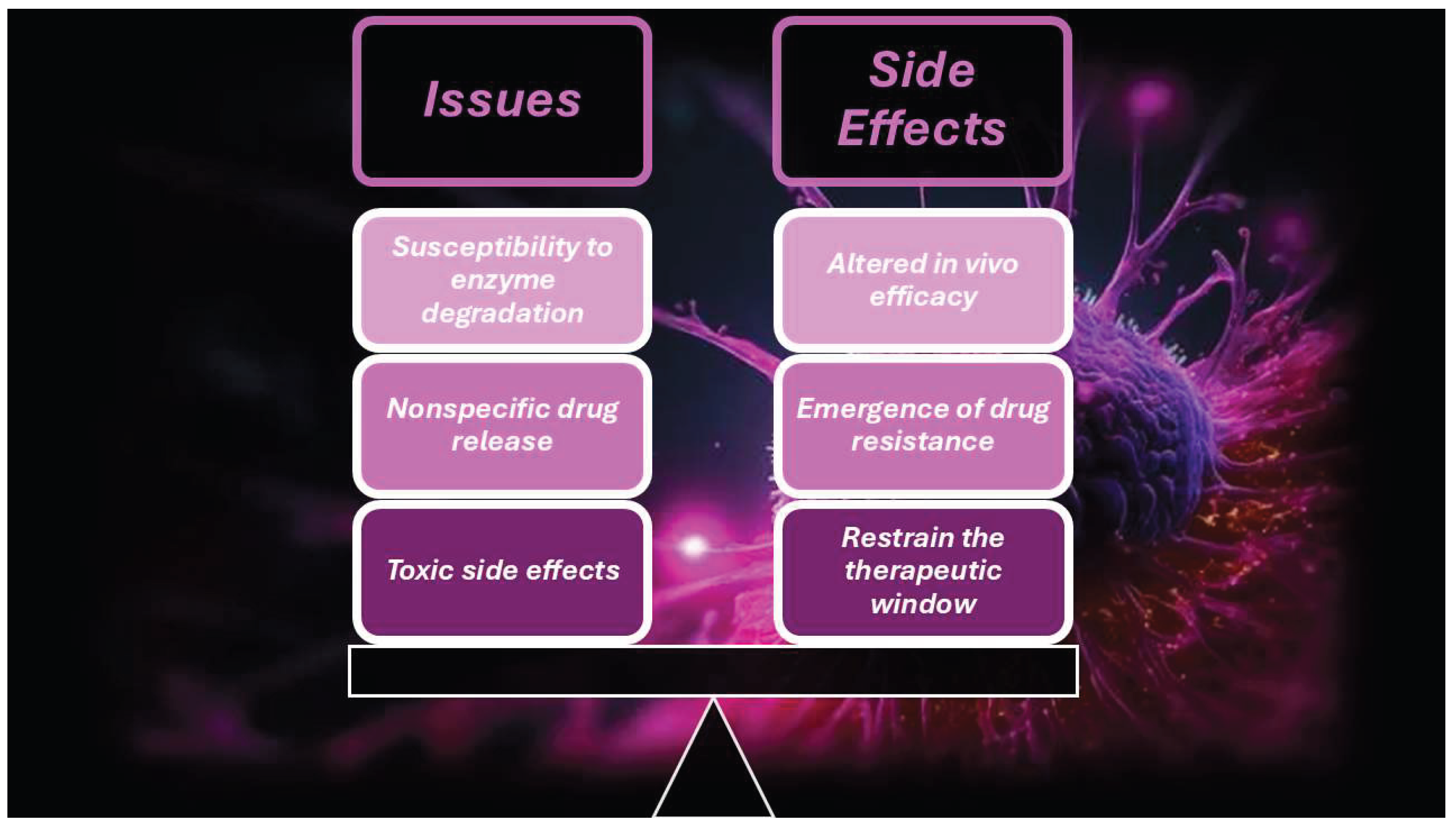
Figure 3.
(A) Schematic representation of -COOH modified SWCNT-PEG-PEI nanocarriers and the DOX-loading process [70]. The image has been reproduced by a paper published in diamond open access under the CC BY 4.0 licence (https://creativecommons.org/licenses/by/4.0/, accessed on 24 May 2025), which permits you to copy and redistribute the material in any medium or format for any purpose, even commercially.
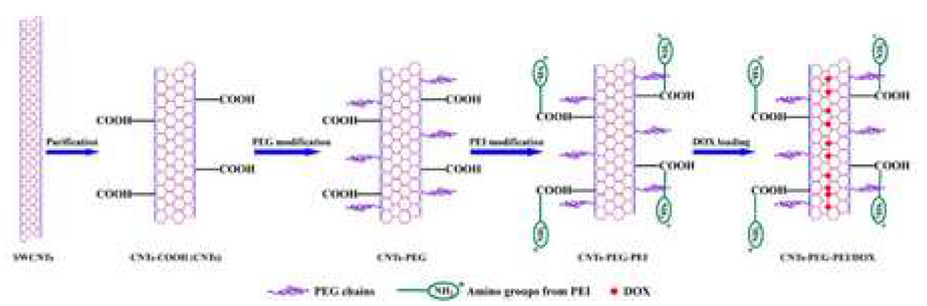

Table 1.
Examples of in vitro and in vivo applications of CNTs-based nanocarriers associated with different types of anticancer therapies.
Table 1.
Examples of in vitro and in vivo applications of CNTs-based nanocarriers associated with different types of anticancer therapies.
| CNTs | Measures | Cell Line/Animal Models | Drug(s) | Concentration/Dose | Results | Refs |
| MWCNTs | L = 110–980 nm | H1299 | MTX | 7.72×10⁻⁵ -1.51×10⁻³ mg/mL | pH dependant selective delivery of MTX to H1299 Low damage to healthy MRC-5 |
[41] |
| MWCNTs | L = 330 nmØ = 30 nm | BM-MSCs, MDA-MB-231 CD44high, CD24low CSCs | Pt-NPs, PBI | 100 µM | Cause cell cycle arrest, diminish drug resistance Impede DNA repair in breast CSCs. |
[49] |
| CNTs | N.R. | Kunming mice | Span, PEG, FA Paclitaxel | 350 mg/kg | Penetrate breast tumours Inhibited development, induced tumour cells death |
[50] |
| SWCNTs | N.R. | HT-29 | Porphyrin, PEG | 25 mg/mL | No antitumour activity | [54] |
| MWCNTs | Ø = 17.1±3.0 nm | C540 Male Balb/c mice | Poly pyrrole | 250 mg/mL, 10 mg/kg | Concentration-dependent cytotoxicity under multi-step ultrasonic irradiation (8.9% cell viability for 250 mg/mL) 75% Necrosis and 50% TVR after 10 days of SDT |
[51] |
| MWCNTs | Ø = 10 nmL = 5–15 µm | Male Balb/c mice | CREKA | MWNTs-PEG 2 mg/kg CMWNTs-PEG 4 mg/kg |
6.4 Times ⬆ accumulation in tumour tissue Xenograft eradicated after 4 cycles of illumination |
[52] |
| MWCNTs | Ø = 5–20 nmL = 1–10 µm | HepG2, HeLa cells C57BL/6J female mice |
PEG-O-CNTs | 0–1000 mL 1 mg/mL injected in tumour at 200 µL/cm3 |
⬆Cell toxicity of PEG-O-CNTs, O-CNTs, pure CNTs Continuous-wave NIR laser diode (808 nm) for 10 min ⬆TVR in animal group (PEG-O-CNTs) |
[53] |
L = Lenth; Ø = diameter; PEG-O-CNTs = oxidised CNTs conjugated to polyethylene glycol (PEG); FA = folic acid; Pt-NPs; PBI = polybenzimidazole; MTX = methotrexate; SDT = sonodynamic therapy; TVR = tumour volume reduction; PTT = photothermal treatment; ⬆ = higher; CSCs = cancer stem cells; BM-MSCs = bone marrow mesenchymal stem cells (BM-MSCs); Pt-NPs = platinum nanoparticles; CREKA peptide = linear polypeptide composed of 5 amino acids arranged by Cys-Arg-Glu-Lys-Ala. 3.1. Carbon Nanotubes-Based Drug Delivery Systems for Target Anticancer Therapy.
Table 2.
Functionalization of CNTs through various molecules and their applications as anticancer agents.
Table 2.
Functionalization of CNTs through various molecules and their applications as anticancer agents.
| CNTs | Functionalizing molecules | Effectiveness | Tumor model | Biocompatibility test | Refs. |
| SWNTs | PEG | ⬆Solubility, prevent particle aggregation, ⬇side effects | Gastric cancer | ⬇Toxicity towards normal tissue | [61] |
| SWNTs/MWNTs | PEI | ⬆Solubility, homogeneity, dispersity, ⬇particle size ⬆Positive charge to interact with siRNA |
Cervical cancer | N/A | [62] |
| SWNTs | HA | ⬆Stability in serum, overcome MDR Target CD44-overexpressing cancer cells |
Ovarian cancer | No cytotoxicity to normal tissue No drop in mice weight |
[63] |
| MWNTs | Chitosan | ⬆Solubility, ⬆cell-penetrating ability, ⬇toxicity | Breast cancer | ⬇Cytotoxicity | [64] |
| MWNTs | PLGA | ⬆Dispersity, ⬇toxicity, provide, tune the temporal release Provide attachment sites for drugs |
Osteosarcoma | ⬇Cytotoxicity in normal cells | [65] |
| SWNTs | CD33 mAB | Recognize and specifically target the GNM-CD33+ cells | Glioblastoma | N/A | [66] |
| SWNTs | IGF1R mAB/HER2 mAB | Target IGF1R and HER2 surface receptors | Breast cancer | ⬇⬇⬇In vitro toxicity in normal cells | [67] |
| SWNTs | RGD peptide | Target αvβ3-expressing cancer cells | MM | ⬇Toxicity in vitro | [68] |
| SWNTs | EGF receptor | Active targeting ability, ⬆uptake of drugs | HSC, NSC | ⬇⬇⬇Toxicity in vitro and in vivo | [69] |
MDR = Multidrug resistance; MM= malignant melanoma; HSC = head squamous carcinoma; NSC = neck squamous carcinoma; enhanced, improved, high, higher; low, lower, decreased, reduced; PEG = polyethylene glycol; PEI =polyethylene amine; IGF-1R = insulin-like growth factor receptor 1R; WS = water solubility; HER2 = human endothelial receptor 2; RGD = arginyl-glycyl-aspartic acid; EGF = epidermal grow factor.The following Figure 3 schematizes the structure of a -COOH polyethylene glycol (PEG) and polyethylene imine (PEI) modified SWCNTs-based nanocarrier, developed by Yang et al., and the doxorubicin (DOX)-loading process [70].
Table 3.
In vitro and in vivo experiments which used modified CNTs as drug and gene delivery systems for various chemotherapeutics and nucleic acids.
Table 3.
In vitro and in vivo experiments which used modified CNTs as drug and gene delivery systems for various chemotherapeutics and nucleic acids.
| Drug/gene | Modified | Cancer type | In vitro | In vivo | CNTs | Therapeutic Outcome | Refs |
| Pt (IV) drug, PD | PL-PEG F | Testicular cancer | Ntera-2 | N.R. | SWCNT | 25 Folds ⬆ cytotoxicity | [80] |
| CP | Covalent amide linkage | Head, neck cancer | HNCC | N.R. | SWCNT | ⬆Effective than free CP and siRNA conjugate | [69] |
| CA | Interior filling | Bladder cancer | EJ28 | N.R. | SWCNT MWCNT | Controlled release properties | [81] |
| OP | PEG600 F | Colorectal cancer | HT29 | N.R. | MWCNT | Delayed cytotoxic activity | [82] |
| DOX | PEGylation F | N.R. | Ascites Sarcoma 180 | N.R. | SWCNT | ⬆Retention of drug in situ ⬇ Effect on other tissues |
[83] |
| Not-F | Breast cancer | MCF-7 | N.R. | MWCNT | ⬆ Cytotoxicity | [84] | |
| P-gp F | Leukaemia | K562 | N.R. | SWCNT | ⬆Cellular uptake 23 folds ⬆ cytotoxicity |
[85] | |
| PTX | PEGylated F | Lung cancers | A549, NCI–H460 | N.R. | SWCNT | ⬆ PTX activity | [86] |
| Ovarian cancer | OVCAR3 | N.R. | SWCNT | Chemosensitizer, ⬆cell death | [87] | ||
| Poly citric acid F | Lung, ovary cancer | A549, SKOV3 | N.R. | MWCNT | ⬆Potency | [88] | |
| PEGylated F | N.R. | N.R. | Murine 4T1 | SWCNT | ⬇⬇⬇Tumour growth 10 folds ⬆ uptake |
[89] | |
| SB-T-1214, TAXO | Biotin F | Lung cancer, leukaemia | L1210FR, L1210 W138 | N.R. | SWCNT | ⬆ Target due to biotin | [90] |
| GEM | Folic acid F | Breast cancer | MCF-7 cells | N.R. | MWCNT | ⬆Cytotoxic than free GEM | [91] |
| PEGylated F | Pancreatic cancer | N.R. | BxPC-3–B/c | ⬇Metastatic lymph nodes | |||
| HIF-1α siRNA | Cationic F | N.R. | N.R. | MiaPaCa 2/HRE MM |
SWCNT | ⬇⬇⬇Tumour growth | [92] |
| Anti-EGFP siRNA | PEI and Pyridinum F | Lung cancer | H1299 | N.R. | MWCNT | ⬆Cytotoxicity | [93] |
| Cyclin A2 siRNA | Amine (NH3+) F | Leukaemia | K562 | N.R. | SWCNT | ⬇Cell proliferation and apoptosis |
[94] |
| PLK 1 siRNA | NH3+ F | Lung cancer | Calu6 | LCXGM | MWCNT | ⬇⬇⬇Tumour | [95] |
| siNEG siRNA | Amidation with PUT SPERD, SPER | Lung cancer | A549 | N.R. | SWCNT MWCNT | ⬇Tumour proliferation | [96] |
| Bcl9l siRNA | Aptamer and Pyr-PEI F | Breast cancer | MDA-MB-231 | N.R. | SWCNT | Targeted silencing activity | [97] |
| GAPDH siRNA | PEI F | Cervical cancer | HeLa | N.R. | SWCNT | Induced cell death | [62] |
| siTOX, siNEG siRNAs |
Cationic DD F | Cervical, lung cancer | HeLa, A549 | N.R. | MWCNT | ⬇Toxicity, effective delivery Gene silencing capabilities | [98] |
| PLK-1 & siTOX siRNAs | NH2-F or cationic liposomes | Breast, cervical, lung, melanoma, prostate, renal, kidney cancers | Calu6, A549, DU145 C-33A, MCF-7, HEK293 HeLa, NIH 3T3 B16F10 |
LCXGM | MWCNT | Apoptosis, ⬇⬇⬇tumour | [99] |
| PLK1 siRNA | NH4+ and Guanidium DD | Cervical cancer | HeLa | N.R. | MWCNT | Significant silencing of PLK-1 | [100] |
| hTERT siRNA+ NIR TPP | PEI F | Prostate cancer | PC-3 | TBM | SWCNT | Severe apoptosis, ⬇⬇⬇ tumour | [101] |
| pDNA | Cationic glyP F | Cervical cancer | HeLa | N.R. | SWCNT | Biocompatible, ⬆ transfection * | [102] |
| p53 plasmids | Ethylenediamine F | Breast cancer | MCF-7 | N.R. | SWCNT | Induced apoptosis | [103] |
| pDNA | Ammonium F | Cervical cancer | HeLa, CHO | N.R. | SWCNT & MWCNT | First reported gene carrier | [104] |
| GFP gene | PAMAM | Cervical cancer | HeLa | N.R. | MWCNT | ⬆ GFP gene transfection | [105] |
| dsDNA | Multi-F | Epithelial cancer | Mouse ovarian epithelial | N.R. | SWCNT | Targeted drug delivery | [106] |
| miRNA | PEI-g-GNR | Cervical cancer | HeLa | N.R. | MWCNT | New nonviral vector for in situ detection | [107] |
| dsODN-NF-kB | Carbodiimide F | Cervical cancer | HeLa | N.R. | SWCNT | Targeted successful deliver | [108] |
| ANT c-myc ODNs | PAMAM | Breast, liver cancer | MCF-7, MDA-MB-435, HepG2 | N.R. | MWCNT | ⬆⬆⬆Gene efficiency | [109] |
| MUC-1 Apt | Carboxylate F | Breast, lung cancer | MCF-7, Calu-6 | N.R. | MWCNT | Aptamer successful integration | [110] |
⬆ = High, higher, improved, enhanced; ⬇ = low, lower, decreased; reduced; ⬆⬆⬆ = strongly enhanced; ⬇⬇⬇ = strongly reduced; suppressed; N.R. = not reported; CP = Cisplatin; CA = carboplatin; OP = oxaliplatin; DOX = doxorubicin; PTX = paclitaxel; TAXO = taxoid; GEM = gemcitabine (GEM); EGPF = enhanced green fluorescent protein; CyA2 = cyclin A2; PLK 1 = polo-like kinase 1; GAPDH = glycerinaldehyd-3-phosphat-dehydrogenase; hTERT = human telomerase reverse transcriptase; pDNA = plasmid DNA; GFP = green fluorescent protein; dsDNA = double stranded; dsODN = double-stranded oligodeoxynucleotides; NF-kB = nuclear factor ‘kappa-light-chain-enhancer’ of activated B-cells; MUC-1 = mucin-1 protein; ANT = antisense; F = functionalization; * = lipofectamine 2000; PAMAM = poly-amidoamine; glyP = glycol-polymer; PEI= poly-ethylenimine; DD = dendron; Pyr = pyrazine; SPER = spermine; SPERD = spermidine; PUT = putrescine; PD = prodrug; LCXGM = xenografted mice; B/c = BALB/c mouse; TBM = tumour bearing mice; MM = mouse model.
Table 4.
Improved biocompatibility and anticancer efficiency of several modified CNTs-based carriers used to deliver in vitro and in vivo anticancer drugs and nucleic acids to treat various tumours.
Table 4.
Improved biocompatibility and anticancer efficiency of several modified CNTs-based carriers used to deliver in vitro and in vivo anticancer drugs and nucleic acids to treat various tumours.
| CNTs | Functionalised molecules | Effectiveness | Tumour model | Biocompatibility test | Refs. |
| MWCNTs | FA | Targeted and pH sensible release of DOX | N.R. | N.R. | [111] |
| SWCNTs | PEG-10–10%PEI/pDNA | Allowed dosage 58-fold ⬇ conventional IC50 ⬆WS, ⬆biological fluids dispersity |
AGS and L929 * | ⬇Toxicity | [61] |
| SWCNTs | PEG-PCL and PEG-PCL-PEI | Faster acidic DOX release; ⬆anticancer efficacy, ⬆WS | MCF-7 * | ⬆Biocompatibility | [112] |
| SWCNTs | ALG, CHI, FA, DOX | Precise magnetic field-dependent CNTs vibration causing cancer cells membranes destruction and DOX release | Lung cancer * | ⬆Biocompatibility | [113] |
| SWCNTs | COOH-, PEG- PEI- DOX | Faster acidic DOX release, ⬆Antitumor activity ⬆Dispersibility, ⬆Affinity for tumour cells. |
MCF-7 * | ⬇Toxicity to normal cells | [70] |
| SWCNTs | PEI-BET-DOX and SUR siRNA | ⬆⬆⬆pH-responsive lysosomal escape of siRNA ⬆Antitumor properties |
HeLa, A549 *,** | ⬇Toxicity to normal cells | [114] |
| SWCNTs | Chim/PEI/5-FU/CNT | ⬆⬆⬆Tissue penetrability, ⬆⬆⬆DL, CT allowed ⬇Invasion and proliferation, apoptosis in 5-FU-RCCs |
Gastric cancer * | ⬆Biocompatibility | [115] |
| SWCNTs | II-NCC | ⬆⬆⬆SWCNTs water dispersion; ⬆ Anticancer effects of CAP | Caco-2 * | ⬆Biocompatibility | [116] |
| MWNTs | iRGD-PEI-MWNT-SS-CD/pAT2 | ⬆WS and complexation capacity by PEI, ⬇⬇⬇ Tumour growth Target smart release in TRE by SS |
A549 *,** | ⬆Biocompatibility | [117] |
| SWCNTs | PEG and PEI | ⬆WS, ⬆ Cytotoxicity | AMJ13 * | ⬇Cytotoxicity to normal cells | [118] |
* In vitro experiments; ** in vivo experiments; DL = drug loading; SS = cystamine; FA = folic acid; PEG = polyethylene glycol; PEI = polyethylene imine; pDNA = plasmid DNA; PCL = polycaprolactone; ALG = sodium alginate; CHI = chitosan; DOX = doxorubicin; BET = betaine; SUR = surviving; siRNA = silencing RNA; Chim = chimera; 5-FU = 5-fluorouracil; II-NCC = N, carboxy cellulose II; iRGD = tumour-homing peptide; CD = candesartan; pAT2 = plasmid AT2; CAP = capecitabine; ⬆ = improving, improved, augmented, high, higher; ⬇ = low, lower, decreased; reduced; ⬆⬆⬆ strongly enhanced, increased; suppression; WS = water solubility; TRE = tumour reducing environment; CT = combination therapy; RCCs = resistant cancer cells. 3.2.1. Carbon Nanotubes-Based Drug Delivery Systems for the Tumor Microenvironment (TME)- and Cite Specific-Target Cancer Therapy.
Table 5.
Some examples of application of CNTs-based DDSs to deliver the transported material to specific intracellular sites and TME constituents.
Table 5.
Some examples of application of CNTs-based DDSs to deliver the transported material to specific intracellular sites and TME constituents.
| CNTs | TT/modulation | DDS | TM | Model | Effectiveness | Refs. |
| SWNTs | Cytoplasm | SWNT-PS/siRNA | PTT, GT | Hela | ⬆ Gene transport capacity, ⬆antitumor activity Controlled gene release |
[126] |
| Cytoplasm | SWNT-CY7-IGF-1Ra | PTT, IT | ASPC-1, BXPC-3 PANC-1 SW1990 * |
Precise tumor-targeting therapy, ⬆ body weight ⬆Survival rate of tumor-bearing mice |
[127] | |
| Mitochondria | SWNTs-PL-PEG-NH2 | TAT | H22 liver cancer | Selective destruction of tumor mitochondria Tumor cell apoptosis |
[128] | |
| CSCs | N.R. | IT | MDA-MB-231 breast cancer | Realize active targetability toward breast CSCs | [124] | |
| MWNTs | Antigen-presenting cells | MWNTs-CpG-αCD40-OVA | IT | B16F10 melanoma | ⬆Co-loading ability of OVA, CpG and anti-CD40 Ig ⬇Tumor growth and metastasis |
[129] |
| Tumor vasculature | iRGD-PEI-MWNT-SS-CD/pAT2 | CT | A549 lung cancer | ⬇⬇⬇Tumor growth and neovascularization | [117] |
TT = Therapeutic target; DDS = drug delivery system; GT = gene therapy; IT = immunotherapy; CT = chemotherapy; * pancreatic cancer cells; CD40 = clusters of differentiation 40; CpG = cytidine-phosphate-guanosine; CSCs = cancer stem cells; IGF-1Ra = insulin-like growth factor-1Ra; MWNTs = multi-wall carbon nanotubes; OVA = ovalbumin; pAT2 = plasmid angiotensin II type 2 receptor; PEI = polyetherimide; PEG = polyethylene glycol; PL = peptide lipid; PTT = photothermal therapy; SWNTs = single-wall carbon nanotubes; SS = disulphide bond; ⬆ = high, higher, improved, enhanced; ⬇ = low, lower reduced; ⬇⬇⬇ = suppression, inhibition.
Table 6.
Application of CNT-based DDSs in targeting various intracellular therapeutic sites and tumour microenvironment (TME) components.
Table 6.
Application of CNT-based DDSs in targeting various intracellular therapeutic sites and tumour microenvironment (TME) components.
| TS | DDS | TM | Tumour model | Effectiveness | Biocompatibility | Refs. |
| Cytoplasm | MWNTs/SOR/siRNA | GT+CT | Liver | ⬆SOR release, ⬆ siRNA stability, ⬆anticancer effects | N/A | [130] |
| Cytoplasm | SWNT-HIF-1α/siRNA | GT | Pancreas | Transfected tumour, activated RNAi response, ⬇⬇⬇TG | Minimal harm to NCs | [92] |
| Cytoplasm | Ox-DWNT/siRNA | GT | Prostate | Prevent survivin creation and induce apoptosis by the release of siRNA into cytoplasm | Good biocompatibility | [131] |
| Nucleus | PEG/SWNTs/DOX | PTT+CT | Breast | ⬆Delivery effectiveness, ⬆DOX localization/accumulation inside nucleus, ⬆⬆ killing of cancer cells |
N/A | [132] |
| Nucleus | SWNTs-carrier | CT | Colorectal | Targeted therapy, regulated medication release | Good biocompatibility | [133] |
| Nucleus | f-SWNTs/p53 | GT | Breast | ⬆Transport and targeting gene into the nucleus, apoptosis | N/A | [103] |
| Mito | MWNT-Rho (PtBzt+BP) | CT | Ovarian | Still ⬇ selective | Still too much side effects | [134] |
| Mito | PEG/CNTs/ABT737 | CT | Lung | ⬆Cell targeting to mitochondria, apoptosis | ⬇Toxic to NHFB NCs than A549 | [135] |
| Mito | P-D-CS-CNTs | PTT | Bladder | ⬆Mito targeting with damage, ROS burst, cancer cells death | N/A | [136] |
| Mito | PL-PEG-SWNT | PAT | Breast | Mito malfunction, mito outer membrane permeabilization | ⬇Toxicity, no harm to EI | [137] |
| ECM | MWNTs | PTT | Epidermoid | ⬇⬇⬇Tumours, ⬇volume, collagen destruction, cell damage | N/A | [138] |
| CCSs | SWNT-Raw, SWNT-COOH | CT | Osteosarcoma | Activated TGFβR1, ⬇⬇⬇ its signalling, ⬇ OSCs population | No toxicity to normal cells | [139] |
| TV | DOX/CD-CNT, CUR/CD-CNT | PTT+CT | Hepatocellular | ⬆ DEE and achieved sustained release of both drugs | No damage to normal cells | [140] |
| TV | iRGD-PEI-MWNT-SS-CD/pAT2 | CT | Lung | ⬆CU, TE, ⬇⬇⬇ angiogenesis, ⬇⬇⬇ tumour growth | No damage or inflammation No damage to tissues § | [117] |
| PD-1/PD-L1 | Rg3-CNT | IT | Breast | Inhibit PD-1/PD-L1 axis and the TNBC cell growth | N/A | [141] |
| IMs | MWNTs-DOX and MWNTs-CpG | IT+CT+PTT | Melanoma | ⬇⬇⬇Tumour growth, ⬆CD4+, CD8+ T cells, ⬆TAM shifting ⬇Tregs in TME | No-toxicity to the organs* | [142] |
Mito = Mitochondria; Rg3 = ginsenoside component of puffed ginseng with anti-cancer activities; RGD = arginyl-glycyl-aspartic acid; ECM = extracellular matrix; CCSs = cancer stem cells; IMs = immune cells; TV = tumour vasculature; TME = tumour microenvironment; DDS = drug delivery system; SOR = sorafenib; HIF-1α = hypoxia-inducible factor-1 alpha; TM = therapeutic modality; PTT = photothermal therapy; PAT = photoacoustic therapy; Ox-DWNT = oxidized double walled carbon nanotubes; CT = chemotherapy; GT = gene therapy; f-SWNTs-p53 = functionalized SWCNTs/plasmid 53 complexes; PEG = polyethylene glycol; Rho/(PtBzt+BP) = Rhodamine-110/ Platinum (IV) prodrug+3-bromopyruvate; ABT737 = selective small molecule B-cell lymphoma 2 (Bcl-2) Homology 3 (BH3) mimetic; CS = chitosan; PL = phospholipid; NCs = normal cells; CUR = curcumin; DOX = doxorubicin; PEI = polyethylene imine; CD = candesartan; pAT2 = plasmid AT2; SS =cystamine; ⬆ = high, higher, improved, enhanced; ⬇ = low, lower reduced; ⬇⬇⬇ = suppression, inhibition; EI = epithelial injury; * = liver, spleen, kidneys, heart and lungs in mice; § = blood, hepatic or kidney function in mice; TG = tumour growth; DEE = drug entrapment efficiency; CU = cellular uptake; TE = transfection efficiency; TGFβR1 = TGFβ type I receptor; TAM = tumour-associated macrophage.
Table 7.
Application of modified CNTs as nanocarrier for the tumour microenvironment (TME)-responsive release of anticancer drugs.
Table 7.
Application of modified CNTs as nanocarrier for the tumour microenvironment (TME)-responsive release of anticancer drugs.
| CNTs | Functionalizing molecules | Effects | TM | Biocompatibility | Refs. |
| MWNT | DOX, Cis, PEG, FA | Stopped Cisplatin release, pH-sensitive release, synergic antitumor impact | L929, MCF-7 | ⬆Biocompatibility | [148] |
| MWNT | MnO2, PEG, Ce6 | MnO2 breakdown of H2O2 produced ROS thus ⬆Ce6-mediated PDT PEG ⬆ WD, ⬆ cell death by ROS, ⬆Ce6 release, MRI-guided PDT and PTT Synergic ⬇ tumour growth, in vitro and in vivo tumour removal |
HeLa | [149] | |
| SWCNTs | DOX-CS/PNIPAAm | ⬆Release at 40 C than 25 C, at pH 5.0 than pH 7.4 ⬆ dual stimuli DOX release (NIR+AE of TME) |
HeLa | [150] | |
| SWNTs | PEG | ⬆ WS, prevent PA | Gastric cancer | ⬇Toxicity to NTs | [61] |
| SWNTs/MWNTs | PEI | ⬆WS, homogeneity and dispersity, ⬇ particle size, ⬆ PC, ⬆interaction with siRNA | Cervical cancer | N/A | [62] |
| SWNTs | HA | ⬆SS, ⬆target CD44-overexpressing cancer cells, overcome MDR | Ovarian cancer | No cytotoxicity to NTs | [63] |
| MWNTs | CS | ⬆Water solubility and cell-penetrating ability | Breast cancer | ⬇Cytotoxicity | [64] |
| MWNTs | PLGA | ⬆Dispersity, ⬆ attachment sites, tuned temporal release | Osteosarcoma | ⬇Cytotoxicity in NC | [65] |
| SWNTs | CD133 mAB | Recognize and specifically target the GNM-CD33+ cells | Glioblastoma | N/A | [66] |
| SWNTs | IGF1R mAB and HER2 mAB | Target IGF1R and HER2 surface receptors | Breast cancer | Negligible toxicity in NCs | [67] |
| SWNTs | RGD peptide | Target αvβ3-expressing cancer cells | MM | ⬇Toxicity in vitro | [68] |
| SWNTs | EGF receptor | Active targeting ability and ⬆ uptake of drugs | HC, NC | Negligible toxicity | [69] |
DOX = Doxorubicine; Cis = cisplatin; PEG = polyethylene glycol; FA = folic acid; MnO2 = manganese dioxide: Ce6 = Chlorin e6; CS/PNIPAAm = chitosan-poly (N-isopropyl acrylamide; TME = tumour microenvironment; DDDS = dual-drug delivery system; MRI = magnetic resonance image; AE = acidic environment; ⬆ = improving, improved, augmented, high, higher; ⬇ = low, lower, decreased; reduced; HA = hyaluronic acid: PA = particles aggregation; NTs = normal tissues; PC = positive charge; NCs = normal cells; PLGA = polylactic-co-glycolic acid; CD33 = cluster of differentiation-133; IGF1R = insulin-like growth factor 1 receptor; HER2 = Human endothelial receptor 2; RGD = arginyl-glycyl-aspartic acid; MM = malignant melanoma; HC = head carcinoma; NC = neck carcinoma; WD = water dispersibility; WS = water solubility; SS = serum stability; MDR = multi drug resistance; N/A = not acquired; TM = tumour model.
Table 8.
Application of modified CNTs in anticancer photothermal therapy (PTT).
| CNTs | Functional molecules | Effectiveness | Tumour model | Observations | Refs. |
| MWCNT | Ag NPs | ⬆ NIR LA at 670 nm | Melanoma B16/F10 cells | ⬆CNT/Ag optical absorption to CNTs and Ag NPs | [181] |
| MWCNT | PEG | ⬆ MWCNTs water dispersibility | Melanoma | ⬇⬇⬇Tumour size in the cancerous mice | [53] |
| MWCNT | –COO, –COOPt, –Pt NPs | -COO ⬆ CNTs solubility, Pt NPs ⬆ LA at 1064nm | Cancer PC3 cells | PP ⬆cytotoxicity due to ⬆temperature of CCs | [161] |
| MWCNT | –COO, –COOAu, –Au NPs | -COO ⬆ CNTs solubility, Au NPs ⬆ LA at 1064nm | MCF7 cells | PP ⬆cytotoxicity due to ⬆temperature of CCs | [160] |
| MWCNT | MTX, PEI, FA | MTX-based ACA, FA-based TP, PEI-based hydrophilicity | MCF7 cells | ⬆Synergistic effect of MTX and PTT (808 nm laser) MWCNT-MTX-PEI-FA are absorbed selectively | [173] |
| MWCNT | PS MAA | Antibody against prostate-specific MA | Prostate cancer LNCaP cells | > 80% cell ablation with a single 30-s (a 532-nm) | [182] |
| MWCNT | Au NSs | ⬆Biocompatibility, ⬆photothermal efficiency | Melanoma B16F10 mice cells | MWCNT/Au NSs have 12.4% ⬆ PE (808 nm) | [154] |
| MWCNT | PEG | ⬆Photothermal transduction (NIR) | Benign prostatic hyperplasia | ⬇Prostate size, apoptosis in PEC | [183] |
| SWCNTs and MWCNTs | PL, SL | BDS with ⬆ PE, TS | HeLa, MRC-5 cells | Muted surviving expression, ⬇⬇⬇TG PE under 808 nm NIR light |
[126] |
| MWCNT | TiO2 NPs | Ability to absorb and scatter the 808 nm NIR light | Melanoma | Selective tumour death, ⬇HR, ⬆necrosis | [184] |
| SWCNT | anti-IGF-1R, CY7 | WS, BS, LT SWCNT-CY7-IGF-1R | PDCA | Target to PDCA for dye imaging-guided PTT | [127] |
⬇⬇⬇ = highly lowered, suppression; ⬆ = improving, improved, augmented, high, higher; ⬇ = low, lower, decreased; reduced; PEG = polyethylene glycol; LA = laser absorption; MTX = methotrexate; PEI =polyethylene amine; FA = folic acid; PS MAA = prostate-specific membrane antigen antibody; Au NSs = gold nano stars; PL = peptide lipid; SL = sucrose laurate; anti-IGF-1R = insulin-like growth factor receptor anti-IGF-1R antibody; CY7 = imaging agent; TiO2 NPs = titanium oxide nanoparticles; WS = water soluble; BS = bio stable; LT = low toxic; BDS = bifunctional delivery system; PDAC = pancreatic ductal adenocarcinoma; PE = photothermal effects; TS = temperature sensitivity; BPH = benign prostatic hyperplasia; PEC = prostatic epithelial cells; PTT = photothermal therapy; PP = plasmon phenomenon; CC = cancer cells; ACA = anticancer activity; TP = targeting power; TG tumour growth; HR = heat resistance.
Table 9.
Application of modified CNTs in anticancer photodynamic therapy (PDT).
| CNTs | Functionalized molecules | Effectiveness | Tumour model | Biocompatibility test | Refs. |
| SWCNT | HA, Ce6 | ⬆SWCNTs stability, WD (HA), Ce6-based ACA by PDT | Colon cancer cells | Colon cancer cells death, ⬆PDT capacity | [165] |
| SWCNT | ZnMCPP | >50% ⬆ singlet and triplet oxygen quantum yields | MCF-7 breast cells | 64% ⬇ CV at 40 M (C1), 97-95% ⬇CV (C2,3) | [163] |
| MWCNT | OEG, DOX, FA | OEG used to link DOX with CNT DOX used as anticancer drug FA act as the system’s targeted ligand |
HeLa, L929, A549 | ⬆Killing of cancer cells when exposed to light. | [185] |
| SWCNT | AA, ZnMCPP | ⬆ Lifetimes, quantum yields, singlet oxygen quantum yields | MCF-7 | 77% cytotoxicity by combined therapy | [164] |
ACA = anticancer activity; ⬆ = improving, improved, augmented, high, higher; ⬇ = low, lower, decreased; reduced, reduction; HA = Hyaluronic acid; Ce6 = chlorine e6; ZnMCPP = zinc mono carboxy phenoxy phthalocyanine; OEG = ethylene glycol oligomers; DOX = doxorubicin; FA = folic acid; AA = ascorbic acid; WD = water dispersity; C1, C2 and C3 = complexes 1, 2 and 3; CV = cell viability.
Table 10.
Applications of CNTs-based drug delivery systems for enhanced phototherapy.
| CNTs | FM | Effectiveness § | TM | Biocompatibility | NIR laser | Refs. |
| MWCNTs | PVPy-S-PEG-FA, DOX | ⬆ATA, ⬆WD, ⬆BCT, ⬆ DL, ⬆PC of CNTs by PVPy, pH-sensitive | HeLa | ⬆Biocompatibility | 808 | [169] |
| MWCNTs | ED | ⬆ATA, ⬆AA of MTX, ⬆CD by SE, MWCNT-ED-MTX = MWCNT-ED-FA* | MCF7 | N.R. | 808 | [173] |
| MWCNTs | Ge, Le | ⬆AA by LH, ⬆biocompatibility, ⬆HDP, ⬆PC | MCF-7 | ⬇Danger to NC | 808 | [171] |
| MWCNTs | MTX, PEI, FA | ⬆ATA, ⬆WD by PEI, ⬆AA of MTX by SE with PPT | MCF-7 | N.R. | 808 | [173] |
| SWCNTs | ANXA5 | ⬆Stimulation of IS, AE by SWCNT-ANXA5 SE | EMT6 BCCs | N.R. | 980 | [155] |
| CNTs | MXene | ⬇CNT aggregation, DOX DL = 85.6%, ⬇⬇⬇HeLa cells by PTT+DOX SE | HeLa | N.R. | 650/808 | [174] |
FM = Functionalizing molecules; TM = tumour model; PVPy = poly-N-vinyl pyrrole; S = ; PEG = polyethylene glycol; ED = ethylenediamine; Ge = gemcitabine; Le = lentinan; FA = folic acid; MTX = methotrexate; PEI = polyethyleneimine; ANXA5 = annexin 5 is a phospholipase A2 and protein kinase C inhibitory protein with calcium channel activity and a potential role in cellular signal transduction, inflammation, growth and differentiation; MXene = tuneable family of 2D carbides and nitrides; ⬆ = enhancing, enhanced, improving, improved, augmented, high, higher; ⬇ = low, lower, decreased; reduced, reduction; BCT = blood circulation time; WD = water dispersibility; ATA = active targeting ability; BCCs = breast cancer cells; AA = anticancer activity; PC = photothermal characteristics; HDP = hydrophilicity; IS = immune system; DL = drug loading; * for anticancer effects; CD = cell death; SE = synergistic effects; LH = localized hypothermia; § = under NIR radiation; AE = abscopal effect; ⬇⬇⬇ = suppression; NCs = normal cells.
Table 11.
Application of modified CNTs in combined PTT and PDT for cancer treatment.
| CNTs | FM | Effectiveness | TM | Biocompatibility | Refs. |
| SWCNHs | ICG | ⬆WD, ⬆photostability, ⬆AA by PTT+PDT SE | 4T1 BCCs * | ⬆Photostability, ⬆biocompatibility. | [175] |
| SWCNHs | Ce6, Gd3+ | ⬆IS, ⬆ATA, ⬆TP, ⬆ISR by PTT+PDT SE in AMMs | 4T1 | N.R. | [177] |
| SWCNHs | Hyp | ⬆WS ⬇4T1 growth by PDT+PTT SE | 4T1 * | ⬆Photostability, ⬆biocompatibility | [176] |
| MWCNT | MnO2, Ce6 | O2+H+ production by MnO2⬇TM, ⬆potential for IG PDT+PTT SE | HeLa * | ⬆Biocompatibility | [178] |
| SWCNT | PEG, Fe3O4, CQDs, DOX, sgc8c | ⬆AA by PTT+PDT+CT by SE | HeLa mice | ⬆Biocompatibility | [179] |
| MWCNT | MnO2, PEG, Ce6 | MnO2 create 1O2, ⬆PTT, ⬇hypoxia, ⬆ PDT, ⬆WD, ⬇⬇⬇ TD by MRI-guided PDT+PTT SE | HeLa * | ⬆Biocompatibility | [149] |
| MWCNT | mTHPC | mTHPC acted as PS, ⬆AA by PDT+PTT SE | SKOV3 OCCs | N.R. | [186] |
| SWCNT | HA, HMME | ⬆WS, HMME = PDT agent, ⬆AA by PDT+PTT SE | B16F10 MMCs | ⬆Biocompatibility | [167] |
SWCNHs = single walled carbon nano horns; ICG = indocyanine green; Ce6 = chlorine e6 photosensitizer; Gd3+ = gadolinio; Hyp = hypericin; PEG = polyethylene glycol; CQDs = carbon quantum dots; DOX = doxorubicin; sgc8c = aptamer; m-THPC = m-tetrahydroxy phenyl chlorin; HA = hyaluronic acid; HMME = hematoporphyrin monomethyl ether; FMs = functionalizing molecules; TM = tumor model; TD = tumor development; ⬆ = enhancing, enhanced, improving, improved, augmented, high, higher; ⬇ = low, lower, decreased; reduced, reduction; WD = water dispersibility; ATA = active targeting ability; TP = tumour penetration; BCCs = breast cancer cells; AA = anticancer activity; ISR = immune system response; SE = synergistic effects; ⬇⬇⬇ = suppression; WS = water solubility; OCC = ovarian cancer cells; CT = chemotherapy; PS photosensitizer; MMCs = mice melanoma cells; AMMs = advanced metastatic malignancies; * in vivo and in vitro experiments; IG = imaging guided; MRI = magnetic resonance imaging; N.R. = not reported.5. Carbon Nanotubes Application in Anticancer Gene Therapy.
Table 12.
CNTs application in anticancer immunotherapy.
| CNTs | FM | effectiveness | TM | Biocompatibility | Refs. |
| MWCNTs | COOH | ⬆WS, ⬆CSA, ⬆CP, ⬆MA | H22 HCCs | N.F. | [208] |
| MWCNTs | Rg3 | Puffed ginseng, ACA, Apoptosis, ⬇PD-L1 expression | TNBC | N.F. | [141] |
| SWCNTs | CpG | ACA in gliomas, ⬇CCP, ⬇invasion/migration | HCCsT116 | ⬆Mice survival rate | [209] |
| MWCNTs | DOX/ CpG | ⬆WD; no change in MWCNTs PS, ⬇TG, ⬆CD4+, ⬆CD8+, ⬆T cells | B16 MCs | Biocompatible to normal cells | [142] |
| SWCNTs | COOH/OVA | Primary immunization/mice, ⬆anti-ovalbumin antibody response | AMBCs | ⬇Toxicity, ⬇inflammation | [210] |
| MWCNTs | PDAEMA | ⬆Design flexibility, ⬆control on SARs, ⬆SE, ⬆CU | B16-F10 | ⬆Biocompatibility | [211] |
FM = Functionalizing molecules; Rg3 = Ginsenoside Rg3; CpG = complex; DOX = doxorubicin; OVA = Ovalbumin; PDAEMA = poly(2-dimethylaminoethylmethacrylate); WS = water solubility; WD = water dispersibility; ⬆ = enhancing, enhanced, improving, improved, augmented, high, higher; ⬇ = low, lower, decreased; reduced, reduction; ACA = anticancer activity; PS = physical structure; SAR = structure activity relationships; HCCs = hepatocarcinoma cells; TNBCs = triple-negative breast cancer; HCCsT116 = human colon cancer cell line T116; MCs = melanoma cells; AMBCs = activated mouse B cells; TM = tumor model; CSA = component system activation; CP = cytokines production; MA = macrophages activation; CCP = cancer cells proliferation; TG = tumor growth; SE = silencing efficiency; CU = cellular uptake; N.F. = not found.
Table 13.
Applications of CNTs in cancer diagnosis.
| CNTs | DM | Effect | Refs. |
| SWCNTs | RI | Spots of RSs were visible in colon-26 cells after 5-days administration of o-SWNTs-PEG | [222] |
| SWCNTs | RI | ⬆SERS-E suitable for labeling and fast RI of biological samples | [223] |
| SWCNTs | MRI | Excellent MRI functions for tumor diagnosis | [224] |
| MWCNTs | MRI | ⬆Longitudinal proton relaxation process, T1 enhanced MRI effect | [225] |
| MWCNTs | US | ⬆Contrast in ultrasonic imaging | [226] |
| MWCNTs | US | Strong, long-lived and ⬆-quality ultrasound signal after sonication treatment | [227] |
| SWCNTs | PAI | Clear and stable PA signals | [228] |
| MWCNTs | PAI | Precise target to the tumor site in vivo, ⬆PA imaging effect | [229] |
| MWCNTs | RNI | Easy and direct SPECT/CT imaging | [230] |
| SWCNTs | RNI | ⬆Tumor tissue accumulation for the subsequent radionuclide imaging | [231] |
| SWCNTs | NIR-FI | ⬆Signal-to-noise performance, ⬆specificity to ovarian tumors and tumor nodules | [232] |
| SWCNTs | NIR-FI | ⬆Resolution intravital tumor vessel images through the thick skin in live mice | [233] |
| MWCNTs | NBSs | ⬆Biosensing ability, wide linear range for detecting miR-21, ⬇⬇⬇detection limit | [234] |
| SWCNTs | NBSs | Broad detection range, ⬇detection limit, ⬆specificity to only OPN in prostate cancer | [235] |
| MWCNTs | NBSs | Simple detection of CDK1, report the enzymatic activity of CDK1 for cancer diagnosis | [236] |
| MWCNTs | NBSs | ⬆Selectivity/sensitivity to HPV-18, early, rapid, easy, and accurate diagnosis of cervical cancer | [237] |
RI = Raman imaging; NBSs = nano-biosensors; NIR-FI = near infrared fluorescence imaging; CNTs in cancer imaging; RNI = radionucleotide imaging; US = ultrasonography; PAI = photoacoustic imaging; MRI = magnetic resonance imaging; DM = diagnostic method; RS = Raman signal; SERS-E = surface-enhanced Raman scattering effect; SPECT = single photon emission computed tomography; CT = computed tomography; ⬆ = strong, enhancing, enhanced, improving, improved, augmented, high, higher; ⬇ = low, lower, decreased; reduced, reduction.
Table 14.
Carbon nanotube-based cancer detection techniques.
| CNTs | Cell line/Biomarkers | Linear Range | LoD | Techniques | Ref. |
| SWCNTs | PSA | N.R. | 250 pg/mL | Electrochemical | [259] |
| SWCNTs | PSA | 0.4–40 pg/mL | 4 pg/mL | Immune sensing | [260] |
| MWCNTs | AFP | 0.02–2.0 ng/mL | 8.0 pg/mL | Immune sensing | [261] |
| MWCNTs | CEA | 0.5–15.0 and 15.0–200 ng/mL | 0.01 ng/mL | Immune sensing | [262] |
| MWCNTs | AFP | 0.1–15.0 and 15.0–200.0 ng/mL | 0.08 ng/mL | Immune sensing | [263] |
| MWCNTs | CA 19-9 | 12.5–270.0 U/mL | 8.3 U/mL | Immune sensing | [264] |
| MWCNTs | hCG | Up to 600 mIU/mL | 14.6 mIU/mL | Electrochemical | [265] |
| MWCNTs | hCG | 0.8–500 mIU/mL | 0.3 mIU/mL | Electrochemical | [266] |
| CNTs | PSA | 1–100 ng/mL | 1.0 ng/mL | Electrochemical | [267] |
| MWCNTs | CA 125 | 1.0–30 and 30–150 U/mL | 0.36 U/mL | Electrochemical | [268] |
| CNTs | AFP | 1–55 ng/mL | 0.6 ng/mL | Immune sensing | [269] |
| MWCNTs | CA19-9 | 0–1000 U/mL | N.R. | Electrochemical | [270] |
| CNTs | GP73 | 0–80 ng/mL | 58.1 pg/mL | Immune sensing | [271] |
| CNTs | AFP | 0–64 ng/mL | 47.1 pg/mL | Immune sensing | [271] |
| CNTs | AKT2 gene | 1 pM–1 μM | 2 fM | Electrochemical | [272] |
| CNTs | CA 125 | 0.001–0.1 ng/mL/0.1–30 ng/mL | 0.5 pg/mL | Electrochemical | [273] |
| CNTs | Cyfra 21-1 | 0.1–10,000 ng/mL | 0.5 ng/mL | Fluorescence | [274] |
| CNTs | HepG2 | 10–105 cells/mL | 5 cells/mL | Electrochemical | [275] |
N.R. = Not reported.
Table 15.
Clinical trials assessing the possible application of CNTs in anticancer therapy.
| Trial Number | Sponsor | Device/Intervention | Problem | Participants | Study Status | Ending year | Ref |
| NCT01773850 * | UNC Lineberger Comprehensive Cancer Center | CNT x-ray source array for SDT of BN | BN | 54 | Completed | 2018 | [286] |
| NCT01420588 ** | Anhui Medical University | Of-AUNPs/CNTs-based chemical nano sensors | PCGLs | 1000 | Completed | 2020 | [287,288] |
SDT = Stationary digital tomosynthesis; BN = breast neoplasm; PCGLs = pre-cancerous gastric lesions; AuNPs = gold nanoparticles; CNTs = carbon nanotubes: Of = organically functionalized; * study title: Stationary carbon nanotube X-ray digital breast tomosynthesis scanner; ** study title: Diagnosis of gastric lesions from exhaled breath and saliva.
Table 16.
Study type assessment.
| Trial Number | Sponsor | Study Type | Ethics Approval | Status | Ref |
| NCT01773850 * | UNC Lineberger Comprehensive Cancer Center | OCPS | |
Completed | [286] |
| NCT01420588 ** | Anhui Medical University | OCPS | |
Completed | [287,288] |
OCPS = Observational cohort prospective study * study title: Stationary carbon nanotube X-ray digital breast tomosynthesis scanner; ** study title: Diagnosis of gastric lesions from exhaled breath and saliva; = positive ethical approval.
= positive ethical approval.
Table 17.
Risk of bias in randomized studies as assessed by the Cochrane Collaboration’s “Risk of Bias” tool.
Table 17.
Risk of bias in randomized studies as assessed by the Cochrane Collaboration’s “Risk of Bias” tool.
| Trial Number | RSG | AC | BPP | BOA | IOD | SR | Ref |
| NCT01773850 | |
|
|
|
|
|
[286] |
| NCT01420588 | |
|
|
|
|
|
[287,288] |
RSG = Random sequence generation; AC = allocation concealment; BPP = blinding of Participants and personnel; BOA = blinding of outcomes assessment; IOD = incomplete outcome data; SR = selective reporting.
Disclaimer/Publisher’s Note: The statements, opinions and data contained in all publications are solely those of the individual author(s) and contributor(s) and not of MDPI and/or the editor(s). MDPI and/or the editor(s) disclaim responsibility for any injury to people or property resulting from any ideas, methods, instructions or products referred to in the content. |
© 2025 by the authors. Licensee MDPI, Basel, Switzerland. This article is an open access article distributed under the terms and conditions of the Creative Commons Attribution (CC BY) license (http://creativecommons.org/licenses/by/4.0/).
Copyright: This open access article is published under a Creative Commons CC BY 4.0 license, which permit the free download, distribution, and reuse, provided that the author and preprint are cited in any reuse.


