Submitted:
28 May 2025
Posted:
28 May 2025
You are already at the latest version
Abstract
Ionizable lipid nanoparticles were a crucial contribution to the effective packaging and delivery of mRNA informational drugs for vaccines and therapeutic applications. However, thermal instability and the need for ultracold storage present significant challenges during distribution and administration, even during non-pandemic periods like epidemics. Ongoing efforts include engineering novel lipids or optimizing mRNA-LNP formulations with different buffers, excipients, and surfactants to enable extended storage at room temperature (RT) or refrigerated conditions (4 ˚C). Methods: In this study, six sugar-surfactant excipient combinations in tris buffer were evaluated for extending the stability of mRNA-LNPs. The experimentation included two phases of screening: first, the evaluation of six formulations under repeated freeze-thaw (−20 °C) cycles, and second, a long-term storage evaluation of the best formulations at RT, 4 °C, − 20 °C and −80 °C. LNPs were formulated with the ionizable lipids ALC-0315 or SM-102. Results: Sucrose-P188 and mannitol-F127 combinations effectively preserved physicochemical properties like encapsulation efficiency (EE), polydispersity index (PDI), and z-average (size) of mRNA-LNPs. Surfactants in the screened formulations reduced the aggregation of LNPs. Storage at −20 °C with sucrose-P188 extended LNP stability beyond ultra-low temperature requirements, showing potential for removing logistical hurdles to improve global delivery by eliminating the need for ultracold storage. Excipients in non-frozen conditions preserved the LNP quality attributes, but the RNA integrity was affected. On the other hand, mRNA degradation was minimized when frozen, but LNP quality was affected, underscoring the challenging trade-off during storage condition optimization. Conclusions: This work highlights the potential of improved formulations to preserve mRNA-LNP functionality and emphasizes the need for further systematic studies on a library of excipient and surfactant combinations, paving the way for stable storage under less stringent conditions.
Keywords:
thermal instability
; ALC-0315
; SM-102
; mRNA-LNP formulations
; excipients
; aggregation
; polydispersity index
; encapsulation efficiency
1. Introduction
Ionizable lipid nanoparticles (LNP) accelerated the approval of mRNA biologics for therapeutic and prophylactic applications by delivering mRNA efficiently in the cell. Previously, the clinical applicability of mRNA was severely limited by the instability of mRNA caused by the innate mRNA degrading responses of the host cell, as well as the inherent susceptibility to hydrolysis. The protective lipid layer in LNPs protected the mRNA from enzymatic degradation by cellular nucleases and facilitated the higher transfection efficiency of mRNA. Thus, LNPs with ionizable lipids enabled the licensing of mRNA-based drugs by efficient packing, overcoming the extra-cellular and intra-cellular barriers associated with RNA delivery. Consequently, these delivery vehicles contributed substantially to the success of the COVID-19 vaccines [1,2]. Despite the successes associated with the mRNA-LNP vaccines, challenges to meet clinical needs persist, even for the recently updated COVID-19 vaccines (Moderna Spikevax, Pfizer-BioNTech Comirnaty) [3] and for the prevention of lower respiratory tract disease (LRTD) caused by respiratory syncytial virus (mRNA vaccine mRESVIA by Moderna Inc.,) [3,4,5]. The challenges limiting the expansion of mRNA-LNP for therapeutic applications include thermal instability and reliance on ultra-cold storage chains to maintain therapeutic efficacy and effective shelf life [6]. These critical bottlenecks restricted field application and larger market penetration of the mRNA-based vaccine treatments into low-to-middle-income countries (LMICs) and prevented equitable distribution during the pandemic.
Multiple degradation mechanisms and their impacts have been described for mRNA-LNPs. Physical degradation, like aggregation or fusion of LNP particles, reduces the in vivo delivery efficiency [7,8]. Chemical degradation pathways, such as hydrolysis and oxidation of the mRNA and/or the lipid species, also hinder product shelf-life [9]. These challenges complicated the development of mRNA-LNP drug products for other priority diseases like cancer (individualized neoantigen cancer therapy), infectious diseases (CMV, EBV, HSV, VZV, HIV, Norovirus, etc.), and neglected, rare diseases like Propionic acidemia, Methylmalonic acidemia, Phenylketonuria, and Glycogen Storage Disease Type 1a [8,10]. Thus, the ongoing research efforts for improving mRNA-LNP products include enhancing the target specificity, delivery efficiency, stability and room-temperature-tolerant formulation.
To this end, two main methods to enhance lipid nanoparticle stability have been explored. The first method is through iterative lipid design, which introduces changes in the lipid chemical structures [2,11]. However, these studies require extensive testing of lipid libraries, and it is unlikely the resulting novel lipids would be used in previously approved products or mRNA therapeutics currently in development. The new generation of LNP preparations must undergo extensive, long-term pre-clinical and clinical evaluation. The second approach builds on the LNP compositions that have already been used in the clinic and focuses instead on the optimization of the buffer formulation and additional excipients to act as protective agents throughout the storage and cellular stresses involved while delivering mRNA vaccines and therapeutics.
Efficient chemical formulations should be purposefully developed using different buffering systems, sugars, surfactants and other excipients that stabilize the mRNA-packed LNP. Ideally, product stability should allow for storage at room temperature for up to 2 months. However, there is a huge gap in the mRNA-LNP-related temperature and stability-associated empirical studies and a huge scope for advancing the existing state-of-the-art approaches to increase the stability and shelf-life of the mRNA-LNPs. Currently, the approved mRNA-LNP products have limited stability at refrigerated and room temperatures (Table 1), impairing the distribution of these vaccines [7] and the need for cold-chain logistics.
Furthermore, during pandemic situations, such as in the case of SARS-COV-2, mRNA vaccines were stored in high concentrations of multiple doses to facilitate mass vaccination [12]. However, for personalized mRNA therapies, rare and orphan diseases, and non-emergency vaccine products, it is decreasingly feasible to employ high-concentration LNP stocks. Thus, achieving stability at reduced LNP concentrations is key to transitioning mRNA-LNP products into various therapeutic field applications. Also, the average time that the integrity of the RNA was maintained at 2-8 ˚C was from a few days to 4 weeks, compared to years or months of stability for other conventional vaccines like protein subunit vaccines, AAV, etc. [13,14].
Another strategy for enhanced stability is to lyophilize the mRNA-LNP product (solid formulations)[15,16]. The conventional vaccines can be stored as a lyophilized powder until the point of use, where they are suspended in buffers or nuclease and pyrogen-free sterile distilled water. However, no lyophilized, stable LNP formulations are commercially available to date. These factors add to the value and significance of building a robust dataset on the stability analysis of LNP formulations.
Herein, this work evaluates the physicochemical stability (EE%, size, PDI) of liquid formulations of mRNA-LNP in the presence of several excipients, both throughout freeze-thaw stresses and multi-week storage at reduced LNP concentrations. The study aims to increase the available data on LNP stability and improve our understanding of the behaviors of these particles across different temperatures and time points. Furthermore, we aimed to identify excipients that aid in preserving mRNA-LNPs both physically (properties associated with the integrity of LNP) and chemically (properties associated with mRNA integrity).
2. Materials and Methods
2.1. mRNA Synthesis and Purification
A DNA construct encoding for eGFP (pGEM4Z-EGFP) was purchased from Addgene (plasmid # 183475, created by Chantal Pichon)[17], and a construct encoding for nLuciferase (pcDNA-LUC-CBR2opt-T7AG-C1) was gifted by the National Research Council of Canada (NRC, Ottawa). The two DNA constructs were transcribed using the in vitro MEGAscript T7 Transcription Kit (Thermo Fisher Scientific, Waltham, MA, USA), following the manufacturer’s protocol. The resulting mRNA was purified by phenol-chloroform purification and quantified using the Quant-it Ribogreen RNA Assay Kit (Thermo Fisher Scientific, Waltham, MA, USA), with the expected final mRNA lengths of 993 nt for eGFP and 2,055 nt for nLuciferase (nLuc) constructs, respectively.
2.2. Lipid Nanoparticle Formulation
mRNA- LNPs were formulated by rapid pipette mixing of the ethanol and aqueous phases. For the ethanol phase, the lipids were resuspended in 100% ethanol at a molar ratio of 50: 10: 38.5: 1.5 (ALC-0315: 1,2-DSPC: cholesterol: ALC-0159) to a total of 1 mg/mL. ALC-0315 and ALC-0159 were acquired from Cayman Chemical (Ann Arbor, MI, USA), while cholesterol and 1,2-distearoyl-sn-glycero-3-phosphocholine were acquired from MilliporeSigma (Burlington, MA, USA). For the aqueous phase, the mRNA was diluted in 50 mM sodium acetate (pH 5.0). The lipid-to-mRNA weight ratio was maintained at 10:1 for all experiments. After formulation, mRNA-LNPs were dialyzed using the Slide-A-Lyzer™ Dialysis Cassettes (Thermo Fisher Scientific, Waltham, MA, USA), 10K MWCO in 1000 volumes of 0.1 M tris buffer (pH 8.0) for at least 2 hours at 4 °C.
2.3. Freeze-Thaw Studies
After dialysis, a batch of LNPs was aliquoted and diluted with appropriate excipients (6 different formulations, Table 2), namely sugars like sucrose, trehalose (Thermo Fisher Scientific, Waltham, MA, USA), Mannitol (BioBasic, Markham, ON, Canada), with surfactants namely kolliphor P188 and pluronic F127 (MilliporeSigma, Burlington, MA, USA) according to concentrations on Table 2 (weight/volume %). Samples were frozen in 1.5 mL plastic Eppendorf tubes at −20 °C for at least 24 hours between thaws. Samples were thawed at room temperature for 30 minutes before characterization. The freeze-thaw studies included 5 freezing and thawing cycles. After testing 6 formulations and the control with nLuc mRNA, the experiment was repeated with two experimental batches using eGFP mRNA for formulation 1, formulation 6 and the control. Baseline characterization (freeze-thaw = 0) of LNPs was done without excipients added in the first screening with nLuc and with excipients added in the second experiment with eGFP. Additional ANOVA analyses were performed with multiple measurements, N=14, to assess the statistical significance of the work and provided as results in the supplementary results, Figures S2 and S3.
2.4. Long-Term Storage
After dialysis, a batch of nLuc-encoding LNPs was first characterized and then aliquoted and diluted with the appropriate excipients for formulation 1, 6 or control according to Table 2. 100 µL aliquots were stored in 1.5 mL plastic Eppendorf tubes at the corresponding temperature (room temperature, 4 °C, −20 °C or −80 °C). They were only removed from storage conditions at the time of analysis. Samples were kept at room temperature for 30 minutes before characterization. Long-term storage studies were performed for up to 8 weeks and analyzed at several intervals (week 0, week 1, week 4, week 8). All the experiments concerning long-term freeze-thaw studies and long-term storage studies were performed using tris buffer, based on the previous studies [18]. Additional ANOVA analyses were performed with multiple measurements, N=14, to assess the statistical significance of the work and provided as results in the supplementary results, Figures S4–S7.
2.5. mRNA-LNP Characterization
RNA encapsulation efficiency (EE%) of the LNP was determined by a modified Quant-it Ribogreen RNA Assay Kit (Thermo Fisher Scientific, Waltham, MA, USA) protocol. Duplicate LNP samples were diluted 1:50 in either 1X TE buffer to determine the unencapsulated RNA concentration or in 2% Triton X-100 to determine the total RNA concentration in a 96-well plate. A standard curve was prepared using the Ribosomal RNA standard provided in the kit. The plate was incubated for 10 min with 2% Triton X-100 at 37 °C to disrupt LNPs before the addition of Ribogreen Reagent into each well, and the Agilent BioTek Synergy HTX MultiMode Microplate Reader (Agilent Technologies, Santa Clara, CA, USA) measured the fluorescence intensity values. EE% was calculated as the difference between the total RNA concentration and the unencapsulated RNA concentration divided by the total RNA concentration. Error bars represent standard deviation between two measurements. Size and PDI were determined by dynamic light scattering (DLS) using the ZetaSizer Nano S90 (Malvern Panalytical, Malvern, United Kingdom). LNPs were brought to room temperature and diluted 1:15 in PBS (WISENT Inc., Saint-Jean-Baptiste, QC, Canada) in a 50 µL disposable cuvette (Sarstedt Inc, Nümbrecht, Germany). The material refractive index (RI) was set to 1.37, and the temperature was maintained at 25 °C. Error bars are plotted based on standard deviation between 14 measurements. Percent changes and fold changes in the quality attributes assessed were calculated as described in Equation 1 and Equation 2, respectively.
2.6. Capillary Electrophoresis for mRNA Integrity
mRNA-LNP samples were treated with 0.1% Triton and incubated at 37 °C for 10 mins to break open particles at the 2-month storage mark. Samples were diluted 10-fold and heated at 70 °C for 2 minutes before performing digital electrophoresis with the 2100 Agilent Bio system using Agilent RNA 6000 Pico Kit (Agilent Technologies, Santa Clara, CA, USA) according to the manufacturer’s instructions. The percentage of the main peak was given by the percentage of total area (%) for the peak at 2000 nt.
3. Results
3.1. Screening LNP Buffer-Excipient Formulations by Freeze-Thaw Cycles
Six formulations were combinatorially designed using two surfactants (P188 and F127) and three sugars (sucrose, trehalose and mannitol) in a tris-buffer solution (Figure 1, Table 2). Tris-buffer was specifically used for this study due to its use in both Moderna [19,20] and Pfizer vaccines, as Pfizer originally used PBS but updated their vaccine formulations to include Tris [21].
The six formulations were evaluated through a freeze-thaw cycle study at −20 °C using ALC-0315—the ionizable lipid used in the FDA-approved BNT162b2 vaccine. The experimental design outlining this screening process is presented in Figure 1 (left).
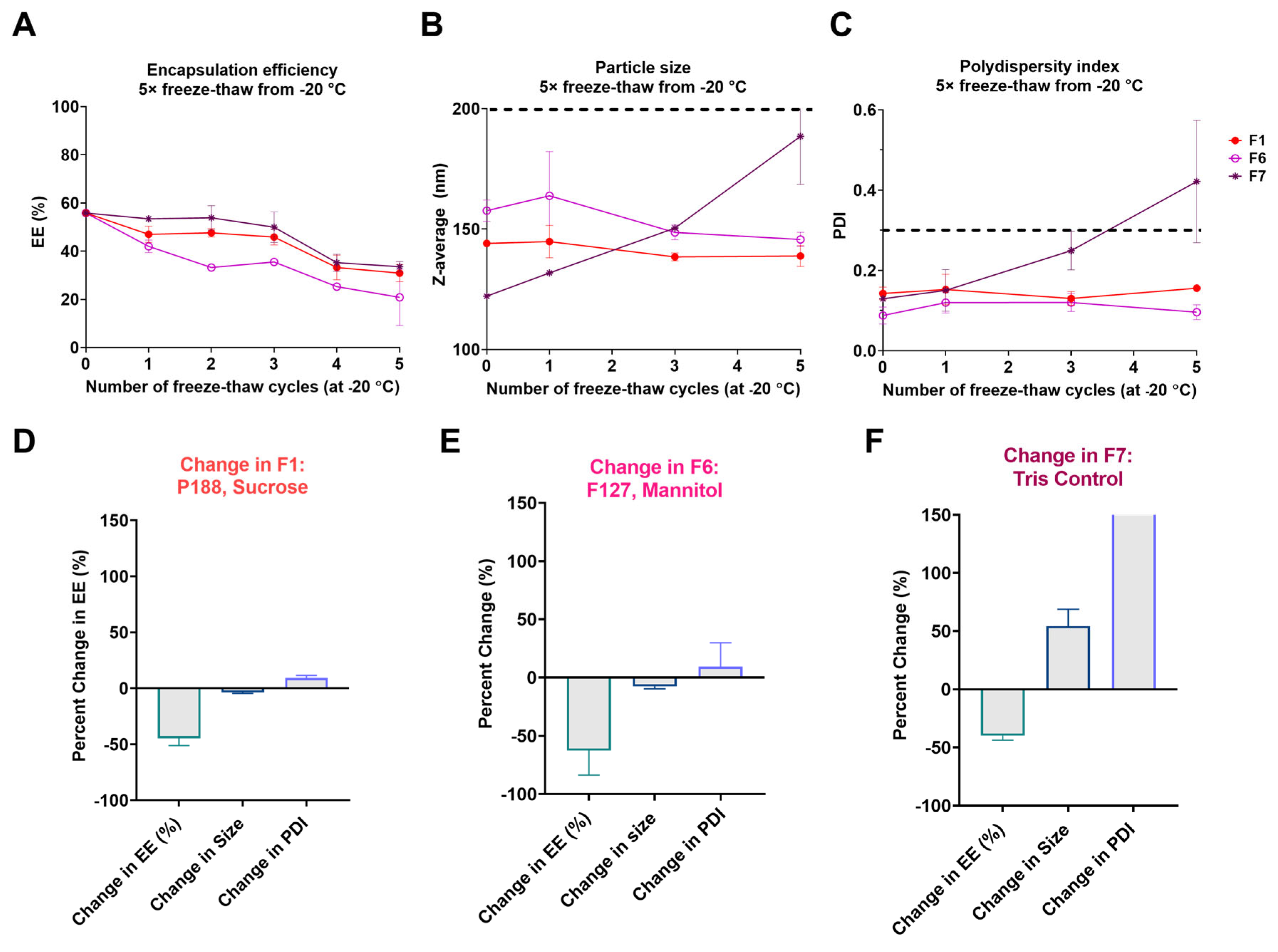
Figure 2 illustrates the results of the change in LNP quality attributes before and after thawing 5 times. It is evident that when no excipients are present, the PDI increases drastically, i.e., the size distribution of the LNP changed dramatically, exceeding an 80% increase in this condition due to the lack of a stabilizing excipient. Moreover, in all the cases examined, an increase in size was observed. This might indicate swelling or aggregation of the particles as a result of temperature cycling [18].
Based on a global evaluation of the change in EE, Z-average, and PDI, the tris-based formulations that demonstrated the least change to LNP configuration after 5 freeze-thaws were F1 (0.5% P188, 8% sucrose) and F6 (0.5% F127, 8% mannitol) (Figure 4D). As such, F1 and F6 were selected as the formulations to be evaluated for long-term stability studies. The screening was also based on a preliminary design of experiment (DoE) analysis performed using JMP toolsets. The DoE analysis is now provided in the supplementary information, SI-1, Figure S1.
Despite the samples exhibiting good PDI and size consistency, the dynamic rearrangement of LNPs during freeze-thaw cycles could compromise their structural integrity. This reconfiguration may result in the loss of encapsulated mRNA, either due to leakage, nuclease activity, or LNP degradation, ultimately reducing the availability of essential mRNA. Thus, among the formulations screened, F1 and F6 (Table 2) showed significantly reduced changes in the PDI over time and good EE throughout the freeze-thaw cycles. Furthermore, compared to the control, LNP formulation with sucrose and mannitol did not significantly vary in EE and size. Interestingly, formulation with trehalose showed the least variation in average size and PDI but performed poorly as an excipient due to the least EE after 5 freeze-thaw cycles (Figure 2). This suggested that among the tested formulations, trehalose did not effectively maintain the stability of the LNP after encapsulation.
To demonstrate the applicability of the compatible formulations (F1 and F6) across other mRNA constructs and to eliminate the possibility of mRNA transcript size and sequence variability that may have caused the stabilizing effect, encapsulation experiments using eGFP mRNA-LNP were tested. The results were consistent with those of nLuc, indicating that after 5 freeze-thaw cycles, less change was observed in the formulated particles than in the tris-only control (Figure 3). Specifically, particle size (Figure 3B) and PDI (Figure 3C) were significantly lower in F1 (size: p = 0.0006; PDI: p = 0.0056 ) and F6 (size: p = 0.0017; PDI: p = 0.0015 ) compared to the control after 5 freeze-thaws (Supplementary Figure S2). The PDI index remained below the acceptable range of 0.3 for both formulated groups, indicating uniformity among these samples. On the other hand, no difference was seen between the control and formulated samples in terms of EE, similar to what was seen with the nLuc (Figure 2). This may indicate that the excipients prevented agglomeration and swelling but not particle breakage.
Figure 3.
Absolute values (thaw-by-thaw data) illustrate EE (A), particle size (B), and PDI (C) for eGFP-encoding mRNA encapsulated in ALC-0315 LNPs within tris-containing formulations. The dotted lines represent the acceptable limits of < 200 nm (B) and < 0.3 PDI (C) according to published standards [22,23]. Percent changes in these physicochemical properties after five iterative freeze-thaw cycles at −20 °C are shown in (D, E, F). Percent changes are calculated relative to the initial LNP characterization before freezing. Error bars represent the standard deviation between duplicate samples. In the bar chart representing the PDI change for the control formulation, values are displayed as exceeding the axis range for consistent comparative visualization.
3.2. Long-Term Stability Assessment of nLuc mRNA-LNPs
nLuc-encoding mRNA-LNPs of selected formulations, F1 and F6, were tested for their long-term stability under non-frozen (Room Temperature and 4 °C) and frozen conditions (−20 °C and −80 °C) for a total duration of two months (8 weeks) alongside a tris-only control as shown in Table 2. To further expand the study, both ALC-0315 and SM-102 were investigated in these conditions. These ionizable lipids were chosen due to their clinical relevance in the FDA-approved SARS-CoV-2 vaccines from Pfizer/BioNTech and Moderna, respectively (Figure 1, right).
For all formulations, the size, PDI and EE properties were recorded before adding excipients, serving as a baseline reference to assess variations in LNP properties over time during storage for 8 weeks. In general, a slight increase in the size of the formulated samples immediately after the addition of the formulation buffer may be attributed to the presence of excipients.
3.2.1. Long-Term Storage at Room Temperature
The stability analysis of mRNA-LNP samples stored at ambient conditions (room temperature) is shown in Figure 4. In general, SM-102 showed higher EE by pipette mixing approach, compared to that of ALC-0315, irrespective of the formulation or the control (Figure 4A). Figure 4B shows that most samples had a z-average of less than 200 nm, within the acceptable limit during the 2 months of storage. However, compared to the initial conditions, the fold-change shown in Figure 4E showed that the particle size in some cases with ALC-0315 doubled. Formulations with SM-102 had a relatively stable property (Figure 4A,E); nevertheless, compared to initial values, fold-change was significantly increased over the course of storage. The PDI measurements also showed similar large variability across all the samples, with a narrow exception of F6 for ALC 0315 and SM102. Furthermore, the formulation with F6 (0.5% F127 and 8% mannitol) effectively limited heterogeneity among the LNPs for samples formulated with ALC-0315 and SM-102 (Figure 4C,F). On the other hand, in some cases, with ALC-0315, the sucrose and P188-containing samples (F1) exceeded a PDI index of 0.2 during their storage at room temperature, demonstrating their inability to maintain the physical qualities (size and PDI) in this storage condition.
Figure 4.
8-week stability evaluation of mRNA-LNP samples stored at room temperature. Absolute values are shown for EE% (A), particle size (B) and PDI (C). The dotted lines represent the acceptable limits of < 200 nm (B) and < 0.3 PDI according to published standards [22,23] (C). Fold-changes for the same attributes are represented in D, E, and F, respectively, with dotted lines representing no change in value (fold-change = 1).
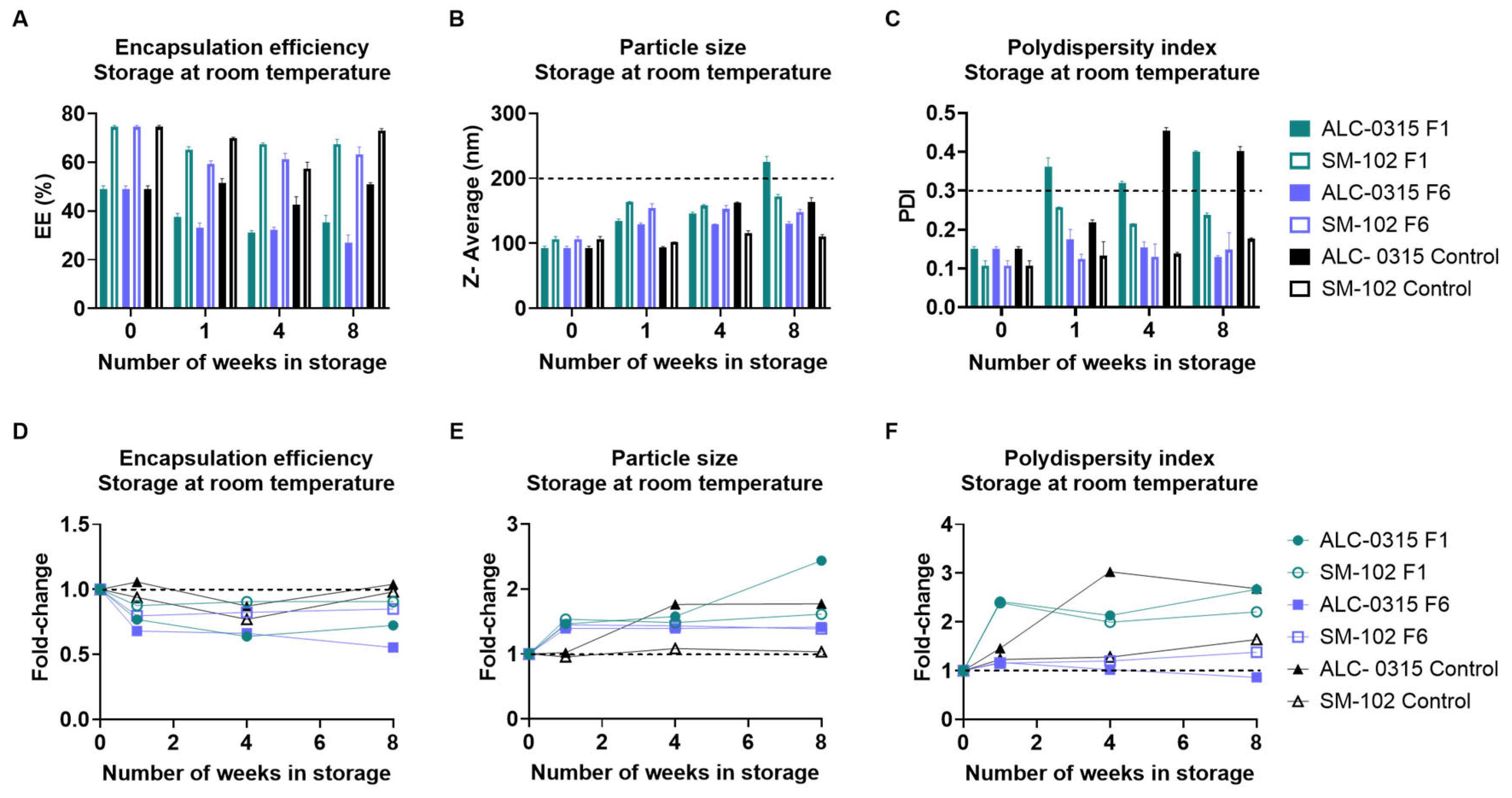
The absolute values in Figure 4 A-C remained within acceptable limits for most formulations, although some conditions affected z-average and PDI beyond acceptable ranges. Although the EE did not decline significantly among the formulated conditions (Figure 4A,D), the encapsulated mRNA within the particle was degraded during the 2 months of their storage and is thus non-functional (Figure 8). Additionally, irrespective of the buffer and excipient composition, the particles formulated with ALC-0315 were consistently unstable regarding changes in their quality attributes, unlike those formulated with SM-102 at room temperature. Thus, the results showed that adding excipients to the mRNA-loaded particles did not improve their stability at room temperature. Surprisingly, particles formulated with SM-102 ionizable lipid in tris-buffer (control) without excipients and surfactants experienced the least changes in the LNP quality attributes than their formulated counterparts (Figure 4). Although the particle quality attributes were relatively stable at room temperature across different formulations, the main concern at this temperature is mRNA degradation (described in detail in the section below, Figure 8).
3.2.2. Long-Term Storage at 4 °C
The mRNA-LNP sample stability was next tested at 4 °C, i.e., under refrigerated conditions (Figure 5). All the analyzed samples, irrespective of the lipid or F1/F6 formulation, showed physical configuration within the acceptable limits (Figure 5B,C). Following the two months of storage at 4 °C, F6 (0.5% F127, 8% mannitol) was identified as successful in maintaining EE in SM-102 mRNA-LNPs as compared to the control particles (Figure 5A). Additionally, the physical quality attributes (size and PDI) of F1 buffer particles were maintained similarly to the control throughout this 2-month storage period (Figure 5B–D). This indicates that the additional surfactants and excipients, in this case, did not contribute to the preservation of LNPs. A size and PDI increase were observed in the F6 buffer within the first week of storage due to the impact of excipient addition, but it remained stable in the weeks following. Overall, another general observation, compared to the RT, is that storage at refrigerated conditions improved EE and PDI properties. Also, similar to the observation at RT, SM-102 performed relatively better compared to the ALC-0315.
3.2.3. Long-Term Storage at −20 °C
The first frozen mRNA-LNP stability study (Figure 6) here was performed at −20 °C, where F1 (0.5% P188, 8% sucrose) maintained the stability of ALC-0315 mRNA-LNPs without much variation through the 8-week period of storage. A notable change is among the EE, which dramatically changed in most screened conditions for ALC-0315 and SM-102 except for the above-mentioned condition of F1-ALC-0315 (Figure 6A). Also, the formulation F6 slightly increased the physical properties during storage for SM102 and ALC-0315 lipids. Furthermore, it appears as though the addition of these excipients reduces the change in the PDI of the particles over the course of 2 months of storage. Despite their growth in size, ALC-0315 LNPs stored in F1 for 2 months did not exceed the acceptable size criteria for mRNA-LNPs and were observed to grow to around 125 nm in size.
3.2.4. Long-Term Storage at −80 °C
Although storage at −80 °C has been deemed a challenge for vaccine distribution, the formulations were studied at this temperature over the course of two months to gain insight into the behaviour of mRNA-LNP particles in this condition. All SM-102 mRNA-LNPs stored during our study demonstrated a large decline in the EE after 1 week of storage (Figure 7A,D). For both ionizable lipids, in storage conditions where a surfactant and sugar are not included, a dramatic increase in the size is observed within one week of storage at −80 °C (Figure 7B,E). These unformulated samples also incurred the largest change in their PDI throughout their storage duration (Figure 7C). These results demonstrate the instability of the lipid nanoparticle shell without excipients at this temperature and the need for the inclusion of excipients at −80 °C to maintain the particle’s physical attributes and reduce aggregation. Despite a general growth of the particle size across conditions, both F1 (0.5% P188, 8% sucrose) and F6 (0.5% F127, 8% mannitol) reduced the changes in size and PDI throughout storage, with F1 outperforming F6 (Figure 7B,C,E,F).
The addition of 8% sucrose and 0.5% P188 to mRNA-LNPs formulated with ALC-0315 minimized changes in their size and the PDI as compared to the tris-only control (Figure 7). The EE of these LNPs, stored with Sucrose and P188, declined by approximately 29% over the course of 2 months at −80 °C, whereas those which were stored without sugar or surfactant experienced a decline of approximately 63.5% in their EE.
3.2.5. mRNA Integrity Analysis
Due to its success in preserving LNP attributes compared to other formulations, F1 ALC-0315 alone was used for capillary gel electrophoresis analysis across different storage temperatures (Figure 8). It was already shown in the study of stability at RT in this study that the physicochemical stability of mRNA-LNPs does not necessarily correlate with mRNA integrity and biological activity [26].
Figure 8.
Capillary electrophoresis for RNA integrity analysis of ALC-0315 mRNA-LNP samples in formulation 1 (F1) or in tris buffer (Control) after 2 months of storage in non-frozen (A) or frozen (B) conditions. nLuc mRNA is expected to have approximately 2000 nt in length. The percentage of the area of the main peak (2000 nt) relative to the total RNA detected is reported below each sample.
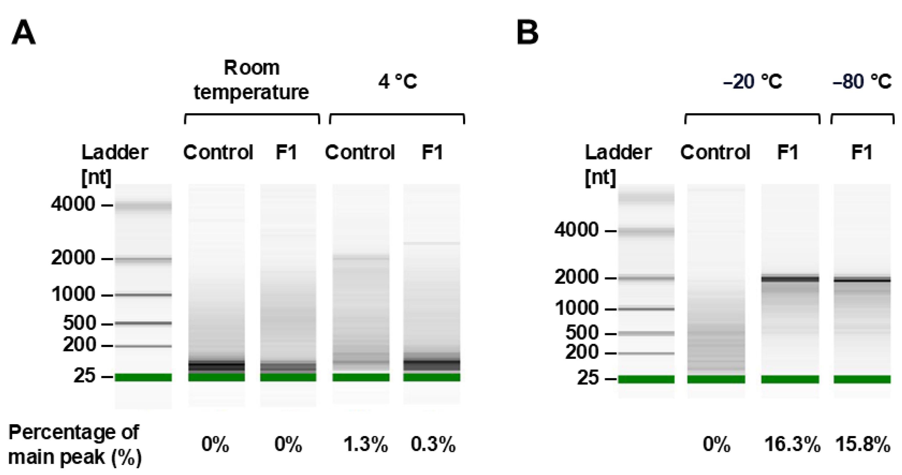
Despite the promising results in preserving LNP attributes, the mRNA integrity was not preserved in non-frozen temperatures (RT, 4 ˚C), as evidenced by the lack of a clear band at 2000 nt, and rather a smear of lower molecular weight species (Figure 8A). Frozen conditions, on the other hand, demonstrated superior RNA quality with a strong band at 2000 nt and less smearing than seen in the unformulated control (Figure 8B). Although the percentage of the main peak was below 20%, indicating RNA integrity is sub-optimal, the detergent and lipids present in the sample may have interfered with this measurement, leading to lower values than expected.
4. Discussion
A primary limitation of the mRNA vaccines and therapeutics in the non-pandemic market is the thermal instability [27]. Specifically, as therapeutics and non-urgent vaccines roll out, dosages may now require longer-term storage and may not need to be stored at high-LNP density multi-dose concentrations.
It is known [8,9] and demonstrated here (Figure 7) that in the absence of excipients and cryoprotectants, mRNA-LNP in tris buffer is unstable and prone to physical degradation. The hypothesis is that the surfactants would reduce aggregation, and sugar would reduce the degradation of the LNP as well as protect the mRNA through freeze-thaw stresses by acting as a cushion layer around the LNP particles, as compared to an unformulated control lacking a surfactant or excipient. The priority quality control attributes for LNPs include EE %, PDI and size. PDI and z-average properties are interrelated, as a change in one will directly affect the other. However, EE % and PDI are not interrelated. For example, LNP particles could aggregate without losing the packed mRNA, leading to higher z-size and PDI. However, this is not ideal, as LNP size influences distribution [28], with particles smaller than 200 nm being hypothesized to circulate to lymphatic nodes by advection while particles 200-500 nm would go through uptake by dendritic cells [28,29]. As such, particles aggregating beyond 200 nm throughout storage could provoke a different response in patients than what would have been validated at < 200 nm. On the contrary, a collapse of LNPs in solution may cause a high loss of EE% with low variation in PDI, which is also not ideal. Thus, a balance between EE% and size-associated properties is critical in choosing the correct formulation.
Sugar-surfactant combinations have non-linear impacts on the EE and PDI: In this study, we screened six formulations based on their ability to minimize changes to the physicochemical characteristics of the ALC-0315 mRNA-LNPs after 5 rounds of freeze-thaw at −20 °C as compared to the particles suspended in tris-only buffer, without excipients and surfactants. We found that sucrose-P188 (F1) and mannitol-F127 (F6)-containing tris buffer effectively prevented physiochemical changes during freeze-thaw stresses and functioned as a cryoprotectant throughout the thermal cycling stresses compared to the control. However, the sugar-surfactant combinations had inconsistent effects on EE% and PDI from one formulation to the other. For example, sucrose-P188 maintained the PDI while mannitol-F127 had limited protection of the LNPs from losing EE (Figure 2). Further, we hypothesize that the sugar used in the formulation has a major role in preventing LNP aggregation. This conclusion is based on the comparison between different sugars and the two surfactants. Surfactants did not directly contribute to EE and PDI. However, among sugars, trehalose negatively affected the EE and PDI, irrespective of the surfactant. Also, among the sugars, sucrose seemed to protect the LNP better than mannitol, even though the use of mannitol as a cryoprotective agent is not new, as is shown to be effective in previous studies [30]. This is consistent with findings for freeze-dried LNPs, where sucrose had the highest cryoprotective effect, followed by mannitol, with no positive effect seen for trehalose [31]. Although some sugars and sugar alcohols have been evaluated as excipients for mRNA-LNPs, the combination of these excipients with surfactants has not been explored to our knowledge, with the exception of one patent by Moderna testing Sucrose and P188 [20,32]. An additional advantage of a formulation containing surfactants is, with the increasing concern against other respiratory diseases and an interest in intranasal delivery, the introduction of surfactants enables potential nebulization applications [33].
Surfactants in excipients impact the LNP size and aggregation: The study used two different surfactants, P188 (8000 Da) and F127 (12500 Da), that function by protecting the LNP from freeze-thaw stresses and aggregation. In a specific case, it has been hypothesized that P188 surfactant would cover the lipid nanoparticles, thereby reducing the free energy and stabilizing the mRNA-LNP complex from collapsing and precipitating [20,34,35,36]. It is commonly observed that many pharmaceutical LNP formulations for cancer drug delivery use surfactant concentrations ranging from 0.5% to 4% (w/v) [37,38,39]. Further, the rationale for choosing F127 and P188 at a concentration of 0.5% is two-fold: A. Both are FDA-approved pharmaceutical ingredients [40], and B. When used in higher concentrations, it can be toxic [39], and inhibit cellular uptake and/or endosomal escape. In the published works [37,38,39,40] concentrations ranging from 0.0005% to up to 4% have been shown to be useful as a lipid nanoparticle stabilizing agent. For example, a published study observed that LNPs were stabilized at low concentrations from 0.05 % (W/V) for drug delivery in ocular sites [39]. In comparison, other studies for preserving oncolytic viral vaccine formulations that reported testing from 0.0005% onwards for gene delivery have also used much lower percentages [41]. Although surfactants have been used with LNPs for other applications, this study aims to fill the gap in formulating mRNA-containing LNPs with these components. Based on these varied reports, we tested a concentration of 0.5%, drawing inspiration from low toxicity and its extensive use for formulating other drugs in solid nanoparticles. In the present study, P188 showed better prevention of aggregation and maintained the average size, with this effect being less prominent in formulations with F127. The possible reason could be due to its inherent surface properties and the length of the polymer chains and molecular weights, since P188 and F127 are similar chemicals that have hydrophilic and hydrophobic properties, with variations in the total length of the polymer chain. The same concentration of P188 (8000 daltons) will have more particles compared to F127 (12500 daltons). As a result, their coverage on the LNP surfaces may be different, leading to such different behaviours during storage stability. This finding is consistent with studies that have indicated that P188, above a concentration of 0.0005% w/v, is an appropriate excipient to protect enveloped viral formulations throughout freeze-thaws [41]. Due to the limited data on the thermostability of mRNA-LNPs, we can draw a comparison between mRNA-LNPs and enveloped viruses since they are both comprised of a lipid-containing envelope that encapsulates genetic material.
LNP stability at RT is an ongoing search: Under the stress conditions of storage at RT, all formulations failed to maintain mRNA integrity. This result is unsurprising as it has been well documented that mRNA is unstable in aqueous conditions at RT due to hydrolytic cleavage of the phosphodiester backbone of the RNA [42]. Previous research has indicated that within the LNP core, approximately 20% of the volume is accounted for by water, implying that the mRNA is exposed to hydrolysis even while encapsulated [9]. Furthermore, at elevated temperatures, the relative humidity of the air has been shown to degrade RNA, and thus, this humidity would need to be more strictly controlled for room temperature storage of these molecules [42]. Furthermore, ribonucleases are known to be increasingly active at elevated temperatures [42]. However, despite mRNA degradation, the physicochemical characteristics of the LNPs were maintained in some of the cases tested, even when stored without excipients. This implies that the instability of mRNA remains the challenge to be surpassed to extend mRNA-LNPs room temperature shelf-life. This is further supported by the specified shelf-life of Onpattro, a siRNA-LNP drug, which can be stored at room temperature for 14 days or be refrigerated for up to 3 years [10,43]. It has been noted that the LNP composition of Onpattro is similar to the mRNA-LNP vaccines, indicating that the limiting factor for storage at ambient temperature is limited by the inherent instability of the mRNA. In contrast to mRNA-LNPs, this siRNA-LNP should not be allowed to freeze throughout its storage duration, most likely to maintain the physical properties of the LNP [44]. Our results emphasized these conflicting needs and stabilities of the lipid nanoparticle component from the mRNA component.
ALC-0315 showed improved stability and shelf life with the use of excipients (sucrose and mannitol). To achieve this aforementioned balance, after 8 weeks of storage at 4 °C, −20 °C, and −80 °C, we identified an improved storage condition for each of these temperatures as compared to the controls. For all conditions, the EE% is higher at the start when using SM-102 than ALC-0315, likely due to the inherent properties of the ionizable lipid components and the lipid mixture used in forming the LNPs. For this study, we used the same pH for encapsulation across all conditions to focus on comparing storage formulations, but pH may have to be optimized for each ionizable lipid according to pKa to achieve higher encapsulation efficiency. The difference in encapsulation efficiency and, consequently, proportion of mRNA payload to lipids, may have interfered in stability of mRNA throughout long-term storage. Besides pH, different starting mRNA concentrations could be tested to further explore this. At 4 °C, the inclusion of mannitol and F127 to SM-102-based lipid nanoparticles demonstrated improved retention of mRNA integrity as well as comparable size and PDI stability to the tris-only control. This represents a progression towards the extension of the 4 °C shelf life of SM-102-based LNPs, which was listed as 30 days [7]. At −20 °C and −80 °C, our results demonstrated that ALC-0315-based LNPs were increasingly stable with the addition of sucrose and P188, both in terms of LNP physicochemical characteristics as well as in terms of mRNA integrity. The ability to store ALC-0315 LNPs at −20 °C for 2 months represents an improvement upon the ultra-cold storage requirements (−80 °C to −60 °C) of the BNT162b2 vaccine from previous studies, which posed many challenges in their shipping and distribution due to this requirement [27]. Furthermore, the results of our freeze-thaw study demonstrate that these excipients were also effective in maintaining the physicochemical characteristics of the LNP throughout several freeze-thaw cycles from −20 °C to room temperature, which would ease restrictions on their shipping conditions.
Despite having identified an improved excipient formulation at −80 °C, this condition proved to be a harsh condition for the maintenance of LNPs. In the inverse case of room temperature, mRNA stored at −80 °C maintained its integrity, but we observed a decrease in the EE% of most particles, indicating leakage from or degradation of the particles themselves. Additionally, we observed size increases and drastic increases in PDI in cases without the inclusion of a cryopreservative, indicating aggregation of particles. This is consistent with the results of Kamiya et al., who observed that in mRNA-LNP samples stored at −80 °C, the EE% decreased by 41.74%, and the size increased by 245.2% as compared to those stored at 4 °C [45]. Our data showed a higher size increase of LNPs stored at −80 °C than at −20 °C, which is also aligned with other studies where −80 °C storage conditions resulted in lower luciferase activity than storage at −30 °C [45] and higher aggregation than storage at −20 °C [46]. This may seem contrary to expectations that mRNA-LNPs would be the most stable when frozen, according to the temperatures chosen for the storage of approved SARS-CoV-2 vaccines [7]. However, it is important to note that the methods for freezing may vary between studies and the procedure performed in the industry. In fact, techniques such as snap-freezing or a gradual change of temperatures may impact how mRNA-LNPs are affected by the freezing process [47,48]. Henderson et al. controlled for this by using a 1 °C/min cooling rate [19], while Kim et al. saw better results with flash-freezing in liquid nitrogen than freezing at −80 °C [46], but many studies do not mention controlled freezing methods.
In summary, in this manuscript, we investigated and presented the preliminary studies on the impact of the choice of sugar and surfactant on mRNA-LNP stability in different storage temperatures and freeze-thaw cycles. To facilitate this comparison, other factors were standardized across conditions, including concentrations of mRNA, lipids and excipients. Additionally, while this preliminary study focused on the physicochemical properties of the particles, further characterization of particle morphology and lipid composiiton through electron microscopy and RNA function through in vitro and in vivo assays could provide more robust explanations for differences observed between storage conditions [46,48]. As such, future perspectives to deepen the understanding of the data discussed include screening other contributing factors and expanding the number of quality attributes assessed.
5. Conclusions
The findings demonstrated that while excipients maintain PDI and structural integrity, as well as EE%, they do not significantly protect mRNA, particularly uncapped mRNA used in this study. Therefore, incorporating such excipients in LNP formulations for R&D labs is advisable to enhance experimental operating conditions. LNPs require excipients during storage for R&D applications and even more so for commercialization to maintain PDI and EE. Although the study examined six different formulations through freeze-thaw cycles and identified the best formulation, a detailed Design of Experiment (DoE) would allow evaluation of LNP stability using a broader range of excipients, buffers, and surfactant-buffer formulations. This study provides valuable data on the freeze-thaw dynamics and long-term storage of mRNA-LNPs, addressing critical stability challenges with the rising interest in mRNA-LNP vaccines and therapeutics. It identifies formulations balancing mRNA and LNP stability at 4 °C, −20 °C, and −80 °C, including surfactants to support future nebulization. These findings aim to improve the accessibility of mRNA-LNP drug products. Future studies may focus on lyophilized formulations for room-temperature storage to address the challenges associated with storing mRNA-LNPs in aqueous conditions.
Supplementary Materials
The following supporting information can be downloaded at: The following supporting information can be downloaded at the website of this paper posted on Preprints.org.
Author Contributions
M.Y.: conceptualization, investigation, formal analysis, writing – original draft preparation. C.H.: conceptualization, investigation. J.P.C.F: investigation, formal analysis, writing – review and editing. M.F.H.K.: formal analysis, writing – review and editing. A.S.P.: investigation, formal analysis, writing – review and editing. A.A.K.: conceptualization, supervision, funding acquisition, writing – review and editing. All authors have read and agreed to the published version of the manuscript.
Funding
M.Y. was supported by a master’s scholarship from the Natural Sciences and Engineering Research Council of Canada (NSERC) and from Fonds de recherche—Nature et Technologies Québec (FRQNT). A.A.K. is partially funded through Natural Sciences and Engineering Research Council (NSERC) RGPIN-2021-02691 and Canada Research Chair Grant Number CRC-240394.
Data Availability Statement
The data presented will be made available through the corresponding author upon request.
Acknowledgments
The authors thank Hiva Azizi and Bassel Akache from the National Research Council of Canada (NRC) for kindly providing material and training. We also thank Nicolas Audet (Imaging and Molecular Biology Platform, McGill University) for Bioanalyzer services and the nanoUQAM facility for access to DLS.
Conflicts of Interest
The authors declare no conflicts of interest.
Abbreviations
The following abbreviations are used in this manuscript:
| mRNA | Messenger ribonucleic acid |
| LNP | Lipid nanoparticle |
| EE | Encapsulation efficiency |
| PDI | Polydispersity index |
| eGFP | Enhanced green fluorescent protein |
| nLuc | NanoLuc luciferase |
| RT | Room temperature |
References
- Labouta, H.I.; Langer, R.; Cullis, P.R.; Merkel, O.M.; Prausnitz, M.R.; Gomaa, Y.; Nogueira, S.S.; Kumeria, T. Role of drug delivery technologies in the success of COVID-19 vaccines: a perspective. Drug Delivery and Translational Research 2022, 12, 2581–2588. [Google Scholar] [CrossRef] [PubMed]
- Vecchio, R.; Gentile, L.; Tafuri, S.; Costantino, C.; Odone, A. Exploring future perspectives and pipeline progression in vaccine research and development. Ann Ig 2024, 36, 446–461. [Google Scholar] [PubMed]
- Anderer, S. FDA Approves Updated COVID-19 Vaccines. JAMA 2024, 332, 1228. [Google Scholar] [CrossRef] [PubMed]
- PRODUCT MONOGRAPH INCLUDING PATIENT MEDICATION INFORMATION SPIKEVAX. Available online: https://covid-vaccine.canada.ca/info/pdf/covid-19-vaccine-moderna-pm-en.pdf (accessed on 11 June 2024).
- PRODUCT MONOGRAPH INCLUDING PATIENT MEDICATION INFORMATION COMIRNATY. Available online: https://covid-vaccine.canada.ca/info/pdf/pfizer-biontech-covid-19-vaccine-pm1-en.pdf (accessed on 11 June 2024).
- Das, R. Update on Moderna’s RSV vaccine, mRESVIA (mRNA-1345), in adults≥ 60 years of age. 2024.
- Crommelin, D.J.; Anchordoquy, T.J.; Volkin, D.B.; Jiskoot, W.; Mastrobattista, E. Addressing the cold reality of mRNA vaccine stability. Journal of pharmaceutical sciences 2021, 110, 997–1001. [Google Scholar] [CrossRef]
- Blenke, E.O.; Örnskov, E.; Schöneich, C.; Nilsson, G.A.; Volkin, D.B.; Mastrobattista, E.; Almarsson, Ö.; Crommelin, D.J. The storage and in-use stability of mRNA vaccines and therapeutics: not a cold case. Journal of pharmaceutical sciences 2023, 112, 386–403. [Google Scholar] [CrossRef]
- Schoenmaker, L.; Witzigmann, D.; Kulkarni, J.A.; Verbeke, R.; Kersten, G.; Jiskoot, W.; Crommelin, D.J. mRNA-lipid nanoparticle COVID-19 vaccines: Structure and stability. International journal of pharmaceutics 2021, 601, 120586. [Google Scholar] [CrossRef]
- Cheng, F.; Wang, Y.; Bai, Y.; Liang, Z.; Mao, Q.; Liu, D.; Wu, X.; Xu, M. Research advances on the stability of mRNA vaccines. Viruses 2023, 15, 668. [Google Scholar] [CrossRef]
- Ripoll, M.; Bernard, M.-C.; Vaure, C.; Bazin, E.; Commandeur, S.; Perkov, V.; Lemdani, K.; Nicolaï, M.-C.; Bonifassi, P.; Kichler, A. An imidazole modified lipid confers enhanced mRNA-LNP stability and strong immunization properties in mice and non-human primates. Biomaterials 2022, 286, 121570. [Google Scholar] [CrossRef]
- Bottger, R. mRNA vaccine stabilization through modulating LNP composition and utilizing drying technologies. 2022.
- Dumpa, N.; Goel, K.; Guo, Y.; McFall, H.; Pillai, A.R.; Shukla, A.; Repka, M.; Murthy, S.N. Stability of vaccines. Aaps Pharmscitech 2019, 20, 1–11. [Google Scholar] [CrossRef]
- Brandau, D.T.; Jones, L.S.; Wiethoff, C.M.; Rexroad, J.; Middaugh, C.R. Thermal stability of vaccines. Journal of pharmaceutical sciences 2003, 92, 218–231. [Google Scholar] [CrossRef]
- Alejo, T.; Toro-Córdova, A.; Fernández, L.; Rivero, A.; Stoian, A.M.; Pérez, L.; Navarro, V.; Martínez-Oliván, J.; de Miguel, D. Comprehensive Optimization of a Freeze-Drying Process Achieving Enhanced Long-Term Stability and In Vivo Performance of Lyophilized mRNA-LNPs. International Journal of Molecular Sciences 2024, 25, 10603. [Google Scholar] [CrossRef] [PubMed]
- Li, M.; Jia, L.; Xie, Y.; Ma, W.; Yan, Z.; Liu, F.; Deng, J.; Zhu, A.; Siwei, X.; Su, W. Lyophilization process optimization and molecular dynamics simulation of mRNA-LNPs for SARS-CoV-2 vaccine. npj Vaccines 2023, 8, 153. [Google Scholar] [CrossRef] [PubMed]
- Perche, F.; Clemençon, R.; Schulze, K.; Ebensen, T.; Guzmán, C.A.; Pichon, C. Neutral lipopolyplexes for in vivo delivery of conventional and replicative RNA vaccine. Molecular Therapy-Nucleic Acids 2019, 17, 767–775. [Google Scholar] [CrossRef] [PubMed]
- Liu, T.; Tian, Y.; Zheng, A.; Cui, C. Design strategies for and stability of mRNA–lipid nanoparticle COVID-19 vaccines. Polymers 2022, 14, 4195. [Google Scholar] [CrossRef]
- Henderson, M.I.; Eygeris, Y.; Jozic, A.; Herrera, M.; Sahay, G. Leveraging biological buffers for efficient messenger RNA delivery via lipid nanoparticles. Molecular pharmaceutics 2022, 19, 4275–4285. [Google Scholar] [CrossRef]
- Moderna, E. Assessment Report COVID-19 Vaccine Moderna. Procedure No; EMEA/H/C/005791/0000. Netherlands: European Medicines Agency: 2021.
- Peter Marks, M.D., Ph.D., Center for Biologics Evaluation and Research. FDA. 2024. Available online: https://www.fda.gov/media/150386/download?attachment.
- Daniel, S.; Kis, Z.; Kontoravdi, C.; Shah, N. Quality by Design for enabling RNA platform production processes. Trends in biotechnology 2022, 40, 1213–1228. [Google Scholar] [CrossRef]
- Mehta, M.; Bui, T.A.; Yang, X.; Aksoy, Y.; Goldys, E.M.; Deng, W. Lipid-based nanoparticles for drug/gene delivery: An overview of the production techniques and difficulties encountered in their industrial development. ACS Materials Au 2023, 3, 600–619. [Google Scholar] [CrossRef]
- Hassett, K.J.; Higgins, J.; Woods, A.; Levy, B.; Xia, Y.; Hsiao, C.J.; Acosta, E.; Almarsson, Ö.; Moore, M.J.; Brito, L.A. Impact of lipid nanoparticle size on mRNA vaccine immunogenicity. Journal of Controlled Release 2021, 335, 237–246. [Google Scholar] [CrossRef]
- Kulkarni, J.A.; Darjuan, M.M.; Mercer, J.E.; Chen, S.; Van Der Meel, R.; Thewalt, J.L.; Tam, Y.Y.C.; Cullis, P.R. On the formation and morphology of lipid nanoparticles containing ionizable cationic lipids and siRNA. ACS nano 2018, 12, 4787–4795. [Google Scholar] [CrossRef]
- Reinhart, A.-G.; Osterwald, A.; Ringler, P.; Leiser, Y.; Lauer, M.E.; Martin, R.E.; Ullmer, C.; Schumacher, F.; Korn, C.; Keller, M. Investigations into mRNA Lipid Nanoparticles Shelf-Life Stability under Nonfrozen Conditions. Molecular Pharmaceutics 2023, 20, 6492–6503. [Google Scholar] [CrossRef]
- Makhijani, S.; Elossaily, G.M.; Rojekar, S.; Ingle, R.G. mRNA-based Vaccines–Global Approach, Challenges, and Could Be a Promising Wayout for Future Pandemics. Pharmaceutical Development and Technology 2024, 1–12. [Google Scholar] [CrossRef] [PubMed]
- Nakamura, T.; Kawai, M.; Sato, Y.; Maeki, M.; Tokeshi, M.; Harashima, H. The effect of size and charge of lipid nanoparticles prepared by microfluidic mixing on their lymph node transitivity and distribution. Molecular pharmaceutics 2020, 17, 944–953. [Google Scholar] [CrossRef] [PubMed]
- Di, J.; Du, Z.; Wu, K.; Jin, S.; Wang, X.; Li, T.; Xu, Y. Biodistribution and non-linear gene expression of mRNA LNPs affected by delivery route and particle size. Pharmaceutical research 2022, 39, 105–114. [Google Scholar] [CrossRef] [PubMed]
- Stark, B.; Pabst, G.; Prassl, R. Long-term stability of sterically stabilized liposomes by freezing and freeze-drying: Effects of cryoprotectants on structure. European journal of pharmaceutical sciences 2010, 41, 546–555. [Google Scholar] [CrossRef]
- Shirane, D.; Tanaka, H.; Nakai, Y.; Yoshioka, H.; Akita, H. Development of an alcohol dilution–lyophilization method for preparing lipid nanoparticles containing encapsulated siRNA. Biological and Pharmaceutical Bulletin 2018, 41, 1291–1294. [Google Scholar] [CrossRef]
- Smith, M.; Almarsson, O.; Brito, L. Stabilized formulations of lipid nanoparticles. 2020.
- Leong, E.W.; Ge, R. Lipid nanoparticles as delivery vehicles for inhaled therapeutics. Biomedicines 2022, 10, 2179. [Google Scholar] [CrossRef]
- Doan, T.N.K.; Davis, M.M.; Croyle, M.A. Identification of film-based formulations that move mRNA lipid nanoparticles out of the freezer. Molecular Therapy-Nucleic Acids 2024, 35. [Google Scholar] [CrossRef]
- Salminen, H.; Helgason, T.; Aulbach, S.; Kristinsson, B.; Kristbergsson, K.; Weiss, J. Influence of co-surfactants on crystallization and stability of solid lipid nanoparticles. Journal of colloid and interface science 2014, 426, 256–263. [Google Scholar] [CrossRef]
- Salminen, H.; Helgason, T.; Kristinsson, B.; Kristbergsson, K.; Weiss, J. Formation of nanostructured colloidosomes using electrostatic deposition of solid lipid nanoparticles onto an oil droplet interface. Food Research International 2016, 79, 11–18. [Google Scholar] [CrossRef]
- Li, M.; Tang, H.; Xiong, Y.; Yuan, Z.; He, L.; Han, L. Pluronic F127 coating performance on PLGA nanoparticles: enhanced flocculation and instability. Colloids and Surfaces B: Biointerfaces 2023, 226, 113328. [Google Scholar] [CrossRef]
- Chokshi, N.V.; Khatri, H.N.; Patel, M.M. Formulation, optimization, and characterization of rifampicin-loaded solid lipid nanoparticles for the treatment of tuberculosis. Drug development and industrial pharmacy 2018, 44, 1975–1989. [Google Scholar] [CrossRef] [PubMed]
- Leonardi, A.; Bucolo, C.; Romano, G.L.; Platania, C.B.M.; Drago, F.; Puglisi, G.; Pignatello, R. Influence of different surfactants on the technological properties and in vivo ocular tolerability of lipid nanoparticles. International journal of pharmaceutics 2014, 470, 133–140. [Google Scholar] [CrossRef] [PubMed]
- Khaliq, N.U.; Lee, J.; Kim, S.; Sung, D.; Kim, H. Pluronic F-68 and F-127 based nanomedicines for advancing combination cancer therapy. Pharmaceutics 2023, 15, 2102. [Google Scholar] [CrossRef] [PubMed]
- Pan, L.; Liu, X.; Fan, D.; Qian, Z.; Sun, X.; Wu, P.; Zhong, L. Study of Oncolytic Virus Preservation and Formulation. Pharmaceuticals 2023, 16, 843. [Google Scholar] [CrossRef]
- Fabre, A.-L.; Colotte, M.; Luis, A.; Tuffet, S.; Bonnet, J. An efficient method for long-term room temperature storage of RNA. European Journal of Human Genetics 2014, 22, 379–385. [Google Scholar] [CrossRef]
- Summary Basis of Decision - Onpattro - Health Canada. Available online: https://hpr-rps.hres.ca/reg-content/summary-basis-decision-detailTwo.php?linkID=SBD00446 (accessed on 11 June 2024).
- Suzuki, Y.; Ishihara, H. Difference in the lipid nanoparticle technology employed in three approved siRNA (Patisiran) and mRNA (COVID-19 vaccine) drugs. Drug Metabolism and Pharmacokinetics 2021, 41, 100424. [Google Scholar] [CrossRef]
- Kamiya, M.; Matsumoto, M.; Yamashita, K.; Izumi, T.; Kawaguchi, M.; Mizukami, S.; Tsurumaru, M.; Mukai, H.; Kawakami, S. Stability study of mRNA-lipid nanoparticles exposed to various conditions based on the evaluation between physicochemical properties and their relation with protein expression ability. pharmaceutics 2022, 14, 2357. [Google Scholar] [CrossRef]
- Kim, B.; Hosn, R.R.; Remba, T.; Yun, D.; Li, N.; Abraham, W.; Melo, M.B.; Cortes, M.; Li, B.; Zhang, Y. Optimization of storage conditions for lipid nanoparticle-formulated self-replicating RNA vaccines. Journal of Controlled Release 2023, 353, 241–253. [Google Scholar] [CrossRef]
- Kafle, U.; Truong, H.Q.; Nguyen, C.T.G.; Meng, F. Development of Thermally Stable mRNA-LNP Delivery Systems: Current Progress and Future Prospects. Molecular Pharmaceutics 2024, 21, 5944–5959. [Google Scholar] [CrossRef]
- Fan, Y.; Rigas, D.; Kim, L.J.; Chang, F.-P.; Zang, N.; McKee, K.; Kemball, C.C.; Yu, Z.; Winkler, P.; Su, W.-C. Physicochemical and structural insights into lyophilized mRNA-LNP from lyoprotectant and buffer screenings. Journal of Controlled Release 2024, 373, 727–737. [Google Scholar] [CrossRef]
Figure 1.
Schematic describing the experimental design and process workflow for screening LNP buffer-excipient formulations by freeze-thaw cycles (left) followed by long-term stability studies (right).
Figure 1.
Schematic describing the experimental design and process workflow for screening LNP buffer-excipient formulations by freeze-thaw cycles (left) followed by long-term stability studies (right).
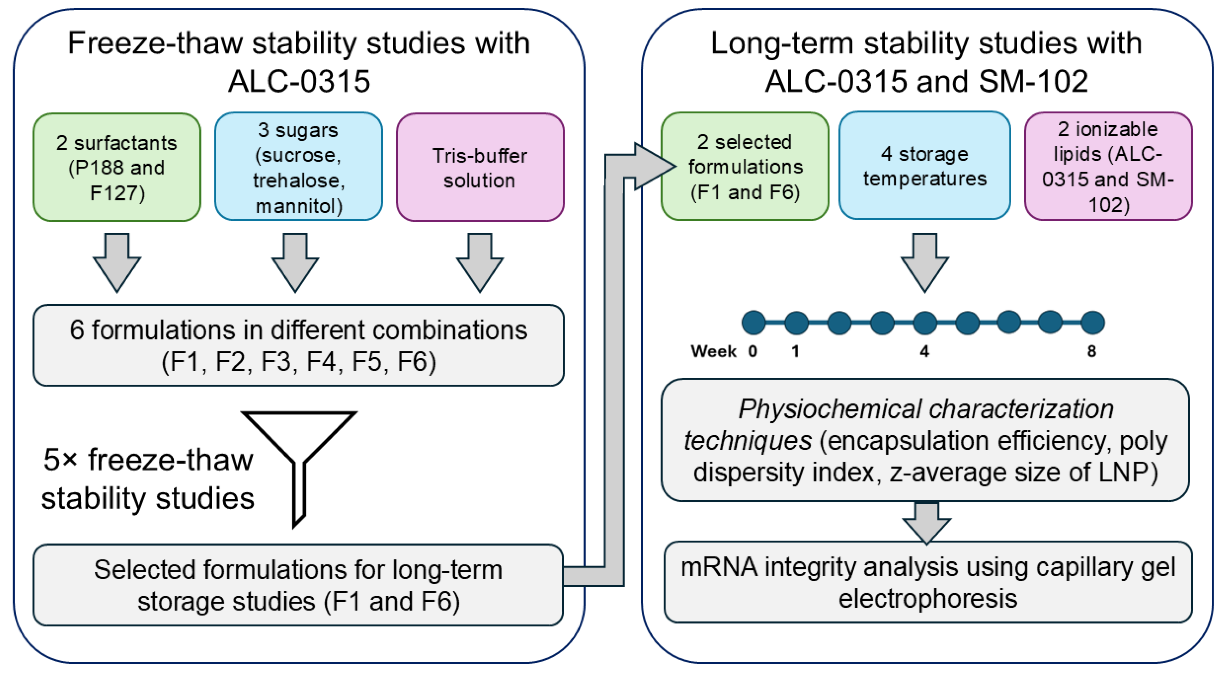
Figure 2.
Screening of LNP surfactant-excipient formulations via freeze-thaw cycles: Absolute values (thaw-by-thaw data) illustrate EE (A), particle size (B), and PDI (C) for nLuc-encoding mRNA encapsulated in ALC-0315 LNPs within tris-containing formulations. The dotted lines represent the acceptable limits of < 200 nm (B) and < 0.3 PDI (C) according to published standards [22,23,24,25]. Percent changes in these physicochemical properties after five iterative freeze-thaw cycles at −20 °C are shown in (D). Percent changes are calculated relative to the initial LNP characterization before freezing. In the percent change graphs, different colours represent different formulation conditions. In the bar chart representing the PDI change for F3 formulation, values are displayed as exceeding the axis range for consistent comparative visualization.
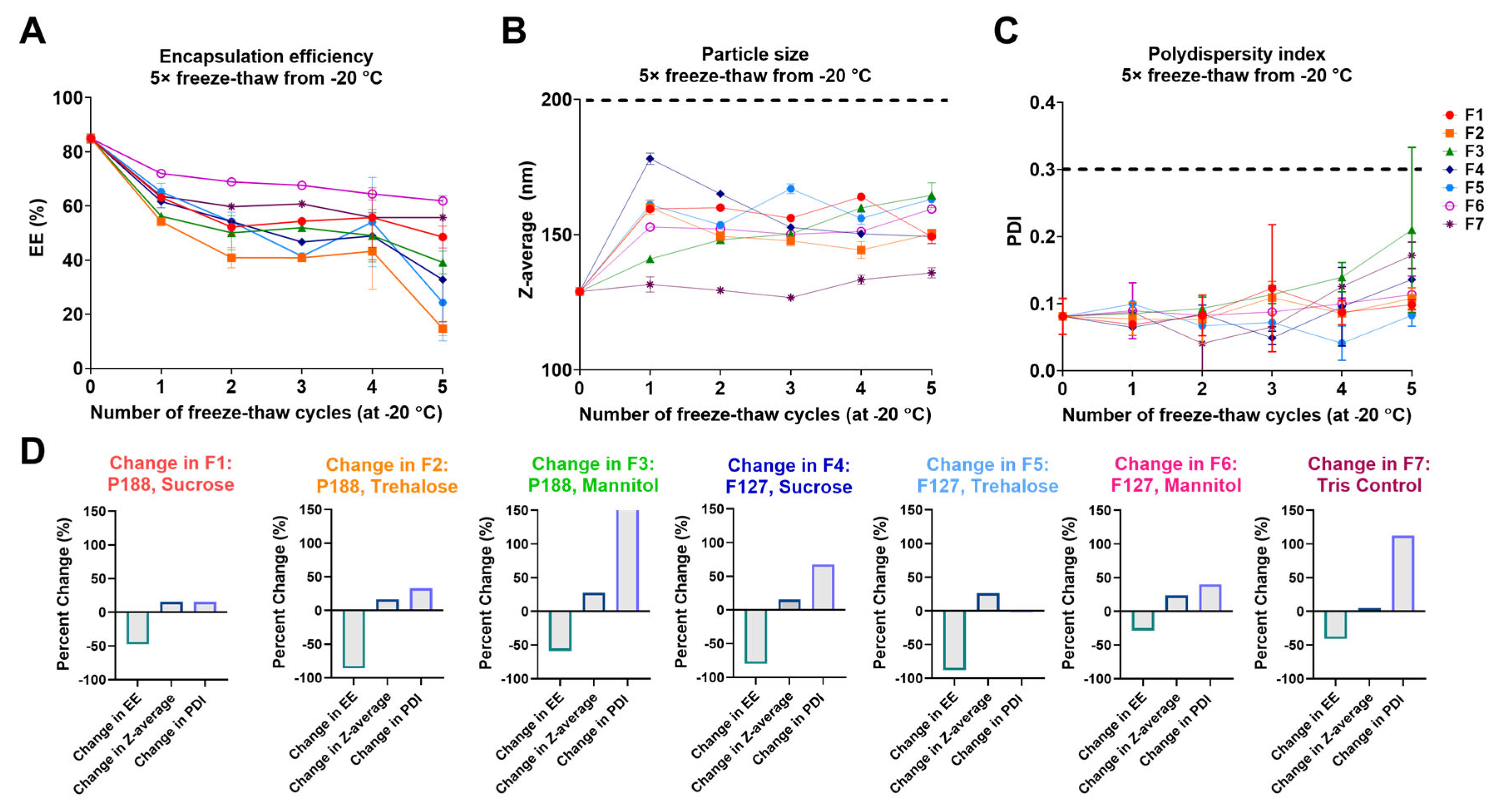
Figure 5.
8-week stability evaluation of mRNA-LNP samples stored at 4 °C. Absolute values are shown for EE% (A), particle size (B) and PDI (C). The dotted lines represent the acceptable limits of < 200 nm (B) and < 0.3 PDI according to published standards [22,23] (C). Fold-changes for the same attributes are represented in D, E, and F, respectively, with dotted lines representing no change in value (fold-change = 1).
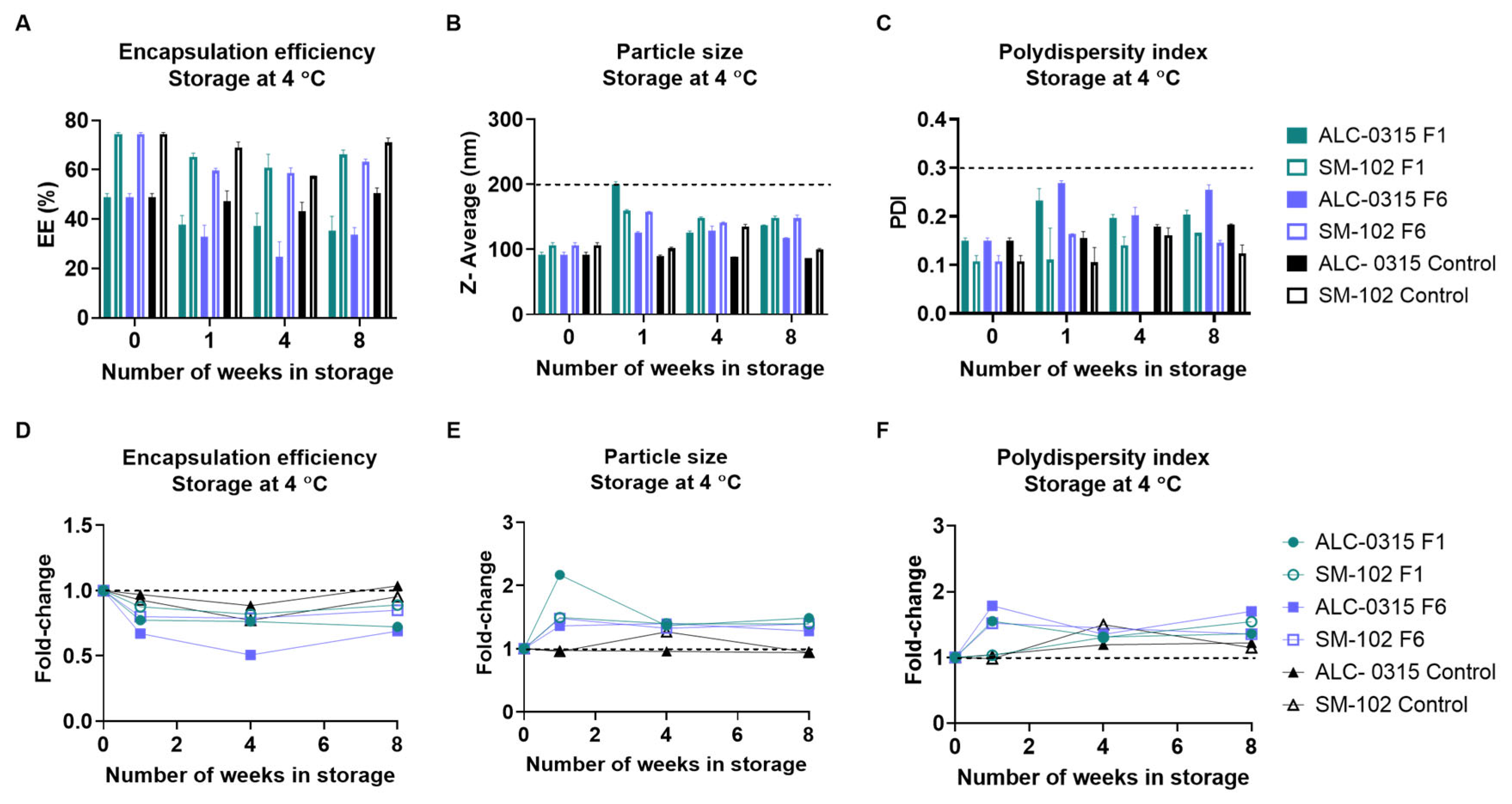
Figure 6.
8-week stability evaluation of mRNA-LNP samples stored at −20 °C. Absolute values are shown for EE% (A), particle size (B) and PDI (C). The dotted lines represent the acceptable limits of < 200 nm (B) and < 0.3 PDI according to published standards [22,23] (C). Fold-changes for the same attributes are represented in D, E, and F, respectively, with dotted lines representing no change in value (fold-change = 1).

Figure 7.
8-week stability evaluation of mRNA-LNP samples stored at −80 °C. Absolute values are shown for EE% (A), particle size (B) and PDI (C). The dotted lines represent the acceptable limits of < 200 nm (B) and < 0.3 PDI according to published standards [22,23](C). Fold-changes for the same attributes are represented in D, E, and F, respectively, with dotted lines representing no change in value (fold-change = 1).
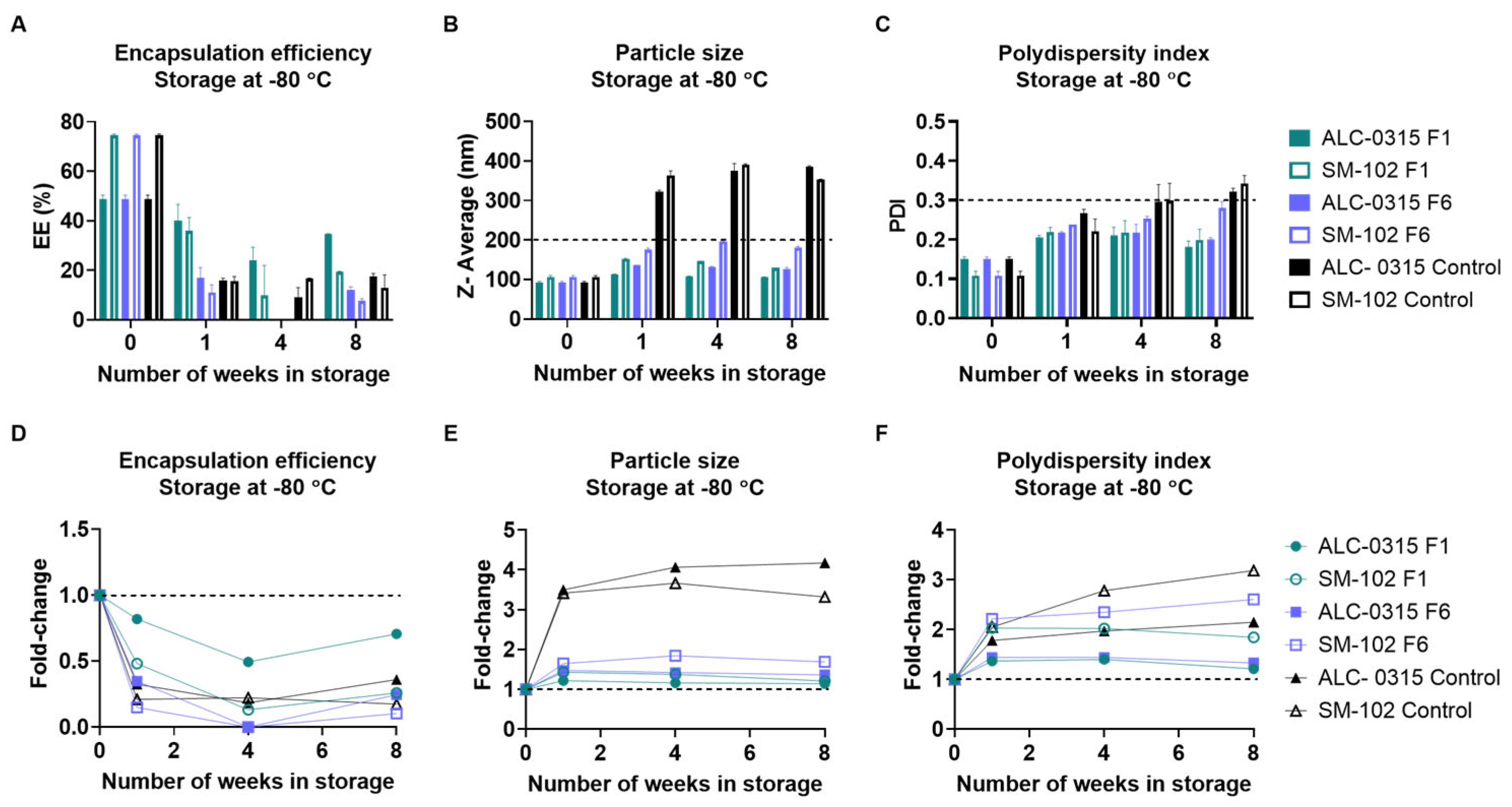

Table 1.
Reported stabilities of mRNA-LNP vaccines approved for use during the SARS-COV-2 pandemic. Table adapted from [7] and modified using [4,5].
| Vaccine Name | Ionizable Lipid | 2-8 °C Shelf Life | Room Temp Shelf Life | Concentration |
|---|---|---|---|---|
| BNT162b2 | ALC-0315 | Up to 5 days | Up to 2 hours | 6-10 doses per vial |
| mRNA-1273 | SM-102 | 30 days | Up to 12 hours | 5-20 doses per vial |
Table 2.
Tris-containing formulations were evaluated in this study. Concentrations listed as percentages are calculated as weight/volume (w/v). All formulations contained a surfactant and sugar as excipients except for the control.
Table 2.
Tris-containing formulations were evaluated in this study. Concentrations listed as percentages are calculated as weight/volume (w/v). All formulations contained a surfactant and sugar as excipients except for the control.
| Buffer | Surfactant | Sugar | Formulation |
|---|---|---|---|
| 0.1 M Tris (pH 8) | 0.5% P188 | 8% Sucrose | F1 |
| 8% Trehalose | F2 | ||
| 8% Mannitol | F3 | ||
| 0.5% F127 | 8% Sucrose | F4 | |
| 8% Trehalose | F5 | ||
| 8% Mannitol | F6 | ||
| None | None | Control (F7) |
Disclaimer/Publisher’s Note: The statements, opinions and data contained in all publications are solely those of the individual author(s) and contributor(s) and not of MDPI and/or the editor(s). MDPI and/or the editor(s) disclaim responsibility for any injury to people or property resulting from any ideas, methods, instructions or products referred to in the content. |
© 2025 by the authors. Licensee MDPI, Basel, Switzerland. This article is an open access article distributed under the terms and conditions of the Creative Commons Attribution (CC BY) license (http://creativecommons.org/licenses/by/4.0/).
Copyright: This open access article is published under a Creative Commons CC BY 4.0 license, which permit the free download, distribution, and reuse, provided that the author and preprint are cited in any reuse.


