
Submitted:
19 May 2025
Posted:
20 May 2025
You are already at the latest version
Abstract
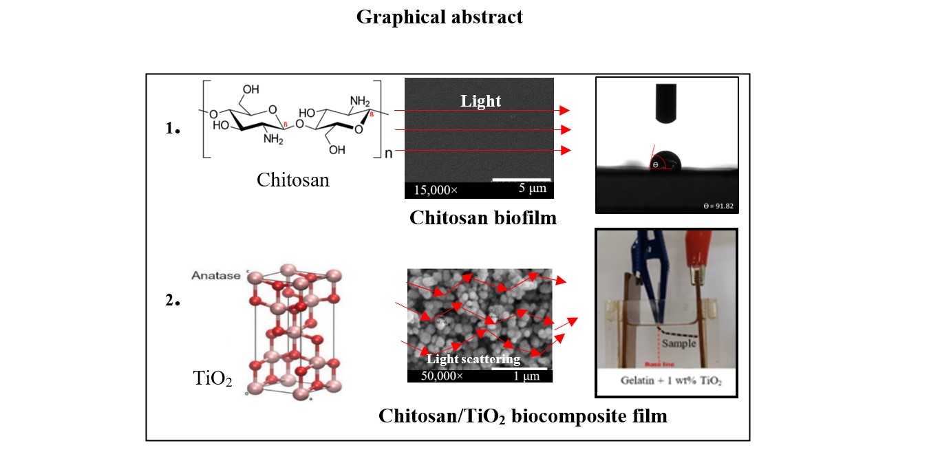
Bio-nanocomposite films were prepared using chitosan, gelatin, and varying concentrations (0, 0.5, 1.0, 2.0, and 5.0 wt%) of nanoparticulate titanium dioxide (TiO2) in acetic acid via the casting process. The incorporation of TiO2 nanoparticles into bio-chitosan films resulted in enhanced ultraviolet absorption and improved physical, mechanical, and electrical properties. Moreover, bio-chitosan films with added TiO2 exhibited antimicrobial properties, enhancing the preservation of packaged products by preventing the growth of microorganisms.
The bio-nanocomposite films with 1.0 wt% TiO2 demonstrated an electroactive response, bending at low electrical field strengths (250 V/mm), while the reference sample without added TiO2 bent at a higher electrical field strength (550 V/mm). This suggests their potential use in electroactive actuator applications, where controlled bending or movement is required. Among the various TiO2 concentrations tested, bio-chitosan nanocomposite films with 0.5% and 1.0 wt% TiO2 (Formulas 7 and 8) demonstrated favorable characteristics. These films (Formulas 7 and 8) exhibited an appealing appearance, absence of tear marks, low bulk density (0.91 ± 0.04 and 0.85 ± 0.18 g/cm3), satisfactory electomechanical response properties at 250 V/m (17.85 ± 2.58 and 61.48 ± 6.97), low percentage of shrinkage (59.95 ± 3.59 and 54.17 ± 9.28), high dielectric constant (1.80 ± 0.07 and 8.10 ± 0.73), and effective UV absorption compared to pure bio-chitosan film without and with gelatin (Formulas 1 and 6).
Keywords:
biofilms
; contact angle
; nanoparticle
; electroactive actuator
; casting process
1. Introduction
Chitosan is a biodegradable natural polymer composed of N-acetyl-2-amino-2-deoxy-D-glucopyranose (acetylated unit) and 2-amino-2-deoxy-D-glucopyranose (deacetylated unit), with repeating units connected by β-(1→4)-glycosidic bonds [1,2]. Sharing similarities with hyaluronic acid and glycosaminoglycans (GAGs), which are components of the cartilage extracellular matrix (ECM) [3], chitosan possesses a linear polymer structure and contains amino groups in its polysaccharide composition. Derived from the deacetylation of chitin [4,5], chitosan exhibits a positive charge due to the presence of amino groups, making it a polyelectrolyte within a pH range of 2-6 [6,7]. The structure of chitosan includes functional groups such as amino (-NH₂), hydroxyl (-OH), and acetyl (-COCH₃) groups. Kumar et al. [7] reported that the amino (-NH₂) groups in chitosan become protonated upon dissolution at pH 6 or below, forming cationic amine groups (-NH₃⁺). This protonation increases intermolecular electric repulsion, resulting in a polycationic, soluble polymer. Several mechanisms have been proposed to explain the antimicrobial activities of chitosan, with the most widely accepted being:
1. Electrostatic interaction with microbial membranes: The positively charged amine groups in chitosan interact with negatively charged microbial cell membranes, altering their barrier properties. This interaction leads to leakage of intracellular contents and ultimately causes cell death, as reported by Hosseinnejad & Jafari, 2018 [8].
2. Chelation of metal ions: Chitosan exhibits chelating properties, selectively binding to metal ions and inhibiting the activity of various metabolic enzymes by blocking their active centers. This process reduces microbial growth.
3. Molecular weight influence: The antimicrobial activity of chitosan is also affected by its molecular weight. High-molecular-weight chitosan forms an impermeable polymeric layer on microbial cell surfaces, altering cell permeability and blocking nutrient entry. Conversely, low-molecular-weight chitosan penetrates the cytosol, binds to cellular DNA, and interferes with mRNA and protein synthesis, leading to cell death, as studied by Hosseinnejad & Jafari, 2018 [8] and Zheng & Zhu, 2003 [9].
Furthermore, Sadaf Samimi, Gharaie et al. described chitosan as a linear, semicrystalline polysaccharide derived from the deacetylation of chitin, the most abundant biodegradable natural polymer after cellulose [10]. The degree of deacetylation (DD), defined by the content of free amino groups in the polysaccharide, is used to differentiate between chitin and chitosan [8]. Chitosan contains free amino groups, making it a positively charged polyelectrolyte within a pH range of 2 to 6. This property enhances its solubility compared to chitin. However, the high viscosity of chitosan solutions complicates their electrospinnability [10]. Dynamic interactions between polymer chains—such as entanglement, hydrophobic interactions, and hydrogen bonding—are crucial for reducing fiber diameters to the nanoscale and ensuring continuous fiber formation. Renowned for its non-toxicity, biocompatibility, and biodegradability, chitosan demonstrates various beneficial properties, including antiviral, antioxidant, antibacterial, and anticancer activities. Moreover, its hydrophilicity enhances interaction with cells [8,9,10,11,12,13]. Due to these characteristics, chitosan finds applications in various industries such as food, cosmetics, and agricultural packaging, as illustrated in Scheme 1. It is also widely used in medical applications such as tissue engineering, wound dressing, facial masks, and drug delivery systems.
Titanium dioxide (TiO2), an inorganic compound, is particularly emphasized in its anatase form due to its higher photocatalytic activity compared to the rutile and brookite structures [10,11,12,13,14,15]. With exceptional properties stemming from its crystallinity [16,17,18,19,20,21,22], the anatase form presents a body-centered tetragonal crystal structure, while rutile exhibits a primitive tetragonal crystal structure, and brookite possesses an orthorhombic crystal structure [16]. As a metal oxide compound, TiO2 showcases remarkable properties, rendering it a versatile material with diverse applications [17,18,19,20,21,22].
Despite chitosan’s structural similarity to cellulose, consisting of -OH and -NH2 groups, it exhibits distinct properties such as polyelectrolyte behavior, optical properties, physicochemical properties, nanocrytallinity, and degradation [22]. The addition of TiO2 nanoparticles increases crystallinity in bio-chitosan naocrystal films, enhancing mechanical, thermal, optical, and physical properties, including film morphology. Furthermore, TiO2 improves gas and solvent barriers, biocompatibility, photocatalytic activity, UV light absorption, non-toxicity, eco-friendliness, and biological-chemical inertness, finding applications in electronic, cosmetic, wound dressing, food preservation, agricultural, healthcare, environmental remediation, and smart packaging industries [23,24,25,26,27]. The combination of these properties makes TiO2 a valuable material in various industries, including cosmetics, healthcare, electronics, environmental remediation, and antimicrobial applications [28,29,30,31,32]. However, while TiO2 is known for its antimicrobial properties, it is essential to consider its toxicity to bacterial cells when used as an antimicrobial agent. Evaluating cytotoxicity and genotoxicity is crucial to ensure the safety of TiO2 [33,34,35,36,37,38]. Various studies have investigated TiO2 cytotoxicity, particularly on different cell lines. For instance, Gea et al. reported low cytotoxicity of TiO2 on a non-tumorigenic lung epithelial cell line (BEAS-2B) using the WST-1 assay method, suggesting minimal detrimental effects on viability [38]. Proquin et al. conducted a study on the influence of TiO2 nanoparticles on colorectal cancer cell lines and found no evidence of cytotoxicity on tested cells using concentrations up to100 μg/cm2, indicating no harmful effects within that concentration range [39]. Given TiO2’s numerous advantages, including biocompatibility, antimicrobial properties, and photocatalytic activity, it has been widely chosen as an additive in biopolymer films. Various biopolymers such as chitosan [17], starch [40], hydroxypropyl methylcellulose [41], gelatin [42], and whey protein [43] have been combined with TiO2 to develop composite films with enhanced properties. Incorporating TiO2’s antimicrobial property into the biopolymer matrix of chitosan nanocrystal films results in composite films with improved physical strength and gas barrier properties. When added to bio-chitosan films for smart composite packaging, TiO2 extends shelf life and serves as a biosensor for harvested fruits [44,45].
Gelatin, a biopolymer acting as a gelling reagent and crosslinking agent, is commonly extracted through the partial or complete hydrolysis of collagen. It is a cost-effective and readily available material [46], derived from various animal byproducts such as pig skin, fish skin, pig and cattle bones, and bovine hides. Possessing desirable mechanical, thermal, and barrier properties, along with biocompatibility and non-toxicity, gelatin is suitable for producing film packaging in the food, agricultural, and medical industries [47,48]. Exhibiting water-binding ability, good film and foam-forming properties via the casting process, and emulsifying properties, gelain’s usage is limited due to its moisture sensitivity, rigidity, brittleness, and tendency to swell and disintegrate. To address these limitations, gelatin is often combined with other biopolymers to modify the elasticity, rigidity, and mechanical properties of biofilms [49].
The preparation of bio-nanocomposite films from chitosan, gelatin, and TiO2 nanoparticles can be achieved through various methods such as casting, coating, electrospinning, and encapsulation [45,46,50]. In the casting method, the biopolymers chitosan and gelatin are dissolved in a suitable solvent, with TiO2 nanoparticles added as an additive to create a film-forming colloidal suspension. This sol is poured into molds at room temperature, initiating chemical reactions involving hydrolysis and condensation processes, such as the sol-gel process, resulting in the formation of semi-rigid films. Once solidified, the films are removed from molds. The casting method, an innovative and relatively simple process, offers the advantage of controlling composition and thickness by adjusting the concentrations of biopolymer and TiO2 nanoparticles. However, casting is just one method for preparing bio-nanocomposite films, with coating, electrospinning, and encapsulation offering different advantages depending on specific application requirements and desired film characteristics.
This study aims to develop a bio-nanocomposite film by incorporating titanium dioxide (TiO2) into chitosan powder dissolved in acetic acid, with the addition of gelatin. The films were prepared using casting and sol-gel processes, and their physical, mechanical, and electrical properties were compared to those of pure chitosan films. Various characterization techniques, including X-ray diffraction, contact angle analysis, ultimate tensile strength testing, and impedance analysis, were employed to evaluate the films, with results presented and discussed in this report.
2. Experimental
2.1. Materials
Chitosan powder, also known as poly (beta-(1,4)-D-glucosamine), with a purity exceeding 98%, was purchased from Sandee Co. Ltd., Thailand. The chitosan powder, obtained through the deacetylation process of chitin, is creamy white, odorless, and consists of fine, agglomerated particles measuring 0.5 mm. Gelatin, a food-grade additive and gelling reagent, was procured from Imperial Co. Ltd., Thailand. It exhibits a white-yellow color, has a fishy odor, and is in the form of a fine powder with a particle size of 30 μm. The purified 100% titanium dioxide nanoparticles were supplied by Ajax Finechem Co. Ltd., Thailand. Titanium dioxide is white, odorless, and exists as a fine powder with particles measuring less than 10 nm. Acetic acid, with a purity of 99.85%, was supplied by Nippon Co. Ltd., Thailand, and is of food-grade quality.
2.2. Instruments
An X-ray diffractometer (XRD; Philips, X’Pert) equipped with a VANTEC-1 detector and double-crystal wide-angle goniometry was utilized to analyze the crystalline structure and phase formation of raw materials (chitosan, titanium dioxide, and gelatin powder) as well as bio-chitosan composite films. The scan speed during data collection was set to 2° 2θ/min, with a 2θ increment of either 0.05° or 0.03°. The scanning range extended from 0° to 80°. CuKα radiation, with a wavelength (λ) of 0.15406 nm, served as the X-ray source. The obtained peak patterns from the XRD analysis were then compared and matched with standard peaks based on the International Center for Diffraction Data (Joint Committee on Powder Diffraction Standards, JCPDS) to identify the patterns and positions of crystalline phases present in both the powder and bio-nanocomposite films.
Fourier-transform infrared (FTIR) spectroscopy (Perkin Elmer, Bruker Alpha E) was employed to assess the chemical functional groups of raw materials (chitosan and gelatin powder) and bio-nanocomposite films. The FTIR analysis covered the range of 500–4,000 cm−1 and was performed using Attenuated Total Reflectance (ATR) mode. A bio-chitosan film without the addition of gelatin and TiO2 served as a reference for comparison with bio-nanocomposite films. The chitosan nanocomposite films, both with and without the addition of TiO2, were analyzed and compared.
The UV-vis spectrophotometer (SHIMADZU, UV-1700 PharmaSpec) was employed to assess the UV transmission properties of the bio-chitosan nanocomposite films. The analysis included measuring the absorbance and transmittance of UV light at various wavelengths within the range of 200-1100 nm.
A Universal Testing Machine (UTM; HOUNSFEILD, H50KS) was used to assess the mechanical properties of bio-chitosan nanocomposite films, both with and without the addition of titanium dioxide nanoparticles. Properties examined included stress, strain, Young’s modulus, and tensile strength. During tensile testing, the UTM applied a maximum load of 900 N at a fixed speed of 50 mm/min. The gauge length, representing the initial length of the film used for measuring strain, was set to 50 mm. Each formulation underwent three tests, and average values with standard deviations were reported.
Crosslink density was determined by immersing various samples in toluene solvent at room temperature (ASTM D-6814-02). Samples, cut into squares (1 cm wide, 3-5 mm thick), were weighed before and after swelling for 72 hours in toluene. Crosslink density was calculated using the Flory-Rehner equations. Each formulation was tested with three samples, and average values with standard deviations were reported.
An Impedance Analyzer (KEYSIGTH, E4980AL) or LCR meter measured electrical properties of the material, including capacitance, electrical conductivity, dielectric constant, dielectric loss, and electromechanical response. Testing covered electrical frequencies ranging from 500 to 20,000 Hz. Each formulation was tested three times, and average values with standard deviations were reported.
A Contact Angle Analyzer (KYOWA, DM-CE 1; Tokyo, Japan) measured the contact angle between a water droplet and the solid sample surface within 2000 ms. Each formulation underwent three tests, and average values with standard deviations were reported. The contact angle provides information about the surface hydrophobicity or hydrophilicity of the samples.
A Scanning Electron Microscope (SEM; HITACHI, SU3500) was used to observe and characterize the micro- and nanostructures of bio-chitosan fiber and membrane samples. To enhance sample conductivity during imaging, a thin layer of gold was sputtered using a sputtering coater (EDARWDS, Pirani 501). SEM images were captured at 10 kV acceleration voltage and magnifications of 15,000 and 50,000 times.
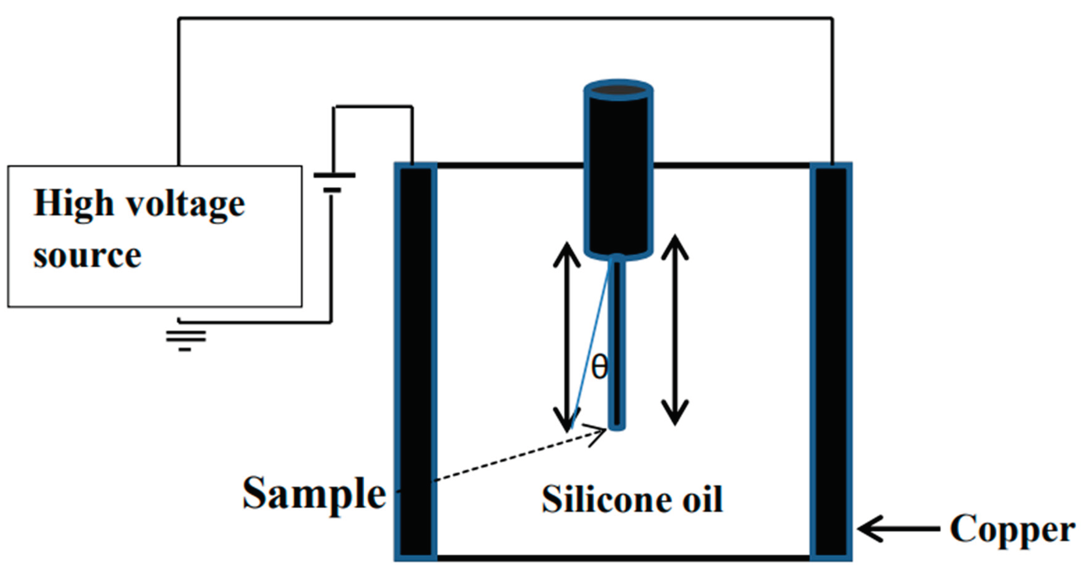
Deflection measurements investigated the electromechanical response of chitosan composite films under an electrical field. Deflections were recorded using photographs and video analysis with a digital image analyzer (Panasonic M3000, Japan). Measurements were conducted at approximately 25°C. The setup involved suspending bio-chitosan nanocomposite films vertically between parallel copper electrodes measuring 40 mm in length, 30 mm in width, and 1 mm in thickness. The entire setup was immersed in a silicone oil bath, as illustrated in Scheme 1 and Scheme 2. In the experiment, a high direct-current voltage was applied in non-contact mode with an electrode gap of 30 mm. Key parameters of interest included displacement of the free bottom end of the samples (denoted as “d”), sample length (l), and degree of bending (θ). The degree of bending (θ) was calculated using the formula θ = arctan (d/l) at various electrical field strengths ranging from 0 to 600 V/mm. All measurements were conducted at room temperature, specifically 25°C.
2.3. Preparation of Bio-Chitosan Film Composites with and without Gelatin Powder and Titanium dioxide (TiO2) Filler for Hydrophobic, Hydrophilic, and Photoelectrical Conductivity Applications
The preparation of the bio-chitosan composite film samples involved ten different formulations, as summarized in Table 1. The first and sixth formulas, referred to as Formulas 1 and 6, served as reference films― with and without gelatin powder―for comparison with the other formulations. Chitosan film was prepared by mixing chitosan powder in an acetic acid solution and forming it via a sol-gel casting process, transitioning from sol to gel within a silicone mold, as shown in Scheme 3. The critical chemical reactions of the sol-gel process are hydrolysis and condensation, which depend on the composition of raw materials, aging temperature and time, pH, and the catalyst. Formulas 2, 3, 4, and 5 were chitosan composite films with 0.5, 1.0, 2.0, and 5.0 wt% titanium dioxide (TiO2) nanoparticles added, respectively. Gelatin was not included in these formulations (Formulas 1-5). Formula 6 consisted of chitosan and gelatin without the addition of titanium dioxide, serving as the reference for Formulas 7-10, which incorporated different amounts of titanium dioxide along with gelatin. The purpose of using Formula 6 as the reference was to compare the properties and performance of additional TiO2 nanopowder in the chitosan composite films (Formulas 7-10) to assess the effects of adding titanium dioxide and gelatin on the film properties. Formulas 7-10 were chitosan composite films with gelatin powder and 0.5, 1.0, 2.0, and 5.0 wt% titanium dioxide (TiO2), respectively. It should be noted that when attempting to prepare chitosan composite films without gelatin and with 2.0 and 5.0 wt% TiO2, film formation was not successful due to phase separation caused by the high amount of TiO2 filler particles. Although Formulas 9 and 10, with 2.0 and 5.0 wt% TiO2 and gelatin powder, formed bio-chitosan composite films with a good appearance, the excess TiO2 powder caused slight tear marks.
2.4. Measurement of Crosslink Density in Bio-Chitosan Film Composites with Titanium Dioxide Filler for Hydrophobic, Hydrophilic, and Photoelectrical Conductivity Applications
To evaluate the crosslink density of the bio-chitosan composite films, the toluene swelling method and the Flory-Rehner equation were employed. It is important to note that the Flory-Rehner equation assumes ideal behavior and may provide only an approximate estimation of the crosslink density. For accuracy and standardized measurements, it is recommended to adhere to the guidelines and specifications outlined in ASTM D-6814-02. The bio-chitosan composite film specimens were cut into small pieces measuring 1x1 cm, with thicknesses ranging from 0.03 to 0.20 cm. Each specimen was individually weighed using an analytical balance before swelling. The pre-weighed specimens were then placed in suitable containers filled with toluene and allowed to swell for 72 hours at room temperature to reach equilibrium swelling. The crosslink density was evaluated using the Flory-Rehner equations (1) and (2):
where Ve is the effective number of chains in a real network per unit volume (mol/cm3), Vr is the polymer volume fraction in the swollen network at the equilibrium, χ1 is the Flory-Huggins interaction parameter for toluene in chitosan (17.92 at 25°C), and V1 is the molecular volume of the solvent (toluene),
approximately 106.3 cm3 /mol.
3. Results and Discussion
3.1. Characteristics and Physical Properties of Chitosan Powder Embedded with Titanium Dioxide for Preparing Bio-Chitosan Nanocomposite Films
Bio-composite films composed of chitosan, with and without the addition of titanium dioxide (TiO2) nanopowder at 0, 0.5, 1.0, 2.0, and 5.0 wt% as an additive, along with gelatin powder as a binder, exhibit a pleasing appearance, as illustrated in Figure 1 and detailed in Table 1. Formula 1 represents a pure chitosan biofilm without gelatin powder, serving as the reference film for comparison with other formulations. It is described as transparent and visually appealing. Similarly, Formula 6 serves as another reference film, where gelatin is added as a binder and crosslink agent to the chitosan biofilm, also resulting in a transparent and visually appealing film.
Both Formulas 1and 6 exhibit transparency and a pleasing appearance. However, Formulas 2, 3, 4, and 5 are chitosan biofilms with different weight percentages of titanium dioxide (0.5, 1.0, 2.0, and 5.0 wt%) without gelatin, and Formulas 7, 8, 9, and 10 contain gelatin as a crosslink agent. These formulations result in opaque films, as depicted in Figure 1d, 1e, 1g, 1h, 1i, and 1j. Unfortunately, Formulas 4, 5, 9 and 10, which contained excess amounts of titanium dioxide (2.0 and 5.0 wt%, respectively) with and without the addition of gelatin, were unable to form biofilms and displayed undesirable properties. These formulations resulted in brittle films with cracking and segregation issues.
All bio-chitosan composite films were characterized, and their physical properties―such as swelling in water and ethanol, percentage of shrinkage, bulk density, and film appearance―were measured and summarized in Table 2. Biofilms from Formulas 1 to 6 exhibited good swelling in water, indicating effective absorption in this solvent. Kumar, B. et al [51] reported that the swelling index of chitosan swelling in water is 104%. However, biofilms from Formulas 7 and 8 showed moderate swelling in water. The high content of TiO2 in these samples may have influenced their swelling behavior, as titanium dioxide (TiO2) is generally known for its water insolubility. TiO2 is a white nanopowder with a refractive index of 2.49 and typically forms the anatase structure, a tetragonal phase. This information is relevant to the characteristics of TiO2 in the context of the bio-chitosan composite films. All bio-chitosan composite films showed no swelling in ethanol, indicating resistance to swelling or disintegration in this solvent. The addition of TiO2 to the bio-chitosan films resulted in a reduction in the percentage of shrinkage, suggesting that the presence of TiO2 enhanced the dimensional stability of the films by decreasing their tendency to shrink. However, when high amounts of TiO2 (2.0 and 5.0 wt%) were added slight tear marks were observed, implying that the excessive TiO2 may compromise the film’s integrity, leading to structural deformation or damage. The percentage of shrinkage was inversely proportional to the amount of TiO2 added to the bio-chitosan films, consistent with the mixture rule, as supported by the SEM results shown in Figure 2. Bio-chitosan films from Formulas 1 and 6, used as references, exhibited clear, transparent films. In contrast, bio-composites with added TiO2 (Formulas 2, 3, 7, 8, 9, and 10) displayed visible TiO2 particle distribution within the bio-chitosan matrix. As the TiO2 content increased, SEM images showed a higher degree of TiO2 distribution, consistent with the films’ physical properties and appearance. Excessive TiO2 content nagatively affected dispersion, leading to brittleness and cracking, particularly in Formulas 4 and 5. In contrast, Formulas 7 and 8 demonstrated smooth biofilm surfaces without tears or fractures. Biocomposite films from Formulas 9 and 10 exhibited small crack, fractures, and slight porosity on the film surface. SEM images at magnifications of 15,000 and 50,000 times revealed the adhesion and distribution of TiO2 nanoparticles within the bio-chitosan matrix, consistent with the SEM results of TiO2 nanoparticles reported by Halawa, A.A. et al. [52].
The FTIR spectra of chitosan, gelatin, and bio-chitosan composite films were recorded in the wavenumber range 500-4000 cm-1 (Figure 3). The FTIR spectrum of raw chitosan showed characteristic peaks corresponding to various chemical functional groups, including O-H (hydroxyl groups), C-H (alkyl groups), N-H (amine groups), and C-O-C (ether linkages) at wavenumbers 3250, 2857-2922, 1585-1647, and 1063 cm-1, respectively. Gelatin, acting as a binder and crosslinking agent, exhibited peaks related to different functional groups, including O-H, C-H, N-H, C-O, and C-N, at wavenumbers 3279, 2854-2922, 1524-1629, 1403, and 1231 cm-1, respectively. The FTIR spectrum of the bio-chitosan film without gelatin and TiO2 (Formula 1) displayed functional groups similar to those of chitosan, such as O-H, -CH2, -CH3, N-H bending, and C-O-C, observed at wavenumbers 3183, 2876, 2920, 1537-1633, 1403, 1016, and 647 cm-1, respectively. These findings are consistent with previous studies by Knidri, H. El et al. [53], Laaraibi A. et al. [54], and Teli M. D. et al. [55]. In Formula 6, the bio-chitosan film displayed an additional peak around 1628 cm-1, corresponding to the amide I band. Peaks around 1528 cm-1 were attributed to N-H bending of the amide II bands, while peaks at 1402 and 1240 cm-1 indicated C-H bending vibrations of -CH2 groups. The amide III band, encompassing C-O-C, C-N, and N-H groups, appeared at wavenumbers 1021-1068 cm-1, suggesting the formation of peptide bond interactions between chitosan and gelatin [56,57,58]. The FTIR spectra of bio-chitosan films with TiO2, both with and without gelatin (Formulas 2, 3, 4, 5, 7, 8, 9, and 10), exhibited small peaks corresponding to the Ti-O-Ti functional group. These peaks appeared at low wavenumbers, approximately 504-523 cm-1 and 680 cm-1, consistent with FTIR results reported by Halawa A.A. et al. [52] and Spoială, A. et al. [59]. When exposed to light and UV radiation, TiO2 nanoparticles incorporated into chitosan films can undergo structural changes, exhibiting behvior similar to photocatalytic conditions that promote charges separation between O2- and Ti4+, producing -•O-Ti3+ [60]. The oxygen radicals generated from TiO2 attacks C-H, C-C, C-O, C-N, and N-H bonds within the chitosan structure, leading to regeneration, activation, and formation of Ti-O-CH3, Ti-O-C, Ti-O=CH2, Ti-O-N, and Ti3+-C bonds through photoexcitation at the film surface. The electrons are then transported away from the Ti sites via the conduction band of TiO2, reaching an intermediate state, as confirmed by FTIR results. The chemical reaction efficiency of TiO2 bonding with chitosan depends on factors such as particle size, particle shape, and the active sites of Ti. The proposed reaction mechanism governing the formation of CH3-O-Ti, C-O-Ti, CH2=O-Ti, and N-O-Ti involves both photolumination and water oxidation reactions. These observations suggest that the combination of chitosan and gelatin results in a homogeneous, well-integrated film, as indicated by the similar FTIR peaks present in both materials. Additionally, the incorporation of TiO2 into the bio-chitosan films leads to characteristic peaks related to Ti-O-Ti, Ti-O-CH3, Ti-O-C, Ti-O=CH2, and Ti-O-N bonds.
The XRD peak patterns of the raw materials (chitosan, gelatin, TiO2) and the bio-chitosan composite films, with and without gelatin and TiO2 powder, are presented in Figure 4. The XRD pattern of the bio-chitosan film reveals a semi-crystalline structure, displaying two distinct crystalline peaks at 2Ɵ angles of approximately 9.5° and 20°. These findings are consistent with previous XRD results reported by Knidri, H. EL et al. [53], Kucukgulmez et al. [61] and Naghibzadeh et al. [62]. The XRD pattern of gelatin also exhibits a semi-crystalline nature, with both amorphous and crystalline regions. The amorphous region is represented by a broad peak around a 2Ɵ angle of 19.5°, while the crystalline region displays a sharp peak at a 2Ɵ angle of 8.0°. These observations align with the XRD results reported by Islam, Md A. A. et al. [63]. The XRD pattern of TiO2 confirms its crystalline structure, specifically the anatase crystal structure, in agreement with the JCPDS standard pattern no. 01-083-5914. The XRD patterns of the bio-chitosan composite films, including Formula 1 (chitosan), Formula 2 (chitosan with 0.5 wt% TiO2), and Formula 6 (chitosan with gelatin), exhibited similar peak positions at 2Ɵ angles of 9°, 12°, and 19°. These positions are consistent with the JCPDS no. 96-210-1328, indicating that the biofilms are primarily composed of organic compounds such as carbon, hydrogen, nitrogen, and oxygen. In contrast, the bio-chitosan composite films in Formulas 3, 7, 8, 9, and 10 displayed sharp peaks at different positions corresponding to JCPDS no. 01-083-5914, with 2Ɵ angles of 10°, 25°, 39°, and 48°, respectively. These patterns suggest the presence of TiO2 in these films, indicating a crystalline structure consistent with the anatase phase.
A study was conducted on the wetting behavior of bio-chitosan composite films with varying amounts of titanium dioxide (TiO2). The contact angle of water droplets on the bio-chitosan composite films was measured at different time intervals, from t0 (initial time) to t2000 (2000 ms), as shown in Figure 5 and reported in Table 3. The contact angle serves as an indicator of wetting behavior, influenced by the zeta potential and surface charge of TiO2 particles on the biofilms. Generally, high contact angles indicate non-wetting behavior, while low contact angles indicate increased wettability. The study found that as the amount of TiO2 in the bio-chitosan films increased, the contact angle of water on the bio-composite films decreased. This indicates that the addition of TiO2 affects physical properties such as transparency, opacity, crystallinity, smoothness, porosity, and hydrophilicity. These changes in wetting behavior have significant implications for applications such as food packaging, wound dressings, cosmetics, and drug delivery, where hydrophobic or hydrophilic properties are critical. At t0, the highest recorded contact angle was 103.98° ± 6.35°, while at t2000, the highest was 92.77° ± 4.54°. In contrast, Formula 8 (5.0 wt% TiO2) exhibited the lowest contact angle at t0 (69.72° ± 6.54°) and at t2000 (40.56° ± 4.17°). Additionally, the study references previous work by Cui, Z. et al. [64], which reported that pure chitosan films typically have contact angles ranging from 76° to 104°. This range suggests that pure chitosan films possess a hydrophobic backbone, making them suitable for water-resistant applications such as packaging, coating, drug release, and biofilms. The decrease in contact angle with TiO2 addition can be attributed to increased surface roughness, crystallinity, and opacity, consistent with SEM results and findings reported by Ling, Y. et al. [65]. This study highlights the potential for tailoring the hydrophilic/hydrophobic properties of bio-chitosan composite films for specific applications through the incorporation of TiO2.
The crosslink density of chitosan composite films, measured in a toluene solution at 27ºC, is reported in Table 4. The study also includes calculations of the crosslink density (Ve) and the volume fraction of polymer in the swollen network at equilibrium with the pure solvent (Vr), using Equations 1 and 2, respectively. According to the study, adding TiO2 with gelatin to bio-chitosan films resulted in good adhesion and increased crosslink density. This improvement is attributed to the unique properties of TiO2 nanoparticles, such as their small size, spherical shape, mechanical strength, UV-light barrier properties, and thermal-optical-antimicrobial characteristics, as mentioned in reference [66]. As the amount of TiO2 added to the bio-chitosan films increased, the crosslink density values also rose. In contrast, pure bio-chitosan films exhibited the lowest crosslink density, reported as 2.263×10⁻² mol/cm3. The study also discussed the percentage of shrinkage, approximately 88.25 ± 2.15%, whereas Formula 8 had the lowest, reported as 28.50 ± 2.35%. Overall, the study suggests that the addition of TiO2 nanoparticles to bio-chitosan films not only improves adhesion but also increases crosslink density, reduced shrinkage, and decreased swelling. These findings highlight the potential of TiO2-based composite films for various applications due to their enhanced properties.
3.2. Mechanical, Optical, and Electrical Properties of Bio-chitosan Composite Films
The mechanical properties of bio-chitosan films, including maximum load, tensile strength, and Young’s modulus, were measured and are presented in Figure 6 and Figure 7. The results suggest that the addition of TiO2 nanoparticles to the bio-chitosan films improves these mechanical properties compared to pure bio-chitosan films. This observation is consistent with other measured physical properties, such as nanostructure, SEM results, contact angle, crosslink density, and percentage of shrinkage. These findings align with those reported by Zhang, X. et al. [67] and Vilela, C. et al. [68], supporting the conclusion that TiO2 nanoparticles enhance the mechanical properties of chitosan films. The incorporation of TiO2 nanoparticles into bio-chitosan films positively affects their mechanical performance, as evidenced by the measured physical properties and supported by related studies. This suggests that TiO2-based composite films have the potential to exhibit superior mechanical properties compared to pure bio-chitosan films, making them suitable for various applications. Formula 8 exhibited the highest tensile strength, at 0.43 MPa, while Formula 1 showed the lowest tensile strength, at 0.10 MPa. However, excessive amounts of TiO2 can lead to brittleness and cracking due to nanoparticle agglomeration and segregation.
The optical properties of bio-chitosan composite films, with and without the addition of TiO2, were measured and are presented in Figure 8. Formulas 1 and 6, which are biofilms without TiO2, exhibited high UV transmittance, reaching 90% in the wavelength range of 200-1000 nm. However, the addition of TiO2 nanoparticles to the biofilms drastically reduced UV transmittance to less than 1.0%. This reduction is attributed to the efficient UV light absorption properties of TiO2 nanoparticles. The incorporation of TiO2 nanoparticles makes bio-chitosan composite films suitable for a wide range of applications, including cosmetics, biomedical products, food packaging, drug delivery, agricultural crop packaging (smart packaging), environmental products, photoactivated films, photocatalysts, and UV protection. Additionally, the presence of TiO2 nanoparticles provides antibacterial benefits through chemical mechanisms activated by UV light. This makes these films useful in medical and dental products, as well as surgical instruments, to maintain sterility and inhibit bacterial growth, as noted in reference [68]. Overall, the addition of TiO2 nanoparticles to bio-chitosan composite films significantly reduces UV transmittance, enhancing their suitability for applications requiring UV protection, photocatalytic properties, and antibacterial effects under UV exposure.
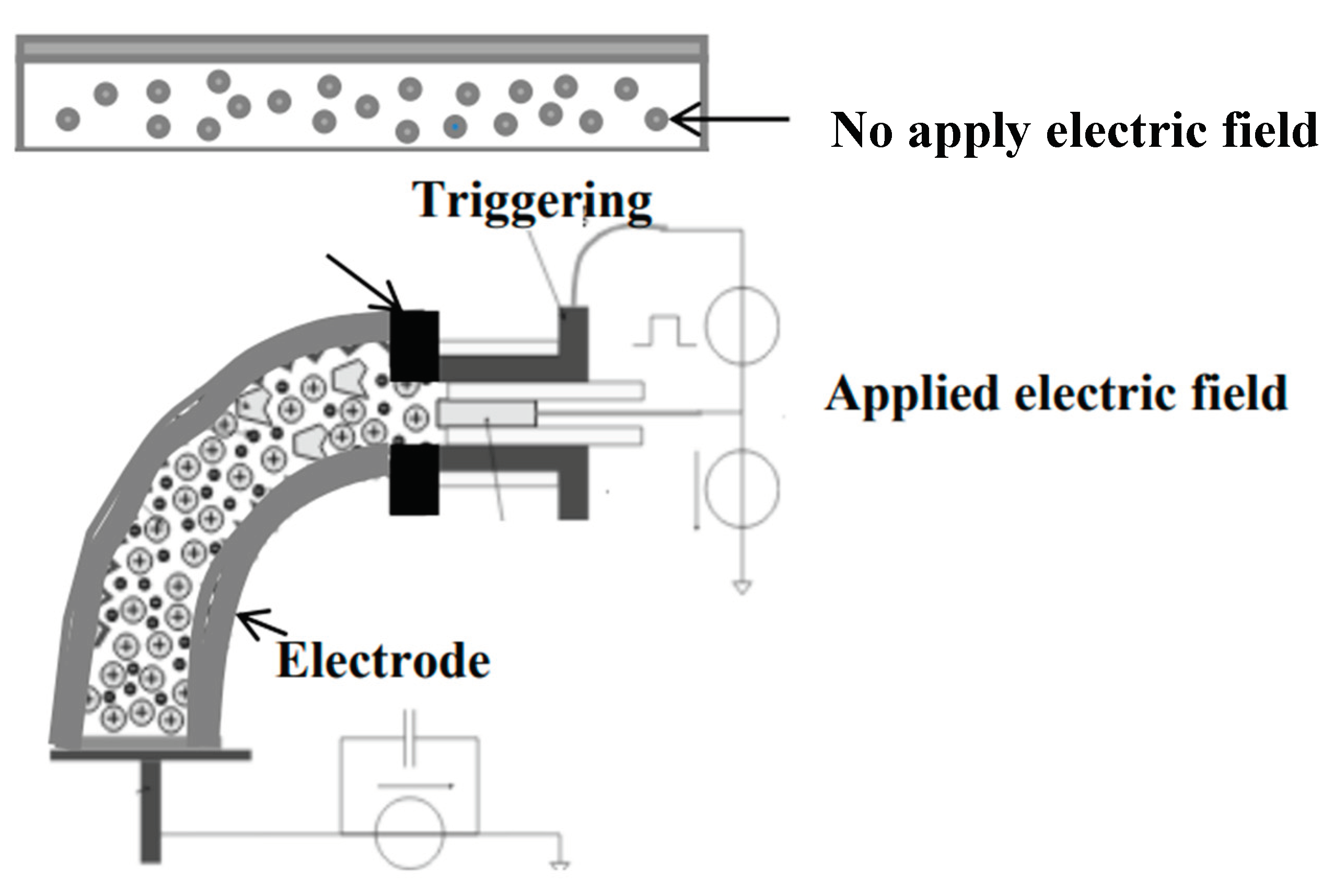
The electrical properties of bio-chitosan composite films―specifically capacitance, dielectric constant, electroactive response, and bending properties―are depicted in Figure 9 and Figure 10 and reported in Table 5 and Table 6, respectively. The capacitance and dielectric constant of bio-chitosan composite films with added TiO2 slightly decrease with increasing frequency. This can be attributed to the polarization of Ti4+ and O2- charges on the surface of the composite films, as illustrated in Scheme 3. These results are consistent with those reported by Gaabour, L.H. [69]. As the amount of TiO2 in the bio-chitosan films increases, the capacitance and dielectric constant also increase. TiO2 has reported capacitance values of approximately 90 F [70], 392 F [71], and 810 F [72], while chitosan exhibits a high capacitance value of approximately 542.63 F, as reported by Anandhavelu, S. [70]. The addition of TiO2 modifies the optical, mechanical, antimicrobial, and electrical properties of bio-chitosan films, following the rule of mixtures [73,74]. Furthermore, the polarization of Ti4+ and O2- charges on the film surface enhances the electromechanical response of the composite films. When subjected to an electric field strength ranging from 0 to 550 V/mm at room temperature (27°C), the films exhibit a higher electromechanical response than bio-chitosan films without TiO2, as shown in Figure 10 and detailed in Table 6. This effect becomes more pronounced with higher TiO2 content, resulting in a reduction of the required electric field strength by approximately four orders of magnitude compared to pure bio-chitosan films. This indicates that the addition of TiO2 enhances the electromechanical response of bio-chitosan composite films under an electric field.
The electromechanical response at electric field strengths ranging from 0 to 550 V/mm varies among different formulations. Formulas 1 and 6, without and with gelatin respectively, showed the lowest electromechanical responses― 22.03° ± 5.34° and 23.81° ± 4.51° at 550 V/mm, respectively. In contrast, Formulas 2, 3, 7, and 8, which contain TiO2 nanopowder, exhibited significant electromechanical responses, with bending angles of 68.35° ± 2.33°, 64.04° ± 2.46°, 70.03° ± 1.53°, and 81.29° ± 0.75° at electric field strengths of 550, 500, 350, and 250 V/mm, respectively. Notably, Formula 8, with a high TiO2 content, demonstrated the best electromechanical response at a low electric field strength of 250 V/mm, with a bending angle of 61.48° ± 6.97°. In summary, the addition of TiO2 nanoparticles to bio-chitosan composite films significantly influences their electrical properties, including capacitance, dielectric constant, and electromechanical response. The results demonstrate the potential for tailoring and enhancing the optical absorption, mechanical strength, antimicrobial activity, and electrical characteristics of bio-chitosan films for a wide range of applications.
4. Conclusions
The integration of titanium dioxide into chitosan powder has been shown to impart desirable characteristics and physical properties to the resulting bio-chitosan nanocomposite films. A comprehensive analysis of their morphological, structural, and mechanical aspects provides a solid foundation for understanding the potential applications of these advanced materials. The findings contribute to ongoing research in the development of sustainable, high-performance biomaterials for diverse industrial and biomedical purposes. Biochitosan composite films can be prepared by incorporating chitosan with gelatin and titanium dioxide nanoparticles. These films exhibit favorable physical, mechanical, optical, and electrical properties. They have an aesthetically pleasing appearance, show minimal tear marks, demonstrate improved wetting behavior, and display a low percentage of shrinkage. The biocomposite films also possess a high crosslink density, excellent UV absorption, and enhanced tensile strength and Young’s modulus. However, it should be noted that adding excessive amounts of TiO2 can be detrimental. The intercalation of TiO2 nanoparticles within the chitosan matrix can lead to agglomeration, resulting in increased hardness, brittleness, and slight tear marks. Nonetheless, one advantage of incorporating Ti4+ and O2- charge polarization is improved resistance to deformation, ensuring good electrical stability and a low electromechanical response under an electric field. Based on the results obtained, bio-chitosan composite films are suitable for various applications, including as biocomposite films or coating in the food, medical, pharmaceutical, agricultural, and petrochemical packaging industries. Additionally, the antimicrobial properties of TiO2 can help protect against microbial growth in the environment, owing to the photocatalytic behavior of titanium dioxide, particularly under ultraviolet (UV) irradiation at both UV-A and UV-B wavelengths.
Author Contributions
Nuchnapa Tangboriboon: Contributed to conceptualization, validation, formal analysis, investigation, and data curation. She was responsible for writing the original draft, reviewing and editing the manuscript, and providing supervision throughout the project. Additionally, she managed project administration and secured funding. Nitchakarn Malichai and Guytawan Wantaha: Contributed to the methodology, validation, formal analysis, and data curation. They were also involved in data visualization. All authors have read and approved the final version of the manuscript submitted.
Data Availability
All data is accessible to the organization, partners, or end-users at any time, as needed.
Acknowledgments
The authors would like to express their gratitude to the Materials Engineering Department at Kasetsart University, Bangkok, Thailand, for providing access to the analytical equipment used in this research. They also acknowledge the support provided to the graduate student by the Faculty of Engineering at Kasetsart University, Bangkok, Thailand.
Conflicts of Interest
The authors declare no conflicts of interest related to the research, publication, or any other aspect of the work described in this manuscript.
Ethics Approval
The authors affirm that they have adhered to accepted ethical standards for conducting research. All study participants provided informed consent and were informed about who would have access to the collected data and how it would be used. Respecting participant confidentiality and ensuring transparency in data handling are integral to maintaining ethical standards in research.
Consent to Participate
The authors confirm that informed consent was obtained from all participants involved in the research. Necessary procedures were followed to ensure participants fully understood the study, their rights, and the implications of their participation. Records obtained consent have been maintained to ensure compliance with ethical guidelines and regulations regarding participant consent in research.
Consent for Publication
If the manuscript is accepted for publication, the authors grant the publisher the license for copyright, providing the publisher with the exclusive right to publish.
References
- Paulino, A.T.; Simionato, J.I.; Garcia, J.C.; Nozaki, J.; Polym, C. 2006, 64, 98.
- Guo, Y.; Chen, X.; Yang, F.; Wang, T.; Ni, M.; Chen, Y.; Yang, F.; Huang, D.; Fu, C.; Wang, S.; Sci, J.F. 2019, 84, 1411.
- Chung, J.H.Y.; Kade, J.; Jeiranikhameneh, A.; Yue, Z.; Mukherjee, P.; Wallace, G.G. ; Bioprinting. 2018, 12, e00031.
- Li, H.; Hu, C.; Yu, H.; Chen, C.; Adv, R.S. 2018, 8, 3736.
- Ahmed, S.; Ali, A.; Sheikh, J.; Macromol, I.J.B. 2018, 116, 849.
- Liu, Y.; Jiang, X.; Wu, J.; Le, X.; Hydrocoll, F. 2016, 52, 564.
- Kumar, S.; Mukherjee, A.; Dutta, J. , Trend Food Sci Technol. 2020, 97, 196.
- Hoseinnejad, M.; Jafari, S.M.; Katouzian, I.; Microbiol, C.R. 2018, 44, 161.
- Zheng, L.Y.; Zhu, J.F. ; Carbohydr Polym. 2003, 54, 527-530.
- Alderman, O.L.G.; Skinner, L.B.; Benmore, C.J.; Tamalonis, A.; Weber, J.K.R.; Review B, P. 2014, 90, 094204.
- Abdou, E.S.; Nagy, K.S.; Elsabee, M.Z.; Technol, B. 2008, 99, 1359.
- Bajaj, M.; Winter, J.; Gallert, C.; Eng J, B. 2011, 56, 51.
- Wang, H.; Gong, X.; Miao, Y.; Guo, X.; Liu, C.; Fan, Y.Y.; Zhang, J.; Niu, B.; Li, W.; Chem, F. 2019, 283, 397.
- Bahal, M.; Kaur, N.; Sharotri, N.; Sud, D.; Technol, A.P. 2019, 1.12. S. Roy, J. -W. Int J Biol Macromol. 2020, 148, 666.
- Higashimoto, S.; Azuma, M. , Appl Catal B Environ. 2009, 89, 557.
- Zhang, W.; Rhim, J.-W.; Packag, F. 2022, 31, 100806.
- Mesgari, M.; Aalami, A.H.; Sahebkar, A. , Int J Biol Macromol. 2021, 176, 530.
- Awwad, A.M.; Amer, M.W.; Salem, N.M.; Abdeen, A.O.; Int, C. 2020, 6, 151.
- Alsohaimi, I.H.; Nassar, A.M.; Elnasr, T.A.S.; Cheba, B.A.; Prod, J.C. 2020, 248, 119274.
- Pugazhendhi, A.; Prabhu, R.; Muruganantham, K.; Shanmuganathan, R.; Natarajan, S. , J Photochem Photobiol B Biol. 2019, 190, 86.
- Hassan, H.; Omoniyi, K.; Okibe, F.; Nuhu, A.; Echioba, E. , J Appl Sci Environ Manag. 2019, 23, 1795.
- Daghrir, R.; Drogui, P.; Robert, D. , Ind Eng Chem Res. 2013, 52, 3581.
- Ali, A.; Ahmed, S. , Int J Biol Macromol. 2018, 109, 273.
- Ullattil, S.G.; Narendranath, S.B.; Pillai, S.C.; Periyat, P.; Eng J, C. 2018, 343, 708.
- Zhang, X.; Liu, Y.; Yong, H.; Qin, Y.; Liu, J.; Liu, J.; Hydrocoll, F. 2019, 94, 80.
- Shafaee, M.; Goharshadi, E.K.; Mashreghi, M.; Sadeghinia, M. , J Photochem Photobiol A Chem. 2018, 357, 90.
- Gumiero, M.; Peressini, D.; Pizzariello, A.; Sensidoni, A.; Iacumin, L.; Comi, G.; Toniolo, R.; Chem, F. 2013, 138, 1633.
- Joy, J.; Mathew, J.; George, S.C. , Int J Hydrog Energy. 2018, 43, 4804.
- Youssef, A.M.; El-Sayed, S.M.; Polym, C. 2018, 193, 19.
- Jbeli, A.; Ferraria, A.M.; Rego, A.M.B.D.; Boufi, S.; Bouattour, S. , Int J Biol Macromol. 2018, 116, 1098.
- Salarbashi, D.; Tafaghodi, M.; Bazzaz, B.S.F.; Polym, C. 2018, 186, 384.
- Xing, Y.; Tang, J.; Li, X.; Huang, R.; Wu, L.; Xu, Q.; Liu, X.; Bi, X.; Prot, J.F. 2022, 85, 597.
- Lin, B.F.; Luo, Y.G.; Teng, Z.; Zhang, B.C.; Zhou, B.; Wang, Q.; Technol, L.W.-F.S. 2015, 63, 1206.
- Kraśniewska, K.; Gniewosz, M.; Sci, J.N. 2012, 62, 199.
- Vejdan, A.; Ojagh, S.M.; Adeli, A.; Abdollahi, M.; Technol, L.W.-F.S. 2016, 71, 88.
- Peighambardoust, S.J.; Peighambardoust, S.H.; Pournasir, N.M.; Pakdel, P. , Food Packag Shelf Life. 2019, 22, 100420.
- Charles, S.; Jomini, S.; Fessard, V.; Bigorgne-Vizade, E.; Rousselle, C.; Michel, C. ; Nanotoxicology. 2018, 12, 357.
- Gea, M.; Bonetta, S.; Iannarelli, L.; Giovannozzi, A.M.; Maurino, V.; Bonetta, S.; Hodoroaba, V.-D.; Armato, C.; Rossi, A.M.; Schilirò, T.; Toxicol, F.C. 2019, 127, 89.
- Proquin, H.; Rodríguez-Ibarra, C.; Moonen, C.G.; Ortega, I.M.U.; Briedé, J.J.; de Kok, T.M.; Van Loveren, H.; Chirino, Y.I. ; Mutagenesis. 2017, 32, 139.
- de M, J., C. E. M. Campos, R. de F. P. M. Moreira, A. R. M. Fritz, Int J Biol Macromol. 2020, 151, 944.
- He, Q. , Int J Biol Macromol. 2016, 84, 153.
- Zolfi, M.; Mousavi, M.; Polym, C. 2014, 109, 118.
- Mohd, H.R.; Nur, A.I. , Int J Biol Macromol. 2020, 153, 1117.
- Masutani, E.M.; Kinoshita, C.K.; Tanaka, T.T.; Ellison, A.K.D.; Yoza, B.A.; Biomater, I.J. 2014, 979636.
- Khodaei, D.; Oltrogge, K.; Hamidi-Esfahani, Z.; Technol, L.W. 2020, 117, 108.
- Luo, Q.; Hossen, M.A.; Zeng, Y.; Dai, J.; Li, S.; Qin, W.; Liu, Y.; Eng, J.F. 2022, 313, 110762.
- Duconseille, A.; Astruc, T.; Quintana, N.; Meersman, F.; Sante-Lhoutellier, V.; Hydrocoll, F. 2015, 43, 360.
- T. Ahmad, A. Ismail, S. A., Ahmad, K. A. Khalil, Y. Kumar, K. D. Adeyemi, A. Q. Sazili, Food Hydrocoll. 2017, 63, 85.
- Andreuccetti, C.; Galicia-García, T.; Gonz, R.; Martínez-Bustos, F.; Grosso, C.R.F.; Resour, P.R. 2017, 8, 11.
- Hosseini, S.F.; Rezaei, M.; Zandi, M.; Farahmandghavi, F.; Hydrocoll, F. 2015, 44, 172.
- Kumar, B.; Castro, M.; Feller, J.F.; Chem, J.M. 2012, 22, 10656.
- Halawa, A.A.; Elshopakey, G.E., M. A. Elmetwally, M. El-Adl, S. Lashen, N. Shalaby, E. Eldomany, A. Farghali, M. Z. Sayed-Ahmed, N. Alam, N. K. Syed, S. Ahmad, and S. Rezk, Sci Rep. 2022, 12, 19667.
- Knidri, H.E.L.; Belaabed, R.; EL, R.; Laajeb, A.; Addaou, A.; Lahsin, A. , J Mater Environ Sci. 2017, 8, 3648.
- Laaraibi, A.; Charhouf, I.; Bennamara, A.; Abourriche, A.; Berrada, M. , J Mater Environ Sci. 2015, 6, 3511.
- Teli, M.D.; Sheikh, J. , Int J Biol Macromol. 2012, 50, 1195.
- Miyanji, P.B.; Semnani, D.; Ravandi, A.H.; Karbasi, S.; Fakhrali, A.; Mohammadi, S.; Technol, P.A. 2021, 1.
- Qian, Y.-F.; Zhang, K.-H.; Chen, F.; Ke, Q.-F.; Mo, X.-M. , J Biomater Sci Polym. 2011, 22, 1099.
- Gharaie, S.S.; Habibi, S.; Nazockdast, H., J Textiles Fibrous Materials. 2018, 1, 1.
- Spoială, A.; Ilie, C.I.; Dolete, G.; Croitoru, A.M.; Surdu, V.A.; Truşcă, R.D.; Motelica, L.; Oprea, O.C.; Ficai, D.; Ficai, A.; Andronescu, E.; Dițu, L.M.; Membr.2022, 12, 12080804.
- Li, W.; He, D.; Hu, G.; Li, X.; Banerjee, G.; Li, J.; Lee, S.H.; Dong, Q.; Gao, T.; Brudvig, G.W.; Waegele, M.M.; Jiang, D.-E.; Wang, D.; Sci, A.C.C. 2018, 4/5, 631.
- Kucukgulmez, A.; Celik, M.; Yanar, Y.; Sen, D.; Polat, H.; Kadak, E.; Chem, F. 2011, 126, 1144.
- Naghibzadeh, M.; Amani, A.; Amini, M.; Esmaeilzadeh, E.; Mottaghi-Dastjerdi, N.; Faramarzi, M.A.; Nanomater, J. 2010, 1.
- Islam, M.A.A.; Rahman, A.F.M.M.; Iftekhar, S.; Salem, K.S.; Sultana, N.; Bari, M.L.; Mater, J.C. 2016, 6, 172.
- Cui, Z.; Beach, E.S. , and P. T. Anastas, Green Chem Lett Rev. 2011, 4, 35. [Google Scholar] [CrossRef]
- Ling, Y., X. Pan, X. Wang, R. Sun, Cellulose. 2014, 21. [CrossRef]
- Roy, S., L. Zhai, H. C. Kim, D. H. Pham, H. Alrobei, and J. Kim, Polymers. 2021, 13, 228.
- Zhang, X.; Xiao, G.; Wang, Y.; Zhao, Y.; Su, H.; Tan, T.; Polym, C. 2017, 169, 101.
- Vilela, C.; Pinto, R.J.B.; Coelho, J.; M. R. M. Domingues, S. Daina, P. Sadocco, S. A. O. Santos, C. S. R. Freire, Food Hydrocoll. 2017, 73, 120.
- Gaabour, L.H.; Adv, A.I. 2021, 11, 105120.
- Anandhavelu, S., V. Dhansekaran, V. Sethuraman, H. J. Park, J Nanosci Nanotechnol. 2017, 17, 1321.
- Zhang, Z. 2023, 352, 135095.
- Kumar, A.J. 2023. 335, 133812.
- Sivanesan, I., J. Gopal, M. Muthu, J. Shin, and J. W. Oh, Polymers. 2021. 13, 2330.
- Callister, W.D.; Rethwisch, D.G. , Materials Science and Engineering. 10th, ISBN: 978-1-119-40549-8, 2018.
Scheme 1.
Deflection measurement setup.
Scheme 2.
Charge polarization arrangement of titanium dioxide (TiO2) in bio-chitosan composite films, functioning as electroactive actuators or compliant electrodes before and after applying an electric field.
Scheme 2.
Charge polarization arrangement of titanium dioxide (TiO2) in bio-chitosan composite films, functioning as electroactive actuators or compliant electrodes before and after applying an electric field.
Scheme 3.
Preparation process of bio-chitosan/gelatin/TiO2 nanocomposite films.
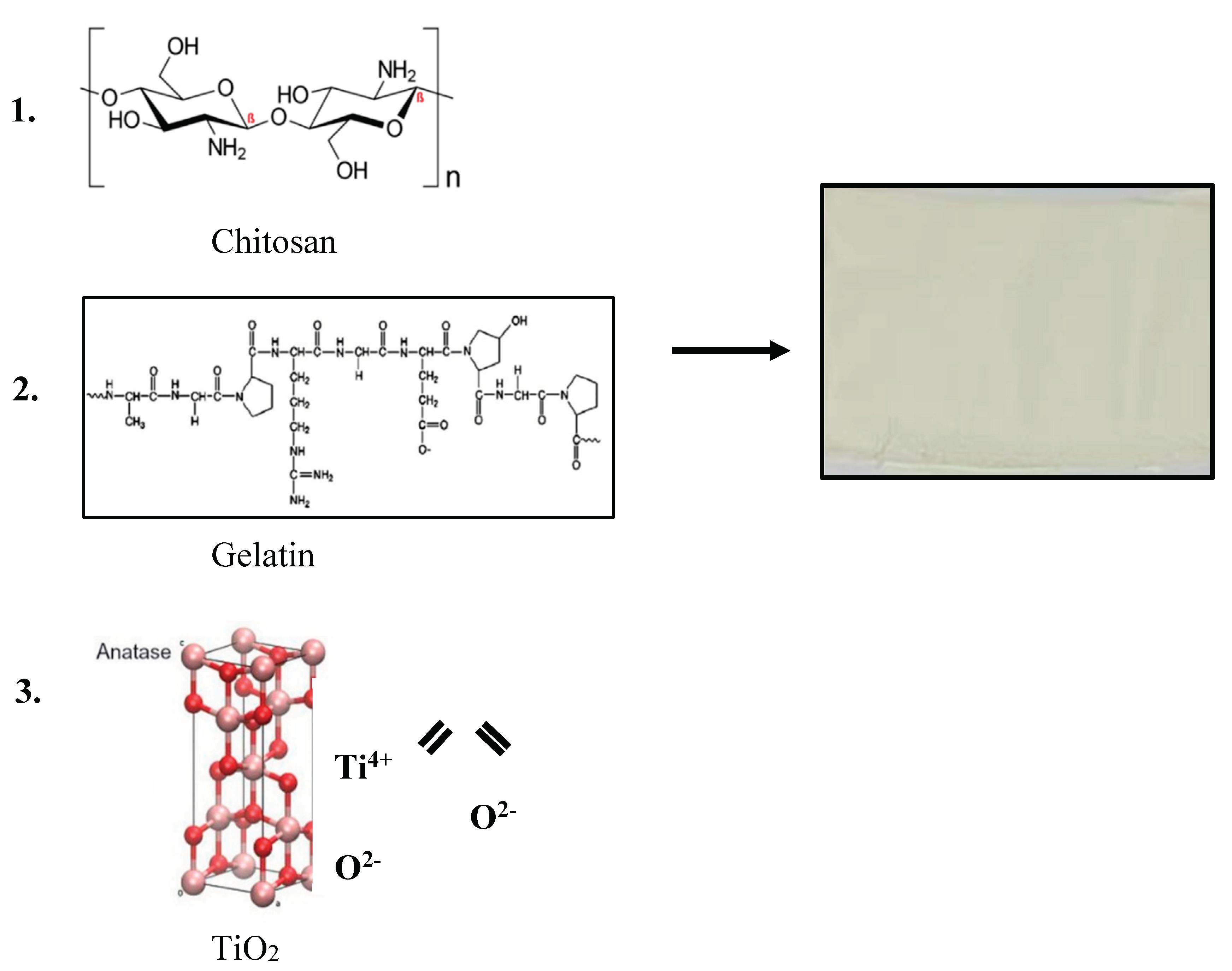
Figure 1.
Appearance of chitosan powder and bio-chitosan composite films with and without the addition of TiO2 powder: a) Chitosan powder; b) Chitosan solution for sol-gel casting films; c) Formula 1 (bio-chitosan film); d) Formula 2 (added 0.5 wt% TiO2); e) Formula 3 (added 1.0 wt% TiO2); f) Formula 6 (added gelatin powder and 0 wt% TiO2); g) Formula 7 (added gelatin powder and 0.5 wt% TiO2); h) Formula 8 (added gelatin powder and 1.0 wt% TiO2); i) Formula 9 (added gelatin powder and 2.0 wt% TiO2); and j) Formula 10 (added gelatin powder and 5.0 wt% TiO2).
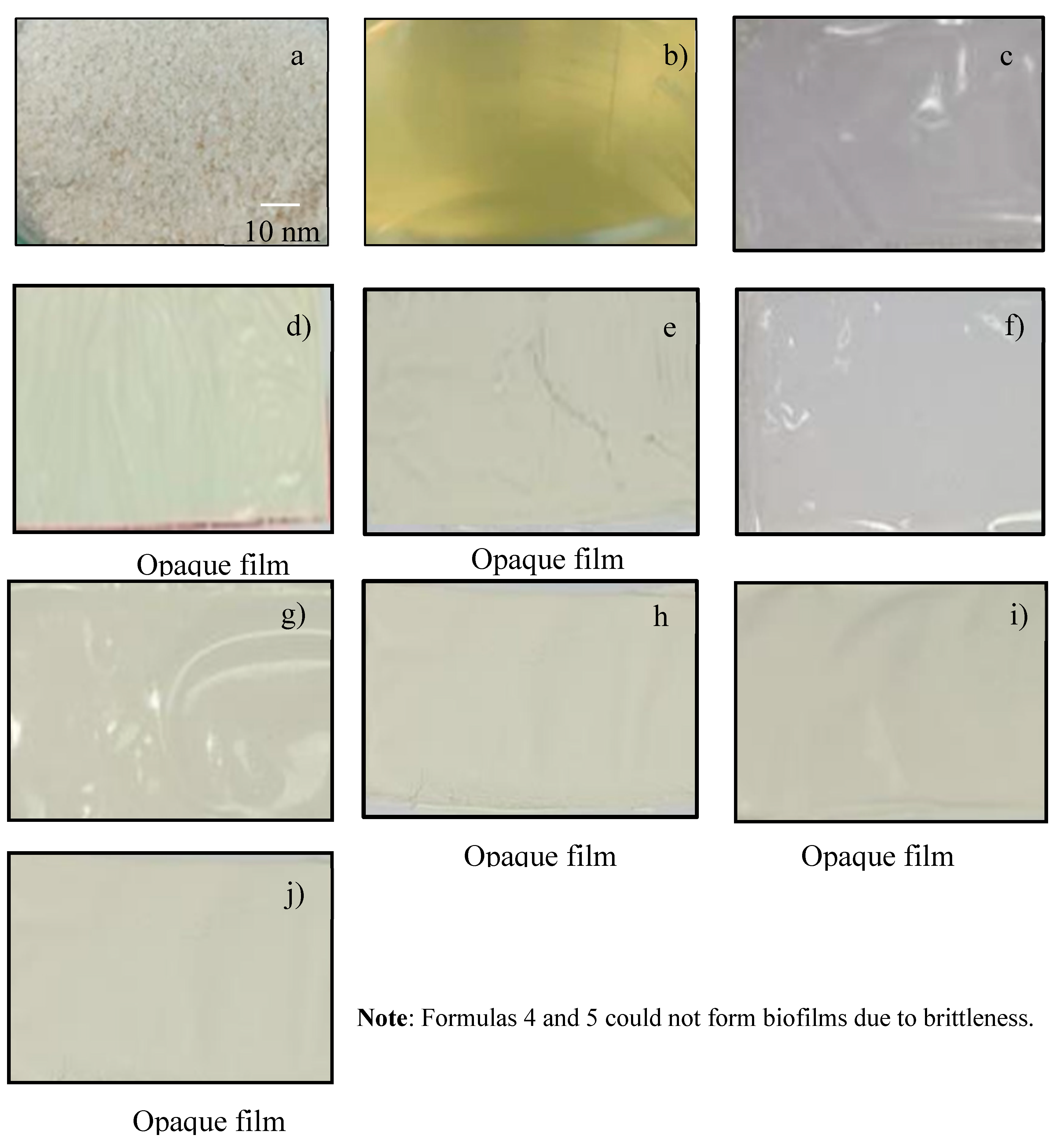
Figure 2.
SEM micrographs of bio-chitosan composite films at magnifications of 15,000 and 50,000 times for: a and a-1) Formula 1; b and b-1) Formula 2; c and c-1) Formula 3; d and d-1) Formula 6; e and e-1) Formula 7; f and f-1) Formula 8; g and g-1) Formula 9; h and h-1) Formula 10. Note: Formulas 4 and 5 could not form biofilms due to brittleness.
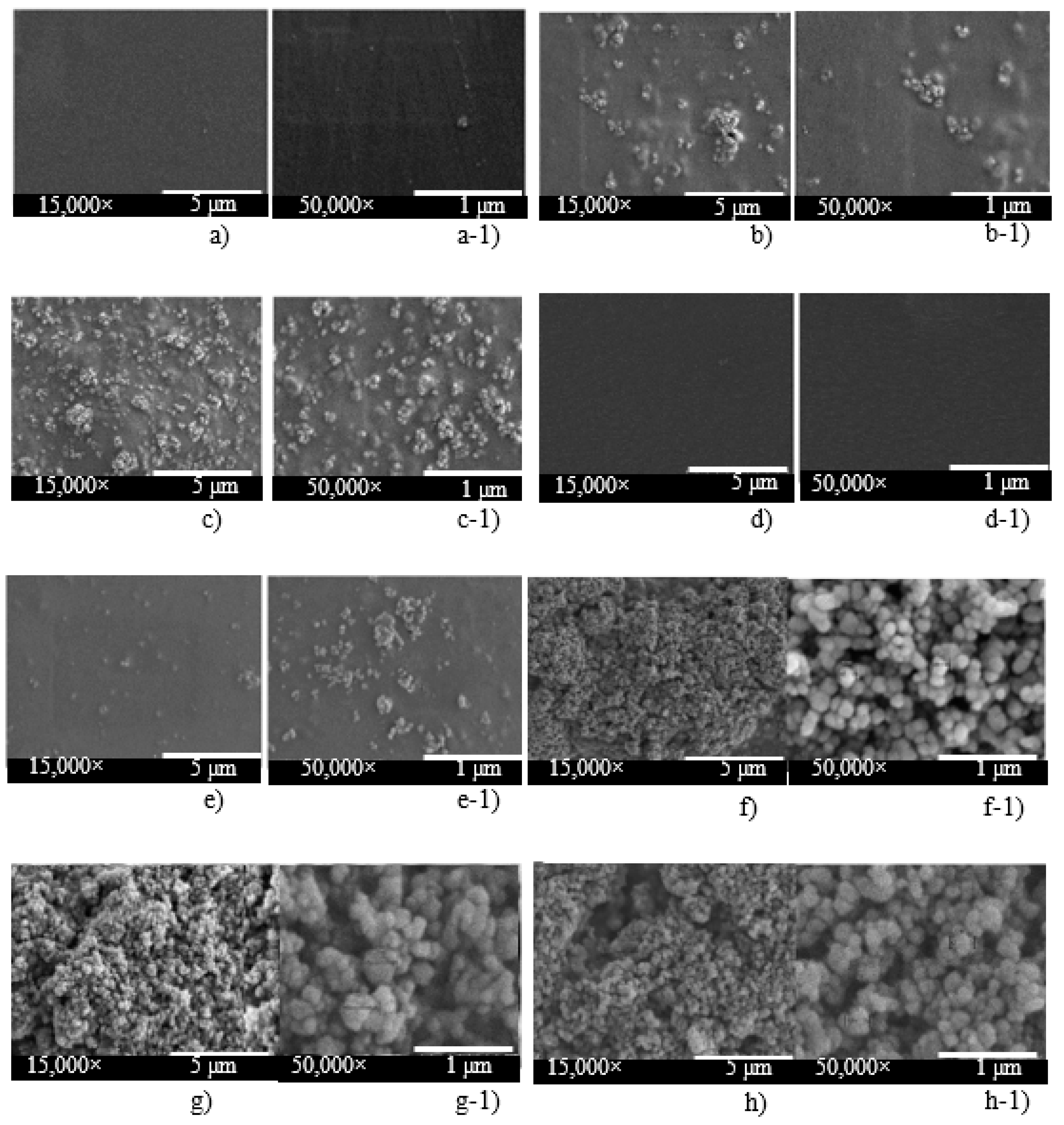
Figure 3.
FTIR spectra of raw materials (chitosan and gelatin powder) and bio-chitosan composite films, measured across the wavenumber range of 500-4000 cm-1. Note: Formulas 4 and 5 could not form biofilms due to brittleness.
Figure 3.
FTIR spectra of raw materials (chitosan and gelatin powder) and bio-chitosan composite films, measured across the wavenumber range of 500-4000 cm-1. Note: Formulas 4 and 5 could not form biofilms due to brittleness.
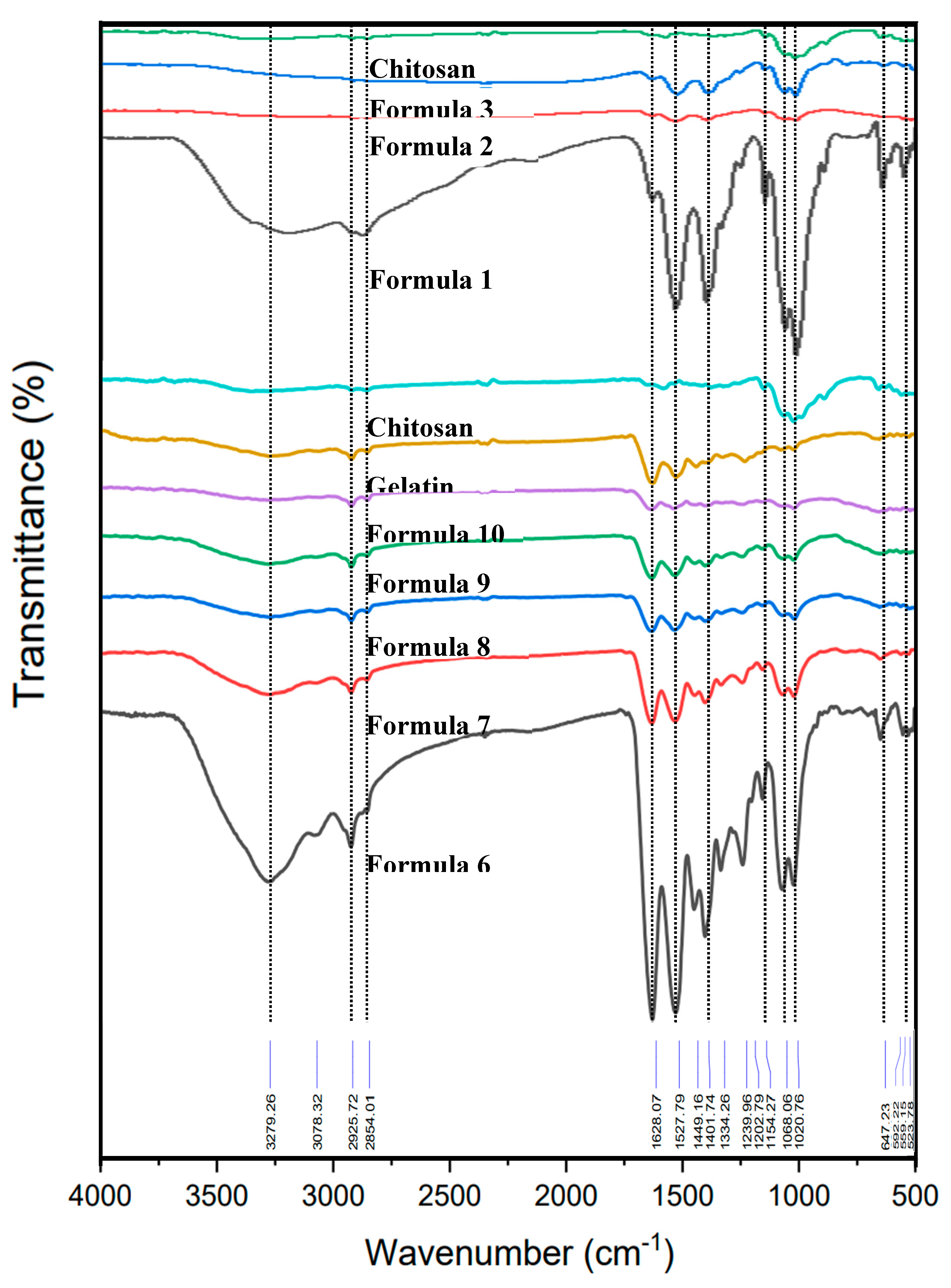
Figure 4.
XRD patterns of raw materials and bio-chitosan composite films. Note: Formulas 4 and 5 could not form biofilms due to brittleness and cracking.
Figure 4.
XRD patterns of raw materials and bio-chitosan composite films. Note: Formulas 4 and 5 could not form biofilms due to brittleness and cracking.
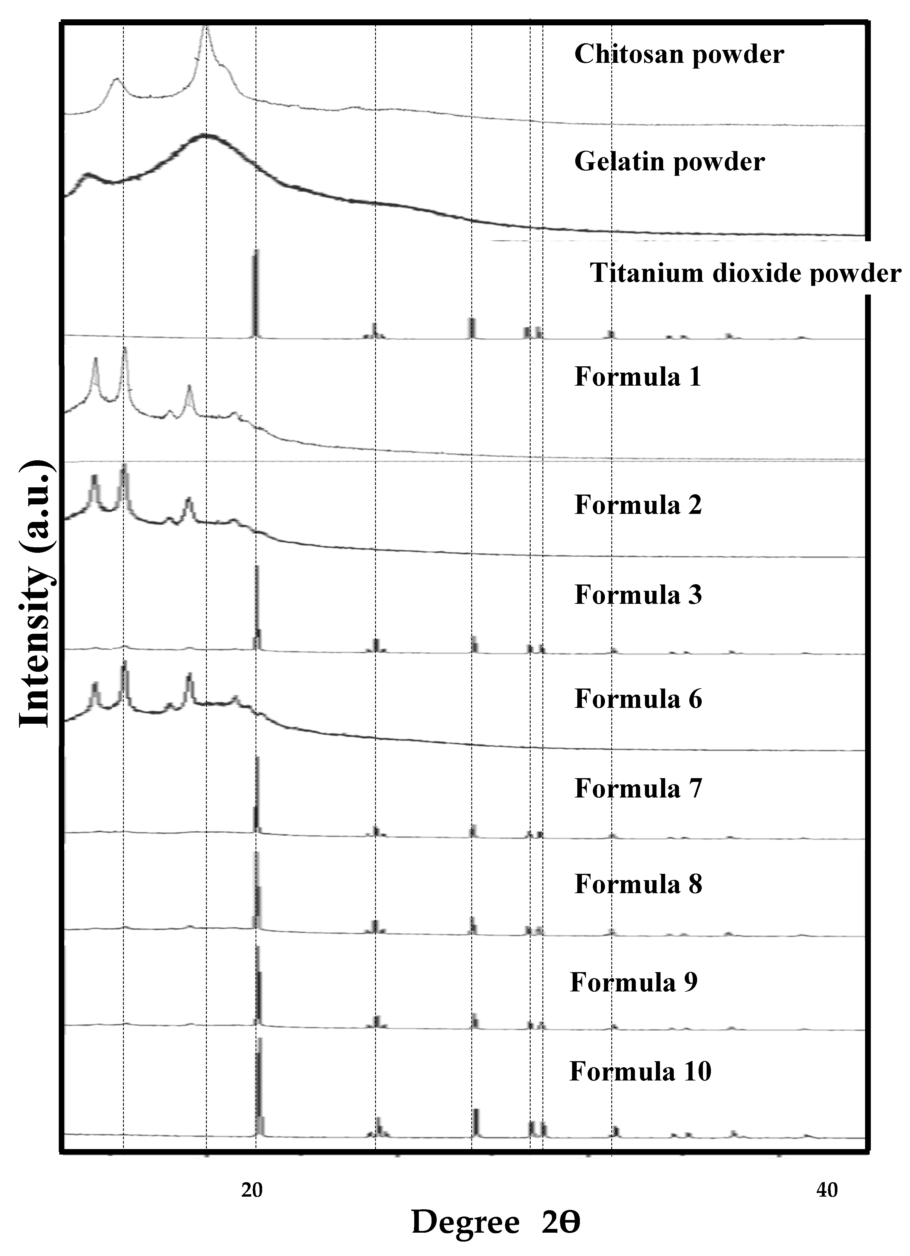
Figure 5.
Contact angles of water droplets on bio-chitosan composite films after 2000 ms: a) Formula 1; b) Formula 2; c) Formula 3; d) Formula 6; e) Formula 7; f) Formula 8; g) Formula 9; and h) formula 10. Note: Formulas 4 and 5 could not form biofilms due to cracking and brittleness.
Figure 5.
Contact angles of water droplets on bio-chitosan composite films after 2000 ms: a) Formula 1; b) Formula 2; c) Formula 3; d) Formula 6; e) Formula 7; f) Formula 8; g) Formula 9; and h) formula 10. Note: Formulas 4 and 5 could not form biofilms due to cracking and brittleness.
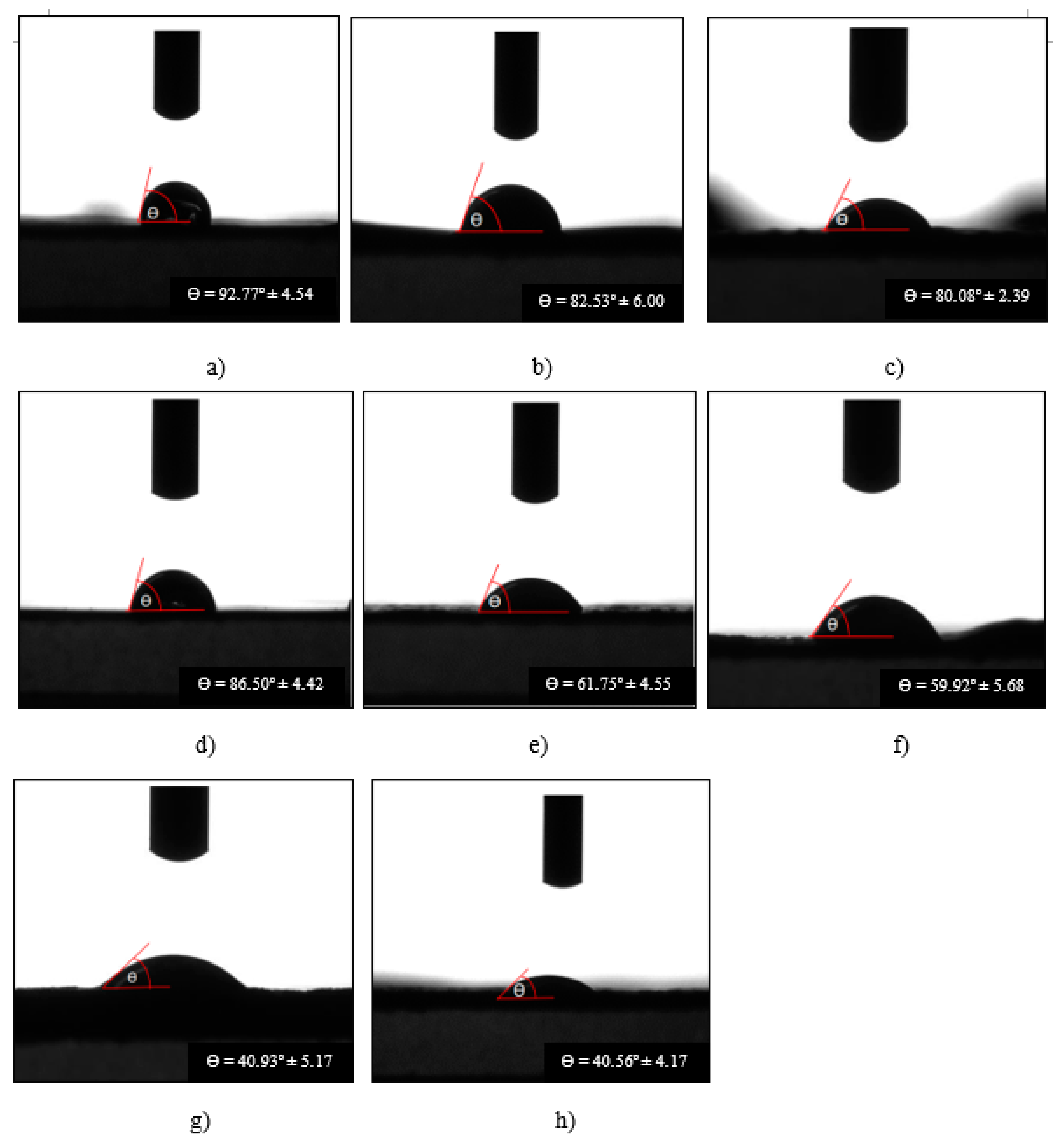
Figure 6.
Mechanical properties of bio-chitosan composite films: a) Maximum tensile load; and b) Tensile strength. Values are presented as average ± standard deviation (S.D.) Note: Formulas 4 and 5 could not form biofilms due to cracking and brittleness.
Figure 6.
Mechanical properties of bio-chitosan composite films: a) Maximum tensile load; and b) Tensile strength. Values are presented as average ± standard deviation (S.D.) Note: Formulas 4 and 5 could not form biofilms due to cracking and brittleness.
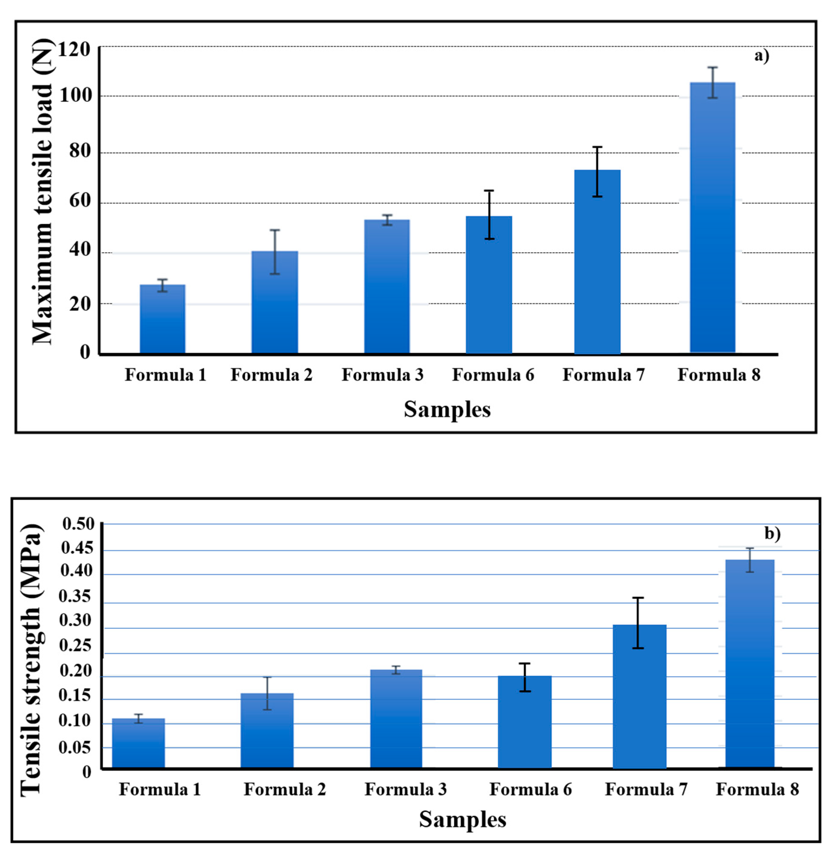
Figure 7.
Tensile Young’s modulus of bio-chitosan composite films, presented as average ± S.D. Note: Formulas 4, 5, 9, and 10 could not form biofilms or be measured due to cracking and brittleness.
Figure 7.
Tensile Young’s modulus of bio-chitosan composite films, presented as average ± S.D. Note: Formulas 4, 5, 9, and 10 could not form biofilms or be measured due to cracking and brittleness.
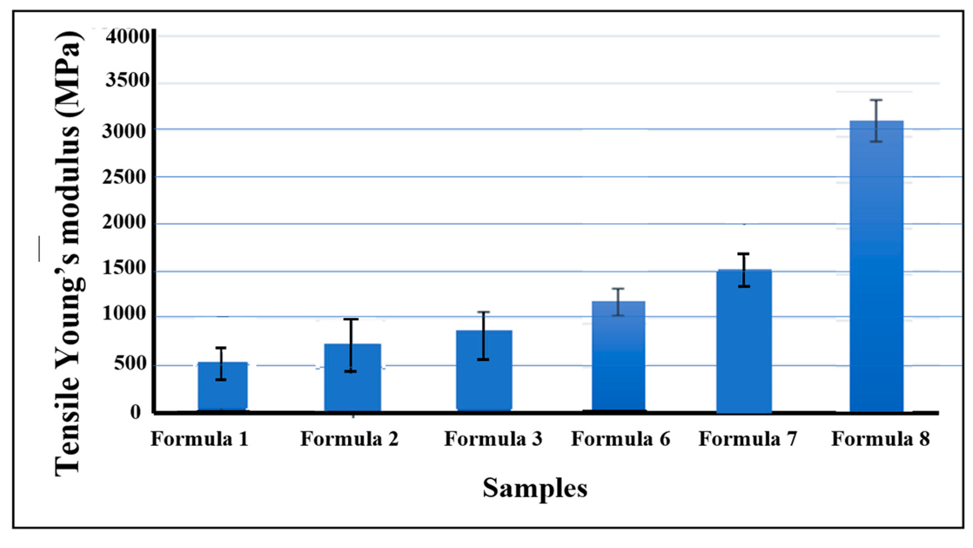
Figure 8.
UV transmisstance through bio-chitosan composite films: a) High transmittance values; b) Low transmittance values. Note: Formulas 4 and 5 could not form biofilms due to cracking and brittleness.
Figure 8.
UV transmisstance through bio-chitosan composite films: a) High transmittance values; b) Low transmittance values. Note: Formulas 4 and 5 could not form biofilms due to cracking and brittleness.
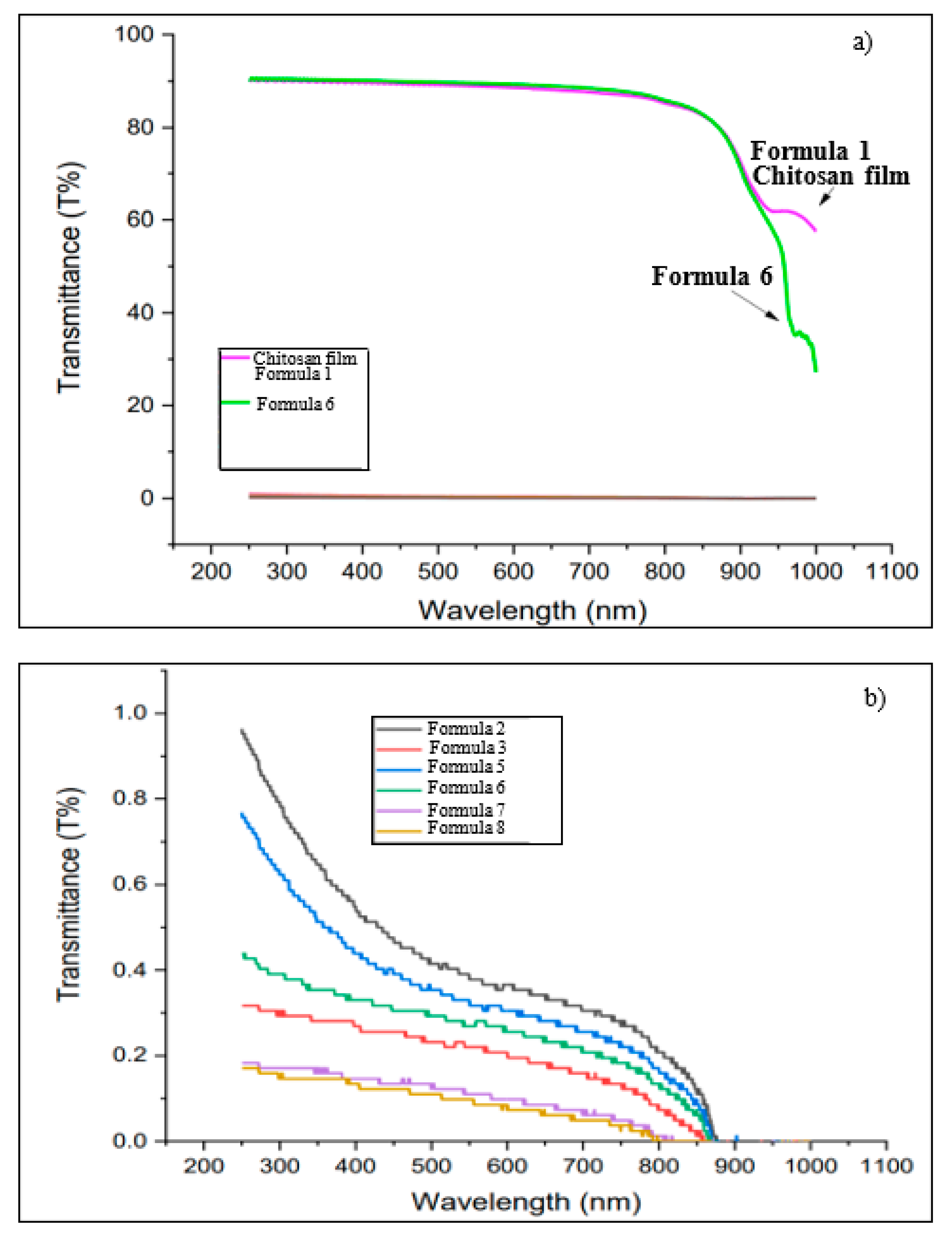
Figure 9.
Electrical properties of bio-chitosan composite films: a) Capacitance vs. frequency; and b) Dielectric constant vs. frequency. Note: Formulas 4 and 5 could not form biofilms due to cracking and brittleness.
Figure 9.
Electrical properties of bio-chitosan composite films: a) Capacitance vs. frequency; and b) Dielectric constant vs. frequency. Note: Formulas 4 and 5 could not form biofilms due to cracking and brittleness.
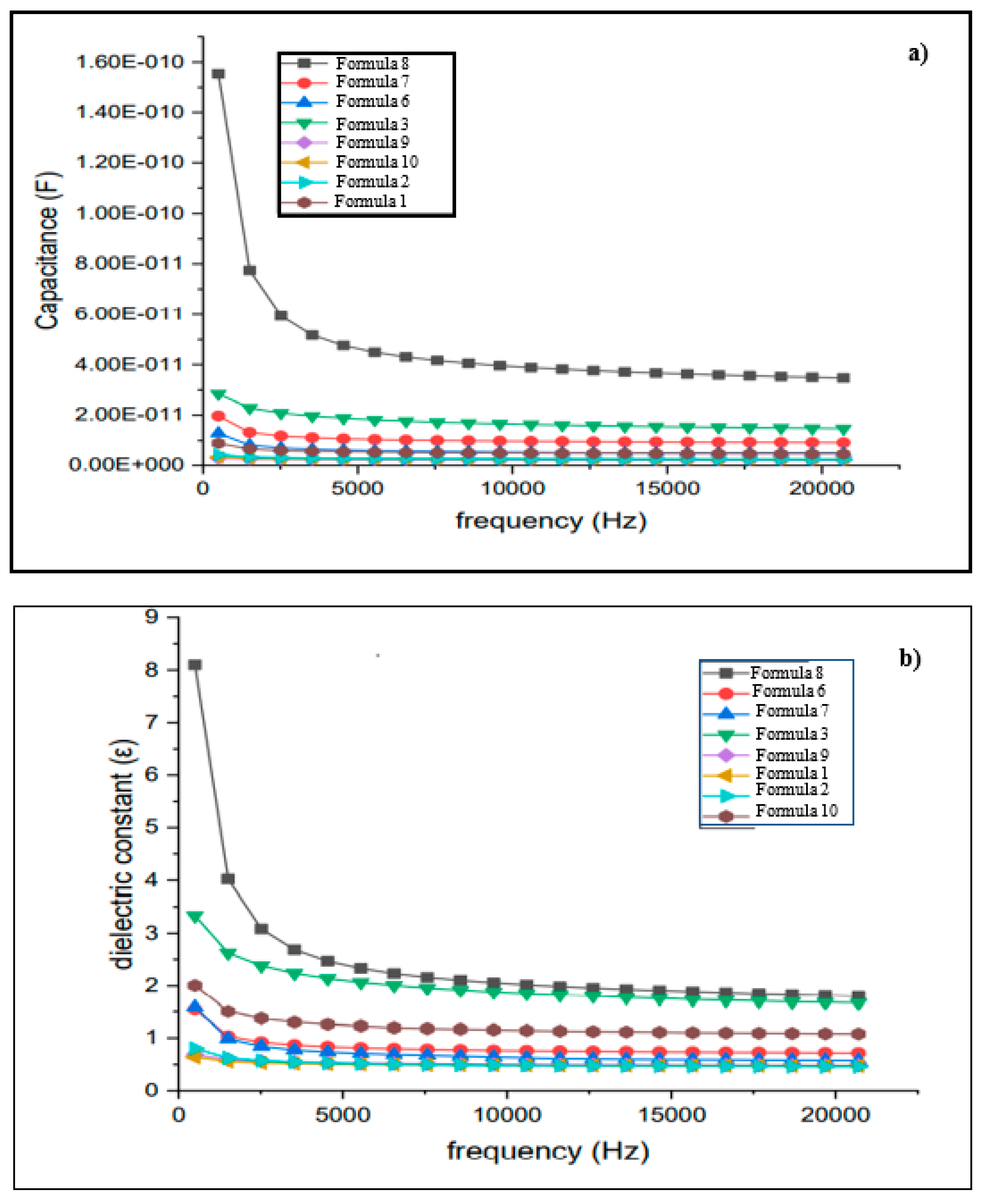
Figure 10.
Electromechanical response of Formulas 1, 2, 3, 6, 7, and 8 in bio-chitosan composite films, measured from 0 V/mm to 550 V/mm: a) at 0 V/mm and b) at the onset of electromechanical response. Note: Formulas 4, 5, 9, and 10 did not exhibit an electroactive response due to excess TiO2, causing brittleness and cracking.


Table 1.
Formulation of bio-chitosan composite films.
| Sample | Formula 1 1Chi-0Gel-0Ti |
Formula 2 1CH-0Gel-0.5Ti |
Formula 3 1Chi-0Gel-1Ti |
Formula 4 1Chi-0Gel-2Ti |
Formula 5 1Chi-0Gel-5Ti |
Formula 6 1Chi-1Gel-0Ti |
Formula 7 1Chi-1Gel-0.5Ti |
Formula 8 1Chi-1Gel-1Ti |
Formula 9 1Chi-1Gel-2Ti |
Formula 10 1Chi-1Gel-5Ti |
|---|---|---|---|---|---|---|---|---|---|---|
| Chitosan powder (g) | 1 | 1 | 1 | 1 | 1 | 1 | 1 | 1 | 1 | 1 |
| Gelatin powder (g) | 0 | 0 | 0 | 0 | 0 | 1 | 1 | 1 | 1 | 1 |
| TiO2 (wt%) | 0 | 0.5 | 1.0 | 2.0 | 5.0 | 0 | 0.5 | 1.0 | 2.0 | 5.0 |
| Acetic acid 1% (ml) | 80 | 80 | 80 | 80 | 80 | 80 | 80 | 80 | 80 | 80 |
Remark: Formulas 1-5 serve as reference samples without the addition of gelatin powder. Formulas 6-10 consist of biocomposite films incorporating nano titanium dioxide powder in conjunction with gelatin. Unfortunately, Formulas 4 and 5 cannot be used to cast bio-chitosan films due to brittleness and cracking. Each formula was prepared in 16 pieces for the purpose of characterizing and measuring their physical, mechanical, and electrical properties. The encoded formula xChi-yGel-zTi represents the weights of chitosan powder (Chi), gelatin powder (Gel), and TiO2 (Ti). For example, 1Chi-0Gel-0Ti means 1 gram of chitosan powder, 0 grams of gelatin powder, and 0 wt% of TiO2 (or 1 g chitosan, 0 g gelatin, and 0 wt% TiO2).
Table 2.
Characteristics and physical properties of bio-chitosan composite films, expressed as average values ± standard deviation (n = 3).
Table 2.
Characteristics and physical properties of bio-chitosan composite films, expressed as average values ± standard deviation (n = 3).
| Sample | Film thickness (mm) |
Swelling in water | Swelling in ethyl alcohol | Percentage of shrinkage after drying (%) |
Bulk density (g/cm3) |
Characteristics |
|---|---|---|---|---|---|---|
| Formula 1 1Chi-0Gel-0Ti |
0.03 ± 0.01 | Excellent swelling | No swelling | 88.25 ± 2.15 | 1.48 ± 0.32 | Transparent, with a good appearance and no tear marks |
| Formula 2 1CH-0Gel-0.5Ti |
0.08 ± 0.02 | Excellent swelling | No swelling | 68.97 ± 7.99 | 0.86 ± 0.15 | Opaque, with a good appearance and no tear marks |
| Formula 3 1Chi-0Gel-1Ti |
0.08 ± 0.01 | Excellent swelling | No swelling | 67.60 ± 3.58 | 0.89 ± 0.18 | Opaque, with a good appearance and no tear marks |
| Formula 6 1Chi-1Gel-0Ti |
0.06 ± 0.01 | Excellent swelling | No swelling | 77.22 ± 4.02 | 1.38 ± 0.13 | Transparent, with a good appearance and no tear marks |
| Formula 7 1Chi-1Gel-0.5Ti |
0.09 ± 0.01 | Excellent swelling | No swelling | 59.95 ± 3.59 | 0.91 ± 0.04 | Opaque, with a good appearance and no tear marks |
| Formula 8 1Chi-1Gel-1Ti |
0.09 ± 0.03 | Excellent swelling | No swelling | 54.17 ± 9.28 | 0.85 ± 0.18 | Opaque, with a good appearance and no tear marks |
| Formula 9 1Chi-1Gel-2Ti |
0.16 ± 0.02 | Moderately swelling | No swelling | 31.63 ± 2.37 | 0.83 ± 0.09 | Opaque with good appearance and minor tear marks |
| Formula 10 1Chi-1Gel-5Ti |
0.17 ± 0.01 | Moderately swelling | No swelling | 28.50 ± 2.35 | 0.90 ± 0.03 | Opaque, with a good appearance and minor tear marks |
Remark: Formulas 4 (1Chi-0Gel-2Ti) and 5 (1Chi-0Gel-5Ti) cannot form bio-chitosan films due to cracking and brittleness. Formulas 1 and 6, serving as reference biofilms without the addition of nano titanium dioxide powder, exhibit the highest percentage of shrinkage. The encoded formula xChi-yGel-zTi represents the weights of chitosan powder (Chi), gelatin powder (Gel), and TiO2 (Ti). For example, 1Chi-0Gel-0Ti means 1 gram of chitosan powder, 0 grams of gelatin powder, and 0 wt% of TiO2 (or 1 g chitosan, 0 g gelatin, and 0 wt% TiO2). Index of chitosan swelling in water 104% [61].
Table 3.
Contact angle on the surface of bio-chitosan composite films, presented as average values ± standard deviation (measured three times).
Table 3.
Contact angle on the surface of bio-chitosan composite films, presented as average values ± standard deviation (measured three times).
| Sample | Contact angle at t = 0 ms (Degree, º) |
Contact angle at t = 2000 ms (Degree, º) |
Decreasing contact angle over time (Degree/ms or º/ms) |
|---|---|---|---|
|
Formula 1 1Chi-0Gel-0Ti |
103.98 ± 6.35 | 92.77 ± 4.54 | 0.58 ± 0.21 |
| Formula 2 1CH-0Gel-0.5Ti |
91.32 ± 4.53 | 82.53 ± 6.00 | 0.43 ± 0.07 |
| Formula 3 1Chi-0Gel-1Ti |
88.24 ± 3.63 | 80.08 ± 2.39 | 0.43 ± 0.05 |
|
Formula 6 1Chi-1Gel-0Ti |
93.34 ± 7.47 | 86.50 ± 4.42 | 0.54 ± 0.09 |
| Formula 7 1Chi-1Gel-0.5Ti |
80.34 ± 7.37 | 61.75 ± 4.55 | 1.44 ± 0.06 |
| Formula 8 1Chi-1Gel-1Ti |
79.23 ± 4.05 | 59.92 ± 5.68 | 1.47 ± 0.28 |
| Formula 9 1Chi-1Gel-2Ti |
73.16 ± 5.50 | 40.93 ± 5.17 | 1.48 ± 0.24 |
| Formula 10 1Chi-1Gel-5Ti |
69.72 ± 6.54 | 40.56 ± 4.17 | 1.51 ± 0.11 |
Remark: Formulas 4 (1Chi-0Gel-2Ti) and 5 (1Chi-0Gel-5Ti) cannot form bio-chitosan films due to cracking and brittleness. Formula 1 and 6, serving as reference biofilms without the addition of nano titanium dioxide powder, exhibit the highest percentage of shrinkage. The encoded formula xChi-yGel-zTi represents the weights of chitosan powder (Chi), gelatin powder (Gel), and TiO2 (Ti). For example, 1Chi-0Gel-0Ti means 1 gram of chitosan powder, 0 grams of gelatin powder, and 0 wt% of TiO2 (or 1 g chitosan, 0 g gelatin, and 0 wt% TiO2).
Table 4.
Crosslink density of bio-chitosan composite films in toluene solution at 27ºC.
| Sample | Percentage of shrinkage (%) |
Bulk density of dry films (g/cm3) |
Weight of dry films (g) |
Weight of absorbed solvent (g) |
Vr | Crosslink density × 10-2 (Ve, mol/cm3) |
|---|---|---|---|---|---|---|
|
Formula 1 1Chi-0Gel-0Ti |
88.25 ± 2.15 | 1.48 ± 0.32 | 0.0265±0.0025 | 0.0017 | 0.9013 | 2.263 |
| Formula 2 1CH-0Gel-0.5Ti |
68.97 ± 7.99 | 0.86 ± 0.15 | 0.0221±0.0020 | 0.0023 | 0.9604 | 4.162 |
| Formula 3 1Chi-0Gel-1Ti |
67.60 ± 3.58 | 0.89 ± 0.18 | 0.0256±0.0026 | 0.0008 | 0.9689 | 4.627 |
|
Formula 6 1Chi-1Gel-0Ti |
77.22 ± 4.02 | 1.38 ± 0.13 | 0.0338±0.0018 | 0.0010 | 0.9550 | 3.912 |
| Formula 7 1Chi-1Gel-0.5Ti |
59.95 ± 3.59 | 0.91 ± 0.04 | 0.0484±0.0072 | 0.0018 | 0.9624 | 4.262 |
| Formula 8 1Chi-1Gel-1Ti |
54.17 ± 9.28 | 0.85 ± 0.18 | 0.0421±0.0059 | 0.0013 | 0.9706 | 4.735 |
| Formula 9 1Chi-1Gel-2Ti |
31.63 ± 2.37 | 0.83 ± 0.09 | 0.0761±0.0139 | 0.0005 | 0.9938 | 7.677 |
| Formula 10 1Chi-1Gel-5Ti |
28.50 ± 2.35 | 0.90 ± 0.03 | 0.0611±0.0032 | 0.0006 | 0.9899 | 6.758 |
Remark: Formulas 4 (1Chi-0Gel-2Ti) and 5 (1Chi-0Gel-5Ti) cannot form a bio-chitosan film due to cracking and brittleness. Formulas 9 and 10 have high crosslink density and are prone to cracking due to the excessive addition of nano titanium dioxide. The encoded formula xChi-yGel-zTi represents the weights of chitosan powder (Chi), gelatin powder (Gel), and TiO2 (Ti). For example, 1Chi-0Gel-0Ti means 1 gram of chitosan powder, 0 grams of gelatin powder, and 0 wt% of TiO2 (or 1 g chitosan, 0 g gelatin, and 0 wt% TiO2). Crosslink density measurement was determined using the toluene-swelling method at room temperature (ASTM D-6814-02). The samples were cut into squares (1 cm wide, 3-5 mm thick). The weight of the specimens was recorded before and after swelling for 72 hours in toluene. Crosslink density was calculated using the Flory-Rehner equations (1) and (2): (1) (2) where Ve is the effective number of chains in the real network per unit volume (mol/cm3), Vr is the volume fraction of polymer in the swollen network in equilibrium with pure solvent (dimensionless), χ1 is the Flory-Huggins interaction parameter for the polymer-solvent interaction (17.92 for toluene in chitosan at 25ºC), the density of toluene is 0.8670 g/cm3, and V1 is the molecular volume of the solvent (toluene 106.3 cm3/mol). Highlight (Formulas 1 and 6) indicates the reference biofilm samples without adding nano titanium dioxide powder to form bio-chitosan composite films.
Table 5.
Electrical properties and dielectric constant of bio-chitosan composite films measured at 27ºC (n =3; average ± standard deviation) and at 500 Hz.
Table 5.
Electrical properties and dielectric constant of bio-chitosan composite films measured at 27ºC (n =3; average ± standard deviation) and at 500 Hz.
| Sample |
Capacitance × 10-10 (F) |
Dielectric constant |
|---|---|---|
|
Formula 1 1Chi-0Gel-0Ti |
0.029 ± 0.062 | 1.54 ± 0.22 |
| Formula 2 1CH-0Gel-0.5Ti |
0.033 ± 0.095 | 1.58 ± 0.54 |
| Formula 3 1Chi-0Gel-1Ti |
0.284 ± 0.011 | 3.33 ± 0.13 |
|
Formula 6 1Chi-1Gel-0Ti |
0.129 ± 0.015 | 1.69 ± 0.17 |
| Formula 7 1Chi-1Gel-0.5Ti |
0.197 ± 0.039 | 1.80 ± 0.07 |
| Formula 8 1Chi-1Gel-1Ti |
1.550 ± 0.150 | 8.10 ± 0.73 |
| Formula 9 1Chi-1Gel-2Ti |
0.090 ± 0.005 | 1.64 ± 0.27 |
| Formula 10 1Chi-1Gel-5Ti |
0.043 ± 0.003 | 2.01 ± 0.70 |
Remark: Formulas 4 (1Chi-0Gel-2Ti) and 5 (1Chi-0Gel-5Ti) cannot form a bio-chitosan film due to cracking and brittleness; hence, they cannot be used to measure electrical properties. Formulas 9 and 10, on the other hand, have high crosslink density and are prone to cracking due to the excessive addition of nano titanium dioxide. The encoded formula xChi-yGel-zTi represents the weights of chitosan powder (Chi), gelatin powder (Gel), and TiO2 (Ti). For example, 1Chi-0Gel-0Ti means 1 gram of chitosan powder, 0 grams of gelatin powder, and 0 wt% of TiO2 (or 1 g chitosan, 0 g gelatin, and 0 wt% TiO2).
Table 6.
Electromechanical response properties of bio-chitosan composite films measured at 27ºC (n =3; average ± standard deviation) under an applied electric field strength ranging from 0 to 550 V/mm.
Table 6.
Electromechanical response properties of bio-chitosan composite films measured at 27ºC (n =3; average ± standard deviation) under an applied electric field strength ranging from 0 to 550 V/mm.
| Electric field strength (V/m) |
Formula 1 1Chi-0Gel-0Ti (Degree, °) |
Formula 2 1CH-0Gel-0.5Ti (Degree, °) |
Formula 3 1Chi-0Gel-1Ti (Degree, °) |
Formula 6 1Chi-1Gel-0Ti (Degree, °) |
Formula 7 1Chi-1Gel-0.5Ti (Degree, °) |
Formula 8 1Chi-1Gel-1Ti (Degree, °) |
Formula 9 1Chi-1Gel-2Ti (Degree, °) |
Formula 10 1Chi-1Gel-5Ti (Degree, °) |
|---|---|---|---|---|---|---|---|---|
| 0 | 0.00 ± 0.00 | 0.00 ± 0.00 | 0.00 ± 0.00 | 0.00 ± 0.00 | 0.00 ± 0.00 | 0.00 ± 0.00 | 0.00 ± 0.00 | 0.00 ± 0.00 |
| 25 | 0.00 ± 0.00 | 0.00 ± 0.00 | 0.00 ± 0.00 | 0.00 ± 0.00 | 0.00 ± 0.00 | 0.00 ± 0.00 | 0.00 ± 0.00 | 0.00 ± 0.00 |
| 50 | 0.00 ± 0.00 | 0.00 ± 0.00 | 0.00 ± 0.00 | 0.00 ± 0.00 | 0.00 ± 0.00 | 3.31 ± 0.17 | 0.00 ± 0.00 | 0.00 ± 0.00 |
| 100 | 0.00 ± 0.00 | 3.37 ± 1.04 | 5.92 ± 0.22 | 0.00 ± 0.00 | 4.64 ± 0.98 | 4.61 ± 2.58 | 4.65 ± 0.85 | 5.23 ± 1.76 |
| 150 | 2.64 ± 0.33 | 4.60 ± 1.65 | 9.07 ± 0.89 | 3.65 ± 0.01 | 6.70 ± 1.74 | 4.97 ± 0.65 | 5.16 ± 0.33 | 5.64 ± 3.33 |
| 200 | 4.02 ± 1.65 | 5.57 ± 1.50 | 10.44±0.73 | 4.23 ± 0.76 | 9.00 ± 1.56 | 32.25±3.56 | 6.21 ± 1.65 | 6.42 ± 3.65 |
| 250 | 4.03 ± 1.00 | 7.22 ± 2.30 | 18.96±3.83 | 4.62 ± 0.83 | 17.85±2.58 | 61.48±6.97 | 6.40 ± 1.00 | 6.50 ± 2.00 |
| 300 | 4.17 ± 1.89 | 9.25 ± 3.12 | 43.67±4.39 | 4.82 ± 0.65 | 38.87±3.01 | 68.58±7.36 | 42.07±0.89 | 8.17 ± 1.89 |
| 350 | 4.91 ± 0.41 | 11.37±3.71 | 54.63±4.16 | 5.25 ± 0.01 | 59.06±3.91 | 72.81±5.57 | 50.09±2.10 | 10.29±2.41 |
| 400 | 5.28 ± 3.35 | 13.59±4.97 | 58.15±1.72 | 8.07 ± 0.89 | 64.77±4.98 | 73.72±5.27 | 52.54±0.35 | 14.84±5.35 |
| 450 | 8.70 ± 4.52 | 17.91±3.93 | 59.34±1.50 | 9.49 ± 0.41 | 67.62±4.37 | 78.02±2.56 | 56.70±5.52 | 19.70±7.52 |
| 500 | 13.58±3.51 | 65.88±5.02 | 62.61±2.23 | 13.84±2.35 | 70.05±2.18 | 79.48±3.28 | 59.34±0.51 | 26.58±6.51 |
| 550 | 22.03±5.34 | 68.35±2.33 | 64.04±2.46 | 23.81±4.51 | 70.03±1.53 | 81.29±0.75 | 60.05±0.30 | 34.03±7.34 |
Remark: Formulas 4 (1Chi-0Gel-2Ti) and 5 (1Chi-0Gel-5Ti) cannot form a bio-chitosan film due to cracking and brittleness; consequently, they cannot be used to measure electrical properties. Formulas 9 and 10 exhibit a low electromechanical response attributed to their brittleness and tendency to crack. The encoded formula xChi-yGel-zTi represents the weights of chitosan powder (Chi), gelatin powder (Gel), and TiO2 (Ti). For example, 1Chi-0Gel-0Ti means 1 gram of chitosan powder, 0 grams of gelatin powder, and 0 wt% of TiO2 (or 1 g chitosan, 0 g gelatin, and 0 wt% TiO2).
Disclaimer/Publisher’s Note: The statements, opinions and data contained in all publications are solely those of the individual author(s) and contributor(s) and not of MDPI and/or the editor(s). MDPI and/or the editor(s) disclaim responsibility for any injury to people or property resulting from any ideas, methods, instructions or products referred to in the content. |
© 2025 by the authors. Licensee MDPI, Basel, Switzerland. This article is an open access article distributed under the terms and conditions of the Creative Commons Attribution (CC BY) license (http://creativecommons.org/licenses/by/4.0/).
Copyright: This open access article is published under a Creative Commons CC BY 4.0 license, which permit the free download, distribution, and reuse, provided that the author and preprint are cited in any reuse.


