
Submitted:
13 May 2025
Posted:
15 May 2025
You are already at the latest version
Abstract
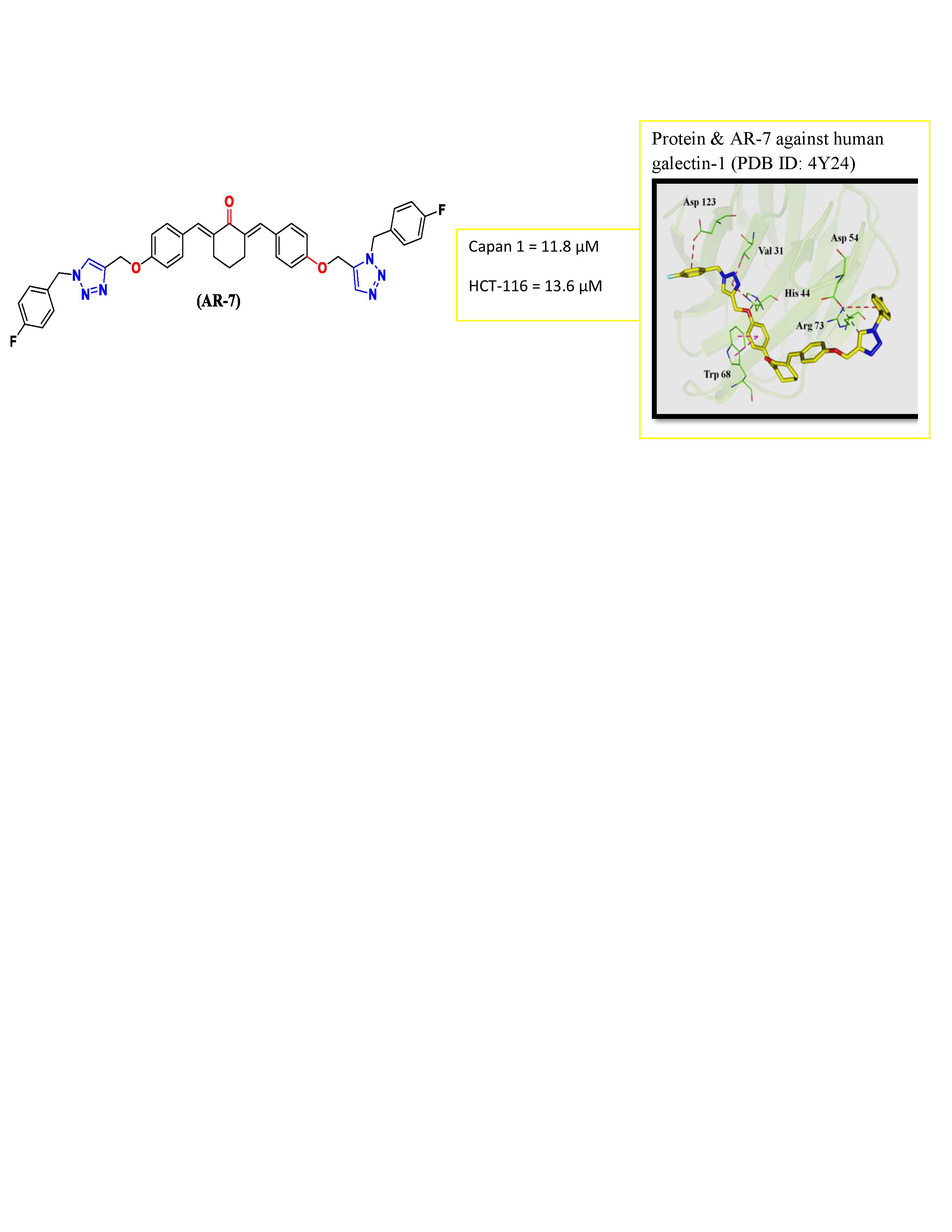
A series of 1,2,3-triazole-linked cyclic ketones (AR 1-14) were synthesized involving the condensation of triazole-aldehydes with cyclic ketones in tetrahydrofuran (THF) and potassium hydroxide (KOH). The synthesized derivatives were evaluated for their in-vitro cytotoxicity against retinal (hTERT RPE-1)-1), pancreatic (Capan-1), myeloid (K-562, Hap-1), colorectal (HCT 116), lung (NCI-H460), lymphoblastic (DND-41), and non-Hodgkin lymphoma (Z-138) cells. Among the tested derivatives, molecule 2,6-bis(4-((1-(4-fluorobenzyl)-1H-1,2,3-triazol-4-yl)methoxy)benzylidene)cyclohexanone (AR-7) exhibited moderate cytotoxic property with an IC₅₀ of 11.8 µM and 13.6 µM against pancreatic adenocarcinoma and colorectal carcinoma cells respectively. Molecular docking against galectin-1 receptor (PDB ID: 4Y24) demonstrated a favourable binding interaction (-7.1 kcal/mol) indicating a strong receptor-ligand affinity.
Keywords:
1
; 2
; 3-Triazole
; cytotoxicity
; galectin-1 (4Y24)
; molecular docking
1. Introduction
Cancer is a group of complex diseases characterized by the uncontrolled growth. In India, the estimated number of new cancer cases in 2022 was 14,61,427, and one in nine individuals is likely to develop cancer during their lifetime. Alarmingly, the incidence of cancer cases in the country is projected to increase by 12.8% by 2025[1]. In recent decades, heterocyclic chemistry has played a pivotal role in the discovery and development of new anticancer agents. Among heterocycles, nitrogen-containing frameworks are particularly valued due to their low toxicity, enhanced reactivity, and strong receptor affinity [2].Considering this, the present study focuses on the design and development of novel 1,2,3-triazole analogs with potential anticancer activity.It was synthesized via a 1,3-dipolar cycloaddition reaction between an azide and an alkyne or aldehyde, under metal-catalyzed or metal-free conditions [3]. It is widely employed in medicinal chemistry as a bioisostere due to its chemical stability and favorable pharmacokinetic properties [4].
Galectins are the promising molecular targets in cancer therapy representing a family of β-galactoside-binding soluble lectins composed of approximately 130 amino acids. These proteins are distributed throughout the cytosol and nucleus, where they mediate various biological functions via N- or O-glycosylation, interacting with carbohydrate recognition domains (CRDs) [5,6,7,8]. Out of the fifteen identified galectins, galectin-1 has garnered significant attention due to its overexpression in several cancers, where it plays a key role in metastasis, tumor progression, and immune-evasion. It binds to glycosylated receptors (such as glycoproteins like CD45, CD43, or integrins) on the surface of immune cells, particularly T cells, dendritic cells, and natural killer (NK) cells [9,10,11,12]. These characteristics make galectin-1 an appealing molecular target for the design of novel anticancer therapeutics.
2. Results and Discussion
2.1. Chemistry
Fourteen derivatives of 1,2,3-triazole-linked cyclic ketones (AR1-14) were synthesized as outlined in the Scheme 1a-d. The synthetic route commenced with the reaction of 4-hydroxybenzaldehyde (1) and propargyl bromide (2) in dry dimethyl formamide (DMF) yielding 4-(prop-2-ynyloxy)benzaldehyde (3) (Scheme 1a). In the subsequent step (Scheme 1b), compound 3 was subjected to a copper(I)-catalyzed azide-alkyne cycloaddition (CuAAC) with different benzyl azides (4a-h) in the presence of sodium ascorbate (C6H7NaO6) and copper sulphate pentahydrate (CuSO₄·5H₂O), affording the corresponding triazole-benzaldehydes (5a-h), Scheme 1b. The final derivatives AR1-14 were synthesized by condensation of 5a-h with cyclic ketones 6 (Scheme 1c) and 7(Scheme 1d) in THF and KOH. The synthesized compounds were confirmed by melting point (M.p), thin-layer chromatography (TLC), Fourier-Transform Infrared (FTIR), and ¹H and ¹³C Nuclear Magnetic Resonance (NMR) spectroscopy. The FTIR spectra of compounds AR 1-14 had shown aromatic C-H stretching vibrations in the range of 3096-3000 cm⁻¹, while aliphatic C-H stretches appeared between 2998-2710 cm⁻¹. The carbonyl (C=O) stretching bands were noted between 1698-1652 cm-¹. The 1H NMR spectra (δ, ppm) showed singlets for triazole ring protons between δ 8.37-8.25 ppm. The aromatic protons appeared in the range δ 8.24-6.44, while benzylic protons were observed between δ 7.58-7.38 ppm. Methylene protons from -OCH₂- and -NCH₂- groups resonated in the ranges of δ 5.80-5.50 and δ 5.24-5.18 ppm, respectively. Methoxy (-OCH₃) protons were evident between δ 3.73-3.62 ppm in compounds AR2, AR3, AR4, AR8, and AR9. Methyl protons in AR6 and AR11 were identified as singlet at δ 2.27 ppm. Cycloalkyl protons resonated broadly in the range of δ 3.05-1.71 ppm. The 13C NMR spectra data for the compounds had displayed peaks in the range of δ 195 to 188 ppm for C=O of cyclic ketones, for aromatic carbons between δ 159-106 ppm, and for methoxy and methyl carbons in the range of δ 62 to 21 ppm.
2.2. Biology
2.2.1. Cytotoxic Study
The in-vitro cytotoxicity study was conducted on different human cancerous and non-cancerous (hTERT RPE-1) cell lines, following established protocols reported in the literature. Dimethyl sulfoxide (DMSO) was employed as the solvent control, while docetaxel and staurosporine were used as reference standards. The IC₅₀ values, as summarized in Table 1, highlight compound AR-7 as the moderately active molecule in the series, exhibiting an IC₅₀ of 11.8 and 13.6 µM against the Capan-1 and HCT-116 cell lines respectively while AR-9 had shown IC50 of 17.4µM against HCT-116 cells. In contrast to docetaxel and staurosporine, none of the other molecules were found to be more cytotoxic to non-cancerous hTERT RPE-1 cells, indicating a higher degree of selectivity for cancerous cells. Molecules AR 1-3 were not tested during the study, hence not included in Table 1.
2.2.2. In-Silico Study
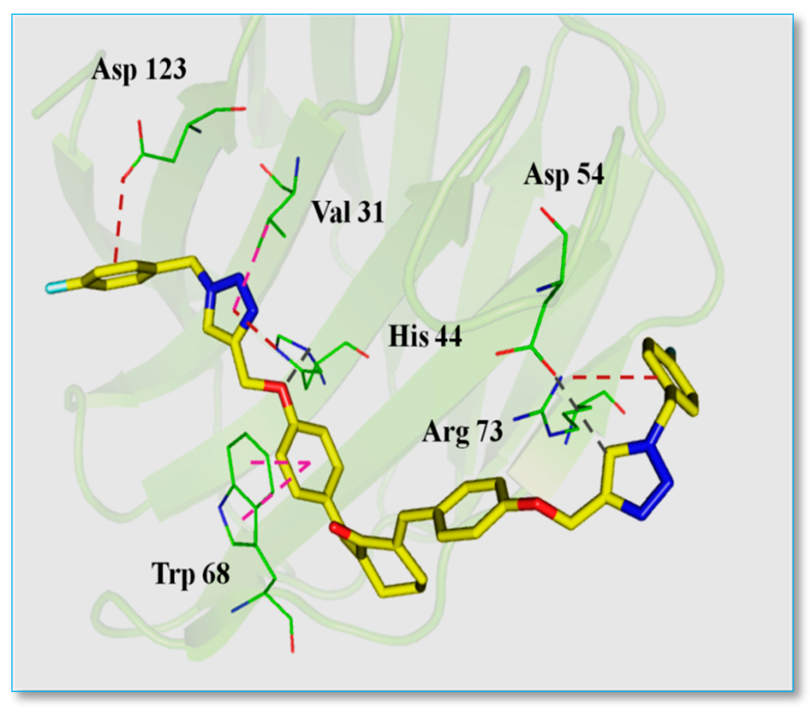
In support of in-vitro cytotoxicity data, the binding mechanism of the most active compound AR-7 was investigated using molecular docking approach. AutoDockVina 1.1.3 was employed to dock the molecule AR-7 into the active site of the target protein, human galectin-1 (PDB ID: 4Y24). The co-crystallized ligand TD-139(bound to galectin-1) was first extracted and prepared using AutoDock Tools. The prepared TD-139 ligand was re-docked into the binding pocket of galectin-1 using the same grid configuration applied for AR-7. The re-docking of TD-139 yielded a conformation closely matching the crystal structure, with a docking score of -7.1 kcal/mol, thereby confirming the reliability of the docking methodology (Figure 1). Subsequent docking of AR-7 revealed a favourable binding pose with a docking score of -7.6 kcal/mol, indicating a potentially stronger interaction compared to TD-139. The CRD) of galectin-1 is comprisedof five carbohydrate-binding sub-sites (CBS) designated A to E (Figure 2). The co-ligand TD-139 was found to interact with sub-sites B, C, D, and E, whereas PR-7, due to its extended structure engaged with sub-sites A, B, C, and E. Detailed interaction analysis showed that:
- The 4-fluorophenyl ring of AR-7 formed an electrostatic interaction with Asp123 in sub-site A.
- The triazole ring exhibited a hydrophobic π-alkyl interaction with Val31 in sub-site B, Trp68 in sub-site C and an electrostatic interaction with His44 in sub-site C.
- The oxygen linker between the triazole and benzene rings participated in hydrogenbonding with His44.
- Another triazole ring formed hydrogen bonding with Asp54 in sub-site A.
- Finally, a fluorophenyl ring at the terminal end of the molecule established an electrostatic π-cation interaction with Arg73 in sub-site A (Figure 3).
Collectively it can be summarized that, AR-7 interacted with all conserved residues in the CBS of galectin-1, similar to TD-139 involving three electrostatic, two hydrogen bonding, and two hydrophobic interactions, supporting its stronger binding affinity and potential for greater inhibition of galectin-1 compared to other synthesized molecules in the series.
Figure 1.
Protein-co-crystal ligand (TD-139) against human galectin-1 (PDB ID: 4Y24). The co-crystallised ligand is shown in pink and its docked pose showed in cyan.
Figure 1.
Protein-co-crystal ligand (TD-139) against human galectin-1 (PDB ID: 4Y24). The co-crystallised ligand is shown in pink and its docked pose showed in cyan.

Figure 2.
The residue belonging to the 5 subsites in CBS of CRD in human Galctin-1 is shown in surface and stick representation. Each subsites shown in different colors. SubsiteA in purple, subsite B in yellow, subsite C in pink, subsite D in orange and subsite E in cyan.
Figure 2.
The residue belonging to the 5 subsites in CBS of CRD in human Galctin-1 is shown in surface and stick representation. Each subsites shown in different colors. SubsiteA in purple, subsite B in yellow, subsite C in pink, subsite D in orange and subsite E in cyan.
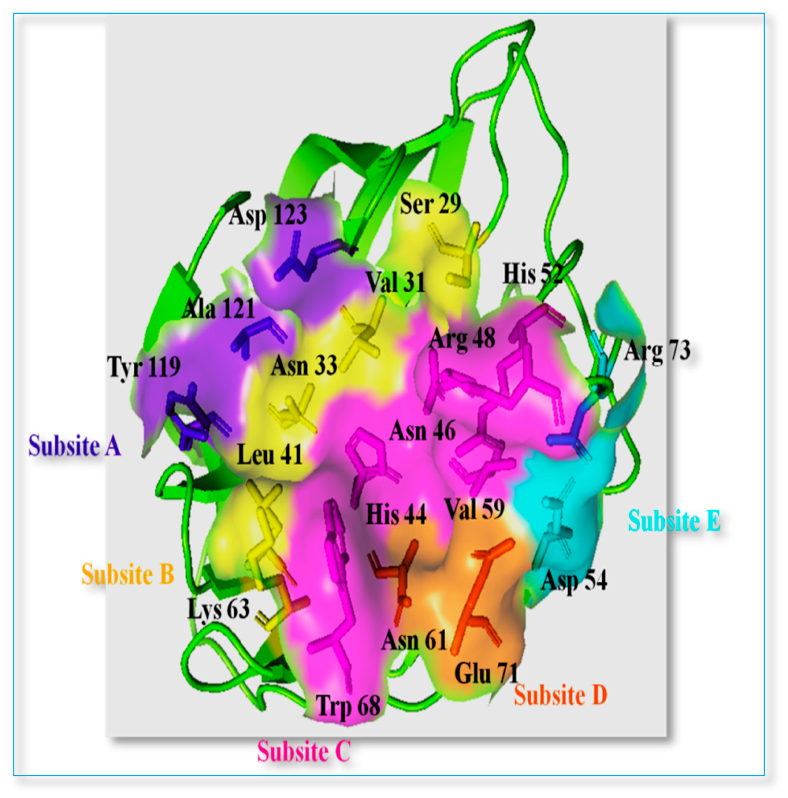
Figure 3.
Protein-AR-7 against human galectin-1 (PDB ID: 4Y24). The compound AR-7 is shown in yellow sticks. Electrostatic, hudrogen and hydrophobic interactions are shown in orange, grey and pink dotted line respectively.
Figure 3.
Protein-AR-7 against human galectin-1 (PDB ID: 4Y24). The compound AR-7 is shown in yellow sticks. Electrostatic, hudrogen and hydrophobic interactions are shown in orange, grey and pink dotted line respectively.
3. Materials and Methods
3.1. Chemistry
3.1.1. General Information and Instrumentation
All reagents and solvents were of analytical grade and used as received unless otherwise stated. The purity of reagents and solvents was confirmed prior to use. The progress of reactions was monitored by TLC using pre-coated Aluchrosep silica gel 60/UV254 plates (Sd Fine-Chem Ltd.). M.p was determined using the open capillary tube method in heavy liquid paraffin and were reported uncorrected. FTIR spectra were recorded using the diffuse reflectance technique with IR-grade potassium bromide (KBr) on a JASCO 460+ spectrometer. The NMR spectra were obtained using Bruker Ultraspec AMX 400 and JEOL RESONANCE spectrometers operating at 400 (1HNMR) and 500 (13C NMR) MHz in deuterated dimethyl sulfoxide (DMSO-d₆) and chloroform (CDCl₃). The chemical shift (δ) values were reported in parts per million (ppm) relative to tetramethylsilane (TMS) as the internal standard. The intermediates 4-(prop-2-ynyloxy)benzaldehyde (3), benzyl azide (4a), 4-nitrobenzyl azide (4b), 4-methylbenzyl azide (4c), 4-chlorobenzyl azide (4d) and the 4-((1-benzyl-1H-1,2,3-triazol-4-yl)methoxy)benzaldehydes (5a-h)were prepared as per the literature [13].The compounds 4-fluoro (4e), 4-methoxy (4f), 3,5-dimethoxy (4g), and 3,4,5-trimethoxybenzyl azide (4h) were synthesized according to literature reference 14.
3.1.2. Synthesis of (2E,6E)-2,6-bis(4-((1-benzyl-1H-1,2,3-triazol-4-yl)methoxy)benzylidene) Cyclic Ketones (AR 1-14).
A solution of triazole aldehyde (5a-h, 1 mmol) in THF (20 mL) was mixed with an equal molar volume of a methanolicKOH solution (20 mL). To this mixture, the corresponding cyclic ketone (6 or 7, 1 mmol) was added gradually with continuous stirring and stirred at 35 °C for 16 h. Progress of reaction was monitored by TLC. Upon completion, the reaction mixture was poured into cold water and neutralized with dilute hydrochloric acid. The resulting precipitate was filtered, washed with cold water, and recrystallized from an ethanol-chloroform mixture (80:20 v/v) to yield AR1-14.
(2E,6E)-2,6-bis(4-((1-benzyl-1H-1,2,3-triazol-4-yl)methoxy)benzylidene)cyclohexanone (AR-1)
Yellow crystals, 70% yield. M.p; 160-162 °C. IR ν (KBr): 3061, 3007, 2945, 2729, 1655, 1596, 1558, 1455, 1297 cm-1. 1H NMR (400 MHz, DMSO-d6):δ 8.29 (2H, s, triazole-H), 7.57 (2H, s, benzyl-H), 7.52 (4H, d, J=8.8, Ar.), 7.39-7.30 (10H, m, Ar.), 7.15 (4H, d, J=8.8, Ar.), 5.61 (4H, s, 2 -OCH2-), 5.19 (4H, s, 2 -N-CH2-), 2.87 (4H, t, 2 -CH2-), J=10.4), 1.71 (2H, p, -CH2-, J=24).
(2E,6E)-2,6-bis(4-((1-(4-methoxybenzyl)-1H-1,2,3-triazol-4-yl)methoxy)benzylidene)cyclohexanone (AR-2)
Yellow amorphous powder, 55% yield, M.p: 128-130 °C. IR ν (KBr): 3072, 3019, 2953, 2836, 1654, 1597, 1551, 1460, 1385, 1277 cm-1. 1H NMR (DMSO-d6); δ 8.25 (2H, s, triazole-H), 7.58 (2H, s, benzyl-H), 7.52 (4H, d, J=7.2, Ar.), 7.31 (4H, d, J=6.8, Ar.), 7.12 (4H, d, J=6.8, Ar.), 6.93 (4H, d, J=6.8, Ar.) 5.53 (4H, s, 2 -OCH2-), 5.18 (4H, s, 2 -N-CH2-), 3.73 (6H, s, 2 -OCH3), 2.87 (4H, q, J=10.4, 2 -CH2-), 1.73 (2H, p, -CH2-, J=19.2) ppm.13C NMR (500 MHz, DMSO-d6): δ 188.62 (s), 159.15 (s), 158.49 (s), 142.68 (s), 135.37 (s), 134.36 (s), 132.22 (s), 131.53 (s), 129.64 (s), 127.90 (s), 124.47 (s), 114.84 (s), 114.13 (s), 113.90 (s), 61.15 (s), 55.13 (s), 52.40 (s), 27.91 (s), 22.46 (s) ppm.
(2E,6E)-2,6-bis(4-((1-(3,5-dimethoxybenzyl)-1H-1,2,3-triazol-4-yl)methoxy)benzylidene)cyclohexanone (AR-3)
Yellow crystals, 61% yield, M.p: 115-118 °C. IR ν (KBr): 3076, 2998, 2840, 1661, 1596, 1560, 1466, 1352, 1295 cm-1. 1H-NMR (400 MHz, DMSO-d6): δ 8.29 (2H, s, triazole-H), 7.57 (2H, s, benzyl-H), 7.51 (4H, d, J=9.2, Ar.), 7.12 (4H, d, J=8.8, Ar.), 6.46-6.44 (6H, m, Ar.), 5.52 (4H, s, 2 -OCH2-), 5.20 (4H, s, 2 -N-CH2-), 3.70 (12H, s, 4 -OCH3), 2.87 (4H, t, 2 -CH2, J=10.8), 1.7 1(2H, p, -CH2-, J=24.8) ppm.
(2E,6E)-2,6-bis(4-((1-(3,4,5-trimethoxybenzyl)-1H-1,2,3-triazol-4-yl)methoxy)benzylidene)cyclohexanone (AR-4)
Yellow crystals, 65% yield, M.p: 180-184°C. IR ν (KBr): 3094, 2996, 2798, 1666, 1591, 1508, 1465, 1387, 1246 cm-1. 1H-NMR (400 MHz, DMSO-d6): δ 8.30 (2H, s, triazole-H), 7.57 (2H, s, benzyl-H), 7.51 (4H, d, J=8.8, Ar.), 7.11 (4H, d, J=8.8, Ar.), 6.69 (4H, s, Ar.), 5.50 (4H, s, 2 -OCH2-), 3.72 (12H, s, 4 -OCH3), 3.62(6H, s, 2 -OCH3), 2.87 (4H, t, 2- CH2, J=10.8), 1.71 (2H, p, -CH2-, J=24.8) ppm.
(2E,6E)-2,6-bis(4-((1-(4-nitrobenzyl)-1H-1,2,3-triazol-4-yl)methoxy)benzylidene)cyclohexanone (AR-5)
Yellow crystals, 58 % yield, M.P; 180-182°C. IR ν (KBr): 3086, 2982, 2714, 1660, 1598, 1558, 1345, 1455, 1464, 1229 cm-1. 1H-NMR (400 MHz, DMSO-d6): δ 8.37 (2H, s, triazole-H), 8.24 (4H, d, J=8.4, Ar.), 7.57 (2H, s, benzyl-H), 7.54-7.47 (8H, m, Ar.), 7.12 (4H, d, J=8.8, Ar.), 5.80 (4H, s, 2 -OCH2-), 5.22 (4H, s, 2 -N-CH2-), 2.86 (4H, t, 2 -N-CH2-J=10.4), 1.71 (2H, p, -CH2-, J=24.8) ppm.13C NMR (500 MHz, DMSO-d6): δ 188.41 (s), 158.45 (s), 147.26 (s), 143.40 (s), 142.92 (s), 135.36 (s), 134.39 (s), 132.24 (s), 131.61 (s), 129.06 (s), 128.29 (s), 125.24 (s), 123.95 (s), 114.88 (s), 113.89 (s), 61.14 (s), 51.94 (s), 27.92 (s), 22.46 (s) ppm.
(2E,6E)-2,6-bis(4-((1-(4-methylbenzyl)-1H-1,2,3-triazol-4-yl)methoxy)benzylidene)cyclohexanone (AR-6)
Creamy crystals, 65% yield, M.p: 148-150°C. IR ν (KBr): 3096, 3027, 2946, 2877, 1654, 1595, 1557, 1456, 1420, 1294 cm-1. 1H-NMR (400 MHz, DMSO-d6): δ 8.25 (2H, s, triazole-H), 7.57 (2H, s, triazole H), 7.51(4H, d, J=8.8, Ar.), 7.21 (4H, d, J=8.0, Ar.), 7.17 (4H, d, J=8.0, Ar.), 7.11 (4H, d, J=8.8, Ar.), 5.54 (4H, s, 2 -OCH2-), 5.18 (4H, s, 2 -N-CH2-), 2.86 (4H, t, 2 -CH2-, J=10.4), 2.27 (6H, s, 2 -CH3), 1.71 (2H, p, -CH2).13C NMR (500 MHz, DMSO-d6): δ 188.60 (s), 158.46 (s), 142.68 (s), 137.50 (s), 135.33 (s), 134.35 (s), 132.96 (s), 132.18 (s), 129.27 (s), 128.23 (s), 127.99 (s), 124.60 (s), 114.84 (s), 61.15 (s), 52.64 (s), 27.87 (s), 22.45 (s), 20.66 (s) ppm.
(2E,6E)-2,6-bis(4-((1-(4-fluorobenzyl)-1H-1,2,3-triazol-4-yl)methoxy)benzylidene)cyclohexanone (AR-7)
Yellow crystals, 67% yield, M.p: 118-120°C. IR ν (KBr): 3084, 3011, 2942, 2834, 1662, 1600, 1564, 1456, 1298 cm-1. 1H-NMR (400 MHz, DMSO-d6): δ 8.29 (2H, s, triazole-H), 7.57 (2H, s, benzyl-H), 7.52 (4H, d, J=8.8, Ar.), 7.41-7.36 (4H, m, Ar.), 7.21 (4H, t, J=16, Ar.), 7.11 (4H, d, J=8.8, Ar.), 5.60 (4H, s, 2 -OCH2-), 5.19 (4H, s, 2 -N-CH2-), 2.87 (4H, t, 2 -CH2-, J=10.4), 1.73 (2H, p, - CH2, J=24.8) ppm.13C NMR (500 MHz, DMSO-d6): δ 188.99 (s), 163.66 (s), 161.57 (s), 159.05 (s), 143.36 (s), 135.94 (s), 134.98 (s), 132.79 (s), 130.93 (s), 130.87 (s), 128.84 (s), 125.29 (s), 116.28 (s), 116.11 (s), 115.45 (s), 61.73 (s), 52.63(s), 28.48 (s), 23.04 (s) ppm.
(2E,6E)-2,6-bis(4-((1-(4-chlorobenzyl)-1H-1,2,3-triazol-4-yl)methoxy)benzylidene)cyclohexanone (AR-8)
Creamy crystals, 60% yield, M.p: 188-190°C. IR ν (KBr): 3082, 3004, 2962, 2837, 1698, 1598, 1513, 1464, 1255 cm-1. 1H-NMR (400 MHz, DMSO-d6): δ 8.26 (2H, s, triazole-H), 7.66 (4H, d, J=7.2, Ar.), 7.40 (2H, s, benzyl-H), 7.31 (4H, d, J=7.2, ar.), 7.16 (4H, d, J=7.2, Ar.), 6.93 (4H, d, J=7.2, Ar.) 5.53 (4H, s, 2 -OCH2-), 5.20 (4H, s, 2 -N-CH2-), 3.73 (6H, s, 2 -OCH3), 3.05 (4H, s, 2 -CH2) ppm.
(2E,5E)-2,5-bis(4-((1-(4-methoxybenzyl)-1H-1,2,3-triazol-4-yl)methoxy)benzylidene)cyclopentanone (AR-9)
Creamy crystals, 58% yield, M.p: 210-212°C. IR ν (KBr): 3072, 3000, 2942, 2837, 1689, 1595, 1508, 1459, 1351, 1243 cm-1. 1H-NMR (400 MHz, DMSO-d6): δ 8.31 (2H, s, triazole-H), 7.65 (4H, d, J=9.2, Ar.), 7.38 (2H, s, benzyl-H), 7.15 (4H, d, J=9.2, Ar.), 6.66 (4H, s, Ar.), 5.50 (4H, s, 2 -OCH2-), 5.22 (4H, s, 2 -N-CH2-), 3.72 (12H, s, 4 -OCH3-), 3.62 (6H, s, 2 -OCH3), 3.04 (4H, s, 2 -CH2-) ppm. 13C NMR (500 MHz, DMSO-d6): δ 189.18 (s), 159.01 (s), 153.59 (s), 143.29 (s), 137.94 (s), 135.91 (s), 134.95 (s), 132.77 (s), 131.86 (s), 128.82 (s), 125.23 (s), 115.43 (s), 106.32 (s), 61.71 (s), 60.55 (s), 56.46 (s), 53.70 (s), 28.36 (s), 23.03 (s) ppm.
(2E,5E)-2,5-bis(4-((1-(3,4,5-trimethoxybenzyl)-1H-1,2,3-triazol-4-yl)methoxy)benzylidene)cyclopentanone (AR-10)
Brown crystals, 68% yield. M.p: 138-140°C. IR ν (KBr): 3095, 3025, 2958, 2711, 1652, 1593, 1552, 1455, 1351, 1295 cm-1. 1H-NMR (400 MHz, DMSO-d6): δ 8.26 (2H, s, triazole-H), 7.65 (4H, d, J=8.8, Ar.), 7.39 (2H, s, benzyl-H), 7.22 (4H, d, J=8, Ar.), 7.18 (4H, d, J=8, Ar.), 7.15 (4H, d, J=8.8. Ar.), 5.55 (4H, s, 2 -OCH2-), 5.20 (4H, s, 2 -N-CH2-), 3.05 (4H, s, 2 -CH2-), 2.27 (s, 6H, 2 -CH3) ppm.13C NMR (500 MHz, DMSO-d6): δ 188.42 (s), 158.46 (s), 142.80 (s), 135.37 (s), 135.00 (s), 134.38 (s), 132.89 (s), 131.53 (s), 129.92 (s), 128.78 (s), 128.26 (s), 124.86 (s), 114.87 (s), 113.94 (s), 61.15 (s), 52.02 (s), 27.9 1(s), 22.47 (s) ppm.
(2E,5E)-2,5-bis(4-((1-(4-methylbenzyl)-1H-1,2,3-triazol-4-yl)methoxy)benzylidene)cyclopentanone (AR-11)
Yellow amorphous mass, % yield 72. M.p: 208-210°C. IR ν (KBr): 3082, 3034, 2989, 2875, 1697, 1632, 1597, 1457, 1377, 1286 cm-1. 1H-NMR (400 MHz, DMSO-d6): δ 8.37 (2H, s, triazole-H), 8.24 (4H, d, J=8.8, Ar.), 7.66 (4H, d, J=8.8, Ar.), 7.54 (4H, d, J=8.8, Ar.), 7.39 (2H, s, benzyl-H), 7.16 (4H, d, J=8.8, Ar.), 5.80 (4H, s, 2 -OCH2-), 5.24 (4H, s, 2 -N-CH2-), 3.05 (4H, s, 2 -CH2-) ppm. 13C NMR (500 MHz, DMSO-d6): δ 195.50 (s), 159.62 (s), 143.21 (s), 138.11 (s), 136.22 (s), 133.58 (s), 133.07 (s), 132.61 (s), 129.88 (s), 128.98 (s), 128.61 (s), 125.27 (s), 115.80 (s), 61.78 (s), 53.23 (s), 26.49 (s), 21.28 (s) ppm.
(2E,5E)-2,5-bis(4-((1-(4-nitrobenzyl)-1H-1,2,3-triazol-4-yl)methoxy)benzylidene)cyclopentanone (AR-12)
Yellow crystals, 65% yield, M.p: 185-187°C. IR ν (KBr): 3078, 2882, 2710, 1665, 1600, 1550, 1350, 1498, 1471, 1220. 1H NMR (400 MHz, DMSO-d6): δ 8.30 (2H, s, triazole-H), 7.66 (4H, d, J=8.8, Ar.), 7.44 (4H, d, J=9.2, Ar.), 7.39 (2H, s, benzyl-H), 7.34 (4H, d, J=9.2 Ar.), 7.15 (4H, d, J=9.2, Ar.), 5.62 (4H, s, 2 -OCH2-), 5.21 (4H, s, 2 -N-CH2-), 3.05 (4H, s, 2 -CH2) ppm.
(2E,5E)-2,5-bis(4-((1-(4-chlorobenzyl)-1H-1,2,3-triazol-4-yl)methoxy)benzylidene)cyclopentanone (AR-13)
Yellow amorphous, 58% yield, M.p: 228-230°C. IR ν (KBr): 3077, 3025, 2921, 2856, 1698, 1622, 1580, 1462, 1365, 1274 cm-1. 1H NMR (400 MHz, DMSO-d6) ppm: δ8.30(s,2H, triazole H), 7.66(d,4H,j=8.8),7.44(d,4H,j=9.2),7.39(s,2H,benzyl-H),7.34(d, 4H,j=9.2),7.15(d, 4H, j=9.2), 5.62(s,4H, 2-OCH2-), 5.21(s,4H, 2-CH2-), 3.05 (s, 4H,cyclopentanyl). 13C NMR (400 MHz, DMSO-d6) ppm: δ 159.01, 142.72, 135.65, 134.99, 132.89, 132.48, 132.02, 129.92, 128.78, 128.42, 124.89, 115.22, 61.19, 52.02, 25.91.
(2E,5E)-2,5-bis(4-((1-(4-fluorobenzyl)-1H-1,2,3-triazol-4-yl)methoxy)benzylidene)cyclopentanone (AR-14)
Yellow crystals, 59% yield, M.p: 208-210 °C. IR ν (KBr): 3040, 2962, 2847, 1692, 1620, 1585, 1472, 1363, 1245 cm-1. 1H-NMR (400 MHz, DMSO-d 6 ):δ 8.30(s,2H, triazole H), 7.66 (d, 4H,j=8.8), 7.41-7.37(m,6H), 7.23-7.13 (td, 8H, j=16,8.8),5.60(s,4H, 2-OCH2-), 5.21(s,4H, 2-CH2-), 3.05(s, 4H, -CH2-CH2-) ppm.13C-NMR (500 MHz, DMSO-d6) ppm: δ 16.91, 159.05, 142.69, 135.63, 132.46, 130.34, 130.27, 124.72, 115.69, 115.52, 115.19, 61.39, 52.03, 25.89.
3.2. Bio-Evaluation
3.2.1. Cancer Cell Lines
The human cancer cell lines Capan-1, HCT-116, NCI-H460, Hap-1, K-562, Z-138 and non cancerous hTERT RPE-1 were obtained from the American Type Culture Collection (ATCC, Manassas, VA, USA). The DND-41 cell line was sourced from the Deutsche Sammlung von Mikroorganismen und Zellkulturen (DSMZ Leibniz-Institut, Braunschweig, Germany). All cell lines were cultured according to the suppliers’ recommendations. Culture media were purchased from Gibco (Gibco Life Technologies, Merelbeke, Belgium) and supplemented with 10% fetal bovine serum (HyClone, Cytiva, MA, USA).For all biological assays, the test compounds and reference drugs were dissolved in DMSO at a concentration of 100 µM.
3.2.2. Cytotoxicity Assays
Adherent cell lines were seeded at densities ranging from 500 to 1500 cells per well in 384-well plates (Greiner Bio-One, Vilvoorde, Belgium). Following overnight incubation, cells were treated with seven different concentrations of the test compounds, ranging from 100 to 0.006 µM. Untreated cell lines (i.e., without compound treatment) were used as negative controls. Suspension cell lines were seeded at densities ranging from 2500 to 5000 cells per well in 384-well culture plates containing the test compounds at the same concentration points. All cell lines were incubated for 72 h with compounds and then analyzed using the CellTiter 96® AQueous One Solution Cell Proliferation Assay (MTS) reagent (Promega, Leiden, The Netherlands) according to the manufacturer’s instructions. Absorbance of the samples was measured at 490 nm using a SpectraMaxPlus 384 (Molecular Devices, CA, USA), and OD values were used to calculate the 50% inhibitory concentration (IC50). Compounds were tested in two independent experiments [15].
3.2.3. In-Silico Study
The binding interaction of the most active compound in the series (AR-7) with galectin-1 protein (PDB ID: 4Y24) was investigated using AutoDockVina 1.1.2. Prior to docking, the protein structure was prepared by removing water molecules, adding hydrogen atoms, and assigning appropriate atom types and partial charges to each atom.The chemical structure of PR-7 was constructed using ChemBioDraw Ultra 12.0, and its geometry was optimized using the MMFF94 force field. Energy minimization was carried out with a maximum of 1000 iterations and a root mean square (RMS) gradient threshold of 0.1 kcal/mol. The minimized structure was further prepared using the AutoDock Tools (ADT) package, where Gasteiger charges were assigned, hydrogens added, and rotatable bonds were defined. Both ligand and receptor structures were saved in .pdbqt format, as required for docking.Docking was performed within a defined binding site using a grid box of 40 × 80 × 40 points with a grid spacing of 0.375 Å. The centre of the grid box was set at coordinates X = 22.081, Y = 0.3, Z = -16.435. The docking simulations were run on a Linux-based multi-core CPU platform. Among the generated poses, the conformation with the lowest binding energy was selected and aligned to the active site of the receptor crystal structure for visualization and interaction analysis [16].
4. Conclusions
A series of fourteen 1,2,3-triazole-linked cyclic ketones (AR 1-14) were synthesized, purified, and structurally characterized. Molecular docking was performed on galectin-1 (PDB ID: 4Y24) with TD-139 as the reference ligand. Among the synthesized derivatives, PR-7 exhibited a docking score of -7.1 Kcal/mol comparable to that of TD-139 (-7.6 kcal/mol) indicating strong binding affinity toward the galectin-1 active site. Cytotoxicity screening was performed across eight human cell lines, including both cancerous and non-cancerous types. PR-7 demonstrated the highest inhibitory effect specifically against the Capan-1 pancreatic cancer cell line. However, none of the synthesized derivatives surpassed the cytotoxic efficacy of standard drugs docetaxel and staurosporine. These findings suggest that further structural optimization is required to enhance the anticancer potency of this scaffold.
Supplementary Materials
The following supporting information can be downloaded at the website of this paper posted on Preprints.org, Figures S1-S16e: 1H-NMR spectra, Figures S17-27d: 13C-NMR spectra.
Author Contributions
Conceptualization& data curation, Dr. Subhas S Karki (SSK); Synthetic investigation, Aranav Kumar (AK)&Arnika Das (AD); Data interpretation and original draftpreparation, Sujeet Kumar (SK); Software, BasavrajMetikurki (BM); In-vitro investigation, Dominique Schols(DS). All authors have read and agreed to the published version of the manuscript.
Funding
This research received no external funding.
Data Availability Statement
The data are available as supplementary file.
Acknowledgments
The authors would like to acknowledge KLE College of Pharmacy and Dr. Prabhakar B. Kore Basic Science Research Centre (Off-Campus) Bengaluru-560010, Karnataka, India for the support to carry-out the submitted work.
Conflicts of Interest
The authors declare no conflicts of interest.
Abbreviations
The following abbreviations are used in this manuscript:
| ADT | AutoDock Tools |
| ATCC | American Type Culture Collection |
| CBS | Carbohydrate-Binding Sub-sites |
| CRD | Carbohydrate Recognition Domain |
| CuAAC | Copper(I)-catalyzed Azide-Alkyne Cycloaddition |
| DMF | Dimethyl Formamide |
| DMSO | Dimethyl Sulfoxide |
| DSMZ | Deutsche Sammlung von Mikroorganismen und Zellkulturen |
| FTIR | Fourier-Transform Infrared |
| M.p | Melting Point |
| NK | Natural Killer |
| NMR | Nuclear Magnetic Resonance |
| OD | Optical Density |
| PDB | Protein Data Bank |
| ppm | Parts Per Million |
| RMS | Root Mean Square |
| THF | Tetrahydrofuran |
| TLC | Thin-Layer Chromatography |
| TMS | Tetramethylsilane |
References
- Sathishkumar, K.; Chaturvedi, M.; Das, P.; Stephen, S.; Mathur, P. Cancer incidence estimates for 2022 & projection for 2025: Result from National Cancer Registry Programme, India. Indian J Med Res 2022, 156, 598–607. [Google Scholar] [PubMed]
- Kumar, A.; Singh, A.K.; Singh, H.; Vijayan, V.; Kumar, D.; Naik, J.; Thareja, S.; Yadav, J.P.; Pathak, P.; Grishina, M.; Verma, A.; Khalilullah, H.; Jaremko, M.; Emwas, A.H.; Kumar, P. Nitrogen containing heterocycles as anticancer Agents: a medicinal chemistry perspective. Pharmaceuticals(Basel) 2023, 14, 299. [Google Scholar] [CrossRef] [PubMed]
- Disha, P.V.; Ruturajsinh, M.V.; Hitendra, M.P. Versatile synthetic platform for 1,2,3-triazole chemistry. ACS Omega 2022, 7, 36945–36987. [Google Scholar]
- Malik, M.S.; Ahmed, S.A.; Althagafi, I.I.; Ansari, M.A.; Kamal, A. Application of triazoles as bioisosteres and linkers in the development of microtubule targeting agents. RSC Med Chem 2020, 11, 327–348. [Google Scholar] [CrossRef] [PubMed]
- Johannes, L.; Jacob, R.; Leffler, H. Galectins at a glance. J Cell Sci 2018, 131, jcs208884. [Google Scholar] [CrossRef] [PubMed]
- Camby, I.; Le Mercier, M.; Lefranc, F.; Kiss, R. Galectin-1: a small protein with major functions. Glycobiology 2006, 16, 137R–157R. [Google Scholar] [CrossRef] [PubMed]
- Cedeno-Laurent, F.; Dimitroff, C.J. Galectin-1 research in T cell immunity: past, present and future. ClinImmunol 2012, 142, 107–16. [Google Scholar] [CrossRef] [PubMed]
- Rabinovich, G. Galectin-1 as a potential cancer target. Br J Cancer 2005, 92, 1188–1192. [Google Scholar] [CrossRef] [PubMed]
- Carlos, P.M.P; Juan, B.C.; Santiago, D.L.; Marcelo, A.M. The structural biology of Galectin-ligand recognition: current advances in modeling tools, protein engineering, and inhibitor design. Front Chem 2019, 7, 1–14. [Google Scholar]
- Sridhar, G.N. Non-carbohydrate galectin-1 inhibitors as promising anticancer agents: design strategies, structure activity relationship and mechanistic insights. Eur J MedChemRep 2024, 11, 100170. [Google Scholar]
- Cousin, J.M.; Cloninger, M.J. The role of galectin-1 in cancer progression, and synthetic multivalent systems for the study of Galectin-1. Int J MolSci 2016, 17, 1566. [Google Scholar] [CrossRef] [PubMed]
- Huang, Y.; Wang, H.C.; Zhao, J.; Wu, M.H.; Shih, T.C. Immunosuppressive roles of galectin-1 in the tumor microenvironment. Biomolecules 2021, 11, 1398. [Google Scholar] [CrossRef] [PubMed]
- Das, A.; Greco, G.; Kumar, S.; Catanzaro, E.; Morigi, R.; Locatelli, A.; Schols, D.; Alici, H.; Tahtaci, H.; Ravindran, F.; Fimognari, C.; Karki, S.S. Synthesis, in-vitro cytotoxicity, molecular docking and ADME study of some indolin-2-one linked 1,2,3-triazole derivatives. ComputBiol and Chem 2022, 97, 107641. [Google Scholar] [CrossRef] [PubMed]
- Shaik, P.S.; Nayak, V.L.; Sultana, F.; SubbaRao, A.V.; Shaik, A.B.; Korrapati, S.B.; Ahmed, K. Design and synthesis of imidazo [2,1-b]thiazole linked triazole conjugates: microtubule-destabilizing agents. Eur J Med Chem 2017, 126, 36–51. [Google Scholar] [CrossRef] [PubMed]
- Walle, T.V.; Theppawong, A.; Grootaert, C.; Jonghe, S.D.; Persoons, L.; Daelemans, D.; Hecke, K.V.; Camp, J.V.; D’hooghe, M. Synthesis and cytotoxic evaluation of monocarbonylcurcuminoids and their pyrazoline derivatives. MonatshChem 2019, 150, 2045–2051. [Google Scholar] [CrossRef]
- Trott, O.; Olson, A.J. AutoDockVina: improving the speed and accuracy of docking with a new scoring function, efficient optimization, and multithreading. J ComputChem 2010, 31, 455–461. [Google Scholar]
Scheme 1.
a: Synthesis of 4-(prop-2-yn-1-yloxy)benzaldehyde(3); b: Synthesis of 4-((1-benzyl-1H-1,2,3-triazol-4-yl)methoxy)benzaldehydes(5a-h); c: 2,6-Bis(4-((1-substituted benzyl-1H-1,2,3-triazol-4-yl)oxy)benzylidene)cyclohexanone (AR 1-8); d: 2,6-Bis(4-((1-substituted benzyl-1H-1,2,3-triazol-4-yl)oxy)benzylidene)cyclopentanone (AR 9-14).
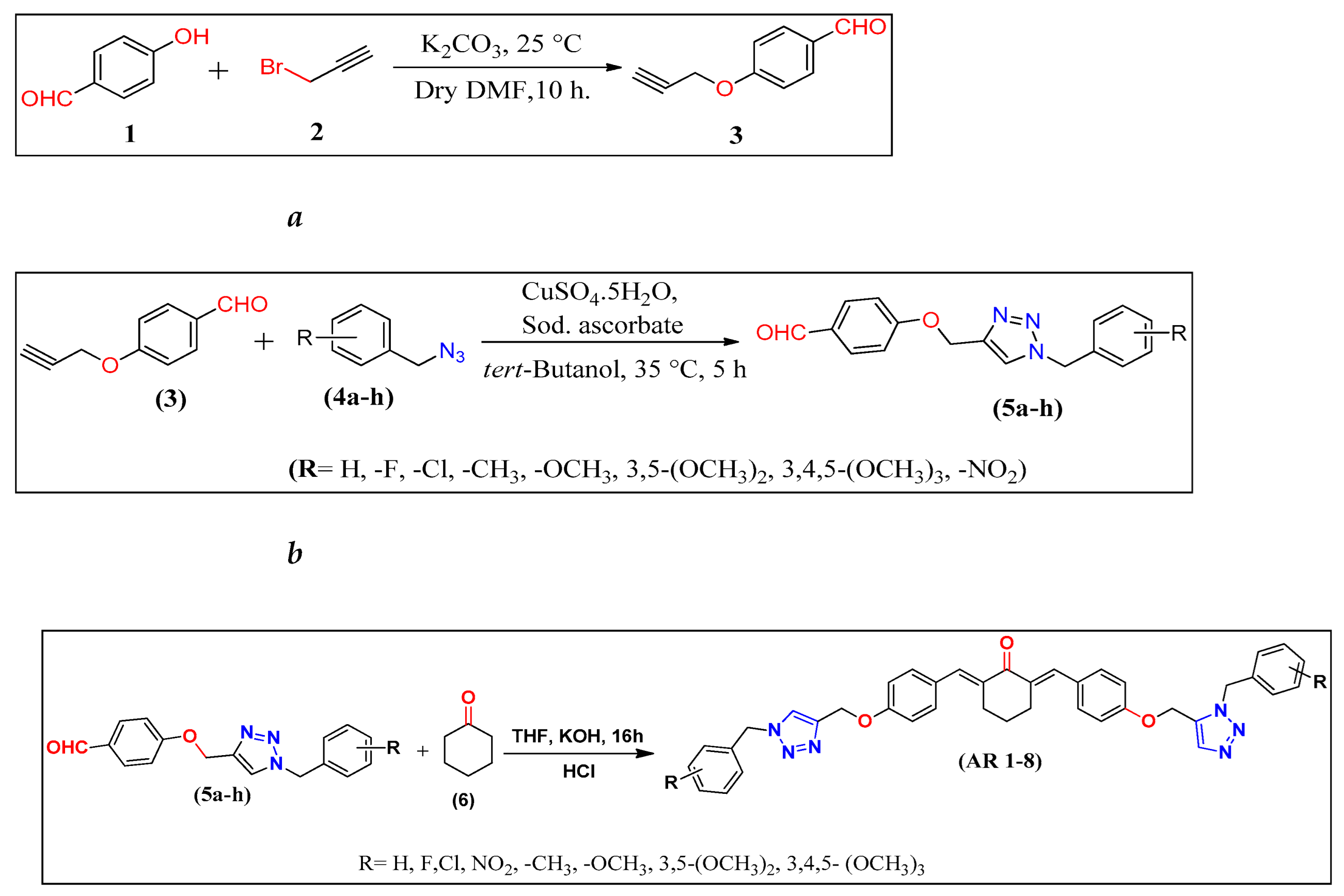
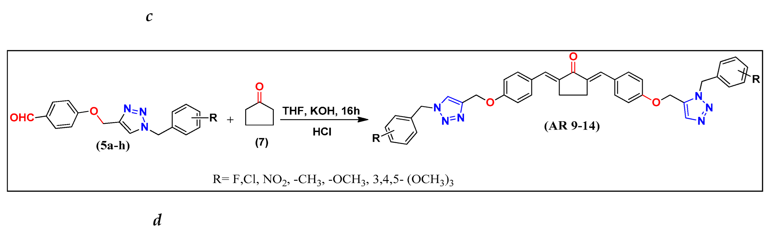

Table 1.
Cytotoxicity data (in-vitro) of synthesized derivatives AR4-14 and reference molecules docetaxel and staurosporine.
Table 1.
Cytotoxicity data (in-vitro) of synthesized derivatives AR4-14 and reference molecules docetaxel and staurosporine.
| Compound | IC50 | ||||||||
| R | hTERT RPE-1 | Capan-1 | Hap-1 | HCT-116 | NCI-H460 | DND-41 | K-562 | Z-138 | |
| retina (non cancerous) | pancreatic adenocarcinoma | chronic myeloid leukemia | colorectal carcinoma | lung carcinoma | acute lymphoblastic leukemia | chronic myeloid leukemia | non-Hodgkin lymphoma | ||
| AR-4 | 3,4,5-tri-OCH3 | >100 | 78.2 | 89.7 | 39.9 | >100 | >100 | >100 | >100 |
| AR-5 | 4-NO2 | >100 | 58.3 | >100 | 47.3 | 24.7 | >100 | >100 | >100 |
| AR-6 | 4-CH3 | 30.9 | 31.5 | 38.2 | 27.7 | 21.2 | 24.4 | >100 | >100 |
| AR-7 | 4-F | 38.1 | 11.8 | 31.6 | 13.6 | 21.9 | >100 | >100 | >100 |
| AR-8 | 4-Cl | >100 | >100 | >100 | >100 | >100 | >100 | >100 | >100 |
| AR-9 | OCH3 | 73.8 | >100 | 84.3 | 17.4 | 76.7 | >100 | >100 | >100 |
| AR-10 | 3,4,5-tri-OCH3 | 48.5 | 31.5 | 45.6 | 80.3 | 40.5 | 68.5 | >100 | >100 |
| AR-11 | 4-CH3 | 40.3 | 25.6 | 42.5 | 29.2 | 20.8 | 37.1 | >100 | >100 |
| AR-12 | 4-NO2 | 53.8 | 20.1 | 43.1 | 20.5 | 47.4 | >100 | >100 | >100 |
| AR-13 | 4-Cl | 42.9 | 47.2 | 51.2 | 48.3 | 53.6 | >100 | >100 | >100 |
| AR-14 | 4-F | 51.8 | 50 | >100 | 67 | 58.1 | >100 | >100 | 40.8 |
| Standard | Docetaxel (nM) | 13.5 | 6.3 | 1.6 | 0.8 | 0.1 | 1.9 | 3.4 | 1.9 |
| Standard | Staurosporine(nM) | 0.4 | 4.6 | 0.3 | 0.3 | 3.2 | 6.4 | 29.8 | 0.3 |
Disclaimer/Publisher’s Note: The statements, opinions and data contained in all publications are solely those of the individual author(s) and contributor(s) and not of MDPI and/or the editor(s). MDPI and/or the editor(s) disclaim responsibility for any injury to people or property resulting from any ideas, methods, instructions or products referred to in the content. |
© 2025 by the authors. Licensee MDPI, Basel, Switzerland. This article is an open access article distributed under the terms and conditions of the Creative Commons Attribution (CC BY) license (http://creativecommons.org/licenses/by/4.0/).
Copyright: This open access article is published under a Creative Commons CC BY 4.0 license, which permit the free download, distribution, and reuse, provided that the author and preprint are cited in any reuse.


