Submitted:
03 May 2025
Posted:
06 May 2025
Read the latest preprint version here
Abstract
Nanobody technology is a promising new approach to cancer research and treatment. Monoclonal antibodies have always been at the heart of targeted treatment, but their large size, cumbersome production, and restricted ability to penetrate tissue cause problems. Nanobodies are derived from camelid heavy-chain antibodies and have intrinsic characteristics that provide distinct benefits, including small size (15 kDa), high stability, and access to inaccessible epitopes. Their ease of production in bacterial systems further enhances their cost-effectiveness compared to conventional antibodies. Their role has been explored, from nanobody-based imaging agents that improve tumor detection to nanobody-drug conjugates that enhance targeted delivery while minimizing off-target effects. In addition, the recent expansion of their role in chimeric antigen receptor T-cell therapies, immune checkpoint blockade, and bispecific T-cell engagers highlights their increasing activity in immunotherapy. Similarly, nanobodies engineering is improving both dendritic cell vaccines and drug delivery through nanoparticle conjugation, expanding the therapeutic panorama. Here we provide remarkable findings about the versatility of Nbs-based strategies in oncology. Through their attractive characteristics, nanobodies’ therapeutics will change the way to treat cancer and provide new perspectives toward more effective and personalized medicine.
Keywords:
nanobodies
; cancer therapies
; nanobodies engineering
Main
The discovery of monoclonal antibodies by Köhler and Milstein in 1975 [1] changed the course of modern medicine, resulting in multiple applications in direct and/or indirect target identification, biotechnology development, clinical diagnostics, and immunotherapy. However, despite the existence of approved and widely used monoclonal antibodies, some inherent characteristics of their structure, such as their size, low structural stability under adverse conditions, the high cost of their production [2], and poor dissemination in tumor tissue, have made their therapeutic application in medical fields such as oncology difficult. In this sense, such concerns have encouraged the scientific community to search for and develop new alternatives.
18 years later, Hamers-Casterman and colleagues [3] uncovered a class of small molecules derived from antibodies composed only of heavy chains and present in the serum of Camelus dromedarius. Such molecules have also been found in cartilaginous fish [4] and are nowadays known as nanobodies (Nbs). Indeed, their unique structure and functional properties, such as small size, the possibility to engineer high-affinity antigen binding, and resistance to extreme conditions, coupled with a low cost of production, make them an interesting platform for XXI century medicine. This review provides relevant information about Nbs technology in cancer. It highlights several applications, from accurate diagnosis to novel therapeutics, and challenges to overcome. These small molecules are modeling cancer therapies and impacting the next generation of strategies in oncotherapy.
How Are Nanobodies Structured, and What Are Their Properties?
Camelids have both antibodies based on conventional heterotetrameric antibodies and only heavy-chain antibodies [5]. Interestingly, these heavy-chain antibodies differ from typical antibodies as they are homodimers with an antigen-binding domain and two constant domains. The independence of these antigen-binding domains is a key aspect of these antibodies and allows them to function independently [6]. Such antigen-binding domains are called Nbs and are smaller (15 kDa) (Figure 1), which provides better penetration depth in tissue. Moreover, these Nbs possess extended CDR1 and CDR3 (complementarity-determining regions), causing an increase in paratope diversity and an additional disulfide bond between CDR1 and CDR3, which provides structural stability. They also have large hydrophilic domains that provide solubility and resistance [6]. These factors contribute to antigen-binding diversity, solubility, and resistance to aggregation; consequently, Nbs are more resistant than antibodies when exposed to altered protease and pH conditions. This stability is beneficial for many routes of administration (Table 1), and all these structural aspects give them a long or rounded structure [2] that, together, facilitates the detection of antigenic epitopes that conventional antibodies would have trouble reaching.
Overview of Nanobody Generation
To date, there are several methods available for the generation of a Nbs library with the goal of producing these small molecules with optimal stability and affinity for their application.
Antigen-specific Nbs can be generated from immune, naive, or synthetic libraries. Immune libraries are typically developed by immunizing animals from the Camelidae family (llamas, camels, and alpacas ) [7]. After immunization, the process involves isolating lymphocytes, purifying mRNA (messenger ribonucleic acid), and amplifying the heavy-chain variable domain sequence by polymerase-chain reaction (PCR). As the Nbs structure does not require glycosylation to maintain their function, the expression is possible in several systems, with E. coli (Escherichia coli) being a standard platform for production. Indeed, one of the major advantages of nanobodies over conventional antibodies is their ease of production and expression in bacteria, making them more cost-effective. Unlike conventional antibodies that require mammalian cells. Second, naive (non-immune) libraries are based on the natural immune response of animals, whereas synthetic libraries are developed using the Nbs scaffold with modifications in the CDRs (in silico), especially CDR3 [8] (Figure 2). These modifications conserve the reading frame of the heavy-chain variable domain and the structural features of Nbs while enabling the rapid generation of billions of CDR variants without the need for animals.
Applications: Nanobody-Based Strategies for Cancer
As the first Nbs therapy approved by the Food and Drug Administration (FDA) in 2019, caplacizumab has been used for the treatment of acquired thrombotic thrombocytopenic purpura [9] and marked an important milestone in Nbs-based strategies (Figure 3).
The diagnosis of tumor imaging is as critical as therapy, as visual knowledge of the tumor's antigen profile is required to maximize therapeutic efficacy. In fact, there are multiple Nbs probes that have been examined with respect to different targets, and one of the more advanced formulations targeting human epidermal growth factor receptor-2 (HER2) is 68-GaNOTA-Anti-HER2, which is used in positron emission tomography/computed tomography [10]. This means these studies have provided the opportunity for using Nbs to detect primary and metastatic tumors without producing adverse effects. Likewise, these data demonstrated the utility of Nbs as diagnostic molecules in cancer. Currently, there are Nbs in preclinical and clinical trials for use as therapeutic tools in cancer [6]. The inherent properties of Nbs make them advantageous for oncology applications. Their nanoscale dimensions allow deep penetration into tumors, and some Nbs can cross the blood-brain barrier, while their modularity allows the development of bispecific or multivalent constructs, antibody-drug conjugates, and delivery systems targeting the tumor microenvironment.
Why Nanobody-Based Cancer Therapies?
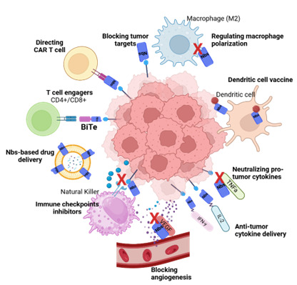
In cancer treatment, Nbs-based therapies have emerged as a promising alternative due to their modular structure and functional versatility (Figure 4). Their ability to target previously inaccessible epitopes, their small size, their low immunogenicity, their high tissue penetration, and the other properties mentioned above expand the treatment possibilities and allow for more precise and effective strategies. For this reason, their therapeutic potential and adaptability in oncology are growing.
Tumor Targets
Roovers and coworkers in 2007 [11], published the first successful in vivo implementation of therapeutic nanobodies for solid tumors. Their anti-EGFR (epidermal growth factor receptor) Nbs effectively delayed tumor growth, and they later developed a biparatopic version that superiorly reduced EGFR activation with comparable efficacy to its monoclonal antibody counterpart, cetuximab [11].
In the same way, Rossotti and colleagues [12] reported that Camelid immunized with EGFR DNA (deoxyribonucleic acid) generated Nbs with improved functionality compared to protein-immunized Nbs. Nbs against EGF (epidermal growth factor) [13], HER2 [14], CAIX (carbonic anhydrase IX) [15], DR5 (death receptor 5) [16], c-Met (mesenchymal-epithelial transition factor ) [17], HGF (hepatocyte growth factor) [18], PA28 (proteasome activator complex) [19], EphA4 (ephrin receptor A4) [20], CEACAM6 (CEA cell adhesion molecule-6) [21], TUFM (mitochondrial translation elongation factor) [22] and protein C receptor [23] have also shown delayed tumor growth.
In addition, synergistic effects can be realized through the utilization of bispecific Nbs targeting CD16 on natural killer cells and EGFR on the epithelial neoplastic cells in the experimental setting revealed synergistic effects which may be translated into clinical benefit. This approach has the effect of augmenting the extent of cellular lysis irrespective of KRAS (Kirsten rat sarcoma viral oncogene homolog) mutational status, and such augmentation has been found to correlate with significantly greater in vivo cytotoxicity against colorectal carcinoma when delivered alongside autologous or allogeneic CD16-expressing natural killer cells [24]. Thus far, anti-CD16 Nbs have been conjugated to Nbs against carcinoembryonic antigen (CEA), MUC-1, or glypican-3 (GPC3), and tumor growth suppression was observed in vivo [25,26,27]. Moreover, there are reports about the application of Nbs in combination with immunotoxins for colon cancer. The conjugation of an EGFR-targeting Nbs with the fungal ribotoxin α-sarcin resulted in the creation of a nano-immunotoxin that demonstrated improved therapeutic efficacy, showing better anti-tumor activity relative to immunotoxin-based treatments alone [28].
Nanobodies as Modulators of Checkpoint Inhibitors
Checkpoint inhibitors revolutionized cancer therapy, most notably with FDA-approved antibodies against (programmed cell death protein 1/programmed death-ligand 1) PD-1/PD-L1 and (cytotoxic t-lymphocyte associated protein 4) CTLA-4. However, their efficacy keeps going limited, which highlights the potential of Nbs to overcome these limitations due to their structural benefits. The use of Nbs as checkpoint blockades has been shown to be effective PDL1, with combination therapies using the avelumab (−L1) showing a greater T-cell response and improved tumor activity [29,30]. Moreover, the anti-CTLA-4 Nbs are revealed to produce potent anti-tumor activity, warranting a novel rationale for the Nbs application for cancer. A fusion protein combining anti-CD47 Nbs with an anti-CD20 antibody to exert a significant anti-tumor efficacy in vivo [29].
There is evidence that Nbs play a crucial role in the regulation of tumor angiogenesis. We know that VEGF (vascular endothelial growth factor) and its corresponding receptors constitute ideal targets for therapeutic inhibition and control of angiogenesis. As such, both monovalent and bivalent nanobodies have successfully prevented VEGF ligand interactions and also inhibited VEGF-induced cellular proliferation in vivo [31]. Additionally, coupling Nbs to a proline-alanine-serine (PAS) sequence improves their in vivo efficacy and pharmacokinetic properties [32]. In vitro effectiveness of a Nbs targeting the anti-VEGF receptor-2 (VEGFR2) in the prevention of capillary-like structure formation has been displayed [33]. Nbs targeting delta-like ligand 4 (DLL4) or CD3 have shown significant inhibition of neovascularization and tumor progression both in vitro and in vivo [33,34].
Dendritic Cell Vaccine Based on Nanobody
Dendritic cells are the most potent of the antigen-presenting cells and are attractive candidates for cancer vaccines. Using the penetration capacity and structural simplicity of nanobodies, studies have explored the implementation of Nbs-based conjugates to enhance dendritic cell-based immunity. That being said, conjugates can enhance antigen uptake by dendritic cells and promote presentation through histocompatibility complexes (MHC). Furthermore, dendritic cells can express nanobodies, which are able to be inserted by lentivirus or adenovirus, provoking the expression of themselves intracellularly or on the surface of these cells [35]. This allows for the stable expression of Nbs directed against checkpoints or their secretion. Nbs-lentiviruses have shown specific transduction into dendritic cells in vitro, but a subsequent study suggested that broad tropism lentiviruses may be more effective in inducing an anti-tumor response [36]. On the other hand, Nbs can be conjugated into nanoparticles (liposomes, polymers, and gold nanoparticles) containing tumor or immunostimulatory antigens. Such nanoparticles can be internalized by dendritic cells through receptor-mediated endocytosis, favoring better activation of dendritic cells. Considering that, Nbs offer a new approach to improving dendritic cell-based vaccines.
Nanobodies Engineering for CAR-T Technology
The adaptation of Nbs to CAR-T cells (Chimeric Antigen Receptor-T Cells) has been evaluated as part of their recognition receptor, or as secretion molecules with immunomodulatory properties to optimize their antitumor effect [37]. Efficacy of chimeric antigen receptor (CAR) T cells has been demonstrated in hematologic malignancies. However, their application to solid tumors has been restricted due to their inherent immunogenicity and the large size of CARs. Various studies have demonstrated the efficacy of utilizing MUC-1, CD7, CD38, VEGFR2, prostate-specific membrane antigen (PSMA), glypican-2 (GPC2) [38,39,40] and T cell receptor (TCR)-like Nbs-CARs in various tumor models [41]. Bispecific Nbs-CARs targeting CD20 and HER2 have also been developed [42].
Xie and coworkers engineered CAR T cells directed against PD-L1 with effective in vivo tumor regression and raised the potential for combination therapies extensions [37]. These anti-PD-L1/CTLA-4 Nbs-secreting CAR T cells have demonstrated enhanced anti-tumor response in vivo and indicate synergistic potential [43]. More recently, other types of CAR structural Nbs have begun to be studied by researchers, and they gave full play to their advantages as alternative CAR-targeting structural domains: the low immunogenicity, high stability, strong specificity and high affinity of Nbs, as well as the simple and feasible development process. Nowadays, several clinical trials using this therapy of Nbs-CAR T (BCMA CAR-T, CD7 CAR-T Cells) have completed their phase I and II, coming up with a good outcome and being a new alternative clinical treatment of cancer [6].
Nanobody-Based Drug Delivery
The combination of nanobodies and drug carriers (liposomes, micelles, albumin- and ferritin-based nanoparticles, and polymer-based multimers) enables active targeting of Nbs-based anticancer drugs to tumor tissues. With them having tumor-specific receptors can be used as carriers for the delivery of drugs to the tumor site, enabling the targeted release and reducing damage to normal cells and reducing side effects. In spite of the preclinical success of cytokine-based therapy, a lot of clinical studies have shown low efficacy due to their short half-life. However, the incorporation of Nbs could impart a new way of improved cytokine therapies. An anti-PD-L1 Nbs fused to either interleukin-2 (IL-2) or interferon-γ (IFNγ) demonstrated in vivo efficacy in treatment-resistant pancreatic tumors [44,45].
Similarly, Nbs have also been created to target chemotactic cytokines that directly impact tumor proliferation, angiogenesis, and metastasis. Such Nbs can block protumor chemokines CXCL10 [46] or fused to anti-tumor chemokines CCL21 [47] that have demonstrated functionality in microfluidic devices but have yet to be tested in vivo.
Nanobody-Drug Conjugates
Antibody-drug conjugates merge the excellent killing capability of small-molecule drugs with the extremely targeted dependence of antibodies. However, the antibody [6] size limits its penetration through the tissue. In comparison, Nbs are small new molecules and have received significant interest in the development of next-generation targeted drug conjugates. Nbs-drug conjugates have the advantage over standard antibody-drug conjugates because they have a greater tumor penetration rate. However, many limitations remain, of which the rapid systemic clearance of the Nbs-drug complex is of particular concern as the complex is sequestered prior to reaching the tumor at concentrations sufficient to have a therapeutic effect. So, patients will need lots of doses over a brief period, so all of this is even more inconvenient. Such pharmacokinetic clearance is reversible by common methods such as molecular weight augmentation through albumin fusion and pegylation [6] to avoid glomerular filtration in the kidneys. Structure-activity relationship engineering such as this is critical in optimizing their pharmacokinetics, cementing their role as powerful components of drug conjugates.
Nanobodies-Based Viral Vectors
Adeno-associated viruses (AAVs) have been further developed to express the Nbs in vivo, which are referred to as Nbs-AAVs. The same approach has generated interest in immunotherapy enabling sustained expression of Nbs treatments in target tissues. This allows for sustained expression of Nbs-based therapies and does not require re-dosing. Antigen targeting has been shown by Nbs-AAVs in previous studies, including, for example, CD38, ARTC2.2, and P2X7 [48], though additional in vivo studies are required. Moreover, tumor vasculature has also been targeted using viral vectors, and recombinant λ (lambda) bacteriophages have shown a significant HER2+ cell growth inhibitory effect in vitro [49,51].
T Cell Engagers
Antibodies targeting CD3 on all T cells were the first to be approved by the FDA for clinical use [9]; however, their initial systemic toxicity led to the development of bi-specific T-cell engagers (BiTE). These are bispecific molecules composed of the variable fragments of both the heavy and light chains of an antibody, with a unique function simultaneously binding an antigen on tumor cells and a surface molecule on T cells to induce tumor lysis [52,53]. Even so, this therapy is associated with antigen loss and immunosuppressive factors, such as the upregulation of immune checkpoints.
With Nbs technology, more compact and improved BiTE have become possible. Li and coworkers [53] created a BiTE (S-Fab) composed of an anti-CEA Nbs and an anti-CD3 fragment-binding antigen [54], with significant T-cell-mediated cytotoxicity in vitro and in vivo. S-Fab was pegylated to prolong its serum half-life while maintaining uncompromised anti-tumor activity [53].
Over the past years, studies have shown that the bi-specific T-cell activator based on three anti-EGFR nanobodies and one anti-CD3 [52] was 15-fold more effective than the same antibody therapy, demonstrating the superiority of Nbs and that innovations in cancer therapy are on the right track [54] (Table 2). Research on the use of Nbs in cancer therapy continues, further enhancing treatment efficacy and reducing toxicity, demonstrating their powerful and promising potential (Table 3).
Limitations of Nanobody-Based Therapy
Nbs-based therapeutics have shown as a promising alternative to traditional monoclonal antibodies, as due to their small size, they have deep tumor penetration and ease of manufacturing. However, the very properties that make them so promising also render them difficult to apply due to their brief half-life in the bloodstream and tumor microenvironment barriers. Such constraints reduce the therapeutic potential since repeated administration is needed to achieve effective drug concentration therein. Although great progress has been made, there is currently no comprehensive strategy that effectively integrates both long circulation and effective targeting tumors.
The immunosuppressive microenvironment of solid tumors poses a barrier for the effectiveness of immunotherapies, including those based on Nbs. Additionally, metabolic changes in the tumor microenvironment impede efficient therapy. Myeloid-derived suppressor cells and regulatory T cells promote an anti-inflammatory microenvironment that impairs immune-mediated attack by Nbs therapies. Although promising results have been achieved with Nbs-based checkpoint inhibitors against PD-L1 and CTLA-4, other strategies are required to counteract tumor-mediated immunosuppression [54]. Therefore, strategies require other approaches, as mentioned above, such as bispecific Nbs, Nbs-drug conjugates and combination therapies including agents capable of remodeling the extracellular matrix or normalizing the tumor vasculature in order to improve drug penetration and retention in the tumor tissues [55]. These emerging strategies also rely on the use of multivalent Nbs to target multiple checkpoints and Nbs-coated dendritic cells to manipulate immune responses directly in the tumor microenvironment. Up to now, engineered multimeric Nbs [56,57] are being evaluated to enhance tumor retention and bidirectionally block multiple immunosuppressive pathways as well as nanoparticle conjugation strategies to increase drug accumulation and circulation time in tumors. On the other hand, Nbs compared to antibodies lack constant fragments (Fc), and thus cannot directly initiate an Fc-mediated immune response. As Nbs are not naturally produced in humans, their therapeutic implementation brings into question their overall safety. Regardless of this approach, Nbs sequence studies have revealed high similarity with human variable domains [58], and combined with their size, structure, and low agglutination, Nbs possess low immunogenicity and are appropriate for human administration. Having said that, new strategies based on Nbs may be the next step to revolutionizing a new wave of cancer therapeutics.
Conclusions
Nbs-based therapies have changed the field of oncology by coming with an innovative alternative relative to conventional antibodies. Their unique structural and functional properties, such as small size, high stability, deep tumor penetration, and low cost of production, have enabled their application in cancer diagnosis, imaging, and targeted therapy. As a result, immune checkpoint inhibition, CAR-T cell engineering, drug delivery, and bispecific T-cell engagers have demonstrated their versatility to cover multiple aspects of cancer treatment. Despite all the advances, the scientific community continues working on the limitations related to the pharmacokinetic properties of Nbs mentioned through this review. That being said, further research is needed to optimize Nbs engineering and, thus, their therapeutic potency. As the field progresses, ongoing clinical trials and innovative approaches, such as multivalent Nbs constructs and combination therapies, are expected to correct these limitations and improve treatment outcomes. As evidenced by existing preclinical studies, Nbs-biotechnology may be the future of oncotherapy. In sum, Nbs-based cancer therapies represent a significant advancement in oncology, promoting a foundation for next-generation targeted treatments. Regardless of the challenges that persist, research and technology will continue to develop the way for their wide clinical application, transforming both cancer and other disease therapies into a more effective and personalized approach.
Acknowledgements
I would like to express my gratitude to Dr. Mario Sergio Valdés-Tresanco, Dr.Aru Narendran and Dr. Ernesta Paola Neri for their guidance and encouragement in the preparation of this article.
Author contributions
Glenda Romero Hernandez: Conceptualization, investigation, writing, and visualization. Carrie Simone Shemanko: Writing review and editing
Competing interests
The authors declare no competing interests.
Additional information
Correspondence and requests for materials should be addressed to Glenda Romero-Hernandez and Carrie Simone Shemanko.
References
- Köhler, G.; Milstein, C. Continuous cultures of fused cells secreting antibody of predefined specificity. Nature 1975, 256, 495–497. [Google Scholar] [CrossRef]
- Ortega-Portilla, P.A.; et al. Nanoanticuerpos: Desarrollo biotecnológico y aplicaciones. TIP. Rev. Esp. Cienc. Quím.-Biol. 2021, 24, e398. [Google Scholar] [CrossRef]
- Hamers-Casterman, C.; et al. Naturally occurring antibodies devoid of light chains. Nature 1993, 363, 446–448. [Google Scholar] [CrossRef] [PubMed]
- Kang, W.; et al. Nanobody conjugates for targeted cancer therapy and imaging. Technol. Cancer Res. Treat. 2021, 20, 15330338211010116. [Google Scholar] [CrossRef]
- Muyldermans, S. Nanobodies: Natural single-domain antibodies. Annu. Rev. Biochem. 2013, 82, 775–797. [Google Scholar] [CrossRef] [PubMed]
- Valdés-Tresanco, M.S.; Molina-Zapata, A., Pose; Moreno, E. Structural insights into the design of synthetic nanobody libraries. Molecules 2022, 27, 2198. [Google Scholar] [CrossRef] [PubMed]
- Alexander, E.; Leong, K.W. Discovery of nanobodies: A comprehensive review of their applications and potential over the past five years. J. Nanobiotechnol. 2024, 22, 661. [Google Scholar] [CrossRef]
- Liu, W.; et al. Recent advances in the selection and identification of antigen-specific nanobodies. Mol. Immunol. 2018, 96, 37–47. [Google Scholar] [CrossRef]
- Duggan, S. Caplacizumab: First Global Approval. Drugs 2018, 78, 1639–1642. [Google Scholar] [CrossRef]
- Keyaerts, M.; et al. Phase I study of 68Ga-HER2-Nanobody for PET/CT assessment of HER2 expression in breast carcinoma. J. Nucl. Med. 2016, 57, 27–33. [Google Scholar] [CrossRef]
- Roovers, R.C.; et al. Efficient inhibition of EGFR signalling and of tumour growth by antagonistic anti-EGFR Nanobodies. Cancer Immunol. Immunother. 2007, 56, 303–317. [Google Scholar] [CrossRef] [PubMed]
- Rossotti, M.A.; et al. Camelid single-domain antibodies raised by DNA immunization are potent inhibitors of EGFR signaling. Biochem. J. 2019, 476, 39–50. [Google Scholar] [CrossRef]
- Guardiola, S.; et al. Blocking EGFR activation with anti-EGF nanobodies via two distinct molecular recognition mechanisms. Angew. Chem. Int. Ed. 2018, 57, 13843–13847. [Google Scholar] [CrossRef]
- Wang, H.; et al. Human domain antibodies to conserved epitopes on HER2 potently inhibit growth of HER2-overexpressing human breast cancer cells in vitro. Antibodies 2019, 8, 25. [Google Scholar] [CrossRef] [PubMed]
- Araste, F.; et al. A novel VHH nanobody against the active site (the CA domain) of tumor-associated, carbonic anhydrase isoform IX and its usefulness for cancer diagnosis. Biotechnol. Lett. 2014, 36, 21–28. [Google Scholar] [CrossRef]
- Sadeghnezhad, G.; et al. Identification of new DR5 agonistic nanobodies and generation of multivalent nanobody constructs for cancer treatment. Int. J. Mol. Sci. 2019, 20, 4818. [Google Scholar] [CrossRef] [PubMed]
- Godar, M.; et al. Dual anti-idiotypic purification of a novel, native-format biparatopic anti-MET antibody with improved in vitro and in vivo efficacy. Sci. Rep. 2016, 6, 31621. [Google Scholar] [CrossRef]
- Vosjan, M.J.W.D.; et al. Nanobodies targeting the hepatocyte growth factor: Potential new drugs for molecular cancer therapy. Mol. Cancer Ther. 2012, 11, 1017–1025. [Google Scholar] [CrossRef]
- Sánchez-Martín, D.; et al. Proteasome activator complex PA28 identified as an accessible target in prostate cancer by in vivo selection of human antibodies. Proc. Natl Acad. Sci. USA 2013, 110, 13791–13796. [Google Scholar] [CrossRef]
- Schoonaert, L.; et al. Identification and characterization of nanobodies targeting the EphA4 receptor. J. Biol. Chem. 2017, 292, 11452–11465. [Google Scholar] [CrossRef]
- Cheng, T.M.; et al. Single domain antibody against carcinoembryonic antigen-related cell adhesion molecule 6 (CEACAM6) inhibits proliferation, migration, invasion and angiogenesis of pancreatic cancer cells. Eur. J. Cancer 2014, 50, 713–721. [Google Scholar] [CrossRef]
- Samec, N.; et al. Glioblastoma-specific anti-TUFM nanobody for in-vitro immunoimaging and cancer stem cell targeting. Oncotarget 2018, 9, 17282–17299. [Google Scholar] [CrossRef] [PubMed]
- Wang, D.; et al. Protein C receptor is a therapeutic stem cell target in a distinct group of breast cancers. Cell Res. 2019, 29, 832–845. [Google Scholar] [CrossRef]
- Wan, R.; et al. Screening and antitumor effect of an anti-CTLA-4 nanobody. Oncol. Rep. 2018, 39, 511–518. [Google Scholar] [CrossRef] [PubMed]
- Rozan, C.; et al. Single-domain antibody-based and linker-free bispecific antibodies targeting FcgRIII induce potent antitumor activity without recruiting regulatory T cells. Mol. Cancer Ther. 2013, 12, 1481–1491. [Google Scholar] [CrossRef]
- Wang, Y.; et al. A GPC3-targeting bispecific antibody, GPC3-S-Fab, with potent cytotoxicity. J. Vis. Exp. 2018, 12, 57588. [Google Scholar]
- Li, Y.; et al. Single domain based bispecific antibody, Muc1-Bi-1, and its humanized form, Muc1-Bi-2, induce potent cancer cell killing in Muc1 positive tumor cells. PLoS ONE 2018, 13, e0191024. [Google Scholar] [CrossRef] [PubMed]
- Homayouni, V.; et al. Preparation and characterization of a novel nanobody against T-cell immunoglobulin and mucin-3 (TIM-3). Iran J. Basic Med. Sci. 2016, 19, 1201–1208. [Google Scholar]
- Ma, L.; et al. Preclinical development of a novel CD47 nanobody with less toxicity and enhanced anti-cancer therapeutic potential. J. Nanobiotechnol. 2020, 18, 12. [Google Scholar] [CrossRef]
- Farajpour, Z.; et al. A nanobody directed to a functional epitope on VEGF, as a novel strategy for cancer treatment. Biochem. Biophys. Res. Commun. 2014, 446, 132–136. [Google Scholar] [CrossRef]
- Khodabakhsh, F.; et al. Development of a novel nano-sized anti-VEGFA nanobody with enhanced physicochemical and pharmacokinetic properties. Artif. Cells Nanomed. Biotechnol. 2018, 46, 1402–1414. [Google Scholar] [CrossRef]
- Behdani, M.; et al. Generation and characterization of a functional nanobody against the vascular endothelial growth factor receptor-2; angiogenesis cell receptor. Mol. Immunol. 2012, 50, 35–41. [Google Scholar] [CrossRef]
- Baharlou, R.; et al. An antibody fragment against human delta-like ligand-4 for inhibition of cell proliferation and neovascularization. Immunopharmacol. Immunotoxicol. 2018, 40, 368–374. [Google Scholar] [CrossRef] [PubMed]
- Khatibi, A.S.; et al. Tumor-suppressing and anti-angiogenic activities of a recombinant anti-CD3ϵ nanobody in breast cancer mice model. Immunotherapy 2019, 11, 1555–1567. [Google Scholar] [CrossRef] [PubMed]
- Goyvaerts, C.; et al. Targeting of human antigen-presenting cell subsets. J. Virol. 2013, 87, 11304–11308. [Google Scholar] [CrossRef]
- Goyvaerts, C.; et al. Antigen-presenting cell-targeted lentiviral vectors do not support the development of productive T-cell effector responses: Implications for in vivo targeted vaccine delivery. Gene Ther. 2017, 24, 370–375. [Google Scholar] [CrossRef]
- Xie, Y.J.; et al. Nanobody-based CAR T cells that target the tumor microenvironment inhibit the growth of solid tumors in immunocompetent mice. Proc. Natl Acad. Sci. USA 2019, 116, 7624–7631. [Google Scholar] [CrossRef] [PubMed]
- An, N.; et al. Anti-multiple myeloma activity of nanobody-based anti-CD38 chimeric antigen receptor T cells. Mol. Pharm. 2018, 15, 4577–4588. [Google Scholar] [CrossRef]
- Hajari Taheri, F.; et al. T cell engineered with a novel nanobody-based chimeric antigen receptor against VEGFR2 as a candidate for tumor immunotherapy. IUBMB Life 2019, 71, 1259–1267. [Google Scholar] [CrossRef]
- Hassani, M.; et al. Construction of a chimeric antigen receptor bearing a nanobody against prostate-specific membrane antigen in prostate cancer. J. Cell Biochem. 2019, 120, 10787–10795. [Google Scholar] [CrossRef]
- Hassani, M.; et al. Engineered Jurkat cells for targeting prostate-specific membrane antigen on prostate cancer cells by nanobody-based chimeric antigen receptor. Iran Biomed J. 2020, 24, 81–88. [Google Scholar] [CrossRef] [PubMed]
- De Munter, S.; et al. Nanobody-based dual-specific CARs. Int. J. Mol. Sci. 2018, 19, 403. [Google Scholar] [CrossRef] [PubMed]
- Xie, Y.J.; et al. Improved anti-tumor efficacy of chimeric antigen receptor T cells that secrete single-domain antibody fragments. Cancer Immunol. Res. 2020, 8, canimm.0734.2019. [Google Scholar] [CrossRef]
- Dougan, M.; et al. Targeting cytokine therapy to the pancreatic tumor microenvironment using PD-L1–specific VHHs. Cancer Immunol. Res. 2018, 6, 389–401. [Google Scholar] [CrossRef]
- Sadeghian-Rizi, T.; et al. Generation and characterization of a functional nanobody against inflammatory chemokine CXCL10, as a novel strategy for the treatment of multiple sclerosis. CNS Neurol. Disord. Drug Targets 2019, 18, 141–148. [Google Scholar] [CrossRef]
- Fang, T.; et al. Remodeling of the tumor microenvironment by a chemokine/anti-PD-L1 nanobody fusion protein. Mol. Pharm. 2019, 16, 2838–2844. [Google Scholar] [CrossRef]
- Eichhoff, A.M.; et al. Nanobody-enhanced targeting of AAV gene therapy vectors. Mol. Ther. Methods Clin. Dev. 2019, 15, 211–220. [Google Scholar] [CrossRef]
- Ahani, R.; et al. Sindbis virus-pseudotyped lentiviral vectors carrying VEGFR2-specific nanobody for potential transductional targeting of tumor vasculature. Mol. Biotechnol. 2016, 58, 738–747. [Google Scholar] [CrossRef] [PubMed]
- Shoae-Hassani, A.; et al. Recombinant λ bacteriophage displaying nanobody towards third domain of HER-2 epitope inhibits proliferation of breast carcinoma SKBR-3 cell line. Arch. Immunol. Ther. Exp. 2013, 61, 75–83. [Google Scholar] [CrossRef]
- Xing, J.; et al. BiHC, a T-cell–engaging bispecific recombinant antibody, has potent cytotoxic activity against HER2 tumor cells. Transl. Oncol. 2017, 10, 780–785. [Google Scholar] [CrossRef]
- Li, L.; et al. A novel bispecific antibody, S-Fab, induces potent cancer cell killing. J. Immunother. 2015, 38, 350–356. [Google Scholar] [CrossRef] [PubMed]
- Harwood, S.L.; et al. ATTACK, a novel bispecific T cell-recruiting antibody with trivalent EGFR binding and monovalent CD3 binding for cancer immunotherapy. Oncoimmunology 2018, 7, e1377874. [Google Scholar] [CrossRef] [PubMed]
- Van Der Linden, R.H.J.; et al. Comparison of physical chemical properties of llama VHH antibody fragments and mouse monoclonal antibodies. Biochim. Biophys. Acta 1999, 1431, 37–46. [Google Scholar] [CrossRef] [PubMed]
- Chen, Y.; Duong Van Hoa, F. Peptidisc-assisted hydrophobic clustering toward the production of multimeric and multispecific nanobody proteins. Biochemistry 2025, 64, 655–665. [Google Scholar] [CrossRef]
- Alexander, E.; Leong, K.W. Discovery of nanobodies: A comprehensive review of their applications and potential over the past five years. J. Nanobiotechnol. 2024, 22, 661. [Google Scholar] [CrossRef]
- Papadopoulos, K.P.; et al. Unexpected hepatotoxicity in a phase I study of TAS266, a novel tetravalent agonistic nanobody® targeting the DR5 receptor. Cancer Chemother. Pharmacol. 2015, 75, 887–895. [Google Scholar] [CrossRef]
- Albert, S.; et al. A novel nanobody-based target module for retargeting T lymphocytes to EGFR-expressing cancer cells via the modular UniCAR platform. Oncoimmunology 2017, 6, e1287246. [Google Scholar] [CrossRef]
- de Bruin, R.C.G.; et al. A bispecific nanobody approach to leverage the potent and widely applicable tumor cytolytic capacity of Vγ9Vδ2-T cells. Oncoimmunology 2017, 7, e1375641. [Google Scholar] [CrossRef]
- Bian, X.; et al. Anti-EGFR-iRGD recombinant protein conjugated silk fibroin nanoparticles for enhanced tumor targeting and antitumor efficiency. Onco Targets Ther. 2016, 9, 3153–3162. [Google Scholar]
- Van De Water, J.A.J.M.; et al. Therapeutic stem cells expressing variants of EGFR-specific nanobodies have antitumor effects. Proc. Natl Acad. Sci. USA 2012, 109, 16642–16647. [Google Scholar] [CrossRef]
- Karges, J.; et al. Synthesis and characterization of an epidermal growth factor receptor-selective RuII polypyridyl–nanobody conjugate as a photosensitizer for photodynamic therapy. ChemBioChem 2019, 21, 531–542. [Google Scholar] [CrossRef] [PubMed]
- Fang, Y.; et al. DR30303, a SMART-VHHBody powered anti-CLDN18.2 VHH-Fc with enhanced ADCC activity for the treatment of gastric and pancreatic cancers. Cancer Res. 2022, 82, 2857. [Google Scholar] [CrossRef]
- Yamamoto, N.; et al. Phase I study of the VEGF/Ang-2 inhibitor BI 836880 alone or combined with the anti-programmed cell death protein-1 antibody ezabenlimab in Japanese patients with advanced solid tumors. Cancer Chemother. Pharmacol. 2023, 91, 469–480. [Google Scholar] [CrossRef] [PubMed]
- Shimizu, T.; et al. Phase I study of envafolimab (KN035), a novel subcutaneous single-domain anti-PD-L1 monoclonal antibody, in Japanese patients with advanced solid tumors. Invest. New Drugs 2022, 40, 1021–1031. [Google Scholar] [CrossRef] [PubMed]
- Li, J.; et al. Subcutaneous envafolimab monotherapy in patients with advanced defective mismatch repair/microsatellite instability-high solid tumors. J. Hematol. Oncol. 2021, 14, 95. [Google Scholar] [CrossRef]
- Papadopoulos, K.P.; et al. First-in-human phase I study of envafolimab, a novel subcutaneous single-domain anti-PD-L1 antibody, in patients with advanced solid tumors. Oncologist 2021, 26, e1514–e1525. [Google Scholar] [CrossRef]
- D’Huyvetter, M.; et al. Phase I trial of ¹³¹I-GMIB-Anti-HER2-VHH1, a new promising candidate for HER2-targeted radionuclide therapy in breast cancer patients. J. Nucl. Med. 2021, 62, 1097–1105. [Google Scholar] [CrossRef]
- Ramlau, R.; et al. P2.06-006 Phase I/II dose escalation study of L-DOS47 as a monotherapy in non-squamous non-small cell lung cancer patients. J. Thoracic Oncol. 2017, 12, S1071–S1072. [Google Scholar] [CrossRef]
- Piha-Paul, S.; et al. A phase I, open-label, dose-escalation study of L-DOS47 in combination with pemetrexed plus carboplatin in patients with stage IV recurrent or metastatic nonsquamous NSCLC. JTO Clin. Res. Rep. 2022, 3, 100408. [Google Scholar] [CrossRef]
- Kater, A.P.; et al. Lava-051, a novel bispecific gamma-delta T-cell engager (Gammabody), in relapsed/refractory MM and CLL: Pharmacodynamic and early clinical data. Blood 2022, 140, 4608–4609. [Google Scholar] [CrossRef]
- Mehra, N.; et al. Early dose escalation of LAVA-1207, a novel bispecific gamma-delta T-cell engager (Gammabody), in patients with metastatic castration-resistant prostate cancer (mCRPC). American Society of Clinical Oncology 2023. [Google Scholar] [CrossRef]
- Martin, T.; et al. Updated results from CARTITUDE-1: Phase Ib/II study of ciltacabtagene autoleucel, a B-cell maturation antigen-directed chimeric antigen receptor T cell therapy, in patients with relapsed/refractory multiple myeloma. Blood 2021, 138, 549. [Google Scholar] [CrossRef]
- Berdeja, J.G.; et al. Ciltacabtagene autoleucel, a B-cell maturation antigen-directed chimeric antigen receptor T-cell therapy in patients with relapsed or refractory multiple myeloma (CARTITUDE-1): A phase Ib/II open-label study. Lancet 2021, 398, 314–324. [Google Scholar] [CrossRef] [PubMed]
- Zhao, W.H.; et al. Four-year follow-up of LCAR-B38M in relapsed or refractory multiple myeloma: A phase I, single-arm, open-label, multicenter study in China (LEGEND-2). J. Hematol. Oncol. 2022, 15, 86. [Google Scholar] [CrossRef]
- van de Donk, N.; et al. B07: Safety and efficacy of ciltacabtagene autoleucel, a chimeric antigen receptor T-cell therapy directed against B-cell maturation antigen in patients with multiple myeloma and early relapse after initial therapy: CARTITUDE-2 results. HemaSphere 2022, 6, 9–10. [Google Scholar] [CrossRef]
- Einsele, H.; et al. P08: CARTITUDE-2 UPDATE: Ciltacabtagene autoleucel, a B-cell maturation antigen–directed chimeric antigen receptor T-cell therapy, in lenalidomide-refractory patients with progressive multiple myeloma after 1–3 prior lines of therapy. HemaSphere 2022, 6, 15. [Google Scholar] [CrossRef]
- Cohen, Y.C.; et al. Efficacy and safety of ciltacabtagene autoleucel (ciltacel), a B-cell maturation antigen (BCMA)-directed chimeric antigen receptor (CAR) T-cell therapy, in lenalidomide-refractory patients with progressive multiple myeloma after 1–3 prior lines of therapy: Updated results from CARTITUDE-2. Blood 2021, 138, 3866. [Google Scholar]
- Agha, M.E.; et al. S185: CARTITUDE-2 cohort B: Updated clinical data and biological correlative analyses of ciltacabtagene autoleucel in patients with multiple myeloma and early relapse after initial therapy. HemaSphere 2022, 6, 86–87. [Google Scholar] [CrossRef]
- Brussel, U.Z. Quantification of 68-GaNOTA-Anti-HER2 VHH1 Uptake in Metastasis of Breast Carcinoma Patients and Assessment of Repeatability (VUBAR)—Pilot Study. ClinicalTrials.gov Identifier: NCT03924466 (2024). 0392. Available online: https://clinicaltrials.gov/study/NCT03924466?a=3.
- Alexion Pharmaceuticals, I. A Phase 3, Randomized, Double-blind, Placebo-controlled, Parallel, Multicenter Study to Evaluate the Safety and Efficacy of ALXN1720 in Adults With Generalized Myasthenia Gravis. ClinicalTrials.gov Identifier: NCT05556096.
Figure 1.
Structure of nanobodies compared to conventional antibodies. (A) Heavy-chain antibodies are homodimers lacking light chains (90 kDa), allowing the isolation of their antigen recognition regions (VhH) in camelids, also known as nanobodies (Nbs) (15 kDa). (B) Conventional antibodies are heterotetramers composed of two heavy and two light chains, forming two identical antigen-binding sites (VH). Each binding site consists of non-covalently linked variable domains. Fc: constant fragment of an antibody. H: heavy chain. L: light chain. Created in https://BioRender.com.
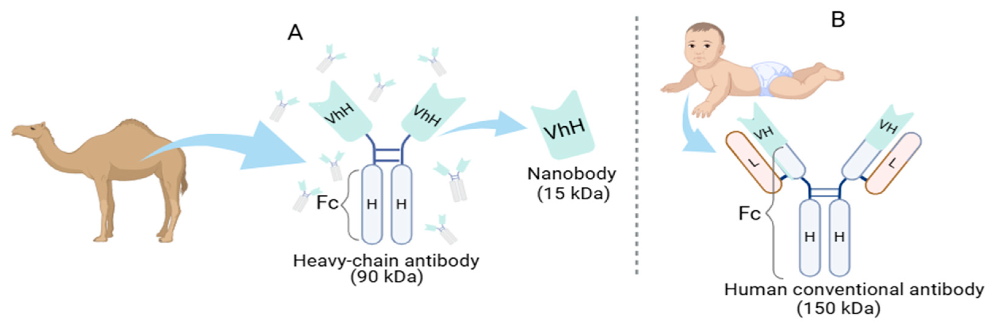
Figure 2.
Generating Antigen-Specific Nbs: A phage-display library approach. A schematic diagram of the different methods of generating Nbs. Camelids are immunized with an antigen to generate an immune library, while a naive library does not require inoculation (natural response). Blood is collected to extract lymphocytes and mRNA, and Nbs sequences are amplified through PCR. In synthetic libraries, a Nbs scaffold is used, and CDRs are randomized. These libraries are transformed into phage vectors and E. coli to display Nbs and produce high-affinity phages. Successful phages are stored for future production. Created in https://BioRender.com.
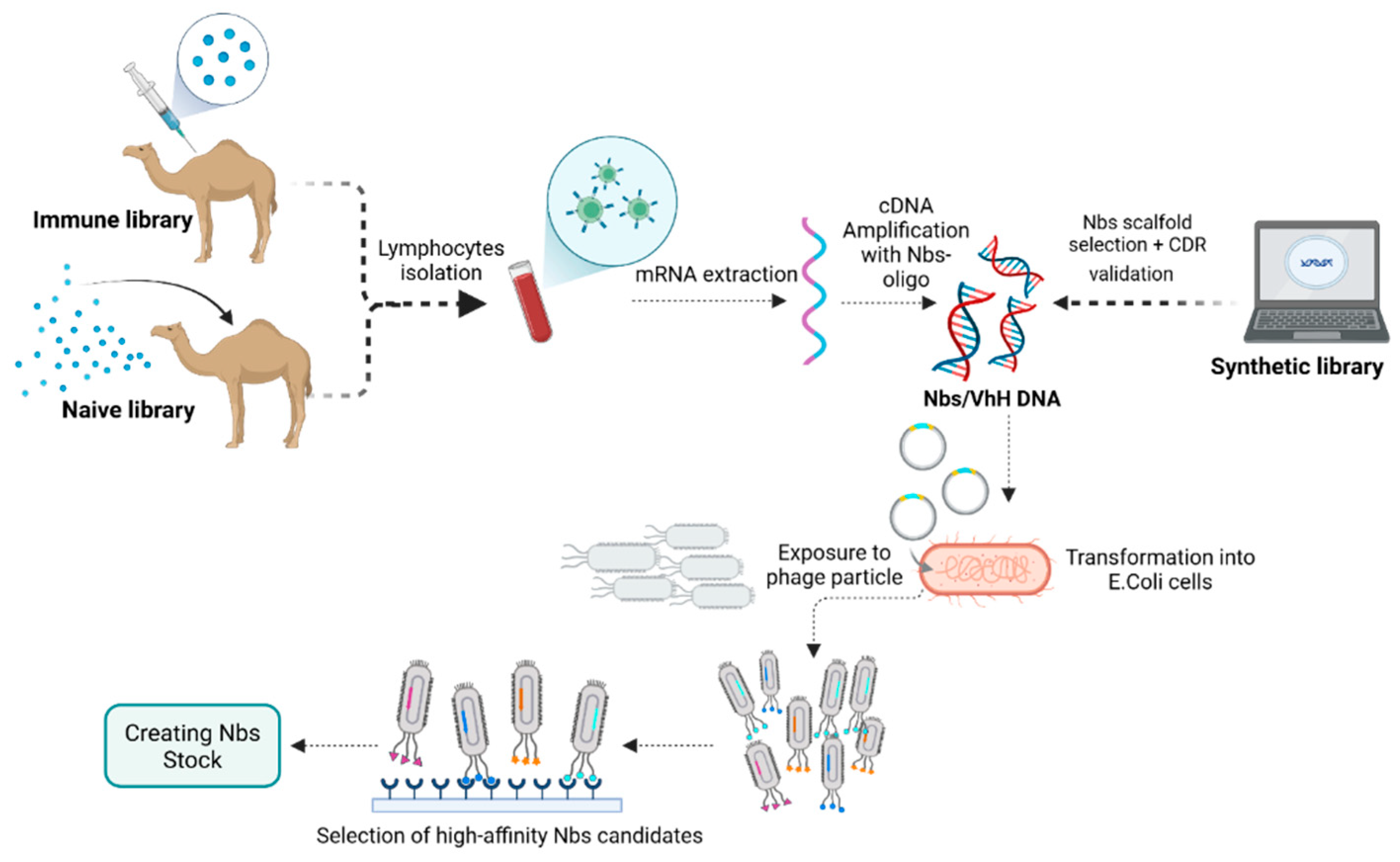
Figure 3.
Scheme of the application potential of the Nbs. (A) Nbs targets in the tumor microenvironment. (B) Use of Nbs in disease diagnosis through techniques such as images, flow cytometry, and fluorescence microscopy. IFN-γ: Interferon-gamma; IL-2: interleukin 2; VEGF: Vascular endothelial growth factor; TNF-α: Tumor necrosis factor-alpha. Created in https://BioRender.com.
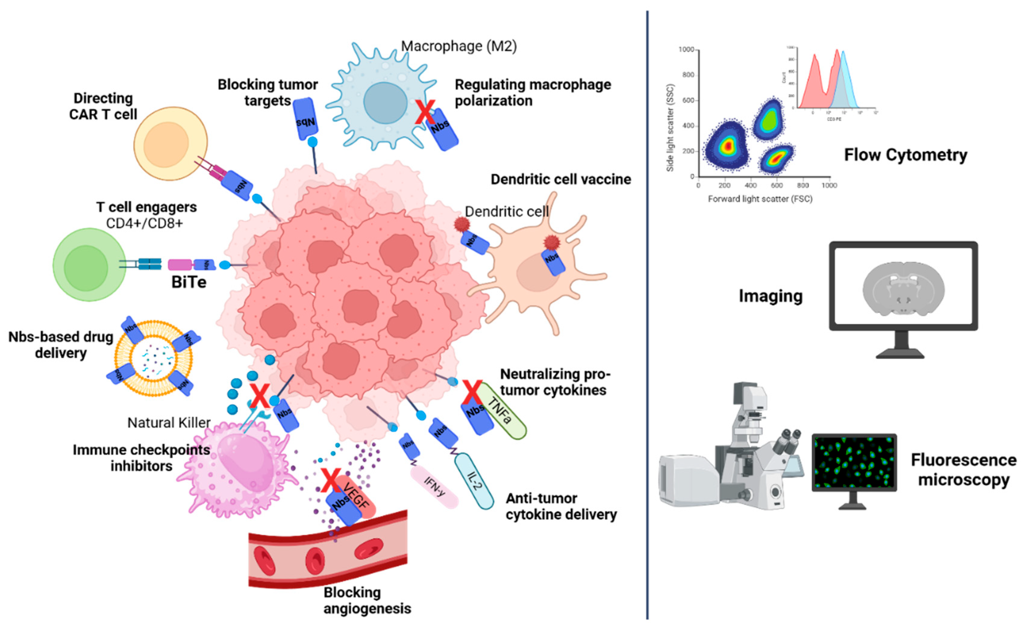

Table 1.
Advantages of nanobodies over conventional antibodies.
| Nanobodies vs. Conventional Antibody | ||
| Structural simplicity | Single-domain structure | Heterotetrameric structure |
| Size | ~15 kDa | ~150 kDa |
| CDR | 3 CDRs (Longer CDR1 and CD3) | 3 CDRs |
| Stability | High. Generally, they are functional at high temperatures and different pH levels | Less stable under extreme temperatures and pH conditions |
| Solubility | High. They are rich in hydrophilic regions, preventing aggregation | Solubility is more variable and lower in some cases due to hydrophobic regions. Such regions increase the risk of aggregation |
| Affinity | High | High |
| Immunogenicity | Low | Higher than nanobodies, can induce immune responses |
| Antigenic Diversity | Both non-planar and planar epitopes | Mainly planar epitopes |
| Efficient Tissue Penetration | High | Low, due to larger size |
| Cost of Production | Low | High |
Table 2.
Other nanobodies-based technologies in cancer.
| Examples | Reference | |
| Targeting Modules (UniCAR) | Nanobody-based targeting modules that effectively retarget UniCAR T cells to induce EGFR+ tumor lysis. | [59] |
| γδ T Cell Activator | Nanobody as part of BiTE, targeting the EGFR and Vγ9Vδ2 T cells receptor stimulated T-cell mediated cytotoxicity against EGFR+ tumor cells in vivo. | [60] |
| Tumor Penetrating Peptides | Nanobodies conjugated to penetrating peptides to improve specificity and penetration. Anti-EGFR nanobodies fused to these peptides have demonstrated antitumor activity in vivo | [61] |
| Nanobody-Secreting Stem Cells | Therapeutic stem cells that secrete either anti-EGFR nanobodies conjugated to tumor necrosis factor-related apoptosis | [62] |
| Nanobodies in Photodynamic Therapy | Nanobody-photosensitizer conjugates demonstrated targeted phototoxicity in vitro and in vivo. Anti-EGFR nanobodies conjugated to a novel RuII polypyridyl complex reported EGFR-specific targeting | [63] |
Table 3.
Nanobodies in clinical studies.
| Clinical Trial ID/Phase | Title | Results | Reference |
| NCT05639153/ Phase I | A Trial to Evaluate Safety, Tolerability, Pharmacokinetics and Preliminary Efficacy of DR30303 in Patients with Advanced Solid Tumors | Completion date-30/04/2024 | [64] |
| NCT03972150/ Phase I | A Study to Find the Best Dose of BI 836880 Alone and in Combination with BI 754091 in Japanese Patients with Different Types of Advanced Cancer | The maximum tolerated dose was not reached. BI 836880 alone and in combination with ezabenlimab had a manageable safety profile with preliminary clinical activity in Japanese patients with advanced solid tumors | [65] |
| NCT03248843/ Phase I | A Study of PD-L1 Antibody KN035 in Japanese Subjects with Locally Advanced or Metastatic Solid Tumors | Well-tolerated with efficacy. Pharmacokinetics data and preliminary anti-tumor response support dose regimens | [66] |
| NCT03667170/ Phase I | KN035 in Subjects with Advanced Solid Tumors | Completion date-15/12/2025 | [67] |
| NCT02827968/ Phase I | Phase 1 Study of Anti-PD-L1 Monoclonal Antibody KN035 to Treat Locally Advanced or Metastatic Solid Tumors | Favorable safety and pharmacokinetic profile, with promising preliminary antitumor activity in patients with advanced solid tumors | [68] |
| NCT02683083/ Phase I | [131I]-SGMIB Anti-HER2 VHH1 in Patients with HER2+ Breast Cancer | No drug-related adverse events with 131I-GMIB-anti-HER2-VHH1, primarily eliminated through the kidneys, stability in circulation, exhibited specific uptake in metastatic lesions in advanced breast cancer patients | [69] |
| NCT02340208/ Phase I/II | A Phase I/II Open-Label, Non-Randomized Dose Escalation Study of Immunoconjugate L-DOS47 | One dose-limiting toxicity (spinal pain) observed, no complete or partial responses were seen, 32 patients achieved stable disease after two treatment cycles, one patient in cohort 9 remained on treatment for 10 cycles without disease progression | [70] |
| NCT02309892/ Phase I | A Phase I, Open Label, Dose Escalation Study of Immunoconjugate L-DOS47 in Combination with Pemetrexed/Carboplatin in Patients with Stage IV (TNM M1a and M1b) Recurrent or Metastatic NSCLC Lung Cancer | L-DOS47 combined with standard pemetrexed, and carboplatin chemotherapy is well tolerated in patients with recurrent or metastatic nonsquamous NSCLC | [71] |
| NCT04887259/ Phase I/IIa | Trial of LAVA-051 in Patients with Relapsed/Refractory CLL, MM, or AML | Completion – 30/12/2024 | [72] |
| NCT05369000/ Phase I/IIa | Trial of LAVA-1207 in Patients with Therapy Refractory Metastatic Castration Resistant Prostate Cancer | Completion—30/03/2024 | [73] |
| NCT03548207/ Phase Ib/2 | A Phase 1b-2, Open-Label Study of JNJ-68284528, A Chimeric Antigen Receptor T-Cell (CAR-T) Therapy Directed Against BCMA in Subjects with Relapsed or Refractory Multiple Myeloma | With a median follow-up of 18 months, results show significant, long-lasting responses in heavily treated multiple myeloma patients, the treatment maintained a manageable safety profile without any new safety concerns | [74,75] |
| NCT03090659/ Phase 1/2 | A Clinical Study of Legend Biotech BCMA-chimeric Antigen Receptor Technology in Treating Relapsed/Refractory (R/R) Multiple Myeloma Patients | Completion—31/12/2023 | [76] |
| NCT04133636/ Phase 2 | A Phase 2, Multicohort Open-Label Study of JNJ-68284528, A Chimeric Antigen Receptor T-Cell (CAR-T) Therapy Directed Against BCMA in Subjects with Multiple Myeloma | Completion—13/11/2028. Interim results—responses with manageable safety, responses in pts with ineffective or insufficient response to autologous stem cell transplantation | [77,78] |
| NCT03924466/ Phase II | Quantification of 68-GaNOTA-Anti-HER2 VHH1 Uptake in Metastasis of Breast Carcinoma Patients and Assessment of Repeatability (VUBAR) – Pilot Study | Completion-31/12/2024 | [79] |
| NCT05556096/ Phase III | A Phase 3, Randomized, Double-blind, Placebo-controlled, Parallel, Multicenter Study to Evaluate the Safety and Efficacy of ALXN1720 in Adults with Generalized Myasthenia Gravis | Completion-07/07/2027 | [80] |
Disclaimer/Publisher’s Note: The statements, opinions and data contained in all publications are solely those of the individual author(s) and contributor(s) and not of MDPI and/or the editor(s). MDPI and/or the editor(s) disclaim responsibility for any injury to people or property resulting from any ideas, methods, instructions or products referred to in the content. |
© 2025 by the authors. Licensee MDPI, Basel, Switzerland. This article is an open access article distributed under the terms and conditions of the Creative Commons Attribution (CC BY) license (http://creativecommons.org/licenses/by/4.0/).
Copyright: This open access article is published under a Creative Commons CC BY 4.0 license, which permit the free download, distribution, and reuse, provided that the author and preprint are cited in any reuse.


