
Submitted:
30 April 2025
Posted:
02 May 2025
You are already at the latest version
Abstract
Rotavirus alphagastroenteritidis (RVA) remains the leading cause of pediatric diarrhea. Although Angola introduced Rotarix, the human monovalent RVA vaccine since 2014 into its routine childhood immunization program, no follow-up study was developed. The aim of this study was to evaluate the distribution of RVA genotypes among children under five years of age, hospitalized with acute gastroenteritis (AGE), after the introduction of rotavirus vaccine. To achieve this goal, stool samples collected between 2021 and 2022 from children under 5 years of age diagnosed with AGE at six hospitals from Luanda Province were analyzed. The RVA-antigen immunochromatographic test (SD Bioline™, Abbott, USA) was performed and positive samples were genotyped. Thirteen strains were aleatory chosen for further Sanger sequencing. The results showed that the G9[P6] was the most prevalent genotype (17.3%), followed by G9P[8] (16.5%), G2P[4] (14.9%), G3P[6] (13.2%), G8P[6] (11.5%), and less frequently G12P[8] (9.1%), G1P[6] (4.1%), and G1P[8] (2.5%). The genotype combinations G3P[6], G8P[6], and G12P[8] were detected for the first time in Luanda Province. In conclusion, the emergence of new genotype combinations supports the need for continuous surveillance to identify the trend of RVA infection and the emergence of new strains circulating in Luanda Province in the post-vaccination period.
Keywords:
Rotavirus alphagastroenteritidis
; Rotavirus A
; Genotyping
; Rotarixâ
; Luanda
; Angola
1. Introduction
Rotavirus alphagastroenteritidis (RVA) is the main causative agent of acute gastroenteritis (AGE) in infants and young children under five years old and remains the most significant pathogen associated with infant mortality in most low- and middle-income countries (LMIC). It is estimated that 30-50% of childhood diarrheal hospitalizations in LMIC are due to RVA infections [1,2]. In 2016, over 128.000 deaths were attributed to RVA infections in children under five years of age [3], and about 90% of these fatalities occurred in LMIC, probably due to limited access to health services, lack of hydration therapy, and malnutrition [1]. In 2016, Angola was among the ten LMIC (India, Pakistan, Kenya, Democratic Republic of Congo, Niger, Angola, Ethiopia, Afghanistan, Nigeria, and Chad) with the highest diarrheal burden due to RVA, and approximately 100 per 100,000 children died before reaching the age of five [4].
Since 2006 several RVA oral live-attenuated vaccines were licensed and pre-qualified by WHO for global use. Since then, RVA vaccination has been introduced in the childhood immunization programs of more than 120 countries worldwide [5]. In this context, the first two approved RVA vaccines were RotaTeq® (RV5; Merck & Co. Inc., Whitehouse Station, NJ, USA) and Rotarix® (RV1; GlaxoSmithKline Biologicals, Rixensart, Belgium). RotaTeq® is a live pentavalent vaccine consisting of a mixture of mono-rearrangements of human and bovine RVA, carrying genes encoding for human RVA proteins G1, G2, G3, G4 and P[8] inserted into the bovine RVA G6P[5] strain while Rotarix® is a monovalent vaccine derived from a human isolate G1P[8] [6]. The latter was introduced in Angola in 2014. The efficacy/effectiveness of the two vaccines was monitored over the years in high-income countries and LMIC. Country differences were reported due to various factors such as malnutrition, breastfeeding, enteric pathogens, histo-blood group antigens, microbiota dysbiosis, and environmental enteropathy [7,8].
Since 2018, two additional vaccines were prequalified by WHO: Rotavac (Bharat Biotech International Ltd, India) and Rotasiil (Serum Institute of India, India). Two additional vaccines are used only nationwide in China (Lanzhou lamb RVA, manufactured by the Lanzhou Institute of Biomedical Products), and Vietnam (Rotavin-M1, manufactured by Polyvac, Vietnam) (https://www.who.int/teams/immunization-vaccines-and-biologicals/policies/position-papers/rotavirus).
Rotavirus belongs to the Sedoreoviridae family and is a large, non-enveloped virus with a triple-layered capsid and a diameter of up to 100 nm. The outer layer contains two capsid proteins, VP4 and VP7, the middle layer consists of the VP6 protein while the inner core includes the VP2 protein associated with VP1 (RNA-dependent RNA polymerase) and VP3 (viral capping enzyme). The viral genome consists of 11 linear double-stranded RNA (dsRNA) segments which are packaged entirely in the inner core layer [9]. The genome encodes six structural proteins (VP1-VP4, VP6 and VP7) and six non-structural proteins (NSP1-NSP6). The structural proteins integrated into the virion determine host specificity and mediate entry into the cell. Non-structural proteins, synthesized during infection, are involved in viral replication, pathogenesis, and inhibition of the host innate immune response, and also include the viral enterotoxin (NSP4) [1].
According to the antigenicity of the VP6 intermediate layer, at least ten different rotavirus species have been identified. Among them, Rotavirus alphagastroenteritidis (RVA), Rotavirus betagastroenteritidis (RVB); Rotavirus tritogastroenteritidis (RVC) and Rotavirus aspergastroenteritidis (RVH) infect both humans and other mammalian species [10].
Based on the antigenic and sequence variations of the two outer capsid proteins, VP7 and VP4, the RVA strains were categorized into G (glycosylated) and P (protease-sensitive) genotypes, respectively, using a dual classification approach [11]. Although 42 G genotypes and 58 P genotypes have been described in humans and animals, only a few combinations of G and P genotypes are predominantly detected in humans [12]. The most frequently detected human rotavirus genotypes are G1P[8], G2P[4], G3P[8], G4P[8], G8P[8], G9P[8], and G12P[8] [12]. G1P[8] predominates in North America, Europe, and Australia, accounting for more than 70% of RVA infections, and is less frequently detected in South America (30%), Asia (30%), and Africa (23%) [13].
The genetic diversity of RVA is driven by interspecies transmission and gene rearrangement events, which represent important mechanisms of rotavirus evolution [14]. Humans can be infected by rotaviruses of animal origin, through direct transmission, or by strains resulting from the exchange of one or more genome segments between human and animal rotaviruses [14]. Several evolutionary studies have shown that human rotavirus strains originated from animal strains of independent ancestry. The evolution and diversity of RVA strains are thought to be driven by various events that can occur in genome segments, including point mutations and genetic rearrangements through interspecies transmission [15]. The impact of these alterations on the effectiveness of the vaccine has yet to be determined [16].
This study is part of a larger investigation regarding rotavirus infection among children admitted with AGE in several public hospitals in Luanda province, Angola [17], and aims to provide baseline information on RVA infection after Rotarix® vaccine introduction, in 2014.
2. Materials and Methods
2.1. Study Design
A cross-sectional hospital-based study was conducted from April 2021 to May 2022 at the pediatric emergency and inpatient services of six public hospitals from Luanda Province, namely Luanda General Hospital (H. Luanda), General Hospital Cajueiros of Cazenga (H. Cajueiros of Cazenga), General Hospital of Kilamba Kiaxi (H. Kilamba Kiaxi), Municipal Hospital of Talatona (H. Talatona), Municipal Hospital of Zango (H. Zango), and Municipal Hospital Cacuaco (H. Cacuaco) [17]. The study involved 1251 children under five years of age, hospitalized with AGE. The primary inclusion criteria for participants were defined by diarrhea (≥3 episodes of loose or liquid stools per 24 hours) and/or vomiting during a maximum of 7 days.
2.2. Sample Collection and RVA Detection
Faecal samples were collected in sterile containers and screened locally for the presence of RVA antigens using the WHO-approved SD BiolineTM Rotavirus immunochromatographic rapid test (Abbott, Chicago, IL, USA), following the manufacturer’s instructions. Samples were stored on-site at 4°C until being transported on ice packs to the National Institute for Health Research (INIS) in Luanda, Angola, where genotyping was performed.
2.3. Genotyping of RVA Strains
Viral RNA was extracted from stool suspensions at 10% (w/v) using the NZY Viral RNA Isolation Kit (NZYTech, Lisbon, Portugal) according to the manufacturer's instructions. The RNA was diluted in RNase-free water (60 µl) and stored at -20º C until further analyses. Synthesis of cDNA was performed by reverse transcription (RT) with random hexamers, using a commercial kit (NZY First-strand cDNA Synthesis Kit, NZYTech, Lisbon, Portugal), according to the manufacturer's instructions. RVA G and P genotyping were done using hemi-nested type specific multiplex PCRs, optimized to detect eight G-types (G1, G2, G3, G4, G8, G9, G10, and G12) and six P-types (P[4], P[6], P[8], P[9], P[10], and P[11]), as described previously [18,19]. The G and P genotypes were assigned according to the amplicon size as visualized under ultraviolet light after electrophoresis on 2% (w/v) agarose gels, stained with GreenSafe Premium (NZYTech, Lisbon, Portugal).
2.4. Sequencing of RVA Strains
Thirteen randomly selected first-round PCR amplicons for VP7 and VP4 genes were sent for DNA sequencing (Sanger method) for further molecular characterization. The selected amplicons corresponded to G and P-typed viruses from each detected genotype, as well as from non-typable strains. DNA sequencing was performed by STABVIDA Laboratories (Costa da Caparica, Portugal) using the corresponding first-round PCR primers.
2.5. Phylogenetic Analysis
The quality of the sequences was manually reviewed and adjusted using the BioEdit v7.2.5 software [20]. All sequences were deposited in GenBank (NCBI) under accession numbers PQ139230 to PQ139236 (VP4 gene sequences) and PQ139237 to PQ139243 (VP7 gene sequences).
Phylogenetic trees were constructed using sequences from this study along with reference sequences retrieved from GenBank. All sequences were aligned using Clustal X 2.1 [21] and matched with GeneDoc 2.7.000 [22]. The software used for phylogenetic tree construction and analysis was MEGA 7.0.23 [23].
The nucleotide substitution pattern used for each tree was based on the model with the lowest Bayesian Information Criterion (BIC) score and was also applied to calculate the identity percentage among the studied sequences. The evolutionary history was inferred using the Maximum Likelihood method [24] with a Bootstrap test of 1000 replicates to assess reliability. To further assess genetic variability, nucleotide distance matrices for each genome segment were generated in MEGA 7.0.23 using the p-distance model, a built-in tool of the software.
2.6. Statistical Analysis
Data were analyzed using SPSS version 22 (IBM Corp, Armonk, NY, USA). The distribution of genotypes is presented as absolute (n) and relative (%) frequencies. The absolute and relative frequencies of categorical variables were determined and the association between proportions was assessed with the Chi-square or Fisher exact tests, when appropriate, and a p<0.05 was considered significant.
2.7. Ethical Considerations
This study was approved by the Independent Ethics Committee of the Faculty of Medicine, Agostinho Neto University, Luanda, Angola (Reference n° 12/2021, 15.01.2021). The parents or legal guardians of each child recruited for the study were informed of the study’s objectives and that participation was voluntary. Children were enrolled in the study after obtaining the informed consent form signed by parents or legal guardians.
3. Results
3.1. High Diversity of RVA Strains Detected in Children Hospitalized with AGE in Luanda Province Public Hospitals
Screening of RVA-positive stool samples was performed using the immunochromatographic rapid test on 1251 hospitalized children with AGE. The overall RVA detection rate was 57.8% (723/1251) [17]. One hundred and twenty-one RVA-positive samples were genotyped, and thirteen (randomly chosen) were confirmed by Sanger sequencing method. The most prevalent G genotype was G9 (38.8%) followed by G2 (14.9%), G3 (14.9%), G8 (12.4%) and G12 (9.1%), while the most prevalent P genotypes were P[6] (47.1%), P[8] (28.1%) and P[4] (14.9%) (Table 1).
The relative frequency of G and P genotypes during the study period is shown in Figure 1. It can be observed that during the study period (2021-2022), G9P[6] (17.4%) was the most frequently found combination. However, G9P[8] (16.5%), G2P[4] (14.9%), G3P[6] (13,2%), G8P[6] (11,6%) and G12P[8] (9,1%) were also commonly detected. It is worth noting that strains such as G1P[6] (4,1%) and G1P[8] (2,5%), predominantly detected in a previous study from Angola [29] were now less frequently found. Various combinations of G or P non-typable strains were identified with frequencies below 2%.
Analysis of frequencies of RVA genotypes in each of the 6 hospitals enrolled in the study, showed some tendencies (Table 2). Genotypes G2P[4] and G9P[8] accounted for 68.7% (11/16) of all genotypes detected in H. Cacuaco, G9P[6] was the most frequent genotype in H. Luanda (34.5%; 10/29), G2P[4] and G9P[8] were the most prevalent in H. Zango (51.6%; 16/31), G12P[8] in H. Talatona (41.1%; 7/17), and G3P[6] and G9P[8] genotypes in H. Kilamba Kiaxi (77.6%; 14/18).
3.2. Genotype Diversity, Age and Severity of Disease
We investigated a possible relationship between genotypes and age or severity of disease. A large majority of genotyped samples (95.8%) belonged to children up to 12 months of age, a proportion similar to that found in the whole set of RVA-positive cases (98.4%; 712/723) (Table 3). Among G genotypes, G9 was the most frequently detected in children less than 1 year old (35.5%), followed by G2 (14.8%), G3 (14.0%), G8 (12.3%), and G12 (9.0%). In addition, the two most frequent P genotypes in this age group, P[6] and P[8], accounted for 75.2% of all analyzed cases (42.9% P[6] and 32.3% P[8]). A chi-square test of independence showed that there is no significant association between the age group and RVA genotypes.
According to the Vesikari scoring system, 90.0% children had severe RVA gastroenteritis (109/121) (Table 4). In this group, G9 was the predominant G genotype (36.6%), followed by G3 (17.4%), G2 (16.5%), G8 (11.9%), G12 (9.2%), and G1 (7.3%). However, the observed frequency differences were not statistically significant (p= 0.14). Moreover, two P genotypes, P[6] (47.1%) and P[8] (28.0%), were found to account for about three quarters of all severe RVA cases. The third most frequent P genotype was P[4] (14.9%). A chi square test of independence was performed, and a statistically significant association was found between P genotype and severity of disease (p < 0.001). However, this association may solely reflect the predominance of P[6] and P[8] genotypes in RVA-infected children and not a specific relation to severe diarrhea cases.
3.3. Phylogenetic Analysis of RVA Strains Identified in Luanda Province Post Vaccine Introduction
In general, the RVA sequences from Luanda Province subjected to phylogenetic analysis exhibit low diversity among them, as they cluster within the same lineages of the different genotypes for both VP4 and VP7.
The phylogenetic analysis of the RVA VP4 gene (Figure 2) reveals that the Luanda Province sequences analyzed in this study form monophyletic clusters belonging to lineage III of genotype P[8] and lineage I.a of genotype P[6]. For both genotypes, all sequences included in this study share between 99.8% and 100% identity.
Regarding genotype P[8], the results indicate that sequences within lineage III exhibit identity percentages ranging from 79.9% (lineage IV) to 94.7% (lineage II) when compared to reference sequences from other lineages. Notably, lineage III includes sequence CSP10 (KT225724), which was circulating in Angola in 2013. This sequence shares a high but incomplete identity (95.6–95.9%) with the 2021–2022 sequences from the same region (DV66HMZ, DV105HMT, and DV117HMT) [29]. Additionally, these sequences display 92.1–92.3% and 86.4–86.7% identity with the vaccine strains RotaTeq® and Rotarix® (the vaccine administered in Angola), respectively.
For genotype P[6], the phylogenetic analysis shows that sequences DV68HKK, DV114HKK, DV120HKK, and DV177HKK cluster with a strain that was circulating in the region in 2012 (C27, KT225683), sharing 97.5% identity [29]. In contrast, the identity analysis of the 2021 Luanda Province sequences with other lineages indicates a significant genetic divergence from subgroup Ib/c (86.6%) and even greater divergence from reference sequences of other lineages, with identity percentages ranging from 54.3% (lineage II) to 73.9% (lineage IV).
The VP7 gene dendrogram (Figure 3) illustrates the clustering of Luanda Province sequences into different genotypes (G3, G8, G9, and G12). The 2021-2022 Luanda Province sequences belonging to genotype G3 (DV68HKK, DV177HKK, and DV114HKK) share 99.1–99.7% identity and cluster within lineage I alongside the RotaTeq® vaccine strain, although with a high but incomplete identity (90.9–92.2%). The 2022 Luanda Province G8 sequences (DV34HECJ and DV540HGL) are identical to each other and exhibit high similarity (98.2%) with sequence CSMI10 (KT225669), belonging to a strain that circulated in Angola in 2012 [29]. Regarding sequence DV117HMT, classified as genotype G12, it clusters within lineage II and shares 95.6% identity with sequence AH6 (KT225672) from lineage III, which corresponds to a strain that was also circulating in Angola in 2012 [29]. Lastly, the 2022 sequence DV72HMCC clusters within lineage VI of genotype G9, distinct from sequence C84 (KT225671), which belongs to a strain found in Angola in 2012 that clusters within lineage III [29].
3.4. Comparison of the Deduced Antigenic Region of VP4 (VP8*) and VP7
Table 5 and Table 6 show the positions of the deduced amino acid sequences corresponding to the antigenic regions of VP4 (VP8*: 8-1, 8-2, 8-3, and 8-4) and VP7 (7-1a, 7-1b, and 7-2), respectively. These tables provide a detailed overview of the amino acid composition of these regions for each analyzed sample.
Concerning the VP4 (VP8*) epitope, we identified seven amino acids conserved across all strains (Table 5): D100, S188, T190, and L103 in region 8-1; T180 and A183 in region 8-2; and N132 in region 8-3. This homology represents 28% of the epitope (7/25 aa).
Within the same genotype, all Luanda Province sequences exhibited 100% similarity in the antigenic region. Furthermore, our results show that P[8] genotype sequences display greater epitope conservation compared to P[6] genotype sequences (60% and 44%, respectively). Finally, sequences belonging to the same lineage displayed higher homology when compared to sequences assigned to other different lineages.
Regarding the P[8] genotype, Luanda Province sequences differed by three amino acids compared to other reference sequences of the same lineage (lineage III), namely in positions 194, 113, and 89. Specifically, we observed a N194D substitution in comparison with strains GER126-08 (Germany, 2008, genotype G12P[8]) and E9779 (France, 2013, genotype G1P[8]); a N113D substitution was observed in comparison with strains CSP10 (Angola, 2013, genotype G1P[8]) and BA20142 (Brazil, 2011, genotype G12P[8]); and a T89N substitution was found in comparison with all other P[8] strains, including the Rotarix® vaccine strain (lineage I of genotype P[8]).
Overall, Luanda P[8] strains exhibited 24% variability in the antigenic region compared to the Rotarix® vaccine strain. Specifically, we identified the following amino acid substitutions: D150E (both acidic), G195N (both polar uncharged), N125S (both polar uncharged), R131S (acidic and polar uncharged, respectively), D135N (acidic and polar uncharged, respectively), and T89N (both polar uncharged).
For the P[6] genotype, Luanda Province strains differed by a single amino acid P114N (non-polar and polar, respectively) compared to strain C27 (G1P[6] genotype), which was circulating in Angola in 2012. In comparison, with the Rotarix® vaccine strain, Luanda Province P[6] strains exhibited 60% variability in the antigenic region.
Concerning the VP7 epitope, we performed a comparison of the antigenic region among the deduced amino acid sequences of the protein, and the results are displayed in Table 6. The limited nucleotide sequence lengths obtained for VP7 in samples DV540HGL (411 nt) and DV34HECJ (409 nt) did not allow analyzing the corresponding complete antigenic regions. Additionally, sequences for the G1P[8] genotype from Luanda Province could not be obtained for comparison with the Rotarix® vaccine strain.
Overall, the results show 20.7% homology in the VP7 epitope across all sequences analyzed, including amino acids T91, W98, Q104, and K291 from region 7-1a; Q201 from region 7-1b; and S190, and G264 from region 7-2.
Sequences from the same genotype exhibit slightly higher, though not complete, homology (82.8% for G12 and G9 genotypes, and 79.3% for G8 genotype). The lowest homology was observed among G3 genotype sequences (62.1%).
Considering the number of missing positions due to nucleotide sequence length limitations, the variability in the VP7 epitope between the Luanda Province sequences and the Rotarix® vaccine strain was 60.7% (G12 strains), 48.3% (G3 strains), 45.5% (G8 strains), and 53.6% (G9 strains).
On the other hand, the sample DV117HMT (G12P[8] genotype), found in Luanda Province in 2021-2022, showed no changes in the antigenic region compared to strain AH6, which circulated in Angola in 2012. Again, considering the absence of amino acid data from the Luanda Province G8 genotype strains (DV540HGL and DV34HECJ), which were found in 2021-2022, a comparison with strain CSMI10 (Angola 2012, G8P[6] genotype), belonging to the same lineage (Lineage V), showed 100% homology (comparison of 10 positions in the antigenic region). Finally, the sample DV72HMCC (Lineage VI of G9P[ND] genotype), found in Luanda Province in 2021-2022, presented a single amino acid change, N100D (polar uncharged to acidic) compared to sample C84 (Angola 2012, G9P[6] genotype).
4. Discussion
This study provides a significant update on the molecular epidemiology of RVA circulating strains among children under five years of age, admitted with AGE at pediatric emergency services after the introduction of Rotarix® vaccine, in 2014. Our results showed a wide diversity of genotype combinations, a major shift in the distribution of genotypes during the study period, and a different dynamic of strains after the introduction of Rotarix® into the Angolan Expanded Program for Immunization.
Some of the identified RVA genotype combinations, G1P[8], G2P[4], G9P[8], and G12P[8], have been previously reported as among the six most common strains detected in humans globally (i.e. G1P[8], G2P[4], G3P[8], G4P[8], G9P[8], and G12P[8]) [30]. However, some uncommon genotypes thought to gain a growing epidemiological relevance in Africa [32], such as G8P[6], were detected with non-negligible frequencies (11.6%). Moreover, some genotypes here detected, such as G3P[6] (13,2%) and G9P[6] (17,6%) have been less commonly reported in Africa until today [31,32].
In our previous studies regarding molecular epidemiology of RVA in four provinces of Angola in 2011-2012 [29] and in the Northwestern Bengo Province in 2012-2013 [33], genotype G1P[8], which is also part of the vaccine makeup, currently administered in Angola, was the most often detected with frequencies of 50% and 47.2%, respectively. Nonetheless, in the present study, the frequency of detection of this genotype was strikingly low (2.5%). This drastic reduction in the circulation of the G1P[8] strain with concomitant replacement by novel emerging RVA strains, such as G3P[8], was earlier reported in Italy, in 2018–2019, after introduction of the Rotarix® vaccine [34]. In the two above-mentioned studies from Angola [29,33], genotype G1P[6] was the second most prevalent, with frequencies around 29%. Herein, the detection rate of this genotype dramatically decreased to 4.1%. An opposite tendency was observed with genotype G9P[6], which was among the less commonly detected in 2012 (1.7%) [29] and has become the most prevalent genotype in Luanda Province after Rotarix® vaccine introduction, with a frequency of 17.3%. Notably, the genotype G3P[6] not identified in previous studies in Angola, was now among the most commonly detected (13.2%). It is important to note that, similarly to our data, earlier studies from Mozambique [35] and Malawi [36], also reported an emergence of G3 strains after the introduction of the Rotarix® vaccine.
We detected six G types of RVA (G1, G2, G3, G8, G9, G12), with G9 being the most frequent genotype in all hospitals (38.8%). Overall, a shift was observed from globally common G-types (G1–G4, G9, G12) to G9, G3, G8 and G12. The decline of G1-type (predominant in the pre-vaccination era) and dominance of G9 was also acknowledged in other African countries such as South Africa [30]. As for P-type, P[6] was recorded in high proportion at all study sites, followed by P[8] and P[4]. Genotype P[6] was now found as the most frequent (47.1%), although P[8] and P[4] were also observed with a significant frequency (28% and 14,9%, respectively). The three P genotypes, P[4], P[6], and P[8] accounted for about 90% of all circulating RVA strains detected after introduction of the Rotarix® vaccine.
When comparing the most prevalent G/P combinations before and after Rotarix® introduction, G9P[6] was the most prevalent genotype combination in this postvaccination study period, while G1P[8] was the most prevalent genotype combination in the pre-vaccination period [29,33]. The genotype combination G9P[6] can be detected worldwide, namely in countries and regions where the vaccine has been introduced [32,37]. However, a different tendency was reported in early epidemiological studies performed in India [38]. These studies showed high detection rates of G9P[6] leading to the inclusion of this genotype in the development of the first Indian rotavirus vaccine.
The reduced percentage of detection of the G1P[8] genotype combination may be attributed to vaccine introduction since Rotarix® is a monovalent vaccine specific to this genotype. Similarly to Angola, other countries that have implemented vaccination programs with the Rotarix® vaccine, also reported a decline in the circulation of G1P[8] genotype combinations. This post-vaccination tendency can be illustrated by the reduction of G1P[8] frequencies, and simultaneous increase of detection of other non-G1P[8] combinations in South Africa [39] or the rise of heterotypic strains, such as G2P[4], in Brasil [40].
In the VP4 (VP8*) epitope, 7 out of 25 amino acids (D100, L103, N132, T180, A183, S188, and T190) were found to be conserved in all strains (28% homology). Both genotypes P[6] and P[8] displayed high homology with a strain circulating in Angola, in 2012, and other reference genotypes. They differed only by 1 and 3 amino acids, respectively, in the VP4 (VP8*) epitope. In addition, further comparison of the P[6] and P[8] epitopes of Luanda Province strains with the Rotarix® vaccine strain showed 60% and 24%, respectively, variability in the antigenic region.
With regard to the VP7 epitope, we could not obtain sequences for the G1P[8] genotype, and for two samples, DV540HGL and DV34HECJ the length of the obtained sequence was limited (409 nt and 411 nt, respectively). Overall, analysis of the deduced amino acid sequences showed 20,7% homology in the VP7 epitope. Furthermore, when compared to the Rotarix® vaccine strain the variability in the VP7 epitope displayed by Luanda Province sequences was 48.3% (G3 strains), 45.5% (G8 strains), 53.6% (G9 strains), and 60.7% (G12 strains).
The eventual impact of the amino acid changes in antigen recognition by the immune system is speculative. However, the observation of amino acid substitutions between different charge groups, nonpolar, polar, and charged, in VP4 (VP8*) and VP7 epitopes may be suggestive of possible conformational modifications. However, the relevance of these putative alterations in epitope conformation is elusive.
The detected changes in amino acids in the epitope regions of circulating strains when compared to the of vaccine strain may also be indicative of the presence of a selective immune pressure. Although speculative, this pressure could accelerate the emergence of new RVA strains capable of evading the immune response. Monitoring this emergence in the post-vaccination era is a task of utmost relevance to reduce the burden of RVA infection, especially in LMIC.
This study has some limitations that should be considered. First, because it was a cross-sectional study, it was not possible to establish causal relationships between the detected genotypes and clinical severity. Second, our sampling was carried out only in public hospitals in the province of Luanda, which may not reflect the situation in other regions of Angola. Third, genetic analysis was performed on a limited number of samples, which may not capture the full diversity of circulating strains. Finally, the use of rapid tests for initial detection of RVA may have reduced sensitivity, especially in cases with low viral load. Nevertheless, our findings are essential for evaluating the effectiveness of childhood immunization against rotavirus, as well as guiding public policies for vaccination and control of gastroenteritis in Angola.
5. Conclusions
This is the first report to describe the circulation of RVA genotypes in Luanda Province of Angola and revealed a change in the circulating genotypes after the introduction of Rotarix® vaccine, in 2014. However, due to the short surveillance period, it is unclear whether the changes observed are due to the introduction of the vaccine or a consequence of the natural variation of the strain. In addition, the emergence of unusual strains such as G9P[8], G3P[6] and G12P[8] has also been observed, which supports the need for continued genomic and epidemiological surveillance in Luanda to monitor changes due to possible vaccine pressure and, consequently, the effect on vaccine efficacy.
Author Contributions
Conceptualization: CI, ZN, CC. Performed laboratory procedures: DGV, EFG, CSS, MB. Methodology: CI, ML, ZN. Analyzed data: DGV, ML, CSB, JRD, CC, CI. Validation: EFG, MB, CI, CC, ZN, ML. Formal analysis: CI, ML, CC, CSB. Investigation: CI, DV. Resources: DGV, ZN, EFG, MB. Data curation: ML, DV. Writing – original draft: DGV, CSB, ML, MB, EFG, ZN, CSS, JRD, CC, CI. Writing – review & editing: JRD, CSB, CSS, CC, CI. Visualization: CI, CC. Supervision: CI, CC, ZN. Project administration: ZN, EFG. Funding acquisition: DGV, EFG.
Funding
This research was funded by Abbott Rapid Diagnostics Group, the National Health Research Institute (INIS), Luanda, Angola, the Centro de Investigação em Saúde de Angola (CISA), Angola and the Gulbenkian Foundation in Lisbon, Portugal. The study sponsors had no role in the design of the study.
Institutional Review Board Statement
The study was conducted in accordance with the Declaration of Helsinki and approved by the Ethics Committee of Faculty of Medicine, Agostinho Neto University (UAN), Luanda, Angola (Deliberation nº 12/2021, 15/01/2021).
Informed Consent Statement
Informed consent was obtained from all subjects involved in the study.
Data Availability Statement
All the sequences obtained in the present work are available in the GenBank repository accession numbers PQ139230 to PQ139236 (VP4 gene sequences) and PQ139237 to PQ139243 (VP7 gene sequences). Any other data is available on request from the authors.
Acknowledgments
The authors thank the Abbott Group for the support that made it possible to carry out the presented work. We also thank to all employees of the six hospitals in Luanda Province, especially the clinical staff for their support and collaboration.
Conflicts of Interest
The authors declare no conflicts of interest.
References
- Crawford, S. E.; Ramani, S.; Tate, J. E.; Parashar, U. D.; Svensson, L.; Hagbom, M.; Franco, M. A.; Greenberg, H. B.; O'Ryan, M.; Kang, G.; Desselberger, U.; Estes, M. K., Rotavirus infection. Nat Rev Dis Primers 2017, 3, 17083.
- Jampanil, N.; Kumthip, K.; Maneekarn, N.; Khamrin, P., Genetic Diversity of Rotaviruses Circulating in Pediatric Patients and Domestic Animals in Thailand. Trop Med Infect Dis 2023, 8, (7).
- Troeger, C.; Khalil, I. A.; Rao, P. C.; Cao, S.; Blacker, B. F.; Ahmed, T.; Armah, G.; Bines, J. E.; Brewer, T. G.; Colombara, D. V.; Kang, G.; Kirkpatrick, B. D.; Kirkwood, C. D.; Mwenda, J. M.; Parashar, U. D.; Petri, W. A., Jr.; Riddle, M. S.; Steele, A. D.; Thompson, R. L.; Walson, J. L.; Sanders, J. W.; Mokdad, A. H.; Murray, C. J. L.; Hay, S. I.; Reiner, R. C., Jr., Rotavirus Vaccination and the Global Burden of Rotavirus Diarrhea Among Children Younger Than 5 Years. JAMA Pediatr 2018, 172, (10), 958-965.
- Tate, J. E.; Burton, A. H.; Boschi-Pinto, C.; Parashar, U. D., Global, Regional, and National Estimates of Rotavirus Mortality in Children <5 Years of Age, 2000–2013. Clin. Infect. Dis. 2016, 62, (suppl 2), S96‐S105.
- Sadiq, A.; Khan, J., Rotavirus in developing countries: molecular diversity, epidemiological insights, and strategies for effective vaccination. Front. Microbiol. 2023, 14, 1297269.
- Burnett, E.; Parashar, U. D.; Tate, J. E., Real-world effectiveness of rotavirus vaccines, 2006-19: a literature review and meta-analysis. Lancet Glob Health 2020, 8, (9), e1195-e1202.
- Desselberger, U., Differences of Rotavirus Vaccine Effectiveness by Country: Likely Causes and Contributing Factors. Pathogens 2017, 6, (4).
- Parker, E. P.; Ramani, S.; Lopman, B. A.; Church, J. A.; Iturriza-Gomara, M.; Prendergast, A. J.; Grassly, N. C., Causes of impaired oral vaccine efficacy in developing countries. Future Microbiol. 2018, 13, (1), 97-118.
- Desselberger, U., Rotaviruses. Virus Res. 2014, 190, 75-96.
- Matthijnssens, J.; Otto, P. H.; Ciarlet, M.; Desselberger, U.; Van Ranst, M.; Johne, R., VP6-sequence-based cutoff values as a criterion for rotavirus species demarcation. Arch. Virol. 2012, 157, (6), 1177-82.
- Matthijnssens, J.; Ciarlet, M.; McDonald, S. M.; Attoui, H.; Banyai, K.; Brister, J. R.; Buesa, J.; Esona, M. D.; Estes, M. K.; Gentsch, J. R.; Iturriza-Gomara, M.; Johne, R.; Kirkwood, C. D.; Martella, V.; Mertens, P. P.; Nakagomi, O.; Parreno, V.; Rahman, M.; Ruggeri, F. M.; Saif, L. J.; Santos, N.; Steyer, A.; Taniguchi, K.; Patton, J. T.; Desselberger, U.; Van Ranst, M., Uniformity of rotavirus strain nomenclature proposed by the Rotavirus Classification Working Group (RCWG). Arch. Virol. 2011, 156, (8), 1397-413.
- RCWG, https://rega.kuleuven.be/cev/viralmetagenomics/virus-classification/rcwg. 2023.
- Sadiq, A.; Bostan, N.; Jadoon, K.; Aziz, A., Effect of rotavirus genetic diversity on vaccine impact. Rev. Med. Virol. 2022, 32, (1), e2259.
- Doro, R.; Farkas, S. L.; Martella, V.; Banyai, K., Zoonotic transmission of rotavirus: surveillance and control. Expert Rev. Anti Infect. Ther. 2015, 13, (11), 1337-50.
- Bányai, K.; Pitzer, V. E., Chapter 2.10 - Molecular Epidemiology and Evolution of Rotaviruses. In Viral Gastroenteritis, Svensson, L.; Desselberger, U.; Greenberg, H. B.; Estes, M. K., Eds. Academic Press: Boston, 2016; pp 279-299.
- Bányai, K.; Gentsch, J., Special issue on ‘Genetic diversity and evolution of rotavirus strains: Possible impact of global immunization programs’. Infect. Genet. Evol. 2014, 28, 375-376.
- Vita, D.; Lemos, M.; Neto, Z.; Evans, M.; Francisco, N. M.; Fortes, F.; Fernandes, E.; Cunha, C.; Istrate, C., High Detection Rate of Rotavirus Infection Among Children Admitted with Acute Gastroenteritis to Six Public Hospitals in Luanda Province After the Introduction of Rotarix((R)) Vaccine: A Cross-Sectional Study. Viruses 2024, 16, (12).
- Gentsch, J. R.; Glass, R. I.; Woods, P.; Gouvea, V.; Gorziglia, M.; Flores, J.; Das, B. K.; Bhan, M. K., Identification of group A rotavirus gene 4 types by polymerase chain reaction. J. Clin. Microbiol. 1992, 30, (6), 1365-73.
- Iturriza-Gomara, M.; Kang, G.; Gray, J., Rotavirus genotyping: keeping up with an evolving population of human rotaviruses. J. Clin. Virol. 2004, 31, (4), 259-65.
- Hall, T. A., BioEdit: a user-friendly biological sequence alignement editor and analysis program for windows 95/98/NT. Nucleic Acids Symp. 1999, 41, 95-98.
- Larkin, M. A.; Blackshields, G.; Brown, N. P.; Chenna, R.; McGettigan, P. A.; McWilliam, H.; Valentin, F.; Wallace, I. M.; Wilm, A.; Lopez, R.; Thompson, J. D.; Gibson, T. J.; Higgins, D. G., Clustal W and Clustal X version 2.0. Bioinformatics 2007, 23, (21), 2947-2948.
- Nicholas, K. B.; Nicholas, H. B.; Deerfield, D. W. In GeneDoc: Analysis and visualization of genetic variation, 1997; 1997.
- Kumar, S.; Stecher, G.; Tamura, K., MEGA7: Molecular Evolutionary Genetics Analysis Version 7.0 for Bigger Datasets. Mol. Biol. Evol. 2016, 33, (7), 1870-4.
- Felsenstein, J., Evolutionary trees from DNA sequences: a maximum likelihood approach. J. Mol. Evol. 1981, 17, (6), 368-76.
- Aoki, S. T.; Settembre, E. C.; Trask, S. D.; Greenberg, H. B.; Harrison, S. C.; Dormitzer, P. R., Structure of Rotavirus Outer-Layer Protein VP7 Bound with a Neutralizing Fab. Science 2009, 324, (5933), 1444-1447.
- Dormitzer, P. R.; Nason, E. B.; Prasad, B. V.; Harrison, S. C., Structural rearrangements in the membrane penetration protein of a non-enveloped virus. Nature 2004, 430, (7003), 1053-8.
- Dormitzer, P. R.; Sun, Z. Y.; Wagner, G.; Harrison, S. C., The rhesus rotavirus VP4 sialic acid binding domain has a galectin fold with a novel carbohydrate binding site. EMBO J. 2002, 21, (5), 885-97.
- Zeller, M.; Patton, J. T.; Heylen, E.; Coster, S. D.; Ciarlet, M.; Ranst, M. V.; Matthijnssens, J., Genetic Analyses Reveal Differences in the VP7 and VP4 Antigenic Epitopes between Human Rotaviruses Circulating in Belgium and Rotaviruses in Rotarix and RotaTeq. J. Clin. Microbiol. 2012, 50, (3), 966-976.
- Esteves, A.; Nordgren, J.; Pereira, J.; Fortes, F.; Dimbu, R.; Saraiva, N.; Mendes, C.; Istrate, C., Molecular epidemiology of rotavirus in four provinces of Angola before vaccine introduction. J. Med. Virol. 2016, 88, (9), 1511-1520.
- Omatola, C. A.; Ogunsakin, R. E.; Olaniran, A. O., Prevalence, Pattern and Genetic Diversity of Rotaviruses among Children under 5 Years of Age with Acute Gastroenteritis in South Africa: A Systematic Review and Meta-Analysis. Viruses 2021, 13, (10).
- Bawa, F. K.; Mutocheluh, M.; Dassah, S. D.; Ansah, P.; Oduro, A. R., Genetic diversity of rotavirus infection among young children with diarrhoea in the Kassena-Nankana Districts of Northern Ghana: a seasonal cross-sectional survey. Pan Afr. Med. J. 2023, 44, 148.
- Munlela, B.; Joao, E. D.; Strydom, A.; Bauhofer, A. F. L.; Chissaque, A.; Chilaule, J. J.; Mauricio, I. L.; Donato, C. M.; O'Neill, H. G.; de Deus, N., Whole-Genome Characterization of Rotavirus G9P[6] and G9P[4] Strains That Emerged after Rotavirus Vaccine Introduction in Mozambique. Viruses 2024, 16, (7).
- Gasparinho, C.; Piedade, J.; Mirante, M. C.; Mendes, C.; Mayer, C.; Vaz Nery, S.; Brito, M.; Istrate, C., Characterization of rotavirus infection in children with acute gastroenteritis in Bengo province, Northwestern Angola, prior to vaccine introduction. PLoS One 2017, 12, (4), e0176046.
- Bonura, F.; Mangiaracina, L.; Filizzolo, C.; Bonura, C.; Martella, V.; Ciarlet, M.; Giammanco, G. M.; De Grazia, S., Impact of Vaccination on Rotavirus Genotype Diversity: A Nearly Two-Decade-Long Epidemiological Study before and after Rotavirus Vaccine Introduction in Sicily, Italy. In Pathogens, 2022; Vol. 11.
- Joao, E. D.; Munlela, B.; Chissaque, A.; Chilaule, J.; Langa, J.; Augusto, O.; Boene, S. S.; Anapakala, E.; Sambo, J.; Guimaraes, E.; Bero, D.; Cassocera, M.; Cossa-Moiane, I.; Mwenda, J. M.; Mauricio, I.; O'Neill, H. G.; de Deus, N., Molecular Epidemiology of Rotavirus A Strains Pre- and Post-Vaccine (Rotarix((R))) Introduction in Mozambique, 2012-2019: Emergence of Genotypes G3P[4] and G3P[8]. Pathogens 2020, 9, (9).
- Mhango, C.; Banda, A.; Chinyama, E.; Mandolo, J. J.; Kumwenda, O.; Malamba-Banda, C.; Barnes, K. G.; Kumwenda, B.; Jambo, K. C.; Donato, C. M.; Esona, M. D.; Mwangi, P. N.; Steele, A. D.; Iturriza-Gomara, M.; Cunliffe, N. A.; Ndze, V. N.; Kamng'ona, A. W.; Dennis, F. E.; Nyaga, M. M.; Chaguza, C.; Jere, K. C., Comparative whole genome analysis reveals re-emergence of human Wa-like and DS-1-like G3 rotaviruses after Rotarix vaccine introduction in Malawi. Virus Evol 2023, 9, (1), vead030.
- Seheri, L. M.; Magagula, N. B.; Peenze, I.; Rakau, K.; Ndadza, A.; Mwenda, J. M.; Weldegebriel, G.; Steele, A. D.; Mphahlele, M. J., Rotavirus strain diversity in Eastern and Southern African countries before and after vaccine introduction. Vaccine 2018, 36, (47), 7222-7230.
- Ramachandran, M.; Das, B. K.; Vij, A.; Kumar, R.; Bhambal, S. S.; Kesari, N.; Rawat, H.; Bahl, L.; Thakur, S.; Woods, P. A.; Glass, R. I.; Bhan, M. K.; Gentsch, J. R., Unusual diversity of human rotavirus G and P genotypes in India. J. Clin. Microbiol. 1996, 34, (2), 436-9.
- Page, N. A.; Seheri, L. M.; Groome, M. J.; Moyes, J.; Walaza, S.; Mphahlele, J.; Kahn, K.; Kapongo, C. N.; Zar, H. J.; Tempia, S.; Cohen, C.; Madhi, S. A., Temporal association of rotavirus vaccination and genotype circulation in South Africa: Observations from 2002 to 2014. Vaccine 2018, 36, (47), 7231-7237.
- Gurgel, R. Q.; Cuevas, L. E.; Vieira, S. C.; Barros, V. C.; Fontes, P. B.; Salustino, E. F.; Nakagomi, O.; Nakagomi, T.; Dove, W.; Cunliffe, N.; Hart, C. A., Predominance of rotavirus P[4]G2 in a vaccinated population, Brazil. Emerg. Infect. Dis. 2007, 13, (10), 1571-3.
Figure 1.
RVA strain diversity in hospitals of Luanda Province (2021/2022). RVA-positive samples were genotyped using a hemi-nested type-specific multiplex PCR protocol optimized to detect eight G-types (G1, G2, G3, G4, G8, G9, G10, and G11) and six P-types (P[4], P[6], P[8], P[9], P[10]. Results are presented as percentage of each G and P combination in relation to the total number of positive samples (n= 121).
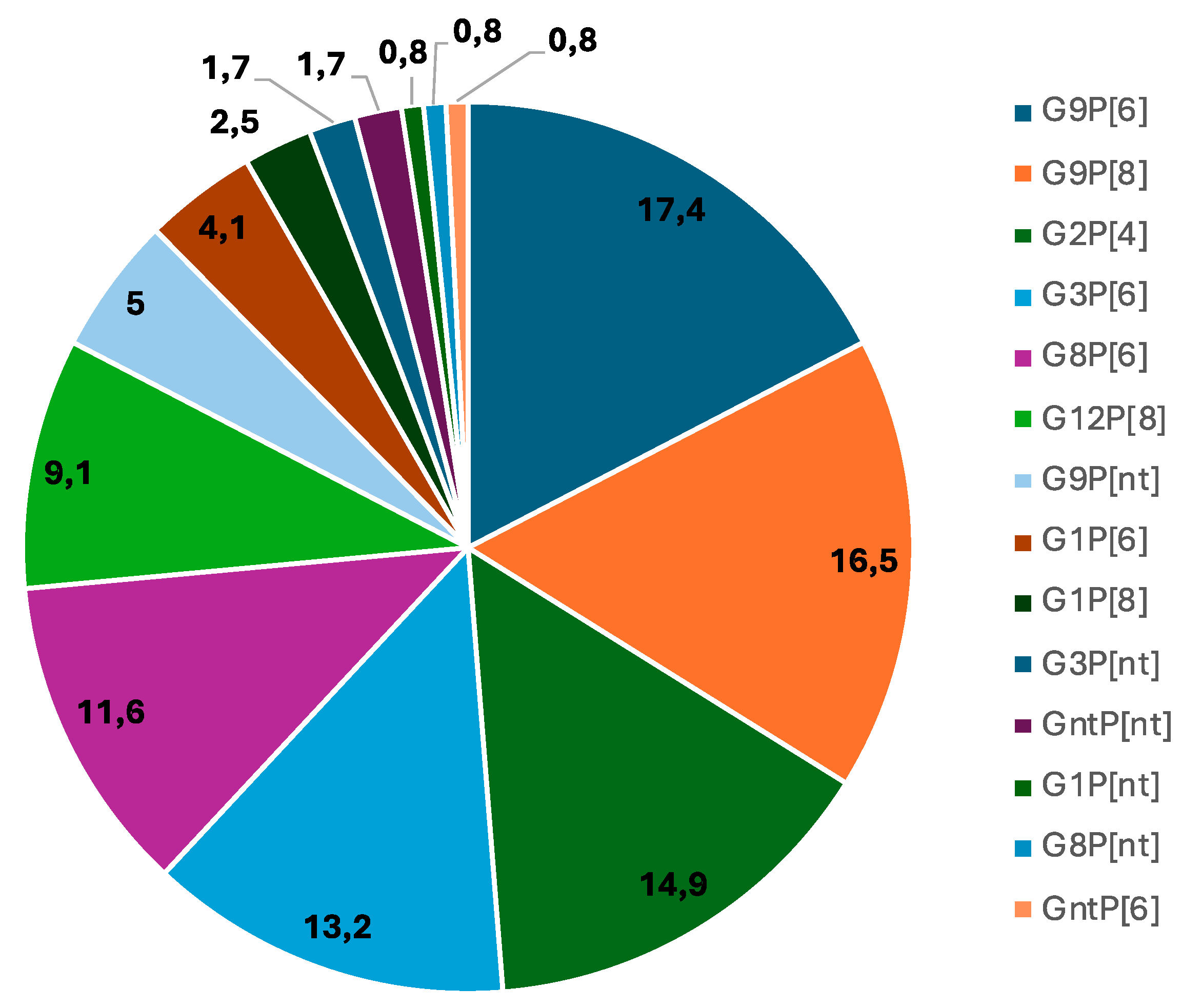
Figure 2.
Molecular Phylogenetic analysis by Maximum Likelihood (ML) method. The ML tree with the highest log likelihood (-3898,30) is shown. The percentage of trees in which the associated taxa clustered together is shown next to the branches. Initial tree(s) for the heuristic search were obtained automatically by applying Neighbor-Join and BioNJ algorithms to a matrix of pairwise distances estimated using the Maximum Composite Likelihood (MCL) approach, and then selecting the topology with superior log likelihood value. A discrete Gamma distribution was used to model evolutionary rate differences among sites (5 categories (+G, parameter = 2,0878)). The rate variation model allowed for some sites to be evolutionarily invariable ([+I], 12,52% sites). The tree is drawn to scale, with branch lengths measured in the number of substitutions per site. The analysis involved 25 nucleotide sequences. All positions containing gaps and missing data were removed. There was a total of 563 positions in the final dataset. Evolutionary analyses were conducted in MEGA7 [24].
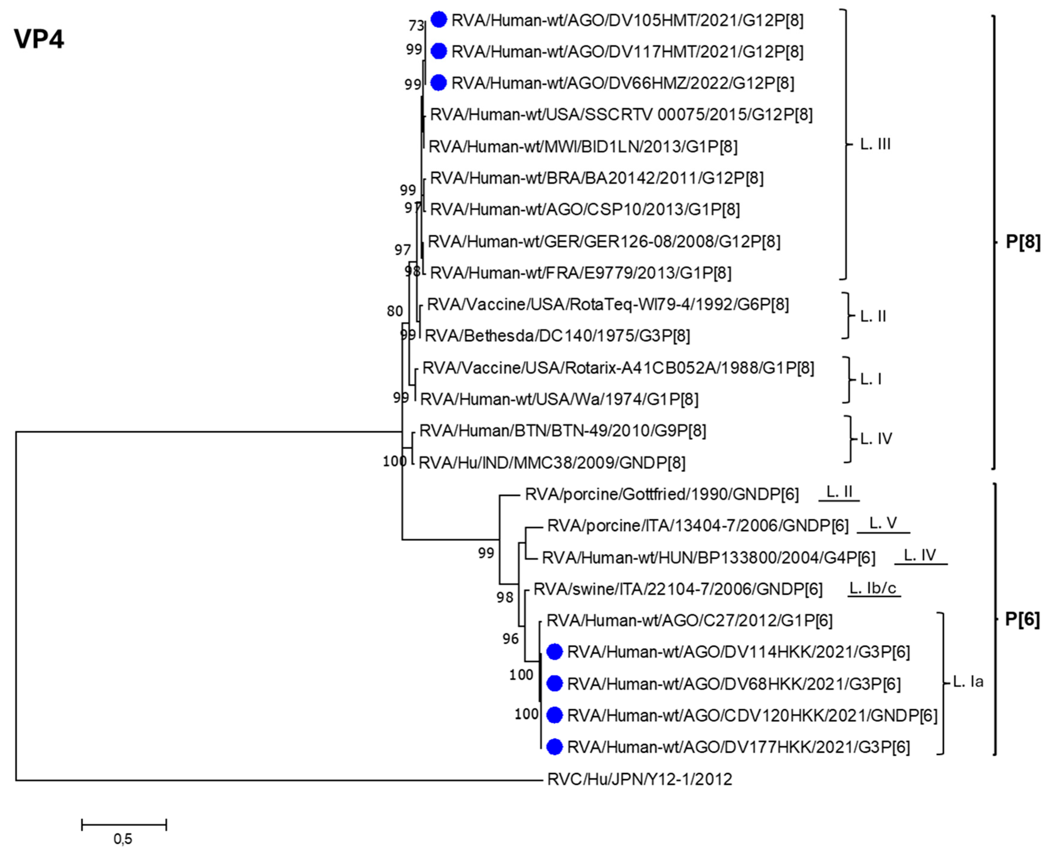
Figure 3.
Molecular Phylogenetic analysis by Maximum Likelihood (ML) method. The ML tree with the highest log likelihood (-2981,70) is shown. The percentage of trees in which the associated taxa clustered together is shown next to the branches. Initial tree(s) for the heuristic search were obtained automatically by applying Neighbor-Join and BioNJ algorithms to a matrix of pairwise distances estimated using the Maximum Composite Likelihood (MCL) approach, and then selecting the topology with superior log likelihood value. A discrete Gamma distribution was used to model evolutionary rate differences among sites (5 categories (+G, parameter = 1,5436)). The rate variation model allowed for some sites to be evolutionarily invariable ([+I], 26,98% sites). The tree is drawn to scale, with branch lengths measured in the number of substitutions per site. The analysis involved 29 nucleotide sequences. All positions containing gaps and missing data were eliminated. There was a total of 354 positions in the final dataset. Evolutionary analyses were conducted in MEGA7 [23].
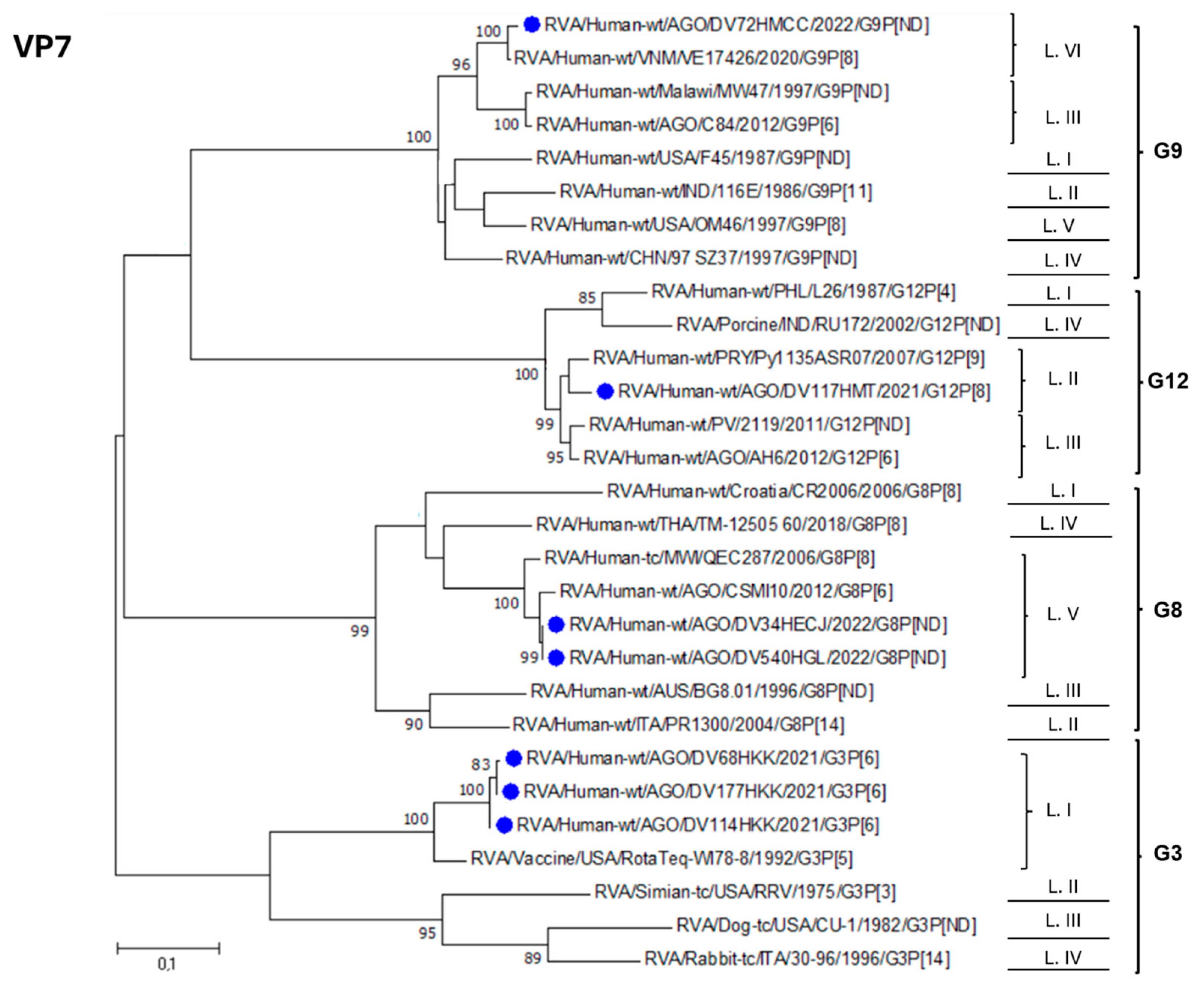

Table 1.
Prevalence of G and P-types in hospitalized children with AGE during the study period (2021-2022).
Table 1.
Prevalence of G and P-types in hospitalized children with AGE during the study period (2021-2022).
| G -type | N | % |
|---|---|---|
| G1 | 9 | 7.4 |
| G2 | 18 | 14.9 |
| G3 | 18 | 14.9 |
| G8 | 15 | 12.4 |
| G9 | 46 | 38.8 |
| G12 | 11 | 9.1 |
| Gnta | 3 | 2.5 |
| Total | 121 | 100 |
| P -type | N | % |
| P4 | 18 | 14.8 |
| P6 | 57 | 47.1 |
| P8 | 34 | 28.0 |
| Pnt b | 12 | 9.9 |
| Total | 121 | 100 |
a Strains that were non-typable for G. b Strains that were non-typable for P.
Table 2.
Genotypic combinations of RVAs circulating in the six hospitals enrolled in the study in Luanda Province (2021/2022).
Table 2.
Genotypic combinations of RVAs circulating in the six hospitals enrolled in the study in Luanda Province (2021/2022).
| Genotype combination |
Cacuaco n (%) |
Luanda n (%) |
Zango n (%) |
Cajueiros n (%) | Talatona n (%) | K. Kiaxi n (%) |
Total n (%) |
|---|---|---|---|---|---|---|---|
| G1P[6] | 0 (0.0) | 2 (6.9) | 3 (9.7) | 0 (0.0) | 0 (00.0) | 0 (0.0) | 5 (4.1) |
| G1P[8] | 0 (0.0) | 0 (0.0) | 0 (0.0) | 0 (0.0) | 3 (17.6) | 0 (0.0) | 3 (2.5) |
| G1Pnt* | 0 (0.0) | 1 (3.4) | 0 (0.0) | 0 (0.0) | 0 (0.0) | 0 (0.0) | 1 (0.8) |
| G2P[4] | 5 (31.2) | 3 (10.3) | 9 (29.0) | 0 (0.0) | 1 (5.9) | 0 (0.0) | 18 (14.9) |
| G3P[6] | 0 (0.0) | 2 (6.9) | 1 (3.2) | 1 (10) | 2 (11.8) | 10 (55.6) | 16 (13.2) |
| G3Pnt* | 0 (0.0) | 0 (0.0) | 1 (3.2) | 0 (0.0) | 0 (0.0) | 1 (5.6) | 2 (1.6) |
| G8P[6] | 1 (6.2) | 3 (10.3) | 3 (9.7) | 4 (40.0) | 2 (11.8) | 1 (5.6) | 14 (11.5) |
| G8Pnt* | 0 (0.0) | 1 (3.4) | 0 (0.0) | 0 (0.0) | 0 (0.0) | 0 (0.0) | 1 (1.7) |
| G9P[6] | 3 (18.8) | 10(34.5) | 5 (16.1) | 1 (10) | 1 (5.9) | 1 (5.6) | 21 (17.3) |
| G9P[8] | 6 (37.5) | 3(10.3) | 7 (22.6) | 0 (0.0) | 0 (0.0) | 4 (22.0) | 20 (16.5) |
| G9Pnt* | 1 (6.3) | 0 (0.0) | 0 (0.0) | 3 (30.0) | 1 (5.9) | 1 (5.6) | 6 (4.9) |
| G12P[8] | 0 (0.0) | 2 (6.9) | 2 (6.5) | 0 (0.0) | 7 (41.1) | 0 (0.0) | 11 (9.1) |
| *GntP[6] | 0 (0.0) | 0 (0.0) | 0 (0.0) | 1 (10) | 0 (0.0) | 0 (0.0) | 1 (0.8) |
| *GntPnt | 0 (0.0) | 2 (6.9) | 0 (0.0) | 0 (0.0) | 0 (0.0) | 0 (0.0) | 2 (1.7) |
| Total | 16 (100) | 29 (100) | 31 (100) | 10 (100) | 17 (100) | 18 (100) | 121 (100) |
*Strains that were non- typable for G or P-type.
Table 3.
Distribution of rotavirus genotypes by age group.
| Age group (months) | |||||
|---|---|---|---|---|---|
| Genotype | 0 – 6 n (%) |
7 - 12 n (%) |
13 – 24 n (%) |
> 24 n (%) |
Total |
| G1P[6] | 3 (3.4) | 2 (6.9) | 0 (0.0) | 0 (0.0) | 5 (4.1) |
| G1P[8] | 3 (3.4) | 0 (0.0) | 0 (0.0) | 0 (0.0) | 3 (2.5) |
| G1PN | 1 (1.1) | 0 (0.0) | 0 (0.0) | 0 (0,0) | 1 (0.8) |
| G2P[4] | 14 (16.1) | 4 (13.8) | 0 (0.0) | 0 (0.0) | 18 (14.9) |
| G3P[6] | 12 (13.8) | 3 (10.3) | 0 (0.0) | 1 (33.3) | 16 (13.2) |
| G3PN | 2 (2.3) | 0 (0.0) | 0 (0.0) | 0 (0.0) | 2 (1.6) |
| G8P[6] | 11 (12.6) | 3 (10.3) | 0 (0.0) | 0 (0.0) | 14 (11.5) |
| G8PN | 1 (1.1) | 0 (0.0) | 0 (0.0) | 0 (0.0) | 1 (1.7) |
| G9P[6] | 9 (10.3) | 8 (27.5) | 2 (100) | 2 (66.7) | 21 (17.3) |
| G9P[8] | 14 (16.1) | 6 (20.7) | 0 (0.0) | 0 (0.0) | 20 (16.5) |
| G9PN | 4 (4.6) | 2 (6.9) | 0 (0.0) | 0 (0.0) | 6 (4.9) |
| G12P[8] | 10 (11.5) | 1 (3.4) | 0 (0.0) | 0 (0.0) | 11 (9.1) |
| *GntP[6] | 1 (1.1) | 0 (0.0) | 0 (0.0) | 0 (0.0) | 1 (0.8) |
| *GntPN | 2 (2.9) | 0 (0.0) | 0 (0.0) | 0 (0.0) | 2 (1.7) |
| Total | 87 (100) | 29 (100) | 2 (100) | 3 (100) | 121 (100) |
*Strains that were non- typable for G.
Table 4.
Distribution of genotype according to the severity of RVA AGE using the Vesikari scoring system.
Table 4.
Distribution of genotype according to the severity of RVA AGE using the Vesikari scoring system.
| Severity of diarrhea | ||||
|---|---|---|---|---|
| Genotype |
Mild (<7) n (%) |
Moderate (7-10) n (%) |
Severe (=> 11) n (%) |
Total n (%) |
| G1P[6] | 0 (0.0) | 0 (0.0) | 5 (4.5) | 5 (4.1) |
| G1P[8] | 0 (0.0) | 1 (10.0) | 2 (1.8) | 3 (2.5) |
| G1Pnt | 0 (0.0) | 0 (0.0) | 1 (0.9) | 1 (0.8) |
| G2P[4] | 0 (0.0) | 0 (0.0) | 18 (16.5) | 18 (14.9) |
| G3P[6] | 0 (0.0) | 1 (10.0) | 15 (15.6) | 16 (13.2) |
| G3PN | 0 (0.0) | 0 (0.0) | 2 (1.8) | 2 (1.6) |
| G8P[6] | 0 (0.0) | 2 (20.0) | 12 (11.0) | 14 (11.5) |
| G8Pnt | 0 (0.0) | 0 (0.0) | 1 (0.9) | 1 (1.7) |
| G9P[6] | 2 (100) | 1 (10.0) | 18 (16.5) | 21 (17.3) |
| G9P[8] | 0 (0.0) | 2 (20.0) | 18 (16.5) | 20 (16.5) |
| G9PN | 0 (0.0) | 2 (20.0) | 4 (3.6) | 6 (4.9) |
| G12P[8] | 0 (0.0) | 1 (10.0) | 10 (9.2) | 11 (9.1) |
| *GntP[6] | 0 (0.0) | 0 (0.0) | 1 (0.9) | 1 (0.8) |
| *GntPN | 0 (0.0) | 0 (0.0) | 2 (1.8) | 2 (1.7) |
| Total | 2 (100) | 10 (100) | 109 (100) | 121 (100) |
*Strains that were non- typable for G or P-type.
Table 5.
VP4 (VP8*) antigenic regions (8-1, 8-2, 8-3 and 8-4), showing the amino acids present in each strain included in the study.
Table 5.
VP4 (VP8*) antigenic regions (8-1, 8-2, 8-3 and 8-4), showing the amino acids present in each strain included in the study.
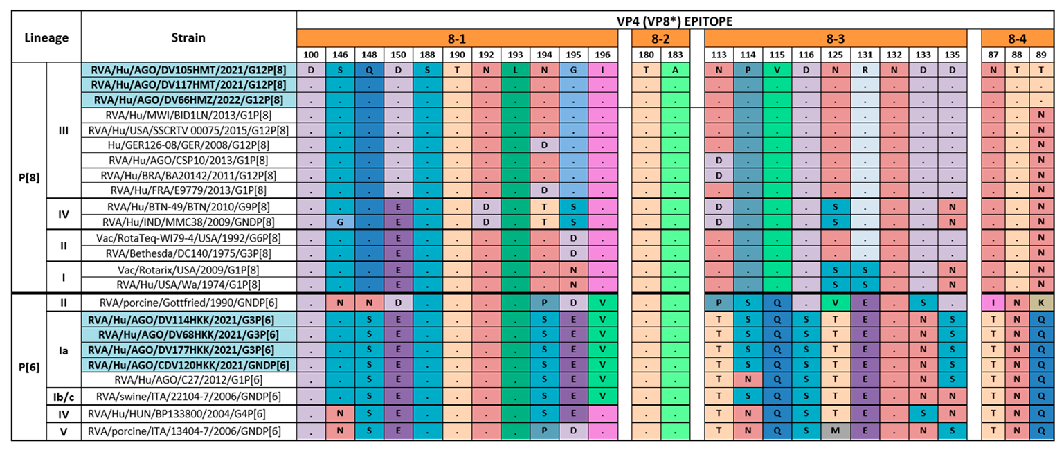 |
Table 6.
VP7 antigenic regions (7-1a, 7-1b, and 7-2), showing the amino acids present in each strain included in the study.
Table 6.
VP7 antigenic regions (7-1a, 7-1b, and 7-2), showing the amino acids present in each strain included in the study.
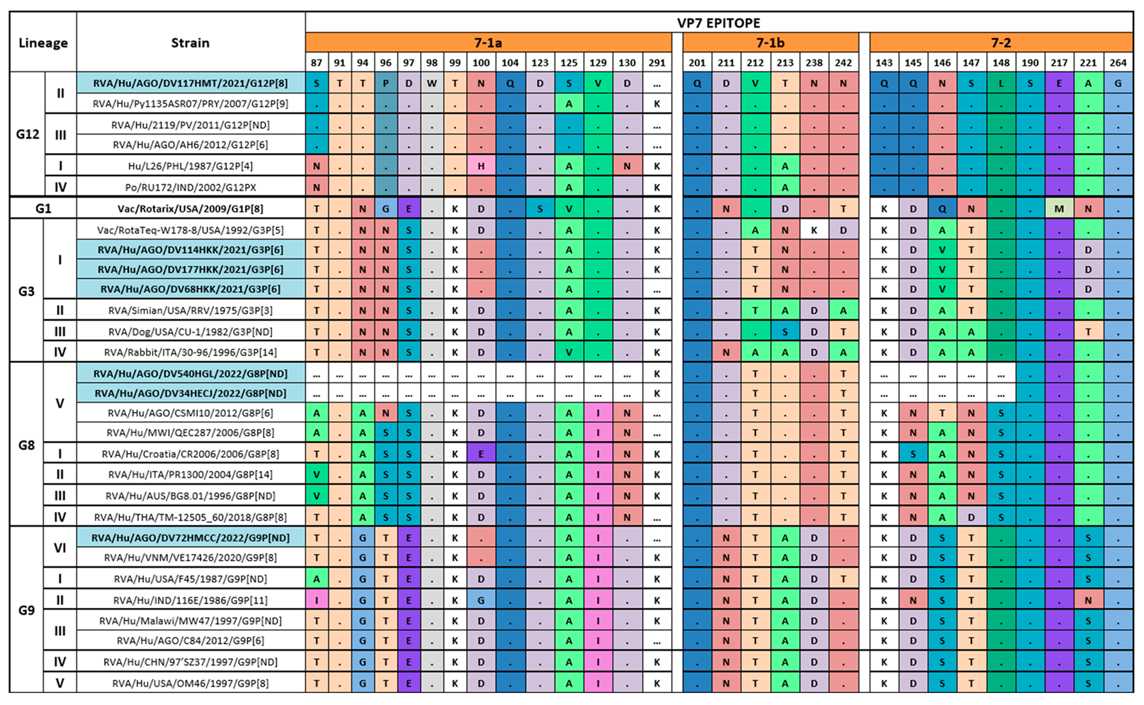 |
Disclaimer/Publisher’s Note: The statements, opinions and data contained in all publications are solely those of the individual author(s) and contributor(s) and not of MDPI and/or the editor(s). MDPI and/or the editor(s) disclaim responsibility for any injury to people or property resulting from any ideas, methods, instructions or products referred to in the content. |
© 2025 by the authors. Licensee MDPI, Basel, Switzerland. This article is an open access article distributed under the terms and conditions of the Creative Commons Attribution (CC BY) license (http://creativecommons.org/licenses/by/4.0/).
Copyright: This open access article is published under a Creative Commons CC BY 4.0 license, which permit the free download, distribution, and reuse, provided that the author and preprint are cited in any reuse.


