
Submitted:
25 April 2025
Posted:
28 April 2025
You are already at the latest version
Abstract
The use of nano-liposomes has increased exponentially since their discovery in the 1960s, primarily for encapsulating medicines or compounds that can improve human health. However, recent studies propose nano-liposomes as vehicles to protect, transport, and subsequently release compounds of various kinds to fortify the properties of foods and cause a prolonged release of encapsulated substances in a specific part of the body. Among the compounds successfully encapsulated are β-carotene, α-carotene, vitamins A, C, and D, lycopene, among others. The encapsulation of extracts with high contents of antioxidant pigments is still to be explored. Therefore, this review aims to compile the compounds that have been successfully encapsulated and have met the specific prolonged release criteria, highlighting areas of research opportunity and application such as biomedical, pharmaceuticals and nutraceutical industries.
Keywords:
Nano-liposomes
; encapsulation
; antioxidant compounds
; affinity
; human health
1. Introduction
Nano-liposomes or nano-liposomal vehicles are molecules composed of a double membrane of phospholipids organized in a lipid bilayer: the polar ends are in the center, trapping a small volume of the aqueous phase, while the apolar tails are arranged outward and interact with the lipophilic ends of the external phospholipids, which position their polar heads outward. This creates a molecule capable of storing both lipophilic and hydrophilic compounds [14,84].
The encapsulation efficiency (EE) of nano-liposomes is higher than those with exclusively hydrophilic properties because the encapsulated substances remain firmly bound to the lipophilic tails [37]. Among the bioactive materials successfully encapsulated are genetic material, proteins, DNA, peptides, vaccines, enzymes, as well as anticancer, antimicrobial, antioxidant, antihemolytic, and anti-inflammatory agents [17]
Recent nano-liposome research has focused on adding them to foods to enhance and fortify the products properties for consumption. Factors determining a successful encapsulation and specific prolonged release include encapsulation efficiency, process yield, prolonged release factor, particle size and Z potential [17].
On the other hand, consumption of antioxidants and natural products is one of the most addressed issues today, as the population is becoming more health conscious. However, it's not just about consumption but the absorption of molecules that provide antioxidants to their biological activity. Beyond protecting pigments from enzymatic and acidic degradation in the digestive system, nano-liposomes aim to ensure the arrival and subsequent release of pigments into the intestine, where the absorption process occurs [34].
One of the current problems in society is people's concern regarding the consumption of functional foods. Due to the current high rates of development of chronic-degenerative diseases such as leukemia, blindness, osteoarthritis, diabetes, cardio-cerebrovascular diseases, and various types of cancer [53], research in food has chosen to explore the encapsulation of biocompounds with antioxidant, anti-inflammatory, antimicrobial, antihemolytic, medicinal and analgesic properties in vesicles formed by phospholipids (nano-liposomes).
Therefore, the objective of this review is to compile compounds with antioxidant, anti-inflammatory, antihemolytic, or photoprotective properties suitable for nano-encapsulation in liposomes, its application in foods and impact on health, as well as to provide a perspective on unexplored areas and future prospects for extracts of different sources.
2. Nano-Liposomes
2.1. Structure and Properties of Nano-Liposomes
In the field of nanotechnology, studies of structures such as nanoemulsions, microemulsions, lipid nanoparticles, biopolymeric nanoparticles, and finally, liposomes, can be found. These studies have focused primarily on using them as encapsulation and prolonged release systems of bioactive compounds, usually used in functional foods [85].
Liposomes are nano or micrometer sized structures (20 nm – 100 µm) whose structural organization is based on the interaction between phospholipids arranged in bilayers or multilayers. Phospholipids, composed of a polar head and a nonpolar tail, are arranged in such a way in an aqueous medium that they form a core where hydrophilic compounds can be encapsulated (aqueous center). Additionally, the nonpolar tails interact with the nonpolar tails of other phospholipids, forming an intermediate layer in which lipophilic compounds can be encapsulated (lipophilic interspace). Finally, the polar heads of the outer phospholipids complete the spherical structure [14,84] (Figure 1).
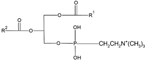
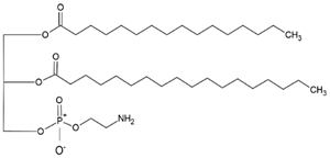
The head determines the surface charge of the liposome (neutral, cationic or anionic) and is generally composed of choline, phosphate and glycerol. On the other hand, the hydrophobic tail is made up of one or two fatty acid chains of 14 - 18 carbons [59]. Phospholipids can be of natural origin (commonly obtained from soy or egg yolk) or synthetic. The most used phospholipids in liposome preparation are shown in Table 1. In addition to phospholipids, liposomes may also contain sterols, with cholesterol being the most common. Cholesterol provides physical and biological stability as it can modify the viscosity or rigidity of the bilayer while reducing its permeability in the presence of biological fluids such as blood. In the absence of cholesterol, the bilayer could experience rupture [45,61].
2.2. Biological Activity of the Encapsulated Compounds
Encapsulated biocompounds can preserve their structure and properties due to the protection of the liposome. This is why this encapsulation process and subsequent addition to foods is considered very effective, both for treating chronic-degenerative diseases and for clinical cases.
Carotenoids are colored natural pigments belonging to a large family of C40 skeleton with eight isoprene molecules. They are classified into xanthophylls and carotenes with the former such as lutein, β-cryptoxanthin and astaxanthin containing one or more oxygen atoms, while the latter such as α- carotene and β-carotene, lycopene and phytoene consisting of hydrogen and carbon atoms [88]. Carotenoid-rich foods have received great attention in human health due to their physiological functions such as antioxidant and anti-cancer as well as the ability to prevent chronic diseases such as age-associated macular degeneration and cardiovascular disease [28,77]. It has been well demonstrated that the functional properties of carotenoids were associated with their chemical structure i.e., the number of conjugated double bonds and the presence of different kinds of end-groups. However, these structural properties are also responsible for the carotenoid’s instability to light, high temperature, oxygen and metal ions, resulting in high susceptibility to oxidation and low bioavailability [28]. Given the multiple health benefits of carotenoids, they are widely used as a natural colorant and antioxidant in both pharmaceutical and food industries to prolong shelf-life in dairy, meat, confectionary, and beverage products. However, carotenoids may undergo loss in functional properties during food processing owing to their instability and interaction with other food ingredients. Also, the presence of digestive enzymes and some other nutrients in vivo as well as pH can alter carotenoid stability [77].
Astaxanthin (AST) is a kind of carotenoid natural pigment that has been widely found in plants, crustaceans shells, flamingos feathers, and microorganisms. Due to its various biological activities, AST has been suggested as an important compound in biochemical research and has great application potential in cosmetics, human nutritional health products, as well as medicines. Unfortunately, the poor water solubility, chemical instability, and low oral bioavailability make challenging to apply AST in food systems. Besides, the amount of natural AST is limited, and how to extract and utilize AST efficiently is in great demand [2]. AST is a non-vitamin A derived ketone lipid soluble carotene [104], which presents a red-orange color and widely exists in many crustacean animals such as shrimp and crab. It is also the highest level product of carotenoid oxygen-containing derivatives [52]. The molecular structure of natural AST contains C=C double chain conjugated olefin structure. The specificity of this structure supplies it an ability to extinguish reactive oxygen species and scavenge free radicals effectively.
As a member of the liposoluble carotenoid family, the special structure of AST may cause the following disadvantages regarding application properties. (1) Hydrophobicity: as a lipophilic substance, AST has a very poor solubility in non-polar solvents. Since there are two hydroxyl groups at each end of AST, and each of them links one hydroxyl group that may interact with fatty acids to form esters. Esterified AST has a stronger hydrophobicity than free AST [28]. (2) Instability: The AST monomers are extremely unstable because of the structures of unsaturated conjugated double bonds. During processing and storage, they are easily degraded and fade under changes in light, temperature, and oxygen content, resulting in further loss of their original biological activity and leading to poor quality and color of final products [104]. The above properties of AST may restrict AST application. To avoid shortcomings and increase the processing adaptability of AST in the food industry, delivery system designing is an effective way in terms of increasing its dispersibility and stability.
Besides, not only do carotenoids have the capacity to improve human health, other biocompounds gives great benefits to human health, such as phenols, alkaloids, and nitrogen and organosulfur compounds. Phenolic compounds are considered secondary metabolites with a wide variety of structures produced by plants. Structurally, phenolic compounds present a benzene ring (C6) with one or more hydroxyl (-OH) group(s), including other functional substituents (glycosides, methyl ethers or esters, etc.) [92].
There are two metabolic pathways through which phenols can be produced in plants: the shikimic acid and the acetic acid pathways. The first produces polyphenols, and the second produces simple phenols. Combining these two pathways produce flavonoids, the most plentiful group of phenolic compounds in plants [74]. Phenolic compounds can be classified in various ways because they consist of various heterogeneous structures, ranging from simple structures to highly polymerized compounds. Based on this, they have been classified into three categories: shortly distributed (e.g., phenols, pyrocatechol, and hydroquinone), widely distributed (e.g., flavonoids and their derivatives, coumarins and phenolic acids), and polymers (e.g., tannin and lignin) [15].
According to their chemical structure, they can also be classified as: soluble (e.g., phenol, flavonoids, and low or medium molecular weight tannins) and insoluble (e.g., condensed tannins and phenolic acids). The first group is not bound to cell membrane compounds, while the second group is bound to cell wall polysaccharides or proteins. This classification is of great importance from a nutritional perspective because the digestion, absorption, and utilization of these compounds largely depend on their solubility. The main interest in phenolic compounds lies in their antioxidant activity, which is associated with beneficial health effects and the prevention of certain diseases. Additionally, they are also used therapeutically for their pharmacological properties. This property entirely depends on the chemical structure, as it may or may not have double bonds or molecules with resonance capacity. Among the phenolic compounds with known high antioxidant activity are flavonoids and tannins [15]. Table 2 presents the structures of the main flavonoids with antioxidant activity.
It has been determined that the high antioxidant capacity of flavonoids is due to their structural configuration, mainly the presence of the hydroxyl group (-OH) in the 3' and 4' positions of the B ring (intermediate), which also confers stability to the compound when it is transformed into a radical by electron donation. This activity is enhanced by the position of the double bonds present in the 2 and 3 carbons of the C ring, together with the carbonyl group in the 4th position, which allows for the movement of the electron between the benzene rings [47].
On the other hand, tannins can be classified into two major groups: hydrolysable tannins and non-hydrolysable tannins, or proanthocyanidins. Hydrolysable tannins have a glucose center or a polyhydric alcohol partially or completely esterified with gallic acid or hexahydroxydiphenic acid, forming gallotannins and ellagitannins, respectively [47].
Proanthocyanidins are polymers of catechins and/or leucoanthocyanidins that are not hydrolysable by acid treatment and are responsible for the astringent properties of plants. They are called proanthocyanidins because of their ability to transform into anthocyanidins. Although the antioxidant capacity of tannins has not been extensively exploited in the food industry, it is known that their activity depends on the degree of polymerization of their chemical structures [71]. Figure 2 shows the structure of a hydrolysable tannin and a non-hydrolysable tannin.
2.3. Bioactive Compounds Encapsulated in Nano-Liposomes
The food industry focuses on the use of liposomes for the encapsulation of bioactive compounds, primarily individual molecules such as retinol, retinoic acid, and retinol ester by retinoids [21,23], as well as carotenoids such as α-carotene, β-carotene, γ-carotene, astaxanthin, violaxanthin, zeaxanthin, and β-cryptoxanthin [24,26,66,76,91]. Phenols have been successfully encapsulated, including gallic acid, protocatechuic acid, caffeic acid, p-coumaric acid and salicylic acid [54,92]. Finally, among the most studied vitamins for encapsulation are A, B, C, D, E and K [27,42,89]. Generally, the compounds mentioned above have biological activity of the antioxidant type. The chemical structures and the type of nanoliposomes in which they were encapsulated are provided in Table 3.
The biological activity of compounds encapsulated in liposomes may not be altered. For example, the astaxanthin’s antioxidant capacity with alpha-tocopherol does not change after they are encapsulated in liposomes. However, it should be noted that nano-liposomes can produce a controlled release of their contents. This leads to different kinetics of the original compound in transport throughout the body. Compounds encapsulated in liposomes must be endocytosed to be transferred inside the cell [43], implying that transport across biological membranes will be subject to this process. This could contrast with the transport of the unencapsulated compound, which may be different (simple diffusion, facilitated diffusion, active transport) and will depend on its physicochemical properties and the biological mechanisms available for that compound.
Almost all the techniques involve the dissolution of phospholipids in an organic solvent followed by the removal of the organic solvent, later in the process. This prior dissolution followed by removal of organic solvent is important for forming of liposomes. The building blocks of liposomes are phospholipids and/or cholesterol. The critical micelle concentration of most used phospholipids is in the nano molar range and the concentration of phospholipids used for liposomes manufacturing is much above the critical micelle concentration. This along with the three-dimensional cylinder like shape of each phospholipid leads to formation of liposomes along with lipid aggregates when phospholipids, as such, are exposed to an aqueous environment.
To make uniform liposomal dispersions, it is important to make thin lipid sheets before exposing them to an aqueous phase or introducing the organic phospholipid solution in a controlled manner in an aqueous environment for the formation of liposomes. This is why all the reported techniques of liposome manufacturing, i.e. solvent evaporation, solvent dispersion/antisolvent addition, or detergent removal, focus on first disaggregating the phospholipids into individual phospholipid molecules followed by exposure to aqueous environment to enable formation of different types of liposomes [3,83].
Thus, transforming from microencapsulation to nanoencapsulation plays a pivotal role in reducing particles to nanosize by employing either top-down or bottom-up methods [60]. Recent advancements in the field of nanoscience and nanotechnology have enabled the preparation of nanoscale functional compounds by encapsulating into a wide variety of nanostructures, including nanoemulsions (NEs), nanoliposomes (NLs), nanocapsules (NCs), nanofibers (NFs), nanoparticles (NPs), solid lipid nanoparticles (SLNPs), nanostructured lipid carriers (NLCs) and supercritical fluid-based nanoparticles [73].
Due to the increasing prevalence rate of chronic diseases, the emerging challenges in delivering functional compounds to target tissues, organs and cells, as well as instability, poor aqueous solubility and bioavailability, and low release and absorption in vivo could not be overcome by microencapsulation techniques. Recent developments in the field of nanotechnology have provided some excellent means to reduce particle size through top-down (high energy method) or bottom-up (self-assembly) processes [70]. Such reduction in particle size has been shown to enhance the stability, targeting ability, bioavailability, and release properties [99]. Most importantly, the reduction in particle size enables penetration into deeper portions of cells or tissues, resulting in high bioavailability [86].
Research studies published within the last five years on nanoencapsulation of various carotenoid compounds mention that it can be done by using different preparation techniques. These studies demonstrated the impact of nanoencapsulation to improve physicochemical property, bioavailability, controlled release and bioactivity. Table 4 and Table 5 summarize various nanosystems used for encapsulation of carotenoids and highlight their advantages as well as disadvantages, respectively.
2.4. Nano-Liposomes Enhanced Foods and Human Health
In recent years, the food industry has played a prominent role in developing functional foods, which provide health benefits beyond the essential nutritional value inherent in the food. These foods contain bioactive components, either chemical compounds naturally present in the food or formed and added during processing, which can exert specific biochemical and physiological functions when consumed by humans [10]. For example, certain lipids in milk have recognized biological properties; among them, conjugated linoleic acid (CLA) can be mentioned. It is a generic term used to describe the mixture of positional and geometric isomers of linoleic acid (C18:2 9c12c) with conjugated double bonds. In recent years, they have gained considerable attention as it is believed that some of these isomers (C18:2 9c, 11t and C18:2 10t, 12c) have beneficial biological effects (reduction of body fat content and increase in muscle mass, stimulation of the immune system, among others) [5].
The deterioration of CLA, especially through oxidation, leads to a decrease in its concentration, loss of bioactivity, and the appearance of unwanted molecules that negatively impact the nutritional and sensory quality of the food. An approach to achieve dairy products enriched in this bioactive compound with good characteristics, without the indicated adverse effects, is the addition of CLA protected by encapsulation, which constitutes a promising alternative. Among encapsulation methods, a very innovative strategy to protect pharmaceutical or food compounds is that of liposomes [5].
In previous study, [100] evaluated the impact of adding a lyophilized powder of liposomes with conjugated linoleic acid (NL-CLA) during yogurt production. They prepared yogurts with CLA in liposomes and control yogurts without CLA. They determined the stability of the fatty acid during storage (21 days at 4°C) and the parameters: pH, acidity, syneresis, microbiological counts (total lactic acid bacteria, molds, and yeasts, total aerobic mesophilic germs), dry residue, fat, and protein content using standardized techniques. Additionally, they observed the microstructure of the yogurts. Adding nano-liposomal vehicles loaded with CLA did not modify the fermentation time; at the end of maturation, the pH and acidity (°D) values remained within appropriate ranges for all yogurts: 4.3-4.4 and 96-99, respectively. The lactic acid bacteria count of the starter reached 10^9 CFU/g, and no contaminating microorganisms were detected. The total solids, protein, and fat contents showed typical values. At the end of storage, yogurts with liposomes exhibited lower syneresis than the controls.
These results correlated with microstructure observations, showing a modification in the protein matrix. The CLA content increased successfully in yogurts with the addition of the liposomal ingredient, as the basal amount of CLA tripled. Thus, the feasibility of applying an ingredient rich in bioactive lipids in the yogurt food matrix was verified.
With the aforementioned information and based on various studies, it can be confirmed that the addition of liposomes to food improves its characteristics and antioxidant properties. All of this is due to the minimal or no modification of the basic structures and the prolonged release of the compounds encapsulated in the liposomes.
The introduction of nano-liposomes enhanced food has sparked great intrigue in society, as they are believed to improve human health, whether through antioxidant, antihemolytic, antimicrobial, anti-inflammatory, photoprotective, and/or anticancer activity and their prolonged liberation property. Innovative encapsulation techniques are currently applied to protect the structure and function of food compounds as well as nutraceutical properties, and to improve their bioavailability. Nano-vehicles or nano-carriers offer the food processor several advantages by ensuring against nutritional loss, incorporating time-release mechanisms into the formulation [100].
[95] in their study, implies that regarding the development and application of nano-liposomes in food technology and food industries, it is considerably less compared to the pharmaceutical and cosmetic industries. However, it is speculated that the greatest advantages will be seen in the agriculture and dairy industries. The limited development is due to the challenges involved in finding safe, low-cost methods for production in the shortest possible time to produce liposomes on a large scale.
Studies to date indicate that the potential of nano-liposomes in food lies in their ability to enhance flavor or in accelerated maturation techniques (e.g., cheese maturation), prolonged and targeted release, the synergistic activity of different encapsulated antioxidant biocompounds and the stabilization of minerals, such as calcium and iron in dairy products [100]. The ability of nanoliposomes to provide targeted delivery of the encapsulated material in specific areas of the food system is highly beneficial for the dairy industry. For example, the employment of proteinase enzymes encapsulated in the lipid vesicles can significantly reduce the time and cost of cheese ripening.
Besides the previous statement, cheese can be improved in their nutritional properties by the addition of vitamin C, D and E, not necessarily in coencapsulation, although it can interact with each other once released. These vitamins has many properties, including: helping protect cells from damage caused by free radicals, collagen formation (necessary for the formation of this protein used to produce skin, tendons, ligaments and blood vessels), strengthening the immune system by helping to keep the immune system strong and reduce the chances of getting sick, regeneration of vitamin E, body calcium absorption (a mineral essential for the formation of strong bones), reduces the risk of cancer, cardiovascular disease, depression and autoimmune diseases [4,97].
Otherwise, minerals can also be encapsulated in nanocarriers, with the aim of avoiding principally iron deficiency, that causes the risk of developing anemia. This condition is mainly due to insufficiency in dietary intake of iron, lack of its bioavailability or both. Iron shortage in blood should not be ignored as it may cause anemia, when the blood hemoglobin level falls below standard levels, a disease that, if not threatened, can evolve to leukemia [32].
Hence, it is necessary to enrich dairy products with iron evading fat oxidation and metallic off-flavor. To achieve this, supplementation of dairy products with iron encapsulated in safe and non-toxic carriers has been developed. Towards this end, highly soluble form of iron (i.e., ferrous sulfate) is preferred due to its cost effectiveness and high bioavailability [100]. Another example of a mineral is magnesium. It is an essential mineral compound, which is associated with lowering the risk of some clinical disorders including cardiovascular disease, hypertension, type 2 diabetes and muscular weakness [33].
3. Conclusions
Nano-liposomes offer significant advantages by protecting encapsulated compounds from degradation by environmental and enzymatic factors, enabling controlled and targeted release in the body. Furthermore, their structural versatility allows for the encapsulation of both hydrophilic and lipophilic compounds, broadening the range of applications in the food, nutraceutical, and pharmaceutical industries. However, challenges remain related to optimizing encapsulation efficiency, storage stability, and site-specific release, aspects that must be addressed for their large-scale implementation. The use of various types of phospholipids increases the range of compounds that can be encapsulated, making this not only an innovative application but also one with a trend of expansion into various scientific fields such as healthcare, food industries, cosmetics, and pharmaceuticals.
4. Prospects
Prospects for the development and application of nanoliposomes in the field of antioxidants are promising. Research on the encapsulation of complex extracts rich in pigments and polyphenols is anticipated, as is the integration of smart release technologies that respond to specific physiological stimuli. Furthermore, the trend toward functional and personalized foods will drive innovation in nanoencapsulation systems tailored to different health needs and population groups. Long-term clinical and safety studies will be essential, as will the development of clear regulations for their use in food and nutraceutical products. Finally, interdisciplinary collaboration between food scientists, chemists, biotechnologists, and healthcare professionals will be key to translating these technological advances into tangible benefits for public health.
Author Contributions
Conceptualization, J.d.J.O.-P. and S.P.A.-M.; writing—original draft preparation, J.G.-M.; writing—review and editing, A.T.B.-M. and R.I.G.-V.; visualization M.A.R.-G. and S.R.-C; and supervision, C.L.D.-T.-S and D.F.-O. All authors have read and agreed to the published version of the manuscript.
Funding
The work was supported by the project CBF2023-2024-3196 from Secretaría de Ciencia, Humanidades, Tecnología e Innovación (SECIHTI).
Institutional Review Board Statement
Not applicable.
Data Availability Statement
No new data were created or analyzed in this study. Data sharing is not applicable to this article.
Acknowledgments
The authors are pleased to acknowledge of Secretaría de Ciencia, Humanidades, Tecnología e Innovación (SECIHTI) for awarding Jonathan García Morales for his doctoral scholarship.
Conflicts of Interest
The authors declare no conflict of interest.
References
- Aman Mohamadi, M.; Farshi, P.; Ahmadi, P.; Ahmadi, A.; Yousefi, M.; Ghorbani, M.; Hosseini, S. M. Encapsulation of Vitamins Using Nanoliposome: Recent Advances and Perspectives. Adv. Pharm. Bull. 2021, 1. [Google Scholar] [CrossRef] [PubMed]
- Ambati, R.; Phang, S.-M.; Ravi, S.; Aswathanarayana, R. Astaxanthin: Sources, Extraction, Stability, Biological Activities and Its Commercial Applications—A Review. Mar. Drugs 2014, 12, 128–152. [Google Scholar] [CrossRef]
- Assil, K. K. Multivesicular Liposomes: Sustained Release of the Antimetabolite Cytarabine in the Eye. Arch. Ophthalmol. 1987, 105, 400. [Google Scholar] [CrossRef]
- Athanassiou, L.; Mavragani, C. P.; Koutsilieris, M. The Immunomodulatory Properties of Vitamin D. Mediterr. J. Rheumatol. 2022, 33, 7. [Google Scholar] [CrossRef]
- Badawy, S.; Liu, Y.; Guo, M.; Liu, Z.; Xie, C.; Marawan, M. A.; Ares, I.; Lopez-Torres, B.; Martínez, M.; Maximiliano, J.-E.; Martínez-Larrañaga, M.-R.; Wang, X.; Anadón, A.; Martínez, M.-A. Conjugated Linoleic Acid (CLA) as a Functional Food: Is It Beneficial or Not? Food Res. Int. 2023, 172, 113158. [Google Scholar] [CrossRef]
- Baek, E. J.; Garcia, C. V.; Shin, G. H.; Kim, J. T. Improvement of Thermal and UV-Light Stability of β-Carotene-Loaded Nanoemulsions by Water-Soluble Chitosan Coating. Int. J. Biol. Macromol. 2020, 165, 1156–1163. [Google Scholar] [CrossRef] [PubMed]
- Baek, K.; Patra, J. K. Novel Green Synthesis of Gold Nanoparticles Using Citrullus Lanatus Rind and Investigation of Proteasome Inhibitory Activity, Antibacterial, and Antioxidant Potential. Int. J. Nanomedicine 2015, 7253. [Google Scholar] [CrossRef]
- Bhalerao, S. S.; Raje Harshal, A. Preparation, Optimization, Characterization, and Stability Studies of Salicylic Acid Liposomes. Drug Dev. Ind. Pharm. 2003, 29, 451–467. [Google Scholar] [CrossRef] [PubMed]
- Bezerra, P. Q. M.; Matos, M. F. R. D.; Ramos, I. G.; Magalhães-Guedes, K. T.; Druzian, J. I.; Costa, J. A. V.; Nunes, I. L. Innovative Functional Nanodispersion: Combination of Carotenoid from Spirulina and Yellow Passion Fruit Albedo. Food Chem. 2019, 285, 397–405. [Google Scholar] [CrossRef]
- Birch, C. S.; Bonwick, G. A. Ensuring the Future of Functional Foods. Int. J. Food Sci. Technol. 2019, 54, 1467–1485. [Google Scholar] [CrossRef]
- Bolla, P. K.; Gote, V.; Singh, M.; Patel, M.; Clark, B. A.; Renukuntla, J. Lutein-Loaded, Biotin-Decorated Polymeric Nanoparticles Enhance Lutein Uptake in Retinal Cells. Pharmaceutics 2020, 12, 798. [Google Scholar] [CrossRef]
- Borba, C. M.; Tavares, M. N.; Macedo, L. P.; Araújo, G. S.; Furlong, E. B.; Dora, C. L.; Burkert, J. F. M. Physical and Chemical Stability of β-Carotene Nanoemulsions during Storage and Thermal Process. Food Res. Int. 2019, 121, 229–237. [Google Scholar] [CrossRef]
- Boura, E.; Nencka, R. Phosphatidylinositol 4-Kinases: Function, Structure, and Inhibition. Exp. Cell Res. 2015, 337, 136–145. [Google Scholar] [CrossRef] [PubMed]
- Bozzuto, G.; Molinari, A. Liposomes as Nanomedical Devices. Int. J. Nanomedicine 2015, 975. [Google Scholar] [CrossRef] [PubMed]
- Bravo, L. Polyphenols: Chemistry, Dietary Sources, Metabolism, and Nutritional Significance. Nutr. Rev. 2009, 56, 317–333. [Google Scholar] [CrossRef]
- Calzada, E.; Onguka, O.; Claypool, S. M. Phosphatidylethanolamine Metabolism in Health and Disease. International Review of Cell and Molecular Biology 2016, 321, 29–88. [Google Scholar] [CrossRef] [PubMed]
- Carugo, D.; Bottaro, E.; Owen, J.; Stride, E.; Nastruzzi, C. Liposome Production by Microfluidics: Potential and Limiting Factors. Sci. Rep. 2016, 6, 25876. [Google Scholar] [CrossRef]
- Chakravarty, P.; Famili, A.; Nagapudi, K.; Al-Sayah, M. A. Using Supercritical Fluid Technology as a Green Alternative During the Preparation of Drug Delivery Systems. Pharmaceutics 2019, 11, 629. [Google Scholar] [CrossRef]
- Chaves, M. A.; Oseliero Filho, P. L.; Jange, C. G.; Sinigaglia-Coimbra, R.; Oliveira, C. L. P.; Pinho, S. C. Structural Characterization of Multilamellar Liposomes Coencapsulating Curcumin and Vitamin D3. Colloids Surf. Physicochem. Eng. Asp. 2018, 549, 112–121. [Google Scholar] [CrossRef]
- Chen, B.-H.; Huang, R.-F. S.; Wei, Y.-J.; Stephen Inbaraj, B. Inhibition of Colon Cancer Cell Growth by Nanoemulsion Carrying Gold Nanoparticles And Lycopene. Int. J. Nanomedicine 2015, 2823. [Google Scholar] [CrossRef]
- Cristiano, M. C.; Cosco, D.; Celia, C.; Tudose, A.; Mare, R.; Paolino, D.; Fresta, M. Anticancer Activity of All- Trans Retinoic Acid-Loaded Liposomes on Human Thyroid Carcinoma Cells. Colloids Surf. B Biointerfaces 2017, 150, 408–416. [Google Scholar] [CrossRef]
- Couto, R.; Alvarez, V.; Temelli, F. Encapsulation of Vitamin B2 in Solid Lipid Nanoparticles Using Supercritical CO 2. J. Supercrit. Fluids 2017, 120, 432–442. [Google Scholar] [CrossRef]
- Cuomo, F.; Ceglie, S.; Miguel, M.; Lindman, B.; Lopez, F. Oral Delivery of All-Trans Retinoic Acid Mediated by Liposome Carriers. Colloids Surf. B Biointerfaces 2021, 201, 111655. [Google Scholar] [CrossRef] [PubMed]
- Dos Santos, P. P.; Andrade, L. D. A.; Flôres, S. H.; Rios, A. D. O. Nanoencapsulation of Carotenoids: A Focus on Different Delivery Systems and Evaluation Parameters. J. Food Sci. Technol. 2018, 55, 3851–3860. [Google Scholar] [CrossRef] [PubMed]
- Dos Santos, P. P.; Paese, K.; Guterres, S. S.; Pohlmann, A. R.; Costa, T. H.; Jablonski, A.; Flôres, S. H.; Rios, A. D. O. Development of Lycopene-Loaded Lipid-Core Nanocapsules: Physicochemical Characterization and Stability Study. J. Nanoparticle Res. 2015, 17, 107. [Google Scholar] [CrossRef]
- Dutta, D.; Dutta, D. Designing a Nanoliposome Loaded with Provitamin A Xanthophyll β-Cryptoxanthin from K. Marina DAGII to Target a Population Suffering from Hypovitaminosis A. Process Biochem. 2024, 138, 97–110. [Google Scholar] [CrossRef]
- Fan, C.; Feng, T.; Wang, X.; Xia, S.; John Swing, C. Liposomes for Encapsulation of Liposoluble Vitamins (A, D, E and K): Comparation of Loading Ability, Storage Stability and Bilayer Dynamics. Food Res. Int. 2023, 163, 112264. [Google Scholar] [CrossRef]
- Focsan, A. L.; Polyakov, N. E.; Kispert, L. D. Supramolecular Carotenoid Complexes of Enhanced Solubility and Stability—The Way of Bioavailability Improvement. Molecules 2019, 24, 3947. [Google Scholar] [CrossRef]
- Foss, B. J.; Sliwka, H.-R.; Partali, V.; Cardounel, A. J.; Zweier, J. L.; Lockwood, S. F. Direct Superoxide Anion Scavenging by a Highly Water-Dispersible Carotenoid Phospholipid Evaluated by Electron Paramagnetic Resonance (EPR) Spectroscopy. Bioorg. Med. Chem. Lett. 2004, 14, 2807–2812. [Google Scholar] [CrossRef]
- García Morales, J.; López Elías, J. A.; Medina Félix, D.; García Lagunas, N.; Fimbres Olivarría, D. Efecto del estrés por nitrógeno y salinidad en el contenido de b-caroteno de la microalga Dunaliella tertiolecta//Effect of nitrogen and salinity stress on the β-carotene content of the microalgae Dunaliella tertiolecta. Biotecnia 2020, 22, 13–19. [Google Scholar] [CrossRef]
- Gasa-Falcon, A.; Arranz, E.; Odriozola-Serrano, I.; Martín-Belloso, O.; Giblin, L. Delivery of β-Carotene to the in Vitro Intestinal Barrier Using Nanoemulsions with Lecithin or Sodium Caseinate as Emulsifiers. LWT 2021, 135, 110059. [Google Scholar] [CrossRef]
- Gaucheron, F. Iron Fortification in Dairy Industry. Trends Food Sci. Technol. 2000, 11, 403–409. [Google Scholar] [CrossRef]
- Gharibzahedi, S. M. T.; Jafari, S. M. The Importance of Minerals in Human Nutrition: Bioavailability, Food Fortification, Processing Effects and Nanoencapsulation. Trends Food Sci. Technol. 2017, 62, 119–132. [Google Scholar] [CrossRef]
- Guimarães, D.; Cavaco-Paulo, A.; Nogueira, E. Design of Liposomes as Drug Delivery System for Therapeutic Applications. Int. J. Pharm. 2021, 601, 120571. [Google Scholar] [CrossRef]
- Hafezi Ghahestani, Z.; Alebooye Langroodi, F.; Mokhtarzadeh, A.; Ramezani, M.; Hashemi, M. Evaluation of Anti-Cancer Activity of PLGA Nanoparticles Containing Crocetin. Artif. Cells Nanomedicine Biotechnol. 2017, 45, 955–960. [Google Scholar] [CrossRef]
- Han, X.; Huo, P.; Ding, Z.; Kumar, P.; Liu, B. Preparation of Lutein-Loaded PVA/Sodium Alginate Nanofibers and Investigation of Its Release Behavior. Pharmaceutics 2019, 11, 449. [Google Scholar] [CrossRef]
- Has, C.; Sunthar, P. A Comprehensive Review on Recent Preparation Techniques of Liposomes. J. Liposome Res. 2020, 30, 336–365. [Google Scholar] [CrossRef] [PubMed]
- Hassane Hamadou, A.; Huang, W.-C.; Xue, C.; Mao, X. Comparison of β-Carotene Loaded Marine and Egg Phospholipids Nanoliposomes. J. Food Eng. 2020, 283, 110055. [Google Scholar] [CrossRef]
- Jain, A.; Sharma, G.; Thakur, K.; Raza, K.; Shivhare, U. S.; Ghoshal, G.; Katare, O. P. Beta-Carotene-Encapsulated Solid Lipid Nanoparticles (BC-SLNs) as Promising Vehicle for Cancer: An Investigative Assessment. AAPS PharmSciTech 2019, 20, 100. [Google Scholar] [CrossRef]
- Janssens, B. ENCAPSULATION OF VITAMIN D3 AND VITAMIN K2 IN CHITOSAN COATED LIPOSOMES.
- Jiao, Y.; Li, D.; Liu, C.; Chang, Y.; Song, J.; Xiao, Y. Polypeptide – Decorated Nanoliposomes as Novel Delivery Systems for Lutein. RSC Adv. 2018, 8, 31372–31381. [Google Scholar] [CrossRef]
- Jiao, Z.; Wang, X.; Yin, Y.; Xia, J. Preparation and Evaluation of Vitamin C and Folic Acid-Coloaded Antioxidant Liposomes. Part. Sci. Technol. 2019, 37, 453–459. [Google Scholar] [CrossRef]
- Kaga, K.; Honda, M.; Adachi, T.; Honjo, M.; Wahyudiono, *!!! REPLACE !!!*; Kanda, H.; Goto, M. Nanoparticle Formation of PVP/Astaxanthin Inclusion Complex by Solution-Enhanced Dispersion by Supercritical Fluids (SEDS): Effect of PVP and Astaxanthin Z-Isomer Content. J. Supercrit. Fluids 2018, 136, 44–51. [Google Scholar] [CrossRef]
- Kanno, K.; Wu, M. K.; Scapa, E. F.; Roderick, S. L.; Cohen, D. E. Structure and Function of Phosphatidylcholine Transfer Protein (PC-TP)/StarD2. Biochim. Biophys. Acta BBA - Mol. Cell Biol. Lipids 2007, 1771, 654–662. [Google Scholar] [CrossRef]
- Kawakami, L. M.; Yoon, B. K.; Jackman, J. A.; Knoll, W.; Weiss, P. S.; Cho, N.-J. Understanding How Sterols Regulate Membrane Remodeling in Supported Lipid Bilayers. Langmuir 2017, 33, 14756–14765. [Google Scholar] [CrossRef] [PubMed]
- Kay, J. G.; Fairn, G. D. Distribution, Dynamics and Functional Roles of Phosphatidylserine within the Cell. Cell Commun. Signal. 2019, 17, 126. [Google Scholar] [CrossRef]
- Koleckar, V.; Kubikova, K.; Rehakova, Z.; Kuca, K.; Jun, D.; Jahodar, L.; Opletal, L. Condensed and Hydrolysable Tannins as Antioxidants Influencing the Health. Mini-Rev. Med. Chem. 2008, 8, 436–447. [Google Scholar] [CrossRef]
- Li, D.; Li, L.; Xiao, N.; Li, M.; Xie, X. Physical Properties of Oil-in-Water Nanoemulsions Stabilized by OSA-Modified Starch for the Encapsulation of Lycopene. Colloids Surf. Physicochem. Eng. Asp. 2018, 552, 59–66. [Google Scholar] [CrossRef]
- Li, W.; Yalcin, M.; Lin, Q.; Ardawi, M.-S. M.; Mousa, S. A. Self-Assembly of Green Tea Catechin Derivatives in Nanoparticles for Oral Lycopene Delivery. J. Controlled Release 2017, 248, 117–124. [Google Scholar] [CrossRef] [PubMed]
- Liu, P.; Shen, J.; Cao, J.; Jiang, W. P-Coumaric Acid-Loaded Nanoliposomes: Optimization, Characterization, Antimicrobial Properties and Preservation Effects on Fresh Pod Pepper Fruit. Food Chem. 2024, 435, 137672. [Google Scholar] [CrossRef]
- Liu, X.; Lin, X.; Wei, Y.; Fei, T.; Hu, X.; Wang, L. Enhancing the Stability and Bio-Accessibility of Carotenoids Extracted from Canistel (Lucuma Nervosa) via Liposomes Encapsulation. LWT 2024, 204, 116455. [Google Scholar] [CrossRef]
- Liu, X.; Shibata, T.; Hisaka, S.; Osawa, T. Astaxanthin Inhibits Reactive Oxygen Species-Mediated Cellular Toxicity in Dopaminergic SH-SY5Y Cells via Mitochondria-Targeted Protective Mechanism. Brain Res. 2009, 1254, 18–27. [Google Scholar] [CrossRef] [PubMed]
- Luna-Guevara, Ma. L.; Luna-Guevara, J. J.; Hernández-Carranza, P.; Ruíz-Espinosa, H.; Ochoa-Velasco, C. E. Phenolic Compounds: A Good Choice Against Chronic Degenerative Diseases. Studies in Natural Products Chemistry 2018, 59, 79–108. [Google Scholar] [CrossRef]
- Machado, A. R.; Pinheiro, A. C.; Vicente, A. A.; Souza-Soares, L. A.; Cerqueira, M. A. Liposomes Loaded with Phenolic Extracts of Spirulina LEB-18: Physicochemical Characterization and Behavior under Simulated Gastrointestinal Conditions. Food Res. Int. 2019, 120, 656–667. [Google Scholar] [CrossRef] [PubMed]
- Mansur, M. C. P. P. R.; Campos, C.; Vermelho, A. B.; Nobrega, J.; Da Cunha Boldrini, L.; Balottin, L.; Lage, C.; Rosado, A. S.; Ricci-Júnior, E.; Dos Santos, E. P. Photoprotective Nanoemulsions Containing Microbial Carotenoids and Buriti Oil: Efficacy and Safety Study. Arab. J. Chem. 2020, 13, 6741–6752. [Google Scholar] [CrossRef]
- Mehrad, B.; Ravanfar, R.; Licker, J.; Regenstein, J. M.; Abbaspourrad, A. Enhancing the Physicochemical Stability of β-Carotene Solid Lipid Nanoparticle (SLNP) Using Whey Protein Isolate. Food Res. Int. 2018, 105, 962–969. [Google Scholar] [CrossRef] [PubMed]
- Nazemiyeh, E.; Eskandani, M.; Sheikhloie, H.; Nazemiyeh, H. Formulation and Physicochemical Characterization of Lycopene-Loaded Solid Lipid Nanoparticles. Adv. Pharm. Bull. 2016, 6, 235–241. [Google Scholar] [CrossRef]
- Noubigh, A.; Aydi, A.; Abderrabba, M. Experimental Measurement and Correlation of Solubility Data and Thermodynamic Properties of Protocatechuic Acid in Four Organic Solvents. J. Chem. Eng. Data 2015, 60, 514–518. [Google Scholar] [CrossRef]
- Nsairat, H.; Khater, D.; Sayed, U.; Odeh, F.; Al Bawab, A.; Alshaer, W. Liposomes: Structure, Composition, Types, and Clinical Applications. Heliyon 2022, 8, e09394. [Google Scholar] [CrossRef]
- Okonogi, S.; Riangjanapatee, P. Physicochemical Characterization of Lycopene-Loaded Nanostructured Lipid Carrier Formulations for Topical Administration. Int. J. Pharm. 2015, 478, 726–735. [Google Scholar] [CrossRef]
- Ortega-Galindo, A. S.; Díaz-Peralta, L.; Galván-Hernández, A.; Ortega-Blake, I.; Pérez-Riascos, A.; Rojas-Aguirre, Y. Los liposomas en nanomedicina: del concepto a sus aplicaciones clínicas y tendencias actuales en investigación. Mundo Nano Rev. Interdiscip. En Nanociencias Nanotecnología 2023, 16, 1e–26e. [Google Scholar] [CrossRef]
- Oviedo-Montiel, H.; Herrera-Cruz, E.; Hoya-Florez, J.; Prieto-Guevara, M.; Estrada-Posada, A.; Yepes Blandón, J. A. Crecimiento y Viabilidad Celular de Microalgas: Efecto Del Medio de Cultivo. Intropica 2020, 15, 126–136. [Google Scholar] [CrossRef]
- Pan, L.; Wang, H.; Gu, K. Nanoliposomes as Vehicles for Astaxanthin: Characterization, In Vitro Release Evaluation and Structure. Molecules 2018, 23, 2822. [Google Scholar] [CrossRef] [PubMed]
- Pan, L.; Zhang, S.; Gu, K.; Zhang, N. Preparation of Astaxanthin-Loaded Liposomes: Characterization, Storage Stability and Antioxidant Activity. CyTA - J. Food 2018, 16, 607–618. [Google Scholar] [CrossRef]
- Păvăloiu, R.-D.; Sha’at, F.; Neagu, G.; Deaconu, M.; Bubueanu, C.; Albulescu, A.; Sha’at, M.; Hlevca, C. Encapsulation of Polyphenols from Lycium Barbarum Leaves into Liposomes as a Strategy to Improve Their Delivery. Nanomaterials 2021, 11, 1938. [Google Scholar] [CrossRef]
- Pereira, A. G.; Otero, P.; Echave, J.; Carreira-Casais, A.; Chamorro, F.; Collazo, N.; Jaboui, A.; Lourenço-Lopes, C.; Simal-Gandara, J.; Prieto, M. A. Xanthophylls from the Sea: Algae as Source of Bioactive Carotenoids. Mar. Drugs 2021, 19, 188. [Google Scholar] [CrossRef]
- Pereira, M. C.; Hill, L. E.; Zambiazi, R. C.; Mertens-Talcott, S.; Talcott, S.; Gomes, C. L. Nanoencapsulation of Hydrophobic Phytochemicals Using Poly (Dl-Lactide-Co-Glycolide) (PLGA) for Antioxidant and Antimicrobial Delivery Applications: Guabiroba Fruit (Campomanesia Xanthocarpa O. Berg) Study. LWT - Food Sci. Technol. 2015, 63, 100–107. [Google Scholar] [CrossRef]
- Pezeshky, A.; Ghanbarzadeh, B.; Hamishehkar, H.; Moghadam, M.; Babazadeh, A. Vitamin A Palmitate-Bearing Nanoliposomes: Preparation and Characterization. Food Biosci. 2016, 13, 49–55. [Google Scholar] [CrossRef]
- Raben, D. M.; Barber, C. N. Phosphatidic Acid and Neurotransmission. Adv. Biol. Regul. 2017, 63, 15–21. [Google Scholar] [CrossRef]
- Rashidinejad, A.; Jafari, S. M. Nanoencapsulation of Bioactive Food Ingredients. In Handbook of Food Nanotechnology; Elsevier, 2020; pp. 279–344. [Google Scholar] [CrossRef]
- Rauf, A.; Imran, M.; Abu-Izneid, T.; Iahtisham-Ul-Haq, *!!! REPLACE !!!*; Patel, S.; Pan, X.; Naz, S.; Sanches Silva, A.; Saeed, F.; Rasul Suleria, H.A. Proanthocyanidins: A Comprehensive Review. Biomed. Pharmacother. 2019, 116, 108999. [Google Scholar] [CrossRef]
- Ravi, H.; Baskaran, V. Biodegradable Chitosan-Glycolipid Hybrid Nanogels: A Novel Approach to Encapsulate Fucoxanthin for Improved Stability and Bioavailability. Food Hydrocoll. 2015, 43, 717–725. [Google Scholar] [CrossRef]
- Rehman, A.; Tong, Q.; Jafari, S. M.; Assadpour, E.; Shehzad, Q.; Aadil, R. M.; Iqbal, M. W.; Rashed, M. M. A.; Mushtaq, B. S.; Ashraf, W. Carotenoid-Loaded Nanocarriers: A Comprehensive Review. Adv. Colloid Interface Sci. 2020, 275, 102048. [Google Scholar] [CrossRef] [PubMed]
- Reis Giada, M. D. L. Food Phenolic Compounds: Main Classes, Sources and Their Antioxidant Power. In Oxidative Stress and Chronic Degenerative Diseases - A Role for Antioxidants; Morales-Gonzalez, J. A., Ed.; InTech, 2013. [CrossRef]
- Roll Zimmer, T. B.; Barboza Mendonça, C. R.; Zambiazi, R. C. Methods of Protection and Application of Carotenoids in Foods - A Bibliographic Review. Food Biosci. 2022, 48, 101829. [Google Scholar] [CrossRef]
- Rostamabadi, H.; Falsafi, S. R.; Jafari, S. M. Nanoencapsulation of Carotenoids within Lipid-Based Nanocarriers. J. Controlled Release 2019, 298, 38–67. [Google Scholar] [CrossRef]
- Rostamabadi, H.; Sadeghi Mahoonak, A.; Allafchian, A.; Ghorbani, M. Fabrication of β-Carotene Loaded Glucuronoxylan-Based Nanostructures through Electrohydrodynamic Processing. Int. J. Biol. Macromol. 2019, 139, 773–784. [Google Scholar] [CrossRef] [PubMed]
- Roy, U. K.; Nielsen, B. V.; Milledge, J. J. Tuning Dunaliella Tertiolecta for Enhanced Antioxidant Production by Modification of Culture Conditions. Mar. Biotechnol. 2021, 23, 482–500. [Google Scholar] [CrossRef]
- Schjoerring-Thyssen, J.; Olsen, K.; Koehler, K.; Jouenne, E.; Rousseau, D.; Andersen, M. L. Morphology and Structure of Solid Lipid Nanoparticles Loaded with High Concentrations of β-Carotene. J. Agric. Food Chem. 2019, 67, 12273–12282. [Google Scholar] [CrossRef]
- Seabra, A.; Durán, N. Nanotoxicology of Metal Oxide Nanoparticles. Metals 2015, 5, 934–975. [Google Scholar] [CrossRef]
- Sengul, A. B.; Asmatulu, E. Toxicity of Metal and Metal Oxide Nanoparticles: A Review. Environ. Chem. Lett. 2020, 18, 1659–1683. [Google Scholar] [CrossRef]
- Shabestarian, H.; Homayouni-Tabrizi, M.; Soltani, M.; Namvar, F.; Azizi, S.; Mohamad, R.; Shabestarian, H. Green Synthesis of Gold Nanoparticles Using Sumac Aqueous Extract and Their Antioxidant Activity. Mater. Res. 2016, 20, 264–270. [Google Scholar] [CrossRef]
- Shah, S.; Dhawan, V.; Holm, R.; Nagarsenker, M. S.; Perrie, Y. Liposomes: Advancements and Innovation in the Manufacturing Process. Adv. Drug Deliv. Rev. 2020, 154–155, 102–122. [Google Scholar] [CrossRef]
- Sharma, D.; Ali, A. A. E.; Trivedi, L. R. An Updated Review On:Liposomes as Drug Delivery System. Pharmatutor 2018, 6, 50. [Google Scholar] [CrossRef]
- Singh, A.; Neupane, Y. R.; Panda, B. P.; Kohli, K. Lipid Based Nanoformulation of Lycopene Improves Oral Delivery: Formulation Optimization, Ex Vivo Assessment and Its Efficacy against Breast Cancer. J. Microencapsul. 2017, 34, 416–429. [Google Scholar] [CrossRef] [PubMed]
- Singh, T.; Shukla, S.; Kumar, P.; Wahla, V.; Bajpai, V. K.; Rather, I. A. Application of Nanotechnology in Food Science: Perception and Overview. Front. Microbiol. 2017, 8, 1501. [Google Scholar] [CrossRef] [PubMed]
- Sotomayor-Gerding, D.; Oomah, B. D.; Acevedo, F.; Morales, E.; Bustamante, M.; Shene, C.; Rubilar, M. High Carotenoid Bioaccessibility through Linseed Oil Nanoemulsions with Enhanced Physical and Oxidative Stability. Food Chem. 2016, 199, 463–470. [Google Scholar] [CrossRef] [PubMed]
- Soukoulis, C.; Bohn, T. A Comprehensive Overview on the Micro- and Nano-Technological Encapsulation Advances for Enhancing the Chemical Stability and Bioavailability of Carotenoids. Crit. Rev. Food Sci. Nutr. 2018, 58, 1–36. [Google Scholar] [CrossRef]
- Sun, X.; Cameron, R. G.; Manthey, J. A.; Hunter, W. B.; Bai, J. Microencapsulation of Tangeretin in a Citrus Pectin Mixture Matrix. Foods 2020, 9, 1200. [Google Scholar] [CrossRef]
- Tan, C.; Feng, B.; Zhang, X.; Xia, W.; Xia, S. Biopolymer-Coated Liposomes by Electrostatic Adsorption of Chitosan (Chitosomes) as Novel Delivery Systems for Carotenoids. Food Hydrocoll. 2016, 52, 774–784. [Google Scholar] [CrossRef]
- Tanaka, Y.; Uemori, C.; Kon, T.; Honda, M.; Wahyudiono, *!!! REPLACE !!!*; Machmudah, S.; Kanda, H.; Goto, M. Preparation of Liposomes Encapsulating β–Carotene Using Supercritical Carbon Dioxide with Ultrasonication. J. Supercrit. Fluids 2020, 161, 104848. [Google Scholar] [CrossRef]
- Tatipamula, V. B.; Kukavica, B. Phenolic Compounds as Antidiabetic, Anti-inflammatory, and Anticancer Agents and Improvement of Their Bioavailability by Liposomes. Cell Biochem. Funct. 2021, 39, 926–944. [Google Scholar] [CrossRef]
- Tirado, D. F.; Palazzo, I.; Scognamiglio, M.; Calvo, L.; Della Porta, G.; Reverchon, E. Astaxanthin Encapsulation in Ethyl Cellulose Carriers by Continuous Supercritical Emulsions Extraction: A Study on Particle Size, Encapsulation Efficiency, Release Profile and Antioxidant Activity. J. Supercrit. Fluids 2019, 150, 128–136. [Google Scholar] [CrossRef]
- Vance, J. E. MAM (Mitochondria-Associated Membranes) in Mammalian Cells: Lipids and Beyond. Biochim. Biophys. Acta BBA - Mol. Cell Biol. Lipids 2014, 1841, 595–609. [Google Scholar] [CrossRef]
- Vasconcelos, A. G.; Valim, M. O.; Amorim, A. G. N.; Do Amaral, C. P.; De Almeida, M. P.; Borges, T. K. S.; Socodato, R.; Portugal, C. C.; Brand, G. D.; Mattos, J. S. C.; Relvas, J.; Plácido, A.; Eaton, P.; Ramos, D. A. R.; Kückelhaus, S. A. S.; Leite, J. R. S. A. Cytotoxic Activity of Poly-ɛ-Caprolactone Lipid-Core Nanocapsules Loaded with Lycopene-Rich Extract from Red Guava (Psidium Guajava L.) on Breast Cancer Cells. Food Res. Int. 2020, 136, 109548. [Google Scholar] [CrossRef]
- Wu, H.; Zhang, H.; Li, X.; Secundo, F.; Mao, X. Preparation and Characterization of Phosphatidyl-Agar Oligosaccharide Liposomes for Astaxanthin Encapsulation. Food Chem. 2023, 404, 134601. [Google Scholar] [CrossRef]
- Xiao, J.; Khan, M. Z.; Ma, Y.; Alugongo, G. M.; Ma, J.; Chen, T.; Khan, A.; Cao, Z. The Antioxidant Properties of Selenium and Vitamin E; Their Role in Periparturient Dairy Cattle Health Regulation. Antioxidants 2021, 10, 1555. [Google Scholar] [CrossRef]
- Yi, J.; Lam, T. I.; Yokoyama, W.; Cheng, L. W.; Zhong, F. Beta-Carotene Encapsulated in Food Protein Nanoparticles Reduces Peroxyl Radical Oxidation in Caco-2 Cells. Food Hydrocoll. 2015, 43, 31–40. [Google Scholar] [CrossRef]
- Yu, H.; Park, J.-Y.; Kwon, C. W.; Hong, S.-C.; Park, K.-M.; Chang, P.-S. An Overview of Nanotechnology in Food Science: Preparative Methods, Practical Applications, and Safety. J. Chem. 2018, 2018, 1–10. [Google Scholar] [CrossRef]
- Zarrabi, A.; Alipoor Amro Abadi, M.; Khorasani, S.; Mohammadabadi, M.-R.; Jamshidi, A.; Torkaman, S.; Taghavi, E.; Mozafari, M. R.; Rasti, B. Nanoliposomes and Tocosomes as Multifunctional Nanocarriers for the Encapsulation of Nutraceutical and Dietary Molecules. Molecules 2020, 25, 638. [Google Scholar] [CrossRef]
- Zhang, Y.; Pu, C.; Tang, W.; Wang, S.; Sun, Q. Gallic Acid Liposomes Decorated with Lactoferrin: Characterization, in Vitro Digestion and Antibacterial Activity. Food Chem. 2019, 293, 315–322. [Google Scholar] [CrossRef] [PubMed]
- Zhao, C.; Wei, L.; Yin, B.; Liu, F.; Li, J.; Liu, X.; Wang, J.; Wang, Y. Encapsulation of Lycopene within Oil-in-Water Nanoemulsions Using Lactoferrin: Impact of Carrier Oils on Physicochemical Stability and Bioaccessibility. Int. J. Biol. Macromol. 2020, 153, 912–920. [Google Scholar] [CrossRef]
- Zhao, L.; Temelli, F.; Curtis, J. M.; Chen, L. Encapsulation of Lutein in Liposomes Using Supercritical Carbon Dioxide. Food Res. Int. 2017, 100, 168–179. [Google Scholar] [CrossRef]
- Zhao, T.; Yan, X.; Sun, L.; Yang, T.; Hu, X.; He, Z.; Liu, F.; Liu, X. Research Progress on Extraction, Biological Activities and Delivery Systems of Natural Astaxanthin. Trends Food Sci. Technol. 2019, 91, 354–361. [Google Scholar] [CrossRef]
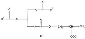
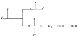
Figure 1.
Comparative scheme between a micelle (A) and a nano-liposome (B).
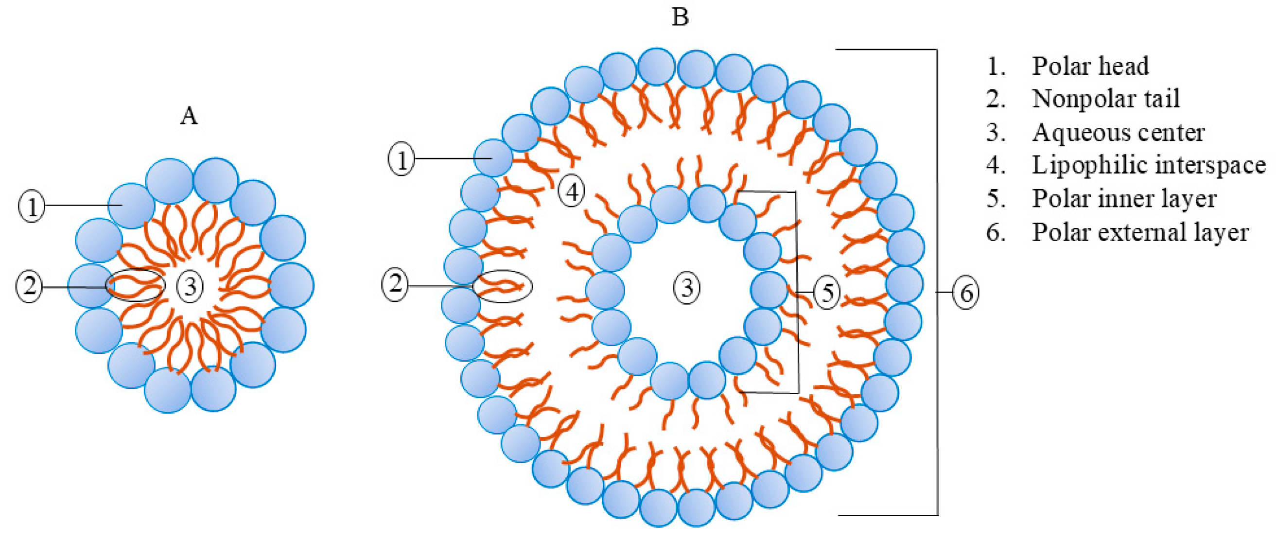
Figure 2.
Chemical structures of a hydrolysable tannin (A) and a non-hydrolysable or proanthocyanidin tannin (B).
Figure 2.
Chemical structures of a hydrolysable tannin (A) and a non-hydrolysable or proanthocyanidin tannin (B).
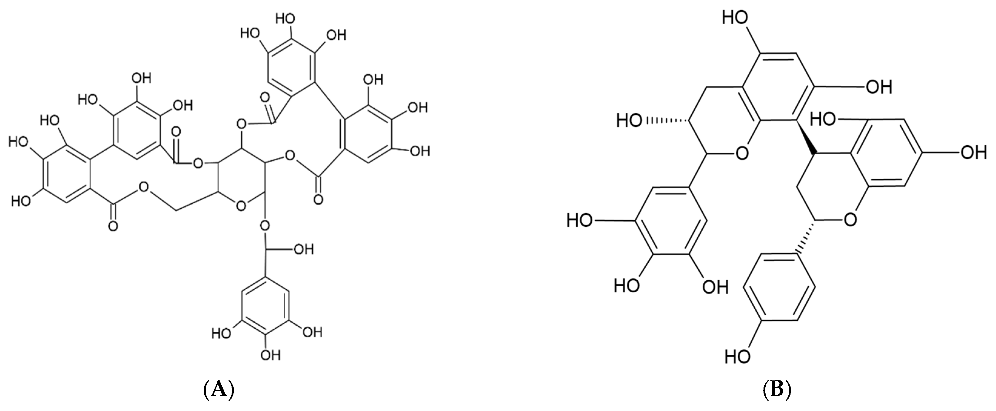

Table 1.
Most used phospholipids in liposome formation.
| Natural | Synthetic | Chemical Structure | Function | Reference |
|---|---|---|---|---|
| Phosphatidy- lcholine (PC) |
Dimyristoylphos- phatidylcholine (DMPC) |
 |
Increase membranes fluidity and eicosanoid production | [44] |
| Phosphatidy- lethanolamine (PE) |
Dioleoylphos- phatidylcholine (DOPC) |
 |
PC precursor, promotes membrane fusion, oxidative phosphorylation and mitochondrial biogenesis | [16] |
| Phosphatidy- lserine (PS) |
Distearoylphos- phatidylcholine (DSPC) |
 |
PE decarboxylation, autophagosomes formation, morphology regulation and dynamics and functions of mitochondria | [94] |
| Phosphatidyl-glycerol (PG) | Dipalmitolphos- phatidylglycerol (DPPG) |
 |
Important role in apoptosis and blood clotting, besides serving as a conduit for the transfer of lipids between organelles | [46] |
| Phosphatidylinositol (PI) | Distearoylphos- phatidylglycerol (DSPG) |
 |
Regulates traffic to and from Golgi apparatus and helps protect against hepatic viruses | [13] |
| Phosphatidic acid (PA) | 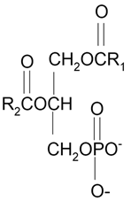 |
Serve as a fusogenic lipid, altering membrane structure and promoting membrane fusion, especially in neurons | [69] |
Table 2.
Chemical structures of the principal classes of flavonoids.
| Flavonoid | Structure |
|---|---|
| Flavones | 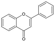 |
| Flavonols | 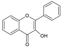 |
| Flavanones | 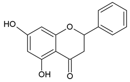 |
| Flavanols | 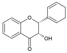 |
| Anthocyanidins | 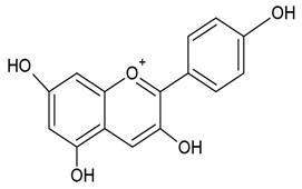 |
| Isoflavones | 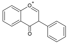 |
Table 3.
Main antioxidant biocompounds encapsulated in nano-liposomes and their chemical structure.
| Retinoids | |||
|---|---|---|---|
| Name | Chemical structure | Type of nano-liposome | Reference |
| Retinol | 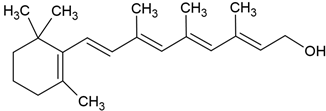 |
Lecithin-cholesterol structure, small unilamellar (20-200 nm), retinol contained in the lipid intermembrane section | [1,68] |
| Retinoic acid | 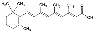 |
Lecithin-cholesterol structure, small unilamellar (20-200 nm), retinoic acid contained in the lipid intermembrane section | [1,68] |
| Retinyl ester | 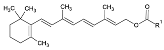 |
Lecithin-cholesterol structure, small unilamellar (20-200 nm), retinyl ester contained in the lipid intermembrane section | [1,68] |
| Carotenoids | |||
| Name | Chemical structure | Type of nano-liposome | Reference |
| α-carotene | 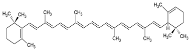 |
Lecithin, cholesterol and polysorbate 80 structure, giant unilamellar size (> 1 µm), α -carotene contained in the lipid intermembrane section | [1,51] |
| β-carotene | 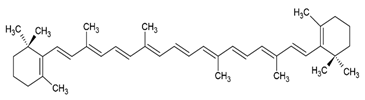 |
Lecithin, cholesterol and polysorbate 80 structure, giant unilamellar size (> 1 µm), β-carotene contained in the lipid intermembrane section | [1,51] |
| γ-carotene | 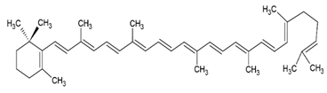 |
Soy, egg or marine lecithin and cholesterol structure, giant unilamellar size (> 1 µm), γ -carotene contained in the lipid intermembrane section | [1,75] |
| Astaxanthin | 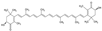 |
Agarose oligosaccharides, phosphatidylcholine, phosphatidyl galactose and/or phosphatidyl neoagarobiose structure, small unilamellar (20-200 nm), astaxanthin contained in the lipid intermembrane section | [28,96] |
| Lutein | 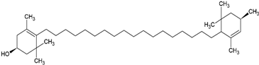 |
Supercritical carbon-dioxide method, small unilamellar size (20-200 nm), lutein contained in the aqueous center | [103] |
| Zeaxanthin | 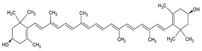 |
Lecithin, cholesterol and polysorbate 80 structure, giant unilamellar size (> 1 µm), zeaxanthin contained in the lipid intermembrane section | [28,50] |
| β-criptoxanthin | 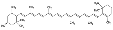 |
Cholesterol and phosphatidylcholine structure, small unilamellar (20-200 nm), β-cryptoxanthin contained in the aqueous center | [26] |
| Phenols | |||
| Name | Chemical structure | Type of nano-liposome | Reference |
| Gallic acid | 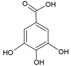 |
Soy lecithin and cholesterol structure, small unilamellar size (20-200 nm), gallic acid contained in the aqueous center | [101] |
| Protocatechuic acid | 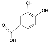 |
Egg yolk phosphatidylcholine and cholesterol structure, small unilamellar size (20-200 nm), protocatechuic acid contained in the aqueous center | [58,65] |
| Caffeic acid | 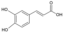 |
Egg yolk phosphatidylcholine and cholesterol structure, small unilamellar size (20-200 nm), protocatechuic acid contained in the aqueous center | [58,65] |
| p-cumaric acid | 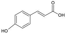 |
Soy lecithin and cholesterol structure, small unilamellar size (20-200 nm), p-coumaric acid contained in the aqueous center | [50] |
| Salicylic acid | 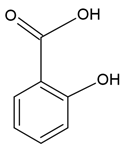 |
Soy lecithin and cholesterol structure, small unilamellar size (20-200 nm), p-coumaric acid contained in the lipid intermembrane section | [8] |
| Vitamins | |||
| Name | Chemical structure | Type of nano-liposome | Reference |
| Vitamin A |  |
Lecithin and cholesterol structure, small unilamellar size (20-200 nm), vitamin A contained in the lipid intermembrane section | [1,68] |
| Vitamin B2 | 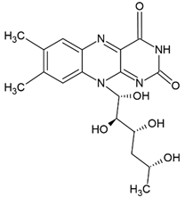 |
Vegetable oil (chia, sunflower and virgin olive) structure, giant unilamellar size (> 1µm), vitamin B2 contained in the lipid intermembrane section | [1,22] |
| Vitamin C | 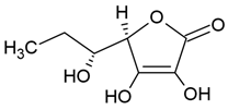 |
Phosphatidylcholine, stearic acid and stearic calcium structure, giant unilamellar (> 1µm), vitamin C contained in the aqueous center | [1,37] |
| Vitamin D3 | 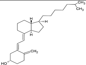 |
Soy phosphatidylcholine and cholesterol structure, giant unilamellar size (> 1µm), vitamin D3 contained in the lipid intermembrane section | [1,19] |
| Vitamin E | 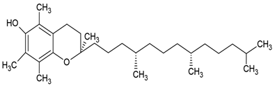 |
Phosphatidylcholine, stearic acid and stearic calcium structure, giant unilamellar (> 1µm), vitamin E contained in the aqueous center | [1,37] |
| Vitamin K |  |
Phosphatidylcholine and cholesterol structure, giant unilamellar size (> 1µm), vitamin K contained in the lipid intermembrane section | [1,40] |
Table 4.
Nanosystems for encapsulation of carotenoids. Obtained and edited from [73].
Table 4.
Nanosystems for encapsulation of carotenoids. Obtained and edited from [73].
| Nanosystem | Carotenoids | Particle Size (nm) | EE (%) | Zeta Potential (mV) | Storage Stability (Days) | References |
|---|---|---|---|---|---|---|
| Nanoemulsions | β-carotene | 218 | NA | 40 | 21 at 37 °C | [6] |
| 143.7 | −38.2 | 30 at 25 °C | ||||
| Microbial carotenoids | 142.1 | NA | 30 at 25 °C | [55] | ||
| Carotenoids | 290 to 350 | −53.4 to −58.8 | 21 at 25 °C | [31] | ||
| β-carotene | 198.4 to 315.6 | −29.9 to −38.5 | 90 at 4, 25, and 37 °C | [12] | ||
| Carotenoids | <200 | −30 to −45 | 35 at 25 °C | [87] | ||
| Lycopene | 145.1 to 161.9 | −19.7 to −20.7 | 1 at 25 °C | [48] | ||
| 200.1 to 287.1 | 61 to 89.1 | 20 to 45 | 42 at 4, 25, and 37 °C | [102] | ||
| Polymeric/biopolymeric NPs | Carotenoids | 153 | 83.7 | NA | NA | [67] |
| 84.4 | >96 | −41.3 to −43.6 | 60 at 41 °C | [9] | ||
| β-carotene | 77.8 to 371.8 | 98.7 to 99.1 | −37.8 to −29.9 | NA | [98] | |
| β-carotene | 70.4 | 97.4 | NA | NA | [76] | |
| Lycopene | 152 | 89 | 58.3 | NA | [49] | |
| ~ 200 | >95 | −36 | 210 at 5 °C | [95] | ||
| 193 | NA | −11.5 | 14 at 25 °C | [25] | ||
| Lutein | <250 | 74.5 | −27.2 | NA | [11] | |
| Lutein | 240 to 340 | ~91.9 | NA | NA | [36] | |
| Crocetin | 288 to 584 | 59.6 to 97.2 | NA | NA | [35] | |
| Fucoxanthin | 200 to 500 | 47 to 90 | 30 to 50 | 6 at 37 °C | [72] | |
| Nanoliposomes/liposomes | Carotenoids | 70 to100 | 75 | −5.3 | NA | [90] |
| β-carotene | 162.8 to 365.8 | ~98 | 64.5 to 42.6 | 70 at 4 °C | [38] | |
| Astaxanthin | 80.6 | 97.6 | 31.8 | 15 at 4 and 25 °C | [63] | |
| 60 to 80 | 97.4 | NA | NA | [64] | ||
| Lutein | 264.8 to 367.1 | 91.8 to 92.9 | −34.3 to −27.9 | NA | [41] | |
| SLNPs and NLCs | β-carotene SLNPs | 200 to 400 | 53.4 to 68.3 | −6.1 to −9.3 | 90 at 5, 25, and 40 °C | [39] |
| <220 | NA | 20 to 30 | 10 at 25 °C | [56] | ||
| 120 | NA | −30 | 56 at 25 °C | |||
| Lycopene SLNPs | 125 to 166 | 86.6 to 98.4 | NA | 60 at 4 °C | [57] | |
| Lycopene NLCs | 157 to 166 | > 99 | −74.2 to −74.6 | 120 at 4, 30, and 40 °C | [60] | |
| 121.9 | 84.50 | −29 | 90 at 25 °C | [85] | ||
| Supercritical fluid-based NPs | Astaxanthin | 150 to 175 | NA | NA | NA | [43] |
| 266 | 84 | NA | NA | [93] | ||
| Metal/metal oxide-based NPs and hybrid nanocomposites | Carotenoids | 20 to 140 | NA | NA | NA | [7] |
| Lycopene | 3 to 5 | −48.5 | 90 at 4 and 25 °C | [20] | ||
| 20.8 | −25.3 | NA | [82] |
1 EE = encapsulation efficiency, NPs = nanoparticles, SLNPs = solid lipid nanoparticles, NA = data not available and NLCs = nanostructured lipid carriers.
Table 5.
The advantages and disadvantages of nanosystems for encapsulation of carotenoids. Obtained and edited from [73].
Table 5.
The advantages and disadvantages of nanosystems for encapsulation of carotenoids. Obtained and edited from [73].
| Nanosystem | Advantages | Disadvantages | References |
|---|---|---|---|
| Nanoemulsions |
|
|
[24,88] |
| Polymeric/biopolymeric NPs |
|
|
[24,28] |
| Nanoliposomes/liposomes |
|
|
[24,73] |
| SLNPs |
|
|
[24,73,88] |
| NLCs |
|
|
[24,88] |
| Supercritical fluid-based NPs |
|
|
[18,73,88] |
| Metal/metal oxide-based NPs and hybrid nanocomposites |
|
|
[80,81] |
1 EE = encapsulation efficiency, NPs = nanoparticles, SLNPs = solid lipid nanoparticles and NLCs = nanostructured lipid carriers.
Disclaimer/Publisher’s Note: The statements, opinions and data contained in all publications are solely those of the individual author(s) and contributor(s) and not of MDPI and/or the editor(s). MDPI and/or the editor(s) disclaim responsibility for any injury to people or property resulting from any ideas, methods, instructions or products referred to in the content. |
© 2025 by the authors. Licensee MDPI, Basel, Switzerland. This article is an open access article distributed under the terms and conditions of the Creative Commons Attribution (CC BY) license (http://creativecommons.org/licenses/by/4.0/).
Copyright: This open access article is published under a Creative Commons CC BY 4.0 license, which permit the free download, distribution, and reuse, provided that the author and preprint are cited in any reuse.


