
Submitted:
21 April 2025
Posted:
27 April 2025
You are already at the latest version
Abstract
Background: Ganoderma lucidum (G. lucidum) polysaccharides (GLPs) are believed to be one of the major bioactive components to promote health benefits. The uridine diphosphate glucose pyrophosphorylase (UGPase) genes are key to regulate the synthesis of GLPs. Therefore, this study sought to characterize the UGPase genes and evaluated the GLPs yield from different sources. Methods: The GLPs were extracted by water soaking alcohol precipitation method and contents were calculated by phenol sulfuric acid method. The whole UGPase gene and an internal reference gene were amplified separately, transferred into plasmid vector, cloned, sequencing and bioinformatics analysis. qPCR was performed to determine the mRNA expression of UGPase gene from different G. lucidum sources. Results: The contents of spore powder and fruiting bodies showed that there was a difference among these different products (P < 0.05). The physicochemical properties of UGPase protein showed that the all G. lucidum from spore powder and fruiting body were similar. Moreover, the relative expression of UGPase mRNA in different sources as well as in the same source demonstrated a significant difference (P < 0.05). The relative expression of GLP and UGPase mRNA in spore powder of the same ground source was higher than those in fruiting bodies (P < 0.05). Conclusion: The quantitative detection of GLP contents and expression of UGPase mRNA levels proposed in the study can be used for the evaluation of the G. lucidum quality.
Keywords:
Ganoderma lucidum
; polysaccharides
; Quantitative real-time PCR
; UGPase
; food adulteration
1. Introduction
Ganoderma lucidum (G. lucidum) is a traditional healthy edible and medicinal fungus known as “Lingzhi” in China. The two species of G. lucidum and G. sinensis recorded in the Chinese Pharmacopoeia, both of which contain a variety of bioactive substances, have high medicinal and nutritional value [1]. The fruiting body and spore powder of G. lucidum are common on the market in China [2]. G. lucidum polysaccharide (GLP) is the secondary metabolite of G. lucidum and the main active substance, which is present in the mycelia and fruiting bodies of G. lucidum and has the effects of lowering blood lipid, blood glucose, and has anti-tumor effects [3]. G. lucidum is mainly cultivated due to the great difference of species and geographical environment in China. The nutritional components including GLP and other active substances are heterogeneous [4].
Uridine diphosphate glucose pyrophosphorylase (UGPase) was first found in yeast cells and has been found in many plant tissues and cells [5]. Most of them are stored in the cytosol, which is the main form of plant activated sugar. The catalytic reaction Glc-1-P+UTP→ UDPG+PPi provides glucose base for the synthesis of cellulose, sucrose and pectin [6,7]. At present, most of the processed G. lucidum products sold in the market are processed pieces, tablets, spore powder or capsules, with a few oral liquids or other healthy products available. Since it is difficult to identify the authenticity of G. lucidum after it is cut into pieces or powdered, it often comes to pass that the tongue of a tree or other fungus is used as the processed G. lucidum [8,9]. The growth environment and processing process of G. lucidum from different places are different, which also leads to the great difference in the quality of G. lucidum products. Therefore, the research on the synthesis pathway of its effective active substances, the functional identification of key enzymes, their metabolic mechanism, and high expression thereof is a key research topic in recent years [10].
To date, UGPase has been recorded from a variety of sources, including eukaryotic yeast, plant systems, and humans [6,7,11]. However, little is known about the functional role of UGPase in G. lucidum. It was speculated that UGPase might be a key sugar-metabolizing enzyme, which may be closely related to the synthesis of GLPs. The purpose of this study was to characterize UGPase gene, the expressions of mRNA, and GLP contents among spore powder and fruiting bodies from different land sources in China.
2. Results
2.1. Determination of the GLP Content in G. lucidum Spore Powder and Fruiting Body Samples
With the mass concentration of glucose as the abscissa and the absorbance as the ordinate, the standard curve was plotted by using the ultraviolet spectrophotometer method (Figure 1): the resulting regression equation is Y = 0.11X + 0.11, R2 = 0.9837.
The standard curve was plotted by using the ultraviolet spectrophotometer method with the mass concentration of glucose as the abscissa and the absorbance as the ordinate. The resulting regression equation is Y = 0.11X + 0.11, R2 = 0.9837.
Determination of the GLP content in G. lucidum spore powder and fruiting body samples entailed sucking 1 mL of G. Lucidum spore powder and fruiting body test solution, measuring its absorbance, and calculating the content of polysaccharide in the solution to be tested according to the standard curve. According to the calculation, the average content of polysaccharide in Yunnan spore powder was 2.887% ± 0.0059%, that of Jilin spore powder was 3.361% ± 0.0046%, and that of Tongrentang spore powder (TRT-sp) was 2.399% ± 0.0053% (Table 1). There was a statistically difference among these products (P < 0.05); the average content of polysaccharide in Hubei fruiting body was 2.446% ± 0.0033%, that in Jilin was 1.809% ± 0.0041%, that in Yunnan was 0.976% ± 0.0036%, and that in Guizhou was 1.325% ± 0.0422%. The difference was statistically significant among these products (P < 0.05) (Table 2).
2.2. Cloning of UGPase PCR Products
TIANSeq HiFi Amplification Mix high-fidelity polymerase was used to clone pUGPase-1956bp and pUGPase-216bp, and the results are shown in Figure 2 and Figure 3a. All the target genes showed clear and bright bands corresponding to the size of the target band, indicating that the cloning of the UGPase gene was successful.
2.3. Sequencing and Bioinformatics Analysis
The cloned plasmids pUGPase-1956bp and pUGPase-216bp were sequenced. The sequencing results were compared with the bioinformatics software BLAST tool and BioEdit. The results showed that the nucleotide sequence of each cloned fragment was 100% the same as the registered sequence (Sequence: KM260167.1) in GenBank, as illustrated in Figure 3b. It is proved that the cloned DNA fragment was the standard fragment of the target gene. Cloned fragments of pUGPase-216bp were stored at -20 °C as a control for subsequent experiments.
We analyzed the physicochemical properties of UGPase protein using ExPASy Protparam online software. The UGPase gene of G. lucidum from spore powder and fruiting body encodes 504 amino acids, with a protein isoelectric point of 6.8, a molecular weight of 56830.1, an instability coefficient of 30.63, an average hydrophobicity of -0.320, and a fat coefficient of 93.41. Analysis of the amino acid composition encoded by the UGPase gene revealed that the UGPase protein consists of 20 types of amino acids, with leucine (Leu) having the highest content at 10.7%, followed by lysine (Lys) at 7.1%, and cysteine (Cys) and tryptophan (Trp) having the lowest content at 0.6%. The results showed that the physico-chemical properties of G. lucidum from spore powder and fruiting body were similar.
2.4. The PCR Amplification Efficiency and Standard Curve
According to the slope of the standard curve, the amplification efficiencies of the target gene UGPase and the internal reference gene 16SrRNA were 100.207% and 100.109%, respectively, close to 100%, consistent with the relative quantitation evinced by 2-ΔΔ calculation requirements of the Ct method (Figure 4a). The reaction index was significantly amplified, and the gradient samples were evenly distributed, indicating that the sample had good repeatability, high amplification efficiency, and accurate detection results. The standard curve of fluorescent quantitative PCR is shown in Figure 4b.
2.5. Comparison of Relative Quantitative Contents of UGase mRNA
Each sample was set with three replicates, and the G. lucidum spore powder and fruiting body cDNA were used as templates for fluorescent quantitative PCR reaction to obtain the fusion curve and amplification curve of the target gene UGPase and internal reference 16S rRNA. In the spore powder group, TRT-sp was selected as the control, and other spore powders were selected as the experimental group to calculate the relative expression of UGPase gene in other spore powders. In the fruiting body group, Yunnan fruiting body (YNfb) was selected as the control group, and the relative expression of UGPase gene in other geographical fruiting bodies was calculated. In addition, in the same provenance, the relative expression of UGPase gene in spore powder was calculated with the fruiting body as the control. This experiment selected 2-ΔΔCt method using TRT-sp as the control group to analyze and calculate the relative expression formula of the target gene as follows: 2-ΔΔCt = 2 - [(Ct purpose - Ct internal parameter) Test - (Ct purpose - Ct internal parameter) Control]. The relative expression of UGPase gene in spore powder and fruiting bodies from different places, as well as in spore powder and fruiting body from the same place, was statistically significant (P < 0.05) (Figure 5a). In addition, it can be found that the relative expression amount of UGPase target gene in the sporopollen group was Jilin > Yunnan > Tongrentang, and the fruiting body was Hubei > Jilin > Guizhou > Yunnan. The relative expression of target gene from the same source was higher in spore powder than in fruiting body, with statistical significance (P < 0.05). The relative quantitative results of UGPase gene in spore powder and fruiting bodies of G. Lucidum from different sources are shown (Figure 5b).
3. Discussion
GLP is considered as the main bioactive component of G. lucidum, and the content of GLP recorded in the Chinese Pharmacopoeia is the key factor used to evaluate the quality of G. Lucidum [12,13]. Due to different climates and geographical environments, different processing techniques used on G. lucidum, and immature separation and purification technology of G. lucidum bioactive substances, there are differences among varieties of G. lucidum, and the content of GLP varies greatly. Therefore, the research on the synthesis pathway of its effective active substances, the functional identification of key enzymes, the metabolic mechanism and the high expression of its active substances has become a key research topic in recent years.
In this experiment, the content of GLP was quantitatively determined by UV visible spectrophotometry. Compared with HPLC, it was fast, simple, and cost-effective. The principle whereby the absorbance of yellow orange compound formed by phenol sulfuric acid and sugar could be detected at 490 nm was used to detect GLP. Finally, the GLP content of different sources was calculated according to the standard curve of glucose. This study collected samples from the main producing areas of G. Lucidum in China, including Changbai Mountain in Jilin Province, Shennongjia Mountain in Hubei Province, Fanjing Mountain in Guizhou Province, and Wenshan Mountain in Yunnan Province. Our laboratory personnel collected G. lucidum spore powder and fruiting body samples in the production area, and 50 samples were identified by experts from the local drug inspection department as meeting the experimental requirements, all of which were artificially cultivated products. The results showed that the GLP content of spore powder from Jilin was the highest (3.361%), and that of fruiting body of G. lucidum from Hubei was the highest (2.446%). The GLP content of spore powder from the same source was two to three times that of the fruiting body. The temperature difference between day and night in summer, severe cold and long winter in Jilin region, unique forest resources, sufficient groundwater, and fertile organic black soil all provided an excellent natural environment for breeding high-quality G. lucidum. The contents of GLP in the fruiting body and spore powder were relatively high in this region, which were consistent with other reports [12,13,14].
G. lucidum spores are the seeds of G. lucidum, which are extremely small oval germ cells ejected from the fungal folds of G. lucidum during its growth and maturity. G. lucidum spore powder has all of the genetic material and health-care functions of G. lucidum. The medicinal value of G. lucidum spores has been paid more attention in recent work: studies have found that G. lucidum spores enhance bodily immunity, inhibiting tumorigenesis, protecting liver injury, and conferring a certain radiation protection [15,16]. At present, the price of G. lucidum spore powder on the market is much higher than that of the fruiting body. This experiment confirmed that the GLP content of spore powder is higher than that of the fruiting body. With the consumers’ in-depth understanding of the medicinal value of G. lucidum spore powder, the G. lucidum spore powder on the market mainly includes ordinary G. lucidum spore powder and G. lucidum spore powder (broken wall); however, the market value of G. lucidum spore powder (broken wall) is significantly higher than that of G. lucidum fruiting body and non-wall-broken G. lucidum spore powder, which has become the mainstream product on the market. The 2020 Chinese G. lucidum Industry Market Research Report shows that there were many domestic production enterprises and varieties of G. lucidum products. According to the information disclosed on the website of the State Administration of Market Supervision and Administration, there were 187 G. lucidum and 1157 G. lucidum spore powder products with drug approval or health food approval, respectively at the end of December 2020. Only G. lucidum spore powder has an annual output value of nearly 12 billion yuan. Driven by interests, black-market dens illegally produce broken wall G. lucidum spore powder products, mainly because non-wall-broken G. lucidum spore powder gets passed off as wall broken G. lucidum spore powder, or the fruiting body powder of G. lucidum was mixed with G. lucidum spore powder [17,18]. Since the implementation of the Technical Specifications for Collection and Processing of G. lucidum Spores in 2012 (National Standard GB/T 29344-2012) in China, the quantity of G. lucidum spores was calculated by microscopic observation as the quality standard. With the continuous upgrading of spore powder collection and deep processing technology, and increasing popular requirements for the quality of spore powder, GLP will be included in the new national standard for spore powder (and supported by the present results).
In recent years, with the development of molecular biology technology, molecular marker technology has emerged to meet changing needs, and has been widely used in the research into medicinal plants, animal genetic diversity, systematics, and taxonomy [19,20]. The application of molecular marker technology in the identification of TCM has allowed some progress, and has manifested its characteristics and advantages in practice, especially in the identification of crude drugs [21,22,23]. DNA molecular markers are present in huge number in various regions of genomic DNA. By comparing the polymorphisms of molecular markers randomly distributed throughout the genome, the diversity of research objects has been comprehensively evaluated and their genetic characteristics revealed [24,25].
In this study, molecular cloning technology was used to analyze the UGPase gene in G. lucidum by fluorescent quantitative PCR and assess the correlation between the overall changes in the relative expression of genes and GLP contents. The relative expression of UGPase gene differed among different sources, similarly, with the same provenance, the relative expression of the UGPase gene in the sporopollen group was several times that in the fruiting body group, suggesting that the UGPase gene may be a positive regulator of GLP [26,27,28].
There are some limitations to the present study: there was a significant difference in the mRNA expression of UGPase gene between different G. lucidum strains owing to this experiment only involving the expression of UGPase gene at the transcription level. The synthesis of GLP is a process involving the regulation of multiple gene expression, which is inseparable from the catalysis of UGPase. The reasons for the difference and the specific mechanism of action warrant further exploration.
4. Materials and Methods
4.1. Collection of Samples
The fruiting bodies of G. lucidum (batch number: 120968-201809), sporoderm-broken spores of G. lucidum (batch number: 121701-201401) and D-anhydrous glucose (batch number: 110833-201908) are the standard products provided by China Institute for Food and Drug Control. A total of seven batches of 65 samples of G. lucidum sporoderm-broken spores and fruiting body from different sources were screened, which were collected in the same season during the January 2022 and October 2024. Yinglan Jin, the chief pharmacist of Jilin Institute for Drug Control, identified the authenticity thereof. Among them, G. lucidum sporoderm-broken spores were purchased from Yunnan Province, Beijing Tongrentang (capsule) had 18 copies in total, and the mass of each copy ranged between 100 g and 250 g. The fruiting bodies of G. lucidum were purchased from Hubei Province, Jilin Province, Yunnan Province, and Guizhou Province, with intact surfaces and masses ranging from 300 g to 500 g. The origin (batch) and sample number are listed in Table 3.
4.2. Determination of GLP Contents in Spore Powder and Fruiting Bodies of G. lucidum
The entire process required three steps. Firstly, we prepared for standard reference and test solution. In a brief, 0.5 g of anhydrous glucose reference substance were weighed and placed it in a 500-mL volumetric flask, added water to dissolve it, diluted it to the scale line, and thence prepared anhydrous glucose with a mass concentration of 1 mg/mL as the storage solution of the reference substance. In the meanwhile, we accurately weighed 2 g of G. lucidum spore powder and 2 g of fruiting body powder of the test sample, put them into a triangular flask, added 50 mL of water, stood the specimens for 30 min, placed them in a boiling-water bath for 2 h, filtered them under hot vacuum, washed the filter residue and filtered it with double-distilled water three times, and transferred the washing solution obtained to a round-bottomed flask. The specimens were rotated in a rotary evaporator for about 20 min, and the filtrate was concentrated to about 20 mL. Taking 20 mL of concentrated solution into a centrifuge tube, we added 30 mL of absolute ethanol, and allowed each specimen to stand overnight before centrifuging at 10,000 rpm for 10 min, discarded the supernatant, dried the specimens at room temperature, dissolved the precipitate in hot double-distilled water, washed and centrifuged the specimens three times, cooled them before placing them in a 500-mL volumetric flask to obtain the test solution. This process was repeated three times for each sample. Secondly, the glucose standard curve was drawn. We measured 0.1, 0.2, 0.3, 0.4, 0.5, and 0.6 mL of standard reference solution into test tubes with stoppers, added double-distilled water to 1.0 mL, added 1.0 mL of 5% phenol solution, shook it well, then slowly injected 5 mL of concentrated sulfuric acid solution along the tube wall, shook it for 6 minutes, occasionally opening the lid to release the gas thus generated, placed it at room temperature for 5 minutes, then heated it in a boiling-water bath for 10 minutes, removed it and cooled it to room temperature. A 1-cm cuvette was used to measure the absorbance at a wavelength of 490 nm and phenol and sulfuric acid were used as solvents for blank controls. A standard curve with the mass concentration of anhydrous glucose (mg/mL) as the abscissa and the absorbance (A) as the ordinate was plotted. Thirdly, we sucked 1 mL of the test sample solution into a tube with a stopper, measured its absorbance, calculated the corresponding GLP concentration from the standard curve regression equation, and further deduced the polysaccharide content of the test sample. The formula is as follows: 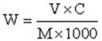 × 100%, where V = 500 mL, C denotes the concentration calculated according to the standard curve, and M represents the mass of the G. lucidum sample; W is the ratio of the GLP content of determined sample to standard contents (%).
× 100%, where V = 500 mL, C denotes the concentration calculated according to the standard curve, and M represents the mass of the G. lucidum sample; W is the ratio of the GLP content of determined sample to standard contents (%).4.3. Bioinformatics Analysis of G. lucidum Genome and Designing a Series of Primers
We conducted a bioinformatics analysis of G. lucidum genome, which were identified and encoded 16,113 genes reported in the model medicinal G. lucidum originated from China (Chen et al, 2013). Besides, the sequence genes of G. lucidum UGPase (Sequence: KM260167.1) and an internal reference gene 16SrRNA mRNA (AF493073.1) were obtained from the NCBI database. Then ProtParam software in ExPASy was applied to predict the relative molecular weight, amino acid contents, and physico-chemical properties of the proteins encoded by UGPase gene. Primer Premier 5.0 software was used to design three pairs of primers.
Primer 1 (upstream: 5’-GGTATGACCGGTGCGAAATC-3’ and downstream: GTAGCGCGACTGATTGAAAGG -3’) was used to amplify the whole UGPase gene.
Primer 2 (upstream: 5’- AGTCGGTTCTTGCCCGTCAA-3’and downstream: 5’- GCTGCGGGTTGATAACGAGC -3’) was applied to amplify the internal reference genes for quantitative analysis of the copy number of the target gene.
Primer 3 (upstream: 5’- GAGAAACGAAGGTTAGGGTAGG-3’and downstream: 5’- CACAAGGCGGAATGGTTATTG-3’) was used to amplify the 16SrRNA gene. The primers were synthesized by Sangon Biotech Co., Ltd (Shanghai, China).
4.4. Total RNA Extraction and cDNA Synthesis
Total RNA extraction and cDNA synthesis were conducted using TaKaRa MinBEST Plant RNA Extraction Kit and PrimeScript™ RT reagent Kit with gDNA Eraser (TaKaRa, Japan) kits according to the instruction method, with three replicates for each sample. RNA integrity was determined by electrophoresis in 1.0% agarose. Total RNA concentration and purity were measured using a nucleic acid analyzer (Q6000, Quawell, USA) at 260 nm and 280 nm. The prepared cDNA was stored at -20 ℃ in a refrigerator/freezer.
4.5. PCR Amplification,Cloning of UGPase Gene of G. Lucidum and Blasting
The PCR reaction system included 2 × Taq PCR MasterMix 10 μL, primer 1.0 μL, cDNA 1.0 μL, and free ion H2O supplemented to 20 μL. PCR reaction conditions include: pre denaturation at 95 ℃ for 3 min; denaturation at 95 ℃ for 30s, annealing at 60 ℃ for 30 s, extension at 72 ℃ for 1 min, 40 cycles; and extension at 72 ℃ for 5 min. For 2% agarose gel electrophoresis, the target band in the agarose gel was cut under ultraviolet light, selected a pGM-T connection kit, T-A connection, transformation, blue and white spot screening and identification, and recombinant plasmid extraction reference [29,30]. Nucleotide sequencing was performed directly on the identified cloning plasmid using an ABI Prism 377 DNA sequencer. Sequence similarity search were compared with BioEdit software, and the NCBI website (www.ncbi.nlm.nih.gov) was accessed for Blast analysis.
4.6. Establishment of Fluorescence Quantitative PCR Reaction System and Conditions
SYBRGreen Ⅱ chimeric fluorescence method was used, and the reverse-transcribed cDNA sample was used as the template of real-time fluorescent quantitative PCR, β- Actin and 16S rRNA were internal reference genes, and the expression of UGPase gene was detected by fluorescence quantitative PCR. The reaction was performed on the PikoReal real-time quantitative PCR instrument (Thermo Fisher, USA), and three parallel experiments were conducted for each sample. The reaction procedure adopted for fluorescent quantitative PCR was as follows: pre denaturation at 94 ℃ for 10 min; denaturation at 95 ℃ for 15 s, annealing at 60 ℃ for 30 s, extension at 72 ℃ for 30 s, 40 cycles, whereupon the melting curve was plotted.
4.7. Preparation of the Fluorescence Quantitative Standard Curve and Counting of Relative Quantitative Contents of G. lucidum
The initial concentration was set to 1 ng/μL of G. lucidum standard cDNA as template, and diluted gradient was set to 10-5 ng/μL 0.4 for each dilution of 10 μL. The detection was conducted according to the fluorescent quantitative PCR reaction system. We used the Ct value as the ordinate and the logarithmic concentration of the template as the abscissa to plot the quantitative standard curve. The linear relationship was used to ascertain whether the amplification efficiency of the duplicate sample data and the initial cDNA template with different copy numbers differed. The R2 value indicates the linear degree of attenuation of the experimental data.
4.8. Statistics Analysis
Three parallel groups were set for each sample, and the Ct values of internal reference gene and target gene were calculated using MS-Excel® software. The relative mRNA level of each target gene was calculated by the Ct method. The data are expressed as mean ± standard deviation. GraphPad Prism 6 software was used to draw a column chart. The ANOVA program in SPSS.19 software was used to analyze the relative expression of target gene mRNA. The least significant difference (LSD) method was used to test the difference between groups. The difference was statistically significant (P < 0.05).
5. Conclusions
The relative expression of GLP and UGPase mRNA in different spore powder and fruiting bodies of G. lucidum differed. The relative expression of GLP and UGPase mRNA in spore powder of the same ground source was higher than those in fruiting bodies. These results suggest that the UGP gene may play an important role in biosynthesis of GLP. UGPase genes show obvious expression advantages in high-GLP producing strains and are positively correlated with the biosynthesis of GLP. This mechanism provides a certain theoretical basis for the in-depth study of the biosynthesis of GLP.
Author Contributions
Conceptualization, M.C.; methodology, X.Y and J.L; software, K.C.; formal analysis, X.Y.; resources, M.C.; writing—original draft preparation, X.Y.; writing—review and editing, X.Y and J.L.; supervision, M.C.; project administration, M.C. and X.Y.; funding acquisition, M.C. All authors have read and agreed to the published version of the manuscript.
Funding
This study received financial support from the Science and Technology DevelopmentProgram of Jilin, China (20200304101YY and 20200301014NY).
Institutional Review Board Statement
Not applicable.
Informed Consent Statement
Not applicable.
Data Availability Statement
The data presented in this study are available on request from the corresponding author. The data are not publicly available due to privacy.
Conflicts of Interest
The authors declare no conflict of interest.
References
- Chinese Pharmacopoeia Commission. Pharmacopoeia of the People’s Republic of China. China Medical Science Press. 2020 Vol, Beijing, p. 1.
- Seweryn, Z., Zia, A., Gamian, A. Health-Promoting of Polysaccharides Extracted from Ganoderma lucidum. Nutrients. 2021, 13 (8):2725.
- Wu, P., Zhang, C., Yin, Y., Zhang, X., Li, Q., Yuan, L., Sun, Y., Zhou, S., Ying, S., Wu, J. Bioactivities and industrial standardization status of Ganoderma lucidum: A comprehensive review. Heliyon. 2024, 10(19):e36987. [CrossRef]
- Zeng, P., Guo, Z., Zeng, X., Hao, C., Zhang, Y., Zhang, M. Molecular mechanisms of bioactive polysaccharides from Ganoderma lucidum (Lingzhi), a review. Int J Biol Macromol. 2020, 150: 765-774.
- Janse van Rensburg, H.C., Van den Ende, W. UDP-Glucose: A Potential Signaling Molecule in Plants?. Front Plant Sci. 2018, 9 (8):2230.
- Chivasa, S., Tomé, D., Slabas, A. UDP-glucose pyrophosphorylase is a novel plant cell death regulator. J Proteome Res. 2013, 12:1743-1753.
- Chambers, J.K., Macdonald, L.E., Sarau, H.M., Ames, R.S., Freeman, K., Foley, J.J.A G protein-coupled receptor for UDP-glucose. J Biol Chem. 2020, 275: 10767-10771.
- Decker, D., Aubert, J., Wilczynska, M., Kleczkowski, L.A. Exploring Redox Modulation of Plant UDP-Glucose Pyrophosphorylase. Int J Mol Sci. 2023,17;24(10):8914. [CrossRef]
- Zhang, J., Shi, X., Cheng, W. Comparison of the Anti-Inflammatory and Antioxidant Activities of Mycelial Polysaccharides from Different Strains of Lingzhi or Reishi Medicinal Mushroom, Ganoderma lucidum (Agaricomycetes). Int J Med Mushrooms.2020, 24(7):77-90.
- Yu, H. Z., Liu, Y. F. Zhou, S. Comparison of the polysaccharides from fruiting bodies, mycelia and spore powder of Ganoderma lingzhi. Mycosystema. 2016, 35 (2): 170-177(in Chinese).
- Guo, C., Guo, D., Fang, L., Sang, T., Wu, J., Guo, C. Ganoderma lucidum polysaccharide modulates gut microbiota and immune cell function to inhibit inflammation and tumorigenesis in colon. Carbohydr Polym. 2021, 67: 118231.
- Sanodiya, B. S., Thakur, G. S., Baghel, R. K. Ganoderma lucidum: a potent pharmacological macrofungus. Curr. Pharm. Biotechnol. 2009, 10: 717-742.
- Fernandes, A., Nair, A., Kulkarni, N., Todewale, N., Jobby, R. Exploring Mushroom Polysaccharides for the Development of Novel Prebiotics: A Review. Int J Med Mushrooms. 2023, 25(2):1-10. [CrossRef]
- Cadar, E., Negreanu-Pirjol, T., Pascale, C., Sirbu, R., Prasacu, I., Negreanu-Pirjol, B.S., Tomescu, C.L., Ionescu, A.M. Natural Bio-Compounds from Ganoderma lucidum and Their Beneficial Biological Actions for Anticancer Application: A Review. Antioxidants (Basel). 2023, 25;12(11):1907. [CrossRef]
- Li, W., Zou, G., Bao, D., Wu, Y. Current Advances in the Functional Genes of Edible and Medicinal Fungi: Research Techniques, Functional Analysis, and Prospects. J Fungi (Basel). 2024, 25;10(5):311. [CrossRef]
- Ahmad, M.F., Ahmad, F.A., Khan, M.I., Alsayegh, A.A., Wahab, S., Alam, M.I., Ahmed, F. Ganoderma lucidum: A potential source to surmount viral infections through β-glucans immunomodulatory and triterpenoids antiviral properties. Int J Biol Macromol. 2021, 30;187:769-779. [CrossRef]
- Zhao, L.Y., Dong, Y. H., Chen, G.T. Extraction, purification, characterization and antitumor activity of polysaccharides from Ganoderma lucidum. Carbohydrate Polymers. 2010, 80(3): 783-789.
- Loyd, A.L., Richter, B.S., Jusino, M.A., Truong, C., Smith, M.E., Blanchette, R.A., Smith, J.A. Identifying the “Mushroom of Immortality”: Assessing the Ganoderma Species Composition in Commercial Reishi Products. Front Microbiol. 2018,16;9:1557. [CrossRef]
- Binns, C.W., Lee, M.K., Lee, A.H. Problems and prospects: public health regulation of dietary supplements. Annu. Rev. Public Health. 2017, 39: 403-420.
- Wang, Q., Xu, M., Zhao, L., Wang, F., Li, Y., Shi, G., Ding, Z. Transcriptome dynamics and metabolite analysis revealed the candidate genes and regulatory mechanism of ganoderic acid biosynthesis during liquid superficial-static culture of Ganoderma lucidum. Microb Biotechnol. 2021, 14(2):600-613. [CrossRef]
- Cortina-Escribano, M., Veteli, P., Wingfield, M.J., Wingfield, B.D., Coetzee, M.P.A., Vanhanen, H., Linnakoski, R. Phylogenetic analysis and morphological characteristics of laccate Ganoderma specimens in Finland. Mycologia. 2024 ,116(6):1046-1062. [CrossRef]
- Fryssouli, V., Zervakis, G.I., Polemis, E., Typas, M.A. A global meta-analysis of ITS rDNA sequences from material belonging to the genus Ganoderma (Basidiomycota, Polyporales) including new data from selected taxa. MycoKeys. 2020, 75:71-143. [CrossRef]
- Pavlik, M., Zhou, S., Zhang, J., Tang, Q., Feng, N., Kurjak, D., Pavlík, M. Jr., Kunca, A. Comparative Analysis of Triterpene Composition between Ganoderma lingzhi from China and G. lucidum from Slovakia under Different Growing Conditions. Int J Med Mushrooms. 2020, 22(8):793-802. [CrossRef]
- Cai, M., Tan, Z., Wu, X., Liang, X., Liu, Y., Xie, Y., Li, X., Xiao, C., Gao, X., Chen, S., Hu, H., Wu, Q. Comparative transcriptome analysis of genes and metabolic pathways involved in sporulation in Ganoderma lingzhi. G3 (Bethesda). 2022, 12(3):jkab448. [CrossRef]
- Liu, D., Sun, X., Diao, W., Qi, X., Bai, Y., Yu, X., Li, L., Fang, H., Chen, Z., Liu, Q., Liang, C. Comparative transcriptome analysis revealed candidate genes involved in fruiting body development and sporulation in Ganoderma lucidum. Arch Microbiol. 2022, 204(8):514. [CrossRef]
- Jia, T., Ge, Q., Zhang, S., Zhang, Z., Liu, A., Fan, S. UDP-Glucose Dehydrogenases: identification, expression, and function analyses in upland cotton (Gossypium Hirsutum). Front Genet. 2020, 11:597890.
- Khadbaatar, S., Bao, H., Gao, X., Huo, H. Study on Differences of Metabolites among Different Ganoderma Species with Comprehensive Metabolomics. J Fungi (Basel). 2024 Jul 27;10(8):524. [CrossRef]
- Brandt, W., Schulze, E., Liberman-Aloni, R., Bartelt, R., Pienkny, S., Carmeli-Weissberg, M., Frydman, A., Eyal, Y. Structural modeling of two plant UDP-dependent sugar-sugar glycosyltransferases reveals a conserved glutamic acid residue that is a hallmark for sugar acceptor recognition. J Struct Biol. 2021,213(3):107777. [CrossRef]
- Duan, S., Ai, J.X., Sun, L., Gao, L., Li, M., Chen, K., Li, D. Development and validation of a rapid kit for authenticity of murine meat in meat products with a species-specific PCR assay. Food Additives & Contaminants: Part A. 2020, 37(4): 552-560.
- Blundell, R., Camilleri, E., Baral, B., Karpiński, T.M., Neza, E., Atrooz, O.M. The Phytochemistry of Ganoderma Species and their Medicinal Potentials. Am J Chin Med. 2023, 51(4):859-882. [CrossRef]
- Rašeta, M., Kebert, M., Mišković, J., Kostić, S., Kaišarević, S., Stilinović N., Vukmirović, S., Karaman, M. Ganoderma pfeifferi Bres. and Ganoderma resinaceum Boud. as Potential Therapeutic Agents: A Comparative Study on Antiproliferative and Lipid-Lowering Properties. J Fungi (Basel). 2024,10(7):501. [CrossRef]
Figure 1.
The glucose standard curve.
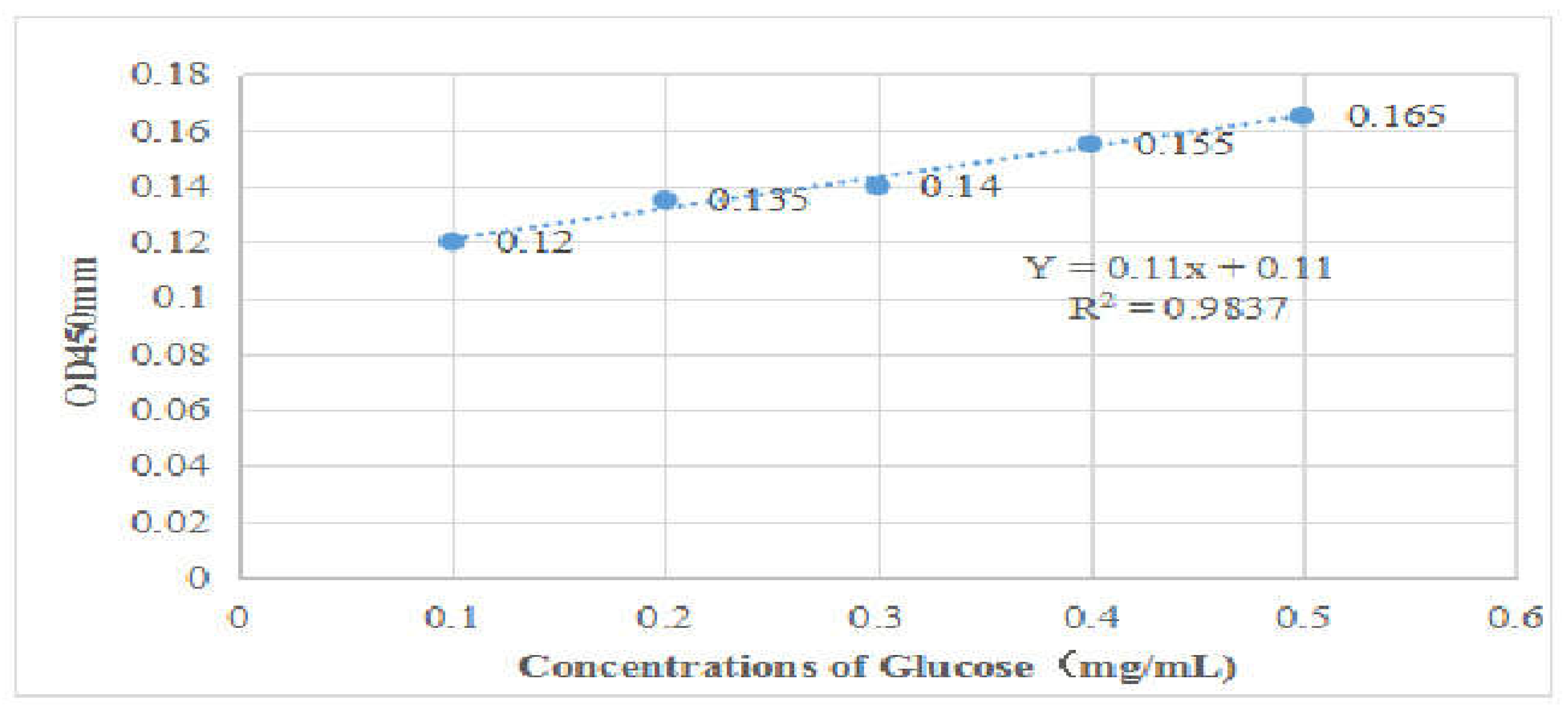
Figure 2.
Agarose gel electrophoretogram of UGPase genes in G. lucidum. M- Marker;1~3-G. lucidum spore powder from different sources;4~7- G. lucidum fruitng from different sources body;8- Negative control. G. lucidum: Ganoderma lucidum
Figure 2.
Agarose gel electrophoretogram of UGPase genes in G. lucidum. M- Marker;1~3-G. lucidum spore powder from different sources;4~7- G. lucidum fruitng from different sources body;8- Negative control. G. lucidum: Ganoderma lucidum
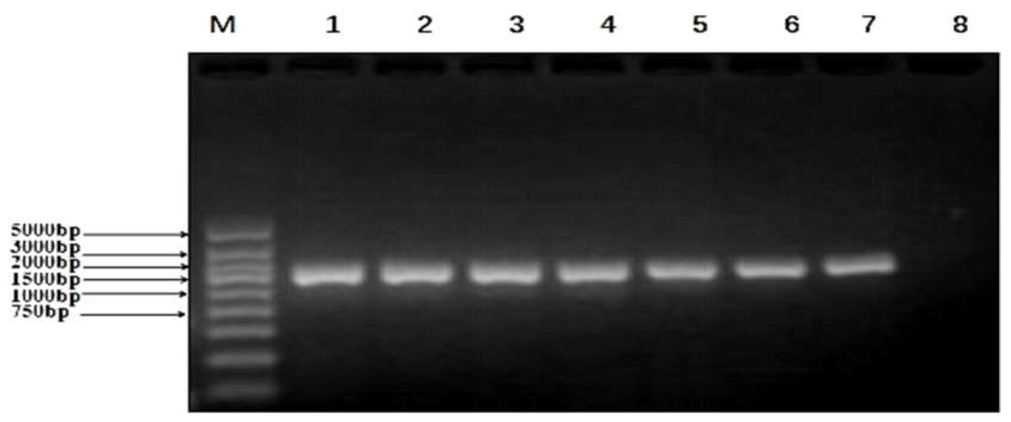
Figure 3.
Cloning and sequencing of UGPase PCR product. A: Agarose gel electrophoretogram of cloning pUGPase-216bp. M- Marker;1~5-G. lucidum spore powder from different sources;6~11- G. lucidum fruitng from different sources body;N- Negative control. G. lucidum: Ganoderma lucidum. B: Sequencing and blast of UGPase PCR product.
Figure 4.
The PCR amplification efficiency and standard curve of fluorescent quantitative PCR. A: The amplification efficiencies of the target gene UGPase and the internal reference gene 16SrRNA were 100.207% and 100.109%, respectively, close to 100%. B: The standard curve of fluorescent quantitative PCR indicated that the sample had good repeatability, high amplification efficiency, and accurate detection results.
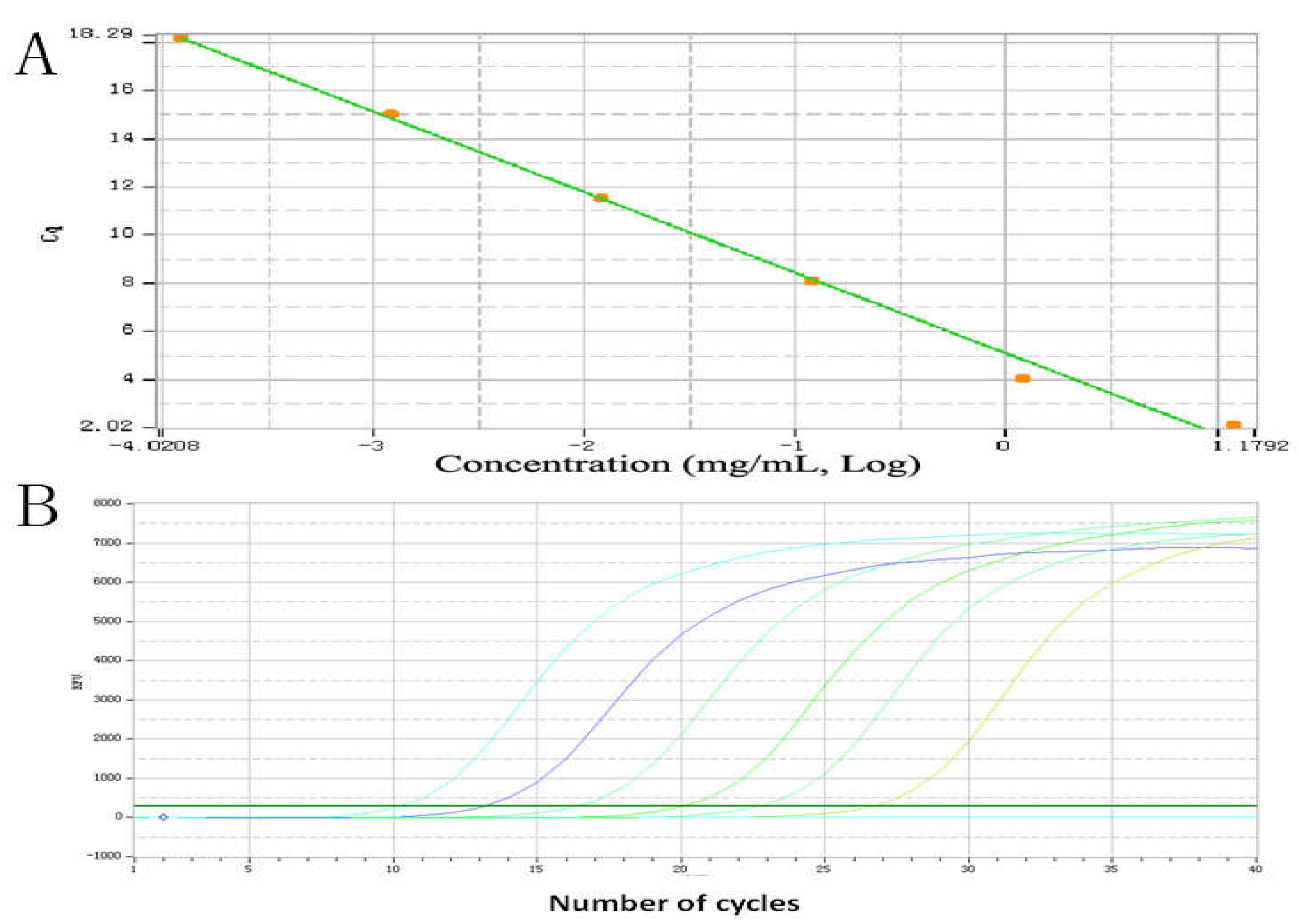
Figure 5.
Comparison of the relative expression of UGPase gene in spore powder and fruiting bodies of G. Lucidum from different places. Relative expression was calculated based on the expression of reference genes. All data represent means ± SE. A: Comparison of the relative expression of UGPase gene in spore powder and fruiting bodies of G. Lucidum from the same source. B: Comparison of the relative expression of UGPase gene in spore powder and fruiting bodies of G. Lucidum from different sources.
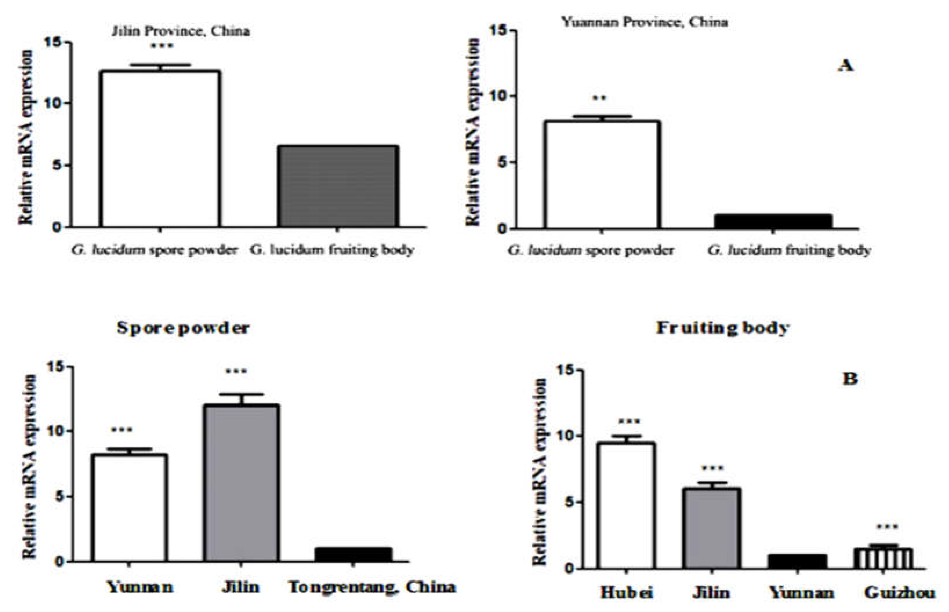

Table 1.
Comparion of relative GLP contents in G. lucidum spore powder from the different source.
| Source | Serial No. | OD450 nm | Concentrations (mg/ml) | Relative GLP Contents (%) |
|---|---|---|---|---|
| GZsp | GZsp-1 | 1.589 | 13.445 | 3.361± 0.0046 |
| GZsp-2 | 1.590 | 13.454 | 3.364± 0.0045 | |
| GZsp-9 | 1.588 | 13.436 | 3.359± 0.0044 | |
| HBsp | HBsp-1 | 1.165 | 9.591 | 2.398± 0.0045 |
| HBsp-2 | 1.162 | 9.564 | 2.391± 0.0046 | |
| HBsp-6 | 1.168 | 9.618 | 2.405± 0.0043 | |
| JLsp | JLsp-2 | 1.589 | 13.445 | 3.361± 0.0042 |
| JLsp-5 | 1.590 | 13.454 | 3.364± 0.0041 | |
| JLsp-6 | 1.588 | 13.436 | 3.359± 0.0045 | |
| TRsp | TRsp-3 | 1.165 | 9.591 | 2.398± 0.0045 |
| TRsp-4 | 1.162 | 9.564 | 2.391± 0.0044 | |
| TRsp-5 | 1.168 | 9.618 | 2.405± 0.0047 | |
| YNsp | YNsp-1 | 1.380 | 11.545 | 2.886± 0.0044 |
| YNsp-2 | 1.378 | 11.527 | 2.882± 0.0043 | |
| YNsp-5 | 1.381 | 11.554 | 2.889± 0.0044 |
G. lucidum: Ganoderma lucidum; YNsp: G. lucidum spore powder from Yuannan Province, China; YNfb: G. lucidum fruitng body from Yuannan Province, China; JLsp: G. lucidum spore powder from Jilin Province, China; YNfb: G. lucidum fruitng body from Jilin Province, China; HBsp: G. lucidum spore powder from Hubei Province, China; HBfb: G. lucidum fruitng body from Hubei Province, China; GZsp: G. lucidum spore powder from Guizhou Province, China; GZfb: G. lucidum fruitng body from Guizhou Province, China; TRspc: G. lucidum spore powder capsule from Beijing, China.
Table 2.
Comparion of relative GLP contents in G. lucidum fruiting body from the different source.
| Source | Serial No. | OD450 nm | Concentrations (mg/ml) | Relative GLP Contents (%) |
|---|---|---|---|---|
| GZfb | GZfb-1 | 0.693 | 5.300 | 1.325± 0.0422 |
| GZfb-2 | 0.692 | 5.291 | 1.323± 0.04012 | |
| GZ-9 | 0.694 | 5.309 | 1.327± 0.0431 | |
| HBfb | HBfb-3 | 1.186 | 9.782 | 2.446 ± 0.0022 |
| HBfb-5 | 1.187 | 9.791 | 2.448 ± 0.0031 | |
| HBfb-6 | 1.186 | 9.782 | 2.446 ± 0.0031 | |
| JLfb | JLfb-5 | 0.904 | 7.217 | 1.804 ± 0.0042 |
| JLfb-7 | 0.905 | 7.227 | 1.807 ± 0.0041 | |
| JLfb-8 | 0.905 | 7.227 | 1.807 ± 0.0043 | |
| YNfb | YNfb-4 | 0.540 | 3.909 | 0.977± 0.0035 |
| YNfb-7 | 0.542 | 3.927 | 0.982± 0.0036 | |
| YNfb-8 | 0.539 | 3.900 | 0.975± 0.0037 |
G. lucidum: Ganoderma lucidum; GZfb: G. lucidum fruitng body from Guizhou Province, China; HBfb: G. lucidum fruitng body from Hubei Province, China;YNfb: G. lucidum fruitng body from Yuannan Province, China; JLfb: G. lucidum fruitng body from Jilin Province, China; TRspc: G. lucidum spore powder capsule from Beijing, China.
Table 3.
Serial number and place of purchase (origin) of Ganoderma lucidum (G. lucidum) samples on the market.
Table 3.
Serial number and place of purchase (origin) of Ganoderma lucidum (G. lucidum) samples on the market.
| Serial No. | Source | Weight (g) |
|---|---|---|
| YNsp1-6 | Yuan Nan Province, China | 250 |
| YNfb1-9 | Yuan Nan Province, China | 500 |
| JLsp1-7 | Jilin Province, China | 250 |
| JLfb1-8 | Jilin Province, China | 500 |
| HBsp1-6 | Hubei Province, China | 250 |
| HBfb1-9 | Hubei Province, China | 500 |
| GZsp1-6 | Guizhou Province, China | 250 |
| GZfb1-9 | Guizhou Province, China | 500 |
| TRspc1-5 | Beijing, China | 100 |
G. lucidum: Ganoderma lucidum. YNsp: G. lucidum spore powder from Yuannan Province, China; YNfb: G. lucidum fruitng body from Yuannan Province, China; JLsp: G. lucidum spore powder from Jilin Province, China; YNfb: G. lucidum fruitng body from Jilin Province, China; HBsp: G. lucidum spore powder from Hubei Province, China; HBfb: G. lucidum fruitng body from Hubei Province, China; GZsp: G. lucidum spore powder from Guizhou Province, China; GZfb: G. lucidum fruitng body from Guizhou Province, China; TRspc: G. lucidum spore powder capsule from Beijing, China.
Disclaimer/Publisher’s Note: The statements, opinions and data contained in all publications are solely those of the individual author(s) and contributor(s) and not of MDPI and/or the editor(s). MDPI and/or the editor(s) disclaim responsibility for any injury to people or property resulting from any ideas, methods, instructions or products referred to in the content. |
© 2025 by the authors. Licensee MDPI, Basel, Switzerland. This article is an open access article distributed under the terms and conditions of the Creative Commons Attribution (CC BY) license (http://creativecommons.org/licenses/by/4.0/).
Copyright: This open access article is published under a Creative Commons CC BY 4.0 license, which permit the free download, distribution, and reuse, provided that the author and preprint are cited in any reuse.


