
Submitted:
09 April 2025
Posted:
10 April 2025
You are already at the latest version
Abstract
Objectives: This study aims to investigate the analgesic potential of extracts from 3 Capsicum species, namely Capsicum chinense (CC), Capsicum frutescens (CF), and Capsicum annuum (CA), using a hot plate test in mice. In addition, the study identified the key bioactive compounds in the extracts through a combined analysis of Fourier Transform Infrared Spectroscopy (FTIR), Gas Chromatography-Mass Spectrometry (GC-MS), and molecular docking studies. Materials and Methods: During the experimental procedures, extracts from the fruit, stem, and leaves of 3 Capsicum species were prepared using methanol, hexane, and distilled water (aquadest) as solvents. The chemical composition of these extracts was profiled and classified through Fourier-transform infrared (FTIR) spectroscopy combined with cluster analysis. Subsequently, the analgesic activity of the samples was assessed in vivo using the hot plate test in mice. Gas chromatography-mass spectrometry (GC-MS) was used to identify the bioactive compounds present in both the most potent and least potent extracts. Molecular docking studies were then conducted to predict the interactions between these compounds and specific analgesic protein targets, namely 5IKQ, 4TLM, and 4EJ4. Results: The results showed that the aquadest fruit extract of CC had the highest analgesic activity, while extracts from CF and CA had minimal effects. GC-MS analysis showed that the extract with minimal effects contained terpenoids, sterols, vitamins, hydrocarbons, and fatty acids. However, CC was rich in alkaloids capsaicin (4.38%) and dihydrocapsaicin, O-acetyl- (2.26%), terpenoids (nerolidyl acetate) (11.32%), phenolics (2-methoxy-phenol) (1.37%), and fatty acid derivatives. Molecular docking studies confirmed that capsaicin and dihydrocapsaicin, O-acetyl- exhibited strong binding affinities with analgesic protein targets 5IKQ (-8.0 kcal/mol), 4TLM (-5.3 and -4.9 kcal/mol), and 4EJ4 (-7.3 and -6.5 kcal/mol) outperforming the reference drug sodium diclofenac (-8.3, -5.9, and -6.6 kcal/mol). Conclusion: This study described the significant analgesic potential of CC extracts, particularly the aquadest fruit extract, as a promising candidate for developing new pain management therapies.
Keywords:
Capsicum extracts
; Analgesic
; FTIR
; GC-MS
; Molecular docking
Introduction
Pain is a subjective sensation triggered by the stimulation of the nervous system, which serves as a protective mechanism against tissue injury or damage [1]. Consequently, effective management is a significant focus in the medical field due to its profound impact on patients’ quality of life [2]. Chronic pain has been reported to affect approximately 20% of the global population. This condition is often associated with reduced physical activity, sleep disturbances, and an increased risk of psychological issues, such as depression and anxiety [3]. In patients with cancer, pain diminishes the quality of life as well as worsens the prognosis by impairing the immune response [4]. A previous study showed that pain caused by diabetic neuropathy was often poorly managed, leading to a higher risk of further complications [5]. Another study reported that musculoskeletal diseases, including osteoarthritis and lower back pain, were a leading cause of disability worldwide, reducing patients' ability to work and perform daily activities [6]. Acute pain resulting from trauma or surgery can also lead to hemodynamic disturbances, increase the risk of complications, such as thrombosis, and prolong the recovery time [7].
To overcome this prevalence, conventional analgesics, including opioids and nonsteroidal anti-inflammatory drugs (NSAIDs), have long been used, but their prolonged usage is often associated with several side effects [8]. Opioids, such as morphine and oxycodone, can lead to physical and psychological dependence, with increasing tolerance requiring higher doses for the same effect [9]. Other side effects include chronic constipation, respiratory depression, nausea, vomiting, and cognitive disturbances [10]. Long-term usage also causes paradoxical hyperalgesia, where pain intensifies with continued use [11].
NSAIDs, including ibuprofen and diclofenac, which are commonly used to reduce pain and inflammation, can cause gastrointestinal disturbances, such as gastritis, ulcers, and bleeding, specifically with prolonged use or in patients with a history of gastric issues [12]. These drugs also carry cardiovascular risks, namely hypertension, myocardial infarction, and congestive heart failure, particularly in vulnerable populations [13]. In addition, long-term use can impact kidney function, leading to analgesic nephropathy, fluid retention, and acute kidney failure in severe cases [14]. Given these diverse risks, there is an increased need for the development of safer and more effective alternative analgesics from natural sources [15].
Capsicum, commonly known as chili pepper, is one such natural source with significant analgesic potential [16]. Capsicum genus comprises several species rich in bioactive compounds, such as capsaicin, dihydrocapsaicin, and flavonoids, which have analgesic activity [17]. The genus comprises approximately 30 species that are widely used and commercially produced [18]. The most studied species are Capsicum annuum (CA), Capsicum frutescens (CF), and Capsicum chinense (CC). CA, known as bell pepper, contains abundant flavonoids and vitamins, making it a potential source of antioxidants [19]. CF, or cayenne pepper, has a higher capsaicin content, supporting its traditional use in pain and inflammation reduction [20]. Meanwhile, CC, or Katokkon chili, contains the highest levels of capsaicin, along with other compounds, such as dihydrocapsaicin and capsiate, which contribute to its analgesic and anti-inflammatory effects [21]. Despite this potential, there are still several study gaps in developing Capsicum as analgesic agent [22].
Several studies have focused on extraction using specific solvents but have not compared the efficacy of different solvents, such as water, methanol, or ethanol, which can affect the composition of the bioactive compounds obtained [23]. In addition, the exploration of different plant parts, including fruit, seeds, and leaves, remains limited, but each part contains bioactive compounds with distinct pharmacological activities [24,25]. Previous studies have shown that there is also a lack of multidisciplinary approaches integrating in vivo testing, chemical identification, and molecular simulations, which offers a more comprehensive understanding of Capsicum analgesic potential [26]. Although there is existing literature on capsaicin as the main compound, the potential of secondary metabolites and their synergistic effects remain underexplored [27].
To address these challenges, there is an urgent need to develop a comprehensive analytical approach. Techniques, such as Fourier Transform Infrared Spectroscopy (FTIR), Gas Chromatography-Mass Spectrometry (GC-MS), and molecular docking studies provide deeper insights into the chemical composition and mechanisms of action of compounds [25]. FTIR can identify the functional groups in the bioactive compounds, while GC-MS allows for the precise identification of the compounds present [28]. Molecular docking helps to evaluate how these compounds interact with target proteins, including the vanilloid receptor 1 (TRPV1) [29], cyclooxygenase (COX), which plays a significant role in pain modulation [30], and N-methyl-D-aspartate (NMDA). Therefore, this study aims to explore analgesic potential of Capsicum through an integrated approach. In vivo biological testing, chemical analysis, and molecular simulations were carried out to provide comprehensive scientific evidence on the effectiveness and mechanisms of action of Capsicum as analgesic.
Materials and Methods Plant Material
2.1. Plant Material
Fresh samples of the stems, fruits, and seeds of CA and CF were obtained from Rosoan Village, Enrekang District, Enrekang Regency, South Sulawesi, Indonesia (coordinates: 3°28′47.53″S, 119°49′21.47″E). Meanwhile, CC samples were sourced from Pangala Village, Rindingallo District, North Toraja Regency, South Sulawesi, Indonesia (coordinates: 2°53′48.73″S, 119°48′40.21″E). Botanical authentication of the collected plant materials was carefully performed at the Botany Laboratory, Department of Biology, Mathematics, and Natural Sciences, Universitas Negeri Makassar, Indonesia. This process ensured the accurate identification and verification of the plant species used in this study [25].
Extraction Methods
Plant samples were dried in an oven (IL80EN) at a stable temperature of 40 to 50°C for 3 days to remove the moisture content. Once dried, the plant materials were ground by applying a high-speed blender (IC-10B) at 25,000 rpm and then sieved through a 40/60 mesh to obtain a fine powder. The resulting powder was stored in airtight containers to prevent contamination until further analysis. Subsequently, the bioactive compounds were extracted using an ultrasonic-assisted method (BRANSON 1800). A total of 20 g of powdered plant material was extracted with 200 mL of distilled water, methanol, or hexane in an ultrasonic bath for 30 minutes. The solvent-to-material ratio was maintained at 1:10 in the process to ensure extraction efficiency. After this procedure, the resulting mixture was filtered, and the solvent was evaporated using a rotary evaporator (BUCHI) or freeze-dryer (BUCHI) to obtain concentrated or dry extracts. The extraction yield was calculated using Formula (1), as described by Yasir et al. [24,25].
FTIR Analysis
FTIR spectroscopy was used to analyze 27 variations of Capsicum extracts. A total of 5 mg of each extract was mixed with 0.5 mg of potassium bromide (KBr) and 5 mL of analytical-grade methanol to prepare the samples. The mixture was homogenized and dried to form pellets, which were then analyzed using a Perkin Elmer Spectrum GX FTIR spectrometer (8400S, Waltham, MA, USA). Spectral data were obtained in the wavelength range of 400 to 4000 cm⁻¹ with 16 scans per sample. To determine the similarities and differences between the extracts, chemometric analysis using cluster analysis (CA) was performed using Minitab 18 software. Grouping was conducted with the complete linkage method, distance was measured using absolute correlation, and the number of clusters was set to 6. CA was applied to group the 27 extracts based on their spectral profiles. Extracts with similarity greater than 80% were considered to have similar chemical properties [25,31]. The analysis revealed 6 identified clusters, which were selected for in vivo analgesic activity testing.
Analgesic Activity
Animals and Ethical Approval
Analgesic activity test of chili extracts (Capsicum spp.) was conducted on 24 male mice (Mus musculus) aged 35 to 60 days, weighing 20 to 35 grams, healthy, and not stressed (normal activity). These animals were divided into 8 groups with 3 mice in each group. Before the treatment, the animals were acclimatized in their cages for 7 days [32] and kept under standard conditions, fasted from food for 24 hours before the experiment, and given access to water only [33]. All experimental processes were approved by Sekolah Tinggi Ilmu Farmasi Makassar ethical committee through letter number: 222/EC.1.1.B/IX/KEPK/2023.
Hot Plate Test
In this study, each mouse was administered a dose of 10 mg per paw. Samples including CC aquadest stem, CA methanol seed, CC aquadest fruit, CC methanol seed, CF hexane fruit, CF methanol fruit, NaCMC 1%, and Voltaren emulgel® were applied to the soles of the test animal’s paws. After a 1-minute exposure, the mouse was placed on a hotplate at 50°C for a maximum of 15 seconds. When the mice showed signs of pain, such as licking their paw, jumping, or shaking, before the 15 seconds were up, the observation was stopped [34]. This response to the heat stimulus was observed for 1 hour and 30 minutes at intervals of 15, 30, 45, 60, 75, and 90 minutes [35,36]. The percentage protection against the thermal pain stimulus was calculated according to the following Formula (2), as described by Hijazi et al. [37].
GC-MS Analysis
GC-MS analysis was carried out at the Chemical Engineering Laboratory, Politeknik Negeri Ujung Pandang (PNUP), Makassar, Indonesia. The samples exhibiting the highest analgesic activity, such as CC extract with aquadest solvent, and a representative extract with no significant effect, namely CC extract with hexane solvent (both 1 g), were dissolved in 5 mL of 96% ethanol (p.a.). The dissolution process was conducted using an ultrasonic device at 55°C for 30 minutes. Subsequently, the solution was filtered using Whatman No. 42 filter paper, and the resulting filtrate was injected into GC-MS Ultra QP 2010 instrument (Shimadzu, Japan). The chromatographic conditions included an injector temperature of 250°C in the splitless mode, a pressure of 76.9 kPa, a carrier gas flow rate of 14 mL/min, and a split ratio of 1:10. In this study, the ion source and interface temperatures were set to 200°C and 280°C, respectively, with a solvent cut-off time of 3 minutes and a mass range of 10 to 890 m/z. The column used was an SH-Rxi-5Sil MS with a length of 30 m and an inner diameter of 0.25 mm. Furthermore, the column temperature program started at 70°C with a hold time of 2 minutes, followed by a temperature increase of 10°C/min until reaching 200°C. This temperature was then increased further at 5°C/min to 280°C with a hold time of 9 min, resulting in a total analysis time of 36 min. The obtained data were analyzed using the NIST 17 and Wiley 9 libraries to identify the major compounds in the extracts with an area percentage greater than 1% [25].
Molecular Docking Studies
Sample Preparation (Virtual Screening)
The major compounds identified were then further analyzed using molecular studies to comprehend their mechanism of action and interaction with target proteins as analgesics. These compounds were identified through GC-MS analysis of CC fruit extracts with aquadest as well as hexane solvents and were subjected to molecular docking. Initially, the structures of these compounds were searched and downloaded from PubChem (accessed on 9 July 2024, link: https://pubchem.ncbi.nlm.nih.gov). This platform provided detailed information on the compound structures, including SMILES (Simplified Molecular Input Line Entry System) data, which could be used to download the 3D structure in PDF for docking purposes. The target proteins for molecular docking were obtained from the Protein Data Bank (accessed on 9 July 2024, link: https://www.rcsb.org) in PDF [25]. In this study, analgesic target proteins used were 4EJ4 [52], 5IKQ [53], and 4TLM [54], which were prepared for the docking process.
Molecular Docking
Ligand was optimized using Chimera 1.17.3 (UCSF Chimera; accessed on 13 July 2024, link: https://www.cgl.ucsf.edu/chimera/). PyMOL version 2.5 (accessed on 13 July 2024, link: https://pymol.org/2) was used to refine protein structures, remove ligands, and eliminate water molecules. The docking process was carried out by applying PyRx AutoDock Vina (accessed on 13 July 2024, link: https://pyrx.sourceforge.io/), which was used to calculate the binding affinity, RMSD, amino acid residues, and bond types between the optimized ligands and receptors. Furthermore, the interaction results between the ligand and receptor were then visualized using the Biovia Discovery Studio Visualizer (accessed on 13 July 2024, link: https://www.3dsbiovia.com). The ligand with the lowest binding energy or the lowest docking score, along with the hydrogen-bonding interactions, was selected as the best candidate [25].
Results and Discussion
Extraction Optimization
The extraction data for 3 chili species, namely CA, CF, and CC using hexane, methanol, and distilled water (aquadest) as solvents revealed significant trends regarding solvent efficiency, variation among plant parts, and interspecies differences. Furthermore, the results indicated that aquadest consistently achieved the highest extraction yields compared to methanol and hexane. This showed that aquadest, as a polar solvent, was particularly effective at extracting polar compounds such as flavonoids, alkaloids, and phenolics, which were abundant in chili plants. Meanwhile, hexane, a nonpolar solvent, produced the lowest yields, reflecting the relatively low content of nonpolar compounds like oils and lipophilic components in the samples [25].
Among the plant parts, fruits consistently demonstrated the highest extraction yields across all species, particularly when extracted with aquadest (Table 1). For instance, CF fruits yielded up to 23.5%, while stems generally exhibited the lowest yields, due to their lower concentrations of bioactive metabolites compared to fruits and seeds. In CA, the highest yield was obtained from fruits using aquadest (18%), with methanol and aquadest depicting similar trends in extracting bioactive compounds from various plant parts. CF demonstrated the highest overall yields, with aquadest extracts yielding 23.5% from fruits and 14.5% from stems. Meanwhile, CC achieved maximum yields of 12.5% and 7% in fruits and stems, respectively, when extracted with aquadest, and methanol provided relatively balanced yields across all parts of CC.
The results were consistent with previous studies that identified water-based solvents as optimal for recovering polar metabolites such as capsaicin and phenolic acids [38]. Methanol, with its dual polarity, was effective at extracting both polar and nonpolar compounds. However, the low yields from hexane were consistent with the limited presence of nonpolar metabolites such as oils and lipids in the chili samples [39]. Chili fruits were known to contain the highest concentrations of secondary metabolites like capsaicinoids, carotenoids, and flavonoids [40], while stems, serving primarily structural roles, typically contained lower metabolite levels.
CF emerged as the standout species, displaying the highest metabolite content, particularly in its fruits extracted with aquadest. Notable yields included CF fruits and stems (23.5% and 14.5%, respectively), CA fruits (18%), CC fruits (12.5%), and CF seeds extracted with methanol (15%). These results emphasized the pharmacological potential of CF as a rich source of bioactive metabolites. For further studies, the extracts were grouped based on the functional groups identified through cluster analysis. Their representative with the highest potential proceeded to in vivo testing using Hot Plate method on mice to assess their analgesic activity.
FTIR Profiling Analysis
FTIR referred to a technique used to identify functional groups in chemical compounds by measuring their vibrational modes, which absorbed specific wavelengths of IR light. These bands observed in FTIR spectra depended on the functional groups present in the compounds [25].
FTIR absorption regions for the compounds identified in the hexane fruit extract of CF indicated distinct functional group characteristics (Figure 1). Oleic acid exhibited absorption at 1700 to 1725 cm⁻¹ (C=O) [41], a broad peak at 2500 to 3300 cm⁻¹ (O-H), and 2800 to 3000 cm⁻¹ (C-H). Esters such as methyl palmitate, methyl linoleate, methyl oleate, and 15-octadecenoic acid methyl ester showed peaks at 1735 to 1750 cm⁻¹ (C=O), 2800 to 3000 cm⁻¹ (C-H, aliphatic), and 1620 to 1680 cm⁻¹ (C=C, alkene, in unsaturated compounds) [45]. N-hexadecanoic acid demonstrated absorption at 1700 to 1725 cm⁻¹ (C=O, carboxylic acid), 2500 to 3300 cm⁻¹ (O-H), and 2800 to 3000 cm⁻¹ (C-H, aliphatic). Similarly, linoleic acid exhibited peaks at 1700 to 1725 cm⁻¹ (C=O, carboxylic acid), 2500 to 3300 cm⁻¹ (O-H), and 1620 to1680 cm⁻¹ (C=C, alkene). N-pentadecylacetamide was characterized by absorption at 1640 to 1690 cm⁻¹ (C=O, amide), 3200 to 3500 cm⁻¹ (N-H, amide), and 2800 to 3000 cm⁻¹ (C-H). Nerolidyl is identified by peaks at 3200 to 3600 cm⁻¹ (O-H, alcohol), 1620 to 1680 cm⁻¹ (C=C, alkene), and 2800 to 3000 cm⁻¹ (C-H). Long-chain alkanes such as heneicosane, pentacosane, hexacosane, and nonacosane exhibited a characteristic alkane signature at 2800 to 3000 cm⁻¹ (C-H). Isofucosterol displayed absorption at 3200 to 3600 cm⁻¹ (O-H, hydroxyl group) and 1600 to 1700 cm⁻¹ (C=C, steroid double bond). Squalene showed peaks at 2800 to 3000 cm⁻¹ (C-H) and 1620 to 1680 cm⁻¹ (C=C, alkene), while dl-α-tocopherol was marked by a broad peak at 3200 to 3600 cm⁻¹ (O-H, phenol) [46], 2800 to 3000 cm⁻¹ (C-H), and 1450 to 1600 cm⁻¹ (C=C, aromatic).
In the aquadest fruit extract of CC, as well as propanoic acid, 2-methyl- was characterized by peaks at 1700 to 1725 cm⁻¹ (C=O, carboxylic acid) and 2500 to 3300 cm⁻¹ (O-H, carboxylic acid). Furthermore, 1-Propanol, 2,2-dimethyl-, and acetate displayed absorption at 1735 to 1750 cm⁻¹ (C=O, ester), 2800 to 3000 cm⁻¹ (C-H, aliphatic), and 3200 to 3600 cm⁻¹ (O-H, alcohol, when free alcohol was present). Phenol, 2-methoxy- exhibited peaks at 3200 to 3600 cm⁻¹ (O-H, phenol), 1450 to 1600 cm⁻¹ (C=C, aromatic), and 1000 to 1300 cm⁻¹ (C-O, ether). Palmitoleic acid shows absorption at 1700–1725 cm⁻¹ (C=O, carboxylic acid), 2500–3300 cm⁻¹ (O-H), and 1620–indicated cm⁻¹ (C=C, alkene). Capsaicin was identified by its characteristic peaks at 3200 to 3600 cm⁻¹ (O-H, phenol) [42], 1650 to 1700 cm⁻¹ (C=O, amide), 2800–3000 cm⁻¹ (C-H), and 1450 to 1600 cm⁻¹ (C=C, aromatic) [43]. Lastly, dihydrocapsaicin, O-acetyl- showed peaks at 1735 to 1750 cm⁻¹ (C=O, ester) and 2800 to 3000 cm⁻¹ (C-H, aliphatic) [44]. These data were integrated with GC-MS and in silico results to validate the presence of these compounds in the extracts and correlate their functional groups with biological activities. The presence of characteristic FTIR peaks could confirm the structural features predicted by GC-MS, enhancing the reliability of compound identification [25].
FTIR profile provided valuable insights into the functional groups and primary components of chili species extracts, including CA, CF, and CC, with variations in the solvents and plant parts, which were essential for chemical characterization and phytochemical studies. The hierarchical cluster analysis based on functional groups detailed the process (Figure 2), where observations were progressively merged into larger clusters based on similarity and distance levels [31]. Initially, the analysis started with 27 distinct clusters, each representing individual observations or small groups, and ultimately resulted in 6 clusters with high similarity levels (>80%) [25,31].
Cluster 1 consisted of extracts from CA hexane stem, CF aquadest fruit, CA methanol stem, CC methanol stem, CF aquadest seed, CA aquadest seed, CA hexane seed, CF aquadest stem, CA methanol seed, and CF hexane fruit, with a similarity of 83.62%. The second one included extracts from CF hexane stem, CF methanol seed, CA methanol fruit, CC methanol fruit, and CF hexane seed, with a similarity of 90.67%. Cluster 3 was composed of extracts from CA aquadest stem, CF methanol stem, and CC aquadest stem, with a similarity of 90.26%. Meanwhile, cluster 4 contained only CC aquadest fruit extract with a similarity of 77.56%, while 5 included extracts from CC hexane stem, CC aquadest seed, CC hexane seed, CC methanol seed, and CA aquadest fruit, with a similarity of 87.58%. Cluster 6 contained extracts from CA hexane fruit, CF methanol fruit, and CC hexane fruit, with a similarity of 99.82%.
The final grouping reflected the hierarchical relationships among the observations, capturing similarities and distances throughout the clustering process [47]. Representatives from each cluster were selected for further analgesic activity testing because clusters with similar functional groups based on FTIR data were likely to exhibit similar pharmacological effects [25].
This novel approach in natural product studies simplified the screening process by focusing on extracts with similar functional group compounds, facilitating the identification of potential candidates without the need to test each extract individually [25]. The extracts selected for analgesic testing were CA methanol seed extract from Cluster 1, CA methanol fruit extract from 2, CA aquadest stem extract from 3, CC aquadest fruit extract from 4, CC methanol seed extract from 5, and CF methanol fruit extract from cluster 6.
Analgesic Activity
The results of analgesic activity test using Hot Plate method revealed significant differences in pain-relieving activity between the positive control (Voltaren Emulgel®), the negative control (NaCMC 1%), and various 10 mg extracts from CF, CC, and CA. Based on the primary effects plot for analgesic activity (%) shown in Figure 3, the ranking from highest to lowest activity included Voltaren emulgel® > CA aquadest fruit extract > CF methanol fruit extract > CF hexane fruit extract > CA aquadest stem extract > CA methanol fruit extract > CC methanol seed extract > NaCMC 1%.
Voltaren emulgel® demonstrated high analgesic efficacy, reaching 100% at the 45-minute mark and maintaining this level through to the 90-minute mark, which indicated strong pain-relieving potential. However, 1% NaCMC did not exhibit significant analgesic activity in the test [48]. Extracts from Capsicum plants, although showing increased analgesic activity over time, still had lower efficacy than the positive control. The highest activity reached 75% for CC aquadest fruit extract at the 90-minute mark. Other extracts, such as CF methanol fruit, CA methanol fruit, and CA aquadest stem, demonstrated relatively lower activities, with 36%, 14%, and 21%, respectively, at the 90-minute mark.
The relationship between analgesic activity results and the previously conducted FTIR data showed that extracts with similar functional groups [49], such as those found in CC aquadest Fruit and CF methanol fruit, exhibited similarities in pharmacological effects, including analgesic activity. In this study, FTIR-based cluster analysis, grouping the extracts according to functional group similarities, indicated that extracts containing similar chemical components were likely to exhibit comparable biological effects [50]. Therefore, the integrative approach using FTIR and cluster analysis facilitated the selection of extracts with better potential, which could be further investigated, although these extracts had not reached the same efficacy levels as the positive control [25].
As the extract with the highest analgesic activity, CC aquadest fruit proceeded to GC-MS testing for a more detailed identification of its chemical components. Furthermore, in silico testing was conducted to analyze the potential bioactive interactions of this extract with 3 key protein targets involved in pain modulation. This included the vanilloid receptor 1 (TRPV1) using structure 4EJ4 [51], the delta opioid receptor, and COX using structure 5IKQ (Human Cyclooxygenase-2) [52], as well as N-methyl-D-aspartate (NMDA) receptor using structure 4TLM (GluN1/GluN2B NMDA receptor, structure 2) [53]. Meanwhile, CF hexane fruit, which demonstrated low analgesic activity, was selected as a representative of extracts with weaker effects for comparison of its chemical content and efficacy. This approach was expected to provide a clearer understanding of the relationship between the chemical composition of the extracts and their analgesic activity and to identify potential candidates for the development of natural pain relief therapies.
GC-MS Metabolite Characterization
The chemical profiling of CF hexane fruit extract and CC aquadest fruit extract, based on GC-MS analysis (Table 2), showed significant differences in their chemical compositions and potential bioactivities [25]. Furthermore, CF hexane fruit extract contained 16 identified compounds with a total area percentage of 75.06%. Notable constituents included nerolidyl (22.64%), a sesquiterpene alcohol with reported anti-inflammatory and analgesic properties [54]. Fatty acids such as n-hexadecanoic acid (6.74%) and linoleic acid (6.88%) contributed to its pharmacological profile, supported by evidence of their anti-inflammatory effects [55,56]. Furthermore, methyl esters such as methyl linoleate (6.66%) and methyl oleate (5.37%) could enhance the membrane permeability and exhibit bioactivity. Squalene (3.43%), a triterpene, provided antioxidant benefits, while dl-α-tocopherol (1.64%), a form of vitamin E, added further antioxidant capacity [57].
In this study, CC aquadest fruit extract contained 15 compounds with a total area percentage of 58.24%. Hydrophilic and semihydrophilic compounds dominate, including n-hexadecanoic acid (12.58%) and nerolidyl acetate (11.32%), both of which exhibited notable anti-inflammatory potential. Linoleic acid ethyl ester (2.37%) and other fatty acid derivatives further enhanced the extract’s anti-inflammatory profile. Capsaicinoids, such as capsaicin (4.38%) and dihydrocapsaicin, O-acetyl (2.26%), were particularly predominant. These compounds were directly associated with analgesic effects through TRPV1 receptor activation [58]. In comparison, CF hexane fruit extract was rich in lipophilic compounds, including fatty acids, esters, and terpenes, which enhanced membrane permeability and exhibited synergistic pharmacological effects [59].
CC aquadest extract emphasized capsaicinoids, which were primarily responsible for its analgesic activity. The presence of fatty acids and terpenes in both extracts suggested overlapping anti-inflammatory mechanisms. However, the distinct hydrophilic and lipophilic compound profiles indicated different potential applications. Meanwhile, CF hexane fruit extract could be more suitable for lipid-based formulations targeting anti-inflammatory effects, CC aquadest extract showed potential for analgesic applications through capsaicin-mediated pathways. These results were consistent with previous studies on chili extracts, describing their potential to manage inflammation and pain [60]. Further investigations, including molecular docking and in vitro studies, were necessary to elucidate their precise mechanisms of action and optimize their therapeutic potential.
Molecular Docking Analysis
Table 3 showed the binding free energy values and amino acid residues interacting with the pain target proteins 5IKQ, 4TLM, and 4EJ4 for the several tested compounds. These data provided insights into molecular interactions between these compounds and pain target proteins, as well as how the results correlated with analgesic activity assays and the chemical profiles of Capsicum extracts identified through GC-MS.
The native ligand (meclofenamic acid) for 5IKQ target protein (Figure 4) had the strongest binding affinity [61], with a bond-free energy of -9.0 kcal/mol, and formed multiple hydrogen bonds with amino acids such as Tyr 385, and Ser 530. This suggested that it strongly inhibited COX-2, potentially acting as an anti-inflammatory or analgesic agent. Diclofenac sodium with a bond-free energy of -8.3 kcal/mol bound to Leu 352, Tyr 358, Ser 530. Although not as strong as meclofenamic acid, but its strong binding suggested that it could act as an effective COX-2 inhibitor, consistent with its clinical use as an anti-inflammatory drug. Capsaicin and dihydrocapsaicin, known for their analgesic effects through TRPV1 receptor activation [62], showed a strong binding affinity (-8.0 kcal/mol). Furthermore, it interacted with Ser 353, and Ser 530, which were key residues included in the enzyme's activity. This reinforced the role of capsaicin and dihydrocapsaicin in pain relief through COX-2 inhibition, supporting its known analgesic properties.
Linoleic acid ethyl ester and nerolidyl acetate, exhibited moderate binding energies (-7.3 to -7.4 kcal/mol) and interacted with residues like Tyr 385, and Ser 530. These interactions contributed to the anti-inflammatory and analgesic effects, potentially modulating COX-2. Oleic acid, heneicosane, isofucosterol, pentacosane, hexacosane, squalene, nonacosane, and dl-α-tocopherol showed weaker binding energies -4.0 to -8.5kcal/mol), and did not form hydrogen bonds (NI). Despite this potential, the compounds could still have biological activity through other mechanisms, such as influencing membrane fluidity or other enzyme pathways. Other fatty acids and esters such as n-hexadecanoic acid, methyl palmitate, methyl linoleate, methyl oleate, and 15-octadecenoic acid, methyl ester, with bond-free energies around -6.2 to -6.9 kcal/mol, formed hydrogen bonds with various residues like Arg 120, and Tyr 385. This suggested their interaction with the target protein, although their binding affinity was lower. Therefore, this analysis suggested that many compounds, specifically those with strong to strong binding affinities (meclofenamic acid, capsaicin, and dihydrocapsaicin), had significant anti-inflammatory and analgesic potential through their interaction with COX-2 protein target.
In Figure 5, the native ligand (4-[(1R,2S)-3-(4-benzylpiperidin-1-yl)-1-hydroxy-2-methylpropyl]phenol) for 4TLM target protein [63] had binding free energy of -6.0 kcal/mol and interacted with the amino acid residue Gly 112 through hydrogen bonds. This suggested its potential for therapeutic applications in pain management or other medicinal uses. Diclofenac sodium had a binding free energy of -5.9 kcal/mol, and it interacted with Ile 133, Ser 108, Tyr 109, and Thr 110. This indicated a moderate interaction, which was consistent with the known mechanism of NSAIDs, binding to COX enzymes to inhibit prostaglandin synthesis, thereby reducing inflammation and pain. Capsaicin and dihydrocapsaicin (with -5.3 and 4.9 kcal/mol) were indicated for their specific interaction with Tyr 109 and Arg 115, which suggested their effective interaction with the protein target and exerted analgesic effects, possibly through the modulation of inflammatory pathways [64]. Oleic acid depicted moderate to strong binding with specific amino acids (Ser 132), which explained their anti-inflammatory or analgesic potential. Fatty acids like oleic acid, methyl palmitate, and methyl linoleate had moderate binding energies (ranging from -4.0 to -4.3 kcal/mol) and were involved in interactions with key amino acids like Ser 132 and Thr 110, which contributed to their potential effects on the protein target. Nerolidyl acetate had slightly stronger interactions with Arg 115, suggesting their potential candidates for the modulation of the target protein. These results suggested that compounds like capsaicin, dihydrocapsaicin, and 4-[(1R,2S)-3-(4-benzylpiperidin-1-yl)-1-hydroxy-2-methylpropyl]phenol could be developed as leads for analgesic drug design, supported by their ability to interact with key residues involved in pain and inflammation. Further studies, including in vitro and in vivo assays, were warranted to validate these molecular interactions and their therapeutic potential.
The native ligand (4bS,8R,8aS,14bR)-7-(cyclopropylmethyl)-5,6,7,8,14,14b-hexahydro-4,8-methano[1]benzofuro[2,3-a]pyrido[4,3-b]carbazole-1,8a(9H)-diol) for the 4EJ4 target protein [65] (Figure 6) had binding free energy of -10.9 kcal/mol and interacted with Asp 128 and Tyr 129. This suggested its potential as a powerful inhibitor, possibly for a receptor involved in inflammatory or neurological pathways. Diclofenac sodium had a moderate binding energy of -6.6 kcal/mol, which reflected its established role as NSAIDs, interacting with Asp 214. Capsaicin and dihydrocapsaicin showed strong binding energies (-7.3 kcal/mol and -6.5 kcal/mol, respectively) and interactions with specific residues (Tyr 219, His 278), which likely played a significant role in their pain-relieving properties through TRPV1 receptor modulation [66]. Oleic acid and other fatty acids (such as linoleic acid, and methyl oleate) exhibited moderate to strong binding energies and interactions with key residues, supporting their potential anti-inflammatory and analgesic properties. Nerolidyl and nerolidyl acetate (with binding energies of -6.5 kcal/mol and -6.3 kcal/mol, respectively) showed significant interactions, despite lacking direct hydrogen bond interactions, suggesting that other non-covalent forces contributed to their activity. This dataset revealed several promising compounds with strong binding affinities and specific amino acid interactions, which supported their potential in treating inflammatory, analgesic, or other related conditions. (4bS,8R,8aS,14bR)-7-(cyclopropylmethyl)-5,6,7,8,14,14b-hexahydro-4,8-methano[1]benzofuro[2,3-a]pyrido[4,3-b]carbazole-1,8a(9H)-diol was evident with the strongest binding, while compounds like capsaicin, dihydrocapsaicin and diclofenac sodium maintained strong interactions with residues known to be associated with their pharmacological effects.
Conclusion
In conclusion, this study aimed to investigate analgesic potential of Capsicum extracts using hot plate test in mice while integrating bioactive compound profiling through FTIR, GC-MS, and molecular docking studies. The results indicated that Capsicum extracts exhibited significant analgesic activity, supported by the presence of several bioactive compounds such as dl-α-tocopherol, nerolidyl acetate, linoleic acid ethyl ester, capsaicin, and dihydrocapsaicin, O-acetyl-. In vivo results from hot plate test demonstrated a dose-dependent reduction in the pain response, which was consistent with the compounds identified in the extracts.
FTIR analysis confirmed the functional groups associated with pain modulation, while GC-MS profiling identified key bioactive metabolites that could contribute to the observed analgesic effects. Notably, capsaicin and dihydrocapsaicin, identified as the most abundant compounds, displayed strong binding affinities to pain-related receptors, as evidenced by molecular docking studies. The docking results indicated that these compounds interact with specific amino acid residues that were associated with the modulation of the pain pathways.
The combination of in vivo and computational studies provided robust evidence for analgesic properties of Capsicum extracts, with capsaicin and its analogs being the key contributors. These results suggested that Capsicum extracts, particularly those rich in capsaicin and dihydrocapsaicin, O-acetyl- had potential as natural analgesic agents, necessitating further exploration for their therapeutic potential in pain management.
Acknowledgments
The authors are grateful to Almarisah Madani University for providing the essential laboratory facilities. Furthermore, the authors are grateful to all collaborators and institutions for their invaluable contributions and technical assistance, which has been instrumental in the completion of this study.
References
- Peghetti, A.; Seri, R.; Cavalli, E.; Martin, V. Pain Management. In Pearls and Pitfalls in Skin Ulcer Management. In Pearls and Pitfalls in Skin Ulcer Management; Maruccia, M., Papa, G., Ricci, E., Giudice, G., Eds.; Springer: Cham, Switzerland, 2023. [Google Scholar] [CrossRef]
- Pandelani, F.F.; Nyalunga, S.L.N.; Mogotsi, M.M.; Mkhatshwa, V.B. Chronic pain: its impact on the quality of life and gender. Front. Pain Res. 2023, 4, 1253460. [Google Scholar] [CrossRef] [PubMed]
- Tabernacki, T.; Gilbert, D.; Rhodes, S.; Scarberry, K.; Pope, R.; McNamara, M.; Gupta, S.; Banik, S.; Mishra, K. The burden of chronic pain in transgender and gender diverse populations: Evidence from a large US clinical database. Eur. J. Pain 2024, 29. [Google Scholar] [CrossRef]
- Tiberio, P.; Balordi, M.; Castaldo, M.; Viganò, A.; Jacobs, F.; Benvenuti, C.; Torrisi, R.; Zambelli, A.; Santoro, A.; De Sanctis, R. Empowerment, Pain Control, and Quality of Life Improvement in Early Triple-Negative Breast Cancer Patients through Pain Neuroscience Education: A Prospective Cohort Pilot Study Protocol (EMPOWER Trial). J. Pers. Med. 2024, 14, 711. [Google Scholar] [CrossRef]
- Blaibel, D.; Fernandez, C.J.; Pappachan, J.M. Acute worsening of microvascular complications of diabetes mellitus during rapid glycemic control: The pathobiology and therapeutic implications. World J. Diabetes 2024, 15, 311–317. [Google Scholar] [CrossRef] [PubMed]
- Tushingham, S.; Cottle, J.; Adesokan, M.; Ogwumike, O.O.; Ojagbemi, A.; Stubbs, B.; Fatoye, F.; O Babatunde, O. The prevalence and pattern of comorbid long-term conditions with low back pain and osteoarthritis in low- and middle-income countries: a systematic review and meta-analysis. Int. J. Heal. Promot. Educ. 2024, 1–25. [Google Scholar] [CrossRef]
- Zanza, C.; Romenskaya, T.; Racca, F.; Rocca, E.; Piccolella, F.; Piccioni, A.; Saviano, A.; Formenti-Ujlaki, G.; Savioli, G.; Franceschi, F.; et al. Severe Trauma-Induced Coagulopathy: Molecular Mechanisms Underlying Critical Illness. Int. J. Mol. Sci. 2023, 24, 7118. [Google Scholar] [CrossRef] [PubMed]
- Tsoupras, A.; Gkika, D.A.; Siadimas, I.; Christodoulopoulos, I.; Efthymiopoulos, P.; Kyzas, G.Z. The Multifaceted Effects of Non-Steroidal and Non-Opioid Anti-Inflammatory and Analgesic Drugs on Platelets: Current Knowledge, Limitations, and Future Perspectives. Pharmaceuticals 2024, 17, 627. [Google Scholar] [CrossRef]
- Badshah, I.; Anwar, M.; Murtaza, B.; Khan, M.I. Molecular mechanisms of morphine tolerance and dependence; novel insights and future perspectives. Mol. Cell. Biochem. 2023, 479, 1457–1485. [Google Scholar] [CrossRef]
- Zimmerman, A.; Laitman, A. Safe Management of Adverse Effects Associated with Prescription Opioids in the Palliative Care Population: A Narrative Review. J. Clin. Med. 2024, 13, 2746. [Google Scholar] [CrossRef]
- Manhapra, A.; MacLean, R.R.; Rosenheck, R.; Becker, W.C. Are opioids effective analgesics and is physiological opioid dependence benign? Revising current assumptions to effectively manage long-term opioid therapy and its deprescribing. Br. J. Clin. Pharmacol. 2023, 90, 2962–2976. [Google Scholar] [CrossRef]
- Alhamadi, N.; Asiri, A.H.; Alshahrani, F.M.; Alqahtani, A.Y.; Al Qout, M.M.; A Alnami, R.; Alasiri, A.S.; Al-Zomia, A.S. Gastrointestinal Complications Associated With Non-steroidal Anti-inflammatory Drug Use Among Adults: A Retrospective, Single-Center Study. Cureus 2022, 14, e26154. [Google Scholar] [CrossRef] [PubMed]
- Liang, S.; Wang, X.; Zhu, X. Insights from pharmacovigilance and pharmacodynamics on cardiovascular safety signals of NSAIDs. Front. Pharmacol. 2024, 15, 1455212. [Google Scholar] [CrossRef]
- LaForge, J.M.; Urso, K.; Day, J.M.; Bourgeois, C.W.; Ross, M.M.; Ahmadzadeh, S.; Shekoohi, S.; Cornett, E.M.; Kaye, A.M.; Kaye, A.D. Non-steroidal Anti-inflammatory Drugs: Clinical Implications, Renal Impairment Risks, and AKI. Adv. Ther. 2023, 40, 2082–2096. [Google Scholar] [CrossRef]
- Bakare, T.T.; Uzoeto, H.O.; Gonlepa, L.N.; Cosmas, S.; Ajima, J.N.; Arazu, A.V.; Ezechukwu, S.P.; Didiugwu, C.M.; Ibiang, G.O.; Osotuyi, A.G.; et al. Evolution and challenges of opioids in pain management: Understanding mechanisms and exploring strategies for safer analgesics. Med. Chem. Res. 2024, 33, 563–579. [Google Scholar] [CrossRef]
- Duranova, H.; Valkova, V.; Gabriny, L. Chili peppers (Capsicum spp.): the spice not only for cuisine purposes: an update on current knowledge. Phytochem. Rev. 2022, 21, 1379–1413. [Google Scholar] [CrossRef]
- Patel, K.; Patel, D.K. Biological Importance, Pharmacological Activities, and Nutraceutical Potential of Capsanthin: A Review of Capsicum Plant Capsaicinoids. Curr. Drug Res. Rev. 2024, 16, 18–31. [Google Scholar] [CrossRef]
- Ferreira, G.D.N.C.; Ferraz, G.V.; da Silva, L.R.; Oliveira, A.C.; de Almeida, L.R.; Costa, M.F.; Silva, R.N.O.; da Silva, V.B.; Lopes, Â.C.d.A.; Gomes, R.L.F. Assessment of phenotypic divergence and hybrid development in ornamental peppers (Capsicum spp.). Genet. Resour. Crop. Evol. 15. [CrossRef]
- Kebu, Z.; Gure, A.; Molole, G.J. Total Phenolic and Flavonoid Content, Lipophilic Components, and Antioxidant Activities of Capsicum annuum Varieties Grown in Omo Nada, Jimma, Ethiopia. Nat. Prod. Commun. 2024, 19, 1934578X241306244. [Google Scholar] [CrossRef]
- Jolayemi, A.; Ojewole, J. Comparative anti-inflammatory properties of Capsaicin and ethylaAcetate extract of Capsicum frutescens linn [Solanaceae] in rats. Afr. Heal. Sci. 2013, 13, 357–361. [Google Scholar] [CrossRef]
- Rastogi, V.; Porwal, M.; Sikarwar, M.S.; Singh, B.; Choudhary, P.; Mohanta, B.C. A review on phytochemical and pharmacological potential of Bhut Jolokia (a cultivar of Capsicum chinense Jacq.). J. Appl. Pharm. Sci. 2014, 14, 79–90. [Google Scholar] [CrossRef]
- Duranova, H.; Valkova, V.; Gabriny, L. Chili peppers (Capsicum spp.): the spice not only for cuisine purposes: an update on current knowledge. Phytochem. Rev. 2021, 21, 1379–1413. [Google Scholar] [CrossRef]
- Lefebvre, T.; Destandau, E.; Lesellier, E. Selective extraction of bioactive compounds from plants using recent extraction techniques: A review. J. Chromatogr. A 2021, 1635, 461770. [Google Scholar] [CrossRef]
- Yasir, B.; Rohman, A. Optimization of Pagoda (Clerodendrum paniculatum L.) Extraction Based By Analytical Factorial Design Approach, Its Phytochemical Compound, and Cytotoxicity Activity. Egypt. J. Chem. 2022, 65, 421–430. [Google Scholar] [CrossRef]
- Yasir, B.; Mus, S.; Rahimah, S.; Tandiongan, R.M.; Klara, K.P.; Afrida, N.; Nisaa, N.R.K.; Risna, R.; Jur, A.W.; Alam, G.; et al. Antimicrobial Profiling of Piper betle L. and Piper nigrum L. Against Methicillin-Resistant Staphylococcus aureus (MRSA): Integrative Analysis of Bioactive Compounds Based on FT-IR, GC-MS, and Molecular Docking Studies. Separations 2024, 11, 322. [Google Scholar] [CrossRef]
- Maharjan, A.; Vasamsetti, B.M.K.; Park, J.-H. A comprehensive review of capsaicin: Biosynthesis, industrial productions, processing to applications, and clinical uses. Heliyon 2024, 10, e39721. [Google Scholar] [CrossRef] [PubMed]
- Wang, F.; Xue, Y.; Fu, L.; Wang, Y.; He, M.; Zhao, L.; Liao, X. Extraction, purification, bioactivity and pharmacological effects of capsaicin: a review. Crit. Rev. Food Sci. Nutr. 2022, 62, 5322–5348. [Google Scholar] [CrossRef]
- Al-Bedairy, M.D.O.; Al-Saadi, Z.S.H.; Al_Majidi, Z.R.K.; Al-Humairi, M.S.K.; Al-groushi, F.A.A.R.; Al-Waeli, M.B.B.M.; Al-Abode, M.A.J.; Al_Aazawei, A.H.N. Anticoagulant, Antioxidant, and Antimicrobial Activity: Biological Activity and Investigation of Bioactive Natural Compounds Using FTIR and GC-MS Techniques. J. Curr. Med. Res. Opin. 2024, 7, 2236–2249. [Google Scholar]
- Amaya-Rodriguez, C.A.; Carvajal-Zamorano, K.; Bustos, D.; Alegría-Arcos, M.; Castillo, K. A journey from molecule to physiology and in silico tools for drug discovery targeting the transient receptor potential vanilloid type 1 (TRPV1) channel. Front. Pharmacol. 2024, 14, 1251061. [Google Scholar] [CrossRef]
- Rudrapal, M.; Eltayeb, W.A.; Rakshit, G.; El-Arabey, A.A.; Khan, J.; Aldosari, S.M.; Alshehri, B.; Abdalla, M. Dual synergistic inhibition of COX and LOX by potential chemicals from Indian daily spices investigated through detailed computational studies. Sci. Rep. 2023, 13, 8656. [Google Scholar] [CrossRef]
- Yasir, B.; Alam, G. Chemometrics-Assisted Fingerprinting Profiling of Extract Variation From Pagoda (Clerodendrum Paniculatum L.) Using Tlc-Densitometric Method. Egypt. J. Chem. 2023, 66, 1589–1596. [Google Scholar] [CrossRef]
- Khan, M.F.; Khodve, G.; Yadav, S.; Mallick, K.; Banerjee, S. Probiotic treatment improves post-traumatic stress disorder outcomes in mice. Behav. Brain Res. 2025, 476, 115246. [Google Scholar] [CrossRef]
- Nachnani, R.; Sepulveda, D.E.; Booth, J.L.; Zhou, S.; Graziane, N.M.; Raup-Konsavage, W.M.; Vrana, K.E. Chronic Cannabigerol as an Effective Therapeutic for Cisplatin-Induced Neuropathic Pain. Pharmaceuticals 2023, 16, 1442. [Google Scholar] [CrossRef] [PubMed]
- Gautam, M.; Yamada, A.; Yamada, A.I.; Wu, Q.; Kridsada, K.; Ling, J.; Yu, H.; Dong, P.; Ma, M.; Gu, J.; et al. Distinct local and global functions of mouse Aβ low-threshold mechanoreceptors in mechanical nociception. Nat. Commun. 2024, 15, 2911. [Google Scholar] [CrossRef]
- Khan, F.A.; Ali, G.; Rahman, K.; Khan, Y.; Ayaz, M.; Mosa, O.F.; Nawaz, A.; Hassan, S.S.U.; Bungau, S. Efficacy of 2-Hydroxyflavanone in Rodent Models of Pain and Inflammation: Involvement of Opioidergic and GABAergic Anti-Nociceptive Mechanisms. Molecules 2022, 27, 5431. [Google Scholar] [CrossRef]
- Gupta, M.; Mazumder, U.; Kumar, R.S.; Gomathi, P.; Rajeshwar, Y.; Kakoti, B.; Selven, V.T. Anti-inflammatory, analgesic and antipyretic effects of methanol extract from Bauhinia racemosa stem bark in animal models. J. Ethnopharmacol. 2005, 98, 267–273. [Google Scholar] [CrossRef] [PubMed]
- Hijazi, M.A.; El-Mallah, A.; Aboul-Ela, M.; Ellakany, A. Evaluation of Analgesic Activity of Papaver libanoticum Extract in Mice: Involvement of Opioids Receptors. Evidence-Based Complement. Altern. Med. 2017, 2017, 8935085. [Google Scholar] [CrossRef]
- Olguín-Rojas, J.A.; Vázquez-León, L.A.; Palma, M.; Fernández-Ponce, M.T.; Casas, L.; Barbero, G.F.; Rodríguez-Jimenes, G.d.C. Re-Valorization of Red Habanero Chili Pepper (Capsicum chinense Jacq.) Waste by Recovery of Bioactive Compounds: Effects of Different Extraction Processes. Agronomy 2024, 14, 660. [Google Scholar] [CrossRef]
- Madhusankha, G.D.M.P.; Siow, L.F.; Thoo, Y.Y. Efficacy of green solvents in pungent, aroma, and color extractions of spice oleoresins and impact on phytochemical and antioxidant capacities. Food Biosci. 2023, 56, 103171. [Google Scholar] [CrossRef]
- Islam, K.; Rawoof, A.; Kumar, A.; Momo, J.; Ahmed, I.; Dubey, M.; Ramchiary, N. Genetic Regulation, Environmental Cues, and Extraction Methods for Higher Yield of Secondary Metabolites in Capsicum. J. Agric. Food Chem. 2023, 71, 9213–9242. [Google Scholar] [CrossRef] [PubMed]
- Wang, L.T.; Chen, Q.; Hong, R.Y.; Kumar, M.R. Preparation of oleic acid modified multi-walled carbon nanotubes for polystyrene matrix and enhanced properties by solution blending. J. Mater. Sci. Mater. Electron. 2015, 26, 8667–8675. [Google Scholar] [CrossRef]
- Colli-Pacheco, J.P.; Rios-Soberanis, C.R.; Moo-Huchin, V.M.; Perez-Pacheco, E. Study of the incorporation of oleoresin Capsicum as an interfacial agent in starch-poly (lactic acid) bilayer films. Polym. Bull. 2022, 80, 9077–9095. [Google Scholar] [CrossRef]
- Ahmad, A.; Ayub, H. Fourier transform infrared spectroscopy (FTIR) technique for food analysis and authentication. In Nondestructive Quality Assessment Techniques for Fresh Fruits and Vegetables; Springer Nature: Singapore, 2022; pp. 103–142. [Google Scholar] [CrossRef]
- Baranska, M.; Schulz, H. Determination of alkaloids through infrared and Raman spectroscopy. Alkaloids: Chem. Biology 2009, 67, 217–255. [Google Scholar] [CrossRef]
- Ahluwalia, V. K. Infrared Spectroscopy. In Instrumental Methods of Chemical Analysis; Springer Nature: Cham, Switzerland, 2023; pp. 179–231. [Google Scholar] [CrossRef]
- Zheng, X.; Hao, Y.; Zhao, M.; Ye, Z.; Zhang, X.; Zhang, K.; Lin, Y.; Liang, S. Efficient enzymatic synthesis of D-α-tocopherol acetate by Carica papaya lipase-catalyzed acetylation of D-α-tocopherol in a solvent-free system. LWT 2024, 202, 116289. [Google Scholar] [CrossRef]
- Bouguettaya, A.; Yu, Q.; Liu, X.; Zhou, X.; Song, A. Efficient agglomerative hierarchical clustering. Expert Syst. Appl. 2015, 42, 2785–2797. [Google Scholar] [CrossRef]
- Cheon, S.; Kim, J.S.; Woo, M.R.; Ji, S.H.; Park, S.; Din, F.U.; Kim, J.O.; Youn, Y.S.; Oh, K.T.; Lim, S.-J.; et al. Establishment of nanoparticle screening technique: A pivotal role of sodium carboxymethylcellulose in enhancing oral bioavailability of poorly water-soluble aceclofenac. Int. J. Biol. Macromol. 2024, 277, 134246. [Google Scholar] [CrossRef]
- Azam, N.K.; Biswas, P.; Khandker, A.; Tareq, M.I.; Tauhida, S.J.; Shishir, T.A.; Bibi, S.; Alam, A.; Zilani, N.H.; Albekairi, N.A.; et al. Profiling of antioxidant properties and identification of potential analgesic inhibitory activities of Allophylus villosus and Mycetia sinensis employing in vivo, in vitro, and computational techniques. J. Ethnopharmacol. 2025, 336, 118695. [Google Scholar] [CrossRef] [PubMed]
- Sampaio, P.N.; Calado, C.C.R. Enhancing Bioactive Compound Classification through the Synergy of Fourier-Transform Infrared Spectroscopy and Advanced Machine Learning Methods. Antibiotics 2024, 13. [Google Scholar] [CrossRef] [PubMed]
- Bian, Y.-M.; He, X.-B.; Jing, Y.-K.; Wang, L.-R.; Wang, J.-M.; Xie, X.-Q. Computational systems pharmacology analysis of cannabidiol: a combination of chemogenomics-knowledgebase network analysis and integrated in silico modeling and simulation. Acta Pharmacol. Sin. 2019, 40, 374–386. [Google Scholar] [CrossRef]
- 52. Zilani, M.N.H.; Islam, M.A.; Biswas, P.; Anisuzzman, M.; Hossain, H.; Shilpi, J.A.; Hasan, M.N.; Hossain, M.G. Metabolite profiling, anti-inflammatory, analgesic potentials of edible herb Colocasia gigantea and molecular docking study against COX-II enzyme. Journal of Ethnopharmacology 2021, 281, 114577. [Google Scholar] [CrossRef]
- Liu, W.; Jiang, X.; Zu, Y.; Yang, Y.; Liu, Y.; Sun, X.; Xu, Z.; Ding, H.; Zhao, Q. A comprehensive description of GluN2B-selective N-methyl-D-aspartate (NMDA) receptor antagonists. Eur. J. Med. Chem. 2020, 200, 112447. [Google Scholar] [CrossRef]
- Ogunro, O.B.; Richard, G.; Izah, S.C.; Ovuru, K.F.; Babatunde, O.T.; Das, M. Citrus aurantium: Phytochemistry, Therapeutic Potential, Safety Considerations, and Research Needs. In Herbal Medicine Phytochemistry: Applications and Trends; Springer International Publishing: Cham, Switzerland, 2023. [Google Scholar] [CrossRef]
- Alves, J.; Gaspar, H.; Silva, J.; Alves, C.; Martins, A.; Teodoro, F.; Susano, P.; Pinteus, S.; Pedrosa, R. Unravelling the Anti-Inflammatory and Antioxidant Potential of the Marine Sponge Cliona celata from the Portuguese Coastline. Mar. Drugs 2021, 19, 632. [Google Scholar] [CrossRef]
- Segovia, S.A.; Vickers, M.H.; Zhang, X.D.; Gray, C.; Reynolds, C.M. Maternal supplementation with conjugated linoleic acid in the setting of diet-induced obesity normalises the inflammatory phenotype in mothers and reverses metabolic dysfunction and impaired insulin sensitivity in offspring. J. Nutr. Biochem. 2015, 26, 1448–1457. [Google Scholar] [CrossRef] [PubMed]
- Yang, J.; Zhang, K.; Bai, S.; Zeng, Q.; Wang, J.; Peng, H.; Xuan, Y.; Su, Z.; Ding, X. Effects of Maternal and Progeny Dietary Vitamin E on Growth Performance and Antioxidant Status of Progeny Chicks before and after Egg Storage. Animals 2021, 11, 998. [Google Scholar] [CrossRef] [PubMed]
- Zhang, W.; Zhang, Y.; Fan, J.; Feng, Z.; Song, X. Pharmacological activity of capsaicin: Mechanisms and controversies (Review). Mol. Med. Rep. 2024, 29, 38. [Google Scholar] [CrossRef] [PubMed]
- Batiha, G.E.-S.; Alqahtani, A.; Ojo, O.A.; Shaheen, H.M.; Wasef, L.; Elzeiny, M.; Ismail, M.; Shalaby, M.; Murata, T.; Zaragoza-Bastida, A.; et al. Biological Properties, Bioactive Constituents, and Pharmacokinetics of Some Capsicum spp. and Capsaicinoids. Int. J. Mol. Sci. 2020, 21, 5179. [Google Scholar] [CrossRef]
- Almotayri, A.; Jois, M.; Radcliffe, J.; Munasinghe, M.; Thomas, J. The effects of red chilli, black pepper, turmeric, and ginger on body weight-A systematic review. Hum. Nutr. Metab. 2020, 19, 200111. [Google Scholar] [CrossRef]
- Orlando, B.J.; Malkowski, M.G. Substrate-selective Inhibition of Cyclooxygeanse-2 by Fenamic Acid Derivatives Is Dependent on Peroxide Tone. J. Biol. Chem. 2016, 291, 15069–15081. [Google Scholar] [CrossRef]
- Cortés-Ferré, H.E.; Guajardo-Flores, D.; La Vega, G.R.-D.; Gutierrez-Uribe, J.A. Recovery of Capsaicinoids and Other Phytochemicals Involved With TRPV-1 Receptor to Re-valorize Chili Pepper Waste and Produce Nutraceuticals. Front. Sustain. Food Syst. 2021, 4, 588534. [Google Scholar] [CrossRef]
- Lee, C.-H.; Lü, W.; Michel, J.C.; Goehring, A.; Du, J.; Song, X.; Gouaux, E. NMDA receptor structures reveal subunit arrangement and pore architecture. Nature 2014, 511, 191–197. [Google Scholar] [CrossRef]
- Laorob, T.; Ngoenkam, J.; Nuiyen, A.; Thitiwuthikiat, P.; Pejchang, D.; Thongsuk, W.; Wichai, U.; Pongcharoen, S.; Paensuwan, P. Comparative effectiveness of nitro dihydrocapsaicin, new synthetic derivative capsaicinoid, and capsaicin in alleviating oxidative stress and inflammation on lipopolysaccharide-stimulated corneal epithelial cells. Exp. Eye Res. 2024, 244, 109950. [Google Scholar] [CrossRef]
- Granier, S.; Manglik, A.; Kruse, A.C.; Kobilka, T.S.; Thian, F.S.; Weis, W.I.; Kobilka, B.K. Structure of the δ-opioid receptor bound to naltrindole. Nature 2012, 485, 400–404. [Google Scholar] [CrossRef]
- Smith, J.; Stillerov, V.T.; Dracinsky, M.; Gaustad, H.L.A.; Lorenzi, Q.; Smrckova, H.; Reinhardt, J.K.; Lienard, M.A.; Bednarova, L.; Sacha, P.; et al. Discovery and isolation of novel capsaicinoids and their TRPV1-related activity. bioRxiv 2024, 2024–10. [Google Scholar] [CrossRef]
Figure 1.
FTIR spectra of Capsicum extracts (CA, Capsicum annuum; CF, Capsicum frutescens; CC, Capsicum chinense) extracted using aquadest, methanol, and hexane from stem, seed, and fruit parts.
Figure 1.
FTIR spectra of Capsicum extracts (CA, Capsicum annuum; CF, Capsicum frutescens; CC, Capsicum chinense) extracted using aquadest, methanol, and hexane from stem, seed, and fruit parts.
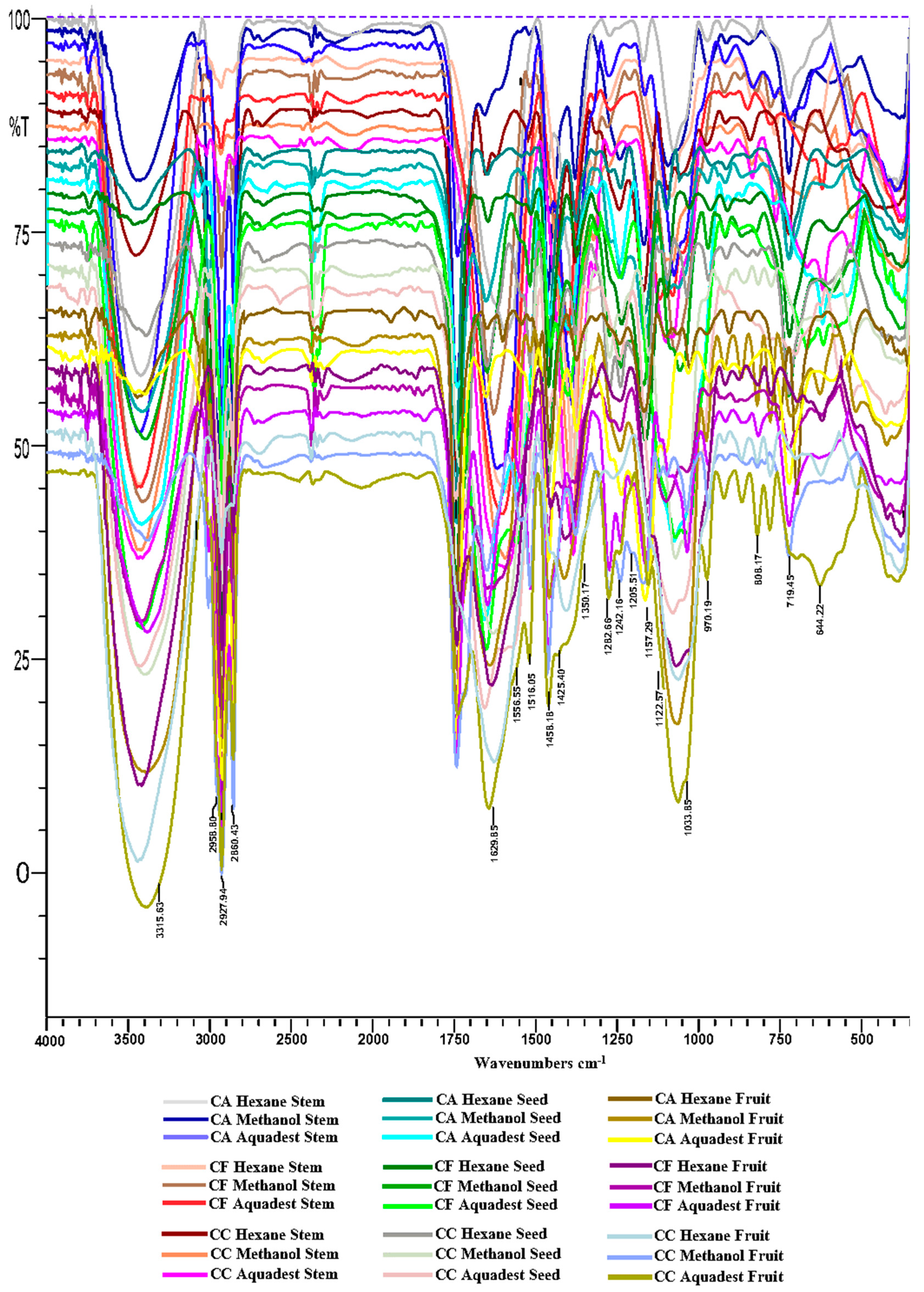
Figure 2.
The dendrogram illustrating the classification of samples through cluster analysis (CA), with samples in clusters 1 to 6 represented by distinct colored lines for each respective cluster.
Figure 2.
The dendrogram illustrating the classification of samples through cluster analysis (CA), with samples in clusters 1 to 6 represented by distinct colored lines for each respective cluster.
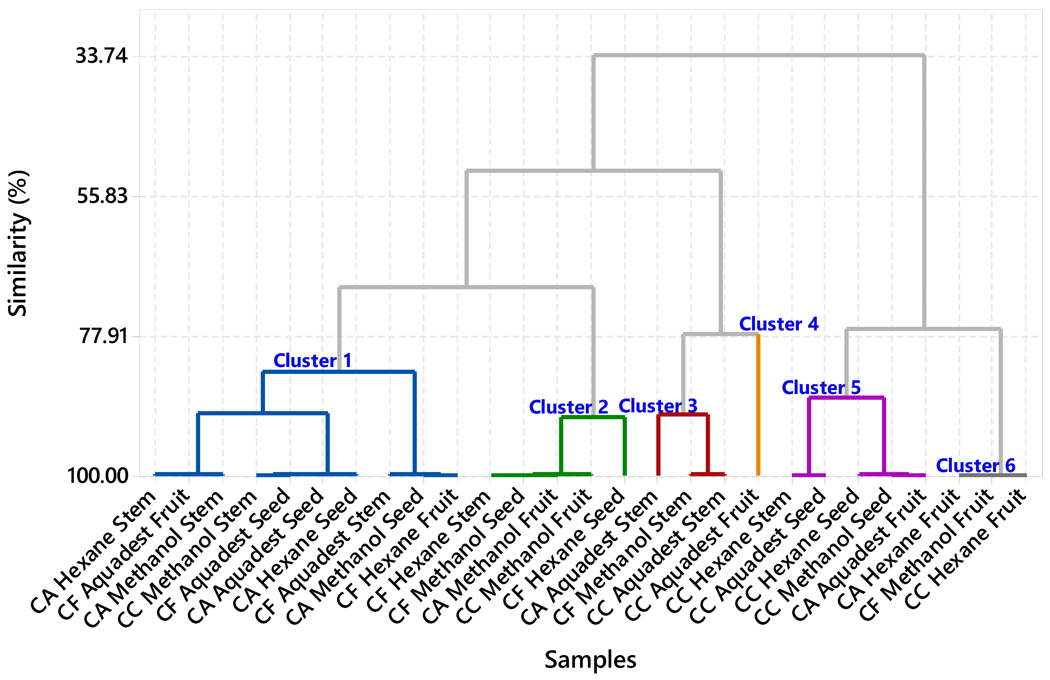
Figure 3.
Main Effects Plot and Interaction Plot Analysis of the Effects of Different Capsicum Extract Variations on Analgesic Activity.
Figure 3.
Main Effects Plot and Interaction Plot Analysis of the Effects of Different Capsicum Extract Variations on Analgesic Activity.
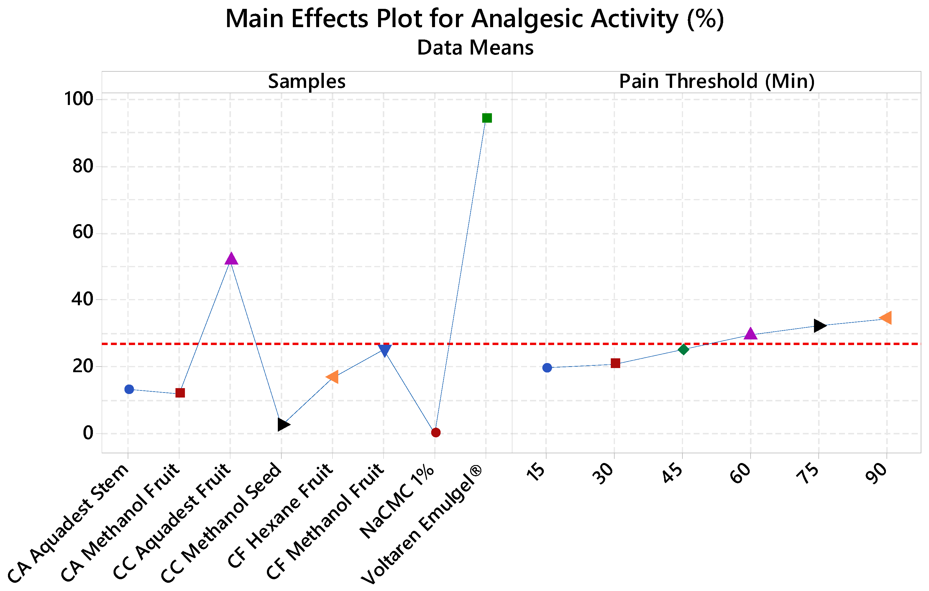

Figure 4.
Structure of the analgesic protein target (4DKI) (C) along with the native ligand (meclofenamic acid) (B). The re-docking of the native ligand (co-crystal) into the analgesic protein target pocket validates the method, resulting in a root mean square deviation (RMSD) value of 0.01 Å (A). Additionally, the interactions of the compounds in the extract, the native ligand, and the positive control with the analgesic protein target were illustrated, emphasizing their binding affinities and interactions within the target pocket.
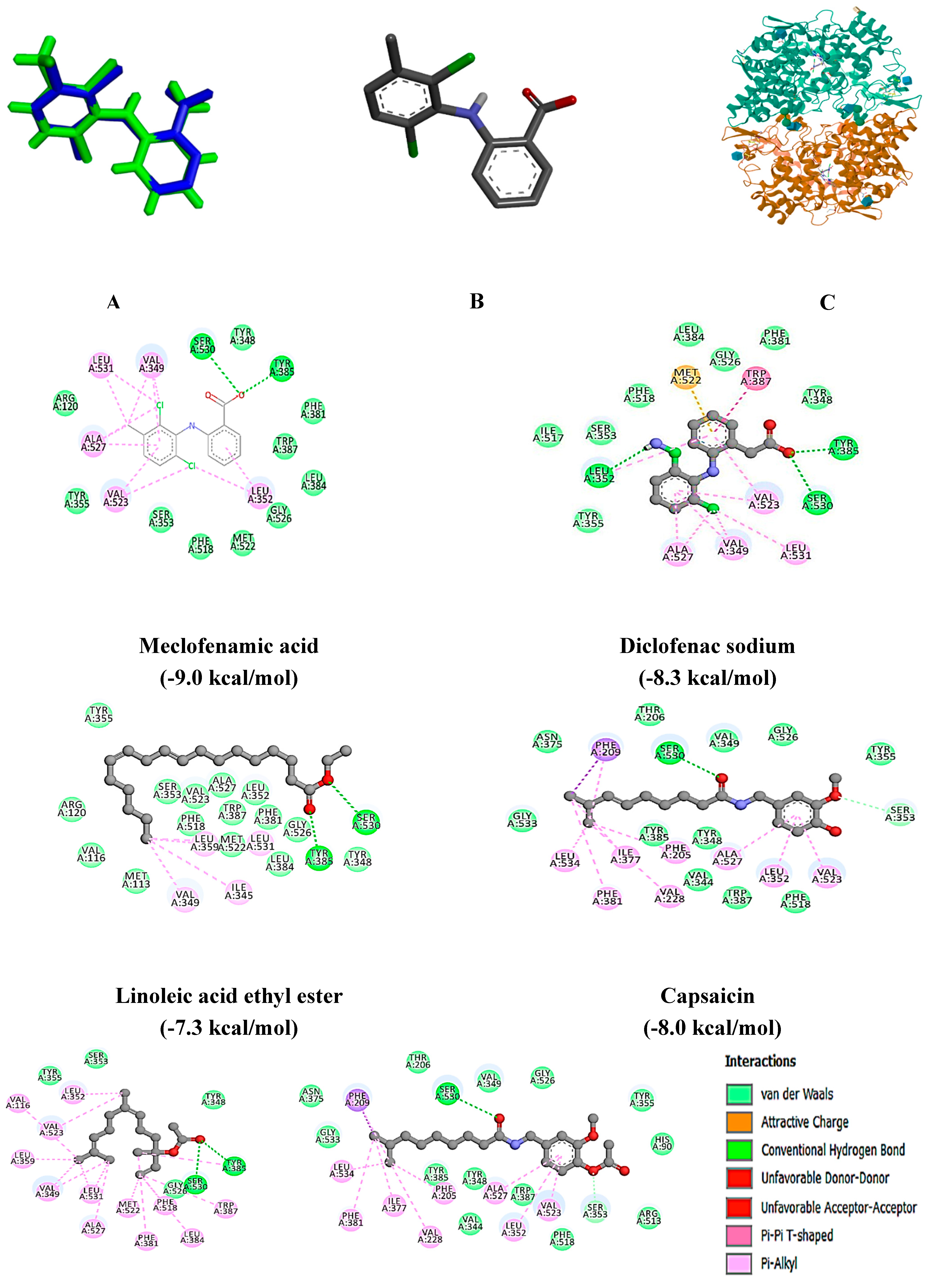

Figure 5.
Structure of the analgesic protein target (4TLM) (F) along with the native ligand (4-[(1R,2S)-3-(4-benzylpiperidin-1-yl)-1-hydroxy-2-methylpropyl]phenol) (E). The re-docking of the native ligand (co-crystal) into the analgesic protein target pocket validates the method, resulting in a root mean square deviation (RMSD) value of 1.321 Å (D). Additionally, the interactions of the compounds in the extract, the native ligand, and the positive control with the analgesic protein target were illustrated, emphasizing their binding affinities and interactions within the target pocket.
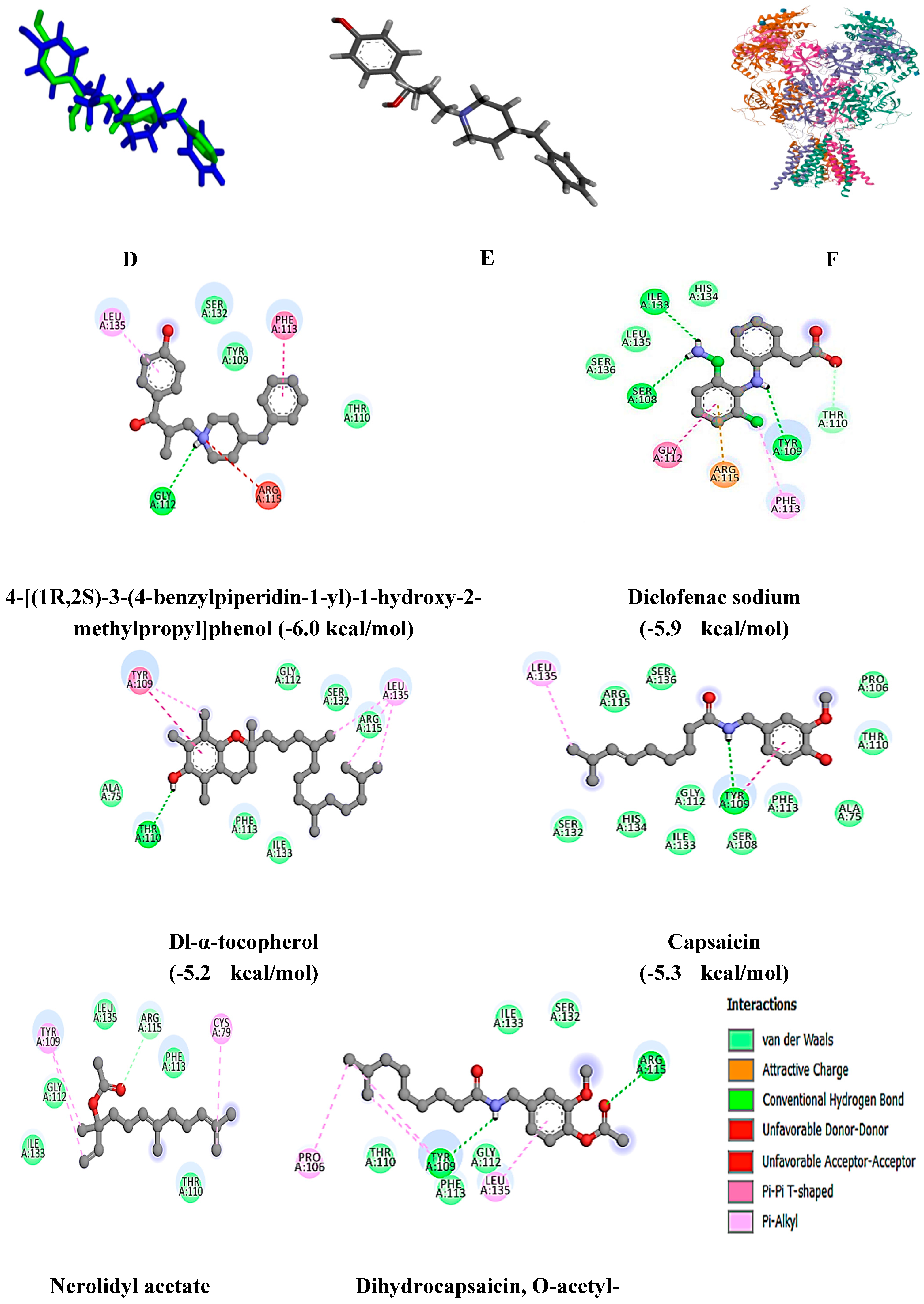

Figure 6.
Structure of the analgesic protein target (4EJ4) (I) along with the native ligand (4bS,8R,8aS,14bR)-7-(cyclopropylmethyl)-5,6,7,8,14,14b-hexahydro-4,8-methano[1]benzofuro[2,3-a]pyrido[4,3-b]carbazole-1,8a(9H)-diol) (H). The re-docking of the native ligand (co-crystal) into the analgesic protein target pocket validates the method, resulting in a root mean square deviation (RMSD) value of 0.01 Å (G). Additionally, the interactions of the compounds in the extract, the native ligand, and the positive control with the analgesic protein target were illustrated, emphasizing their binding affinities and interactions within the target pocket.
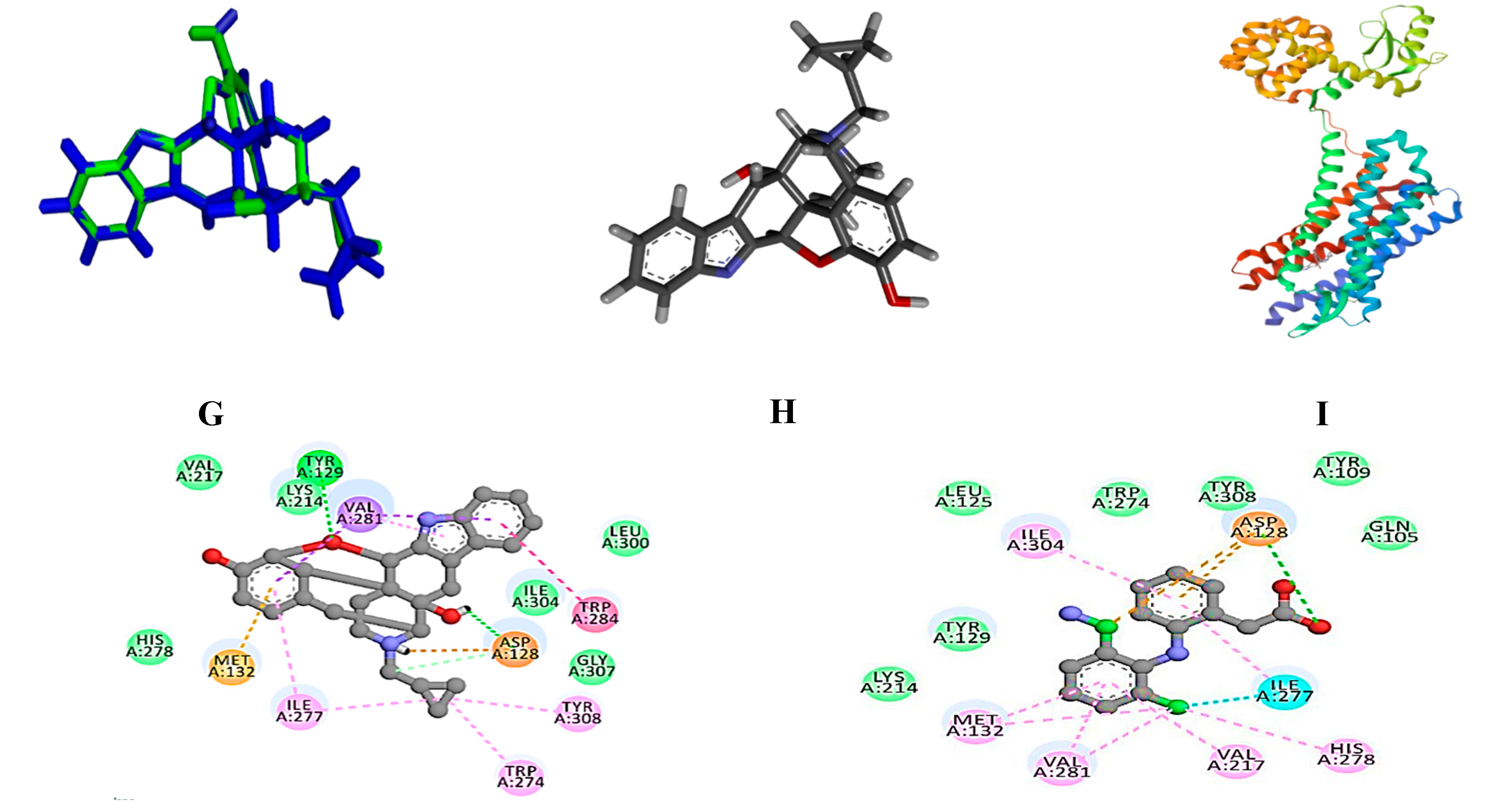
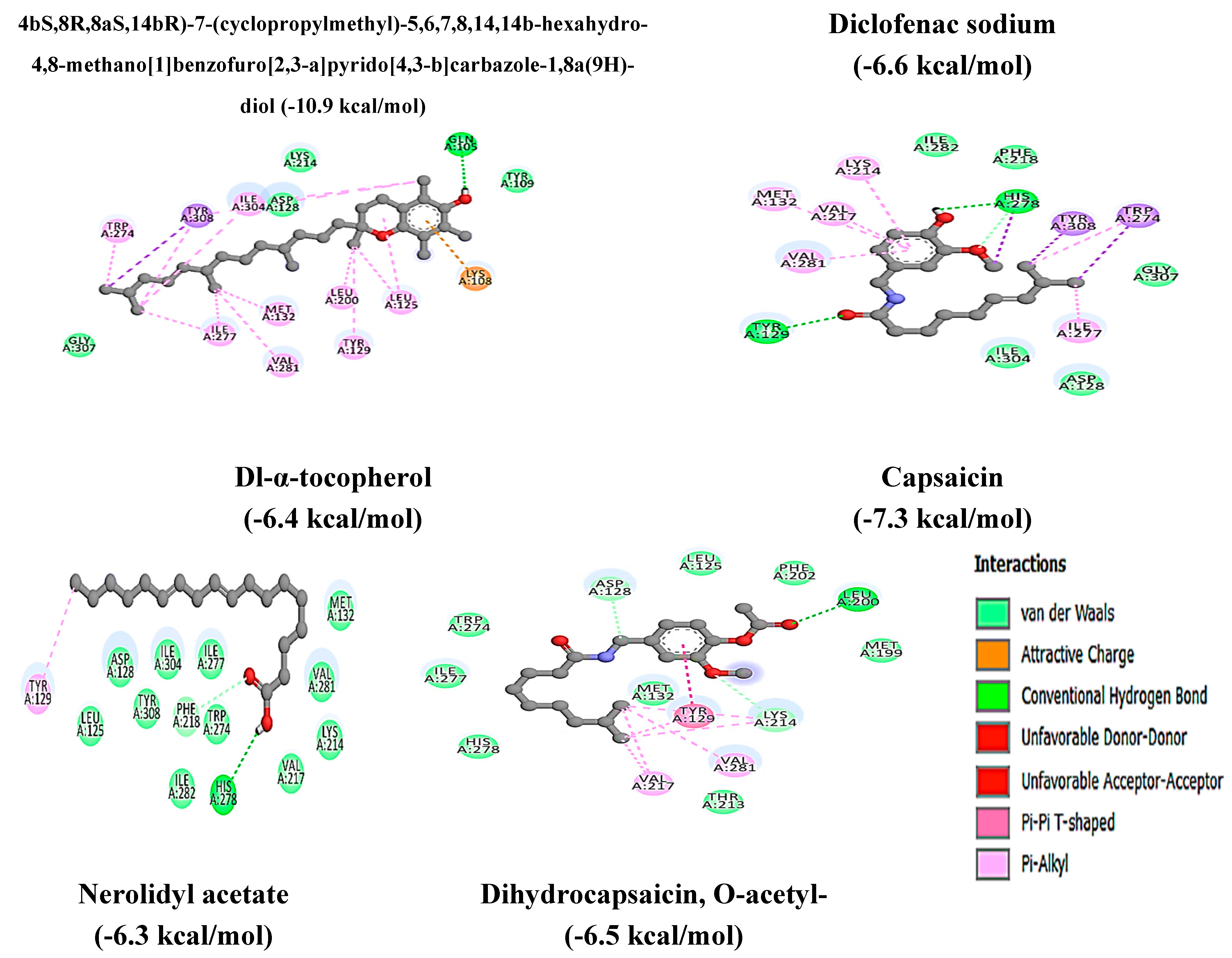

Table 1.
Percentage yield of extracts from different species, solvents and parts of Capsicum plants.
Table 1.
Percentage yield of extracts from different species, solvents and parts of Capsicum plants.
| Code | Samples | Plant Parts | Solvent |
Extract (g) | Yield (%) |
|---|---|---|---|---|---|
| CA | Capsicum annuum | Stem | Hexane | 0.1 | 0.5 |
| Methanol | 1.1 | 5.5 | |||
| Aquadest | 2.4 | 12 | |||
| Seed | Hexane | 2 | 10 | ||
| Methanol | 1 | 5 | |||
| Aquadest | 0.8 | 4 | |||
| Fruit | Hexane | 1 | 5 | ||
| Methanol | 2 | 10 | |||
| Aquadest | 3.6 | 18 | |||
| CF | Capsicum frutescens | Stem | Hexane | 0.2 | 1 |
| Methanol | 2 | 10 | |||
| Aquadest | 2.9 | 14.5 | |||
| Seed | Hexane | 1 | 5 | ||
| Methanol | 2 | 10 | |||
| Aquadest | 0.7 | 3.5 | |||
| Fruit | Hexane | 1 | 5 | ||
| Methanol | 3 | 15 | |||
| Aquadest | 4.7 | 23.5 | |||
| CC | Capsicum chinense | Stem | Hexane | 0.1 | 0.5 |
| Methanol | 1.7 | 8.5 | |||
| Aquadest | 1.4 | 7 | |||
| Seed | Hexane | 1 | 5 | ||
| Methanol | 1 | 5 | |||
| Aquadest | 0.8 | 4 | |||
| Fruit | Hexane | 1 | 5 | ||
| Methanol | 2 | 10 | |||
| Aquadest | 2.5 | 12.5 |
Table 2.
GC-MS analysis of compounds in hexane extract of Capsicum frutescens fruit and aquadest extract of Capsicum chinense fruit.
Table 2.
GC-MS analysis of compounds in hexane extract of Capsicum frutescens fruit and aquadest extract of Capsicum chinense fruit.
| Compouds Detected | Molecular formula | MW (g/mol) | Pubchem (CID) | RT (M in) | SI (%) | Area (%) |
|---|---|---|---|---|---|---|
| Capsicum frutescens (CF) hexane fruit extract | ||||||
| Oleic acid | C18H34O2 | 282 | 445639 | 19.025 | 88 | 1.06 |
| Methyl palmitate | C17H34O2 | 270 | 8181 | 21.127 | 97 | 3.90 |
| N-hexadecanoic acid | C16H32O2 | 256 | 985 | 22.129 | 95 | 6.74 |
| Methyl linoleate | C19H34O2 | 294 | 5284421 | 24.938 | 96 | 6.66 |
| Methyl oleate | C19H36O2 | 296 | 5364509 | 25.084 | 94 | 5.37 |
| 15-octadecenoic acid, methyl ester | C19H36O2 | 296 | 5364490 | 25.217 | 85 | 2.26 |
| Linoleic acid | C18H32O2 | 280 | 5280450 | 26.271 | 93 | 6.88 |
| N-pentadecylacetamide | C17H35NO | 269 | 12009452 | 26.659 | 85 | 6.59 |
| Nerolidol | C15H26O | 222 | 8888 | 29.086 | 86 | 22.64 |
| Heneicosane | C21H44 | 296 | 12403 | 29.417 | 94 | 3.10 |
| Isofucosterol | C29H48O | 412 | 5281326 | 29.892 | 89 | 1.23 |
| Pentacosane | C25H52 | 352 | 12406 | 33.233 | 97 | 1.06 |
| Hexacosane | C26H54 | 366 | 12407 | 36.600 | 96 | 1.08 |
| Squalene | C30H50 | 410 | 638072 | 38.342 | 98 | 3.43 |
| Nonacosane | C29H60 | 408 | 12409 | 39.667 | 97 | 1.42 |
| Dl-α-tocopherol | C29H50O2 | 430 | 2116 | 44.167 | 96 | 1.64 |
| Total | 75.06 | |||||
| Capsicum chinense (CC) aquadest fruit extract | ||||||
| Propanoic acid, 2-methyl- | C4H8O2 | 88 | 6590 | 4.700 | 87 | 3.50 |
| 1-Propanol, 2,2-dimethyl-, acetate | C7H14O2 | 130 | 13552 | 4.906 | 87 | 7.73 |
| Phenol, 2-methoxy- | C7H8O2 | 124 | 460 | 8.894 | 92 | 1.37 |
| (6E)-8-methyl- 6-nonenoic acid | C10H18O2 | 170 | 5365959 | 13.225 | 94 | 1.16 |
| Hexadecanoic acid, methyl ester | C17H34O2 | 270 | 8181 | 21.044 | 96 | 1.89 |
| Palmitoleic acid | C16H30O2 | 254 | 445638 | 21.668 | 97 | 1.31 |
| N-hexadecanoic acid | C16H32O2 | 256 | 985 | 22.076 | 95 | 12.58 |
| 9,12-Octadecadienoic acid (Z,Z)- | C19H34O2 | 294 | 5284421 | 26.080 | 96 | 1.68 |
| Oleic acid | C18H34O2 | 282 | 445639 | 26.167 | 85 | 1.59 |
| 7-tetradecenal, (Z)- | C14H26O | 210 | 5364468 | 26.270 | 87 | 2.19 |
| Linoleic acid ethyl ester | C20H36O2 | 308 | 5282184 | 26.372 | 94 | 2.37 |
| Octadecanoic acid | : C18H36O2 | 284 | 5281 | 26.717 | 93 | 2.91 |
| Nerolidyl acetate | C17H28O2 | 264 | 5363426 | 28.996 | 86 | 11.32 |
| Capsaicin | C18H27NO3 | 305 | 1548943 | 34.746 | 97 | 4.38 |
| Dihydrocapsaicin, O-acetyl- | C20H31NO4 | 349 | 91715808 | 35.182 | 89 | 2.26 |
| Total | 58.24 | |||||
Note: MW (molecular weight); PubChem (PubChem compound identifier); RT (retention time); SI (similarity index).
Table 3.
Bond-free energy values and amino acid residues binding to analgesic protein targets (5IKQ, 4TLM, and 4EJ4).
Table 3.
Bond-free energy values and amino acid residues binding to analgesic protein targets (5IKQ, 4TLM, and 4EJ4).
| Compounds | Protein Target | Bond-free energy (kcal/mol) | H-bond Interaction |
|---|---|---|---|
| Meclofenamic acid | 5IKQ | -9.0 | Tyr 385, Ser 530 |
| Diclofenac sodium | -8.3 | Leu 352, Tyr 358, Ser 530 | |
| Oleic acid | -6.7 | NI | |
| Methyl palmitate | -6.6 | Arg 120, Tyr 355 | |
| N-hexadecanoic acid | -6.5 | Arg 120, Tyr 355 | |
| Methyl linoleate | -6.9 | Arg 120, Tyr 355 | |
| Methyl oleate | -6.7 | Arg 120 | |
| 15-octadecenoic acid, methyl ester | -6.9 | Arg 120 | |
| Linoleic acid | -6.9 | Arg 120 | |
| N-pentadecylacetamide | -6.7 | Arg 120 | |
| Nerolidol | -6.9 | Arg 120 | |
| Heneicosane | -6.7 | NI | |
| Isofucosterol | -4.0 | NI | |
| Pentacosane | -7.2 | NI | |
| Hexacosane | -7.1 | NI | |
| Squalene | -8.5 | NI | |
| Nonacosane | -7.5 | NI | |
| Dl-α-tocopherol | -8.8 | NI | |
| Propanoic acid, 2-methyl- | -4.3 | ALA 151, ARG 496 | |
| 1-Propanol, 2,2-dimethyl-, acetate | -4.9 | ALA 527 | |
| Phenol, 2-methoxy- | -5.3 | MET 522, VAL 523, ALA 527 | |
| >(6E)-8-methyl- 6-nonenoic acid | >-6.3 | TYR 385, SER 530 | |
| >Hexadecanoic acid, methyl ester | -6.5 | ARG 120, TYR 355 | |
| Palmitoleic acid | >-6.7 | ARG 120, TYR 355 | |
| 9,12-octadecadienoic acid (Z,Z)- | -6.9 | ARG 120, TYR 355 | |
| 7-tetradecenal, (Z)- | -6.2 | ARG 120, TYR 355 | |
| Linoleic acid ethyl ester | -7.3 | TYR 385, SER 530 | |
| Octadecanoic acid | -6.8 | ARG 120 | |
| Nerolidyl acetate | -7.4 | TYR 385, SER 530 | |
| Capsaicin | -8.0 | SER 353, SER 530 | |
| Dihydrocapsaicin, O-acetyl- | -8.0 | SER 353, SER 530 | |
| 4-[(1R,2S)-3-(4-benzylpiperidin-1-yl)-1-hydroxy-2-methylpropyl]phenol | 4TLM | -6.0 | Gly 112 |
| Diclofenac sodium | -5.9 | Ile 133, Ser 108, Tyr 109, Thr 110 | |
| Oleic acid | -4.3 | Ser 132 | |
| Methyl palmitate | -4.2 | Thr 110 | |
| N-hexadecanoic acid | -3.7 | Gly 112, Leu 135 | |
| Methyl linoleate | -4.3 | Thr 110 | |
| Methyl oleate | -3.9 | NI | |
| 15-octadecenoic acid, methyl ester | -4.0 | NI | |
| Linoleic acid | -4.2 | NI | |
| N-pentadecylacetamide | -4.5 | Ser 132, Ile 133 | |
| Nerolidol | -4.9 | NI | |
| Heneicosane | -3.9 | NI | |
| Isofucosterol | -6.0 | NI | |
| Pentacosane | -3.7 | NI | |
| Hexacosane | -3.4 | NI | |
| Squalene | -4.5 | NI | |
| Nonacosane | -3.6 | NI | |
| Dl-α-tocopherol | -5.2 | Thr 110 | |
| Propanoic acid, 2-methyl- | -3.5 | Ser 108, His 134, Leu 135, Ser 136 | |
| 1-Propanol, 2,2-dimethyl-, acetate | -3.8 | Tyr 109, Gly 112, Leu 135, Ser 136 | |
| Phenol, 2-methoxy- | -4.3 | His 134 | |
| (6E)-8-methyl- 6-nonenoic acid | -3.4 | Gly 112 | |
| Hexadecanoic acid, methyl ester | -3.7 | Gly 112, Leu 135 | |
| Palmitoleic acid | -4.2 | NI | |
| 9,12-octadecadienoic acid (Z,Z)- | -4.2 | NI | |
| 7-tetradecenal, (Z)- | -4.0 | NI | |
| Linoleic acid ethyl ester | -4.2 | Arg 115 | |
| Octadecanoic acid | -3.8 | Ile 133 | |
| Nerolidyl acetate | -4.9 | Arg 115 | |
| Capsaicin | -5.3 | Tyr 109 | |
| Dihydrocapsaicin, O-acetyl- | -4.9 | Tyr 109, Arg 115 | |
| (4bS,8R,8aS,14bR)-7-(cyclopropylmethyl)-5,6,7,8,14,14b-hexahydro-4,8-methano[1]benzofuro[2,3-a]pyrido[4,3-b]carbazole-1,8a(9H)-diol | 4EJ4 | -10.9 | Asp 128, Tyr 129 |
| Diclofenac sodium | -6.6 | Asp 128 | |
| Oleic acid | -5.6 | Gln 105, Lys 108, Tyr 109 | |
| Methyl palmitate | -4.8 | NI | |
| N-hexadecanoic Acid | -5.3 | Gly 307, Tyr 308 | |
| Methyl linoleate | 6.0 | Met 199, Leu 200 | |
| Methyl oleate | -5.7 | NI | |
| 15-octadecenoic acid, methyl ester | -5.3 | His 278 | |
| Linoleic acid | -5.4 | Phe 218, His 278 | |
| N-pentadecylacetamide | -5.2 | Asp 128, Ile 304 | |
| Nerolidol | -6.5 | NI | |
| Heneicosane | -5.2 | NI | |
| Isofucosterol | -7.7 | NI | |
| Pentacosane | -5.2 | NI | |
| Hexacosane | -5.0 | NI | |
| Squalene | -6.8 | NI | |
| Nonacosane | -5.3 | NI | |
| Dl-α-tocopherol | -6.4 | Gln 105 | |
| Propanoic acid, 2-methyl- | -4.1 | Asp 128, Tyr 308 | |
| 1-Propanol, 2,2-dimethyl-, acetate | -4.6 | NI | |
| (6E)-8-methyl- 6-nonenoic acid | -5.0 | NI | |
| Hexadecanoic acid, methyl ester | -5.3 | Gly 307, Tyr 308 | |
| Palmitoleic acid | -5.6 | Phe 218, His 278 | |
| 9,12-octadecadienoic acid (Z,Z)- | -5.5 | Lys 214, His 278 | |
| 7-tetradecenal, (Z)- | -5.4 | NI | |
| Linoleic acid ethyl ester | -5.8 | NI | |
| Octadecanoic acid | -5.7 | Gln 105, Tyr 308 | |
| Nerolidyl acetate | -6.3 | His 278, Phe 218 | |
| Capsaicin | -7.3 | Tyr 219, His 278 | |
| Dihydrocapsaicin, O-acetyl- | -6.5 | Asp 128, Lys 214, Leu 200 |
Note: “NI” indicates that the amino acid residues did not interact with the analgesic protein target (5IKQ, 4TLM, and 4EJ4) via H-bond interactions and were not found in the native ligand interactions.
Disclaimer/Publisher’s Note: The statements, opinions and data contained in all publications are solely those of the individual author(s) and contributor(s) and not of MDPI and/or the editor(s). MDPI and/or the editor(s) disclaim responsibility for any injury to people or property resulting from any ideas, methods, instructions or products referred to in the content. |
© 2025 by the authors. Licensee MDPI, Basel, Switzerland. This article is an open access article distributed under the terms and conditions of the Creative Commons Attribution (CC BY) license (http://creativecommons.org/licenses/by/4.0/).
Copyright: This open access article is published under a Creative Commons CC BY 4.0 license, which permit the free download, distribution, and reuse, provided that the author and preprint are cited in any reuse.


