Preprint
Article

This version is not peer-reviewed.

Sanguinarine-Chelerythrine from Coptis chinensis Offers Analgesic and Anti-Inflammatory Effects Without Gastrotoxicity

A peer-reviewed article of this preprint also exists.

Submitted:

25 December 2024

Posted:

26 December 2024

You are already at the latest version

Abstract

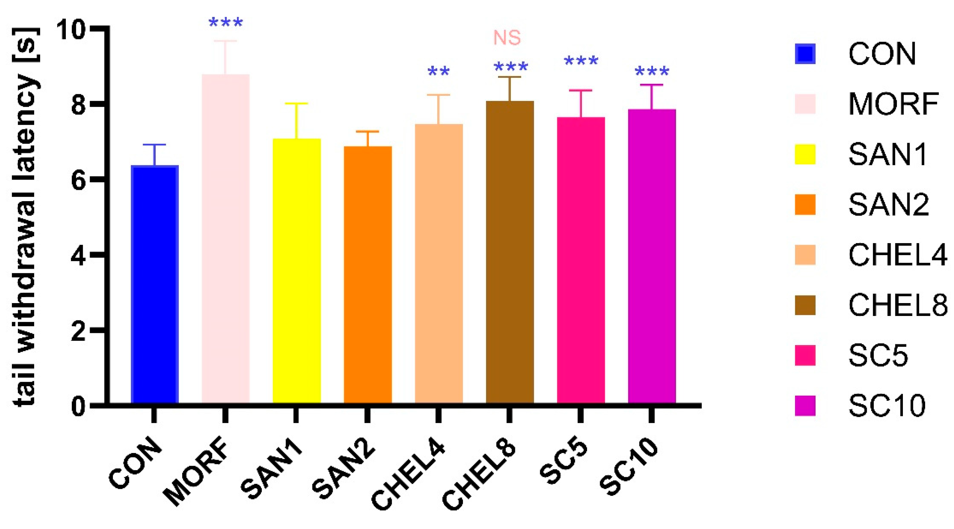
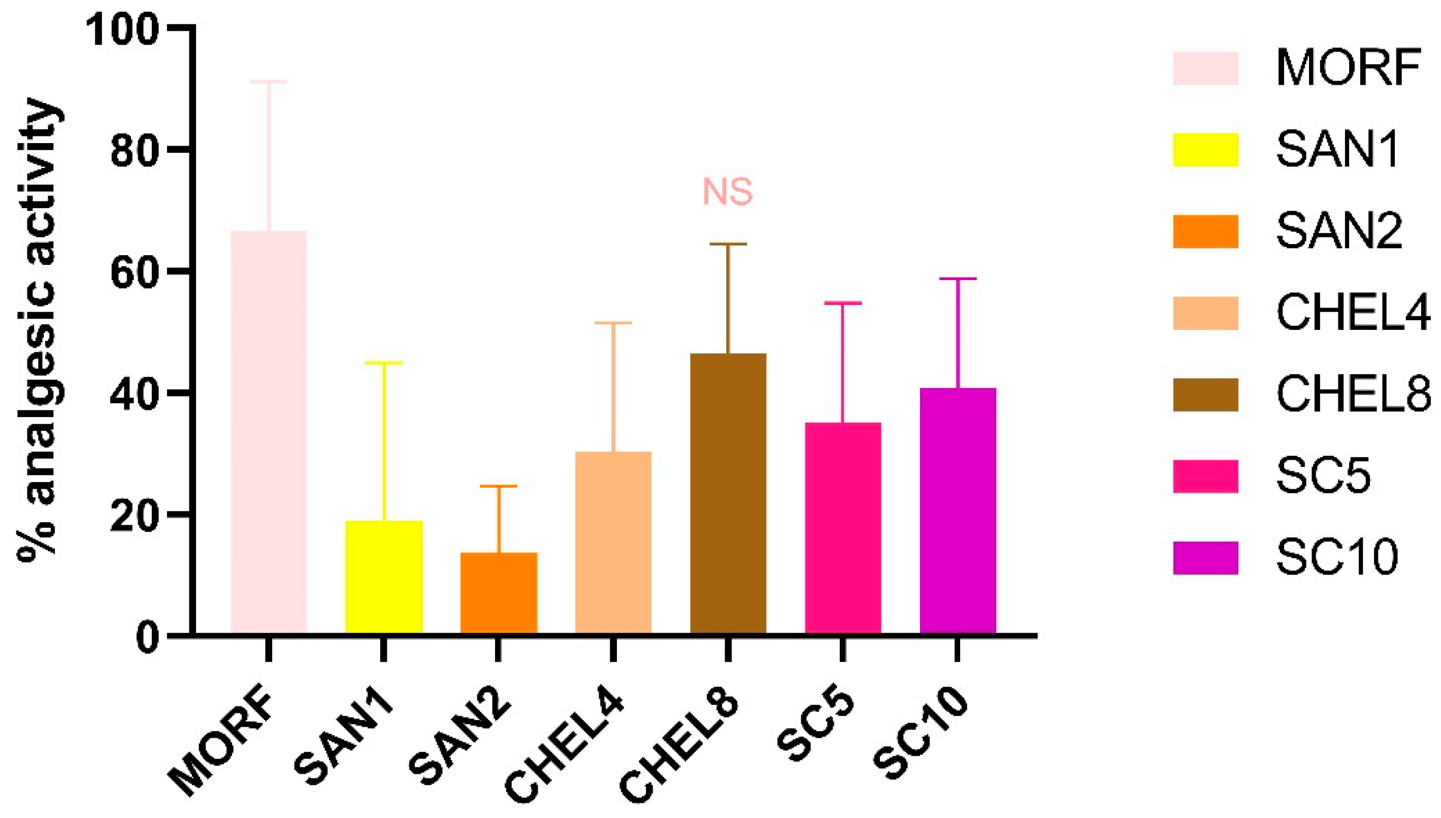
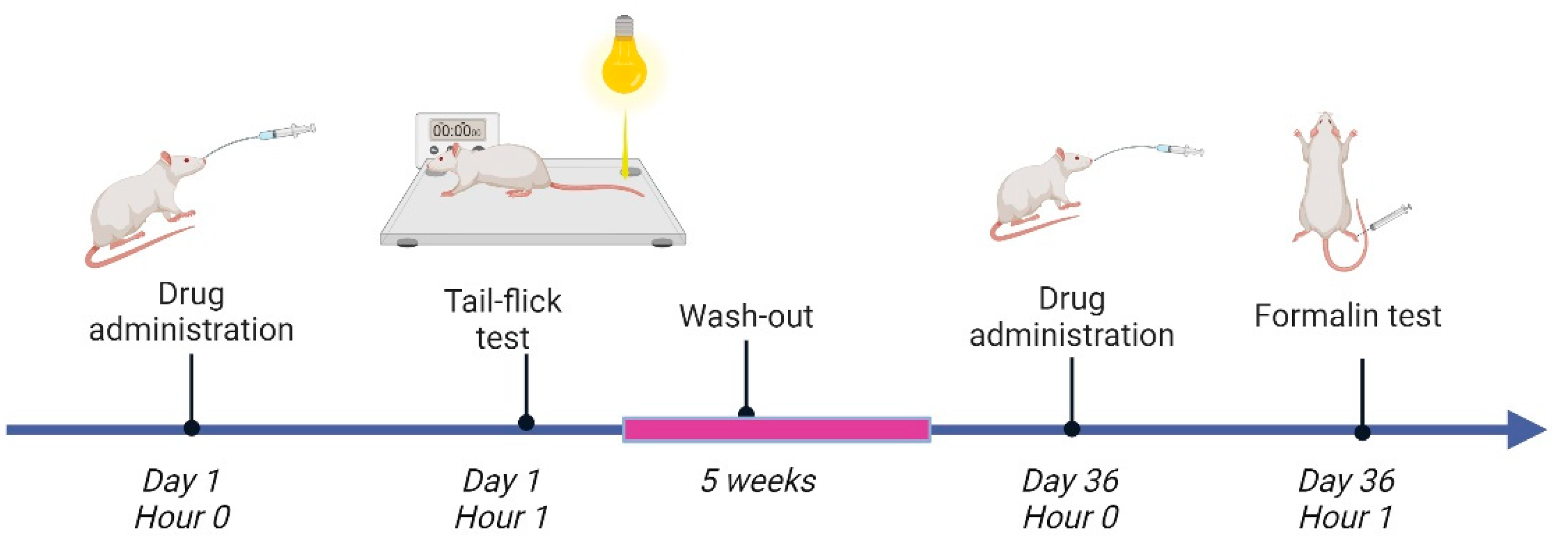
Background: Pain is a major clinical and socioeconomic problem worldwide and existing therapies are not always effective and are often associated with the burden of adverse effects that limit their usage. Natural compounds are an important group of pharmaceuticals that may be used in pain management. We aimed to investigate the analgesic activity of the sanguinarine-chelerythrine from C. chinensis. Methods: The analgesic and anti-inflammatory activity of sanguinarine-chelerythrine fraction of C. chinensis (SC 5 and10mg/kg), sanguinarine (SAN 1 and 2 mg/kg) and chelerythrine (CHEL 4 and 8 mg/kg ) was assessed in the tail-flick and formalin tests. Microscopic and macroscopic examination of stomach mucosae was performed. TNFα and MMP-9 levels were measured with ELISA kits. Results: Morfine (MORF), CHEL and SC prolongated the tail withdrawal latency and with comparable analgesic activity of MRF and CHEL 8 mg/kg. MORF, CHEL 8 mg/kg, and SAN 2 mg/kg ameliorated the pain reaction in the neurogenic phase of the formalin test. In the inflammatory phase of the formalin test, all tested substances exerted analgesic activity. SAN, CHEL and SC reduced additionally TNFα and MMP-9 secretion.. Conclusions: Our results confirmed analgesic effects of CHEL and SC with CHEL analgesic activity comparable to MORF. All investigated substances exerted significant anti-inflammatory activity without concomitant gastrotoxicity.

Keywords: 
;  ;  ;  

1. Introduction

Pain is a major clinical and socioeconomic problem worldwide [1,2,3]. International Association for the Study of Pain (IASP) defines pain as “an unpleasant sensory and emotional experience associated with, or resembling that associated with, actual or potential tissue damage” [4]. Pain significantly impacts individuals’ quality of life and increases the costs to the health system. Additionally, chronic pain belongs to the leading causes of disability and disease burden globally [5]. It is important to bear in mind that untreated or ineffectively treated pain is associated with an increased prevalence of comorbidities. Chronic persistent pain has been associated, among others, with the development of reduced cognitive function, insomnia, sexual dysfunction, and various depressive and anxiety disorders [6]. However, existing therapies are not always effective and are often associated with the burden of adverse effects that limit their usage. Nonsteroidal anti-inflammatory drugs (NSAIDs) are very efficient in the treatment of inflammatory pain, but gastrointestinal side effects often restrict their administration. Opioid drugs are another effective analgesic option, however, although they do not increase the risk of gastric bleeding, their long-term administration is associated with a significant risk of drug abuse. Therefore, it is extremely important to search for new safer therapeutic options, and natural compounds are an important group of pharmaceuticals that may be used in pain management.
Rhizomes of Coptis chinensis are known in Chinese Pharmacopoeia as Coptidis Rhizoma (CR) or Huang Lian [7]. CR belongs to one of the most important components of many traditional Chinese formulations that have been used in the treatment of diarrhea, other gastrointestinal disorders, diabetes, toothache, inflammatory disorders, fever, and skin diseases [8]. Shengyang Sanhuo Decoction containing CR is prescribed in Traditional Chinese Medicine, among others, in the treatment of neuropathic pain [9], and its effectiveness is attributed to the inhibition of TNFα, IL-6, and CRP production. Various contemporary studies confirmed the anti-inflammatory [10,11,12] and analgesic [7] activity of C. chinensis. However, most studies focus on berberine as the most abundant isoquinoline alkaloid present in CR [13,14]. CR contains over 100 various chemical constituents, including, among others, berberine, sanguinarine, and chelerythrine, which are the main bioactive ingredients [8]. It has been suggested that the analgesic activity of CR may be attributed to the inhibition of TRPV1 (transient receptor potential cation channel subfamily V member 1) and NF-κB (nuclear factor kappa-light-chain-enhancer of activated B cells) overexpression and the activation of the JNK/p38 MAPK (c-Jun N-terminal kinase/p38 mitogen-activated protein kinase) pathways [9]. However, further studies investigating the activity of individual constituents are needed.
Alkaloids are nitrogen-containing chemical compounds that are found in plants, and several investigations have demonstrated their various biological activity. Alkaloids found in CR can be divided into several subgroups according to their structures. Berberine, the most abundant alkaloid, is a protoberberine derivative, whereas sanguinarine and chelerythrine are benzophenanthridine (BZD) quaternary amine alkaloids (Figure 1).
Plants producing BZDs have played an important role in traditional folk medicine for centuries. Both sanguinarine and chelerythrine have demonstrated anti-inflammatory, anticancer, and antiviral activity in various studies [15,16]. It has also been reported that sanguinarine interferes with nerve impulse transmission due to its inhibitory effects on choline acetyltransferase activity and hindering of nicotinic, muscarinic, and serotonin receptors activity [15]. Some authors reported also that sanguinarine may ameliorate neuropathic pain [17,18,19]. Although the chemical structure of sanguinarine and chelerythrine is similar, it was reported that they exert different effects on glycine transporters [20], which are promising targets for analgesic therapy. Therefore, it seems to be of great relevance to compare the analgesic activity of both alkaloids.
In our previous study, we demonstrated the anti-inflammatory activity of the sanguinarine-chelerythrine fraction of the C. chinensis extract in the carrageenan paw edema test [21]. In the reported study, we aimed to investigate the analgesic activity of the sanguinarine-chelerythrine fraction of the C. chinensis extract and broaden the knowledge on the activity of both alkaloids by comparison of the analgesic and anti-inflammatory activity of isolated constituents and their mixture. The doses investigated in the reported study were based on the previous results [21].

2. Results

2.1. Tail-Flick Test

There was a significant prolongation of the tail withdrawal latency in the groups receiving morphine, chelerythrine, and the investigational fraction of C. chinensis containing sanguinarine and chelerythrine compared to the negative control group (MORF: 8.79 ± 0.89 s, CHEL4: 7.48 ± 0.77 s, CHEL8: 8.08 ± 0.65 s, SC5: 7.66 ± 0.71 s, and SC10: 7.87 ± 0.65 s vs. CON: 6.39 ± 0.54 s, respectively), whereas there was no significant prolongation of tail-flick latency in groups receiving sanguinarine (SAN1: 7.08 ± 0.04 s, SAN2: 6.88 ± 0.40 s). Additionally, no significant difference in the tail withdrawal latency between MORF and CHEL8 groups was detected (Figure 2).
Analysis of the percentage of analgesic activity (Figure 3) revealed that there is no significant difference in analgesic activity between morphine 10 mg/kg and chelerythrine 8 mg/kg (66,6 % ± 24.6 % vs. 46.7% ± 18.1%). In all the other groups, significantly lower analgesic activity was detected. Percentage of analgesic activity of sanguinarine (1 and 2 mg/kg), chelerythrine 4 mg/kg, and investigational fraction of C. chinensis containing sanguinarine and chelerythrine 5 and 10 mg/kg was significantly lower than in the animals receiving morphine 10 mg/kg (SAN1: 19.0 % ± 26.0%, SAN2: 13.7% ± 11.1%, CHEL4: 30.3% ± 21.2%, SC5: 35.1% ± 19.7%, and SC10: 40.9% ± 17.9%, respectively).

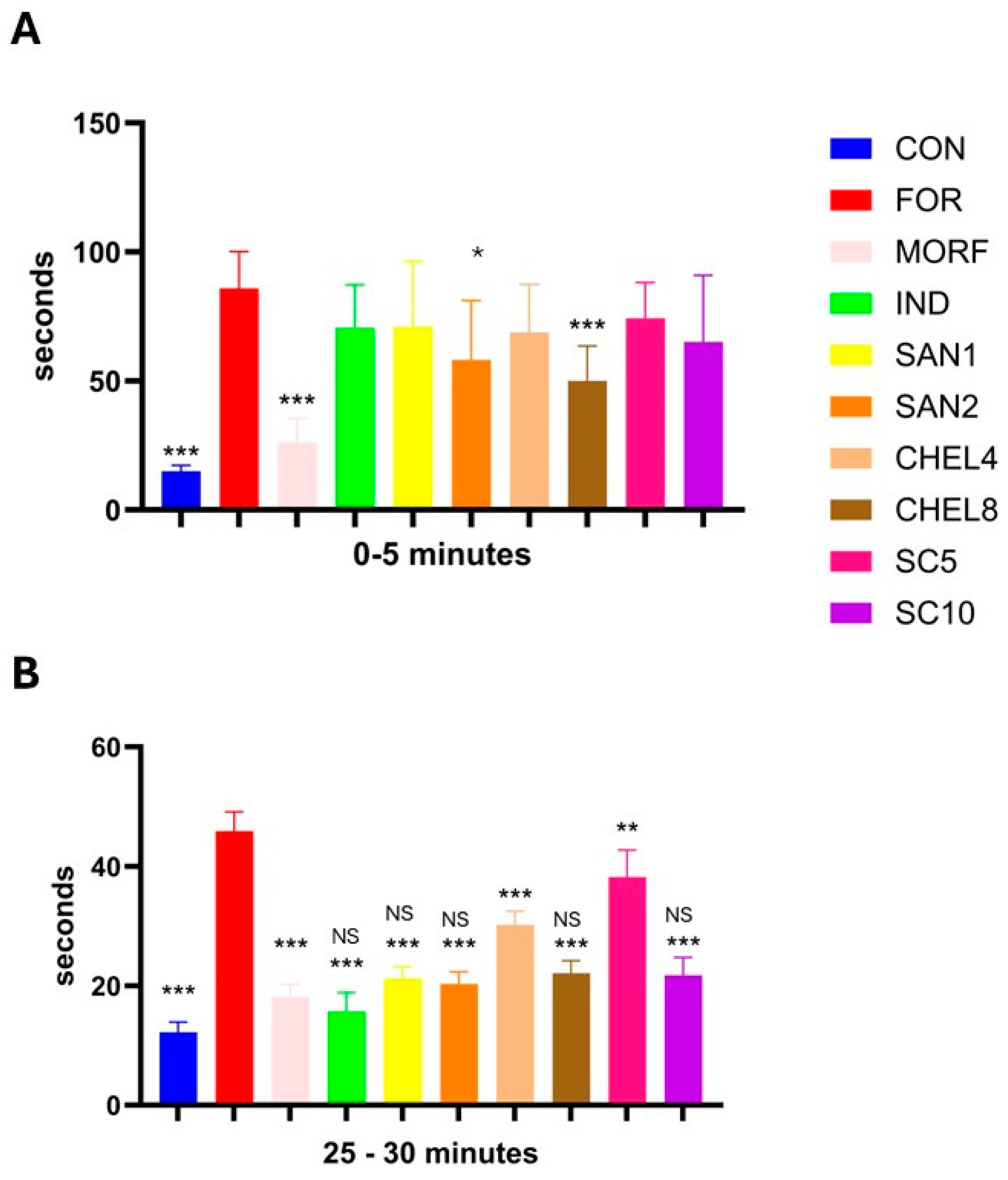
2.2. Formalin Test (Figure 4)

Formalin significantly prolonged the pain reaction in both phases, the neurogenic and the inflammatory one, during the formalin test compared to the control group that received a saline injection (87 ± 13 s vs. 14 ± 3 s, and 45 ± 3 s vs. 12 ± 2 s, respectively). Only morphine 10 mg/kg, chelerythrine 8 mg/kg, and sanguinarine 2 mg/kg ameliorated the pain reaction in the neurogenic phase of the formalin test (MORF: 26 ± 7 s, CHEL8: 50 ± 9 s, and SAN2 58 ± 14 s). However, the analgesic response to morphine was significantly more pronounced than the response to the investigated alkaloids. In the inflammatory phase of the formalin test, a significant reduction of pain response was observed in all groups receiving investigated substances (CHEL, SAN, and SC), as well as in the groups receiving morphine and indomethacin as reference drugs.
Figure 4. Effects of SC, SAN, and CHEL on the pain reaction (the duration of licking, flinching, shaking, and biting in seconds) during the neurogenic (A - early; 0 – 5 min) and inflammatory (B - late; 25 - 30 min) phase of the nociceptive reaction in the formalin test. Experimental groups: CON – a negative control group, FOR – a positive control group, MORF – animals receiving morphine 10 mg/kg, IND – animals receiving indomethacin 10 mg/kg, SAN1 – animals receiving sanguinarine 1 mg/kg, SAN2 – animals receiving sanguinarine 2 mg/kg, CHEL4 – animals receiving chelerythrine 4 mg/kg, CHEL8 – animals receiving chelerythrine 8 mg/kg, SC5 - animals receiving investigational fraction of C. chinensis containing sanguinarine and chelerythrine 5 mg/kg, SC10 - animals receiving investigational fraction of C. chinensis containing sanguinarine and chelerythrine 10 mg/kg. * - p < 0.05 vs. FOR, ** - p < 0.01 vs. FOR, *** - p < 0.001 vs. FOR, NS – not significant vs. MORF.
Preprints 144143 g004

2.3. MMP-9 and TNFα in Paw Homogenates

Formalin injection induced inflammation of paw tissues, leading to an increased concentration of MMP-9 and TNFα in paw homogenates (Table 1). This was ameliorated by the administration of indomethacin, a reference anti-inflammatory drug. Reduction of inflammatory response was also observed in animals receiving tested alkaloids and the investigational fraction of C. chinensis. Additionally, there was no significant difference in inhibitory effect on MMP-9 and TNFα secretion between the IND group and groups receiving higher CHEL, SAN, and SC doses.
Experimental groups: CON – a negative control group, FOR – a positive control group, MORF – animals receiving morphine 10 mg/kg, IND – animals receiving indomethacin 10 mg/kg, SAN1 – animals receiving sanguinarine 1 mg/kg, SAN2 – animals receiving sanguinarine 2 mg/kg, CHEL4 – animals receiving chelerythrine 4 mg/kg, CHEL8 – animals receiving chelerythrine 8 mg/kg, SC5 - animals receiving investigational fraction of C. chinensis containing sanguinarine and chelerythrine 5 mg/kg, SC10 - animals receiving investigational fraction of C. chinensis containing sanguinarine and chelerythrine 10 mg/kg.
^ - p < 0.05 vs. CON, ^^ - p < 0.01 vs. CON, * - p < 0.05 vs. FOR, ** - p < 0.01 vs. FOR, *** - p < 0.001 vs. FOR, NS p > 0.05 vs. IND.

2.4. Histopathological Assessment of Gastric Mucosa

To assess the gastric safety profile of the investigated compounds, macroscopic and microscopic examinations of the stomachs were performed. Macroscopic and microscopic examinations of the gastric mucosa did not reveal any significant gastric injuries in the experimental groups.

3. Discussion

In this study, we assess the antinociceptive effects of sanguinarine chloride (SAN), chelerythrine chloride (CHEL), and sanguinarine-chelerythrine fraction of Coptis chinensis extract (SC) in the rat model. Our goal is not only to evaluate the effectiveness of the above compounds but also to compare their analgesic properties and safety of use. The most important findings are that in the case of the groups receiving chelerythrine and the sanguinarine-chelerythrine fraction of C. chinensis extract, there was a significant prolongation of the tail withdrawal latency compared to the negative control group in the tail-flick test, and also there was no relevant difference in the analgesic activity in the group receiving a higher dose of chelerythrine and morphine. Moreover, in the formalin test, we have shown that in both of the mentioned groups – MORF and CHEL8 – there was a significant reduction in the pain reaction cumulative time in the early, neurogenic phase, while in the late inflammatory phase we observed such a decrease in several research groups, especially receiving higher doses of the tested substances – sanguinarine, chelerythrine, and sanguinarine-chelerythrine fraction of C. chinensis extract.
To our knowledge, this is the first experiment to compare the analgesic and anti-inflammatory activity of C. chinensis extract and the two alkaloid components administered separately. The current work is a continuation of our previous one, in which we proved, among others, that the sanguinarine-chelerythrine fraction of C. chinensis extract possesses anti-inflammatory activity comparable to indomethacin and does not cause damage to the gastric mucosa [21]. In the reported study we try to answer the question of whether the natural mixture of alkaloids from the extract has a stronger or weaker effect than the corresponding doses of substances administered separately and to assess the dose-effect dependency. Additionally, to assessment of anti-inflammatory activity in the current study we assess the analgesic effect of the investigated agents, that was not studied before.
In the tail-flick test, we observed that the reaction time to pain stimuli in the form of a heat source was significantly longer in animals from the MORF, CHEL8, SC5, and SC10 groups compared to the CON control group. The substances administered to the animals: morphine 10 mg/kg, chelerythrine 8 mg/kg, and C. chinensis extract at both tested doses, i.e., 5 mg/kg and 10 mg/kg, respectively, showed thereby a relevant analgesic effect, substantially extending the tail withdrawal latency. We also obtained a similar result, although to a slightly lesser degree, in the group receiving a lower dose of chelerythrine (4 mg/kg). Nevertheless, the above results can be summarized with a conclusion that in all research groups receiving chelerythrine or extract, regardless of the dose, we obtained a statistically significant extension of the tail withdrawal latency, confirming the promising antinociceptive potential of the tested substances. The tail withdrawal latency time, especially in the CHEL8 and SC10 groups, was prolonged by about 1,5 seconds compared to the control group. With the average time for the CON group of 6.39 s, it increases time latency by 26,4% and 23,2%, respectively. The most positive result in such a comparison was certainly obtained in the MORF group - an extension of almost 2,5 seconds, i.e. 37,6%.
At the same time, in both research groups receiving sanguinarine, SAN1 and SAN2 (1 mg/kg and 2 mg/kg), we did not note a significant result, which at first glance may indicate that of both components of the extract fraction, it is the chelerythrine that is the more influential one in the context of the analgesic effect. However, it is worth remembering that the doses of both active substances administered separately differ four times (1 mg/kg and 2 mg/kg for sanguinarine and 4 mg/kg and 8 mg/kg for chelerythrine), therefore, a clear comparison of the activity of both compounds is impossible to make. The use of the above doses of mono-administered substances results from the composition of the extract, in which both compounds occur approximately in the proportion 1+4, and our attempt to precisely determine the properties of the extract, which is the basic research material in this study.
Due to the application of such proportion, we could observe further interesting results. Firstly, the latency time in the CHEL4 group (corresponding to a lower dose of extract) was slightly shorter than in the SC5 group, but in the case of the CHEL8 group, it was longer than in the SC10 group. Additionally, in the SAN1 group, the latency time was longer than in the SAN2 group. It may suggest a decreasing role of sanguinarine in obtaining an analgesic effect with an enhancement in the dose of the extract. This is, of course, a far-reaching conclusion based on the available data, but it is certainly a hypothesis worth further exploring. Secondly, the antinociceptive effect of the extract was not the sum of the impacts of sanguinarine and chelerythrine, and simply adding the results obtained for both substances administered separately did not translate into the effect of the corresponding doses of the extract. This was an expected result, but we achieved its specific experimental validation.
Translating the above observations into the analysis of analgesic activity, three research groups: MORF, CHEL8, and SC10 were characterized by the highest average values ​​of analgesic activity, 66.6%, 46.7%, and 40.9%, respectively. And even though the result of the SC10 group was relatively high, it was statistically different from MORF. There was no relevancy in the comparison of MORF vs. CHEL8. It is therefore justified to conclude that the analgesic activity of both groups is comparable, i.e. the use of chelerythrine at a dose of 8 mg/kg gives, to some extent, a similar antinociceptive effect to that of morphine at a dose of 10 mg/kg. The C. chinensis extract, although weaker in comparison, provides grounds for further analysis with the potential to obtain promising results. The findings presented above may suggest the extract’s dose-dependency, so it can be assumed that increasing the dose of the extract may enhance the analgesic activity and obtain an effect at least comparable to morphine in 10 mg/kg. However, these are only the authors' speculations that require further confirmation.
One of the probable mechanisms of the analgesic effect of the tested alkaloids is the impact on nociceptive signals by modulating glycine transporters. Jursky et al. found that chelerythrine and sanguinarine selectively inhibit the glycine transporter GlyT1 with similar activity in the low micromolar range. GlyT1 inhibition by sanguinarine was irreversible, whereas the chelerythrine effect washed out over time [20]. Another mechanism – inhibition of the microglia and p38 MAPK signaling pathway activation – was confirmed in the neuropathic pain studies [19,22].
Outcomes corresponding to ours were obtained in several studies but were mainly concerned with chelerythrine. Wen et al. confirmed attenuation of pertussis toxin (PTX) - induced thermal hyperalgesia by i.a. chelerythrine, and associated this effect with protein kinase C inhibition. Additionally, they observed that chelerythrine could alleviate the PTX-induced reduction of morphine analgesia [23]. Similar results, including a complete reversion of morphine antinociceptive tolerance by a small dose of chelerythrine pretreatment, were described in other reports [24,25,26]. Sanguinarine, in turn, was frequently mentioned as an antimicrobial agent [27,28,29,30,31]. Our assumption about the potential analgesic activity of such a combination of alkaloids is, to a certain extent, reflected in research on another herb, Zanthoxyli Radix, a dried root of Zanthoxylum nitidum, is a raw material of plant origin, containing both sanguinarine and chelerythrine, used in traditional Chinese medicine as an analgetic [32,33].
In the formalin test, we divided the analysis into two phases: early, neurogenic, during which we observed the reaction to pain in the form of licking, flinching, shaking, and biting during the first 5 minutes after formalin application, and late, inflammatory, in which we made similar observations during the 25-30 minutes from application. In the early phase, we noted a relevant reduction in the pain reaction cumulative time compared to the group receiving only formalin in the MORF, CHEL8, and CON groups. We also observed relatively noticeable shorter reaction times in the SAN2 and SC10 groups, but it did not translate to statistically significant differences. We can, therefore, conclude that there is a consistency of results, where positive outcomes, more or less comparable to the usage of morphine, are obtained especially in the CHEL8 group, as well as SC10, where also once again the result was more favorable than in the case of SC5.
In the late, inflammatory phase, the cumulative reaction time in each of the research groups was significantly shorter compared to the FOR group, and in the case of each of the analyzed substances - regardless of whether it was sanguinarine, chelerythrine or C. chinensis extract - the result was noticeably more favorable in groups receiving a higher dose of the tested compound. The largest positive difference (i.e. the greatest reduction in the pain reaction time) in the comparison between a higher and lower dose of the tested substance was noted in the case of the extract. These results are consistent with our hypothesis about the extract’s dose-effect dependency, where, once again, the higher dose turned out to be more effective and also reflected the outcomes in the mono-alkaloid research groups. In this phase of pain reaction, we did not observe any significant differences between the MORF, IND, SAN1, SAN2, CHEL8, and SC10 groups. It may indicate that the analgesic effect of sanguinarine, chelerythrine, or an extract containing both of these alkaloids is directly related to the anti-inflammatory effect of these substances. This statement is reflected in various other reports [34,35,36,37] and is also consistent with our previous study [21].
In the TNFα assay, we observed a relevant increase in the positive control group (FOR) compared to the negative control (CON). Of the tested compounds, indomethacin showed the most effective reduction of TNFα level. However, in all groups receiving the tested alkaloids, regardless of the dose and compound, we noted significantly lower levels of TNFα than in the FOR group to varying degrees. Once again, higher doses of the tested substances were characterized by greater efficacy with outcomes not statistically different from the IND group. The lowest level of TNFα in the groups receiving alkaloids was observed in the SC10 group, which may suggest, at least in part, the beneficiary effect resulting from the cumulative action of both sanguinarine and chelerythrine. There are several reports of the beneficial impact of C. chinensis derivatives on TNF-α levels, often involving traditional Asian medicine products that include Coptis. However, this positive effect in any study is rarely directly linked to sanguinarine or chelerythrine action [38,39,40,41,42].
TNFα is a multifunctional cytokine with a strong pro-inflammatory effect and plays a crucial role in the pathogenesis of many diseases. It has an impact on diverse developmental and immunological processes, including inflammation, differentiation, cell communication, lipid metabolism, and apoptosis [43,44,45]. TNF-α is considered a key factor in the initiation and maintenance of neuropathic pain [46]. It triggers cytokine storm and boosts a cascade of other cytokines in pain-related pathways, thereby inducing and modulating neuropathic pain by facilitating peripheral (i.a., by sensitizing nociceptors) and central sensitization [47,48]. It also plays an important role in regulating pain communication between the immune system and the brain [49].
In the assessment of matrix metalloproteinase-9, we observed a relevant increase in the positive control group compared to the negative control. In all groups receiving the tested alkaloids or the extract, the level of MMP-9 was significantly lower compared to the FOR group. Interestingly, enzyme concentrations were lower in groups with higher doses of sanguinarine and chelerythrine administered separately (which seems to be a logical consequence), but in the SC10 group, the effect of reducing MMP-9 level was lesser than in the SC5 group. In our previous report on the extract, we also noted a decrease in the MMP-9 level compared to the group treated with a pro-inflammatory factor, in that case, carrageenan. One of the assumptions of the current study was to expand the research by including groups receiving the tested alkaloids separately to perform a comparative analysis. The results obtained in this assay foreclose to draw a clear conclusion and also disturb the previously stated hypothesis of the dose dependency of the extract. Further research is needed to thoroughly verify and obtain repeatable results. However, it is worth noting that in all research groups receiving alkaloids, regardless of whether the substances were administered separately or in the form of an extract containing both tested substances, and regardless of the dose used, the level of MMP-9 did not differ statistically from the level in the IND group. This means that the potential of these substances in the late, inflammatory phase of the pain reaction is comparable to the effect of indomethacin, one of the strongest anti-inflammatory drugs from the NSAID group.
Matrix metalloproteinase-9 is an endopeptidase, also known as gelatinase B or type IV collagenase, which plays a substantial role in trauma reaction after tissue injury, among others, through interaction with various cytokines and growth factors, including TNF-α. It is particularly important in the case of neuropathic pain, mechanical allodynia, thermal hyperalgesia, inflammation, and oedema in many different tissues [50,51]. Increased MMP-9 may contribute to the degradation of various protein components of the extracellular matrix, including type IV collagen and gelatin, through a wide range of physiological and pathophysiological processes involving tissue remodeling [52,53,54]. Evaluation of MMP-9 levels, one of the important pro-inflammatory factors, harmonizes with the outcomes of the pain reaction analysis in the late, inflammatory phase, suggesting that the analgesic activity of the C. chinensis extract containing the sanguinarine-chelerythrine fraction results mainly from the anti-inflammatory properties of the assessed alkaloids. Interestingly, the anti-inflammatory or analgesic effects of the derivatives acquired from C. chinensis, e.g., dried rhizome or extract, were often associated primarily with the activity of another alkaloid present in this herb, berberine [13,55,56,57,58,59]. Our promising results shed new light on this issue, proving that sanguinarine and chelerythrine may be important constituents of these effects.
During our experiment, we did not observe any significant damage to the gastric mucosa, both macroscopically and microscopically. We also obtained similar results in our previous study on extract’s properties [21]. Many commonly used analgesic drugs belong to the group of NSAIDs, which, especially in the form of COX-1 inhibitors, may harm the gastric mucosa. Therefore, searching for agents comparable in efficacy but safer to use is more than necessary. The sanguinarine-chelerythrine fraction of C. chinensis extract may be a promising alternative to this approach. Moreover, Lin et al. reported that sanguinarine is not only harmless for the gastrointestinal mucosa but may even effectively reverse the inflammatory lesions induced by indomethacin in rats’ small intestines [52]. In turn, Shi et al. stated that sanguinarine may improve intestinal health by enhancing intestinal antioxidant ability, alleviating intestinal barrier damage, and ameliorating intestinal microbiota homeostasis, which was confirmed in the grass carp model [60].
As the greatest limitation of our study, we consider the lack of application of a third, larger dose of the extract. However, this is a conclusion made a posteriori, based on the results obtained, which showed that a larger dose of the extract would possibly allow achieving outcomes fully comparable to the effects of two positive control compounds, indomethacin and morphine while maintaining safety for the gastric mucosa.

4. Materials and Methods

4.1. Chemicals and Materials

The following drugs and chemicals were used in the experiment: sanguinarine chloride (Extrasynthese, France) (SAN), chelerythrine chloride (Extrasynthese, France) (CHEL), morphine sulphate 10 mg/ml (Morphini Sulfas WZF ®, Zaklady Farmaceutyczne Polpharma, Starogard Gdanski, Poland), indomethacin (Sigma-Aldrich, Steinheim, Germany); 0.9% saline solution (Zaklady Farmaceutyczne Polpharma, Starogard Gdanski, Poland); xylazine 20 mg/ml (Sedazin®, Biowet, Pulawy, Poland); ketamine 100 mg/ml (Biowet, Pulawy, Poland); formalin 37% sol. (Chempur, Piekary Slaskie, Poland). Other used chemicals were included in the commercially available kits.

4.2. Plant Compounds Preparation

The sanguinarine-chelerythrine fraction of C. chinensis extract (SC) was used in the tail-flick and formalin tests in rats. The fraction containing both alkaloids was isolated as a mixture (0.2:1 w/w) from C. chinensis rhizoma (19g/100g yield) as described previously [61].

4.3. Animals

The study was conducted on one hundred male Wistar rats (weighing 171.9 ± 22.5 g) purchased from the Animal Research Centre at Wroclaw Medical University (Wroclaw, Poland). Rats were housed in pairs in transparent polypropylene cages under standard conditions of temperature (21-23 ̊C), humidity (60-70%), and a light-dark cycle (12:12 h). Animals were fed with a standard rodent diet (LSM, Agropol, Motycz, Poland). Access to food and water was ad libitum.

4.4. Drug Administration

Acclimated animals were randomized into ten experimental groups of ten animals each: two control groups (a negative control group CON and a positive control group FOR) receiving 0.9% saline solution intragastrically (i.g.) (3 ml/kg), two groups receiving an investigational fraction of C. chinensis containing sanguinarine and chelerythrine (5 and 10 mg/kg i.g. in saline solution 3 ml/kg) named SC5 and SC10 respectively, two groups receiving sanguinarine (1 and 2 mg/kg i.g. in saline solution 3 ml/kg) named SAN1 and SAN2 respectively, two groups receiving chelerythrine (4 and 8 mg/kg i.g. in saline solution 3 ml/kg) named CHEL4 and CHEL8 respectively, and two groups receiving reference drugs: indomethacin (IND) (10 mg/kg i.g. in saline solution 3 ml/kg) and morphine (MORF) (10 mg/kg i.g. in saline solution 3 ml/kg). The doses of the investigational fraction of C. chinensis were established based on the previously reported study investigating the anti-inflammatory properties of the fraction (Danielewski et al., 2022). The administered doses of sanguinarine and chelerythrine were calculated as equivalents of their content in the corresponding doses of the investigational fraction of C. chinensis. The investigated substances were administered intragastrically one hour before the tail-flick or formalin tests, as shown in Figure 5. There was a five-week long wash-out period between both tests.

4.5. Tail-Flick Test

To evaluate the antinociceptive effects of investigated substances, a tail flick apparatus (TF-01, Porfex, Bialystok, Poland) was used. One hour after administration of the appropriate experimental substance (saline solution in the negative control group, morphine in a group receiving a reference drug, and sanguinarine, chelerythrine or investigational fraction of C. chinensis containing sanguinarine and chelerythrine in experimental groups), the animal was briefly restrained with the hand, with its tail extended in a slot of variable width supplied with a groove that guarantees a precise placement and enables free movement. A heat stimulus of fixed intensity was applied to the cutaneous skin of the mid-tail (4.0-6.0 cm from the tip of the tail), and the time required for the animal to flick its tail from the stimulus was recorded. The tail-flick latency was defined as the time (in seconds) for the rat to withdraw its tail from the radiant heat source. The cut-off time was 10 s to prevent tissue damage (Szandruk-Bender et al., 2020). The result was blindly assessed by an experienced investigator.
The percentage of the analgesic activity was calculated with the following formula:
%   a n a l g e s i c   a c t i v i t y   = 100 × t a i l   w i t h d r a w a l   l a t e n c y b a s i c   l a t e n c y c u t   o f f   t a i l   w i t h d r a w a l   l a t e n c y b a s i c   l a t e n c y
In the above formula, the basic latency was defined as the average tail withdrawal latency in the control group receiving saline solution.

4.6. Formalin-Test

One hour after administration of the appropriate experimental substance (saline solution in negative and positive control groups, morphine and indomethacin in reference groups, and sanguinarine, chelerythrine or investigational fraction of C. chinensis containing sanguinarine and chelerythrine in experimental groups), 50 µl of 5% formalin (positive control group, reference groups receiving FOR and IND, and all animals receiving investigated substances) or 50 µl of saline solution (negative control group – CON) was injected subcutaneously into the dorsal surface of the right hind paw using a microsyringe with a 26-gauge needle. 5% formalin solution was obtained by dilution of the commercially available 37% formalin solution with normal saline solution. The pain reaction was measured as the duration of licking, flinching, shaking, and biting (in seconds) of the injected paw during the neurogenic (early; 0–5 min) and inflammatory (late; 25–30 min) phase of the nociceptive reaction. The time of pain response was recorded with a handheld stopwatch by 2 independent blinded observers.

4.7. Isolation of Right Hind Paw

After euthanasia, the right hind paws were immediately cut off. The oedematous tissue was isolated, homogenized and the obtained supernatant was frozen for further enzyme-linked immunosorbent assay (ELISA). The concentration of tumour necrosis factor alpha (TNFα), and matrix metalloproteinase 9 (MMP-9) in the supernatant was assessed with commercial ELISA Kits (Nori Rat TNF alfa ELISA Kit, Genorise Scientific Inc., Glen Mills, USA; and Nori Rat MMP-9 ELISA Kit, Genorise Scientific Inc., Glen Mills, USA respectively ) according to the manufacturer’s instructions.

4.8. Macro- and Microscopic Examination of Gastric Mucosa

After euthanasia, the abdomen was opened and the stomach was excised. The removed stomach was opened along greater curvature and gastric content was rinsed with 5 ml of distilled water. Before macroscopic examination, the cleaned stomach was preserved in 0.1 M phosphate saline buffer (1:4 (w/v), pH 7.4). Then, the stomach was fixed in 4% buffered formalin, embedded in paraffin, and cut into 4 µm-thick slices, which were mounted on the glass slides and stained by the routine hematoxylin-eosin (H&E) method.
Damage of the gastric mucosa was assessed in macro- and microscopic examination. The severity of macroscopically visible changes in the mucous membrane was evaluated using the J-scoring method, classifying the erosions as follows: no erosions = 0; 0-1 mm in diameter = 1; 1-2 mm = 2; greater than 2 mm in diameter = 3. The sum of these measured areas in each animal was described as the gastric index (Dehpour et al., 1999).
Histopathological changes of all stomach specimens were examined in a blinded way by the experienced pathologist. The inflammation process and the damage of the gastric mucosa were assessed independently. The severity of the inflammation was assessed using 0-3 scale (0 – no inflammation, 1 – mild inflammation, 2 – moderate, and 3 – severe inflammation). The severity of the damage of gastric mucosa was assessed using 0-3 scale (0 – no damage, 1 – superficial erosion, 2 – submucous ulceration, 3 – ulceration in muscularis propria). The cumulative microscopic gastric index was defined as the sum of the inflammation and damage score.

4.9. Statistical Analysis

All experimental data are presented as mean values ± standard deviation (SD). Statistical differences between studied parameters were analysed using one-way analysis of variance (ANOVA) and NIR Fischer post hoc test. All statistical analyses were performed with Statistica v. 13.1 (Tibco Software, Palo Alto, USA) with statistical significance set at p-value < 0.05.

5. Conclusions

Pain is a common therapeutic problem and nonsteroidal anti-inflammatory drugs, and opioid analgesic are not deprived of side effects. In our study we found thet the sanguinarine and chelerythrine fraction of natural origin possess significant analgesic activity. To our best knowledge, this is the first study comparing the analgesic and anti-inflammatory activity of sanguinarine-chelerythrine fraction from C. chinensis extract and two alkaloid components administered separately. Our results confirmed analgesic effects of chlerythrine and sanginurine-chelerythrine fraction with chelerythrine analgesic activity comparable to morphine. Additionally all investigated substances exerted significant anti-inflammatory activity without concomitant gastrotoxicity. Summing up, sanguinarine-chelerythrine from C. chinensis is a promising candidate for further research on new analgesic drugs.

Author Contributions

Conceptualization, B.N. and S.Z.; methodology, S.Z., A.M.-L., M.S.-B., W.S., M.W., P.Z..; validation, B.N. and M.D. formal analysis, B.N..; investigation, M.D., A.M.-L., M.S.-B., W.S., T.S. B.N, P.Z.; resources, B.N., M.D., S.Z.; data curation, B.N., M.D., A. M.-L., M.S.-B., W.S.; writing—original draft preparation, B.N., M.D.; writing—review and editing, B.N., M.D., S.Z., A.M..; visualization, B.N.; supervision, A.S.; project administration, B.N.; funding acquisition, A.S. All authors have read and agreed to the published version of the manuscript.

Funding

This research was funded by WROCLAW MEDICAL UNIVERSITY, grant numbers: SUBZ.080.22.076 and SUBZ.080.24.021.

Institutional Review Board Statement

The study protocol was approved by the First Local Ethics Committee for Animal Experiments in Wroclaw, Poland (No 086/2021). All animal experiments were performed in accordance with ARRIVE guidelines and the EU Directive 2010/63/EU for animal experiments.

Informed Consent Statement

Not applicable.

Data Availability Statement

Data available on request.

Acknowledgments

In this section, you can acknowledge any support given which is not covered by the author contribution or funding sections. This may include administrative and technical support, or donations in kind (e.g., materials used for experiments).

Conflicts of Interest

The authors declare no conflicts of interest.

References

  1. Henschke, N.; Kamper, S.J.; Maher, C.G. The epidemiology and economic consequences of pain. Mayo Clin. Proc. 2015, 90, 139–147. [CrossRef]
  2. Mills, S.E.E.; Nicolson, K.P.; Smith, B.H. Chronic pain: a review of its epidemiology and associated factors in population-based studies. Br. J. Anaesth. 2019, 123, e273–e283. [CrossRef]
  3. Yong, R.J.; Mullins, P.M.; Bhattacharyya, N. Prevalence of chronic pain among adults in the United States. Pain 2022, 163, E328–E332. [CrossRef]
  4. International Association for the Study of Pain Terminology Working Group IASP Revises Its Definition for the First Time Since 1979. Int. Assoc. Study Pain 2020, 4.
  5. Vos, T.; Abajobir, A.A.; Abate, K.H.; Abbafati, C.; Abbas, K.M.; Abd-Allah, F.; Abdulkader, R.S.; Abdulle, A.M.; Abebo, T.A.; Abera, S.F.; et al. Global, regional, and national incidence, prevalence, and years lived with disability for 328 diseases and injuries for 195 countries, 1990–2016: a systematic analysis for the Global Burden of Disease Study 2016. Lancet 2017, 390, 1211–1259. [CrossRef]
  6. Meier, T.A.; Refahi, M.S.; Hearne, G.; Restifo, D.S.; Munoz-Acuna, R.; Rosen, G.L.; Woloszynek, S. The Role and Applications of Artificial Intelligence in the Treatment of Chronic Pain. Curr. Pain Headache Rep. 2024, 28, 769–784. [CrossRef]
  7. Tjong, Y.; Ip, S.; Lao, L.; Fong, H.H.S.; Sung, J.J.Y.; Berman, B.; Che, C. Analgesic Effect of Coptis chinensis rhizomes (Coptidis Rhizoma) Extract on Rat Model of Irritable Bowel Syndrome. J. Ethnopharmacol. 2011, 135, 754. [CrossRef]
  8. Yang, C. -N.; Kok, S. -H.; Wang, H. -W.; Chang, J.Z. -C.; Lai, E.H. -H.; Shun, C. -T.; Yang, H.; Chen, M. -H.; Hong, C. -Y.; Lin, S. -K. Simvastatin alleviates bone resorption in apical periodontitis possibly by inhibition of mitophagy-related osteoblast apoptosis. Int. Endod. J. 2019, 52, 676–688. [CrossRef]
  9. Guo, W.; Zhang, J.; Feng, Y. Treatment of neuropathic pain by traditional Chinese medicine: An updated review on their effect and putative mechanisms of action. Phyther. Res. 2024, 38, 2962–2992. [CrossRef]
  10. Lin, S.; Huang, J.; Zheng, L.; Liu, Y.; Liu, G.; Li, N.; Wang, K.; Zou, L.; Wu, T.; Qin, L.; et al. Glucocorticoid-Induced Osteoporosis in Growing Rats. Calcif. Tissue Int. 2014, 95, 362–373. [CrossRef]
  11. Park, S.M.; Min, B.G.; Jung, J.Y.; Jegal, K.H.; Lee, C.W.; Kim, K.Y.; Kim, Y.W.; Choi, Y.W.; Cho, I.J.; Ku, S.K.; et al. Combination of Pelargonium sidoides and Coptis chinensis root inhibits nuclear factor kappa B-mediated inflammatory response in vitro and in vivo. BMC Complement. Altern. Med. 2018, 18, 1–13. [CrossRef]
  12. Yang, Z.; Grinchuk, V.; Ip, S.P.; Che, C.-T.; Fong, H.H.S.; Lao, L.; Wu, J.C.; Sung, J.J.; Berman, B.; Shea-Donohue, T.; et al. Anti-Inflammatory Activities of a Chinese Herbal Formula IBS-20 In Vitro and In Vivo. Evidence-Based Complement. Altern. Med. 2012, 2012, 1–12. [CrossRef]
  13. Hashemzaei, M.; Rezaee, R. A review on pain-relieving activity of berberine. Phyther. Res. 2021, 35, 2846–2853. [CrossRef]
  14. Meng, J.; Qiu, S.; Zhang, L.; You, M.; Xing, H.; Zhu, J. Berberine Alleviate Cisplatin-Induced Peripheral Neuropathy by Modulating Inflammation Signal via TRPV1. Front. Pharmacol. 2022, 12, 1–14. [CrossRef]
  15. Laines-Hidalgo, J.I.; Muñoz-Sánchez, J.A.; Loza-Müller, L.; Vázquez-Flota, F. An Update of the Sanguinarine and Benzophenanthridine Alkaloids’ Biosynthesis and Their Applications. Molecules 2022, 27. [CrossRef]
  16. Valipour, M.; Zarghi, A.; Ebrahimzadeh, M.A.; Irannejad, H. Therapeutic potential of chelerythrine as a multi-purpose adjuvant for the treatment of COVID-19. Cell Cycle 2021, 20, 2321–2336. [CrossRef]
  17. Chi, H.; Zhang, X.; Chen, X.; Fang, S.; Ding, Q.; Gao, Z. Sanguinarine is an agonist of TRPA1 channel. Biochem. Biophys. Res. Commun. 2021, 534, 226–232. [CrossRef]
  18. Li, P.; Wang, Y.X.; Yang, G.; Zheng, Z.C.; Yu, C. Sanguinarine Attenuates Neuropathic Pain in a Rat Model of Chronic Constriction Injury. Biomed Res. Int. 2021, 2021, 2–9. [CrossRef]
  19. Yu, C.; Li, P.; Wang, Y.X.; Zhang, K.G.; Zheng, Z.C.; Liang, L.S. Sanguinarine Attenuates Neuropathic Pain by Inhibiting P38 MAPK Activated Neuroinflammation in Rat Model. Drug Des. Devel. Ther. 2020, 14, 4725–4733. [CrossRef]
  20. Jursky, F.; Baliova, M. Differential effect of the benzophenanthridine alkaloids sanguinarine and chelerythrine on glycine transporters. Neurochem. Int. 2011, 58, 641–647. [CrossRef]
  21. Danielewski, M.; Zielińska, S.; Matuszewska, A.; Słupski, W.; Włodarczyk, M.; Jęśkowiak, I.; Wiatrak, B.; Kowalski, K.; Jezierska-Domaradzka, A.; Ziółkowski, P.; et al. Sanguinarine-Chelerythrine Fraction of Coptis chinensis Exerts Anti-inflammatory Activity in Carrageenan Paw Oedema Test in Rats and Reveals Reduced Gastrotoxicity. Oxid. Med. Cell. Longev. 2022, 2022, 1–12. [CrossRef]
  22. Li, P.; Wang, Y.-X.; Yang, G.; Zheng, Z.-C.; Yu, C. Sanguinarine Attenuates Neuropathic Pain in a Rat Model of Chronic Constriction Injury. Biomed Res. Int. 2021, 2021, 1–8. [CrossRef]
  23. Wen Zhi-Hong, Chang Yi-Chen, W.C.-S. Implications of intrathecal pertussis toxin animal model on the cellular mechanism of neuropathic pain syndrome. Acta Anaesthesiol Sin 2003, 41, 187–196.
  24. Wu, G.J.; Wen, Z.H.; Chang, Y.C.; Yang, S.N.; Tao, P.L.; Wong, C.S. Protein kinase C inhibitor chelerythrine attenuates the morphine-induced excitatory amino acid release and reduction of the antinociceptive effect of morphine in rats injected intrathecally with pertussis toxin. Life Sci. 2006, 78, 1801–1807. [CrossRef]
  25. Matsushita, Y.; Ishikawa, M.; Abe, K.; Utsunomiya, I.; Chikuma, T.; Hojo, H.; Hoshi, K.; Quock, R.M.; Taguchi, K. Involvement of the protein kinase Cgamma isoform in development of tolerance to nitrous oxide-induced antinociception in mice. Neuroscience 2007, 148, 541–547. [CrossRef]
  26. Bohn, L.M.; Lefkowitz, R.J.; Caron, M.G. Differential Mechanisms of Morphine Antinociceptive Tolerance Revealed in βArrestin-2 Knock-Out Mice. J. Neurosci. 2002, 22, 10494–10500. [CrossRef]
  27. Hagel, J.M.; Beaudoin, G.A.W.; Fossati, E.; Ekins, A.; Martin, V.J.J.; Facchini, P.J. Characterization of a Flavoprotein Oxidase from Opium Poppy Catalyzing the Final Steps in Sanguinarine and Papaverine Biosynthesis. J. Biol. Chem. 2012, 287, 42972–42983. [CrossRef]
  28. Huang, F.C.; Kutchan, T.M. Distribution of morphinan and benzo[c]phenanthridine alkaloid gene transcript accumulation in Papaver somniferum. Phytochemistry 2000, 53, 555–564. [CrossRef]
  29. Alcantara, J.; Bird, D.A.; Franceschi, V.R.; Facchini, P.J. Sanguinarine biosynthesis is associated with the endoplasmic reticulum in cultured opium poppy cells after elicitor treatment. Plant Physiol. 2005, 138, 173–183. [CrossRef]
  30. Agarwal, P.; Pathak, S.; Lakhwani, D.; Gupta, P.; Asif, M.H.; Trivedi, P.K. Comparative analysis of transcription factor gene families from Papaver somniferum: identification of regulatory factors involved in benzylisoquinoline alkaloid biosynthesis. Protoplasma 2016, 253, 857–871. [CrossRef]
  31. Zulak, K.G.; Khan, M.F.; Alcantara, J.; Schriemer, D.C.; Facchini, P.J. Plant Defense Responses in Opium Poppy Cell Cultures Revealed by Liquid Chromatography-Tandem Mass Spectrometry Proteomics. Mol. Cell. Proteomics 2009, 8, 86–98. [CrossRef]
  32. Li, L.; Zhao, D.; Du, K. ze; Li, J.; Fang, S.; He, J.; Tian, F.; Chang, Y. A vortex-enhanced magnetic solid phase extraction for the selective enrichment of four quaternary ammonium alkaloids from Zanthoxyli Radix. J. Chromatogr. B. Analyt. Technol. Biomed. Life Sci. 2023, 1217. [CrossRef]
  33. Lin, Q.; Pu, H.; Guan, H.; Ma, C.; Zhang, Y.; Ding, W.; Cheng, X.; Ji, L.; Wang, Z.; Wang, C. Rapid identification and pharmacokinetic studies of multiple active alkaloids in rat plasma through UPLC-Q-TOF-MS and UPLC-MS/MS after the oral administration of Zanthoxylum nitidum extract. J. Pharm. Biomed. Anal. 2020, 186, 113232. [CrossRef]
  34. Yang, S.C.; Hsu, C.Y.; Chou, W.L.; Fang, J.Y.; Chuang, S.Y. Bioactive Agent Discovery from the Natural Compounds for the Treatment of Type 2 Diabetes Rat Model. Molecules 2020, 25, 5713. [CrossRef]
  35. Sun, Q.; Li, W.; Li, H.; Wang, X.; Wang, Y.; Niu, X. Preparation, Characterization and Anti-Ulcer Efficacy of Sanguinarine Loaded Solid Lipid Nanoparticles. Pharmacology 2017, 100, 14–24. [CrossRef]
  36. Fan, L.; Fan, Y.; Liu, L.; Tao, W.; Shan, X.; Dong, Y.; Li, L.; Zhang, S.; Wang, H. Chelerythrine Attenuates the Inflammation of Lipopolysaccharide-Induced Acute Lung Inflammation Through NF-κB Signaling Pathway Mediated by Nrf2. Front. Pharmacol. 2018, 9. [CrossRef]
  37. Mikołajczak, Ł.P.; Kędzia, B.; Ożarowski, M.; Kujawski, R.; Bogacz, A.; Bartkowiak-Wieczorek, J.; Białas, W.; Gryszczyńska, A.; Buchwald, W.; Szulc, M.; et al. Evaluation of anti-inflammatory and analgesic activities of extracts from herb of Chelidonium majus L. Cent. Eur. J. Immunol. 2015, 4, 400–410. [CrossRef]
  38. Lan, Y.; Wang, H.; Wu, J.; Meng, X. Cytokine storm-calming property of the isoquinoline alkaloids in Coptis chinensis Franch. Front. Pharmacol. 2022, 13. [CrossRef]
  39. Hu, S.; Wang, J.; Liu, E.; Zhang, X.; Xiang, J.; Li, W.; Wei, P.; Zeng, J.; Zhang, Y.; Ma, X. Protective effect of berberine in diabetic nephropathy: A systematic review and meta-analysis revealing the mechanism of action. Pharmacol. Res. 2022, 185. [CrossRef]
  40. Tong, Y.; Wang, R.; Liu, X.; Tian, M.; Wang, Y.; Cui, Y.; Zou, W.; Zhao, Y. Zuojin Pill ameliorates chronic atrophic gastritis induced by MNNG through TGF-β1/PI3K/Akt axis. J. Ethnopharmacol. 2021, 271. [CrossRef]
  41. Zhu, L.; Li, X. jia; Gu, C.; Gao, Y.; Zhang, C. sheng; Wang, L. yu; Chen, N. hong; Li, G. Mongolian medicine Wenguanmu ointment treats eczema by inhibiting the CKLF-1/NF-κB pathway. J. Ethnopharmacol. 2023, 313, 116549. [CrossRef]
  42. Al-Romaima, A.; Guan, X.; Qin, X.; Liao, Y.; Qin, G.; Tang, S.; Feng, J. Topical Application of Chinese Formula Yeliangen Promotes Wound Healing in Streptozotocin-Induced Diabetic Rats. J. Diabetes Res. 2022, 2022, 1193392. [CrossRef]
  43. Wang, Y.; Che, M.; Xin, J.; Zheng, Z.; Li, J.; Zhang, S. The role of IL-1β and TNF-α in intervertebral disc degeneration. Biomed. Pharmacother. 2020, 131. [CrossRef]
  44. Sethi, J.K.; Hotamisligil, G.S. Metabolic Messengers: tumour necrosis factor. Nat. Metab. 2021 310 2021, 3, 1302–1312. [CrossRef]
  45. Pan, H.; Li, H.; Guo, S.; Wang, C.; Long, L.; Wang, X.; Shi, H.; Zhang, K.; Chen, H.; Li, S. The mechanisms and functions of TNF-α in intervertebral disc degeneration. Exp. Gerontol. 2023, 174, 112119. [CrossRef]
  46. Duan, Y.; Li, Q.; Zhou, Y.; Chen, S.; Li, Y.; Zang, Y. Activation of the TNF-α-Necroptosis Pathway in Parvalbumin-Expressing Interneurons of the Anterior Cingulate Cortex Contributes to Neuropathic Pain. Int. J. Mol. Sci. 2023, 24. [CrossRef]
  47. Duan, Y.W.; Chen, S.X.; Li, Q.Y.; Zang, Y. Neuroimmune Mechanisms Underlying Neuropathic Pain: The Potential Role of TNF-α-Necroptosis Pathway. Int. J. Mol. Sci. 2022, Vol. 23, Page 7191 2022, 23, 7191. [CrossRef]
  48. Shibata, S.; Tagashira, H.; Nemoto, T.; Kita, S.; Kita, T.; Shinoda, Y.; Akiyoshi, K.; Yamaura, K.; Iwamoto, T. Perineural treatment with anti-TNF-α antibody ameliorates persistent allodynia and edema in novel mouse models with complex regional pain syndrome. J. Pharmacol. Sci. 2023, 153, 1–11. [CrossRef]
  49. Son, J.Y.; Ju, J.S.; Kim, Y.M.; Ahn, D.K. TNF-α-Mediated RIPK1 Pathway Participates in the Development of Trigeminal Neuropathic Pain in Rats. Int. J. Mol. Sci. 2022, 23. [CrossRef]
  50. Escolano-Lozano, F.; Gries, E.; Schlereth, T.; Dimova, V.; Baka, P.; Vlckova, E.; König, S.; Birklein, F. Local and Systemic Expression Pattern of MMP-2 and MMP-9 in Complex Regional Pain Syndrome. J. pain 2021, 22, 1294–1302. [CrossRef]
  51. An, J.; Yang, H.; Zhang, Q.; Liu, C.; Zhao, J.; Zhang, L.; Chen, B. Natural products for treatment of osteoporosis: The effects and mechanisms on promoting osteoblast-mediated bone formation. Life Sci. 2016, 147, 46–58.
  52. Tseng, K.Y.; Wang, H.C.; Cheng, K.F.; Wang, Y.H.; Chang, L.L.; Cheng, K.I. Sciatic Nerve Intrafascicular Injection Induces Neuropathy by Activating the Matrix Modulators MMP-9 and TIMP-1. Front. Pharmacol. 2022, 13. [CrossRef]
  53. Shih, Y.C.; Chen, P.Y.; Ko, T.M.; Huang, P.H.; Ma, H.; Tarng, D.C. Mmp-9 deletion attenuates arteriovenous fistula neointima through reduced perioperative vascular inflammation. Int. J. Mol. Sci. 2021, 22, 5448. [CrossRef]
  54. Kelppe, J.; Thorén, H.; Haglund, C.; Sorsa, T.; Hagström, J. MMP-7, -8, -9, E-cadherin, and beta-catenin expression in 34 ameloblastoma cases. Clin. Exp. Dent. Res. 2021, 7, 63–69. [CrossRef]
  55. Zhou, R.; Huang, Y.; Tian, C.; Yang, Y.; Zhang, Z.; He, K. Coptis chinensis and Berberine Ameliorate Chronic Ulcerative Colitis: An Integrated Microbiome-Metabolomics Study. Am. J. Chin. Med. 2023, 51, 2195–2220. [CrossRef]
  56. Zan, Y.; Kuai, C.X.; Qiu, Z.X.; Huang, F. Berberine Ameliorates Diabetic Neuropathy: TRPV1 Modulation by PKC Pathway. 2017, 45, 1709–1723. [CrossRef]
  57. Zhang, S.; Xu, P.; Zhu, Z.; Zhou, L.; Li, J.; Zhou, R.; Kan, Y.; Li, Y.; Yu, X.; Zhao, J.; et al. Acetylation of p65Lys310 by p300 in macrophages mediates anti-inflammatory property of berberine. Redox Biol. 2023, 62. [CrossRef]
  58. Li, Z.; Geng, Y.N.; Jiang, J.D.; Kong, W.J. Antioxidant and Anti-Inflammatory Activities of Berberine in the Treatment of Diabetes Mellitus. Evidence-Based Complement. Altern. Med. 2014, 2014, 289264. [CrossRef]
  59. Prieto, J.M.; Schinella, G.R. Anti-Inflammatory and Antioxidant Chinese Herbal Medicines: Links between Traditional Characters and the Skin Lipoperoxidation “Western” Model. Antioxidants (Basel, Switzerland) 2022, 11. [CrossRef]
  60. Shi, Y.; Liu, Y.; Xie, K.; Zhang, J.; Wang, Y.; Hu, Y.; Zhong, L. Sanguinarine Improves Intestinal Health in Grass Carp Fed High-Fat Diets: Involvement of Antioxidant, Physical and Immune Barrier, and Intestinal Microbiota. Antioxidants (Basel, Switzerland) 2023, 12. [CrossRef]
  61. Zielinska, S.; Wójciak-Kosior, M.; Dziagwa-Becker, M.; Glensk, M.; Sowa, I.; Fijalkowski, K.; Ruranska-Smutnicka, D.; Matkowski, A.; Junka, A. The activity of isoquinoline alkaloids and extracts from chelidonium majus against pathogenic bacteria and Candida sp. Toxins (Basel). 2019, 11. [CrossRef]
Figure 1. Chemical structure of investigated alkaloids. A – Sanguinarine, B - Chelerythrine.
Preprints 144143 g001
Figure 2. Effects of SC, SAN, and CHEL on the latency time during the tail-flick test. Experimental groups: CON – a negative control group, MORF – animals receiving morphine 10 mg/kg, SAN1 – animals receiving sanguinarine 1 mg/kg, SAN2 – animals receiving sanguinarine 2 mg/kg, CHEL4 – animals receiving chelerythrine 4 mg/kg, CHEL8 – animals receiving chelerythrine 8 mg/kg, SC5 - animals receiving investigational fraction of C. chinensis containing sanguinarine and chelerythrine 5 mg/kg, SC10 - animals receiving investigational fraction of C. chinensis containing sanguinarine and chelerythrine 10 mg/kg. ** - p < 0.01 vs. CON, *** - p < 0.001 vs. CON, NS – not significant vs. MORF.
Preprints 144143 g002
Figure 3. Percentage of analgesic activity of SC, SAN, and CHEL during the tail-flick test. Experimental groups: MORF – animals receiving morphine 10 mg/kg, SAN1 – animals receiving sanguinarine 1 mg/kg, SAN2 – animals receiving sanguinarine 2 mg/kg, CHEL4 – animals receiving chelerythrine 4 mg/kg, CHEL8 – animals receiving chelerythrine 8 mg/kg, SC5 - animals receiving investigational fraction of C. chinensis containing sanguinarine and chelerythrine 5 mg/kg, SC10 - animals receiving investigational fraction of C. chinensis containing sanguinarine and chelerythrine 10 mg/kg. NS – not significant vs. MORF.
Preprints 144143 g003
Figure 5. Outline of the experimental protocol. On Day 1 and 36 animals received investigated substances 1 hour prior to the tail-flick test and the formalin test, respectively.
Preprints 144143 g005
Table 1. Effects of SC, SAN, and CHEL on MMP-9 and TNFα concentration in paw homogenates. .
CON FOR MORF IND SAN1 SAN2 CHEL4 CHEL8 SC5 SC10
MMP-9 [pg/ml] 536.3 ± 84.8** 805.9 ± 160.3 ^^ 676.0 ± 131.2 551.9 ± 117.7 ** 552.2 ± 136.4 **, NS 528.6 ± 83.7 **, NS 572.8 ± 125.2 *, NS 526.4
± 74.3 **, NS
537.9 ± 172.6 **, NS 592.3 ± 58.6 *, NS
TNFα [pg/ml] 29.72 ± 12.39* 43.09 ± 9.91 ^ 34.11 ± 12.86 11.75 ± 2.81 *** 24.07 ± 7.04 ** 19.93 ± 3.42 ***, NS 30.44 ± 8.58 * 24.37 ± 4.91 **, NS 23.30 ± 3.89 ** 19.0.4 ± 2.70 ***, NS
Disclaimer/Publisher’s Note: The statements, opinions and data contained in all publications are solely those of the individual author(s) and contributor(s) and not of MDPI and/or the editor(s). MDPI and/or the editor(s) disclaim responsibility for any injury to people or property resulting from any ideas, methods, instructions or products referred to in the content.
Copyright: This open access article is published under a Creative Commons CC BY 4.0 license, which permit the free download, distribution, and reuse, provided that the author and preprint are cited in any reuse.
Prerpints.org logo

Preprints.org is a free preprint server supported by MDPI in Basel, Switzerland.

Subscribe

Disclaimer

Terms of Use

Privacy Policy

Privacy Settings

© 2025 MDPI (Basel, Switzerland) unless otherwise stated