Submitted:
03 April 2025
Posted:
04 April 2025
You are already at the latest version
Abstract
Fiber optic sensors (FOSs) have developed as a transformative technology in healthcare, often offering unparalleled accuracy and sensitivity in monitoring various physiological and biochemical parameters. Their applications range from tracking vital signs to guiding minimally invasive surgeries, enabling advancements in medical diagnostics and treatment. However, the integration of FOSs into biomedical applications faces numerous challenges. This article describes some of the challenges for adopting FOSs for biomedical purposes, exploring technical and practical obstacles, and examining innovative solutions. Major challenges include biocompatibility, miniaturization and addressing signal processing complexities as well as meeting regulatory standards. Through outlining solutions to the stated challenges, it is intended that this article will therefore provide a better understanding of FOSs technology in biomedical settings and their implementation. A wider appreciation of the technology provided in this article will ultimately lead to enhancing patient care and improved medical outcomes.
Keywords:
Fiber optic sensors
; biomedical applications
; biocompatibility
; glucose measurement
; signal processing
1. Introduction
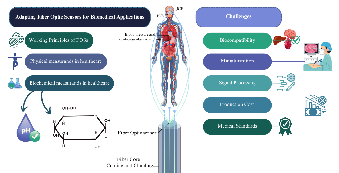
As sensor technology continues to advance, Fiber Optic Sensors (FOSs) have gained prominence for their accuracy, high sensitivity, and resistance to electromagnetic interference. These characteristics make them particularly attractive for biomedical applications, where accurate and reliable measurements are crucial. From monitoring physiological parameters to aiding in minimally invasive surgeries, fibre optic sensors hold the potential to revolutionize medical diagnostics and treatment [1,2].
Despite their promise, the adoption of fibre optic sensors in the biomedical field requires several obstacles to overcome. The integration of these sensors into medical devices requires overcoming challenges related to biocompatibility, miniaturization, and effective an efficient signal processing. Additionally, ensuring the robustness, real time responsivity and reliability of FOSs in the dynamic and often harsh environments of the human body presents another layer of complexity. Also, their use requires meeting International Standards which can also be challenging in the pathway to commercialisation [3,4,5].
This article investigates the specific challenges encountered in the adoption of fibre optic sensors for biomedical applications. It examines the challenges associated with FOS implementation, with a focus on different medical standards, assessing their limitations and discussing key physical and biochemical measurands relevant to biomedical applications.
1.1. Background
FOSs operate by transmitting light through optical fibers, where changes in properties such as intensity, phase, or wavelength indicate specific physiological conditions. In the biomedical field, FOSs enable real-time monitoring of vital signs, including pressure and temperature, facilitate biochemical detection, and support minimally invasive surgical procedures. Despite their advantages, several challenges hinder their widespread adoption in healthcare. Key issues include ensuring biocompatibility, scaling up manufacturing to meet industry standards, and developing efficient real-time signal processing algorithms. Overcoming these barriers is critical for integrating FOS technology into routine medical diagnostics and treatment, where it holds the potential to enhance accuracy and reliability in patient care [6,7].
Polymer-based (or plasic) optical fibre sensors (POFSs) represent a significant advancement in FOS technology, offering flexible and adaptable sensing solutions. These sensors can operate based on various principles, including waveguide-based [8,9] mechanisms, luminescence [10], surface plasmon resonance (SPR) [11], and optical fiber-based sensing, and they are suitable for both single-point and multi-point applications [12,13,14,15,16]. A particular focus is placed on polymer optical fiber sensors due to their applications in biosensing. The use of polymeric materials, such as biodegradable, biocompatible, hydrophilic, stimuli-responsive, conductive, and molecularly imprinted polymers, further enhances biomedical sensing capabilities. Additionally, various fabrication techniques for polymer optical fibers (POFs), including thermal drawing, extrusion, laser writing, microfabrication, and advanced coatings are included in Figure 1 as a classification on different types of fibers and fabrication techniques [1,17,18].
1.2. Working Principles of FOSs
FOSs vary widely in terms of their physical appearance and characteristics and their operating principles are correspondingly diverse. The primary FOS classification splits into intrinsic and extrinsic sensors [12]. Intrinsic FOSs rely on the light-matter interaction occurring wholly within the fibre itself, where changes in light properties (intensity, phase, polarization, or wavelength) occur due to external influences such as temperature, pressure, or strain. Extrinsic fibre optic sensors, on the other hand, use the optical fibre merely to transmit light to and from an external sensing element. Fiber Bragg Gratings (FBGs) [20,21] have represented a revolution in sensing using optical fibres. They operate by reflecting specific wavelengths of light that shift in response to strain or temperature changes. Interferometric sensors (of which the FBG is an example), generally measure phase changes in light caused by external perturbations [3,22,23]. The FBGs interaction with light as it passes through the grating planes depends on the Bragg condition and the first-order Bragg condition can be stated as below:
wheredenotes the effective refractive index, while represents the grating period, and stands for the Bragg wavelength.
There exist several different types of FOSs and Polymer-based optical fibre (POF) sensors based on different working principles which cannot be included in this article owing to page length restrictions. e.g., fluorescence-based sensors [24,25] detect variations in fluorescence emitted by certain materials subject to external light excitation, and are often used for biochemical measurements. Other types of FOSs include interferometric-based devices such as Fabry-Perot [22,26], Surface Plasmon Resonance (SPR) [27,28], Optical Coherence Tomography (OCT) [29,30] utilized in imaging applications and often incorporate specialist fibre e.g., Photonic Crystal Fiber (PCF) [24,31]. They all leverage the interaction between light and the external environment to provide sensitive and accurate measurements, in a wide range applications [23,32,33,34,35]. They can be categorized in various ways, including fibre type, application, and sensing mechanisms, as outlined in Table 1.
Also, fiber optic sensors (FOSs) can be classified based on their measurands into physical and biochemical types. Physical FOSs detect parameters including pressure, temperature, and strain by analyzing optical signal changes. Biochemical FOSs identify specific analytes, such as glucose or pH, e.g., using functionalized coatings. This classification highlights their versatility in biomedical and industrial applications. This review focuses on their applications in biomedical sensing.
1.3. Physical Measurands in Healthcare
FOSs are well suited for providing measurements of various physical measurands and are becoming increasingly accepted for patient monitoring and diagnostics. These measurands include temperature, pressure, strain, flow, liquid level, displacement, vibration, rotation, radiation, and biochemical markers. For example body temperature measurements are vital for tracking temperature fluctuations during surgeries, post-operative care, and critical care settings, ensuring patient stability and the early detection of infections [53,54]. Pressure sensors are extensively used to monitor several medical pressure parameters including blood pressure, Intracranial Pressure (ICP) [55,56], and Intraocular Pressure (IOP) [57,58], which are critical for managing conditions including hypertension, Traumatic Brain Injuries (TBI) [59], and glaucoma [58] respectively. Strain sensors are employed to monitor respiration by measuring chest wall movements, offering valuable data for respiratory therapy [3], sleep studies, and managing conditions including asthma or chronic obstructive pulmonary disease (COPD) [4,60]. By providing real-time data, fibre optic sensors offer enhanced clinical decision-making, improved patient outcomes, and contribute to the advancement of personalized medicine [61,62].
Biomechanical measurands encompass the physical parameters of the human body where the focus is on physical structure and movement, and accurate measurement is crucial for various successful monitoring. FOSs can measure strain and deformation in tissues and organs, providing critical data for orthopaedic and rehabilitation applications [1,2]. For instance, it is possible to monitor the stress and strain on bones and joints during physical activities, aiding in the assessment and treatment of musculoskeletal disorders. Additionally, they have been used for posture monitoring and ulcer formation detection in patients who are required to use a wheelchair [63]. Furthermore, these sensors are employed in the development of prosthetics and wearable devices, providing real-time feedback on the mechanical performance and interaction with the body. By measuring these biomechanical parameters, FOSs support the diagnosis, treatment, and rehabilitation of various conditions and advancing the field of biomechanics in healthcare [4,5,64,65,66,67].
1.4. Biochemical Measurands in Healthcare
All biochemical measurands could be considered vital parameters in healthcare, providing essential information about the whole physiological and metabolic states of patients. FOSs are increasingly used to measure these biochemical markers with high sensitivity and specificity. One significant application of FOSs is in continuous glucose monitoring (CGM), particularly for diabetes management. These sensors can measure glucose levels in blood or interstitial fluid, providing real-time data that aids in maintaining optimal glycemic control [68,69,70,71,72]. pH monitoring is used in assessing metabolic conditions and the body’s acid-base balance, which is vital in some critical care setting and surgical procedures [73,74,75,76]. Additionally, fibre optic sensors are used for multiple blood component detection [69], to detect specific proteins [77,78], enzymes [71,79,80], and hormones [31,81,82], aiding in the diagnosis and monitoring of various diseases, such as cancer [83,84,85] and hormonal imbalances [32,86]. These sensors can be functionalized to detect biomarkers at the molecular level, enabling early disease detection and the tracking of treatment efficacy. By measuring these biochemical parameters, fibre optic sensors provide data that present real time monitoring opportunities, enhancing diagnostic accuracy and treatment monitoring which are directly related to biochemical measurands [32,87,88].
FOSs are adept at detecting various substances in both gas and liquid phases. In the gas phase, FOSs can detect a wide range of gases including oxygen, carbon dioxide, and Volatile Organic Compounds (VOCs) [89,90,91,92] with high sensitivity. In respiratory monitoring [60,93] and detecting gases in environmental health studies. In the liquid phase, FOSs are widely used for measuring biochemical substances, such as glucose, electrolytes, and pH levels in bodily fluids like blood, urine, and saliva [86,94,95,96]. This capability is essential for continuous glucose monitoring in diabetic patients and assessing kidney function or urinary protein [95,97,98]. Accurate, real-time measurements can be achieved using different sensing mechanisms, based on different responsive materials. These sensors also provide access to a vast range of use cases as described in Table 2 [24,99,100]. Table 2 provides an overview of recently developed FOSs designed for pH measurement and glucose detection, highlighting their fibre type and detection range with the responsive material.
FOSs have also demonstrated potential in the monitoring of glucagon-like peptide-1 (GLP-1), a critical biomarker in glucose metabolism and insulin regulation. The ability to continuously monitor GLP-1 levels offers significant advantages in the management of diabetes and other metabolic disorders. A genetically encoded sensor (GLPLight1) has been developed by engineering a circularly permuted green fluorescent protein into the human GLP-1 receptor (GLP1R), Figure 2. This sensor accurately detects receptor conformational activation in response to pharmacological ligands, as indicated by its fluorescence signal. [111,112].
Ensuring accurate parameter values is paramount in sensor technology, particularly in the case of fiber optic sensors for biomedical applications. A comprehensive approach to achieving high accuracy must span the entire lifecycle of sensor development and deployment as shown in the example step graph of Figure3. This entails fundamental materials research during the design phase to comprehend material properties, transduction mechanisms, and device physics, ultimately leading to optimized materials and device structures. Iteration between scientific inquiry and engineering optimization enhances sensor accuracy, alongside considering factors such as the 3S’s (size, speed, and sensitivity).
Fabricated device accuracy and consistency is pivotal in moving towards manufacturability and deployment. Additionally, large-scale validation employing standardized procedures and benchmarking against gold-standard measurements are indispensable for obtaining reliable calibration curves [114].
2. Challenges for FOSs in Biomedical Applications
The development of biodegradable and biocompatible optical fibres for biomedical applications presents several key challenges. Firstly, material selection is critical, as the fibres must degrade safely in the body without releasing toxic byproducts. Natural or synthetic polymers, such as polylactic acid (PLA) or phosphate-based glasses, must be carefully evaluated for their mechanical properties and degradation rates to match specific biomedical requirements. Secondly, fabrication techniques such as thermal drawing, extrusion, or 3D printing must be optimized to ensure that the fibres maintain structural integrity during use while also being scalable for clinical applications. Thirdly, fibre design plays a significant role; features like microstructured or step-index profiles must balance light-guiding efficiency with mechanical flexibility and controlled biodegradability. Finally, application-specific considerations must be addressed, such as ensuring effective light delivery in photodynamic therapy or accurate biosensing under dynamic physiological conditions. PLA-based fibres have shown promise due to their adjustable degradation rates and biocompatibility. However, challenges such as mechanical fragility and inconsistent degradation in physiological environments necessitate further research. Similarly, phosphate-based glass fibres are attractive for their complete dissolution in biological fluids, but optimizing their mechanical properties without compromising their optical performance remains a significant challenge. These challenges highlight the need for continued advancements in material science and fibre design to achieve reliable, biodegradable, and biocompatible optical fibres for diverse biomedical applications [115]. Figure 4 illustrates a schematic representation of the four key challenges encountered in the development of biodegradable and biocompatible optical fibres.
2.1. Biocompatibility
Ensuring non-toxicity for any material to be used in fabricating sensors is one of the primary challenges to achieving biocompatibility. The selection of materials needs to ensure that the FOSs are mechanically safe, non-toxic and do not provoke an immune response when implanted in or used on the human body. This requires extensive testing to ensure that the materials do not cause inflammation, allergic reactions, or any other adverse effects [5,116]. This is generally conducted by regulating bodies such as the Food and Drug Administration (FDA) in the USA (section 2.5 of this article).
FOSs often require coatings to enhance biocompatibility. These coatings must be able to withstand the harsh biological environment without degrading. Ensuring long-term stability and functionality of these coatings is crucial for the reliable performance and regulatory approval of the sensors. Furthermore, sensors often need to endure various sterilization methods such as autoclaving, gamma irradiation, or chemical sterilization. A major challenge remains to design sensors that maintain their performance characteristics post-sterilization, as these processes can sometimes compromise sensor integrity and functionality [20,117]. Table 3 presents an overview of biomaterials used in the fabrication of optical fibres, highlighting their base material, advantages and disadvantages based on literature.
2.2. Miniaturization, Durability and Longevity
Reducing the size of fibre optic sensors without sacrificing sensitivity or accuracy is an ongoing significant challenge. Miniaturized sensors must still be capable of delivering accurate and reliable measurements. Also, small-sized sensors need to be seamlessly integrated into medical devices and systems, often requiring custom design solutions. This integration must ensure that the sensors do not interfere with the overall device performance and that they are easy to incorporate into existing medical infrastructure [87,131].
Producing miniaturized sensors at scale while maintaining high quality and consistency presents additional manufacturing challenges. Advanced fabrication techniques and stringent quality control measures are necessary to address these issues. FOSs often need to maintain their functionality over extended periods, particularly in chronic disease management and long-term monitoring applications. This requires materials and designs that are resistant to degradation over time [6,98,132].
Sensors must be able to withstand the dynamic and often harsh conditions encountered within the human body, such as movement, pressure changes, and exposure to various bodily fluids. Ensuring mechanical robustness while maintaining sensor sensitivity is a critical challenge [18,133].
Ideally, fibre optic sensors used in medical applications should require minimal maintenance. Developing sensors that can operate reliably over long periods without the need for frequent recalibration or replacement is essential for their practical use [134,135]. Also, it is highly advantageous if the sensor can be delivered inside a standard medical catheter as this often overcomes the problem of mechanical robustness [136,137,138].
2.3. Signal Processing, Data Integration, and Interoperability
Biological environments are often inherently noisy e.g., external electromagnetic interference from scanning equipment (MRI and CT), which can interfere with the signals detected by fibre optic sensors. If it is not possible to make the sensor immune to these sources of interference, developing advanced algorithms and signal processing techniques to filter out this noise becomes crucial for accurate measurements. The signals from FOSs often require sophisticated interpretation, especially when monitoring dynamic biological processes. This necessitates the development of advanced computational models and machine learning algorithms to accurately analyse and interpret the data [139,140,141].
Many types of medical applications require real-time data processing and feedback. Ensuring that the sensor systems can handle the computational load and providing timely, accurate information is a significant and ongoing technical challenge. FOSs must be compatible with existing healthcare IT systems and electronic health records (EHRs). Ensuring seamless integration and data interoperability is therefore essential for effective use in clinical settings [142,143].
Developing standardized data protocols to ensure that data from fibre optic sensors can be easily shared and interpreted across different platforms and systems is crucial. This includes ensuring sound data security and maintaining patient privacy. Finally, at this stage providing user-friendly interfaces that allow healthcare professionals to easily interact with and interpret data from fibre optic sensors is important for their adoption. This involves developing intuitive software and visualization tools [87,132,144].
2.4. Production Cost and Manufacturing
High production costs can be a barrier to the widespread adoption of fibre optic sensors. Developing cost-effective manufacturing processes without compromising quality and performance is crucial for making the sensors affordable [37,88]. Additionally, it may be necessary to accommodate further scaling up of production while maintaining consistency and reliability. The latter requires advanced manufacturing techniques and stringent quality control measures to ensure that each sensor meets the required standards. Finally, achieving economies of scale to achieve lower costs involves not only improving manufacturing processes but also increasing market demand and production volumes. This can too be challenging in the early stages of technology adoption [145,146].
2.5. Medical Standards and Regulatory Approval
Medical and/or biomedical FOSs must meet stringent regulatory standards set by organizations such as the FDA (Food and Drug Administration) and EMA (European Medicines Agency) [147,148,149,150,151,152]. This involves extensive testing to demonstrate safety, efficacy, and reliability.
Ensuring compliance with international standards for medical devices is critical. This includes adhering to ANSI (American National Standards Institute) /AAMI (Association for the Advancement of Medical Instrumentation) or ISO standards such as AAMI/ISO 10993, ISO 13485, AAMI TIR42: Technical Information Report (TIR), not a formal standard, but rather guidance for evaluating biocompatibility in alignment with ISO 10993. It helps manufacturers interpret biocompatibility requirements for regulatory compliance. [147,153,154,155].
Also, regulatory frameworks governing medical devices within the European Union include the EU Medical Devices Regulation (MDR) and the In Vitro Diagnostic Regulation (IVDR) [150,151,153]. The process of obtaining regulatory approval can be lengthy and complex, requiring a long lead time and resources. This can delay the introduction of new fibre optic sensor technologies to the market and their wider application. Table 4 outlines the regulatory standards governing the use of fibre optic sensors in medical applications.
3. Conclusions
FOSs have been transformative in healthcare, offering high accuracy and versatility in monitoring physiological and biochemical parameters including temperature, pressure, strain, and biochemical markers, enhancing diagnostics and patient outcomes. However, the widespread adoption of FOS technology in healthcare faces several critical challenges, including biocompatibility, miniaturization, durability, and robust signal processing. Addressing these challenges requires interdisciplinary collaboration across materials science, engineering, and medical practice to develop reliable, scalable, and clinically viable sensor systems.
Innovative advancements in biocompatible materials, fabrication techniques, and signal processing algorithms continue to push the boundaries of what FOSs can achieve in medicine. Standardization and regulatory approval remain key hurdles that must be overcome to facilitate their transition from research laboratories to commercial medical devices. Future research efforts should focus on enhancing sensor integration within existing medical systems, improving long-term reliability, and developing AI-driven analytical methods for accurate data interpretation.
By overcoming these challenges, fibre optic sensors have the potential to revolutionize biomedical sensing, paving the way for more precise diagnostics, personalized treatment plans, and improved patient outcomes. As the field advances, the synergy between optical sensor technology and emerging biomedical innovations will shape the future of healthcare, making real-time, minimally invasive, and monitoring an integral part of medical practice.
Acknowledgments
Authors expand sincere gratitude to colleagues and collaborators who provided valuable insights and support throughout this research. This research is supported by a postgraduate research bursary from the Atlantic Technological University.
Conflicts of Interest
The authors declare no conflicts of interest.
Abbreviations
The following abbreviations are used in this manuscript:
| FOSs | Fiber Optic Sensors (FOSs) |
| POSs | Polymer-based optical sensors |
| OF | Optical Fiber |
| SPR | Surface Plasmon Resonance |
| POFs | Polymer Optical Fibers |
| FBGs | Fiber Bragg Gratings |
| OCT | Optical Coherence Tomography |
| IOP | Intraocular Pressure |
| PCF | Photonic Crystal Fiber |
| SMF | Single-Mode Fiber |
| FPI | Fabry-Pérot Interferometer |
| MMF | Multi-Mode Fiber |
| MEMs | Micro-Electro-Mechanical Systems |
| LSPR | Localized Surface Plasmon Resonance |
| LMR | Lossy Mode Resonance |
| HST | Hollow Silica Tube |
| PID | Proportional Integral Derivative |
| PANi | Polyaniline |
| TFBG | Tilted Fiber Bragg Grating |
| PAAm | Polyacrylamide |
| GO | Graphene Oxide |
| GOD | Glucose Oxidase |
| LPFG | Long-Period Fiber Grating |
| TOFI | Tapered Optical Fiber Interferometer |
| 3-APBA | 3-Aminophenylboronic Acid |
| LDOF | Lossy Dielectric Optical Fiber |
| HBF | high-birefringence fibre |
| PLA | polylactic acid |
| FDA | Food and Drug Administration |
| PEG | Polyethylene Glycol |
| POC | Poly (Octamethylene Citrate) |
| POMC | Poly (Octamethylene Maleate Citrate) |
| PVC | Polyvinyl Chloride |
| SU-8 | Negative Photoresist Polymer |
| PLLA | Poly (L-Lactic Acid) |
| PDLLA | Poly (D, L-Lactic Acid) |
| PLGA | Poly (L-Lactic-Co-Glycolic Acid) |
| PDLGA | Poly (D, L-Lactic-Co-Glycolic Acid) |
| PCL | Poly (ε-Caprolactone) |
| PGs | Phosphate Glass |
| PDMS | Polydimethylsiloxane |
| PAA | Polyacrylic Acid |
| AG | Agarose Hydrogel |
| AuNPs | Gold Nanoparticles |
| MRI | Magnetic Resonance Imaging |
| CT | Computed Tomography |
| EHRs | Electronic Health Records |
| EMA | European Medicines Agency |
| ANSI | American National Standards Institute |
| AAMI | Association for the Advancement of Medical Instrumentation |
| TIR | Technical Information Report |
| MDR | Medical Devices Regulation |
| IVDR | In Vitro Diagnostic Regulation |
References
- Ngiejungbwen, L.A.; Hamdaoui, H.; Chen, M.Y. , “Polymer optical fiber and fiber Bragg grating sensors for biomedical engineering Applications: A comprehensive review,” Opt Laser Technol, vol. 170, no. 23, p. 110187, 2024. 20 October. [CrossRef]
- Rahuman, M.A.A.; et al. , “Recent Technological Progress of Fiber-Optical Sensors for Bio-Mechatronics Applications,” Technologies, vol. 11, no. 6, 2023. [CrossRef]
- Bartnik, K.; Koba, M.; Śmietana, M. , “Advancements in optical fiber sensors for in vivo applications – A review of sensors tested on living organisms,” Measurement, vol. 224, no. 23, p. 113818, 2024. 20 November. [CrossRef]
- Padha, B.; Yadav, I.; Dutta, S.; Arya, S. , “Recent Developments in Wearable NEMS/MEMS-Based Smart Infrared Sensors for Healthcare Applications,” ACS Appl Electron Mater, vol. 5, pp. 5386–5411, 2023. [CrossRef]
- Zhang, X.; Wang, C.; Zheng, T.; Wu, H.; Wu, Q.; Wang, Y. , “Wearable Optical Fiber Sensors in Medical Monitoring Applications: A Review,” Sensors, vol. 23, no. 15, 2023. [CrossRef]
- Yu, L.; Kim, B.J.; Meng, E. , “Chronically implanted pressure sensors: Challenges and state of the field,” Sensors (Switzerland), vol. 14, no. 11, pp. 20620–20644, 2014. [CrossRef]
- Presti, D.L.O.; et al. , “Fiber Bragg Gratings for Medical Applications and Future Challenges: A Review,” IEEE Access, vol. 8, pp. 156863–156888, 2020.
- Wang, J.; Dong, J. , “Optical waveguides and integrated optical devices for medical diagnosis, health monitoring and light therapies,” Sensors (Switzerland), vol. 20, no. 14, pp. 1–33, 2020. [CrossRef]
- Peng, C.; et al. , “Optical Waveguide Refractive Index Sensor for Biochemical Sensing,” Appl Sci, vol. 13, no. 6, 2023. [CrossRef]
- Leiner, M.J.P. , “Luminescence chemical sensors for biomedical applications: scope and limitations,” Anal Chim Acta, vol. 255, no. 2, pp. 209–222, 1991. [CrossRef]
- Englebienne, P.; Van Hoonacker, A.; Verhas, M. , “Surface plasmon resonance: Principles, methods and applications in biomedical sciences,” Spectroscopy, vol. 17, no. 2–3, pp. 255–273, 2003. [CrossRef]
- Grattan, L.S.; Meggitt, B.T. , Optical Fiber Sensor Technology: Fundamentals. Springer US, 2010. [Online]. Available: https://books.google.com/books?
- Pirzada, M.; Altintas, Z. , “Recent progress in optical sensors for biomedical diagnostics,” Micromachines, vol. 11, no. 4, 2020. [CrossRef]
- Liu, X.; Zhang, X.; Liu, Y.; Liu, Z.; Peng, W. , “Multi-point fiber-optic refractive index sensor by using coreless fibers,” Opt Commun, vol. 365, pp. 168–172, 2016. [CrossRef]
- Pendão, C.; Silva, I. , “Optical Fiber Sensors and Sensing Networks: Overview of the Main Principles and Applications,” Sensors, vol. 22, no. 19, 2022. [CrossRef]
- Xiong, L.; Zhong, H.; Wan, S.; Yu, J. , “Single-point curved fiber optic pulse sensor for physiological signal prediction based on the genetic algorithm-support vector regression model,” Opt Fiber Technol, vol. 82, p. 103583, 2024. [CrossRef]
- Nagar, M.A.; Janner, D. , “Polymer-Based Optical Guided-Wave Biomedical Sensing: From Principles to Applications,” 2024.
- Soge, A.O.; Dairo, O.F.; Sanyaolu, M.E.; Kareem, S.O. , “Recent developments in polymer optical fiber strain sensors: A short review,” J Opt, vol. 50, no. 2, pp. 299–313, 2021. [CrossRef]
- Nagar, M.A.; Janner, D. , “Polymer-Based Optical Guided-Wave Biomedical Sensing: From Principles to Applications,” Photonics, vol. 11, no. 10, 2024. [CrossRef]
- Zhang, Z.; Zhang, C.; Zuo, S. , “A Novel Bioinspired Whisker Sensor for Gastrointestinal Endoscopy,” IEEE/ASME Trans Mechatronics, vol. PP, pp. 1–11, 2023. [CrossRef]
- Theodosiou, A. , “Recent Advances in Fiber Bragg Grating Sensing,” Sensors (Basel), vol. 24, no. 2, 2024. [CrossRef]
- Zhu, C.; et al. , “Advances in Fiber-Optic Extrinsic Fabry-Perot Interferometric Physical and Mechanical Sensors: A Review,” IEEE Sens J, vol. 23, no. 7, 2023. [CrossRef]
- Elsherif, M.; et al. , “Optical Fiber Sensors: Working Principle, Applications, and Limitations,” Adv Photonics Res, vol. 3, no. 11, 2022. [CrossRef]
- Paget, B.M.; et al. , “A review on photonic crystal fiber based fluorescence sensing for chemical and biomedical applications,” Sensors Actuators B Chem, vol. 400, no. PB, p. 134828, 2024. [CrossRef]
- Azmi, A.N.; et al. , “Review of Open Cavity Random Lasers as Laser-Based Sensors,” ACS Sensors, vol. 7, no. 4, pp. 914–928, 2022. [CrossRef]
- Domingues, M.F.; et al. , “Optical Fibre FPI End-Tip based Sensor for Protein Aggregation Detection,” 2022 IEEE Int Conf E-Health Networking, Appl Serv Heal 2022, pp. 129–134, 2022. [CrossRef]
- Wang, F.; et al. , “3D fiber-probe surface plasmon resonance microsensor towards small volume sensing,” Sensors Actuators B Chem, vol. 384, no. 22, p. 133647, 2023. 20 December. [CrossRef]
- Mi, H.; Wang, Y.; Jin, P.; Lei, L. , “Design of a ultrahigh-sensitivity SPR-based optical fiber pressure sensor,” Optik (Stuttg), vol. 124, no. 21, pp. 5248–5250, 2013. [CrossRef]
- Samimi, K.; et al. , “Optical coherence tomography of human fetal membrane sub-layers during loading,” Biomed Opt Express, vol. 14, no. 6, p. 2969, 2023. [CrossRef]
- Hui, P.C.; et al. , “Implantable self-aligning fiber-optic optomechanical devices for in vivo intraocular pressure-sensing in artificial cornea,” J Biophotonics, vol. 13, no. 7, pp. 1–13, 2020. [CrossRef]
- Nithish, A.N.; et al. , “Terahertz Women Reproductive Hormones Sensor Using Photonic Crystal Fiber With Behavior Prediction Using Machine Learning,” IEEE Access, vol. 11, no. May, pp. 75424–75433, 2023. [CrossRef]
- Gupta, B.D.; Pathak, A.; Shrivastav, A.M. , “Optical Biomedical Diagnostics Using Lab-on-Fiber Technology: A Review,” Photonics, vol. 9, no. 2, 2022. [CrossRef]
- Uniyal, A.; Srivastava, G.; Pal, A.; Taya, S.; Muduli, A. , “Recent Advances in Optical Biosensors for Sensing Applications: a Review,” Plasmonics, vol. 18, no. 2, pp. 735–750, 2023. [CrossRef]
- Katrenova, Z.; Alisherov, S.; Abdol, T.; Molardi, C. , “Status and future development of distributed optical fiber sensors for biomedical applications,” Sens Bio-Sensing Res, vol. 43, no. 23, p. 100616, 2024. 20 September. [CrossRef]
- Butt, M.A.; Kazanskiy, N.L.; Khonina, S.N.; Voronkov, G.S.; Grakhova, E.P.; Kutluyarov, R.V. , “A Review on Photonic Sensing Technologies: Status and Outlook,” Biosensors, vol. 13, no. 5, 2023. [CrossRef]
- Zhang, S.; et al. , “An optical fiber pressure sensor with ultra-thin epoxy film and high sensitivity characteristics based on blowing bubble method,” IEEE Photonics J, vol. 13, no. 1, 2021. [CrossRef]
- Jauregui-Vazquez, D.; et al. , “Low-pressure and liquid level fiber-optic sensor based on polymeric Fabry–Perot cavity,” Opt Quantum Electron, vol. 53, no. 5, pp. 1–12, 2021. [CrossRef]
- Wang, S.; Wang, J.; Li, W.; Liu, Y.; Li, J.; Jia, P. , “A MEMS-Based High-Fineness Fiber-Optic Fabry–Perot Pressure Sensor for High-Temperature Application,” Micromachines, vol. 13, no. 5, 2022. [CrossRef]
- An, C.L.I.; et al. , “LSPR optical fiber biosensor based on a 3D composite structure of gold nanoparticles and multilayer graphene films,” vol. 28, no. 5, pp. 6071–6083, 2020.
- Susheel, S.S.A.; Esakki, P.V.T.K.; Sivanantha, M.A. , “A novel surface plasmon based photonic crystal fiber sensor,” Opt Quantum Electron, vol. 52, no. 6, pp. 1–12, 2020. [CrossRef]
- Silva, T.B.; Cleumar, A.; Melo, A.; Cleumar, A.; Melo, A.S.M. , “of a D-Shaped Optical Fiber Investigation of a D-Shaped Optical Fiber Investigation of a D-Shaped Optical Fiber Sensor with Investigation of a Graphene D-Shaped Overlay Optical Fiber Sensor with Graphene Overlay Sensor with Graphene Overlay Sensor with,” 2018. [CrossRef]
- Tien, C.; Lin, H.; Su, S. , “High Sensitivity Refractive Index Sensor by D-Shaped Fibers and Titanium Dioxide Nanofilm,” vol. 2018, 2018. [CrossRef]
- Iesheng, T.W.U.; et al. , “Surface plasmon resonance biosensor based on gold-coated side-polished hexagonal structure photonic crystal fiber,” vol. 25, no. 17, pp. 227–231, 2017.
- Haque, E.; Hossain, A.; Ahmed, F. , “Surface Plasmon Resonance Sensor Based on Modified D -Shaped Photonic Crystal Fiber for Wider Range of Refractive Index Detection,” vol. 18, no. 20, pp. 8287–8293, 2018. [CrossRef]
- Access, O. , “Design of a Fiber Optic Biosensor for Cholesterol Detection in Human Blood Design of a Fiber Optic Biosensor for Cholesterol Detection in”. [CrossRef]
- Heng, Y.U.C.; Ang, Y.I.W.; Ong, Z.I.S. , “High-sensitivity optical fiber temperature sensor based on a dual-loop optoelectronic oscillator with the Vernier effect,” vol. 28, no. 23, pp. 35264–35271, 2020.
- Cao, H.; Li, D.; Zhou, K.; Chen, Y. , “Demonstration of a ZnO-Nanowire-Based Nanograting Temperature Sensor,” vol. 13, no. 1, pp. 1–7, 2023. [CrossRef]
- Zhou, N.; et al. , “MEMS-based reflective intensity-modulated fiber-optic sensor for pressure measurements,” Sensors (Switzerland), vol. 20, no. 8, 2020. [CrossRef]
- Chen, Y.; et al. , “Fiber-Tip Fabry-Perot Cavity Pressure Sensor With UV-Curable Polymer Film Based on Suspension Curing Method,” IEEE Sens J, vol. 22, no. 7, pp. 6651–6660, 2022. [CrossRef]
- Qureshi, K.K. , “Detection of Plantar Pressure Using an Optical Technique,” 2021 7th Int Conf Eng Appl Sci Technol ICEAST 2021 - Proc, pp. 77–80, 2021. [CrossRef]
- Yoshida, T.; Tsuruoka, N.; Haga, Y.; Kinoshita, H.; Lee, S.S.; Matsunaga, T. , “Automatic irrigation system with a fiber-optic pressure sensor regulating intrapelvic pressure for flexible ureteroscopy,” Sci Rep, vol. 13, no. 1, pp. 1–11, 2023. [CrossRef]
- Han, J.; Chen, M.; Wen, J.; Yang, T.; Dong, Y. , “High sensitivity fiber optic esophageal pressure sensor based on OFDR,” J Phys Conf Ser, vol. 2581, no. 1, 2023. [CrossRef]
- Shang, C.; et al. , “Soft Biomimetic Fiber-Optic Tactile Sensors Capable of Discriminating Temperature and Pressure,” ACS Appl Mater Interfaces, vol. 15, no. 46, pp. 53264–53272, 2023. [CrossRef]
- Zhang, H.; Cong, B.; Zhang, F.; Qi, Y.; Hu, T. , “Simultaneous measurement of refractive index and temperature by Mach–Zehnder cascaded with FBG sensor based on multi-core microfiber,” Opt Commun, vol. 493, no. 20, p. 126985, 2021. 20 December. [CrossRef]
- Narayan, V.; Mohammed, N.; Savardekar, A.R.; Patra, D.P.; Notarianni, C.; Nanda, A. , “Noninvasive Intracranial Pressure Monitoring for Severe Traumatic Brain Injury in Children: A Concise Update on Current Methods,” World Neurosurg, vol. 114, pp. 293–300, 2018. [CrossRef]
- He, C.; Teng, C.; Xiong, Z.; Lin, X.; Li, H.; Li, X. , “Intracranial pressure monitoring in neurosurgery: the present situation and prospects,” Chinese Neurosurg J, vol. 9, no. 1, pp. 1–12, 2023. [CrossRef]
- Mimura, M.; Akagi, T.; Kohmoto, R.; Fujita, Y.; Sato, Y.; Ikeda, T. , “Measurement of vitreous humor pressure in vivo using an optic fiber pressure sensor,” Sci Rep, vol. 13, no. 1, pp. 1–6, 2023. [CrossRef]
- Raveendran, R.; et al. , “Current Innovations in Intraocular Pressure Monitoring Biosensors for Diagnosis and Treatment of Glaucoma—Novel Strategies and Future Perspectives,” Biosensors, vol. 13, no. 6, 2023. [CrossRef]
- Ordookhanian, C.; Nagappan, M.; Elias, D.; Kaloostian, P.E. , “Management of Intracranial Pressure in Traumatic Brain Injury,” Trauma Brain Inj - Pathobiol Adv Diagnostics Acute Manag, 2018. [CrossRef]
- Zhao, C.; et al. , “Recent advances in fiber optic sensors for respiratory monitoring,” Opt Fiber Technol, vol. 72, no. April, p. 103000, 2022. [CrossRef]
- Tosi, D.; Poeggel, S.; Iordachita, I.; Schena, E. , Fiber Optic Sensors for Biomedical Applications. Elsevier Inc., 2018. [CrossRef]
- Poeggel, S.; et al. , “Recent improvement of medical optical fibre pressure and temperature sensors,” Biosensors, vol. 5, no. 3, pp. 432–449, 2015. [CrossRef]
- González-Cely, A.X.; Diaz, C.A.R.; Callejas-Cuervo, M.; Bastos-Filho, T. , “Optical fiber sensors for posture monitoring, ulcer detection and control in a wheelchair: a state-of-the-art,” Disabil Rehabil Assist Technol, vol. 0, no. 0, pp. 1–18, 2023. [CrossRef]
- Najafzadeh, A.; et al. , “Application of fibre bragg grating sensors in strain monitoring and fracture recovery of human femur bone,” Bioengineering, vol. 7, no. 3, pp. 1–14, 2020. [CrossRef]
- De Tommasi, F.; Romano, C.; Presti, D.L.; Massaroni, C.; Carassiti, M.; Schena, E. , “FBG-Based Soft System for Assisted Epidural Anesthesia: Design Optimization and Clinical Assessment,” Biosensors, vol. 12, no. 8, 2022. [CrossRef]
- Wang, X.; et al. , “Highly Sensitive Strain Sensor Based on Microfiber Coupler for Wearable Photonics Healthcare,” Adv Intell Syst, vol. 5, no. 5, 2023. [CrossRef]
- Zhao, J.; et al. , “Wearable Optical Sensing in the Medical Internet of Things (MIoT) for Pervasive Medicine: Opportunities and Challenges,” ACS Photonics, vol. 9, no. 8, pp. 2579–2599, 2022. [CrossRef]
- Elsherif, M.; Hassan, M.U.; Yetisen, A.K.; Butt, H. , “Hydrogel optical fibers for continuous glucose monitoring,” Biosens Bioelectron, vol. 137, no. May, pp. 25–32, 2019. [CrossRef]
- Qu, W.; et al. , “Application of Optical Fiber Sensing Technology and Coating Technology in Blood Component Detection and Monitoring,” Coatings, vol. 14, no. 2, 2024. [CrossRef]
- Elsherif, M.; Hassan, M.U.; Yetisen, A.K.; Butt, H. , “Biosensors and Bioelectronics Hydrogel optical fi bers for continuous glucose monitoring,” Biosens Bioelectron, vol. 137, no. April, pp. 25–32, 2019. [CrossRef]
- Zhang, J.; Mai, X.; Hong, X.; Chen, Y.; Li, X. , “Optical fiber SPR biosensor with a solid-phase enzymatic reaction device for glucose detection,” Sensors Actuators B Chem, vol. 366, no. May, p. 131984, 2022. [CrossRef]
- Li, Y.; Luo, S.; Gui, Y.; Wang, X.; Tian, Z.; Yu, H. , “Difunctional Hydrogel Optical Fiber Fluorescence Sensor for Continuous and Simultaneous Monitoring of Glucose and pH,” Biosensors, vol. 13, no. 2, 2023. [CrossRef]
- Werner, J.; Belz, M.; Klein, K.; Sun, T.; Grattan, K.T.V. , “Fiber optic sensor designs and luminescence-based methods for the detection of oxygen and pH measurement,” Measurement, vol. 178, no. 20, p. 109323, 2021. 20 December. [CrossRef]
- Gong, J.; Tanner, M.G.; Venkateswaran, S.; Stone, J.M.; Zhang, Y.; Bradley, M. , “Analytica Chimica Acta A hydrogel-based optical fi bre fl uorescent pH sensor for observing lung tumor tissue acidity,” Anal Chim Acta, vol. 1134, pp. 136–143, 2020. [CrossRef]
- Khan, R.R.; Watekar, A.V.; Kang, S. , “Fiber-Optic Biosensor to Detect pH and Glucose,” IEEE Sens J, vol. 18, no. 4, pp. 1528–1538, 2018. [CrossRef]
- Steinegger, A.; Wolfbeis, O.S.; Borisov, S.M. , “Optical Sensing and Imaging of pH Values: Spectroscopies, Materials, and Applications,” 2020. [CrossRef]
- Paltusheva, Z.U.; Ashikbayeva, Z.; Tosi, D. , “Highly Sensitive Zinc Oxide Fiber-Optic Biosensor for the Detection of CD44 Protein,” 2022.
- Song, H.; Wang, Q.; Zhao, W. , “A novel SPR sensor sensitivity-enhancing method for immunoassay by inserting MoS 2 nanosheets between metal film and fiber,” vol. 132, no. March, 2020. [CrossRef]
- Cai, J.; Liu, Y.; Shu, X. , “Long-Period Fiber Grating Sensors for Chemical and Biomedical Applications,” Sensors, vol. 23, no. 1, 2023. [CrossRef]
- Chen, X.; et al. , “Tapered Fiber Bioprobe Based on U-Shaped Fiber Transmission for Immunoassay,” Biosensors, vol. 13, no. 10, 2023. [CrossRef]
- Vijayalakshmi, D.; Manimegalai, N.A.C.T.; Alzahrani, F.A. , “Photonic crystal fiber - based biosensor for detection of women reproductive hormones,” Opt Quantum Electron, vol. 55, no. 5, pp. 1–13, 2023. [CrossRef]
- Villegas-cantoran, D.S.; et al. , “Quantification of hCG Hormone Using Tapered Optical Fiber Decorated with Gold Nanoparticles,” pp. 1–11, 2023.
- Mohammed, N.A.; Khedr, O.E.; El-Rabaie, E.S.M.; Khalaf, A.A.M. , “Early detection of brain cancers biomedical sensor with low losses and high sensitivity in the terahertz regime based on photonic crystal fiber technology,” Opt Quantum Electron, vol. 55, no. 3, pp. 1–21, 2023. [CrossRef]
- Perspectives, F. , “Overview of Optical Biosensors for Early Cancer Detection:,” 2023.
- Sun, D.; Ran, Y. , “Label-Free Detection of Cancer Biomarkers Using,” 2017. [CrossRef]
- Arcadio, F.; Seggio, M.; Pitruzzella, R.; Zeni, L.; Bossi, A.M.; Cennamo, N. , “An Efficient Bio-Receptor Layer Combined with a Plasmonic Plastic Optical Fiber Probe for Cortisol Detection in Saliva,” Biosensors, vol. 14, no. 7, 2024. [CrossRef]
- Naresh, V.; Lee, N. , “A review on biosensors and recent development of nanostructured materials-enabled biosensors,” Sensors (Switzerland), vol. 21, no. 4, pp. 1–35, 2021. [CrossRef]
- Leitão, C.; et al. , “Cost-Effective Fiber Optic Solutions for Biosensing,” Biosensors, vol. 12, no. 8, 2022. [CrossRef]
- Prasanth, A.; Meher, S.R.; Alex, Z.C. , “Metal oxide thin films coated evanescent wave based fiber optic VOC sensor,” Sensors Actuators A Phys, vol. 338, p. 113459, 2022. [CrossRef]
- Pathak, A.K.; Viphavakit, C. , “A review on all-optical fiber-based VOC sensors: Heading towards the development of promising technology,” Sensors Actuators A Phys, vol. 338, p. 113455, 2022. [CrossRef]
- Buszewski, B.; Grzywinski, D.; Ligor, T.; Stacewicz, T.; Bielecki, Z.; Wojtas, J. , “Detection of Volatile Organic Compounds as Biomarkers in Breath Analysis by Different Analytical Techniques,” Bioanalysis, vol. 5, no. 18, pp. 2287–2306, 2013. [CrossRef]
- Grantzioti, E.; Pissadakis, S.; Konstantaki, M. , “Optical Fiber Volatile Organic Compound Vapor Sensor With ppb Detectivity for Breath Biomonitoring,” IEEE Sens J, vol. 25, no. 5, pp. 8224–8229, 2025. [CrossRef]
- Rohan, R.; Venkadeshwaran, K.; Ranjan, P. , “Recent advancements of fiber Bragg grating sensors in biomedical application: a review,” J Opt, no. 2, 2023. [CrossRef]
- Tentor, F.; et al. , “Development of an ex-vivo porcine lower urinary tract model to evaluate the performance of urinary catheters,” Sci Rep, vol. 12, no. 1, pp. 1–17, 2022. [CrossRef]
- Guo, T.; et al. , “Highly sensitive detection of urinary protein variations using tilted fiber grating sensors with plasmonic nanocoatings,” Biosens Bioelectron, vol. 78, pp. 221–228, 2016. [CrossRef]
- Xiong, H.; et al. , “Recent advances in biosensors detecting biomarkers from exhaled breath and saliva for respiratory disease diagnosis,” Biosens Bioelectron, vol. 267, no. 24, p. 116820, 2025. 20 September. [CrossRef]
- Mulyanti, B.; et al. , “SPR-Based Sensor for the Early Detection or Monitoring of Kidney Problems,” vol. 2022, 2022. [CrossRef]
- Vogt, B. , “Catheter-Free Urodynamics Testing: Current Insights and Clinical Potential,” Res Reports Urol, vol. 16, no. January, pp. 1–17, 2024. [CrossRef]
- Zhang, Y.; et al. , “Multiplexed optical fiber sensors for dynamic brain monitoring,” Matter, vol. 5, no. 11, pp. 3947–3976, 2022. [CrossRef]
- Zhou, B.; Fan, K.; Li, T.; Luan, G.; Kong, L. , “A biocompatible hydrogel-coated fiber-optic probe for monitoring pH dynamics in mammalian brains in vivo,” Sensors Actuators B Chem, vol. 380, no. January, p. 133334, 2023. [CrossRef]
- Aldaba, A.L.; González-vila, Á.; Debliquy, M.; Lopez-amo, M.; Caucheteur, C. , “Sensors and Actuators B: Chemical Polyaniline-coated tilted fiber Bragg gratings for pH sensing,” Sensors Actuators B Chem, vol. 254, pp. 1087–1093, 2018. [CrossRef]
- Zhao, Y.; Lei, M.; Liu, S.; Zhao, Q. , “Sensors and Actuators B: Chemical Smart hydrogel-based optical fiber SPR sensor for pH measurements,” Sensors Actuators B Chem, vol. 261, pp. 226–232, 2018. [CrossRef]
- Jiang, B.; et al. , “Sensors and Actuators B: Chemical Label-free glucose biosensor based on enzymatic graphene oxide-functionalized tilted fiber grating,” Sensors Actuators B Chem, vol. 254, pp. 1033–1039, 2018. [CrossRef]
- Li, Y.; et al. , “Sensors and Actuators B: Chemical Immobilized optical fiber microprobe for selective and high sensitive glucose detection,” Sensors Actuators B Chem, vol. 255, pp. 3004–3010, 2018. [CrossRef]
- Wu, C.W. , “S-shaped long period fiber grating glucose concentration biosensor based on immobilized glucose oxidase,” Optik (Stuttg), vol. 203, no. 19, p. 163960, 2020. 20 September. [CrossRef]
- Li, J.X.; Zhang, W.H.; Tong, Z.R.; Liu, J.W. , “Fiber optic sensor modified by graphene oxide–glucose oxidase for glucose detection,” Opt Commun, vol. 492, no. January, p. 126983, 2021. [CrossRef]
- Li, X.; Gong, P.; Zhang, Y.; Zhou, X. , “Label-Free Micro Probe Optical Fiber Biosensor for Selective and Highly Sensitive Glucose Detection,” IEEE Trans Instrum Meas, vol. 71, pp. 1–8, 2022. [CrossRef]
- Ujah, E.; Lai, M.; Slaughter, G. , “Ultrasensitive tapered optical fiber refractive index glucose sensor,” Sci Rep, vol. 13, no. 1, pp. 1–8, 2023. [CrossRef]
- Lee, S.-L.; Kim, J.; Choi, S.; Han, J.; Lee, Y.W. , “Optical glucose detection using birefringent long-period fiber grating functionalized with graphene oxide,” Opt Eng, vol. 60, no. 8, p. 87102, Aug. 2021. [CrossRef]
- Rahaman, M.E.; Jibon, R.H.; Ahsan, M.S.; Ahmed, F.; Sohn, I.B. , “Glucose Level Measurement Using Photonic Crystal Fiber–based Plasmonic Sensor,” Plasmonics, vol. 17, no. 1, pp. 1–11, 2022. [CrossRef]
- Huang, Z.; et al. , “Glucose-sensing glucagon-like peptide-1 receptor neurons in the dorsomedial hypothalamus regulate glucose metabolism,” Sci Adv, vol. 8, no. 23, 2022. [CrossRef]
- Duffet, L.; et al. , “Optical tools for visualizing and controlling human GLP-1 receptor activation with high spatiotemporal resolution,” Elife, vol. 12, pp. 1–20, 2023. [CrossRef]
- Mirdita, M.; Schütze, K.; Moriwaki, Y.; Heo, L.; Ovchinnikov, S.; Steinegger, M. , “ColabFold: making protein folding accessible to all,” Nat Methods, vol. 19, no. 6, pp. 679–682, 2022. [CrossRef]
- Luo, Y.; et al. , “Technology Roadmap for Flexible Sensors,” ACS Nano, vol. 17, no. 6, pp. 5211–5295, 2023. [CrossRef]
- Gierej, A.; Geernaert, T.; Van Vlierberghe, S.; Dubruel, P.; Thienpont, H.; Berghmans, F. , “Challenges in the fabrication of biodegradable and implantable optical fibers for biomedical applications,” Materials (Basel), vol. 14, no. 8, 2021. [CrossRef]
- Zhang, P.; Kim, J.W.; Gehlbach, P.; Iordachita, I.; Kobilarov, M. , “Autonomous Needle Navigation in Retinal Microsurgery: Evaluation in ex vivo Porcine Eyes,” Proc - IEEE Int Conf Robot Autom, vol. 2023-May, pp. 4661–4667, 2023. [CrossRef]
- Liu, Y.; et al. , “Miniature fiber-optic tip pressure sensor assembled by hydroxide catalysis bonding technology,” Opt Express, vol. 28, no. 2, p. 948, 2020. [CrossRef]
- Liu, Z.; Zhang, Z.F.; Tam, H.; Tao, X. , “Multifunctional Smart Optical Fibers: Materials, Fabrication, and Sensing Applications,” pp. 1–24, 2019.
- Schyrr, B.; Boder-pasche, S.; Ischer, R.; Smajda, R.; Voirin, G. , “Sensing and Bio-Sensing Research Fiber-optic protease sensor based on the degradation of thin gelatin films,” Sens BIO-SENSING Res, vol. 3, pp. 65–73, 2015. [CrossRef]
- Mcdonald, S.R.; Tao, S. , “Analytica Chimica Acta An optical fiber chlorogenic acid sensor using a Chitosan membrane coated bent optical fiber probe,” Anal Chim Acta, vol. 1288, no. 23, p. 342142, 2024. 20 July. [CrossRef]
- Marpu, S.B.; Benton, E.N. , “Shining Light on Chitosan: A Review on the Usage of Chitosan for Photonics and Nanomaterials Research,” pp. 1–38, 2018. [CrossRef]
- Suna, F.A.N.; Yi, Z.; Xiangyu, H.; Lihong, G.; Huili, S. , “Silk materials for medical, electronic and optical applications,” vol. 62, no. 6, pp. 903–918, 2019.
- Fujiwara, E.; Engineering, M.; De Campinas, U.E. , “Recent developments in agar - based optical devices,” MRS Commun, vol. 14, no. 3, pp. 237–247, 2024. [CrossRef]
- Arefnia, F.; Zibaii, M.I.; Layeghi, A.; Rostami, S. , “Citrate polymer optical fiber for measuring refractive index based on LSPR sensor,” Sci Rep, pp. 1–19, 2024. [CrossRef]
- Chen, Z.; Lee, J. , “Biocompatibility of SU-8 and Its Biomedical Device Applications,” 2021.
- Manvi, P.K.; Beckers, M.; Mohr, B.; Seide, G.; Gries, T.; Bunge, C. , Chapter 3. Polymer fiber-based biocomposites for medical sensing applications. Elsevier Inc., 2019. [CrossRef]
- Han, S.; Shin, G. , “Biodegradable Optical Fiber in a Soft Optoelectronic Device for Wireless Optogenetic Applications,” 2020.
- Raghunandhan, R.; et al. , “Sensors and Actuators B: Chemical Chitosan / PAA based fiber-optic interferometric sensor for heavy metal ions detection,” vol. 233, pp. 31–38, 2016. [CrossRef]
- Liang, D.; et al. , “Silk Fibroin - Based Wearable All - Fiber Multifunctional Sensor for Smart Clothing,” Adv Fiber Mater, vol. 4, no. 4, pp. 873–884, 2022. [CrossRef]
- Rabizah, S.; et al. , “Enhancing fibre optic sensor signals via gold nanoparticle-decorated agarose hydrogels,” Opt Mater (Amst), vol. 143, no. August, p. 114247, 2023. [CrossRef]
- Tarar, A.A.; Mohammad, U.; Srivastava, S.K. , “Wearable skin sensors and their challenges: A review of transdermal, optical, and mechanical sensors,” Biosensors, vol. 10, no. 6, 2020. [CrossRef]
- Li, L.; Li, Y.; Yang, L.; Fang, F.; Yan, Z.; Sun, Q. , “Continuous and Accurate Blood Pressure Monitoring Based on Wearable Optical Fiber Wristband,” IEEE Sens J, vol. 21, no. 3, pp. 3049–3057, 2021. [CrossRef]
- Ates, H.C.; et al. , “End-to-end design of wearable sensors,” Nat Rev Mater, vol. 7, no. 11, pp. 887–907, 2022. [CrossRef]
- Zawawi, M.A.; O’Keffe, S.; Lewis, E. , “Intensity-modulated fiber optic sensor for health monitoring applications: A comparative review,” Sens Rev, vol. 33, no. 1, pp. 57–67, 2013. [CrossRef]
- Raju, B.; Kumar, R.; Dhanalakshmi, S.; Dooly, G.; Duraibabu, D.B. , “Review of fiber optical sensors and its importance in sewer corrosion factor analysis,” Chemosensors, vol. 9, no. 6, pp. 1–29, 2021. [CrossRef]
- Meena, K.V.; Sankar, A.R. , “Biomedical Catheters with Integrated Miniature Piezoresistive Pressure Sensors: A Review,” IEEE Sens J, vol. 21, no. 9, pp. 10241–10290, 2021. [CrossRef]
- Friedemann, M.; et al. , “In-Vivo Animal Trial of a Fiber-Optic Pressure Sensor Probe with Distributed Sensing Points for the Diagnosis of Lumbar Spinal Stenosis,” Proc World Congr Electr Eng Comput Syst Sci, pp. 1–9, 2023. [CrossRef]
- Gan, L.; Wang, J.; Xie, L.; Zhou, Y. , “A High Precision Triaxial Force Sensor Based on Fiber Bragg Gratings for Catheter Ablation,” IEEE Trans Instrum Meas, vol. 73, pp. 1–11, 2023. [CrossRef]
- Sadek, I.; Biswas, J.; Abdulrazak, B. , “Ballistocardiogram signal processing: a review,” Heal Inf Sci Syst, vol. 7, no. 1, pp. 1–23, 2019. [CrossRef]
- Cibula, E.; Pevec, S.; Lenardic, B.; Pinet, E.; Donlagic, D. , “Miniature all-glass robust pressure sensor,” Opt Express, vol. 17, no. 7, p. 5098, 2009. [CrossRef]
- Zhao, Z.; Chen, J.; Yang, J.; Jiang, Q. , “Photonic sensor with radio frequency power detection for body pressure monitoring,” Optoelectron Lett, vol. 19, no. 12, pp. 752–755, 2023. [CrossRef]
- Yi, L.; Hou, B.; Liu, X. , “Optical Integration in Wearable, Implantable and Swallowable Healthcare Devices,” ACS Nano, vol. 17, no. 20, pp. 19491–19501, 2023. [CrossRef]
- Fisher, C.; et al. , “Perspective on the integration of optical sensing into orthopedic surgical devices,” J Biomed Opt, vol. 27, no. 01, pp. 1–15, 2022. [CrossRef]
- Ochoa, M.; Algorri, J.F.; Roldan-Varona, P.; Rodriguez-Cobo, L.; Lopez-Higuera, J.M. , “Recent advances in biomedical photonic sensors: A focus on optical-fibre-based sensing,” Sensors, vol. 21, no. 19, 2021. [CrossRef]
- Xavier, M.S.; et al. , “Soft Pneumatic Actuators: A Review of Design, Fabrication, Modeling, Sensing, Control and Applications,” IEEE Access, vol. 10, pp. 59442–59485, 2022. [CrossRef]
- Shen, F.; Ai, M.; Li, Z.; Lu, X.; Pang, Y.; Liu, Z. , Pressure measurement methods in microchannels: advances and applications, vol. 25, no. 5. Springer Berlin Heidelberg, 2021. [CrossRef]
- Bills, E. , “Risk management for IEC 60601-1 third edition.,” Biomed Instrum Technol, vol. 40, no. 5, pp. 390–2, 2006, [Online]. Available: http://www.ncbi.nlm.nih. 1707. [Google Scholar]
- Tettey, F.; Parupelli, S.K.; Desai, S. , “A Review of Biomedical Devices: Classification, Regulatory Guidelines, Human Factors, Software as a Medical Device, and Cybersecurity,” Biomed Mater Devices, vol. 2, no. 1, pp. 316–341, 2024. [CrossRef]
- Wirges, M.; Funke, A.; Serno, P.; Knop, K.; Kleinebudde, P. , “Development and in-line validation of a Process Analytical Technology to facilitate the scale up of coating processes,” J Pharm Biomed Anal, vol. 78–79, pp. 57–64, 2013. [CrossRef]
- Parliament, T.E.; Union, C.O.T.E. , “Regulation (EU) 2017/746 of the European parliament and of the council on in vitro diagnostic medical devices,” Off J Eur Union, vol. 5, no. 5, pp. 117–176, 2017.
- Rivera, S.C.; et al. , “Advancing UK Regulatory Science Strategy in the Context of Global Regulation: a Stakeholder Survey,” Ther Innov Regul Sci, vol. 55, no. 4, pp. 646–655, 2021. [CrossRef]
- Wilson, D.J. , “Polio,” Polio, vol. 2013, no. 90, pp. 1–184, 2009. 19 June. [CrossRef]
- Becker, S.H. , Approved American National Standards, vol. 100, no. 10. 2012. [CrossRef]
- “Iso 13485:,” no. January, p. 13485, 2010.
- Alden, A.E. , “Approved American National Standards,” SMPTE J, vol. 90, no. 5, pp. 415–464, 2012. [CrossRef]
- Standardization, I.O.F. ; ISO 13485:2016 - Medical devices - Quality management systems - Requirements for regulatory purposesNo Title. Geneva, Switzerland, 2022.
Figure 1.
Different type of polymer-based FOSs with fabrication techniques, Adopted Idea: [19].
Figure 1.
Different type of polymer-based FOSs with fabrication techniques, Adopted Idea: [19].
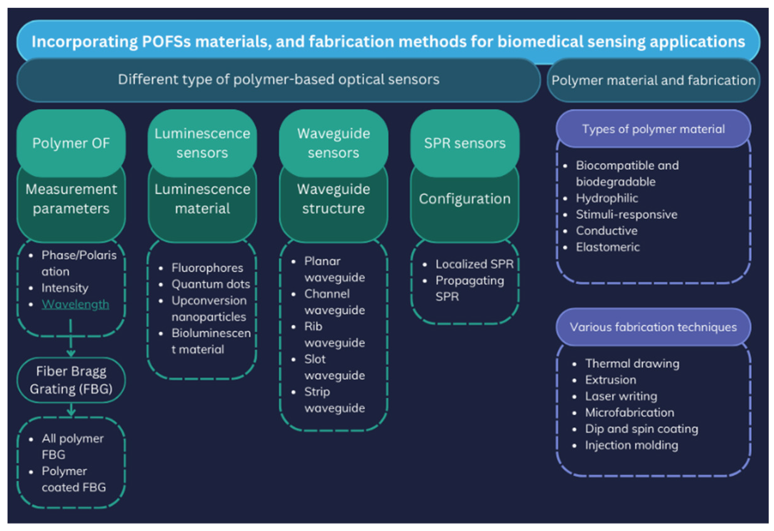
Figure 2.
The development and optical properties of GLPLight1 were examined using structural modeling and fluorescence imaging. a) The structural model, generated using Alphafold [113], depicts the human glucagon-like peptide-1 receptor (GLP1R) in gold, the circularly permuted green fluorescent protein (cpGFP) in green, and mutagenesis target residues in magenta. b) Fluorescence imaging in HEK293T cells and primary cortical neurons demonstrated an increase in fluorescence intensity. Pixel-wise ΔF/F0 images further confirmed these fluorescence changes, supporting the sensor’s effectiveness in detecting GLP-1 interactions. Image source: [112].
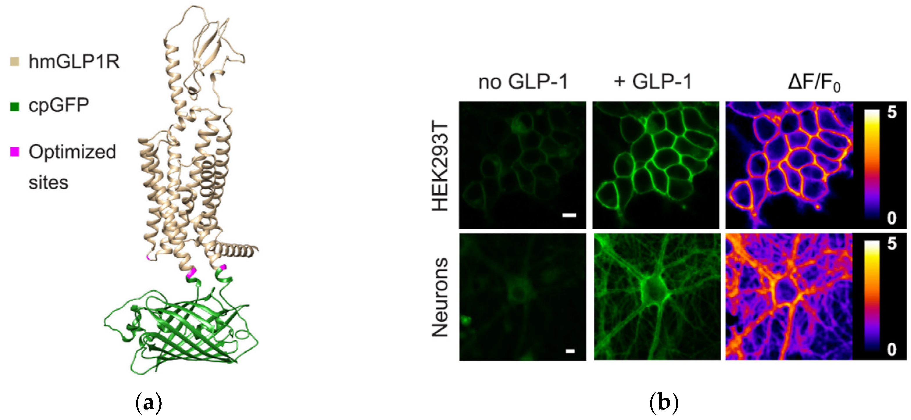
Figure 3.
Step graph of an approach to sensor accuracy assurance during the design, manufacturing, validation, and deployment of a sensor technology, factors to consider are highlighted in blue in the first and in the last steps, Adopted with permission from: [114]. Copyright 2025 American Chemical Society.
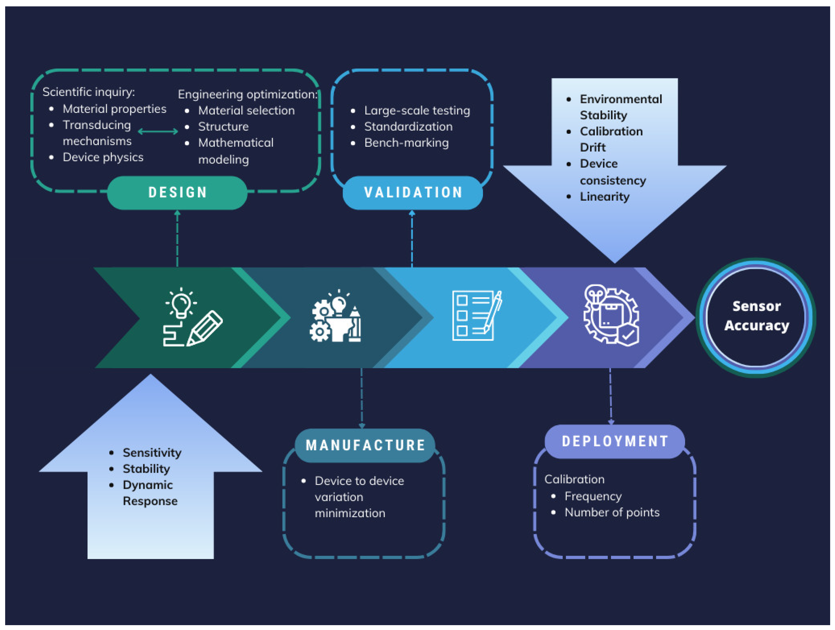
Figure 4.
Schematic puzzle of the four major challenges during developing biodegradable and biocompatible optical fibres, Adopted Idea [115].
Figure 4.
Schematic puzzle of the four major challenges during developing biodegradable and biocompatible optical fibres, Adopted Idea [115].


Table 1.
Recently introduced FOSs based on the fiber type and their application with assessment on their sensing mechanisms.
Table 1.
Recently introduced FOSs based on the fiber type and their application with assessment on their sensing mechanisms.
| Fiber Type | Application | Sensitivity | Sensing Mechanism | Ref. |
|---|---|---|---|---|
| SMF | Pressure | 263.15 pm/kPa | FPI | [36] |
| OF | Pressure (IOP) | Low baseline drift (<2.8 mmHg) over >4.5 years | FPI with OCT | [30] |
| MMF | Pressure | 2.49 nm/kPa | Interference-based sensing | [37] |
| OF | Pressure/temperature | 55.468 nm/MPa (pressure), 0.01859 nm/°C (temperature) | FPI with MEMs | [38] |
| U-shaped MMF | Biosensing | 1251.44 nm/RIU | LSPR | [39] |
| PC fibre | Biosensing | 12,000 nm/RIU and 16,000 nm/RIU |
SPR | [40] |
| D-shaped OF | Biosensing | 5161 nm/RIU | SPR | [41] |
| D-shaped OF | Biosensing | 4122 nm/RIU | LMR | [42] |
| D-shaped PC fibre | Biosensing | 21,700 nm/RIU | SPR | [43] |
| D-shaped PC fibre | Biosensing | 20,000 nm/RIU | SPR | [44] |
| Plastic OF | Cholesterol detection | 140 mg/dL to 250 nm/dL | - | [45] |
| SMF | Temperature | 210.25 KHz/◦C | Vernier effect | [46] |
| Fiber tip integrated ZnO-nanowire-nanograting | Temperature | 0.066 nW/◦C | Bragg reflection | [47] |
| MMF with spherical end | Pressure/temperature | 0.139 mV/kPa (pressure), 0.87 mV/°C (temperature) | RI modulation using MEMS-based silicon | [48] |
| SMF with a Hollow Silica Tube (HST) | Pressure | 396 pm/kPa | FPI | [49] |
| SMF with FBG | Pressure | 1.466 pm/kPa | FBG array | [50] |
| Ultra-miniature fiber-optic sensor | Pressure (IPP) | (r ≥ 0.7, p < 0.001) | Diaphragm-based FO integrated with a proportional–integral–derivative (PID) | [51] |
| Distributed OF | Pressure | 65.920 μϵ/kPa | Axial strain change detection with a sensitizing structure | [52] |
Table 2.
Overview of recent FOSs for pH measurement, glucose measurement/detection.
| Sensing application | Responsive material with fiber type | Detection range | Ref. |
|---|---|---|---|
| pH | PANi with TFBG | 2-12 | [101] |
| PAAm hydrogel with SPR | 8-10 | [24,99,100,102] | |
| gold nanoparticle-functionalized fiber-optic probes with FPI | 2-12 | [75] | |
| Hydrogel + polymer microarrays with miniature optical fiber | 5.5-8 | [74] | |
| glucose | GO/GOD with LPFG | 0–8mM | [103] |
| GOD with TOFI | 0.0–166.67mM | [104] | |
| 3-APBA with LDOF | 0–50mM | [68] | |
| GO with LPFG | 0 ∼ 1 wt% | [105] | |
| GO/GOD with PCF | 10 g/L to 70 g/L | [106] | |
| SPR with Microsphere optical fiber | 0–200 mg/dL | [107] | |
| Gold nanoparticles (AuNPs) and LSPR with TOF | 5–45 wt% | [108] | |
| GO/GOD with PS-LPFG inscribed on high-birefringence fiber (HBF) | 5–25 mM | [109] | |
| Gold-coated plasmonic layer with PCF | Not specified | [110] | |
| SPR with enzymatic reaction | 0–400 mg/d | [71] | |
| gold nanoparticle-functionalized fiber-optic probes with FPI | 1 μM – 1 M | [75] |
| Material Type | Material Example | Advantages | Disadvantages | Ref. |
|---|---|---|---|---|
| Natural | Proteins: silk Polysaccharides: alginate, cellulose, agarose, chitosan gelatine |
biocompatibility and biodegradability |
limited design flexibility, restricted availability and quantity, batch-to-batch variability, low mechanical strength, and potential immunogenicity | [119,120,121,122,123] |
| Synthetic | Hydrogels: Polyethylene Glycol (PEG), Pluronic (Poloxamer) Citrate-based elastomers: poly (octamethylene citrate) (POC), poly (octamethylene maleate citrate) (POMC), Polymer-Based: Polyvinyl Chloride (PVC), SU-8 (Negative Photoresist Polymer), poly (L-lactic acid) (PLLA), poly (D, L-lactic acid) (PDLLA), poly (L-lactic-co-glycolic acid) (PLGA), poly (D,L-lactic-co-glycolic acid) (PDLGA), poly-”-caprolactone (PCL) Inorganic materials: calcium-phosphate glass (PGs) Silicon-Based Materials: Silicon, Polydimethylsiloxane (PDMS) |
adaptable and flexible structure, tunable biodegradability, and customizable physical, mechanical, and chemical characteristics | biocompatibility should be verified and confirmed, rigidness and brittleness for glass |
[17,115,124,125,126,127] |
| Hybrid Biomaterials (Natural & Synthetic) | Chitosan and Polystyrene Membranes/PAA Silk Fibroin Film, Agarose hydrogel (AG) with gold nanoparticles (AuNPs) | biocompatibility, mechanical strength and tunable properties for PAA, controlled permeability, chemical resistance | limited flexibility, surface modification required for some, degradation issues, Processing complexity, for AuNPs agglomeration of AuNPs and limited long-term stability | [121,122,128,129,130] |
Table 4.
Regulatory Standards for Fiber Optic Sensors in Medical Applications [147,148,149,150,151,152,153,154,155,156].
| Regulatory Body | Standard/Guideline | Scope and relevance to FOSs |
|---|---|---|
| FDA (USA) | FDA Medical Device Approval Process | Safety, efficacy, and reliability assessment ensure FOSs meet regulatory requirements before market approval |
| EMA (EU) | Medical Devices Regulation (MDR) and In Vitro Diagnostic Regulation (IVDR) | Regulation of general medical devices in the EU, governs the safety and performance |
| ISO | ISO 13485/ ISO 10993 | Quality management system for medical devices/ Biocompatibility evaluation of medical device |
| AAMI/ANSI | AAMI TIR42 | Guidance on biocompatibility evaluation which supports compliance with ISO 10993 for medical FOSs |
Disclaimer/Publisher’s Note: The statements, opinions and data contained in all publications are solely those of the individual author(s) and contributor(s) and not of MDPI and/or the editor(s). MDPI and/or the editor(s) disclaim responsibility for any injury to people or property resulting from any ideas, methods, instructions or products referred to in the content. |
© 2025 by the authors. Licensee MDPI, Basel, Switzerland. This article is an open access article distributed under the terms and conditions of the Creative Commons Attribution (CC BY) license (http://creativecommons.org/licenses/by/4.0/).
Copyright: This open access article is published under a Creative Commons CC BY 4.0 license, which permit the free download, distribution, and reuse, provided that the author and preprint are cited in any reuse.


