Submitted:
30 March 2025
Posted:
31 March 2025
You are already at the latest version
Abstract
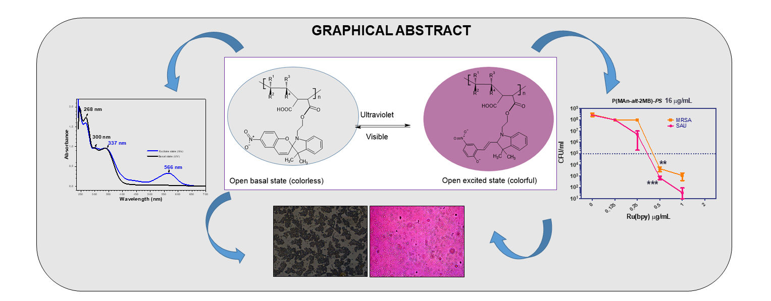
This work focuses on the design of polymeric systems that utilize hydrophilic polymers, with the primary aim of adapting these materials for biological applications. The study further evaluates the effectiveness of photoactive polymers in photodynamic therapy (PDT). It details the synthesis and characterization of photoactive copolymers derived from maleic anhydride (MAn) combined with vinyl monomers such as 2-methyl-2-butene (MB) and 1-octadecene (OD), as well as the organic compound 1-(2-hydroxyethyl)-3,3-dimethylindoline-6-nitrobenzopyran (SP). The two novel optically active alternating polymeric systems, poly(maleic anhydride-alt-octadecene) and poly(maleic anhydride-alt-2-methyl-2-butene), were functionalized with SP through an esterification process in a 1:1 monomer feed ratio, using pyridine as a catalyst. This methodology incorporated approximately 100% of the photoactive molecules into the main acrylic chain to prepare the alternating copolymers. These copolymers were characterized by UV-visible, FTIR, and 1H-NMR spectroscopy and optical and thermal properties. When exposed to UV light, the photoactive polymer films can develop a deep blue color (566 nm in the absorption spectra). Finally, the study also assesses their capacity for photodynamic antimicrobial action in organic film. Notably, the photoactive P(MAn-alt-2MB)-PS significantly enhances the photodynamic antimicrobial activity of the photosensitizer Ru(bpy) against two bacterial strains, reducing the minimum inhibitory concentration (MIC) from 2 µg/mL to 0.5 µg/mL. Therefore, 4 times less photosensitizer is required when mixed with the photoactive polymer to inhibit the growth of antibiotic-sensitive and resistant bacteria.
Keywords:
light-sensitive materials
; antimicrobial activity
; spiropyrans stimulus-responsive copolymers
; morphological surface characteristic
1. Introduction
Materials with photochromic properties are recognized as excellent candidates for various technological applications, including optical information storage devices, optical switches, and other materials that exhibit photosensitivity when exposed to light of the appropriate frequency and intensity [1].
In this regard, the synthesis and copolymerization of photoactive monomers derived from N-(2-methacryloyloxyethyl)-6-nitrospirobenzopyranindoline and methyl methacrylate by atom transfer radical polymerization have been reported [2]. Indeed, some of these compounds have evolved into convenient building blocks for producing commercial ophthalmic lenses, as well as the development of switches and memories, stimulating intense and up-and-coming interdisciplinary research.
Furthermore, a new optically active polymeric methacrylate was synthesized and investigated, in which the (S)-3-hydroxypyrrolidinyl group, linked through the nitrogen atom to an azopyridine chromophore, has been incorporated, in combination with a polymeric methacrylate that incorporates the photoisomerizable spiropyran chromophore in the side chain. This system can be modulated in an acidic medium by the protonation of the azopyridine fractions through the photoisomerization of the spiropyran component, allowing it to behave as a chiroptical switch [3].
Similarly, the synthesis of 1`, 3’, 3’-trimethylspiro [2H-1]-benzopyran-2,2’-indoline derivatives with H, Br, I, and NO2 substituents at the 6- and 8-positions of the benzopyran ring has been reported [4]. These compounds were obtained by the condensation of 1,3,3-trimethyl-2-methylindoline with 2-hydroxybenzaldehyde, which is substituted in five positions with several electron-withdrawing groups. Among these, the 6-nitro-spiro benzopyran indoline showed the highest photochromatic activity. It was also established that the absorption of the compound in the visible region of the spectrum, in the excited state, is independent of solvent polarity; however, the extent of stability of the colored form increases with the increase of solvent polarity [4]. On the other hand, the search for a variety of new functional materials, such as films with highly regular patterning, has received significant interest due to their potential use in material technology [5,6], where porous films might find application in membranes, [7] in photonic[8] and/or optoelectronic devices [9]. Regarding the exploration of light-responsive molecules in devices typically requires their immobilization on a surface through a linker that does not interfere with the structures’ light-switching behaviour. This has been achieved for photoswitchable molecules by forming self-assembled monolayers (SAMs) [10] and bilayers [11], as well as by incorporating SAMs into polymer films [12,13] and polymer beads. The reversibility of these macroscopic properties results from photoinduced transformations at the molecular level [14,15]. These processes can be explored to tune the optical signals, thereby offering the opportunity to design and implement photonic devices for optical processing based on molecular components [15]. The change in chemical structure allows it to absorb in a specific region of the spectrum, typically in the visible range, from which it returns to its basal state after a short period under the influence of radiation or thermal stimulus [16,17]. The terms “positive” and “negative” photochromism are generally used to indicate photoinduced coloration and decolouration processes, respectively. However, both transformations must be reversible by definition [17].
Spiropyrans (SP), first described by Fischer and Hirshberg in 1962, [18] are one of the most studied classes of photoswitchable compounds. The irradiation of SP compounds with near-UV light [19] or their electro-oxidation [20] induces the heterolytic cleavage of the spiro carbon-oxygen bond, leading to ring opening and conversion into merocyanine (MC). The intense absorption in the visible region of the open-form MC has led to the advanced study of SP compounds in photochromic, [16] molecular optoelectronic, [21] optobioelectronic systems, [22] and chemical sensing [23].
In this sense, it is essential to highlight a potential application of these materials in biomedicine due to the existence of bacteria that have been progressively accumulating with a high resistance to antibiotics, which results in progressively fewer antimicrobial alternatives for effective treatment; for this reason, there is a need for materials that helps to complement the treatment with antibiotics. In this regard, the emergence of multidrug resistance (MDR) for pathogenic bacteria is one of the most pressing global threats to human health in the 21st century. In the US, almost 23,000 persons die annually due to antibiotic-resistant infections [24]. As bacteria have accumulated progressively more resistance factors, this results in progressively fewer antimicrobial alternatives for effective treatment [25]. Hence, the availability of new treatments becomes indispensable to prevent morbidity and mortality caused by MDR-infectious agents. In this regard, photodynamic therapy (PDT) has the advantage of being locally activated using phototherapy devices [26]. The PDT utilizes photosensitizer molecules, oxygen, and light to induce non-specific photooxidative stress, which kills bacteria. Bacteria that represent a severe threat to public health are the Gram-positive Staphylococcus aureus. S. aureus is one of the most significant MDR bacteria responsible for health-associated infections (HAIs) (30%) [27,28,29,30]. Moreover, MDR strains are responsible for the more prevalent HAI infections associated with surgical wound infection, urinary tract infection (UTI), and pneumonia [31,32,33,34].
This work aims to contribute to the design and implementation of photonic devices for optical processing based on molecular and macromolecular components, and subsequently to evaluate the utility of photoactive polymers in photodynamic therapy (PDT). This work describes the design, synthesis, characterization, and application of two novel photoactive copolymers derived from de maleic anhydride (MAn) with vinyl monomers such as 2-methyl-2-butene (MB) and 1-octadecene (OD) and the organic photosensitive compound -(2-hydroxyethyl)-3,3-dimethylindoline-6-nitrobenzopyran (SP). Both optically active alternating polymeric systems, poly(maleic anhydride-alt-octadecene) and poly(maleic anhydride-alt-2-methyl-2-butene), were functionalized with SP through an esterification reaction. The optically active polymer carries a photoisomerizable spiropyran chromophore in the side chain linked through the oxygen atom to the polymer chain. We focused on determining its ability to inhibit bacterial growth of S. aureus, it’s possible use as a solid matrix in combination with a photosensitizer, and its utility in sanitary pads or plasters to complement antibiotic treatment. The light-emitting properties of the polymers were characterized by blue and violet colors when exposed to UV light and were subsequently used in an assay to verify their antimicrobial properties in photodynamic therapy (PDT). These macromolecular components exhibited an evident photon transfer process; based on this behavior, it was investigated with a photosensitizer molecule for PDT. These copolymers were characterized by spectroscopic techniques, such as FT-IR and 1H-NMR, optical microscopy, UV-visible spectroscopy, and thermal analysis techniques, such as TGA.
2. Experimental Part
2.1. Characterization
The absorption spectra of the films were recorded at 25 ºC between 250-700 nm using a Perkin Elmer Lambda 35 spectrophotometer. The FT-IR spectrums were recorded on a Perkin-Elmer Spectrum-Two spectrometer with a UATR unit coupled in the range of 4000 to 500 cm-1 with a resolution of 1 cm-1. Photoluminescence (PL) measurements were performed at room temperature by a fluorescence spectrometer system (Perkin Elmer, model L 55). The number averages (Mn), weight average (Mw) molecular weights, and polydispersity (Mw/Mn) of the polymers were determined by size exclusion chromatography (SEC) using a Shimatzu LC 20 instrument equipped with RI detector. An optical microscopy LEICA Model DM2000 LED with a camera LEICA MFC 170 HD was used. The camera was set to an automatic exposure of 500.00 ms, saturation of 120, and gamma 0.00. The image surface was 549.45 μm x 412.09 μm. For image acquisition under fluorescence, the aperture was 1/3 and the focus 3/3. The films were prepared using a chamber Darwin model PH9-DA with relative humidity (RH) control of 75% at 25 °C.
2.2. Procedure of Photochromic Agent Synthesis
The synthesis of the photochromic agent 1-(2-hydroxyethyl)-3,3-dimethylindoline-6-nitrobenzopyran (SP) is carried out in two steps (a and b), as described below, see Figure 1. The synthesis yield is 70.6%, and the physical data, as determined by FTIR and ¹H NMR, conform to those reported in the consulted literature [35].
a) Synthesis and characterization of 1-(2-hydroxyethyl)-2,3,3-trimethylindolenine bromide salt: The procedure was carried out in a Schlenk tube in which 4.0 mL (25 mmol) of 2,3,3-trimethylindolenine was added, then 1.8 mL (25 mmol) of 2-bromoethanol in 3.16 mL (35 mmol) of 2-butanone as solvent. Subsequently, the mixture was degassed under an inert atmosphere, frozen with liquid nitrogen, and then subjected to several cycles of vacuum and thawing. The synthesis tube was placed in an oil bath at 78 °C with constant stirring for 10 hours. Then, at room temperature, the mixture was filtered, yielding a pink solid. This was purified by extraction in a benzene soxhlet for 24 hours until the solution became colorless, yielding 70.6 %. The chemical shifts show the following signals at (δ, ppm, CDCl3): 1.65 (9H, singlet, -CH3-); 3.15 (1H, singlet, -OH); 4.20 (2H, triplet, CH2-OH); 4.89 (2H, triplet, CH2-N); 7.57 (4H, multiplet, aromatic ring). The signals confirm the molecular structure of 1- (2-hydroxyethyl) -2,3,3-trimethylindolenine bromide.
b) Synthesis and characterization of 1- (2-hydroxyethyl) -3,3-dimethylindoline-6-nitrobenzopyran: In a 250 mL ball (three-mouth) equipped with a magnetic stirrer and condenser, was added 2 g (7.04 mmol) of 1- (2-hydroxyethyl) -2,3,3-trimethylindolenine bromide, 1.2 g (7.04 mmol) of 2-hydroxy-5-nitrobenzaldehyde, 4 mL of triethylamine (28.16 mmol) in 20 mL of ethanol and heating until boiling (78 °C) for 4 hours in an oil bath. After 4 hours of reaction, it was allowed to cool, and the ethanol was evaporated using a rotary evaporator.
The product was extracted in a separator funnel with a solution of 10% HCl and chloroform in equal volumes to recover the organic phase where it is located. After this, the product was dried in the presence of magnesium sulfate, filtered, and the chloroform evaporated, yielding 80.3 %. The product obtained was purple crystals.
Moreover, TFIR exhibits the following signals vibration bands υ (cm-1, KBr): 1088 (flexion -C-O-C-); 1335 (symmetric stretch Ar-NO2); 1510 (asymmetric stretching Ar-NO2); 1603 (stretching -C=C-); 1930-1836 (Ar-H aromatic overtones); 2961 (stretching C-H; CH-, -CH2-, -CH3); 3068 (stretching =C-H); 3365 (O-H stretch -CH2OH). The 1H-NMR exhibits the following chemical shifts at (δ, ppm, CDCl3): 1.12 [3H, singlet, -CH3-];1.22 [3H, singlet, -CH3-];1.53 [1H, singlet, -OH]; 3.46 [2H, multiplet, CH2-N]; 3.71 [2H, triplet, CH2-OH]; 5.84 [1H, doublet, H (i)]; 6.57 [1H, doublet, H (h)]; 6.71 [1H, doublet, H (g)]; 6.84 [2H, triplet, H (e), H (f)] 7.01 [1H, doublet, H (d)]; 7.18 [1H, triplet, H (c)];7.97-7.92 [2H, multiplet, H (a) and H (b)]. The signals observed in the spectrum are characteristic of the photochromic compound 1-(2-hydroxyethyl)-3,3-dimethylindoline-6- nitrobenzo pyran.
2.3. Characterization by UV-Visible Spectrophotometry
The colored state of the compound in solution was reached after irradiating the quartz cell with an ultraviolet light for 5 minutes. A solution of 1x10⁻⁵ M of 1- (2-hydroxyethyl) -3,3-dimethylindoline-6-nitrobenzopyran (SP) was prepared, using chloroform as a solvent; 2 mL of this solution was deposited in a quartz cell, this was placed in the ultraviolet spectrophotometer cell holder, then determined the wavelength values of the compound.
As can be seen, 1- (2-hydroxyethyl) -3,3-dimethylindoline-6-nitrobenzopyran compound, after being irradiated by ultraviolet light, changes from a basal state, colorless (closed) to an excited state colored (open). The unimolecular reactions of the organic molecules with photochromic properties involve ring-closing and opening steps or trans → cis and cis → trans isomerization, see Figure 1.
2.4. Preparation of Photoactive Alternating Copolymer
The alternating copolymers P(MAn-alt-OD) and P(MAn-alt-2MB) were carried out at 1:1 monomers feed ratio in the THF solution. The polymerization reaction was initiated using 0.5 mol-% benzoyl peroxide (BPO) as the initiator agent in solution. The general procedure for 1:1 monomers feed ratio was as follows: 20 mL of THF was transferred to septum-copped, nitrogen-purged flasks containing 3.171 g (12.50 mmol) of MAn, 1.196 g (12.50 mmol) of comonomers (OD/2MB), and 140.5 mg (0.580 mol-%) of the initiator was added. The ampoule was degassed with freeze cycles and sealed under a high vacuum. The monomers and initiator in solution were taken to a thermoregulated bath at 80 °C to initiate copolymer formation through free radical polymerization for 8 h. The products were removed from the systems using methanol/ethanol respectively, then the methanol/ethanol was evaporated from the balloon with a rotary evaporator, then chloroform (CHCl3) was added, and the solution was deposited in a separator funnel. Extraction of the soft yellow color product was carried out respectively with a 10% HCl solution and chloroform in a ratio of 1:1, twice. After the resulting organic phase, a second extraction was carried out with chloroform and 10% sodium carbonate (1:1). Posteriorly, anhydrous magnesium sulfate was added to the resulting organic phase, and it was left to dry for 30 minutes to recover the yellow crystals respectively. The yield of poly(MAn-alt-OD) and poly(MAn-alt-2MB) copolymers was 70.0 % and 80.0 %, respectively. Copolymers with molecular weights of 18,200 and 17,300 and polydispersity index of 1.49 and 1.68 were obtained. Finally, the dried copolymers were characterized by FT-IR spectroscopy, and thermal and optical properties.
Subsequently, the poly(MAn-alt-OD) and poly(MAn-alt-2MB) copolymer were functionalized with photochromic compound 1-(2-hydroxyethyl) -3,3-dimethylindoline-6-nitrobenzopyran (SP) using a trans-esterification reaction. The experimental procedure was carried on a 100 mL three-mouth ball equipped with a magnetic stirrer and condenser, 2.0 g (0.036 mmol) of copolymers, 0.013 g (0.036 mmol) of 1- (2-hydroxyethyl) -3,3-dimethylindoline-6-nitrobenzopyran and one drop of H2SO4 were added in 5 mL of tetrahydrofuran and heating for 4 hours. Finally, a purple/yellow color product was obtained.
2.5. Determination of the Antimicrobial Photodynamic Property
Staphylococcus aureus bacteria were cultured in solid or liquid Trypticase soy medium as needed. In a liquid medium, they were grown to an optical density OD600=0.2-0.4, and for photodynamic therapy (PDT), bacteria were adjusted to 1x10⁷ colony-forming units (CFU)/mL. The bacteria were mixed with the photoactive polymers P(MAn-alt-OD)-SP or P(MAn-2MB)-SP with/without photosensitizer Ru(bpy) [36], in various concentrations. Excitation was performed in a light box with 17 mW/cm2 for 10 min with a blue LED lamp (450-460 nm) equivalent to 61.2 J/cm2. After excitation, bacterial viability was determined by broth microdilution and colony counting on plates after 16-20 h of incubation in Muller-Hinton medium. Bacterial viability is expressed as the mean ±SD in CFU/mL. The MIC of the photosensitizer was determined by mixing 1x10⁷ CFU/mL of bacteria with increasing concentrations of the photosensitizer Ru(bpy) between 0.125 – 4 μg/mL.
3. Results and Discussion
The photoactive copolymers were obtained containing SP moiety with photoluminescence properties. The copolymers were synthesized in an approximate molar ratio of 1:1, as shown in Figure 2, and their functionalized copolymers are according to Figure 3.
3.1. Characterization of Alternating Copolymer
3.1.1. The Characterization by FT-IR and 1H-NMR
FT-IR spectra for P(MAn-alt-OD) shows the following vibration bands υ (cm-1, KBr): at 2957.5 (stretching =C-H); 2855.0 (stretching C-H; CH-, -CH2-, -CH3); 1780.0-1720.0 (symmetric stretching >C=O; -CO-O-CO- MAn); 1707.5 (stretching C=O; -COOH); 1467.0 (bending C-H; -CH2-, CH3). FT-IR spectra of SP shows the following vibration bands υ (cm-1, KBr): at 3386.0 (O-H stretch; CH2OH); 2960.0 (stretching =C-H); 2860.0 (stretching C-H; CH-, -CH2-, -CH3); 1780.0-1720.0 (Ar-H aromatic overtones); 1612.5 (stretching -C=C- Ar); 1485.0 (asymmetric stretching N-O; -N=O); 812.5 and 747.5 (C-H stretching; polysubstituted H-Ar). While, the FT-IR spectrum of P(MAn-alt-OD)-SP functionalized copolymer exhibited characteristic absorption bands at 2957.5; 2855.0 (stretching C-H; CH-, -CH2-, -CH3); 1780.0 (stretching >C=O asymmetric; -CO-O-CO- MAn); 1720.0 (stretch C=O; -COOR ester); 1612.5 (stretch C=C; Ar); 1485.0 (N-O stretch; -N=O); 1467.0 (C-H bending; -CH2-, CH3); 812.5 and 747.5 (C-H stretching; polysubstituted H-Ar). These signals are characteristic of the photochromic compound SP, which confirms its incorporating in P(MAn-alt-OD)-SP functionalized copolymer.
FT-IR spectra for P(MAn-alt-2MB) shows the following vibration bands TF-IR ν(cm-1, KBr): at 2985.0–2855.0 (stretching C-H; CH-, -CH2-, -CH3); 1857.5 (stretching >C=O asymmetric; -CO-O-CO- MAn); 1780.0 (stretching >C=O symmetric; -CO-O-CO- MAn); 1719.0 (stretching C=O; -COOH); 1480.0 y 1390.0 (bending C-H; -CH2-, CH3).
FT-IR spectra of SP shows the following vibration bandsν (cm-1, KBr):at 3382.0 (tension O-H; -OH from SP); 2985.0 – 2855.0 (stretching C-H; CH-, -CH2-, -CH3); 1612.5 (stretching C=C; Ar); 1485.0 (stretching N-O; -N=O); 807.5 y 750.0 (stretching C-H; H-Ar, polysubstituted). While, the FT-IR spectrum of P(MAn-alt-2MB)-SP functionalized copolymer exhibited characteristic absorption bands at ν(cm-1, KBr): 2985.0 – 2855.0 (stretching C-H; CH-, -CH2-, -CH3); 1857.5 (stretching >C=O symmetrical; -CO-O-CO- MAn); 1780 stretching >C=O asymmetric; -CO-O-CO- MAn); 1728.0 (stretching C=O; -COOR éster); 1612.5 (stretching C=C; Ar); 1485.0 (stretching N-O; -N=O); 807.5 y 750.0 (stretching C-H; H-Ar polysubstituted). Table 1 summarizes the main vibration bands in the FT-IR spectra for the functionalized copolymers.
3.1.2. Characterization by NMR 1H- of the Copolymers P(MAn-alt-OD)-SP and P(MAn-alt-OD)-SP
NMR1H- spectrum (δ ppm, CHCl3) of the P(MAn-alt-1OD)-SP copolymer shows the following signals at: 8.00 (2H, m, Ha and Hb); 7.20(1H, t, Hd); 7.10(1H, d, Hc); 6.90(2H, m, He and Hf); 6.77(1H, d, Hh); 6.66(1H, d, Hi); 5.88(1H, d, Hg); 3.79(2H, m, -CH2O-); 3.40(2H, m, -CH2N<); 2.80 to 0.4 (48H, m, 3x –CH<, 18x –CH2- and 3x –CH3), see Figure 4a). The characterization of P(MAn- alt-OD) show the following signals: δ (ppm, DMSO-d6): 2.2 to 1.7 (2H, m width, MAn); 1.6 and 0.6 (40H, S wide, >CH-, -CH2-, -CH3). NMR1H- spectrum (δ ppm, CHCl3) of P(MAn-alt- 2MB)-SP: the spectrum shows the following signals at: 8.01(2H, m, Ha y Hb); 7.19(1H, t, Hd); 7.11(1H, d, Hc); 6.90(2H, m, He y Hf); 6.75(1H, d, Hh); 6.67(1H, d, Hi); 5.89(1H, d, Hg); 3.77(2H, m, -CH2O-); 3.40(2H, m, -CH2N<); 2.80 a 0.5(18H, m, 3x –CH< y 5x –CH3) (see Figure 4b).
3.1.3. Degrees of Functionalization (Copolymer Composition)
The degree of functionalization of the two copolymers was determined by designing systems of equations that relate the quantities of aliphatic protons, methylene protons, and/or aromatic protons of the copolymer matrices with their integrals and their chemical shifts in 1H NMR spectroscopy. Managing to establish the following equations (1 – 4).
where
- = Number of aliphatic protons in the fraction X.
- = Number of aliphatic protons in the fraction Y
- = Number of methylene protons
- = Number of aromatic protons
- = Integral aliphatic protons
- = Integral aromatic protons
- = Integral methylene protons
- = Numerical value, mathematical operation of η1, η2, η4, ,.
- = Numerical value, mathematical operation of η1, η2, η3, ,
- : Comonomer fraction in X
- : Comonomer fraction in Y
3.1.4. Functionalization of P (MAn-alt-OD)-SP and P(MAn-alt-2MB)-SP
To establish the copolymer composition of the functionalized copolymers, the integrals of each aliphatic fraction of the copolymers were considered versus the fractions of the numerical values , andand the degree of functionalization was determined. As a result, similar values for X and Y were obtained by both methods (see Table 2). The results obtained for fractions X and Y showed that the functionalization of the MAn comonomer (fraction X) is 40% and 49%, respectively.
3.1.5. Characterization by UV-Visible Spectrophotometry
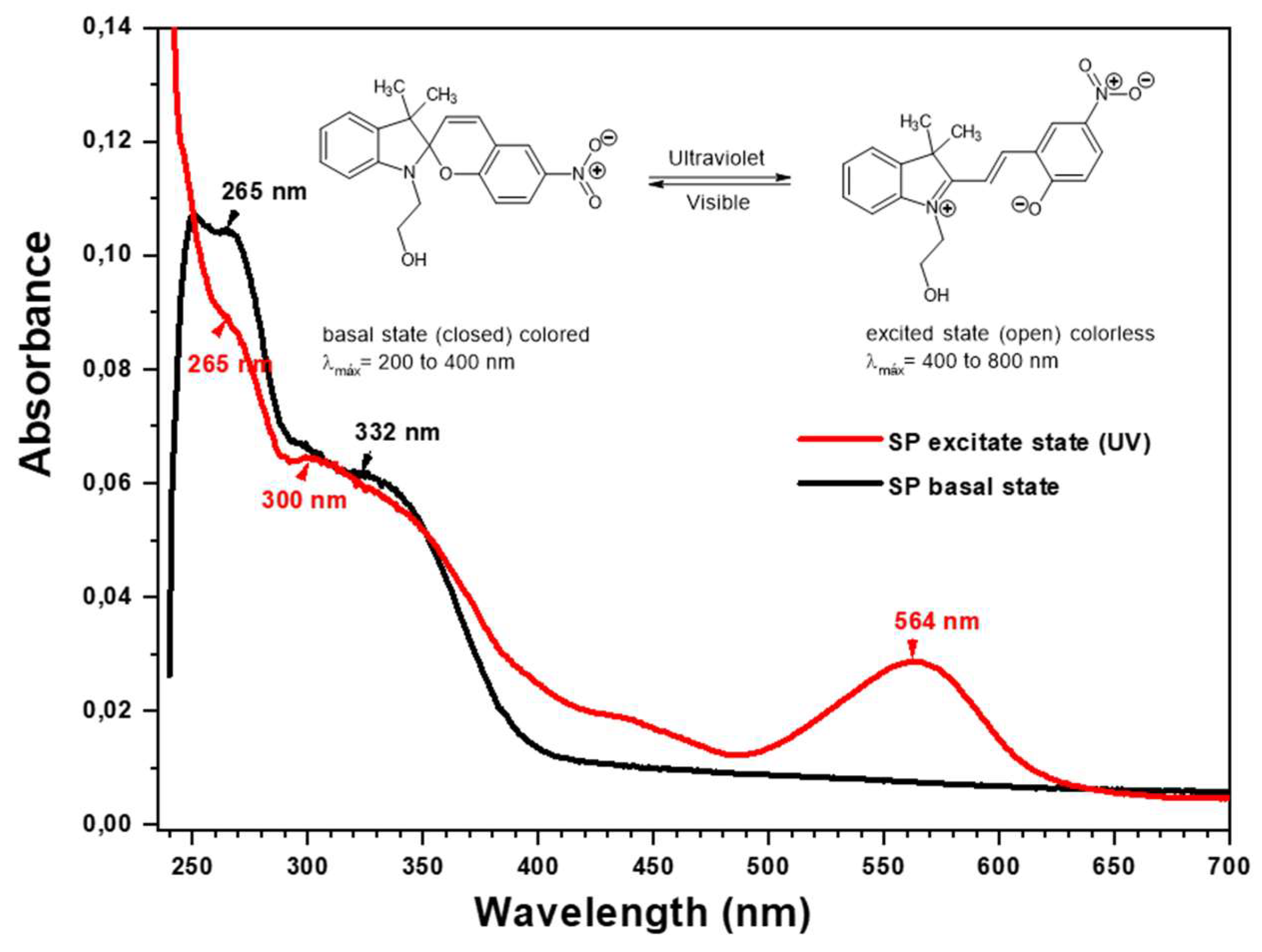
Figure 5 shows the photochromic equilibria of the functionalized copolymers; when irradiated by ultraviolet light, they change from a colorless basal state (closed) to a colorful excited state (open).
Figure 5, it was possible to observe the isomeric equilibrium of the photochromism of 1-(2-hydroxyethyl)-3,3-dimethylindoline-6-nitrobenzopyran (SP). The basal UV spectrum of the photochromic agent SP presents two λmax at 265 nm and 332 nm, and the spectrum in its excited state presents three λmax 265 nm, 300 nm, and 564 nm. These data demonstrate the photochromic isomeric balance since the λmax 265 nm and 332 (300) nm are maintained, and a new λmax is formed at 564 nm, evidenced by the visible blue coloration.
Figure 6.
Absorption changes on the photoactive agent (3 mg·mL-1, in THF), a) before irradiation (black line) and after irradiation at 365 nm (red line).
Figure 6.
Absorption changes on the photoactive agent (3 mg·mL-1, in THF), a) before irradiation (black line) and after irradiation at 365 nm (red line).
On the other hand, the solution of photoactive copolymers showed photoinduced reversible interconversion upon UV-Vis irradiation. As a result, the absorbance of the photochromic solution can be modulated simply by turning an ultraviolet source on and off. The spectrophotometry of functionalized copolymers with SP generates two isomeric structures in equilibrium. These are formed when the sample in solution is irradiated with ultraviolet light at 385 nm, moving from the colorless basal (molecular state closed) to a colorful (blue) (molecular excited open state) (see Figure 7).
Figure 7 shows the effect of irradiating the functional polymers P(MAn-alt-2MB)-SP and P(MAn-alt-OD)-SP on the absorption spectrum. After irradiation, the photoinduced formation of the colored state “merocyanine” (MC), is responsible for the significant increases of absorbance in the visible region at 560 and 566 nm, respectively, indicating its photoconversion from SP to MC (open conformation) [37,38]. Table 3 summarizes the λmax in the ground state and excited state of the UV-VIS spectra of the photochromic agent and the functionalized copolymers.
The basal spectrum of the functionalized copolymer P(MAn-alt-OD)-SP presents a λmax at 320 nm and, in its excited state, presents two λmax at 318 nm and a new signal at 560 nm. These data demonstrate the photochromic isomeric equilibrium of the copolymer, which is evidenced by the visible blue coloration. Moreover, the basal spectrum of the functionalized copolymer P(MAn-alt-2MB)-SP presents three λmax at 268 nm, 300 nm, and 337 nm, whereas in its excited state, it presents four λmax at 268 nm, 300 nm, 337 nm, and at 566 nm (a new signal). Therefore, the photochromic balance of the SP agent is maintained in the functionalized copolymer. The λmax close to 560 nm shows the breaking of the spiro bond (N-C-O-C) coming from the benzopyran of the photochromic agent and the functionalized copolymers, forming an isomeric structure called merocyanin (MC) that has conjugated π bond systems (conjugated polyenes), with a great resonance effect induced by the nitro group capable of absorbing energy and being excited in the visible light region. This is due to the decrease in the energy of the electronic transition of the double bond system from π to π* where the excited state has more polarity than the basal state, and the effect of radiation is mostly effective, showing a large bathochromic shift and a hypochromic effect of lesser intensity. Furthermore, the electronic transition π to π* is very likely to occur from a basal singlet state to an excited singlet state since the spin of the excited electron is unpaired with its antiparallel spin, and its relaxation mechanism is fast and reversible (see Figure 7). While the electronic excitation π to π* at a triplet level requires a “forbidden” spin transition to be carried out, which implies that it is unlikely to form, since the spin of the excited electron is unpaired with its spins. parallel and its relaxation mechanism is slow and not very reversible.
PDT requires a light irradiation of a specific wavelength to excite the PS moiety. As is exhibited in Figure 7, upon irradiation, the PS moiety in its lowest energy level (ground singlet state, π) is changed to the short-lived excited singlet state (1π*), which can be converted to the long-lived excited triplet state, (3π*. In the presence of ambient oxygen, the triplet state can undergo two types of reaction mechanisms: I) through transfer electrons to form toxic reactive oxygen species (such as peroxide, H2O2, and hydroxyl radicals, etc.): II) where it involves an energy transfer ground state triplet oxygen to produce highly reactive singlet oxygen (1O2). The two reactions can take place simultaneously. However, not all highly conjugated unsaturated organic molecules can undergo the intersystem crossing to produce the triplet state necessary for photochemical reactions to occur [39].
The optical band gap Eg of the photoactive copolymers were estimated from UV-Vis measurements before and after irradiation at 365 nm wavelength, using the Tauc plot equation: (αhν) = A (hν – Eg)m where α is the absorption coefficient, h the Planck’s constant, ν frequency of light, A constant, m constant related to the type of optical transition and Eg the band gap. The absorption coefficient is estimated from α = 2.303 A/d, where A is the absorbance, and d is the compactness of the film [40,41]. The value of m=1/2 was used in the calculations because it was considered direct optical transition, [42] for all the compounds, therefore, (αhν)2 versus the hν, the photon energy, was plotted, and the linear part of the figure was extrapolated by a straight line to (αhν)2 = 0 to estimate the band gap. Table 4 shows both photoactive copolymers’ band gap (Eg) values estimated before and after irradiation. The band gap value for P(MAn-alt-2MB)- SP is higher than for P(MAn-alt-OD)- SP. After irradiation, the band gap value for the P(MAn-alt-2MB)- SP decreases at 3.78 eV. The same behavior is observed for the P(MAn-alt-OD)- SP. The reduction of the band gap could be due to the development of a new transitional level beneath the conduction band [40].
3.2. Thermal Decomposition Analysis by TGA
The results of the thermal behaviour by TGA of both copolymers and their functionalized derivatives are presented in Figure 8, which are expressed in percentage mass losses (%) as a function of the increase in temperature. Additionally, the extrapolated thermal decomposition temperature (TDTe) is reported. The thermograms of the copolymers exhibited higher thermal stability than those functionalized with the photochromic agent. The copolymers exhibited two TDTe; the first temperature is attributed to the decomposition of the hydrocarbon chain, and the second TDT is attributed to the decomposition of the maleic anhydride ring. On the other hand, the functionalized polymer P(MAn-alt-OD)-SP exhibited two TDTe (137.0 °C and 357.0 °C); the first temperature is attributed to the decomposition of the hydrocarbon chain of the copolymer and the second at 357.0 °C show lower stability of the maleic anhydride ring functionalized with the SP agent, which is evidenced by a 75% loss of mass, seeFigure 8 a). Otherwise, the thermogram of the P(MAn-alt-2MB) exhibited two TDTe (145.0 °C and 276.0 °C), and the second TDT exhibited a total decomposition of the copolymer with a mass loss that reaches 99%. At the same time, the photoactive P(MAn-alt-2MB)-SP exhibited three TDTe (140.0 °C, 220.0 °C and 381.0 °C), the two first are attributed to the decomposition of the hydrocarbon chain of the copolymer and the third at 381.0°C demonstrates greater stability of the maleic anhydride ring with the photochromic agent, which is evidenced by a 55% loss of mass (see Figure 8 b).
3.3. Characterization by Fluorescence Optical Microscopy
Figure 9 shows the surface of P(MAn-alt-OD)-SP and P(MAn-alt-2MB)-SP films before and after UV irradiation at 3 g·L-1 (in CS2), respectively. When exposed to visible light, the images exhibit structured surfaces and show red luminescence. After irradiation, the photoinduced formation of the colored state “merocyanine” (MC) is observed due to the SP moiety incorporated into the polymer chain. The optical images of the surface films show a specific pore array on the surface. The SP moieties grafted to the chain polymer are expected to lead to a different organized surface, with a different distribution in the whole surface (Figure 9 a, b, c, d).
3.4. Determination of Antimicrobial Properties Based on Photodynamic Therapy (PDT).
Since the optically active polymers, P(MAn-alt-OD)-SP and P(MAn-alt-2MB)-PS, present macromolecular components with an evident photon transfer process, its utility for antimicrobial photodynamic therapy (aPDT) was investigated. The PDT is a technique employing the photoactive copolymer (PS) in the presence of the photosensitizer, Ru(bpy), followed by light irradiation with a specific wavelength that can excite the photosensitizer and PS moiety to cause production of cytotoxic ROS in the presence of ambient molecular oxygen. The utilization of ROS produced during irradiation application can exert lethal effects on microbial pathogens. As seen in Figure 10 a), the photosensitizer Ru(bpy) presents a minimum inhibitory concentration (MIC) of 2 μg/mL against the bacterial strains sensitive Staphylococcus aureus (SAU) and methicillin-resistant S. aureus (MRSA). The MIC of Ru(bpy) was significantly (p < 0.01) improved when mixed with the polymer P(MAn-alt-2MB)-PS (Figure 10 b), decreasing the MIC to 0.5 μg/mL. This means that 4 times less photosensitizer is required when mixed with P(MAn-alt-2MB)-PS to inhibit the growth of both antibiotic-sensitive and resistant bacteria. On the other hand, P(MAn-alt-OD)-SP shows no significant improvement in the photodynamic activity of the Ru(bpy) photosensitizer (data non-shown). Therefore, the polymer P(MAn-alt-2MB)-SP significantly improves the bactericidal photodynamic activity of the photosensitizer Ru(bpy) against both bacterial strains, decreasing the MIC from 2 μg/mL to 0.5 μg/mL.
4. Conclusions
According to the results obtained, it is possible to conclude that the preparation of two new alternating copolymers, P(MAn-alt-OD) and P(MAn-alt-2MB) and their photoactive polymer P(MAn-alt-OD)-SP and P(MAn-alt-2MB)-SP were achieved employing free radical polymerization using maleic anhydride with the octadecene and 2-methyl-2-butene comonomers. Moreover, as part of the photochemical evaluation, the results indicate that the photoactive properties of the photochromic compound are slightly altered when incorporated into the polymer chain. The optical and morphological properties of the photoactive copolymers and their relationship with each other were analyzed, indicating that the compounds exhibit an evident photon transfer process, characterized by a change in color after irradiation. For P(MAn-alt-2MB)-SP, the band gap decreased from 4.53 to 3.78 eV after UV irradiation. Moreover, incorporating the SP moiety into the alternating copolymer P(MAn-alt-OD)-SP does not affect the band gap. On the other hand, the film copolymers also exhibited light-sensitive behavior under UV light irradiation, showing changes in their optical and morphological properties. The photoactive polymer P(MAn-alt-2MB)-PS significantly enhances the photodynamic antimicrobial activity of the photosensitizer Ru(bpy) against bacterial strains, decreasing the minimum inhibitory concentration (MIC) from 2 μg/mL to 0.5 μg/mL. It is possible to conclude that these materials could be very attractive for their use in manufacturing light-sensitive antimicrobial films due to their optical response.
Acknowledgements
The authors thank FONDECYT [grant number 1240357], Project funded by the Research Continuity, year 2023, code PY LCLI23-02, Universidad Tecnológica Metropolitana. 1241555 (awarded to C.E.P.); ANID.
References
- Paras, N.; Prasad, D. J. W. Introduction_to_Nonlinear_Optical_Effect, ilustrada; Wiley, 1991; Ed. [Google Scholar]
- García, A. E.; Elizalde, L. E.; Guillén, L.; De los Santos, G.; Medellín, D. I. Síntesis y evaluación fotocromática de compuestos espirobenzopiránicos. J Mex Chem Soc 2004, 48, 269–274. [Google Scholar]
- Angiolini, L.; Benelli, T.; Giorgini, L.; Raymo, F. M. Chiroptical Switching Based on Photoinduced Proton Transfer between Homopolymers Bearing Side-Chain Spiropyran and Azopyridine Moieties. Macromol Chem Phys 2008, 209(19), 2049–2060. [Google Scholar] [CrossRef]
- de los Santos, G.; Elizalde, L. E.; Castro, B.; García, A. E.; Medellín, D. I. Sociedad Química de México : [Revista]. Revista de la Sociedad Química de México 1996, 48, 332–337. [Google Scholar]
- Nunes, S. P.; Behzad, A. R.; Hooghan, B.; Sougrat, R.; Karunakaran, M.; Pradeep, N.; Vainio, U.; Peinemann, K.-V. Switchable PH-Responsive Polymeric Membranes Prepared via Block Copolymer Micelle Assembly. ACS Nano 2011, 5(5), 3516–3522. [Google Scholar] [CrossRef] [PubMed]
- Hilke, R.; Pradeep, N.; Madhavan, P.; Vainio, U.; Behzad, A. R.; Sougrat, R.; Nunes, S. P.; Peinemann, K. V. Block Copolymer Hollow Fiber Membranes with Catalytic Activity and PH-Response. ACS Appl Mater Interfaces 2013. [CrossRef]
- Amado, F. D. R.; Gondran, E.; Ferreira, J. Z.; Rodrigues, M. A. S.; Ferreira, C. A. Synthesis and Characterisation of High Impact Polystyrene/Polyaniline Composite Membranes for Electrodialysis. J Memb Sci 2004, 234, (1–2). [Google Scholar] [CrossRef]
- Wijnhoven, J. E. G. J.; Vos, W. L. Preparation of Photonic Crystals Made of Air Spheres in Titania. Science (1979) 1998, 281(5378), 802–804. [Google Scholar] [CrossRef] [PubMed]
- Imada, M.; Noda, S.; Chutinan, A.; Tokuda, T.; Murata, M.; Sasaki, G. Coherent Two-Dimensional Lasing Action in Surface-Emitting Laser with Triangular-Lattice Photonic Crystal Structure. Appl Phys Lett 1999, 75(3), 316–318. [Google Scholar] [CrossRef]
- KATSONIS, N.; LUBOMSKA, M.; POLLARD, M.; FERINGA, B.; RUDOLF, P. Synthetic Light-Activated Molecular Switches and Motors on Surfaces. Prog Surf Sci 2007, 82, 407–434. [Google Scholar] [CrossRef]
- Hugel, T.; Holland, N. B.; Cattani, A.; Moroder, L.; Seitz, M.; Gaub, H. E. Single-Molecule Optomechanical Cycle. Science (1979) 2002, 296(5570), 1103–1106. [Google Scholar] [CrossRef]
- Byrne, R. J.; Stitzel, S. E.; Diamond, D. Photo-Regenerable Surface with Potential for Optical Sensing. J Mater Chem 2006, 16(14), 1332. [Google Scholar] [CrossRef]
- Lee, J.; Kwon, T.; Kim, E. Electropolymerization of an EDOT-Modified Diarylethene. Tetrahedron Lett 2007, 48(2), 249–254. [Google Scholar] [CrossRef]
- Raymo, F. M.; Tomasulo, M. Optical Processing with Photochromic Switches. Chemistry – A European Journal 2006, 12, 3186–3193. [Google Scholar] [CrossRef]
- Yildiz, I.; Deniz, E.; Raymo, F. M. Fluorescence Modulation with Photochromic Switches in Nanostructured Constructs. Chem Soc Rev 2009, 38(7), 1859. [Google Scholar] [CrossRef] [PubMed]
- Berkovic, G.; Krongauz, V.; Weiss, V. Spiropyrans and Spirooxazines for Memories and Switches. Chem Rev 2000, 100(5), 1741–1754. [Google Scholar] [CrossRef]
- Samat, A.; Lokshin, V. Thermochromism of Organic Compounds. In Organic Photochromic and Thermochromic Compounds; Kluwer Academic Publishers: Boston; pp. 415–466. [CrossRef]
- Heiligman-Rim, R.; Hirshberg, Y.; Fischer, E. 29. Photochromism in Some Spiropyrans. Part III. The Extent of Phototransformation. Journal of the Chemical Society (Resumed) 1961, 156. [Google Scholar] [CrossRef]
- Malik, M. Z. A.; Nazri, S. A. A. A.; Zainuddin, M. T.; Islam, N. Z. M.; Aziz, N. M. A. N. A.; Isha, K. M. Effects of Papain Incorporation on the Photo-Transformation Stability of 5-Bromo-8-Methoxy-6-Nitro Bips. Adv Mat Res 2014, 879, 73–78. [Google Scholar] [CrossRef]
- Wagner, K.; Byrne, R.; Zanoni, M.; Gambhir, S.; Dennany, L.; Breukers, R.; Higgins, M.; Wagner, P.; Diamond, D.; Wallace, G. G.; Officer, D. L. A Multiswitchable Poly(Terthiophene) Bearing a Spiropyran Functionality: Understanding Photo- and Electrochemical Control. J Am Chem Soc 2011, 133(14), 5453–5462. [Google Scholar] [CrossRef]
- Willner, I.; Willner, B. Layered Molecular Optoelectronic Assemblies. J Mater Chem 1998, 8(12), 2543–2556. [Google Scholar] [CrossRef]
- Willner, I.; Willner, B. Photoswitchable Biomaterials as Grounds for Optobioelectronic Devices. Bioelectrochemistry and Bioenergetics 1997, 42(1), 43–57. [Google Scholar] [CrossRef]
- Scarmagnani, S.; Walsh, Z.; Slater, C.; Alhashimy, N.; Paull, B.; Macka, M.; Diamond, D. Polystyrene Bead-Based System for Optical Sensing Using Spiropyran Photoswitches. J Mater Chem 2008, 18(42), 5063. [Google Scholar] [CrossRef]
- Lahsoune, M.; Boutayeb, H.; Zerouali, K.; Belabbes, H.; El Mdaghri, N. Prévalence et État de Sensibilité Aux Antibiotiques d’Acinetobacter Baumannii Dans Un CHU Marocain. Med Mal Infect 2007, 37(12), 828–831. [Google Scholar] [CrossRef]
- Magiorakos, A.; Srinivasan, A.; Carey, R. B.; Carmeli, Y.; Falagas, M. E.; Giske, C. G.; Harbarth, S.; Hindler, J. F. Bacteria : An International Expert Proposal for Interim Standard Definitions for Acquired Resistance. 2011. [Google Scholar]
- Liu, Y.-Y.; Wang, Y.; Walsh, T. R.; Yi, L.-X.; Zhang, R.; Spencer, J.; Doi, Y.; Tian, G.; Dong, B.; Huang, X.; Yu, L.-F.; Gu, D.; Ren, H.; Chen, X.; Lv, L.; He, D.; Zhou, H.; Liang, Z.; Liu, J.-H.; Shen, J. Emergence of Plasmid-Mediated Colistin Resistance Mechanism MCR-1 in Animals and Human Beings in China: A Microbiological and Molecular Biological Study. Lancet Infect Dis 2016, 16(2), 161–168. [Google Scholar] [CrossRef]
- Paczosa, M. K.; Mecsas, J. Klebsiella Pneumoniae: Going on the Offense with a Strong Defense. Microbiology and Molecular Biology Reviews 2016, 80(3), 629–661. [Google Scholar] [CrossRef]
- Ko, W. C.; Paterson, D. L.; Sagnimeni, A. J.; Hansen, D. S.; Von Gottberg, A.; Mohapatra, S.; Casellas, J. M.; Goossens, H.; Mulazimoglu, L.; Trenholme, G.; Klugman, K. P.; McCormack, J. G.; Yu, V. L. Community-Acquired Klebsiella Pneumoniae Bacteremia: Global Differences in Clinical Patterns. Emerg Infect Dis 2002, 8(2), 160–166. [Google Scholar] [CrossRef] [PubMed]
- Lakhundi, S.; Zhang, K. Crossm. 2018, 31, 1–103. [Google Scholar]
- Diekema, D. J.; Pfaller, M. A.; Schmitz, F. J.; Smayevsky, J.; Bell, J.; Jones, R. N.; Beach, M. Survey of Infections Due to Staphylococcus Species: Frequency of Occurrence and Antimicrobial Susceptibility of Isolates Collected in the United States, Canada, Latin America, Europe, and the Western Pacific Region for the SENTRY Antimicrobial Surveillanc. Clinical Infectious Diseases 2001, 32. [Google Scholar] [CrossRef] [PubMed]
- Podschun, R.; Ullmann, U. Klebsiella Spp. as Nosocomial Pathogens. Clin Microbiol Rev 1998, 11(4), 589–603. [Google Scholar] [CrossRef]
- Nadasy, K. A.; Domiati-Saad, R.; Tribble, M. A. Invasive Klebsiella Pneumoniae Syndrome in North America. Clinical Infectious Diseases 2007, 45(3), e25–e28. [Google Scholar] [CrossRef]
- Giulieri, S. G.; Tong, S. Y. C.; Williamson, D. A. Using Genomics to Understand Meticillin-and Vancomycin-Resistant Staphylococcus Aureus Infections. Microb Genom 2020, 6. [Google Scholar] [CrossRef]
- Zhang, R.; Wang, F.; Kang, J.; Wang, X.; Yin, D.; Dang, W.; Duan, J. Prevalence of Multidrug Resistant Gram-Positive Cocci in a Chinese Hospital over an 8-Year Period; 2015; Vol. 8. www.ijcem.com/.
- Pizarro, G.; Alavia, W.; González, K.; Díaz, H.; Marambio, O.; Martin-Trasanco, R.; Sánchez, J.; Oyarzún, D.; Neira-Carrillo, A. Design and Study of a Photo-Switchable Polymeric System in the Presence of ZnS Nanoparticles under the Influence of UV Light Irradiation. Polymers (Basel) 2022, 14(5), 945. [Google Scholar] [CrossRef] [PubMed]
- Li, F.; Collins, J. G.; Keene, F. R. Ruthenium Complexes as Antimicrobial Agents. Chem Soc Rev 2015, 44(8), 2529–2542. [Google Scholar] [CrossRef] [PubMed]
- Raymo, F. M.; Tomasulo, M. Electron and Energy Transfer Modulation with Photochromic Switches. Chem Soc Rev 2005, 34(4), 327. [Google Scholar] [CrossRef] [PubMed]
- Raymo, F. M.; Yildiz, I. Luminescent Chemosensors Based on Semiconductor Quantum Dots. Physical Chemistry Chemical Physics 2007, 9(17), 2036. [Google Scholar] [CrossRef]
- Hu, X.; Huang, Y.-Y.; Wang, Y.; Wang, X.; Hamblin, M. R. Antimicrobial Photodynamic Therapy to Control Clinically Relevant Biofilm Infections. Front Microbiol 2018, 9. [Google Scholar] [CrossRef]
- Nayak, D.; Choudhary, R. B. Augmented Optical and Electrical Properties of PMMA-ZnS Nanocomposites as Emissive Layer for OLED Applications. Opt Mater (Amst) 2019, 91, 470–481. [Google Scholar] [CrossRef]
- Singh, R.; Choudhary, R. B.; Kandulna, R. Optical Band Gap Tuning and Thermal Properties of PMMA-ZnO Sensitized Polymers for Efficient Exciton Generation in Solar Cell Application. Mater Sci Semicond Process 2019, 103, 104623. [Google Scholar] [CrossRef]
- Kole, A. K.; Gupta, S.; Kumbhakar, P.; Ramamurthy, P. C. Nonlinear Optical Second Harmonic Generation in ZnS Quantum Dots and Observation on Optical Properties of ZnS/PMMA Nanocomposites. Opt Commun 2014, 313, 231–237. [Google Scholar] [CrossRef]
Figure 1.
Photoinduced interconversion of 1- (2-hydroxyethyl) -3,3-dimethylindoline-6-nitrobenzopyran by UV-Vis irradiation.
Figure 1.
Photoinduced interconversion of 1- (2-hydroxyethyl) -3,3-dimethylindoline-6-nitrobenzopyran by UV-Vis irradiation.

Figure 2.
General reaction for the synthesis of 1-(2-hydroxyethyl)-3,3-dimethylindoline-6-nitrobenzopyran (AF). Name and acronyms used for copolymers with maleic anhydride.(MAn).
Figure 2.
General reaction for the synthesis of 1-(2-hydroxyethyl)-3,3-dimethylindoline-6-nitrobenzopyran (AF). Name and acronyms used for copolymers with maleic anhydride.(MAn).
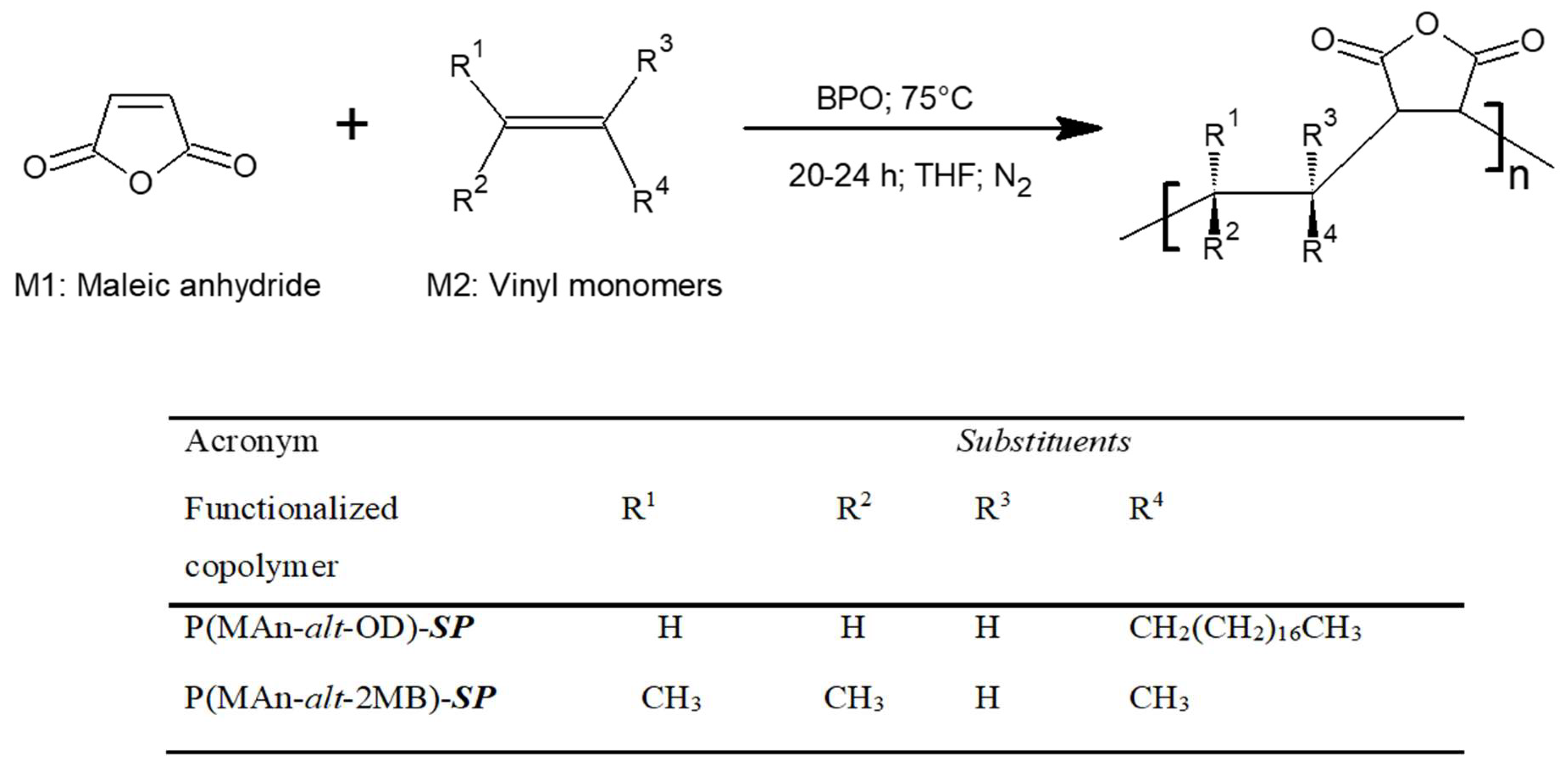
Figure 3.
General reaction of the copolymer functionalization. Name and acronyms used for functionalized copolymers with maleic anhydride (MAn).
Figure 3.
General reaction of the copolymer functionalization. Name and acronyms used for functionalized copolymers with maleic anhydride (MAn).
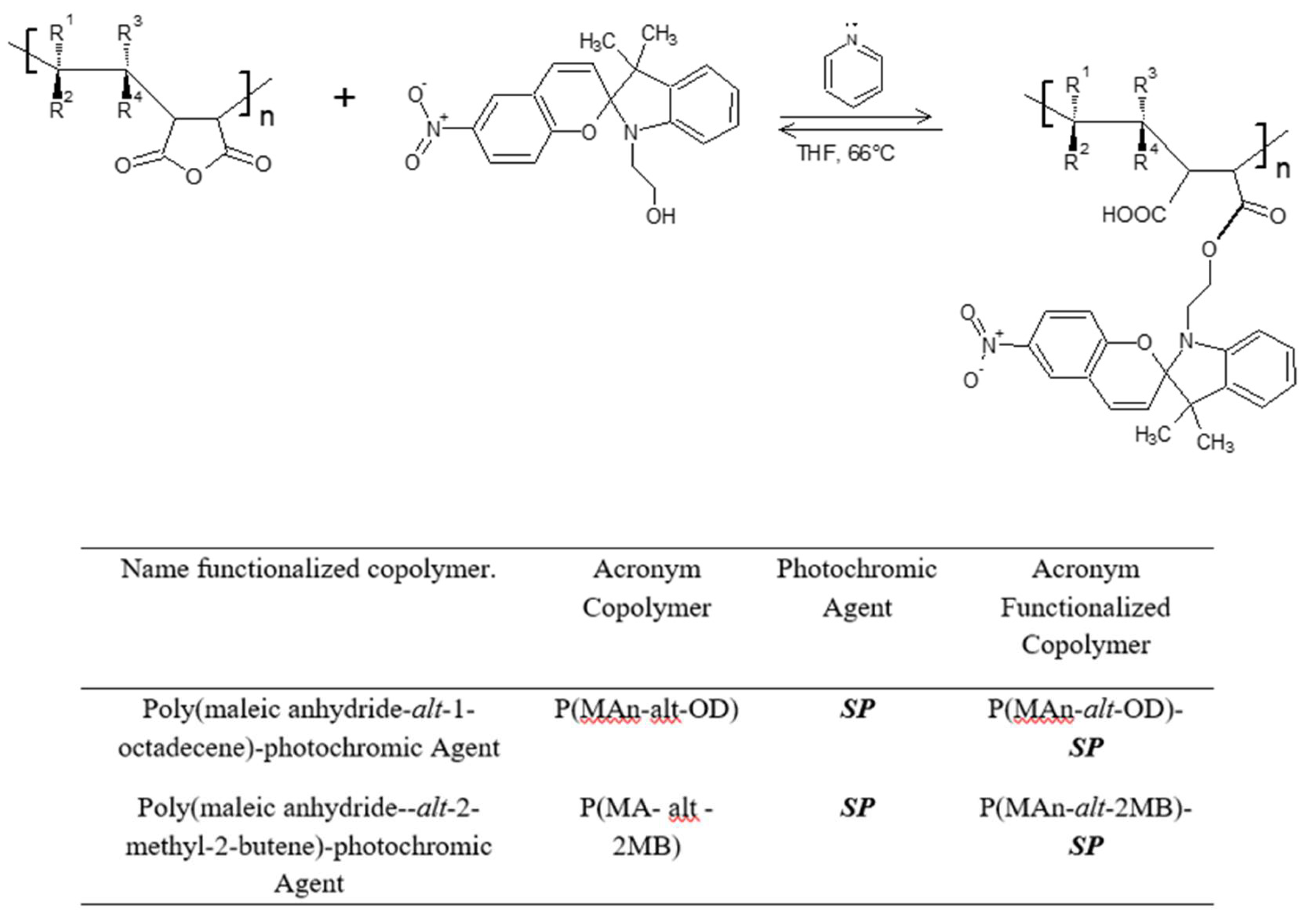
Figure 4.
a) NMR1H- spectrum of the P(MAn-alt-1OD)-SP copolymer. b) NMR 1H-spectrum of the P(MAn-alt-2MB)-SP copolymer.
Figure 4.
a) NMR1H- spectrum of the P(MAn-alt-1OD)-SP copolymer. b) NMR 1H-spectrum of the P(MAn-alt-2MB)-SP copolymer.
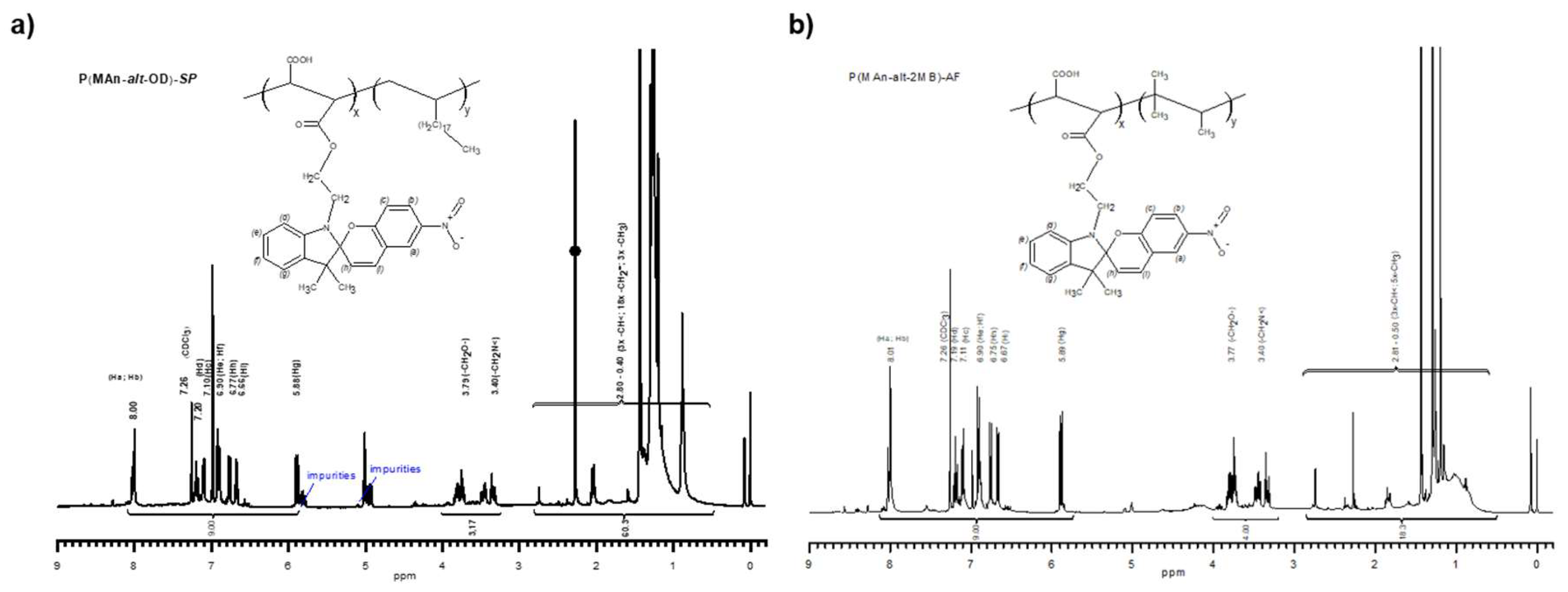
Figure 5.
Photochromism reaction of functionalized copolymers.
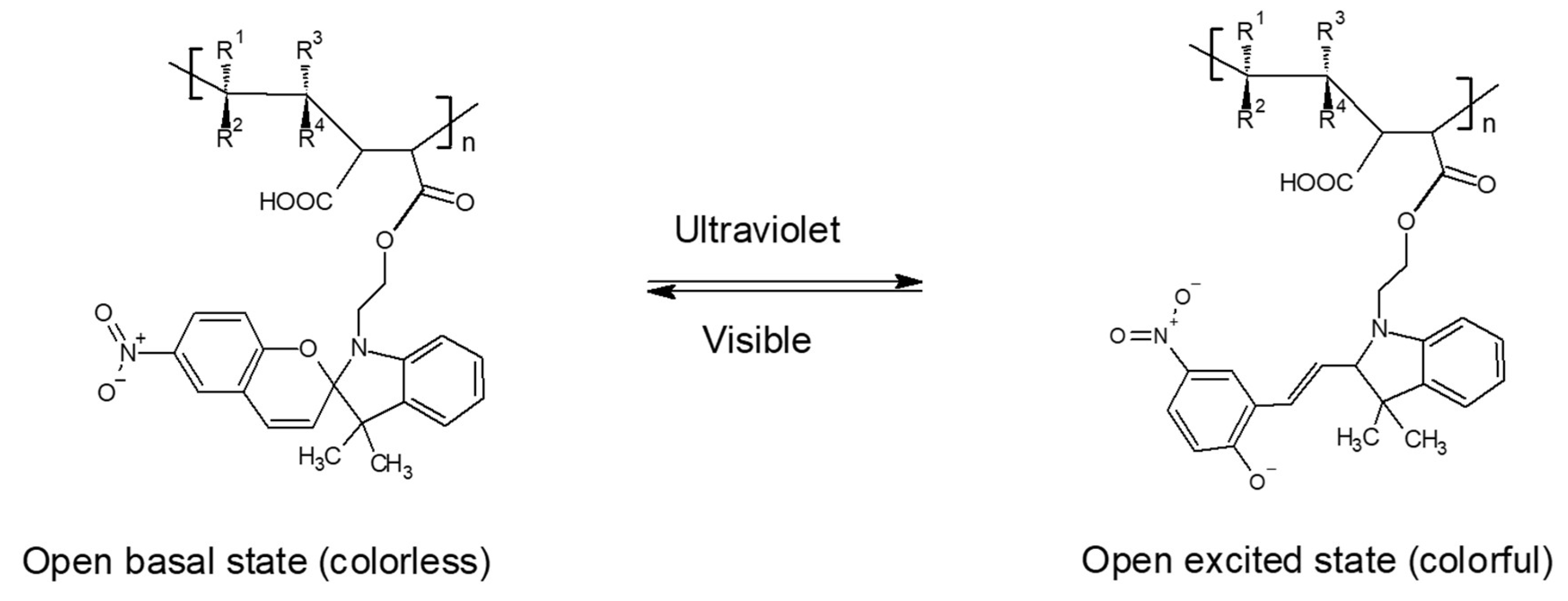
Figure 7.
Absorption changes in the photoactive-polymer solution (3 g/L in THF) (a) before irradiation and (b) after irradiation at 365 nm.
Figure 7.
Absorption changes in the photoactive-polymer solution (3 g/L in THF) (a) before irradiation and (b) after irradiation at 365 nm.
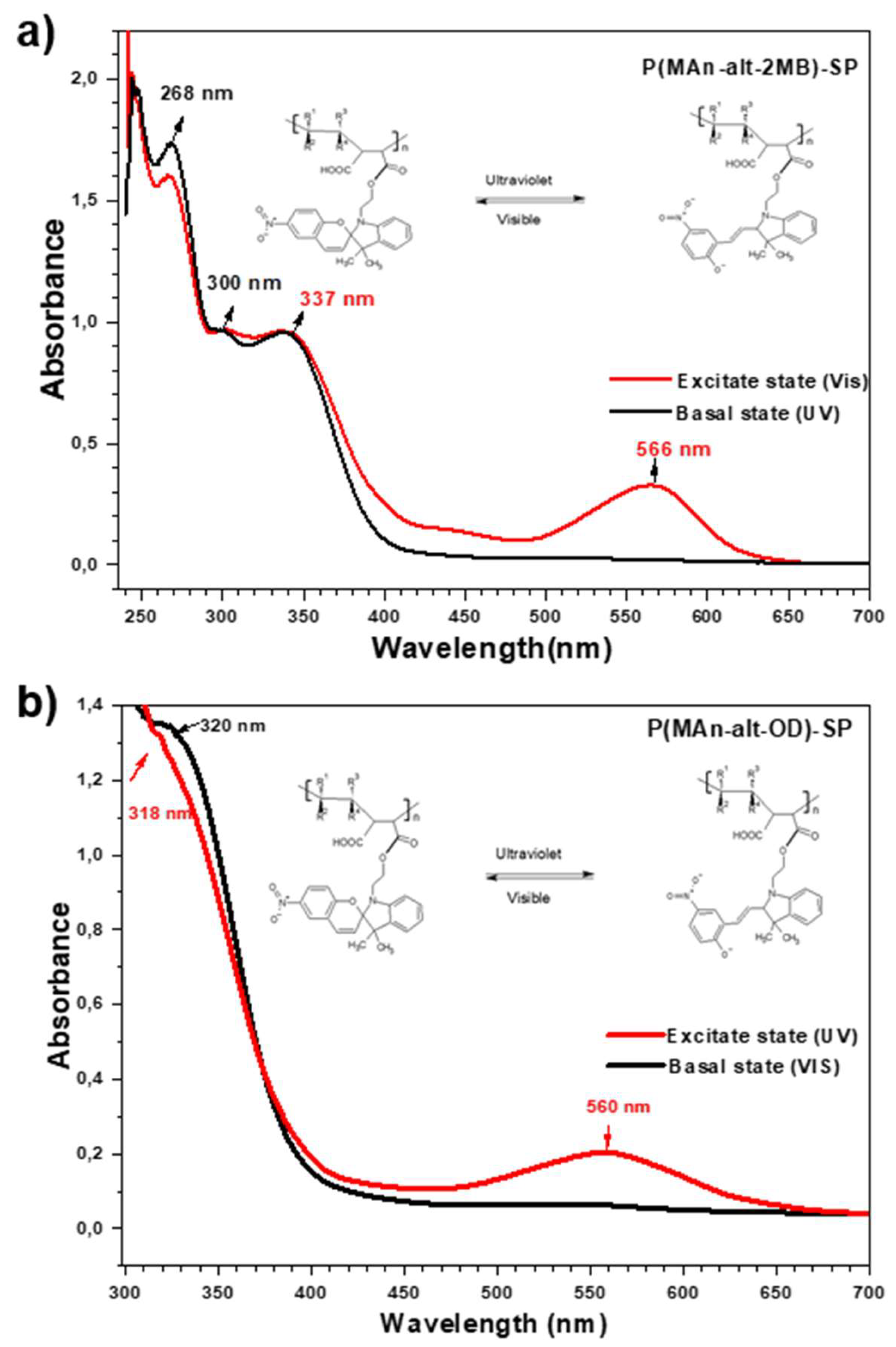
Figure 8.
TGA: Thermograms of the copolymers and photoactive copolymers a) P(MAn-alt-OD) and P(MAn-alt-OD)-SP and b) P(MAn-alt-2MB) and P(MAn-alt-2MB)-SP.
Figure 8.
TGA: Thermograms of the copolymers and photoactive copolymers a) P(MAn-alt-OD) and P(MAn-alt-OD)-SP and b) P(MAn-alt-2MB) and P(MAn-alt-2MB)-SP.
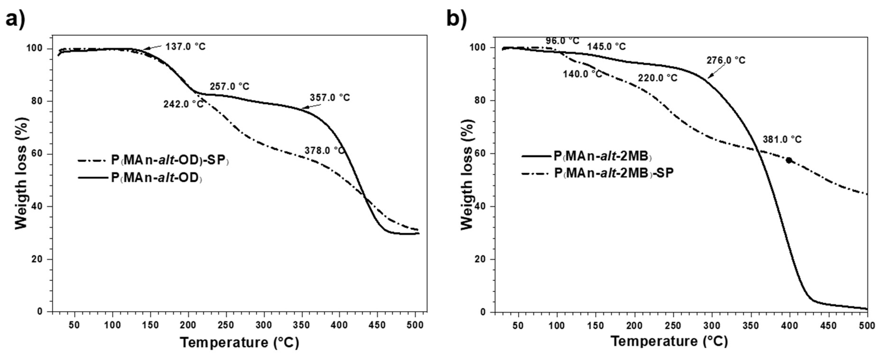
Figure 9.
Optical microscope images (OMI) at 40X magnification in CS2. a) Before and b) after irradiation for P(MAn-alt-OD)-SP film; and c) Before and d) after irradiation for P(MAn-alt-2MB)-SP film Enlarged images (inset zoom) (e, f).
Figure 9.
Optical microscope images (OMI) at 40X magnification in CS2. a) Before and b) after irradiation for P(MAn-alt-OD)-SP film; and c) Before and d) after irradiation for P(MAn-alt-2MB)-SP film Enlarged images (inset zoom) (e, f).
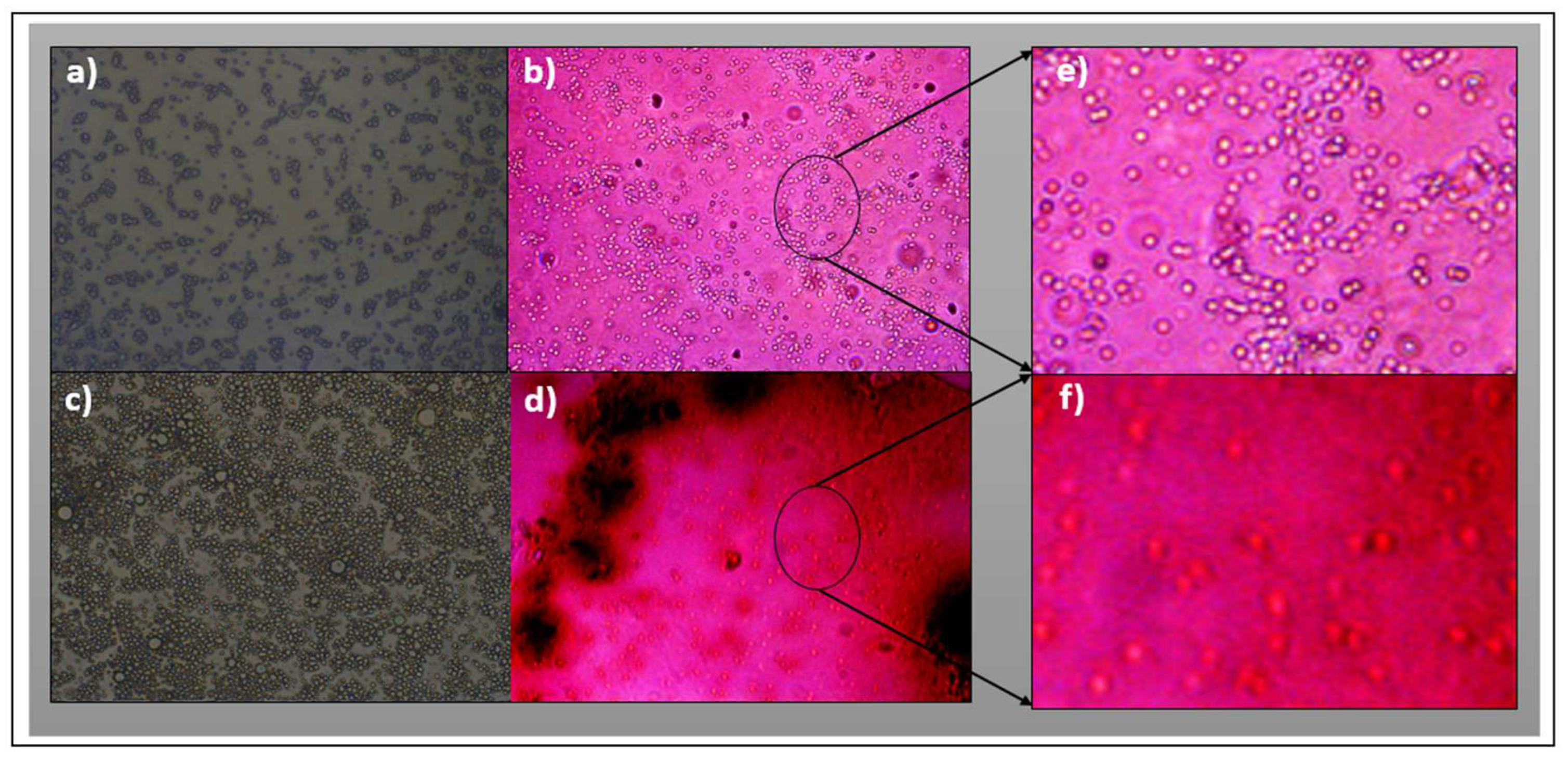
Figure 10.
Antimicrobial photodynamic properties of a) photosensitizer Ru(bpy) and b) Ru(bpy) combined with P(MAn-alt-2MB)-PS. Methicillin-susceptible (SAU) and methicillin-resistant (MRSA) Staphylococcus aureus bacterial strains were treated with PDT. The photosensitizer Ru(bpy) MIC was determined at 0 – 4 μg/mL a), and the Ru(bpy) MIC reduction was evaluated by mixing with 16 µg/mL P(MAn-alt-2MB)-PS b). Bacterial viability is expressed as log10 of mean ± SD were ** =p<0.01; ***=p<0.001 Two-tail t-test compared to control bacteria in the dark.


Table 1.
Main vibration bands of the functionalized copolymers.
| Maleic Anhydride (MAn) | COOH | Ester-COOR | -NO2 | ||
| Copolymer | Asymmetric (C=O) | Symmetrical (C=O) | Tension (C=O) | tension (C=O) | Tension (N-O) |
| P(MAn-alt-OD) | --- | 1780.0 | 1707.5 | --- | --- |
| P(MAn-alt-OD)-SP | --- | 1780.0 | --- | 1720.0 | 1460.0 |
| P(MAn-alt-2MB) | 1857.0 | 1780.0 | 1719.0 | --- | --- |
| P(MAn-alt-2MB)-SP | 1857.0 | 1780.0 | --- | 1728.0 | 1460.0 |
The asymmetric and symmetric tension bands for the MAn comonomer ring appear at 1857.0 cm-1 and 1780.0 cm-1, evidencing that a fraction of the MAn ring remains in the functionalized copolymer. While the functionalization with the photochromic agent is evidenced by the tension bands of the carbonyl (C=O) of the ester at 1720.0 cm-1 and 1728.0 cm-1 for the copolymers P(MAn-alt-OD)-SP and P (MAn-alt-2MB)-SP respectively, in addition to the N-O (–NO2) bond strain bands at 1460.0 cm-1. The carboxylic acid carbonyl tension bands present in both copolymers are not present in the functionalized copolymers.
Table 2.
Degrees of functionalization of P(MAn-alt-OD)-SP and P(MAn-alt-OD)-SP.
| Name | n1 | n2 | n3 | n4 | Ialiph | Imeth | Iar | X | Y |
| P(MAn-alt-OD)-SP | 8.00 | 10.0 | 4.00 | 9.00 | 48.0 | 6.06 | --- | 0.40 | 0.60 |
| P(MAn-alt -OD)-SP | 8.00 | 10.0 | 4.00 | 9.00 | 48.0 | --- | 2.53 | 0.40 | 0.60 |
| P(MAn-alt-2MB)-SP | 8.00 | 10.0 | 4.00 | 9.00 | 18.3 | 4.00 | --- | 0.49 | 0.51 |
| P(MAn-alt -2MB)-SP | 8.00 | 10.0 | 4.00 | 9.00 | 18.3 | --- | 9.00 | 0.49 | 0.51 |
Table 3.
Maximum wavelength (λmax) of the photochromic agent and the functionalized copolymers.
| Compounds | λmax basal state (nm) | λmax excited state (nm) | |||||
| Photochromic agent | 265 | 332 | -- | 265 | 300 | -- | 564 |
| P(MAn-alt-OD)-SP | 320 | -- | -- | 318 | -- | -- | 560 |
| P(MAn-alt-2MB)-SP | 268 | 300 | 337 | 268 | 300 | 337 | 566 |
Table 4.
Optical Band gap (Eg )*.
| Case | Eg (eV) | |
| before irradiation | after irradiation | |
| Photochromic agent (SP) | 3.00 | 2.00 |
| P(MAn-alt-OD)-SP | 3.73 | 3.02 |
| P(MAn-alt-2MB)-SP | 4.53 | 3.78 |
| * The Eg was estimated from Tauc plot equation41 , before and after irradiation at 365 nm. | ||
Disclaimer/Publisher’s Note: The statements, opinions and data contained in all publications are solely those of the individual author(s) and contributor(s) and not of MDPI and/or the editor(s). MDPI and/or the editor(s) disclaim responsibility for any injury to people or property resulting from any ideas, methods, instructions or products referred to in the content. |
© 2025 by the authors. Licensee MDPI, Basel, Switzerland. This article is an open access article distributed under the terms and conditions of the Creative Commons Attribution (CC BY) license (http://creativecommons.org/licenses/by/4.0/).
Copyright: This open access article is published under a Creative Commons CC BY 4.0 license, which permit the free download, distribution, and reuse, provided that the author and preprint are cited in any reuse.


