
Submitted:
26 March 2025
Posted:
26 March 2025
You are already at the latest version
Abstract
Background/Objectives: Three-dimensional cell culture systems are relevant in vitro models for studying cellular behavior. In this regard, the present study investigates the interaction between human osteoblast-like cells and 3D-printed scaffolds mimicking physiological and osteoporotic bone structures under simulated microgravity conditions. The objective is to assess the effects of scaffold architecture and dynamic culture conditions on cell adhesion, proliferation, and meta-bolic activity, with implications for both osteoporosis research and space medicine.
Methods: Poly (lactic acid) (PLA) scaffolds with physiological (P) and osteoporotic-like (O) tra-becular architectures were 3D printed by means of the used deposition modeling (FDM) tech-nology. Morphometric characterization was performed using micro-computed tomography (µCT). Human osteoblast-like SAOS-2 and U2OS cells were cultured on the scaffolds under static and dynamic simulated microgravity conditions using a rotary cell culture system (RCCS). Cell viability, adhesion, and metabolic activity were evaluated through BrdU, WST-1, and ELISA assays, with TNF-α secretion assessed to determine biocompatibility.
Results: Both scaffold models supported osteoblast-like cell adhesion and growth, with enhanced colonization observed on the high-porosity O scaffolds under dynamic conditions. The dynamic environment facilitated increased surface interaction, amplifying the effects of scaffold archi-tecture on cell behavior. No inflammatory response was detected, confirming scaffold biocom-patibility.
Keywords:
3D printed bone-like scaffolds
; Biomimetics
; Microgravity conditions
1. Introduction
Three-dimensional cell culture systems have transformed biological research, particularly in the field of human health, by offering a more realistic representation of the native tissue microenvironment compared to traditional two-dimensional in vitro cultures [1]. These systems enhance cell–cell and cell–matrix interactions, thanks to their spatial organization and biomimetic scaffold surfaces, making them invaluable tools for applications in tissue engineering and regenerative medicine. In the context of bone tissue engineering, the development of advanced 3D platforms is essential for overcoming the limitations of conventional medical prostheses, which can significantly impact the recipient's quality of life. The possibility to deal with ad hoc devices aimed to replicate the natural extracellular matrix (ECM), a highly complex and functional structure, can effectively support cellular growth, differentiation, and integration with surrounding tissues [2]. Recent advancements in 3D printing technologies, such as fused deposition modeling (FDM), have enabled the fabrication of scaffolds that mimic the intricate microarchitecture of native bone, including healthy and pathological conditions like osteoporosis[3]. A comprehensive research strategy can be then planned by integrating biomimetic, biocompatible, and bioresorbable scaffolds with dynamic bioreactors, such as rotary cell culture systems (RCCS), that represent a cutting-edge 3D in vitro means capable of closely reproduce the native bone tissue microenvironment [4]. RCCS bioreactors create low-shear fluidic field with a continuous suspension of scaffolds to simulate microgravity conditions, enhancing nutrients and oxygen diffusion, and mimic in vivo conditions, fostering cell growth, differentiation, and metabolic activity within 3D scaffolds. Moreover, the combination of RCCS with scaffolds enriched with functional fillers, such as β-tricalcium phosphate, known for its osteoinductive and anti-osteoclastogenic properties, could provide a powerful platform for advancing bone tissue engineering applications. This approach not only enhances cellular response but also holds great potential for developing innovative therapies, paving the way for improved treatment strategies and preclinical research models.
Bone is a dynamic and hierarchically structured tissue that provides structural support, facilitates movement, and protects internal organs[5,6]. It consists of two main types: trabecular and cortical bone. Trabecular bone, which accounts for approximately 20% of the skeleton, is characterized by a sponge-like structure with interconnected trabeculae. The porosity ranges from 50% to 90%, significantly influencing mechanical properties, such as modulus and compressive strength [7]. Cortical bone, in contrast, is denser, with a porosity of about 10%, forming the outer layer of bones and providing resistance to bending and torsion. This structure has evolved to respond to a number of different conditions, including external ones like Earth’s gravity, where mechanical loading plays a pivotal role to maintain bone strength through a dynamic remodeling process involving osteocytes, osteoblasts, and osteoclasts. Microgravity environments, such as those encountered during spaceflight, disrupt this remodeling process, leading to significant bone mass loss, particularly in weight-bearing regions like the femur and pelvis. Astronauts lose approximately 1.8–2% of bone mass per month in microgravity, increasing the risk of fractures [8]. In this context, bioreactors, based on rotary cell culture systems can provide an effective in vitro platform to study these effects and develop countermeasures. Starting from this assumption, we designed and fabricated 3D-printed scaffolds using FDM to replicate both physiological and osteoporotic bone architectures. Bone-like scaffolds were located within a RCCS bioreactor to be seeded and cultured with osteoblast-like cells (SAOS-2 and U2OS) under both static and dynamic conditions. The study’s primary objectives were: i) to evaluate the biocompatibility and functionality of the scaffolds using assays to measure cell proliferation, adhesion, and inflammatory response; ii) to assess how scaffold internal microarchitecture, porosity and fluid dynamics in the RCCS influence cellular behavior, particularly growth and metabolic activity; and iii) to optimize scaffold design for simulated microgravity conditions, hypothesizing that higher porosity enhances cell growth and metabolic activity by increasing surface area and improving fluid dynamics. The collected findings are expected to highlight the transformative potential of combining advanced 3D cell culture technologies with dynamic bioreactor systems, bridging the gap between in vitro models and in vivo applications. This investigation provides valuable insights into bone remodeling induced by microgravity, paving the way for innovative therapeutic strategies that may address a range of challenges, from mitigating bone loss during long-term spaceflight to treating conditions like osteoporosis more effectively. This integrated platform serves as a step forward in creating next-generation tissue engineering solutions for both research and clinical applications.
2. Materials and Methods
2.1. Biomimetic Scaffold Design
The design and the preliminary analysis of the proposed scaffolds were performed using Meshmixer software (Meshmixer version 2018, Autodesk, San Rafael, CA, USA). Starting from a previous approach[9], an updated model was prepared to simulate the osteoporotic-like trabecular bone tissue. Biomimetic bone scaffolds were realized to deal with a physiological (P) and an osteoporotic (O) model, according to the parameters summarized in Table 1. The trabecular network was generated using an algorithm based on Delaunay triangulation.
2.2. 3D Printing Set-Up
Each scaffold file (Figure 1) was imported in IdeaMaker software (IdeaMaker version 3.6.1, Raise 3D Inc., Irvine, CA, USA) to slice the model and drive the Raise 3D N2 printer (Raise 3D Inc., Irvine, CA, USA). A commercial polylactic acid (PLA) filament (FILOALFA, Turin, Italy) was extruded at 205 °C through a 0.4 mm diameter nozzle, the build platform temperature was set at 60 °C.
2.3. Micro-Tomographic Analysis
µ-CT is a non-destructive technique, which allows us to obtain qualitative and quantitative information on the internal structure of the investigated samples. Each scaffold model was therefore analyzed to evaluate the printed microstructure and verify the conformity to the design criteria. For this aim, the microtomograph Skyscan 1072 (Bruker microCT, Kontich, Belgium) was used, powering the X-ray tube at 40 kV and 248 μA, and setting pixel dimensions at 14.65 x 14.65 μm (corresponding to 20x magnification, and 180° rotation with a step of 0.45°). The 3D reconstruction of the sample was performed using the NRecon software (Version 1.7.0; Bruker microCT, Kontich, Belgium). The CT-Analyser software (Version 1.16.9.0; Bruker microCT, Kontich, Belgium) was employed to elaborate the collected data and calculate the histomorphometric parameters.
2.4. Cell Culture
The human osteosarcoma cell lineage SAOS-2 and U2OS (purchased from the American Type Culture Collection, ATCC, HTB-85 and HTB-96 Rockville, MD, USA) were selected to in vitro assess scaffold biocompatibility, cell growth and adhesion. Before starting the experiments, cells were seeded on a plastic Petri dish and placed in a humidified incubator at 37 °C and 5% CO2. Cells were cultured in high-glucose Dulbecco’s modified Eagle’s Medium (DMEM; Euroclone, Milan. Italy), completed with 10% heat-inactivated fetal bovine serum (FBS; Euroclone, Milan, Italy), 2 mM L-glutamine (Sigma, Darmstadt, Germany), 1.0 unit ml-1 penicillin (Sigma, Darmstadt, Germany), and 1.0 mg ml-1 streptomycin (Sigma, Darmstadt, Germany). Scaffolds were sterilized with 70% ethanol solution and accurately washed before cell seeding.
2.5. RCCS Bioreactor
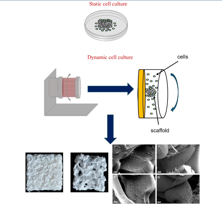
Dynamic cell cultures in microgravity conditions were carried out using the Rotary Cell Culture System™ 4SCQ (Synthecon Incorporated, 8044 El Rio, Houston, Texas 77054). The RCCS bioreactor is shown in Figure 2, the cell culture chamber is horizontally rotated to constantly suspend the inoculated cells and any support (e.g., scaffolds) in the culture medium. The optimal suspension condition was achieved by setting a rotational speed of 45 rpm.
2.6. Biological Assays
For the static culture, SAOS-2 and U2OS were seeded on the 3D printed scaffolds at a density of 2x10⁴ cell/cm2 and cultured in an incubator at 5% CO2 and 37 °C. At day 4, scaffolds were removed for the characterization. The scaffolds were specifically sized (4x4x4 mm3) to be cultured in a suspension mode into the RCCS bioreactor to balance the force acting on the scaffolds themselves (i.e., gravity, buoyancy, centrifugal and drag forces). For the SAOS-2 and U2OS cultures in simulated microgravity, P and O scaffolds were inserted into the RCCS bioreactor chambers and cells were directly seeded into the chamber filled with culture medium, at density of 2.0x10⁴ cell/cm2. Bubbles were removed using a syringe. The bioreactor was located into an incubator at 5% CO2 and 37 °C. On day 4, scaffolds were removed for the characterization.
2.6.1. Cell Proliferation and Metabolic Activity Analysis
Cell proliferation was evaluated by Bromodeoxyuridine incorporation assay (BrdU, Cell Proliferation Kit; Roche Diagnostics), while cell metabolic activity was quantified by a colorimetric assay based on the oxidation of water-soluble tetrazolium salts (Cell Proliferation Reagent WST-1; Roche Diagnostics). The absorbance of 100 µl of supernatant was measured in an ELISA (Enzyme-Linked ImmunoSorbent Assay) reader (VICTOR3 multilabel readers; PerkinElmer, Waltham, Massachusetts) at 450 nm.
2.6.2. TNF-α Secretion Analysis
Quantification of Tumor necrosis factor-α (TNF-α) released by the cells was obtained by ELISA development kit (PeproTech® EC Ltd., UK) at 405 nm. Supernatants from SAOS-2 and U2OS cells grown on the scaffolds were collected at 96 h of culture, centrifuged at 1200 rpm for 5 min and stored at -80 °C until use. TNF-α release was measured in culture medium using human ELISA development kit (PeproTech® EC Ltd., UK), according to the manufacturer’s instructions. Cell supernatants and human recombinant standards were serially diluted in 1x PBS/0.05% Tween-20/0.1% BSA (Sigma-Aldrich®, Dorset, UK). Interleukin bindings were measured by biotin-avidin detection, followed by incubation with chromogen 2,2'-azino-bis (3-ethylbenzothiazoline-6-sulphonic acid (Sigma-Aldrich®, Dorset, UK). Colorimetric response was monitored at 405 nm.
2.6.3. Scanning Electron Microscopy (SEM) Analysis
Scanning electron microscopy (SEM) analysis was carried out to confirm the results obtained by cell growth assays. The cells grown on the scaffolds were fixed with 4% paraformaldehyde for 10 min and the dried specimens were coated by an evaporated Au thin film (10 nm) before analysis with a ZEISS SIGMA 300 field emission SEM. The morphological analysis was verified at an accelerating voltage of 5 kV by using the secondary electron (SE) detector.
2.7. Statistical Analysis
MedCalc software was used for statistical analysis, data was analyzed by T-student test and the significance level adopted for all analyses was p < 0.05.
3. Results
3.1. Scaffold Assessment
The nominal porosity of the scaffold models was measured by means of the “analysis” tool of the 3D modelling software Meshmixer 3.5. The P scaffold porosity was 53.77%, being within the physiological range of 50% - 90%[10], while the O model was characterized by a porosity level of 82.96%. The bone porosity may change depending on the skeletal site and can increase up to 90% in osteoporotic conditions[11]; in this study, the design of the O scaffolds allowed to reach the highest porosity with thin trabeculae and an interconnected bone-like structure in the required osteoporotic range as previously reported[12,13,14].
3.2. 3D Printed Scaffolds
The 3D printed PLA scaffolds are shown in Figure 3, where it is possible to observe the different microarchitectures of both the physiological and osteoporotic-like constructs.
3.3. Morphometric Analysis
Each scaffold was analyzed by μ-CT to study the internal microstructure and quantify the compliance of the design criteria to the printed cases. Quantitative analysis was carried out elaborating the μ-CT data to present the main bone histomorphometric parameters, for this study, as a result (Table 2 and Table 3) and comparing them with the corresponding values from literature.
The data obtained from the μ-CT analysis confirmed a good agreement with literature data, especially for the osteoporotic case. This was not strictly verified for the physiological model which can be related to the resolution limits of the 3D printer, particularly for the parameters related to trabeculae.
3.4. Cell Growth and Metabolic Activity Study in Microgravity Conditions
Simulated microgravity was investigated to evaluate the interaction of osteoblast-like-cells with the physiological and osteoporotic-like PLA scaffolds (P and O models). Two osteoblast-like cell lines, SAOS-2 and U2OS, were cultured in simulated microgravity conditions with both PLA scaffolds, using the RCCS bioreactor, to investigate their capability of colonizing and growing on the constructs by means of BrdU and WST-1 colorimetric assay, in comparison to those grown in 2D static conditions (CTR). These cell lines, that are derived from human osteosarcomas with different differentiation levels, have been used in several studies for bone tissue engineering. SAOS-2 cells have an osteoblast phenotype with characteristics similar to human primary osteoblasts and present a high level of ALP activity and mineralization ability[19,20]. U2OS cells instead exhibit properties of mesenchymal cells[21] and have a very low level of ALP and mineralization capacity[22]. The SAOS-2 and U2OS cells cultured for 4 days in simulated dynamic conditions showed a positive interaction with the polymer substrates which were able to support both cell growth and metabolic activity processes (Figure 4 and Figure 5).
The P scaffolds were characterized by a slight increase in cell colonization and metabolic activity in simulated microgravity conditions, compared to the static one, while a higher growth and metabolic response were observed for the O model, in which this difference between static and dynamic culture, obtained by BrdU and WST-1 tests, were statistically significant (p < 0.05). BrdU and WST-1 results in static culture revealed a decreasing trend from the less porous to the more porous model. This trend could be due to the scaffold morphology: the P model, with an effective porosity of about 44.7%, prevent cells from settling to the bottom, allowing cells to better adhere on the scaffold surface and within the internal trabecular network, resulting in a higher cell growth after 4 days, consistent with a report by[23]. Instead, the O model, due to its osteoporotic-like structure (about 80.53% of porosity), induced a lower cell interaction with the printed microarchitecture, settling at the bottom through the large pore network with a lower cell adhesion as proven by the BrdU and WST-1 assays. In microgravity conditions, the scaffolds and cells continuously get in touch and the high porosity of the O scaffolds, by increasing the available surface area, improves the cells’ attachment. For this reason, the osteoblast-like cells (SAOS-2 and U2OS) seeded on high porosity scaffolds (O) and cultured in microgravity conditions showed a higher cell colonization compared to static conditions, but also to low porosity substrates (P).
3.5. Cell Adhesion Study in Microgravity Conditions
The adhesion and migration ability of cells into the scaffolds after culture in the RCCS bioreactor were studied by scanning electron microscopy (SEM), for SAOS-2 cells, which are routinely used for investigations on osteoblast-biomaterials interaction. The images reported in Figure 6 confirmed a random porous microarchitecture. SAOS-2 cells appear well attached to the scaffold surfaces and many of them are concentrated around the pore wall, demonstrating the ability to colonize and penetrate the scaffolds, which increased in the samples grown under microgravity conditions.
3.6. TNF-α Secretion Analysis
The biomaterial-cells interaction was assessed in terms of biocompatibility in both static and microgravity conditions. The pro-inflammatory TNF-α levels secreted by SAOS-2 and U2OS cells grown on P and O scaffolds were evaluated by ELISA assay. Figure 7 shows the TNF-α concentration, measured as absorbance level at 405 nm for all conditions studied at 4 days of culture.
Evaluating the secretion levels of TNF-α, no experimental group produced detectable amounts of this proinflammatory factor: the concentration in the samples resulted undetectable compared to the measured absorbance and the standard curve of TNF-α concentration. This outcome states that the scaffolds do not induce an inflammatory response after 96 hours of culture, further confirming the biocompatibility of the substrates.
4. Discussion
Three-dimensional cell culture systems represent a major advancement in biomedical research, providing an improved in vitro model to study cellular behavior in a more physiologically relevant microenvironment; in addition, their integration with dynamic bioreactors has opened new opportunities in tissue engineering and regenerative medicine[24]. In this study, we investigated the interaction between osteoblast-like cells and 3D-printed scaffolds that mimic physiological and osteoporotic bone structures under simulated microgravity conditions. Bone remodeling is significantly affected by microgravity, inducing a faster resorption process than ossification[25]. Gravity and mechanical stimuli e.g., tensile and compressive stresses, fluid-exerted shear stress, and hydrostatic pressure concur to sustain the maintenance of healthy tissues and cells[26] and these input conditions need to be investigated in detail to develop specific countermeasures. In this regard, additive manufacturing may be a suitable technology to prepare tissue replicas to foster bone research in 3D conditions. The possibility to modulate the architecture of the bone microenvironment, in terms of pore size, porosity, trabecular arrangement, can be promptly realized to test the biological output of different bone-related cells[27]. To promote an advancement in the field and perform in vitro analyses resembling bone physiopathology, PLA bone scaffolds with increasing porosity from low physiological value to osteoporotic range were designed and 3D printed by fused deposition modeling. The morphometric characteristics of the P model, measured by μ-CT, were slightly different from literature data, related, in this case, to the physiological range of the human proximal ulna[15]; these differences can be ascribed to the resolution limit of the 3D printer. Regarding the design values selected for the O model, these were set to create a biomimetic trabecular architecture whose structural parameters fall within the osteoporotic range referred to the femoral head[11,14,16,17,18], which is a skeletal site dramatically prone to osteoporosis both on Earth and in weightlessness. Regarding biological assessment, the results showed that both osteoblast-like cell lines used, positively interacted with all PLA substrates[28], demonstrating that the fabricated scaffolds were biocompatible and able to support cell growth, adhesion and metabolic activity without an inflammatory response, as proven by BrdU, WST-1 and ELISA assays. SAOS-2 and U2OS cells, deriving from human osteosarcomas with different levels of differentiation, represent well-established models for bone tissue engineering investigations. They can be used for studying the osteogenesis process and how it could be affected by different treatments. SAOS-2 cell has the capability to produce mineralized matrix and express osteocyte marker genes, and the potential to mimic the interaction between primary human osteoblasts and biomaterials[9]. U2OS has a lower osteoblastic differentiation level and exhibits mesenchymal cell properties[21]. For this reason, they were chosen in this work, to evaluate their different biological behavior in microgravity conditions. Results obtained by BrdU and WST-1 assay highlighted that scaffold morphology, mainly porosity, affects cell adhesion and growth when comparing static and dynamic cell culture conditions. This effect is greater in microgravity conditions because the 3D culture environment and the fluid motion support cells to interact with a larger surface area of the substrate, thus amplifying the effect of the different scaffold porosity on cell adhesion and viability[29,30]. Particularly, the best cell colonization and metabolic response occurs for cells cultured within the O model in the RCCS bioreactor, suggesting that the combination of this culture condition and the most porous scaffold (O model) provides the best cell-scaffold interaction, probably due to a suitable fluid flow condition established within the substrate framework. This finding underscores the importance of dynamic culture conditions, which allow cells to interact with a larger surface area, improving attachment and viability. Furthermore, the absence of detectable TNF-α secretion suggests that the 3D-printed PLA scaffolds used in this study are biocompatible and do not elicit an inflammatory response [28,31]. The combination of additive manufacturing and rotary cell culture systems (RCCS) enabled the creation of biomimetic platforms with precise control over scaffold microarchitecture, offering an innovative system to study bone remodeling and for tissue engineering applications. The advantages of 3D cell culture models are particularly evident when studying bone tissue engineering strategies. Traditional 2D cell cultures fail to replicate the complex structural and mechanical properties of bone, limiting their relevance in preclinical research. The RCCS bioreactor system, by mimicking a low-shear microgravity environment, provides a more biomimetic approach for investigating bone cell behavior, particularly in the context of osteoporosis and microgravity-induced bone loss. The enhanced nutrient diffusion and dynamic fluid conditions within the RCCS further contribute to a more physiologically relevant culture environment. The collected results can have important implications for both space research and terrestrial biomedical applications. The ability to simulate bone loss conditions in microgravity offers a valuable tool for studying the mechanisms of osteoporosis and developing countermeasures. Additionally, the use of high-porosity scaffolds in a dynamic 3D culture system may provide novel insights into optimizing scaffold design for tissue engineering applications. Future research should focus on refining scaffold materials and architectures, as well as investigating the role of interstitial fluid dynamics in regulating osteogenic activity. By leveraging advanced 3D cell culture systems and dynamic bioreactors, this research bridges the gap between in vitro models and in vivo applications. The integration of biomimetic scaffolds with simulated microgravity conditions presents a promising strategy for improving bone tissue engineering approaches (9), ultimately contributing to the development of more effective therapies for osteoporosis and other degenerative bone disorders.
5. Conclusions
Data from the biological assays of this work show that a 3D printed PLA scaffolds, with an osteoporotic bone-like structure, rather than worsening cell growth, improves cells response when cultured in a RCCS bioreactor to simulate weightlessness. This outcome can be probably related to the fluid dynamics established within the scaffold which contribute to enhancing the cell response. A 3D printed osteoporotic-like scaffold may be regarded as a potential model to assess possible countermeasures to stimulate and improve cell activity, simulating bone disorders when exposed to dynamic culture conditions. RCCS bioreactor can represent an in vitro suitable set-up for this aim thanks to the 3D culture capability, fluid dynamics, nutrient transfer and gas exchange that allow to perform more effective and specific studies with respect to the conventional static approach. These results need to be further refined as microgravity-simulating bioreactors and osteoporotic bone models could be used to better analyze the effects of fluid flow in the osteo-activity regulation as a means to design a potential protocol to treat microgravity and/or age-related osteoporosis. Biological data also suggest that it is probably feasible to achieve a fluid condition suitable for osteogenic activities even in bone tissues with higher porosity, supporting future research projects to be focused on the role of the interstitial fluid flow in osteoporotic bone.
Author Contributions
Conceptualization, M.L, C.D and A.L; methodology, G.G, E.Z, L.P; software, E.S.; validation, E.Z., G.G. and E.S.; formal analysis, E.Z and G.G.; investigation, E.Z.,G.G., L.P, R.P. and A.C.; resources, M.L and A.L.; data curation, M.L and G.G .; writing—original draft preparation, M.L.; writing—review and editing, M.L, A.L., E.Z, E.S. C.D.and C.A.; supervision, M.L, A.L and C.D.; project administration, M.L.; funding acquisition, M.L and A.l.. All authors have read and agreed to the published version of the manuscript.
Funding
This research was funded by MUR DSB.AD006.371.010 / Invecchiamento attivo e in salute (FOE 2022) IFT. This research was co-financed by European Union—Next Generation EU, within the PNRR project “Rome Technopole—Innovation Ecosystem” (ECS00000024, CUP B83C22002890005).
Institutional Review Board Statement
Not applicable.
Informed Consent Statement
Not applicable.
Data Availability Statement
The original contributions presented in this study are included in the article; further inquiries can be directed to the corresponding authors.
Acknowledgments
The authors would like to thank Francesca Villani, Matilde Paggiolu and Pamela Papa for their administrative and technical support.
Conflicts of Interest
The authors declare no conflicts of interest.
References
- Habanjar, O.; Diab-Assaf, M.; Caldefie-Chezet, F.; Delort, L. 3D Cell Culture Systems: Tumor Application, Advantages, and Disadvantages. Int J Mol Sci 2021, 22. [Google Scholar] [CrossRef] [PubMed]
- Wang, Z.; Sun, Y.; Li, C. Advances in 3D printing technology for preparing bone tissue engineering scaffolds from biodegradable materials. Front Bioeng Biotechnol 2024, 12, 1483547. [Google Scholar] [CrossRef] [PubMed]
- Winarso, R.; Anggoro, P.W.; Ismail, R.; Jamari, J.; Bayuseno, A.P. Application of fused deposition modeling (FDM) on bone scaffold manufacturing process: A review. Heliyon 2022, 8, e11701. [Google Scholar] [CrossRef] [PubMed]
- Hidaka, M.; Su, G.N.; Chen, J.K.; Mukaisho, K.; Hattori, T.; Yamamoto, G. Transplantation of engineered bone tissue using a rotary three-dimensional culture system. In Vitro Cell Dev Biol Anim 2007, 43, 49–58. [Google Scholar] [CrossRef]
- Boskey, A. Mineralization, Structure and Function of Bone. 2006; pp. 201–212.
- Weiner, S.; Wagner, H.D. The material bone: Structure mechanical function relations. Annu Rev Mater Sci 1998, 28, 271–298. [Google Scholar] [CrossRef]
- Salgado, A.J.; Coutinho, O.P.; Reis, R.L. Bone tissue engineering: state of the art and future trends. Macromol Biosci 2004, 4, 743–765. [Google Scholar] [CrossRef]
- Vernikos, J.; Schneider, V.S. Space, gravity and the physiology of aging: parallel or convergent disciplines? A mini-review. Gerontology 2010, 56, 157–166. [Google Scholar] [CrossRef]
- Zenobi, E.; Merco, M.; Mochi, F.; Ruspi, J.; Pecci, R.; Marchese, R.; Convertino, A.; Lisi, A.; Del Gaudio, C.; Ledda, M. Tailoring the Microarchitectures of 3D Printed Bone-like Scaffolds for Tissue Engineering Applications. Bioengineering (Basel) 2023, 10. [Google Scholar] [CrossRef]
- Singh, S.; Bray, T.J.P.; Hall-Craggs, M.A. Quantifying bone structure, micro-architecture, and pathophysiology with MRI. Clin Radiol 2018, 73, 221–230. [Google Scholar] [CrossRef]
- Molino, G.; Dalpozzi, A.; Ciapetti, G.; Lorusso, M.; Novara, C.; Cavallo, M.; Baldini, N.; Giorgis, F.; Fiorilli, S.; Vitale-Brovaron, C. Osteoporosis-related variations of trabecular bone properties of proximal human humeral heads at different scale lengths. J Mech Behav Biomed 2019, 100. [Google Scholar] [CrossRef]
- Ciarelli, T.E.; Fyhrie, D.P.; Schaffler, M.B.; Goldstein, S.A. Variations in three-dimensional cancellous bone architecture of the proximal femur in female hip fractures and in controls. J Bone Miner Res 2000, 15, 32–40. [Google Scholar] [CrossRef] [PubMed]
- Sandino, C.; McErlain, D.D.; Schipilow, J.; Boyd, S.K. Mechanical stimuli of trabecular bone in osteoporosis: A numerical simulation by finite element analysis of microarchitecture. J Mech Behav Biomed Mater 2017, 66, 19–27. [Google Scholar] [CrossRef]
- Porrelli, D.; Abrami, M.; Pelizzo, P.; Formentin, C.; Ratti, C.; Turco, G.; Grassi, M.; Canton, G.; Grassi, G.; Murena, L. Trabecular bone porosity and pore size distribution in osteoporotic patients - A low field nuclear magnetic resonance and microcomputed tomography investigation. J Mech Behav Biomed Mater 2022, 125, 104933. [Google Scholar] [CrossRef]
- Viveen, J.; Perilli, E.; Zahrooni, S.; Jaarsma, R.L.; Doornberg, J.N.; Bain, G.I. Three-dimensional cortical and trabecular bone microstructure of the proximal ulna. Arch Orthop Trauma Surg 2023, 143, 213–223. [Google Scholar] [CrossRef]
- Vale, A.C.; Pereira, M.F.C.; Maurício, A.; Amaral, P.; Rosa, L.G.; Lopes, A.; Rodrigues, A.; Caetano-Lopes, J.; Vidal, B.; Monteiro, J.; et al. Micro-computed tomography and compressive characterization of trabecular bone. Colloid Surface A 2013, 438, 199–205. [Google Scholar] [CrossRef]
- Nikodem, A. Correlations between structural and mechanical properties of human trabecular femur bone. Acta Bioeng Biomech 2012, 14, 37–46. [Google Scholar] [CrossRef] [PubMed]
- Ozan, F.; Pekedis, M.; Koyuncu, S.; Altay, T.; Yildiz, H.; Kayali, C. Micro-computed tomography and mechanical evaluation of trabecular bone structure in osteopenic and osteoporotic fractures. J Orthop Surg (Hong Kong) 2017, 25, 2309499017692718. [Google Scholar] [CrossRef]
- Prideaux, M.; Wijenayaka, A.R.; Kumarasinghe, D.D.; Ormsby, R.T.; Evdokiou, A.; Findlay, D.M.; Atkins, G.J. SaOS2 Osteosarcoma Cells as an In Vitro Model for Studying the Transition of Human Osteoblasts to Osteocytes. Calcified Tissue Int 2014, 95, 183–193. [Google Scholar] [CrossRef]
- Czekanska, E.M.; Stoddart, M.J.; Richards, R.G.; Hayes, J.S. In Search of an Osteoblast Cell Model for Research. Eur Cells Mater 2012, 24, 1–17. [Google Scholar] [CrossRef]
- Mornet, E.; Stura, E.; Lia-Baldini, A.S.; Stigbrand, T.; Ménez, A.; Le Du, M.H. Structural evidence for a functional role of human tissue nonspecific alkaline phosphatase in bone mineralization. J Biol Chem 2001, 276, 31171–31178. [Google Scholar] [CrossRef]
- Orimo, H.; Shimada, T. Effects of phosphates on the expression of tissue-nonspecific alkaline phosphatase gene and phosphate-regulating genes in short-term cultures of human osteosarcoma cell lines. Mol Cell Biochem 2006, 282, 101–108. [Google Scholar] [CrossRef]
- Zhou, X.Q.; Zhou, G.; Junka, R.; Chang, N.X.; Anwar, A.; Wang, H.Y.; Yu, X.J. Fabrication of polylactic acid (PLA)-based porous scaffold through the combination of traditional bio-fabrication and 3D printing technology for bone regeneration. Colloid Surface B 2021, 197. [Google Scholar] [CrossRef]
- Aishwarya, P.; Agrawal, G.; Sally, J.; Ravi, M. Dynamic three-dimensional cell-culture systems for enhanced applications. Curr Sci India 2022, 122, 149–160. [Google Scholar] [CrossRef]
- Zerath, E. Effects of microgravity on bone and calcium homeostasis. Adv Space Res 1998, 21, 1049–1058. [Google Scholar] [CrossRef]
- LeBlanc, A.; Matsumoto, T.; Jones, J.; Shapiro, J.; Lang, T.; Shackelford, L.; Smith, S.M.; Evans, H.; Spector, E.; Ploutz-Snyder, R.; et al. Bisphosphonates as a supplement to exercise to protect bone during long-duration spaceflight. Osteoporosis Int 2013, 24, 2105–2114. [Google Scholar] [CrossRef] [PubMed]
- Leong, K.F.; Cheah, C.M.; Chua, C.K. Solid freeform fabrication of three-dimensional scaffolds for engineering replacement tissues and organs. Biomaterials 2003, 24, 2363–2378. [Google Scholar] [CrossRef] [PubMed]
- da Silva, D.; Kaduri, M.; Poley, M.; Adir, O.; Krinsky, N.; Shainsky-Roitman, J.; Schroeder, A. Biocompatibility, biodegradation and excretion of polylactic acid (PLA) in medical implants and theranostic systems. Chem Eng J 2018, 340, 9–14. [Google Scholar] [CrossRef]
- Ingram, M.; Techy, G.B.; Saroufeem, R.; Yazan, O.; Narayan, K.S.; Goodwin, T.J.; Spaulding, G.F. Three-dimensional growth patterns of various human tumor cell lines in simulated microgravity of a NASA bioreactor. In Vitro Cell Dev-An 1997, 33, 459–466. [Google Scholar] [CrossRef]
- Jin, F.; Zhang, Y.; Xuan, K.; He, D.; Deng, T.; Tang, L.; Lu, W.; Duan, Y. Establishment of three-dimensional tissue-engineered bone constructs under microgravity-simulated conditions. Artif Organs 2010, 34, 118–125. [Google Scholar] [CrossRef]
- Rasal, R.M.; Janorkar, A.V.; Hirt, D.E. Poly(lactic acid) modifications. Prog Polym Sci 2010, 35, 338–356. [Google Scholar] [CrossRef]
Figure 1.
Preview printing of the P scaffold model (left panel) and the O scaffold model (right panel).
Figure 1.
Preview printing of the P scaffold model (left panel) and the O scaffold model (right panel).
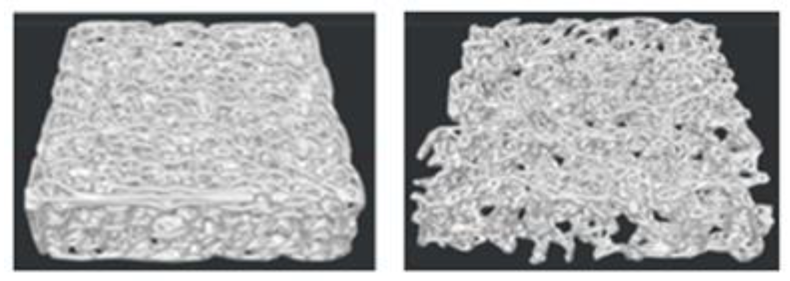
Figure 2.
RCCS bioreactor model used in this study. Static conditions (left panel) and Dynamic conditions (right panel).
Figure 2.
RCCS bioreactor model used in this study. Static conditions (left panel) and Dynamic conditions (right panel).
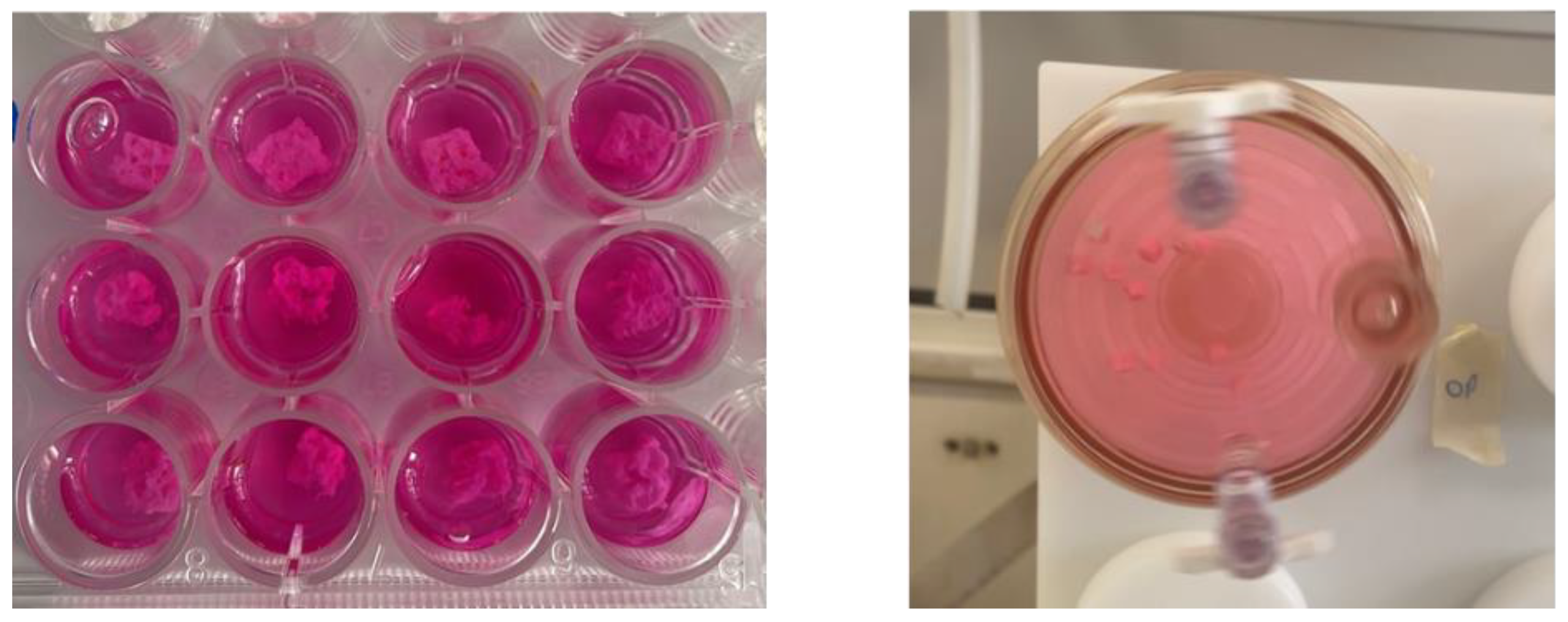
Figure 3.
3D printed PLA scaffolds: physiological P model (left panel) and osteoporotic O model (right panel).
Figure 3.
3D printed PLA scaffolds: physiological P model (left panel) and osteoporotic O model (right panel).
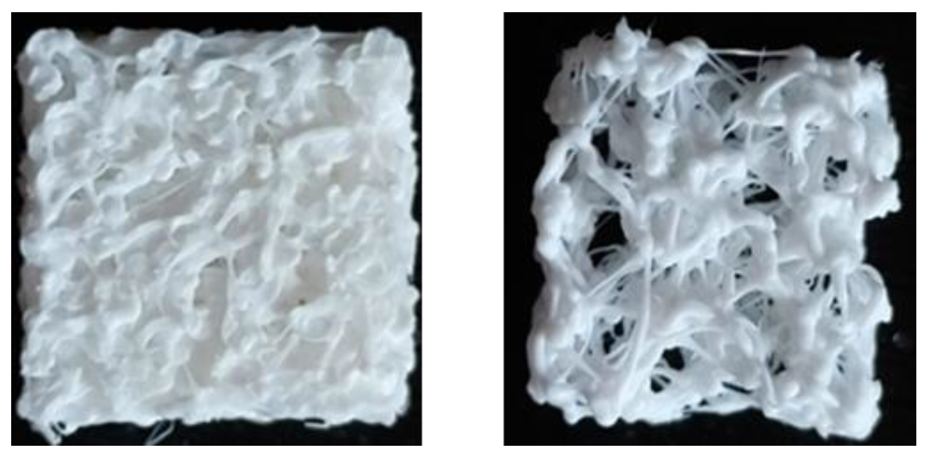
Figure 4.
Analysis of cell metabolic activity measured with the WST-1 assay at day 4 for both cell lines.
Figure 4.
Analysis of cell metabolic activity measured with the WST-1 assay at day 4 for both cell lines.
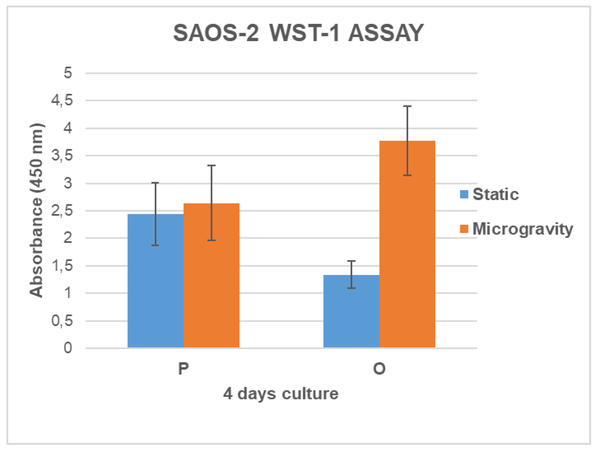
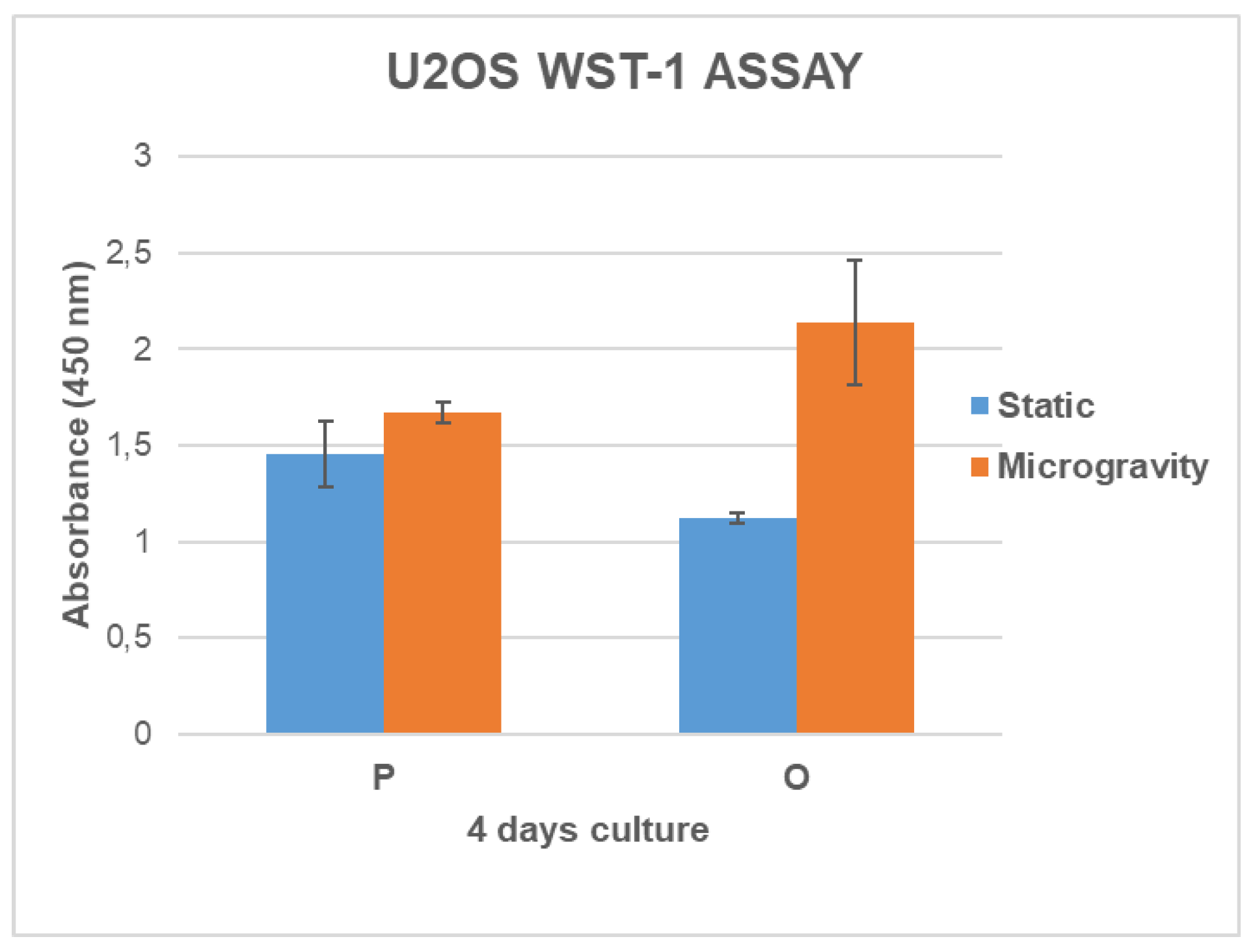
Figure 5.
Analysis of cell growth measured with the BrdU assay at day 4 for both cell lines.
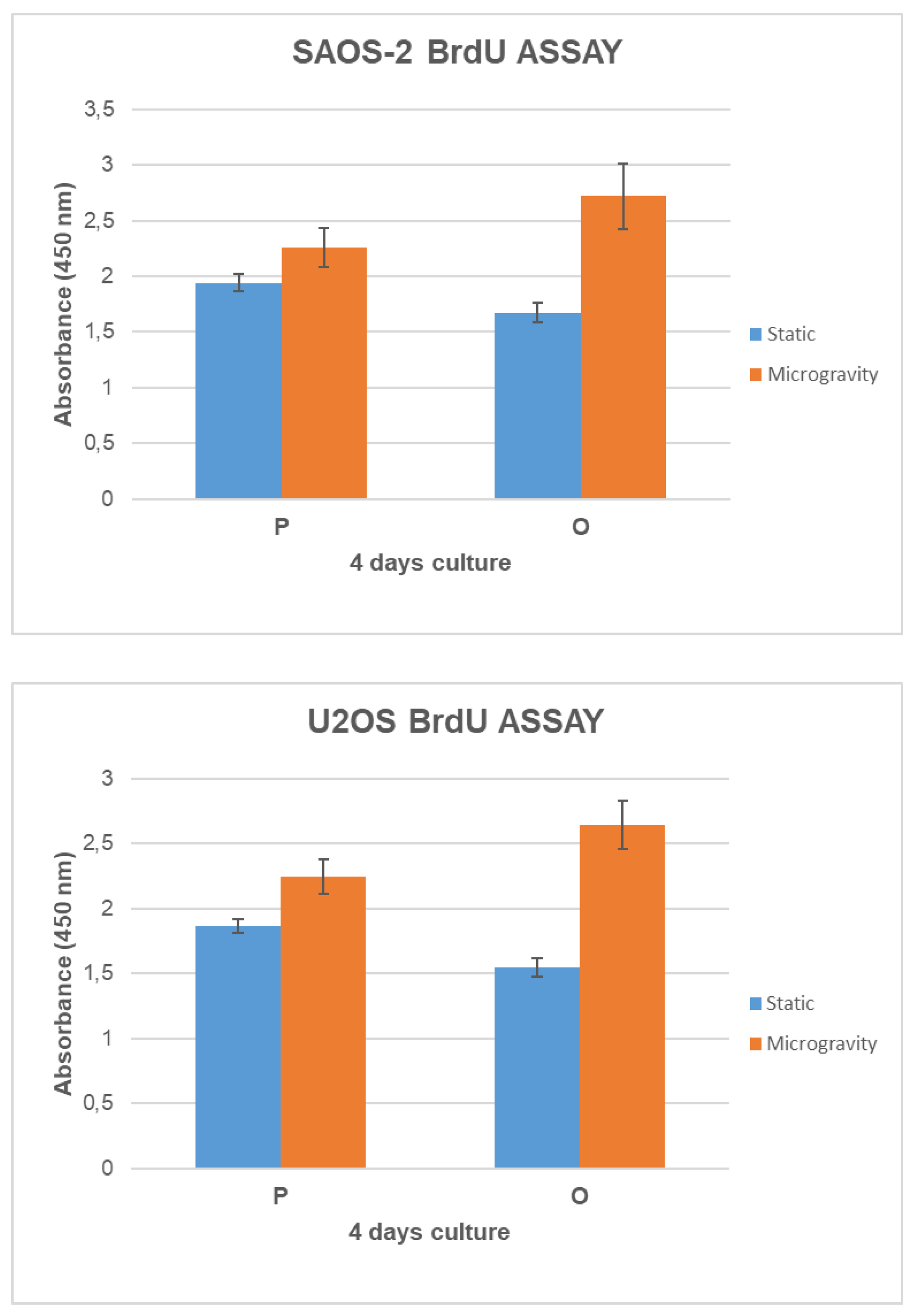
Figure 6.
SEM Analysis of SAOS-2 cells grown on P and O scaffold models for 4 days.
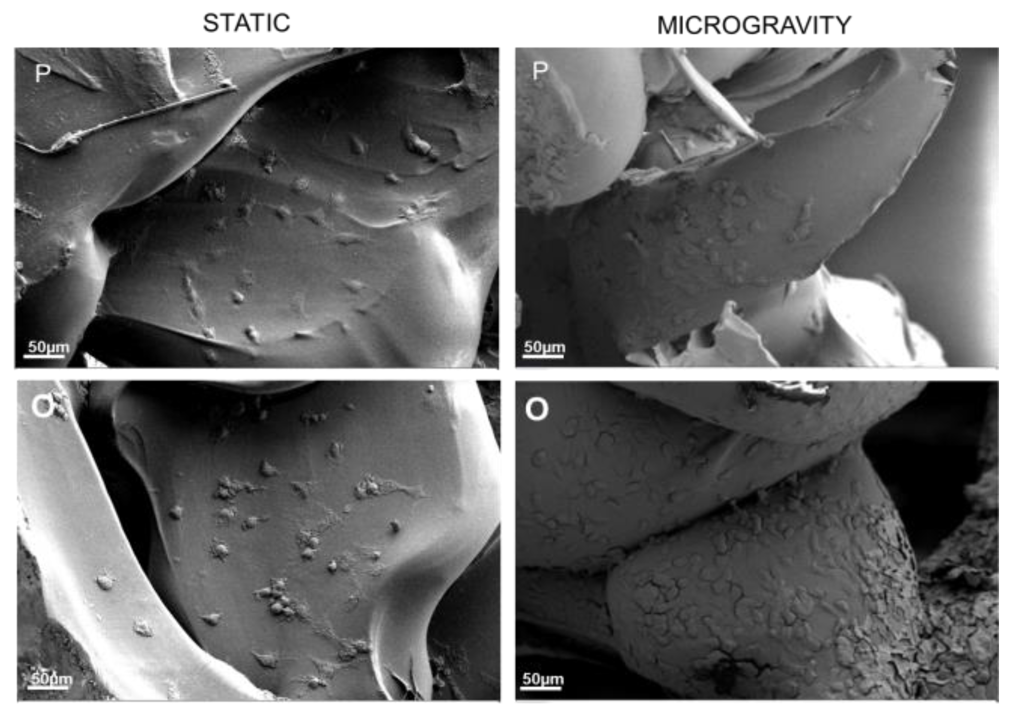
Figure 7.
SAOS-2 and U2OS TNF-α concentration analysis grown on P and O scaffold models and comparing static and microgravity conditions.
Figure 7.
SAOS-2 and U2OS TNF-α concentration analysis grown on P and O scaffold models and comparing static and microgravity conditions.
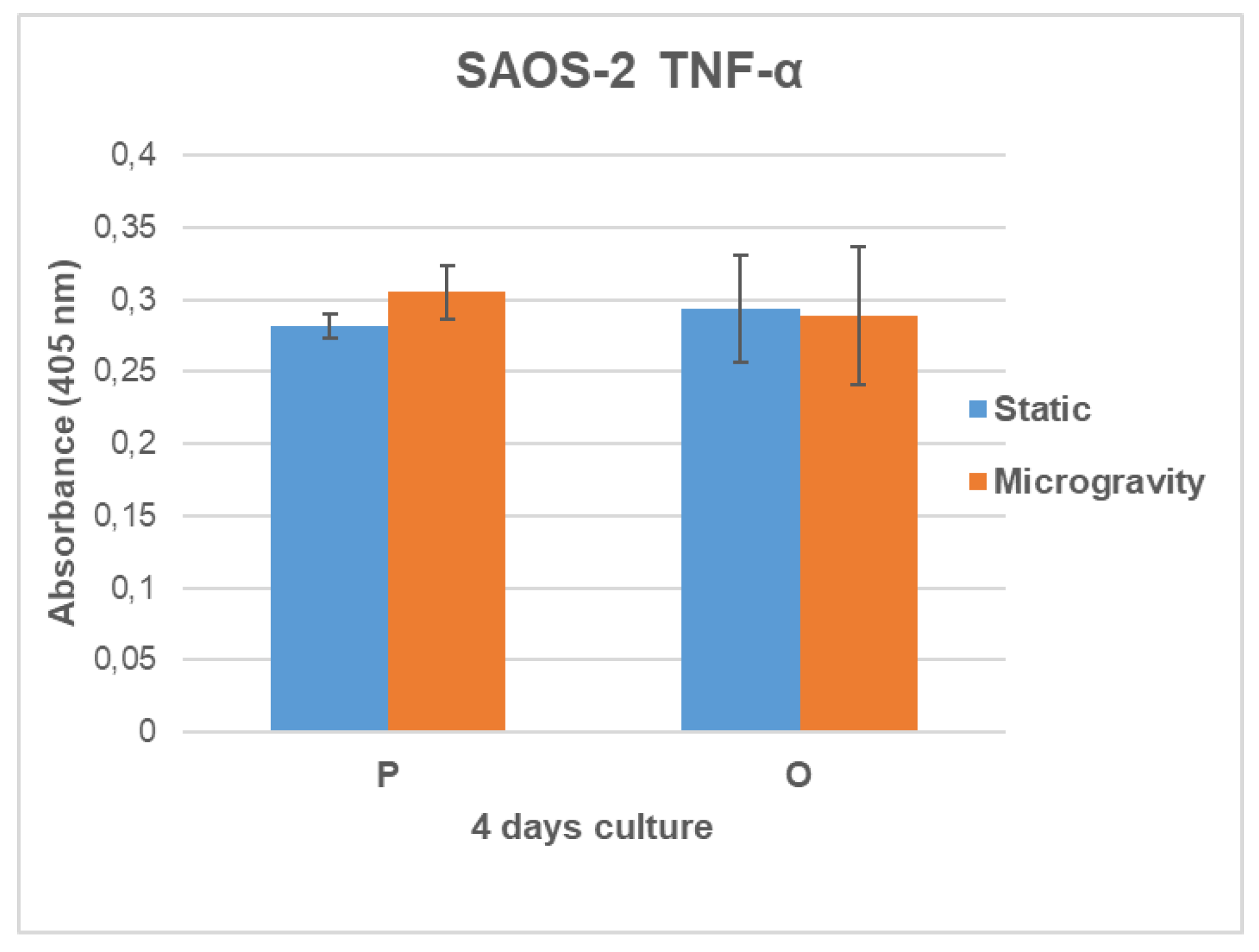
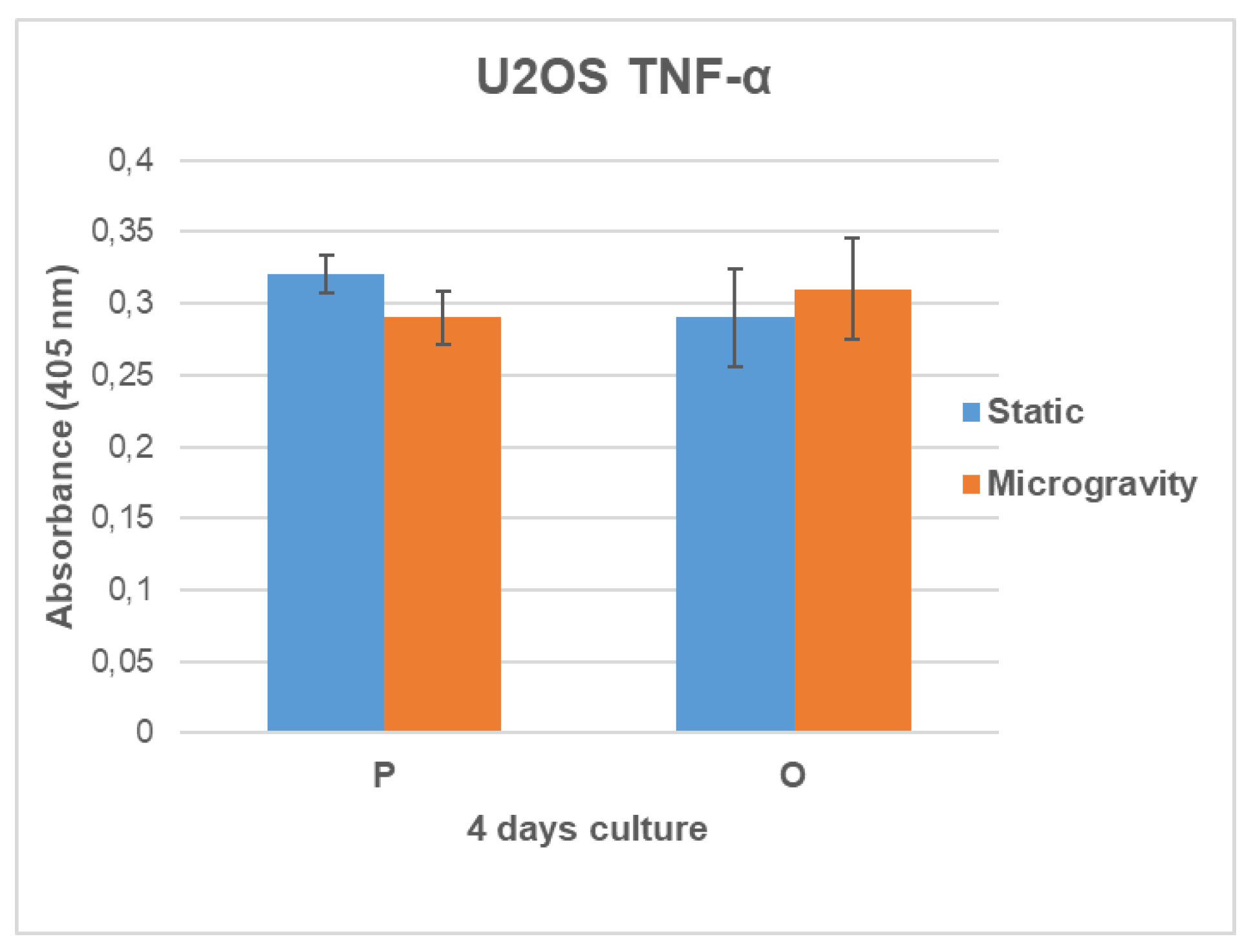

Table 1.
Parameters used for CAD modelling: Pore dimension (PD), pore spacing (PS), trabecular thickness (TT) and trabecular spacing (TS).
Table 1.
Parameters used for CAD modelling: Pore dimension (PD), pore spacing (PS), trabecular thickness (TT) and trabecular spacing (TS).
| SAMPLE | PD (µm) | PS (µm) | TT (µm) | TS (µm) |
|---|---|---|---|---|
| P | 700 | 700 | 200 | 600 |
| O | 800 | 500 | 150 | 800 |
Disclaimer/Publisher’s Note: The statements, opinions and data contained in all publications are solely those of the individual author(s) and contributor(s) and not of MDPI and/or the editor(s). MDPI and/or the editor(s) disclaim responsibility for any injury to people or property resulting from any ideas, methods, instructions or products referred to in the content. |
© 2025 by the authors. Licensee MDPI, Basel, Switzerland. This article is an open access article distributed under the terms and conditions of the Creative Commons Attribution (CC BY) license (http://creativecommons.org/licenses/by/4.0/).
Copyright: This open access article is published under a Creative Commons CC BY 4.0 license, which permit the free download, distribution, and reuse, provided that the author and preprint are cited in any reuse.


