
Submitted:
20 March 2025
Posted:
24 March 2025
You are already at the latest version
Abstract
Engineered nano- and microparticles are considered as promising tools in biomedical applications, such as imaging, sensing, and drug delivery. Protein adsorption on these particles in biological media is an important factor affecting their properties, cellular interactions, and biological fate. Understanding the parameters determining the efficiency and pattern of protein adsorption is crucial for the development of effective biocompatible particle-based applications. This review focuses on the influence of the morphological and physicochemical properties of particles on protein adsorption, including the pattern and amount of the adsorbed protein species, as well as the relative abundance of proteins with specific functions or physicochemical parameters. The effects of functionalization of the particle surface with polyethylene glycol, zwitterions, zwitterionic polymers, or proteins (precoating) on the subsequent protein adsorption are analyzed. In addition, the dependences of protein adsorption on the protein species, biological buffers, fluids, tissues, and other experimental conditions are considered. The influence of protein adsorption on the targeting efficiency of particle-based delivery systems is also discussed. Finally, the effect of the adsorbed protein corona on the interaction of the engineered micro- and nanoparticles with cells and the roles of specific proteins adsorbed on the particle surface in the recognition of the particles by the immune system are discussed.
Keywords:
protein adsorption
; nanoparticles
; microparticles
; protein corona
; particle–cell interactions
; drug delivery
; targeting
1. Introduction
Protein adsorption on nanoparticles (NPs) and microparticles (MPs) is widely recognized as an important factor in the development of particle-based biomedical applications. NPs and MPs of different types are used in drug delivery [1,2], imaging [3], sensing [4,5], and theranostics [6]. However, protein adsorption on particles after their introduction into biological media leads to the formation of a so-called protein or biomolecular corona, which endows particles with biological identity [7]. The protein corona alters the properties of the particles and determines the biological response, including their blood clearance, cellular uptake, biodistribution, and toxicity, as well as the immune response to them [8]. Therefore, deep understanding of the particle properties affecting the protein adsorption can facilitate the rational design of particle-based biomedical applications.
The formation of the protein corona has been shown for a broad range of NPs and MPs, including gold [9,10], silica [11,12,13], poly(lactic-co-glycolic acid) (PLGA), [14,15,16] and superparamagnetic iron oxide NPs [17]; various polymeric particles, polymersomes, and nanogels [18,19,20]; polystyrene (PS) [21,22,23], protein [24], and lipid [25,26,27] NPs. The structural and surface properties of the particles determine their interaction with components of biological media, specifically proteins. The spectrum and amount of the adsorbed proteins depend on the particle size, shape, morphology, structure, and stiffness, as well the physicochemical characteristics of their surface, including the hydrophobicity/hydrophilicity, charge, and functionalization. Experimental conditions, including the incubation medium (protein solution, serum-containing culture medium, or pure serum or plasma), dispersion medium [28], and washing procedure [29], also influence protein–particle interactions. The composition of the adsorbed proteins may affect the biological response to particles, such as cellular uptake and blood clearance rate, as well as the particle toxicity and targeting efficiency. A variety of approaches have been developed to reduce or modify protein adsorption: surface modification with polyethylene glycol (PEG), its derivatives, and zwitterionic compounds; precoating particles with specific proteins; and changing the surface chemistry so that the particles bind desirable proteins.
In this review, we analyse the dependence of protein adsorption on the morphological and physicochemical properties of the particles. In addition, different approaches to surface modification reducing protein adsorption are discussed, as well as strategies of using the protein corona for improving the biological performance of the particles. Finally, we discuss how the protein adsorption onto the engineered particles affects their interactions with living cells, with special focus on their uptake by cells, including immune cells, and specific binding of functionalized particles with target cells.
2. Dependence of Protein Adsorption on the Particle Morphology
2.1. Size and Shape
The size of NPs and MPs is one of the most studied among the parameters affecting protein adsorption on the particles. Regarding NPs, it is generally accepted that smaller NPs, with a larger surface curvature, adsorb proteins more weakly due to steric effects [30]. On the other hand, comparison of protein adsorption on NPs and submicroparticles of different sizes shows that particles with a smaller size and, hence, larger relative surface area adsorb more proteins per unit weight. For example, greater adsorption of bovine serum protein (BSA) has been observed on smaller chitosan NPs [31], However, the amount of adsorbed proteins normalized by surface area rises with increasing particle size, which has been shown for gold, PS, silica, elastin-like polypeptide (ELP), and solid lipid NPs [12,25,32,33,34,35,36] (Figure 1, Panel 1A). Larger particles exhibit greater adsorption of high-density lipoproteins (HDLs) [37]. The size of engineered particles affects not only the quantity of adsorbed proteins, but also the composition of the protein corona. In an early study, the similarity of protein coronas formed on 50- and 100-nm neutral PS NPs in human plasma was high (~80%), but the effect of size was pronounced in the cases of amino- and carboxyl-modified NPs [21]. This study also demonstrated a complex relationship between the protein corona composition and characteristics of the particles. However, most proteins adsorbed on silica NPs from soluble yeast protein extracts were similar for NPs of three different sizes [36]. In a study on 50- and 500-nm silica NPs, the particles of both sizes had similar protein profiles, their uptake by macrophages being reduced after the protein corona formed [12] (Figure 1, Panel 1B). In another study, smaller silica NPs tended to adsorb lower-molecular-weight proteins, probably due to the surface curvature effect [34]. Analysis of the adsorption of plasma proteins on ELP nanoparticles showed that three major proteins (albumin, IgG, and complement factor 3) were adsorbed on all types of particles, whereas the profile of other proteins varied. ELP NPs of different sizes were also found to inhibit blood clotting, the effect of smaller NPs being stronger [35].
The effect of adsorption on the protein conformation also depends on the particle size. For example, a study on the interaction of 30- to 1000-nm particles with proteins showed only the particles larger than 200 nm caused conformational changes of adsorbed BSA and myoglobin [38]. Furthermore, PS particles with different sizes had different effects on amyloid fibril formation from hen egg-white lysozyme [39].
The effect of particle shape on the adsorption of serum proteins was studied using spherical, and faceted silica particles [40]. The protein coverage and corona composition of the faceted particles were found to strongly depend on the number of exposed facets particle and aspect ratio; the spherical particles formed a more homogenous corona with a higher albumin content. In another study, rod-shaped silica particles adsorbed three and four times more protein from plasma and serum, respectively, than spherical ones of the same composition [41]. Comparison of protein adsorption on gold NPs with four shapes (spheres, rods, stars, and cages) and three surface modifications (methoxy, carboxy, and amino groups) showed that the cage-shaped NPs nanostars adsorbed the largest and the smallest amount of protein adsorption, respectively [10]. The compositions of the protein corona formed on nanostars and nanospheres were similar to each other and considerably differed from the composition of the corona on nanocages. Interestingly, the shape of particles more strongly affected the composition of the protein corona than the surface functional groups. Experiments in vivo with star- and rod-shaped gold NPs of different sizes demonstrated that the composition of the protein corona formed on them depended on both their size and shape [9] (Figure 1, panel 2).
2.2. Surface Morphology
Surface morphology of particles (e.g., the presence and size of pores) considerably affects the protein adsorption. For example, a smoother surface tends to adsorb more protein than porous and etched surfaces do [12,22]. Cellular uptake of PS MPs with smooth or etched surface was found to depend on BSA adsorption, the effect of etching being more distinct in the case of larger particles [22]. Porous silica particles were shown to adsorb larger amounts of low-molecular-weight proteins than dense particles, which was explained by the size-exclusion effect [34]. More detailed investigation of pore size effect on the adsorption of proteins revealed nonlinear dependence and postulated the existence of a critical pore size for each particular protein at which both the adsorption capacity is significantly enhanced [42].
Thus, the particle size, shape, and surface morphology can affect both the quantity and composition of the adsorbed proteins. Understanding the effect of particle morphology is crucial in designing tailor-made nano- and microcarriers for biomedical applications.
2.3. Structure
Comparison of MPs with different structures, e.g., core/shell polymeric MPs and capsules (obtained by removing the MP core) showed differences in the composition of their protein coronas [43]. Both types of microstructures functionalized with monoclonal antibodies retained the targeting capacity if they had an additional protein layer on their surface; however, the relative number of cells targeted by the core/shell particles was larger than those targeted by the capsules (90% and 70%, respectively). The capsules were found to adsorb considerably more proteins from both human serum and plasma than the core/shell particles [44]. The composition of the protein corona also varied between the two types of microstructures: core/shell MPs tended to bind a larger amount of apolipoproteins, whereas the capsules adsorbed relatively more complement factors and immunoglobulins.
Overall, the structure of microparticles is also an important parameter whose variation may lead to noticeable changes in the protein quantity and corona composition, which, in turn, may affect the MP interactions with cells.
3. Dependence of Protein Adsorption on the Physicochemical Properties of Particles
3.1. Composition
The chemical nature of NPs and MPs if of high importance for their interaction with proteins. Differences in proteins adsorption on methacrylic acid co-methyl methacrylate (MAA) and polymethyl methacrylate (PMMA) microbeads were analyzed using human serum and plasma [45]. This study showed variations in protein adsorption profiles, including that of complement proteins, which were related to weaker complement activation by MAA beads. Three types of alginate microspheres, namely, sulphated alginate, high G alginate, and poly-L-lysine coated alginate ones, adsorbed different amounts of complement and coagulation factors, which were correlated with their different inflammatory and fibrotic profiles [46].
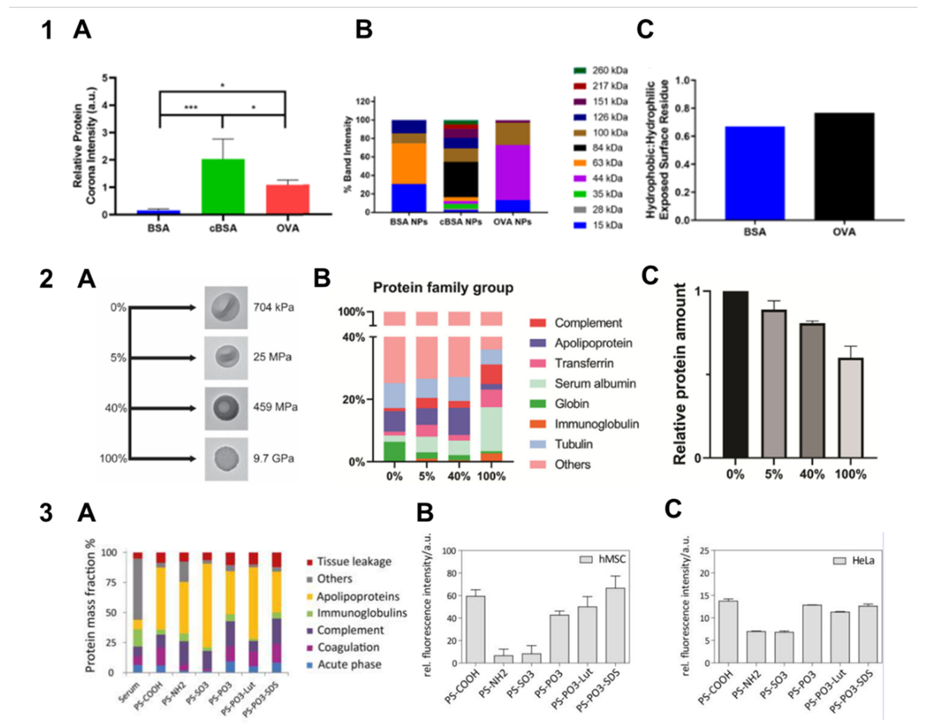
Hydrophobic interactions are considered to be some of the main driving forces of protein adsorption [47]. Therefore, hydrophobicity of the particle material is an important factor of protein–particle interactions. Comparison of the compositions of protein coronas formed on PLGA, cholesterol, or hybrid PLGA–cholesterol NPs showed different fingerprints of the three types of particles [15]. Engineered ovalbumin particles adsorbed more protein than BSA NPs, which was attributed to the experimentally determined higher hydrophobicity of ovalbumin NP surface [24] (Figure 2, Panel 1). In addition, a higher affinity of serum proteins to more hydrophobic particles and different protein corona compositions were found in a study on poly(ε-caprolactone) (PCL) and PLGA nanoparticles [16. Poly(methyl methacrylate-co-styrene) particles with a higher styrene content bound larger amounts of the BSA, IgG, and fibronectin proteins [19].
Furthermore, differences in the phospholipid composition of lipid nanodiscs were found to be related to variations of protein adsorption capacity and affinity to apolipoprotein E (ApoE) [26]. Nanodiscs with a higher capacity for ApoE exhibited better brain targeting in mice, which agrees with previous data [48]. In addition, slight changes in complement activation were detected for these lipid NPs, which influenced particle stability. Analysis of protein adsorption on various lipid NPs demonstrated differences in protein corona related to changes in lipid composition [27]. NPs containing negatively charged lipids had an apolipoprotein-rich corona, which was associated with a more efficient delivery to tumors compared with a vitronectin-rich corona.
Differences in the molecular weight of the monomers used for the fabrication of microparticles also influence the formation of the protein corona. For example, high-molecular-weight chitosan NPs bound less protein than low-molecular-weight NPs with a size of about 100 nm, which was explained by a higher structural rigidity of the polymer [31]. In addition, the polymer chain flexibility of elastomer microspheres was found to affect protein adsorption from an fetal blood serum (FBS)-supplemented cell medium [49].
Figure 2.
Dependence of protein adsorption on the physicochemical properties of particles. Panel 1. The effect of the nanoparticle composition and charge on protein adsorption. The protein coronas were analyzed by gel electrophoresis and nanoliquid chromatography–mass spectrometry. (A) Relative intensities of the adsorbed protein coronas on albumin, cationic albumin, and ovalbumin cross-linked nanoparticles. The average intensities were measured using densitometry and compared against the average intensity of the protein ladder in each image (n = 4; * p < 0.05 (one-way ANOVA), *** p < 0.001 (one-way ANOVA); (B) average distribution of the molecular weight bands identified in the nanoparticle protein coronas (n = 4); (C) the ratio of the hydrophobic to hydrophilic exposed surface residues in albumin and ovalbumin. Abbreviations: BSA, bovine serum albumin; cBSA, cationic BSA; OVA, ovalbumin. Adapted with permission from Ref. [24]. Panel 2. The effect of particle stiffness on protein adsorption. (A) Types of silica nanoparticles with different tetraethyl orthosilicate (TEOS) to triethoxy vinyl silane (TEVS) ratios (0% TEOS/100% TEVS, 5% TEOS/95% TEVS, 40% TEOS/60% TEVS, and 100% TEOS/0% TEVS) and their stiffnesses (Young moduli); (B) the results of liquid chromatography–mass spectrometry analysis of silica capsules incubated with FBS for 3 h, with protein family group coverage up to 50%; (C) relative total protein amounts in different groups of the nanoparticles, with the highest protein amount taken to be 1. The mean ± SD values from three independent replicates are shown. Abbreviations: FBS, fetal blood serum. Adapted with permission from Ref. [50]. Panel 3. The effect of surface functionalization on protein adsorption. (A) Distribution of the seven major protein groups in human serum and in the protein corona of differently functionalized polystyrene nanoparticles presented as protein mass fractions (%); (B) nanoparticle uptake by human mesenchymal stem cells quantitatively assessed by flow cytometry; (C) nanoparticle uptake in HeLa cells quantitatively assessed by flow cytometry. Abbreviations: PS, polystyrene; Lut, Lutensol AT50; SDS, sodium dodecyl sulphate. Adapted with permission from Ref. [23].
Thus, the composition of particles can affect the abundance of biologically relevant proteins adsorbed on their surface and, hence, the physiological response to them. In some cases, enrichment of particles with specific proteins can improve their targeting properties.
3.2. Stiffness
Detailed analysis of the formation of the protein corona on silica NPs with different stiffnesses (Young modulus ranging from 704 kPa to 9.7 GPa) showed a decrease in the amount of adsorbed proteins with increasing stiffness [50]. The adsorbed protein profile varied between these NPs, with higher amounts of complement factor C3 and immunoglobulins adsorbed on the stiffest particles, which correlated with elevated macrophage uptake (Figure 2, Panel 2). Similarly, hydrogel NPs with different stiffnesses (Young modulus ranging from 45 kPa to 760 MPa) coated with a PEGylated lipid bilayer exhibited differences in the protein corona composition upon incubation with mouse plasma [51]. Apolipoprotein A-I was identified as a protein whose relative abundance in the protein corona was correlated with the blood clearance rate. Interestingly, this pattern was retained if the composition of the particle outer shell was changed, which proves the importance of the particle stiffness for protein adsorption and subsequent biological effects.
3.3. Surface Chemistry
In a pioneering study on protein adsorption onto PS particles of two sizes with unmodified, carboxyl-modified, and amino-modified surfaces [21]. Both parameters (size and surface modification) were found to affect the composition of the protein corona. Analysis of protein adsorption from human serum onto PS particles with different surface functional groups (–COOH, –NH2, –SO3, and –PO3) [23] showed that the PS–COOH particles adsorbed twice as much protein molecules as the other types of PS NPs. All types of particles predominantly adsorbed apolipoproteins and complement factors, the particles functionalized with –NH2 and –SO3 exhibiting the highest enrichment with these substances. The highest abundance of ApoH was observed on the PS–COOH and PS–PO3 particles, whose cellular uptake was also the highest. In contrast, preferential adsorption of ApoA4, ApoC3, complement factor C3, and serum albumin was correlated with a decreased cellular uptake (Figure 2, Panel 3). Later, protein adsorption on PS–COOH and PS–NH2 surfaces was studied using a serum-containing cell culture medium [52]. The surface functionalized with –COOH favored adsorption of proteins related to integrin signaling and promoted anti-inflammatory response from macrophages. Recently, the formation and evolution of the protein corona was studied using nanovesicles modified with glycosylated polyhydroxy polymer that had different amino-to-hydroxyl group ratios in their surface coating [53]. The NPs with the maximum NH2-to-OH ratio exhibited a decreased immunoglobulin adsorption, which was accompanied by prolonged blood circulation, and a strong adsorption of CD44 and osteopontin, which promoted the NP internalization by cancer cells. A study of protein adsorption on poly(styrene)-block-poly(ethylene glycol) (PS-b-PEG) nanocarriers and their cellular uptake showed that introduction of cationic or anionic groups to the surface of the NPs resulting in a shift of their ζ-potential significantly affected these processes. [54] Functionalization with the cationic polymer poly(N,N-dimethilaminoethyl methacrylate) with a 1% PS-b-PEG substitution enhanced protein adsorption on the NPs and facilitated their nonspecific cell binding, whereas the NPs modified with the anionic poly(acrylic acid) (PAA) displayed weak protein adsorption and specific binding to macrophages. Comparison of protein adsorption on protein particles consisting of BSA and cationic BSA (with primary amines substituted for carboxyl groups) (with carboxyl groups substituted for primary amines) showed that the cationic particles bound significantly more proteins from FBS [24]. The protein coronas of the two types of particles distinctly differed, the corona of the cationic BSA NPs being more diverse. However, both BSA and cationic BSA NPs were readily taken up by macrophages. Analysis of the adsorption of human serum and plasma proteins on PLA and PLGA NPs showed an important role of surface groups (acidic or ester) in determining the quantity and profile of the adsorbed proteins [55]. The quantity of these functional groups affected only the amount of the adsorbed proteins. The effect of the particle hydrophobicity (estimated by the lactic acid content) on both quantity and profile of proteins was smaller; however, some proteins bound preferentially to hydrophobic or hydrophilic NPs.
Adsorption of proteins from human plasma on three types of colloidal NPs (soft core/multishell NPs and cationic (Eudragit RS) and anionic (ethyl cellulose) rigid NPs) and three types of nanogels (the thermoresponsive dendritic-polyglycerol and two amino-functionalized nanogels) [20]. The cationic and anionic rigid NPs adsorbed the greatest amount of proteins from human plasma due to their high surface charges. These NPs also had the most variable corona and exhibited low cellular uptake and drug release rates. The core/multishell NPs with a neutral charge absorbed small amounts of albumin, but the presence of the protein corona still impaired their cellular uptake. All the nanogels adsorbed little protein due to their high hydrophilicity. Cellular uptake of nanogels by primary human macrophages was not affected by the presence of protein corona, but cytokine release from them was enhanced.
Comparison of BSA adsorption on different types of silica particles showed that the distribution of surface silanol (–SiOH) groups affected this process more strongly than the surface charge [56].
Formation of the protein corona on the surface of nonfunctionalized particles and those functionalized with –COOH and –NH2 groups with sizes of 50, 100, 200, 500, and 1000 nm was studied using an in vitro model of gastrointestinal digestion with subsequent incubation in serum-containing cell culture medium [57]. The in vitro digestion increased the macrophage uptake of neutral 50-, 100-, and 200-nm particles, but not neutral 500- or 1000-nm NPs or charged 100-nm particles. Liquid chromatography–tandem mass spectrometry (LC-MS/MS) showed that the digestion affected the composition of the protein corona formed upon incubation with serum, the presence of specific proteins (complement and coagulation cascade proteins) in the corona correlating with cellular uptake. This study showed that both charge and size of particles determined their interaction with proteins, thus affecting their biological interaction, which agrees with earlier data [21]. In addition, a recent study demonstrated that nanoparticle size and charge determined the pattern of adsorption of saliva proteins [58].
Overall, surface properties play an important role in determining the interaction of particles with proteins. In general, particles with a higher surface charge adsorb more proteins, and cationic groups tend to bind with proteins more effectively. Not only the surface charge, but also the presence of some functional groups (e.g., amino, hydroxyl, and carboxyl ones) has turned out to be an important factor determining the adsorption of specific proteins regulating the biological response to the particles.
Table 1 summarizes the main experimental data on the effects of the particle morphology and physicochemical characteristics on protein adsorption.
4. Surface Modifications Affecting Protein Adsorption on Particles
Various surface modifications are used to control the protein adsorption on NPs and MPs. Modification of particle surface with hydrophilic polymers or zwitterions is usually used to reduce the amount of adsorbed proteins (antifouling or protein repellent surfaces) and avoid nonspecific cellular uptake, imparting them with the so-called stealth properties. Precoating of particles with certain proteins or protein mixtures allows altering further protein adsorption from biological media and affecting cytotoxicity and cellular uptake. Another promising type of surface modification is used to enhance the adsorption of specific proteins, which leads to alteration of the biological properties of the particles, such as biodistribution and specific binding. The results of recent studies on different particle modifications and their effects on protein adsorption are summarized in Table 2.
4.1. Surface Modification with Hydrophilic Polymers
Poly(ethylene glycol) is widely used for particle surface modification due to its hydrophilicity and biocompatibility, as well as because it increases the colloidal stability and blood circulation time of the particles. Recently, PEGylation has been applied to different types of particles, such as PLGA [59], layered double hydroxide (LDH) [60], silica [61,62,63], and strontium sulfite [64] NPs, hydroxyethyl starch (HES) nanocarriers [65,66], gold NPs [33,67], to substantially decrease the protein adsorption rate. Various PEG derivatives are also used for modification of particles, e.g., the poly ethylene glycol and allyl glycidyl ether (PEG-b-AGE) block copolymer have been used for coating iron oxide nanoparticles [68]; the PEG-based surfactant Lutensol AT50, for coating PS NPs [69]; and chitosan-g-methoxy poly(ethylene glycol), for surface modification of hydrogel microcapsules [70]. Apart from the protein repelling effect, PEGylated particles are also characterized by a high colloidal stability [60,65,68] and low of nonspecific cellular uptake rate [68,71].
PEGylation not only reduces the amount of proteins adsorbed from biological media, but also affects the composition of the protein corona. PEG- and poly(ethyl ethylene phosphate) (PEEP)-coated PS nanocarriers were shown to form protein coronas enriched with apolipoproteins, such as ApoA1 and clusterin (apolipoprotein J, ApoJ) [72]. A high clusterin content of the protein corona derived from plasma was found to be associated with a weak uptake of PEG- and PEEP-modified nanocarriers by RAW264.7 macrophages, with the predominant role of this protein in preventing particle–cell interaction confirmed in experiments on the cellular uptake of clusterin-coated PEG-NPs (Figure 3, Panel 1). In the case of PEGylated PLGA particles 100 and 200 nm in size, a decreased abundance of proteins related to immune response was observed [59]. Functionalization of strontium sulfite NPs with biotinylated PEG has been reported to increase the relative albumin abundance and reduce the diversity of adsorbed proteins in the protein corona [64].
The molecular weight and length of PEG chains used for modification of NPs directly affect the amount of adsorbed proteins. For instance, the adsorption rate and quantity of adsorbed proteins on PEGylated gold NPs increased as the PEG molecular weight increased in the series 5000 < 10000 < 30000 Da [73]. The effect of the PEG molecular weight on the protein composition of the corona was also demonstrated for PEG-coated gold NPs [74]. The relative abundances of albumin and transferrin were higher on the surface of NPs modified with 550-Da PEG compared with those modified with 350- and 1000-Da PEGs. Serum albumin is known to act as a dysopsonin, whereas transferrin can enhance binding of NPs with various cancer cells due to overexpression of the Tf receptor on them. Correspondingly, doxorubicin-conjugated NPs modified with 55-Da PEG were found to have the highest cytotoxicity against TfR-expressing HepG2 cells and tumor targeting effect in mice among all formulations tested [74]. The conformation of PEG (brush or mushroom) [75] is determined by both the molecular weight and the grafting density of PEG on the particle surface. This factor also influences protein adsorption and subsequent cellular uptake. It was shown that modification of ovalbumin nanocarriers with PEG in the brush conformation led to a decrease in serum albumin adsorption and an increase in clusterin adsorption, which resulted in a low macrophage uptake [71]. The effect of PEG modification on protein adsorption and particle interaction with cells varies in different types of the particles. For example, the protein coronas formed on PEGylated mesoporous silica particles and PEGylated liposomes in blood plasmas from different individual human donors considerably differed in composition, which determined differences in their interaction with immune cells. In contrast, PEG-only capsules (obtained by removing the core particle after PEGylation) interacted weakly if at all with immune cells, irrespective of the plasma donor [76].
The effect of particle PEGylation on the formation of the protein corona can be used to improve the targeting ability of the particles. For example, specific binding of PEGylated gold NPs conjugated with Herceptin was preserved after incubation with serum, in contrast to NPs without PEG coating [67]. Conjugation of the outer layer of PEG with targeting ligands is also a suitable approach to ensuring specific binding to cells. For instance, HES nanocarriers modified with PEG conjugated to mannose retained the capacity for binding with C-type lectin and dendritic cells after incubation with plasma. [65 In addition, the protein corona formed on Onyvide (a PEGylated liposomal drug) promoted the drug uptake by PANC-1 cells [77].
However, a serious drawback of using PEGylated nanocarriers is the production of anti-PEG antibodies by the immune system correlated with accelerated blood clearance [78]. Furthermore, the presence of anti-PEG IgM antibodies in mice serum altered the composition of protein corona formed on the particles, presumably due to activation of the complement cascade [79]. In addition, the presence of anti-PEG antibodies can substantially reduce drug efficacy and cause hypersensitivity reactions and other side effects [80]. This calls for the search of alternatives to PEG.
One of the compounds considered as a PEG alternative is hyperbranched polyglycerol (PG), a non-immunogenic biocompatible hydrophilic polymer [80]. PG-modified NPs of various types and sizes have proved to be more resistant to protein adsorption than the PEG-modified counterparts. [81 PG-modified particles almost completely evaded macrophage uptake upon incubation in 55% serum or cell culture medium supplemented with 10% FBS. However, PG modification of liposomes enhanced their uptake by macrophages, whereas PEG modification decreased the uptake rate [82]. Note that protein adsorption was insignificant on both PG- and PEG-functionalized liposomes, as well as unmodified ones. This effect can be explained by the predominant role of the liposome surface chemistry in determining interactions with cells.
Poly(phosphoester) was used for modification of PS NPs as a biodegradable alternative to PEG. This resulted in a reduced protein adsorption and decreased macrophage uptake upon incubation with plasma [83].
Modification of NPs with poly(2-ethyl-2-oxazoline) efficiently in reduced protein adsorption on them and their nonspecific cellular uptake [84].
Thus, PEGylation is a well-studied polymer modification of various types of particles that suppresses protein adsorption and alters the composition of the protein corona resulting in a reduced nonspecific cellular uptake of the particles. However, the biomedical use of this approach has some limitation, including toxicity and the formation of anti-PEG antibodies. Several polymers with improved properties, such as non-immunogenicity and low toxicity, have been tested as alternatives to PEG and exhibited a comparable efficiency in stealth modification of particles.
4.2. Surface Modification with Zwitterionic Polymers
As an alternative for PEG antifouling coating in biomedical applications, zwitterionic polymers have gained much attention, mainly because they tend to form a hydration layer on their surface. [85] In addition, zwitterions are low-toxic, do not induce inflammation or antibody production, and have a very low hemolytic activity and relatively long circulation half-time in mice (40 h) [86]. Zwitterionic compounds include low-molecular-weight zwitterions, such as amino acids, sulfobetaine and carboxybetaine derivatives, and polymeric zwitterionic materials. Poly-zwitterions can be subdivided into monomeric ones, with monomers carrying both positive and negative charges, such as poly(sulfobetaine), poly(2-methacryloyloxylethyl phosphorylcholine), poly(sulfobetaine methacrylate), and mixtures of positively and negatively charged monomers [87].
Cysteine and other amino acids can be used as low-molecular-weight zwitterionic ligands. Experiments using silica NPs modified with cysteine as a zwitterion and biotin as a model targeting ligand demonstrated reduction of protein adsorption and effective targeting of NPs after incubation in 10% or 50% human plasma [88]. Cysteine, lysine, and arginine functionalization of silica NPs were shown to make them resistant to protein adsorption from a BSA solution and FBS. [89] Recently, zwitterionic peptides with various amino acid sequences and compositions were used to modify gold NPs and were found to be promising antifouling surface functionalities [90]. The protein adsorption pattern was found to depend more on the charge motif and sequence than on the amino acid composition, and it influenced the rate of NP uptake by macrophages.
Another low-molecular-weight zwitterion, organosiloxane, used for modification of silica particles, inhibited protein adsorption on them and ensured their high stability [91]. Further development of this approach led to the use of bi-functionalized zwitterionic/–COOH silica NPs, which preserved antifouling properties and could conjugate with biomolecules [92]. Magnetic silica NPs functionalized with amino and alkene functional groups along with the betaine zwitterion also exhibited reduced adsorption of BSA, lysozyme, and FBS proteins while retaining the capacity for interacting with biomolecules. [93 However, double functionalization of silica NPs with zwitterion (sulfobetaine) and amino, mercapto, or carboxylic functional groups imparted antifouling properties to them but hindered the interaction between the functional groups and cells [94].
Successful reduction of protein adsorption was demonstrated in experiments on sulfobetaine modification of silica NPs [62,95] and 4-(4-hydroxymethyl-3-methoxyphenoxy)-butyric acid (HMPB) NPs loaded with anticancer drugs [96]. The latter NPs had high targeting capability and biocompatibility. Carboxybetaine coating of silica NPs reduced protein adsorption by as much as 94%, depending on conjugation method [97].
Modification with polymeric zwitterions is also successfully used to suppress protein adsorption on particles. Magnetic gold NPs coated with zwitterionic polymers adsorbed significantly less protein than those coated with PEG, polyethylenimine (PEI), and polyallylamine hydrochloride [98]. Modification of core/shell Fe3O4@SiO2 NPs with PEI and zwitterionic 2-methacryloyloxyethyl phosphorylcholine suppressed protein adsorption in a BSA solution and FBS [99]. Phosphorylcholine methacrylate (MPC) coating reduced protein adsorption on gold NPs in serum and bronchoalveolar lavage fluid (BALF) and increased their cellular uptake rate by A549 cells after the treatment with these biological fluids [100]. The amount of BSA adsorbed on poly(glycidyl methacrylate) (PGMA) microspheres noticeably decreased after modification of their surface with the PMPDSAH poly-zwitterion [101]. In core/shell gelatin methacrylate MPs coated with carboxybetaine methacrylate (CBMA), sulfobetaine methacrylate (SBMA) and MPC, nonspecific protein adsorption, cell adhesion, and inflammation response in vitro were significantly inhibited [102] (Figure 3, Panel 2). The zwitterion-coated MPs implanted in mice resisted the body response for as long as four months.
The rates of adsorption of basic and acidic proteins on silica NPs were shown to depend on the degree of quaternization of the carboxybetaine copolymer coating them [103]. Comparison of the protein coronas formed on silica NPs, poly(2-methacryloyloxyethyl phosphorylcholine) (PMPC)-coated hybrid silica NPs, and PMPC replica particles upon incubation in human serum showed that the bare silica NPs adsorbed a wider range of proteins than the zwitterion-coated particles [104,105]. Differences in the adsorption profile between PMCP-coated and bare silica particles were confirmed in another study [106], with PMPC-coated particles forming only a “soft” corona, i.e., a layer of loosely bound proteins.
However, the adsorption of negatively charged proteins (BSA and fibrinogen) on PCL–(N-(3-sulfopropyl-N-methacryloxyethy-N,N-diethylammonium betaine) (PCL–PDEAPS) particles was more pronounced than on PCL–PEG particles, whereas the adsorption of lysozyme was relatively low in both cases [107]. Combination of a zwitterionic ligand with tetraethylene glycol for functionalization of the nanodiamond surface resulted in a high colloidal stability and suppression of protein adsorption [108].
In summary, various monomeric and polymeric zwitterionic coatings of NPs and MPs significantly decrease the protein adsorption rate. However, some drawbacks of this approach compared with PEG coating have been reported, such as hindering of the interaction between surface functional groups and living cells.
4.3. Surface Modification with Proteins
One of the most common proteins used for surface precoating is serum albumin (BSA or HSA). Formation of the protein corona consisting of BSA on poly-3-hydroxybutyrate-co-3-hydroxyhexanoate (PHBHHx) NPs allowed reducing the blood clearance time and cytotoxicity [109]. After subsequent incubation in serum, lower levels of IgG adsorption and complement component C4b generation were observed. In a study on single protein coronas formed on polymersomes, the viability of cells taking up these particles was increased if the polymersomes were preliminarily incubated with IgG, lysozyme, and BSA [110]. The effect of BSA precoating on the cellular uptake of particles seems to depend on the cell line used in the study. BSA coating increased the uptake of gelatin–oleic acid NPs by HEK293 cells but decreased their uptake by A459 cells in a cell culture media containing FBS. [111 Albumin pre-adsorption on LDH NPs enhanced their uptake by Chinese hamster ovary (CHO) cells [112]. A preformed corona of BSA modified with the RGDyK cyclic peptide on si-VEGF-loaded chitosan-based NPs reduced the protein adsorption from serum and enhanced the capacity for targeting cancer cells and efficiencies of the delivery to the tumor and suppression of angiogenesis in vivo [113,114]. The binding of sialyl Lewis A-targeted PLGA and chitosan–PLGA particles with endothelial cells in the presence of plasma was improved after coating with HSA [115]. SDS-PAGE analysis of proteins adsorbed on HSA-coated particles showed higher intensities of bands with molecular weights of 75 and 150 kDa, probably corresponding to histidine-rich glycoprotein and IgG. Precoating with FBS weakened NP interaction with leukocytes more effectively than precoating with BSA or treatment with bovine serum, the effect correlating with a lower total protein adsorption rate and abundance of immunoglobulins [116]. BSA coating of PS NPs inhibited protein adsorption more strongly than modification with PEG or chitosan [117].
Precoating of silica NPs with γ-globulin and HSA as an opsonin and a dysopsonin, respectively, was used to modify the protein corona composition [118]. Indeed, coating with HSA reduced the adsorption of coagulation factors, e.g., fibrinogen, which could enhance immune response, whereas coating with γ-globulins increased adsorption of immunoglobulins and complement factors. However, coating with γ-globulins did not enhance macrophage uptake of the NPs via Fc-mediated phagocytosis, probably due to shielding of immunoglobulins with other adsorbed proteins. Precoating with a single protein, such as BSA, myoglobin, β-lactoglobulin, lysozyme, or fibrinogen, on PS particles also affected the composition of the protein corona after incubation with serum proteins [119]. The protein of the primary corona was still detectable after incubation with serum proteins, which evidences the stability of the artificial protein coating. The enhancing effect of lysozyme precoating on particle–cell interactions was shown for 200-nm PS particles, but not for 3-µm ones. Casein precoating was found to significantly decrease the amount of protein adsorbed on starch-coated poly(methyl methacrylate-co-acrylic acid) NPs; accordingly, casein-coated NPs conjugated with an aptamer exhibited a high delivery efficiency in vitro and in vivo [120].
Pre-adsorption of antibodies on PS and HES NPs formed stable targeting moieties unaffected by incubation in human serum or plasma [121]. In addition, it was shown that the presence of targeting antibodies on the surface of polymeric NPs favored the activation of the classical complement pathway, in contrast to untargeted NPs, which activated the alternative pathway [122]. Regarding complement activation, the use of complement factor H for particle coating reduced complement activation by silica NPs [123], in accordance with previous studies postulating the stealth effect of this protein [124,125].
Preliminary formation of a protein corona consisting of glutathione transferase fused with affibody on metal–organic framework NPs led to noticeably reduced protein adsorption on them and effective targeting in vitro and in vivo [126] (Figure 4). Combination of this approach with collagenase coating enabled deep tissue penetration of NPs and tumor growth inhibition [127].
For lipid nanocarriers consisting of 1,2-dioleoyl-3-trimethylammonium propane and DNA, spontaneous enrichment with vitronectin after incubation with human plasma was shown, which enhanced their uptake by cancer cells expressing the integrin receptor [128].
Apolipoproteins are another family of proteins known to noticeably affect particle–cell interaction. For example, precoating of PS-COOH nanoparticles with ApoA4 or ApoC3 weakened their cellular uptake, whereas coating with ApoH enhanced this process [23]. Surface coating of PS and HES NPs with clusterin (ApoJ) before incubation with IgG-enriched plasma prevented preferential adsorption of immunoglobulins, which was associated with reduced cellular internalization [129].
Another type of artificial protein coronas formed on NPs includes coronas consisting of protein fractions of human plasma [130]. It was found that preincubation of PS and PS-NH2 particles with IgG and HSA depleted plasma fractions prevented aggregation of NPs placed into plasma. In addition, preliminary coating with the HSA-containing plasma fraction facilitated cellular uptake of NPs. Preincubation of PS particles with IgG depleted plasma before adding to macrophages (RAW264.7) in a plasma-supplemented cell culture significantly decreased their cellular uptake [131].
5. Other Factors Affecting Protein Adsorption
5.1. Protein Type
Along with the properties of the particles, the physicochemical characteristics of proteins, such as their molecular weight, isoelectric point, and hydrophobicity, also affect protein adsorption. Therefore, the type of protein used in a study should be taken into account in interpreting the results. Most studies use BSA, HSA, fibrinogen, or immunoglobulin G as a model protein (Table 1). One of the approaches to revealing the influence of the protein properties on its adsorption is modification of the native protein by attaching functional groups. HSA with additional carboxylic or amino groups were shown to differ from the native protein in the affinity to negatively charged QDs used as model NPs [132]. These results demonstrate the importance of the local protein charge for its interaction with NPs. On the other hand, experiments with three proteins with different sizes and charges (BSA, IgG, and lysozyme) showed that all of them could be adsorbed on negatively charged polymersomes [110]. Experimental data demonstrated that interaction between polymersomes and proteins were mainly driven by hydrogen bonding and van der Waals interactions, the role of electrostatic interactions being less important. Regarding hydrophilicity of proteins, two different modes of adsorption on nanomicelles were demonstrated [133]. The hydrophobic bovine hemoglobin interacted with nanomicelles in the insertion mode, and the hydrophilic BSA and lysozyme, in the surface adsorption mode, with surface charge determining the differences in behavior between these two hydrophilic proteins.
5.2. Incubation Medium
In order to better understand the interaction of NPs and MPs with biomolecules in the complex physiological environment, derivatives of biological fluids, such as blood serum and plasma, are commonly used in protein corona studies. A study of protein adsorption on PLGA and PLA NPs using serum and plasma as incubation media showed only minor differences in the profile and quantity of adsorbed proteins [55]. However, noticeable variations differences between the protein adsorption profiles of copolymer methacrylate beads incubated in serum and plasma were demonstrated [45]. In the case of PS-NH2 particles, the composition of protein corona formed upon incubation in human serum and plasma differed substantially due to the high rate of fibrinogen adsorption in plasma [134]. The same study showed that the particle–cell interactions after the formation of the protein corona were strongly influenced by the anticoagulant heparin. Similarly, the sizes and compositions of the protein corona on silica NPs incubated with serum and plasma differed from each other, with greater amounts of coagulation and complement system proteins adsorbed from plasma than from serum [135]. The NPs incubated with plasma were also more readily taken up by RAW264.7 macrophages, but not NIH 3T3 fibroblasts.
To sum up, serum as an incubation medium is advantageous due to the absence of coagulation proteins, whereas plasma better mimics the physiological conditions. In most cases, the compositions of protein coronas formed on particles incubated with serum and plasma differ, which usually results in differences in their cellular uptake.
An approach to more accurate simulation of physiological conditions is using either cell-conditioned [136] or flowing (dynamic) incubation media to mimic blood flow. Particles whose protein coronas have formed under the static and dynamic conditions differ with respect to their macrophage uptake and stimulation of cytokine secretion. In addition, the influence of the type of washing medium on the composition of the protein corona formed on magnetic nanocarriers was shown [29]. Variations of the composition of the NP protein corona formed in FBS were also demonstrated when different media were used for the dispersion of the NPs before incubation [28].
Liposomes incubated in either static or flowing FBS were found to differ in the protein corona composition, which further confirmed the importance of more accurate modelling of in vivo conditions [137]. Another example is considerable differences in the protein coronas formed on PS NPs modified with transferrin receptor–targeting ligands that were incubated under the in vitro and in vivo conditions [138]. Fibrinogen was one of the most abundant proteins in the in vitro corona, whereas it accounted for less than 1% of the in vivo corona. In contrast, the amounts of adsorbed albumin and clusterin were higher in the in vivo corona. Similarly, the protein composition of the corona formed on magnetic NPs in vivo was more diverse than that of the in vitro corona [139]. In order to better mimic the in vivo conditions, the authors of that study suggested the ex vivo approach, with NPs incubated with freshly isolated blood for 1 min. In this case, the composition of the protein corona was similar to that of the in vivo corona.
In summary, protein adsorption on NPs and MPs is strongly affected by experimental conditions, including the incubation medium and overall experimental design. To deeper understand the protein corona formation and its biological consequences, it is necessary to accurately select optimal experimental conditions imitating physiological conditions.
6. Effect of Protein Adsorption on Particle–Cell Interactions
6.1. Targeted Delivery
The influence of protein adsorption on engineered NPs and MPs for targeted delivery is crucial for the development of effective delivery systems. Binding of transferrin-functionalized NPs with both soluble transferrin receptor and A549 cells expressing this receptor was decreased after adsorption of proteins [140]. The targeting ability of NPs functionalized with transferrin was lost after the formation of the protein corona in vitro but partly retained after its formation in vivo [141]. Note that transferrin–NPs preserved the targeting specificity towards brain tumor cells after crossing the blood–brain barrier. Protein adsorption from human serum significantly suppressed the interaction of affibody-functionalized, polymer-coated silica particles with SK-OV-3 cells [142]. This effect of protein adsorption on the specific binding of particles can be explained by shielding of ligands by adsorbed proteins, which prevents them from interacting with the receptor.
However, polymeric particles and capsules conjugated with monoclonal antibodies were reported to retain their targeting ability after protein adsorption [43]. The effect of protein adsorption on the interaction of hyaluronic acid (HA)-modified metal–phenolic network (MPN) capsules depended on the molecular weight of HA: the capsules modified with 13-kDa HA lost their targeting ability, whereas those modified with 230-kDa HA exhibited only a 5% reduction of specific binding [143]. In the case of Fn14-targeted NPs, the reduction of cellular uptake after incubation with serum was greater in NPs functionalized with the Fab fragment than in those functionalized with the full-length antibody [144].
6.2. Cellular Uptake
Considerable differences in cellular uptake of silica NPs were observed in serum-free and complete culture media [145]. In the serum-containing media, the protein corona was formed that decreased adhesion and internalization of the particles by A549 cells. Preincubation of polymeric NPs with plasma also reduced their uptake by Telo-RF and HeLa cells [146]. Formation of the protein corona on lipid and silica NPs reduced their uptake by RAW 264.7 macrophages [147]. Adsorption of proteins on polymer particles in an FBS-supplemented cell medium had different effects on the association of the particles with THP-1 monocytes and with macrophage-like cells differentiated from them [148]. The particles with a BSA-rich protein corona were internalized by THP-1 monocytes less efficiently than bare particles. On the other hand, the rate of particle internalization by the differentiated macrophage-like cells did not change after adsorption of proteins on the particles.
The presence of adsorbed proteins on the particle surface can affect the cell entry mechanism. After incubation of liposomes with human plasma and formation of a protein corona containing a large portion of apolipoproteins, HeLa cells mainly took up the liposomes via clathrin-mediated endocytosis [149]. In the case of gold nanorods modified with cetriltrimethylammonium bromide (CTAB) or poly-diallyldimethylammonium chloride (PDDAC), the mechanisms of particle internalization by macrophages were different for plasma-incubated and pristine nanorods [150]. The uptake of plasma-incubated nanorods was decreased after pre-treatment of the cells with anti-CD36 antibodies, which indicated the involvement of the CD36 scavenger receptor, probably due to the presence of apolipoproteins and transport proteins on the nanorods surface. It was shown that different mechanisms were involved in the NP cellular uptake after the formation of a protein corona in media with high and low concentrations of serum, which was explained by differences in the corona composition [151]. Experiments with silica NPs showed that the presence of apolipoprotein B on the NP surface determines the NP recognition by the LDL receptor, whereas surface IgG was recognized by the Fc-gamma receptor I [152]. The secondary structure of the adsorbed proteins was also shown to determine the type of receptors that bind the protein-coated NPs [153]. In the case of anionic NPs, adsorbed BSA retained its native conformation, which allowed the binding of the NP–BSA complexes by albumin receptors. In contrast, adsorption of BSA on cationic NPs led to BSA denaturation and subsequent binding of binding of the NP–BSA complexes by scavenger receptors.
The role of individual proteins adsorbed on the surface of particles in modulating cellular uptake is being intensely studied. For example, PS NP coating with ApoA4 and ApoC3 considerably reduced their uptake by human mesenchymal stem cells, whereas coating with ApoH enhanced the cellular uptake [23]. A negative correlation between ApoA4 adsorption on mesoporous silica–PEG NPs and their association with cells (monocytes and B cells) was reported [76].
The protein coronas of NPs and MPs can facilitate their use as vehicles for targeted drug delivery. For example, the apolipoprotein-rich corona formed on lipid NPs ensured their targeted delivery to HepG2 tumor cells [27]. Clusterin (ApoJ) was identified as the key component of a protein corona providing the stealth effect (reduction of nonspecific cellular uptake) of PEGylated PS NPs [72] and unmodified silver and silica nanocarriers [154].
The presence of immunoglobulins and complement proteins (C4B and C3) on the particle surface was found to facilitate association of the particles with immune cells (monocytes and B cells) [76]. Adsorption of C1q on NPs promoted opsonization with the C3 component and subsequent recognition by leukocytes and macrophages [155]. Binding of IgG to the surface of different nanomedicines was shown to induce deposition of the C3 component [156]. The adsorption of plasma proteins on mesoporous silicon and PMPC particles proved to be required for their association with blood cells (monocytes, granulocytes, and B cells) [105]. Precoating with IgG promoted association of particles with phagocytic cells. Enrichment of the protein corona with complement proteins was more pronounced on mesoporous silicon particles than on PMPC ones and was positively correlated with their association with B cells.
7. Conclusions and Outlook
Understanding of protein adsorption on engineered NPs and MPs is crucial for the development of efficient and safe biomedical applications. Here, we considered the influence of the main physicochemical parameters of the particles on the characteristics of protein adsorption. The particle size is directly related with the amount of the adsorbed protein. In most cases, larger particles bind more protein. The amounts of different proteins adsorbed on particles may depend on the particle size more or less strongly and may affect the cellular uptake of the particles. The shape, surface morphology, and surface structure of particles are also important characteristics whose changes may cause quantitative and qualitative differences in the protein adsorption process. The particle composition considerably influences both the total amount of the adsorbed proteins and their proportions in the protein corona. The protein corona composition affects the biological responses to the particles, such as complement activation. Particle surface chemistry and charge is a key factor determining the interaction with proteins. Generally, the higher the surface charge of particles, the higher the protein adsorption rate. In addition, the presence of specific functional groups influences the composition of the formed protein corona and subsequent cellular interactions. In conclusion, the characteristics of the protein corona depend on the combination of different properties of particles.
Among approaches to reducing or modifying the protein adsorption, modification of the particle surface with hydrophilic polymers, such as PEG or PG, is one of the most popular. The presence of PEG on the surface of nanocarriers affect not only the amount of adsorbed proteins, but also preferential adsorption of some proteins, e.g., apolipoproteins. Modification of the surface with zwitterionic compounds is a promising approach to conferring protein repellent properties to particles. Alternatively, precoating of particles with some proteins allows the modulation of subsequent protein adsorption and particle–cell interactions. In some cases, modification of particles with specific proteins enables specific targeting and biodistribution of particles.
Adsorption of proteins on particles influences their interaction with cells in different ways. The binding efficiency and specificity can be preserved in the presence of the protein corona, but also can be impeded after adsorption of proteins, probably due to the shielding effect of protein molecules. The effect on cellular uptake depends on the type of cells and the profile of proteins adsorbed on the surface of the particles, because different proteins may either enhance or suppress the interaction with specific receptors on the cell surface.
The diversity of biomedical tools based on NPs and MPs currently developed and the large number of factors affecting protein adsorption call for new methods for predicting the composition of the protein corona and biological effects of particle–protein complexes. Recently, a model based on machine learning and meta-analysis has been suggested that predicts the functional composition of the protein corona and cellular recognition of NPs [157]. Machine learning algorithms have also been used for predicting the relative amounts of proteins in the protein corona of NPs [158] In addition, a software package has been developed enabling the prediction of the protein corona composition on NPs immersed in complex biological fluids [159].
Overall, the understanding of factors influencing protein adsorption on particles and various approaches to its reduction or modification can help improving the design of NPs and MPs for biomedical applications.
Author Contributions
Conceptualization, A.S. and I.N.; data curation, E.G.; funding acquisition, A.K.; project administration, A.S., supervision, I.N.; validation, A.K.; writing—original draft, E.G.; writing—review and editing, all authors. All authors have read and agreed to the published version of the manuscript.
Funding
This study was funded by the Russian Science Foundation, grant no. 22-75-10103.
Data Availability Statement
No primary research results, software or code have been included, and no new data other than those presented in this publication were generated or analyzed as part of this review.
Acknowledgments
I.N. & A.S. acknowledge the Université de Reims Champagne and the Graduate School NANO-PHOT (École Universitaire de Recherche, PIA3, contract ANR-18-EURE-0013). We thank Vladimir Ushakov for proofreading the manuscript.
Conflicts of Interest
The authors declare no conflicts of interest.
Abbreviations
The following abbreviations are used in this manuscript:
| NP | Nanoparticle |
| MP | Microparticle |
| PLGA | Poly(lactic-co-glycolic acid) |
| PS | Polystyrene |
| PEG | Polyethylene glycol |
| BSA | Bovine serum protein |
| ELP | Elastin-like polypeptide |
| HDL | High-density lipoprotein |
| MAA | Methacrylic acid co-methyl methacrylate |
| PMMA | Polymethyl methacrylate |
| PCL | Poly(ε-caprolactone) |
| ApoE | Apolipoprotein E |
| FBS | Fetal blood serum |
| PS-b-PEG | Poly(styrene)-block-poly(ethylene glycol) |
| LDH | Layered double hydroxide |
| HES | Hydroxyethyl starch |
| PEG-b-AGE | Allyl glycidyl ether |
| PEEP | Poly(ethyl ethylene phosphate) |
| ApoJ | Apolipoprotein J |
| HMPB | 4-(4-hydroxymethyl-3-methoxyphenoxy)-butyric acid |
| PEI | Polyethylenimine |
| MPC | Phosphorylcholine methacrylate |
| BALF | Bronchoalveolar lavage fluid |
| PGMA | Poly(glycidyl methacrylate) |
| CBMA | Carboxybetaine methacrylate |
| SBMA | Sulfobetaine methacrylate |
| PMPC | Poly(2-methacryloyloxyethyl phosphorylcholine) |
| PCL–PDEAPS | PCL–(N-(3-sulfopropyl-N-methacryloxyethy-N,N-diethylammonium betaine) |
| PHBHHx | Poly-3-hydroxybutyrate-co-3-hydroxyhexanoate |
| CHO | Chinese hamster ovar |
| CTAB | Cetriltrimethylammonium bromide |
| PDDAC | Poly-diallyldimethylammonium chloride |
References
- Nifontova, G.; Tsoi, T.; Karaulov, A.; Nabiev, I.; Sukhanova, A. Structure–function relationships in polymeric multilayer capsules designed for cancer drug delivery. Biomater. Sci. 2022, 10, 5092–5115. [Google Scholar] [CrossRef] [PubMed]
- Afzal, O.; Altamimi, A.S.A.; Nadeem, M.S.; Alzarea, S.I.; Almalki, W.H.; Tariq, A.; Mubeen, B.; Murtaza, B.N.; Iftikhar, S.; Riaz, N.; et al. Nanoparticles in drug delivery: From history to therapeutic applications. Nanomaterials 2022, 12, 4494. [Google Scholar] [CrossRef] [PubMed]
- Siddique, S.; Chow, J.C.L. Application of nanomaterials in biomedical imaging and cancer therapy. Nanomaterials 2020, 10, 1700. [Google Scholar] [CrossRef] [PubMed]
- Mazetyte-Stasinskiene, R.; Köhler, J.M. Sensor micro and nanoparticles for microfluidic application. Appl. Sci. 2020, 10, 8353. [Google Scholar] [CrossRef]
- Grasso, G.; Colella, F.; Forciniti, S.; Onesto, V.; Iuele, H.; Siciliano, A.C.; Carnevali, F.; Chandra, A.; Gigli, G.; del Mercato, L.L. Fluorescent nano-and microparticles for sensing cellular microenvironment: past, present and future applications. Nanoscale Adv. 2023, 5, 4311–4336. [Google Scholar] [CrossRef]
- Xu, Y.; Zheng, H.; Schumacher, D.; Liehn, E.A.; Slabu, I.; Rusu, M. Recent advancements of specific functionalized surfaces of magnetic nano-and microparticles as a theranostics source in biomedicine. ACS Biomater. Sci. Eng. 2021, 7, 1914–1932. [Google Scholar] [CrossRef]
- Monopoli, M.P.; Åberg, C.; Salvati, A.; Dawson, K.A. Biomolecular coronas provide the biological identity of nanosized materials. Nat. Nanotechnol. 2012, 7, 779–786. [Google Scholar] [CrossRef]
- Cai, R.; Chen, C. The crown and the scepter: roles of the protein corona in nanomedicine. Adv. Mater. 2019, 31, 1805740. [Google Scholar] [CrossRef]
- García-Álvarez, R.; Hadjidemetriou, M.; Sánchez-Iglesias, A.; Liz-Marzán, L.M.; Kostarelos, K. In vivo formation of protein corona on gold nanoparticles. The effect of their size and shape. Nanoscale 2018, 10, 1256–1264. [Google Scholar] [CrossRef]
- Bewersdorff, T.; Glitscher, E.A.; Bergueiro, J.; Eravci, M.; Miceli, E.; Haase, A.; Calderón, M. The influence of shape and charge on protein corona composition in common gold nanostructures. Mater. Sci. Eng. C 2020, 117, 111270. [Google Scholar] [CrossRef]
- Tenzer, S.; Docter, D.; Kuharev, J.; Musyanovych, A.; Fetz, V.; Hecht, R.; Schlenk, F.; Fischer, D.; Kiouptsi, K.; Reinhardt, C.; et al. Rapid formation of plasma protein corona critically affects nanoparticle pathophysiology. Nat. Nanotechnol. 2013, 8, 772–781. [Google Scholar] [CrossRef] [PubMed]
- Saikia, J.; Yazdimamaghani, M.; Hadipour Moghaddam, S.P.; Ghandehari, H. Differential protein adsorption and cellular uptake of silica nanoparticles based on size and porosity. ACS Appl. Mater. Interfaces 2016, 8, 34820–34832. [Google Scholar] [CrossRef] [PubMed]
- Clemments, A.M.; Botella, P.; Landry, C.C. Spatial mapping of protein adsorption on mesoporous silica nanoparticles by stochastic optical reconstruction microscopy. J. Am. Chem. Soc. 2017, 139, 3978–3981. [Google Scholar] [CrossRef] [PubMed]
- Sempf, K.; Arrey, T.; Gelperina, S.; Schorge, T.; Meyer, B.; Karas, M.; Kreuter, J. Adsorption of plasma proteins on uncoated PLGA nanoparticles. Eur. J. Pharm. Biopharm. 2013, 85, 53–60. [Google Scholar] [CrossRef]
- Ottonelli, I.; Duskey, J.T.; Genovese, F.; Pederzoli, F.; Caraffi, R.; Valenza, M.; Tosi, G.; Vandelli, M.A.; Ruozi, B. Quantitative comparison of the protein corona of nanoparticles with different matrices. Int. J. Pharm. X 2022, 4, 100136. [Google Scholar] [CrossRef]
- Ndumiso, M.; Buchtová, N.; Husselmann, L.; Mohamed, G.; Klein, A.; Aucamp, M.; Canevet, D.; D’Souza, S.; Maphasa, R.E.; Boury, F.; et al. Comparative whole corona fingerprinting and protein adsorption thermodynamics of PLGA and PCL nanoparticles in human serum. Colloids Surfaces B Biointerfaces 2020, 188, 110816. [Google Scholar] [CrossRef]
- Blume, J.E.; Manning, W.C.; Troiano, G.; Hornburg, D.; Figa, M.; Hesterberg, L.; Platt, T.L.; Zhao, X.; Cuaresma, R.A.; Everley, P.A.; et al. Rapid, deep and precise profiling of the plasma proteome with multi-nanoparticle protein corona. Nat. Commun. 2020, 11, 3662. [Google Scholar] [CrossRef]
- de Oliveira, F.A.; Albuquerque, L.J.C.; Castro, C.E.; Riske, K.A.; Bellettini, I.C.; Giacomelli, F.C. Reduced cytotoxicity of nanomaterials driven by nano-bio interactions: Case study of single protein coronas enveloping polymersomes. Colloids Surf. B Biointerfaces 2022, 213, 112387. [Google Scholar] [CrossRef]
- Seifert, B.; Baudis, S.; Wischke, C. Composition-Dependent Protein–Material Interaction of Poly (Methyl Methacrylate-co-styrene) Nanoparticle Series. Int. J. Mol. Sci. 2023, 24, 16390. [Google Scholar] [CrossRef]
- Obst, K.; Yealland, G.; Balzus, B.; Miceli, E.; Dimde, M.; Weise, C.; Eravci, M.; Bodmeier, R.; Haag, R.; Calderón, M.; et al. Protein corona formation on colloidal polymeric nanoparticles and polymeric nanogels: impact on cellular uptake, toxicity, immunogenicity, and drug release properties. Biomacromolecules 2017, 18, 1762–1771. [Google Scholar] [CrossRef]
- Lundqvist, M.; Stigler, J.; Elia, G.; Lynch, I.; Cedervall, T.; Dawson, K.A. Nanoparticle size and surface properties determine the protein corona with possible implications for biological impacts. Proc. Natl. Acad. Sci. U. S. A. 2008, 105, 14265–14270. [Google Scholar] [CrossRef] [PubMed]
- Chen, B.; Wu, Z.; Tian, M.; Feng, T.; Yuanwei, C.; Luo, X. Effect of surface morphology change of polystyrene microspheres through etching on protein corona and phagocytic uptake. J. Biomater. Sci. Polym. Ed. 2020, 31, 2381–2395. [Google Scholar] [CrossRef] [PubMed]
- Ritz, S.; Schöttler, S.; Kotman, N.; Baier, G.; Musyanovych, A.; Kuharev, J.; Landfester, K.; Schild, H.; Jahn, O.; Tenzer, S.; et al. Protein corona of nanoparticles: distinct proteins regulate the cellular uptake. Biomacromolecules 2015, 16, 1311–1321. [Google Scholar] [CrossRef] [PubMed]
- Pustulka, S.M.; Ling, K.; Pish, S.L.; Champion, J.A. Protein nanoparticle charge and hydrophobicity govern protein corona and macrophage uptake. ACS Appl. Mater. Interfaces 2020, 12, 48284–48295. [Google Scholar] [CrossRef]
- Wang, W.; Huang, Z.; Li, Y.; Wang, W.; Shi, J.; Fu, F.; Huang, Y.; Pan, X.; Wu, C. Impact of particle size and pH on protein corona formation of solid lipid nanoparticles: A proof-of-concept study. Acta Pharm. Sin. B 2021, 11, 1030–1046. [Google Scholar] [CrossRef]
- Pan, F.; Liu, M.; Li, G.; Chen, B.; Chu, Y.; Yang, Y.; Wu, E.; Yu, Y.; Lin, S.; Ding, T.; et al. Phospholipid type regulates protein corona composition and in vivo performance of lipid nanodiscs. Mol. Pharm. 2024, 21, 2272–2283. [Google Scholar] [CrossRef]
- Chen, D.; Parayath, N.; Ganesh, S.; Wang, W.; Amiji, M. The role of apolipoprotein-and vitronectin-enriched protein corona on lipid nanoparticles for in vivo targeted delivery and transfection of oligonucleotides in murine tumor models. Nanoscale 2019, 11, 18806–18824. [Google Scholar] [CrossRef]
- Strojan, K.; Leonardi, A.; Bregar, V.B.; Križaj, I.; Svete, J.; Pavlin, M. Dispersion of nanoparticles in different media importantly determines the composition of their protein corona. PLoS One 2017, 12, e0169552. [Google Scholar] [CrossRef]
- Brückner, M.; Simon, J.; Jiang, S.; Landfester, K.; Mailänder, V. Preparation of the protein corona: How washing shapes the proteome and influences cellular uptake of nanocarriers. Acta Biomater. 2020, 114, 333–342. [Google Scholar] [CrossRef]
- Bilardo, R.; Traldi, F.; Vdovchenko, A.; Resmini, M. Influence of surface chemistry and morphology of nanoparticles on protein corona formation. Wiley Interdiscip. Rev. Nanomed. Nanobiotechnol. 2022, 14, e1788. [Google Scholar] [CrossRef]
- Suvarna, M.; Dyawanapelly, S.; Kansara, B.; Dandekar, P.; Jain, R. Understanding the stability of nanoparticle–protein interactions: effect of particle size on adsorption, conformation and thermodynamic properties of serum albumin proteins. ACS Appl. Nano Mater. 2018, 1, 5524–5535. [Google Scholar] [CrossRef]
- Lima, T.; Bernfur, K.; Vilanova, M.; Cedervall, T. Understanding the lipid and protein corona formation on different sized polymeric nanoparticles. Sci. Rep. 2020, 10, 1129. [Google Scholar] [CrossRef] [PubMed]
- Xiao, W.; Xiong, J.; Zhang, S.; Xiong, Y.; Zhang, H.; Gao, H. Influence of ligands property and particle size of gold nanoparticles on the protein adsorption and corresponding targeting ability. Int. J. Pharm. 2018, 538, 105–111. [Google Scholar] [CrossRef] [PubMed]
- Clemments, A.M.; Botella, P.; Landry, C.C. Protein adsorption from biofluids on silica nanoparticles: corona analysis as a function of particle diameter and porosity. ACS Appl. Mater. Interfaces 2015, 7, 21682–21689. [Google Scholar] [CrossRef]
- Bahniuk, M.S.; Alshememry, A.K.; Unsworth, L.D. Human plasma protein adsorption to elastin-like polypeptide nanoparticles. Biointerphases 2020, 15, 021007. [Google Scholar] [CrossRef]
- Marichal, L.; Klein, G.; Armengaud, J.; Boulard, Y.; Chédin, S.; Labarre, J.; Pin, S.; Renault, J.P.; Aude, J.C. Protein corona composition of silica nanoparticles in complex media: Nanoparticle size does not matter. Nanomaterials 2020, 10, 240. [Google Scholar] [CrossRef]
- Jang, G.J.; Jeong, J.Y.; Kang, J.; Cho, W.; Han, S.Y. Size dependence unveiling the adsorption interaction of high-density lipoprotein particles with PEGylated gold nanoparticles in biomolecular corona formation. Langmuir 2021, 37, 9755–9763. [Google Scholar] [CrossRef]
- Satzer, P.; Svec, F.; Sekot, G.; Jungbauer, A. Protein adsorption onto nanoparticles induces conformational changes: Particle size dependency, kinetics, and mechanisms. Eng. Life Sci. 2016, 16, 238–246. [Google Scholar] [CrossRef]
- Chen, Y.; Liu, Q.; Yang, F.; Yu, H.; Xie, Y.; Yao, W. Submicron-size polystyrene modulates amyloid fibril formation: From the perspective of protein corona. Colloids Surf. B Biointerfaces 2022, 218, 112736. [Google Scholar] [CrossRef]
- Kuschnerus, I.; Giri, K.; Ruan, J.; Huang, Y.; Bedford, N.; Garcia-Bennett, A. On the growth of the soft and hard protein corona of mesoporous silica particles with varying morphology. J. Colloid Interface Sci. 2022, 612, 467–478. [Google Scholar] [CrossRef]
- Visalakshan, R.M.; González García, L.E.; Benzigar, M.R.; Ghazaryan, A.; Simon, J.; Mierczynska-Vasilev, A.; Michl, T.D.; Vinu, A.; Mailänder, V.; Morsbach, S.; et al. The influence of nanoparticle shape on protein corona formation. Small 2020, 16, 2000285. [Google Scholar] [CrossRef] [PubMed]
- Qiao, L.; Zhou, Q.; Du, K. Protein adsorption to diethylaminoethyl-dextran grafted macroporous cellulose microspheres: A critical pore size for enhanced adsorption capacity and uptake kinetic. Sep. Purif. Technol. 2023, 314, 123588. [Google Scholar] [CrossRef]
- Dai, Q.; Yan, Y.; Ang, C.S.; Kempe, K.; Kamphuis, M.M.J.; Dodds, S.J.; Caruso, F. Monoclonal antibody-functionalized multilayered particles: targeting cancer cells in the presence of protein coronas. ACS Nano 2015, 9, 2876–2885. [Google Scholar] [CrossRef] [PubMed]
- Gerasimovich, E.; Kriukova, I.; Shishkov, V.V.; Efremov, Y.M.; Timashev, P.S.; Karaulov, A.; Nabiev, I.; Sukhanova, A. Interaction of serum and plasma proteins with polyelectrolyte microparticles with core/shell and shell-only structures. ACS Omega 2024, 9, 29739–29750. [Google Scholar] [CrossRef]
- Wells, L.A.; Guo, H.; Emili, A.; Sefton, M.V. The profile of adsorbed plasma and serum proteins on methacrylic acid copolymer beads: Effect on complement activation. Biomaterials 2017, 118, 74–83. [Google Scholar] [CrossRef]
- Coron, A.; Fonseca, D.M.; Sharma, A.; Slupphaug, G.; Strand, B.L.; Rokstad, A.M.A. MS-proteomics provides insight into the host responses towards alginate microspheres. Mater. Today Bio 2022, 17, 100490. [Google Scholar] [CrossRef]
- Yu, Y.; Luan, Y.; Dai, W. Int. Dynamic process, mechanisms, influencing factors and study methods of protein corona formation. J. Biol. Macromol. 2022, 205, 731–739. [Google Scholar] [CrossRef]
- Dal Magro, R.; Ornaghi, F.; Cambianica, I.; Beretta, S.; Re, F.; Musicanti, C.; Rigolio, R.; Donzelli, E.; Canta, A.; Ballarini, E.; et al. ApoE-modified solid lipid nanoparticles: A feasible strategy to cross the blood-brain barrier. J. Control. Release 2017, 249, 103–110. [Google Scholar] [CrossRef]
- Vyner, M.C.; Amsden, B.G. Polymer chain flexibility-induced differences in fetuin A adsorption and its implications on cell attachment and proliferation. Acta Biomater. 2016, 31, 89–98. [Google Scholar] [CrossRef]
- Tengjisi; Hui, Y.; Fan, Y.; Zou, D.; Talbo, G.H.; Yang, G.; Zhao, C.X. Influence of nanoparticle mechanical property on protein corona formation. J. Colloid Interface Sci. 2022, 606, 1737–1744. [Google Scholar] [CrossRef]
- Li, M.; Jin, X.; Liu, T.; Fan, F.; Gao, F.; Chai, S.; Yang, L. Nanoparticle elasticity affects systemic circulation lifetime by modulating adsorption of apolipoprotein AI in corona formation. Nat. Commun. 2022, 13, 4137. [Google Scholar] [CrossRef] [PubMed]
- Buck, E.; Lee, S.; Stone, L.S.; Cerruti, M. Protein adsorption on surfaces functionalized with COOH groups promotes anti-inflammatory macrophage responses. ACS Appl. Mater. Interfaces 2021, 13, 7021–7036. [Google Scholar] [CrossRef] [PubMed]
- Miao, Y.; Li, L.; Wang, Y.; Wang, J.; Zhou, Y.; Guo, L.; Zhao, Y.; Nie, D.; Zhang, Y.; Zhang, X.; et al. Regulating protein corona on nanovesicles by glycosylated polyhydroxy polymer modification for efficient drug delivery. Nat. Commun. 2024, 15, 1159. [Google Scholar] [CrossRef] [PubMed]
- Wilson, B.K.; Yang, H.; Prud’homme, R.K. Polyelectrolyte-Doped Block Copolymer-Stabilized Nanocarriers for Continuous Tunable Surface Charge. ACS Appl. Nano Mater. 2024, 7, 11071–11079. [Google Scholar] [CrossRef]
- Spreen, H.; Behrens, M.; Mulac, D.; Humpf, H.U.; Langer, K. Identification of main influencing factors on the protein corona composition of PLGA and PLA nanoparticles. Eur. J. Pharm. Biopharm. 2021, 163, 212–222. [Google Scholar] [CrossRef]
- Catalano, F.; Alberto, G.; Ivanchenko, P.; Dovbeshko, G.; Martra, G. Effect of silica surface properties on the formation of multilayer or submonolayer protein hard corona: Albumin adsorption on pyrolytic and colloidal SiO2 nanoparticles. J. Phys. Chem. C 2015, 119, 26493–26505. [Google Scholar] [CrossRef]
- Brouwer, H.; Porbahaie, M.; Boeren, S.; Busch, M.; Bouwmeester, H. The in vitro gastrointestinal digestion-associated protein corona of polystyrene nano-and microplastics increases their uptake by human THP-1-derived macrophages. Part. Fibre Toxicol. 2024, 21, 4. [Google Scholar] [CrossRef]
- Dorsch, A.; Förschner, F.; Ravandeh, M.; da Silva Brito, W.A.; Saadati, F.; Delcea, M.; Wende, K.; Bekeschus, S. Nanoplastic size and surface chemistry dictate decoration by human saliva proteins. ACS Appl. Mater. Interfaces 2024, 16, 25977–25993. [Google Scholar] [CrossRef]
- Partikel, K.; Korte, R.; Stein, N.C.; Mulac, D.; Herrmann, F.C.; Humpf, H.U.; Langer, K. Effect of nanoparticle size and PEGylation on the protein corona of PLGA nanoparticles. Eur. J. Pharm. Biopharm. 2019, 141, 70–80. [Google Scholar] [CrossRef]
- Cao, Z.; Adnan, N.N.M.; Wang, G.; Rawal, A.; Shi, B.; Liu, R.; Liang, K.; Zhao, L.; Gooding, J.J.; Boyer, C.; et al. Enhanced colloidal stability and protein resistance of layered double hydroxide nanoparticles with phosphonic acid-terminated PEG coating for drug delivery. J. Colloid Interface Sci. 2018, 521, 242–251. [Google Scholar] [CrossRef]
- Sun, N.; Jia, Y.; Bai, S.; Yang, Y.; Dai, L.; Li, J. Spatial mapping and quantitative evaluation of protein corona on PEGylated mesoporous silica particles by super-resolution fluorescence microscopy. J. Colloid Interface Sci. 2024, 653, 351–358. [Google Scholar] [CrossRef] [PubMed]
- Galdino, F.E.; Picco, A.S.; Sforca, M.L.; Cardoso, M.B.; Loh, W. Effect of particle functionalization and solution properties on the adsorption of bovine serum albumin and lysozyme onto silica nanoparticles. Colloids Surf. B Biointerfaces 2020, 186, 110677. [Google Scholar] [CrossRef] [PubMed]
- von Baeckmann, C.; Kählig, H.; Lindén, M.; Kleitz, F. On the importance of the linking chemistry for the PEGylation of mesoporous silica nanoparticles. J. Colloid Interface Sci. 2021, 589, 453–461. [Google Scholar] [CrossRef] [PubMed]
- Karim, M.E.; Chowdhury, E.H. PEGylated Strontium Sulfite nanoparticles with spontaneously formed surface-embedded protein Corona restrict off-target distribution and accelerate breast tumour-selective delivery of siRNA. J. Funct. Biomater. 2022, 13, 211. [Google Scholar] [CrossRef]
- Kang, B.; Okwieka, P.; Schöttler, S.; Winzen, S.; Langhanki, J.; Mohr, K.; Opatz, T.; Mailänder, V.; Landfester, K.; Wurm, F.R. Carbohydrate-based nanocarriers exhibiting specific cell targeting with minimum influence from the protein corona. Angew. Chemie—Int. Ed. 2015, 54, 7436–7440. [Google Scholar] [CrossRef]
- Kang, B.; Okwieka, P.; Schöttler, S.; Seifert, O.; Kontermann, R.E.; Pfizenmaier, K.; Musyanovych, A.; Meyer, R.; Diken, M.; Sahin, U.; et al. Tailoring the stealth properties of biocompatible polysaccharide nanocontainers. Biomaterials 2015, 49, 125–134. [Google Scholar] [CrossRef]
- Dai, Q.; Walkey, C.; Chan, W.C.W. Polyethylene glycol backfilling mitigates the negative impact of the protein corona on nanoparticle cell targeting. Angew. Chemie—Int. Ed. 2014, 53, 5093–5096. [Google Scholar] [CrossRef]
- Li, Y.; Lin, R.; Wang, L.; Huang, J.; Wu, H.; Cheng, G.; Zhou, Z.; MacDonald, T.; Yang, L.; Mao, H. PEG-b-AGE polymer coated magnetic nanoparticle probes with facile functionalization and anti-fouling properties for reducing non-specific uptake and improving biomarker targeting. J. Mater. Chem. B 2015, 3, 3591–3603. [Google Scholar] [CrossRef]
- Martínez-Negro, M.; Russo, D.; Prévost, S.; Teixeira, J.; Morsbach, S.; Landfester, K. Poly (ethylene glycol)-based surfactant reduces the conformational change of adsorbed proteins on nanoparticles. Biomacromolecules 2022, 23, 4282–4288. [Google Scholar] [CrossRef]
- Zheng, J.N.; Xie, H.G.; Yu, W.T.; Liu, X.D.; Xie, W.Y.; Zhu, J.; Ma, X.J. Chitosan-g-MPEG-modified alginate/chitosan hydrogel microcapsules: A quantitative study of the effect of polymer architecture on the resistance to protein adsorption. Langmuir 2010, 26, 17156–17164. [Google Scholar] [CrossRef]
- Li, M.; Jiang, S.; Simon, J.; Paßlick, D.; Frey, M.L.; Wagner, M.; Mailänder, V.; Crespy, D.; Landfester, K. Brush conformation of polyethylene glycol determines the stealth effect of nanocarriers in the low protein adsorption regime. Nano Lett. 2021, 21, 1591–1598. [Google Scholar] [CrossRef] [PubMed]
- Schöttler, S.; Becker, G.; Winzen, S.; Steinbach, T.; Mohr, K.; Landfester, K.; Mailänder, V.; Wurm, F.R. Protein adsorption is required for stealth effect of poly (ethylene glycol)-and poly (phosphoester)-coated nanocarriers. Nat. Nanotechnol. 2016, 11, 372–377. [Google Scholar] [CrossRef] [PubMed]
- Perera, Y.R.; Xu, J.X.; Amarasekara, D.L.; Hughes, A.C.; Abbood, I.; Fitzkee, N.C. Understanding the adsorption of peptides and proteins onto PEGylated gold nanoparticles. Molecules 2021, 26, 5788. [Google Scholar] [CrossRef] [PubMed]
- Cui, T.; Ma, Y.; Yang, J.Y.; Liu, S.; Wang, Z.; Zhang, F.; Wang, J.; Cai, T.; Dong, L.; Hong, J.; et al. Protein corona-guided tumor targeting therapy via the surface modulation of low molecular weight PEG. Nanoscale 2021, 13, 5883–5891. [Google Scholar] [CrossRef]
- Zalba, S.; ten Hagen, T.L.M.; Burgui, C.; Garrido, M.J. Stealth nanoparticles in oncology: Facing the PEG dilemma. J. Control. Release 2022, 351, 22–36. [Google Scholar] [CrossRef]
- Ju, Y.; Kelly, H.G.; Dagley, L.F.; Reynaldi, A.; Schlub, T.E.; Spall, S.K.; Bell, C.A.; Cui, J.; Mitchell, A.J.; Lin, Z.; et al. Person-specific biomolecular coronas modulate nanoparticle interactions with immune cells in human blood. ACS Nano 2020, 14, 15723–15737. [Google Scholar] [CrossRef]
- Papi, M.; Caputo, D.; Palmieri, V.; Coppola, R.; Palchetti, S.; Bugli, F.; Martini, C.; Digiacomo, L.; Pozzi, D.; Caracciolo, G. Clinically approved PEGylated nanoparticles are covered by a protein corona that boosts the uptake by cancer cells. Nanoscale 2017, 9, 10327–10334. [Google Scholar] [CrossRef]
- Zhang, P.; Sun, F.; Liu, S.; Jiang, S. Anti-PEG antibodies in the clinic: Current issues and beyond PEGylation. J. Control. Release 2016, 244, 184–193. [Google Scholar] [CrossRef]
- Grenier, P.; de, O. Viana, I.M.; Lima, E.M.; Bertrand, N. Anti-polyethylene glycol antibodies alter the protein corona deposited on nanoparticles and the physiological pathways regulating their fate in vivo. J. Control. Release 2018, 287, 121–131. [Google Scholar] [CrossRef]
- Thi, T.T.H.; Pilkington, E.H.; Nguyen, D.H.; Lee, J.S.; Park, K.D.; Truong, N.P. The importance of poly (ethylene glycol) alternatives for overcoming PEG immunogenicity in drug delivery and bioconjugation. Polymers (Basel). 2020, 12, 298. [Google Scholar] [CrossRef]
- Zou, Y.; Ito, S.; Yoshino, F.; Suzuki, Y.; Zhao, L.; Komatsu, N. Polyglycerol grafting shields nanoparticles from protein corona formation to avoid macrophage uptake. ACS Nano 2020, 14, 7216–7226. [Google Scholar] [CrossRef] [PubMed]
- Weber, C.; Voigt, M.; Simon, J.; Danner, A.K.; Frey, H.; Mailänder, V.; Helm, M.; Morsbach, S.; Landfester, K. Functionalization of liposomes with hydrophilic polymers results in macrophage uptake independent of the protein corona. Biomacromolecules 2019, 20, 2989–2999. [Google Scholar] [CrossRef] [PubMed]
- Müller, J.; Bauer, K.N.; Prozeller, D.; Simon, J.; Mailänder, V.; Wurm, F.R.; Winzen, S.; Landfester, K. Coating nanoparticles with tunable surfactants facilitates control over the protein corona. Biomaterials 2017, 115, 1–8. [Google Scholar] [CrossRef] [PubMed]
- Koshkina, O.; Westmeier, D.; Lang, T.; Bantz, C.; Hahlbrock, A.; Würth, C.; Resch-Genger, U.; Braun, U.; Thiermann, R.; Weise, C.; et al. Tuning the surface of nanoparticles: impact of poly (2-ethyl-2-oxazoline) on protein adsorption in serum and cellular uptake. Macromol. Biosci. 2016, 16, 1287–1300. [Google Scholar] [CrossRef]
- García, K.P.; Zarschler, K.; Barbaro, L.; Barreto, J.A.; O’Malley, W.; Spiccia, L.; Stephan, H.; Graham, B. Zwitterionic-coated “stealth” nanoparticles for biomedical applications: recent advances in countering biomolecular corona formation and uptake by the mononuclear phagocyte system. Small 2014, 10, 2516–2529. [Google Scholar] [CrossRef]
- Lin, W.; Ma, G.; Ji, F.; Zhang, J.; Wang, L.; Sun, H.; Chen, S. Biocompatible long-circulating star carboxybetaine polymers. J. Mater. Chem. B 2015, 3, 440–448. [Google Scholar] [CrossRef]
- Khung, Y.L.; Narducci, D. Surface modification strategies on mesoporous silica nanoparticles for anti-biofouling zwitterionic film grafting. Adv. Colloid Interface Sci. 2015, 226, 166–186. [Google Scholar] [CrossRef]
- Safavi-Sohi, R.; Maghari, S.; Raoufi, M.; Jalali, S.A.; Hajipour, M.J.; Ghassempour, A.; Mahmoudi, M. Bypassing protein corona issue on active targeting: Zwitterionic coatings dictate specific interactions of targeting moieties and cell receptors. ACS Appl. Mater. Interfaces 2016, 8, 22808–22818. [Google Scholar] [CrossRef]
- Wang, H.; Cheng, F.; Shen, W.; Cheng, G.; Zhao, J.; Peng, W.; Qu, J. Amino acid-based anti-fouling functionalization of silica nanoparticles using divinyl sulfone. Acta Biomater. 2016, 40, 273–281. [Google Scholar] [CrossRef]
- Overby, C.; Park, S.; Summers, A.; Benoit, D.S.W. Zwitterionic peptides: Tunable next-generation stealth nanoparticle modifications. Bioact. Mater. 2023, 27, 113–124. [Google Scholar] [CrossRef]
- Hu, F.; Chen, K.; Xu, H.; Gu, H. Functional short-chain zwitterion coated silica nanoparticles with antifouling property in protein solutions. Colloids Surf. B Biointerfaces 2015, 126, 251–256. [Google Scholar] [CrossRef] [PubMed]
- Hu, F.; Chen, K.; Xu, H.; Gu, H. Design and preparation of bi-functionalized short-chain modified zwitterionic nanoparticles. Acta Biomater. 2018, 72, 239–247. [Google Scholar] [CrossRef] [PubMed]
- Estupiñán, D.; Bannwarth, M.B.; Mylon, S.E.; Landfester, K.; Muñoz-Espí, R.; Crespy, D. Design and preparation of bi-functionalized short-chain modified zwitterionic nanoparticles. Nanoscale 2016, 8, 3019–3030. [Google Scholar] [CrossRef] [PubMed]
- Loiola, L.M.D.; Batista, M.; Capeletti, L.B.; Mondo, G.B.; Rosa, R.S.M.; Marques, R.E.; Bajgelman, M.C.; Cardoso, M.B. Shielding and stealth effects of zwitterion moieties in double-functionalized silica nanoparticles. J. Colloid Interface Sci. 2019, 553, 540–548. [Google Scholar] [CrossRef]
- Jung, S.; Park, S.; Choi, D.; Hong, J. Efficient drug delivery carrier surface without unwanted adsorption using sulfobetaine zwitterion. Adv. Mater. Interfaces 2020, 7, 2001433. [Google Scholar] [CrossRef]
- Bai, H.; Sun, Q.; Kong, F.; Dong, H.; Ma, M.; Liu, F.; Wang, C.; Xu, H.; Gu, N.; Zhang, Y. Zwitterion-functionalized hollow mesoporous Prussian blue nanoparticles for targeted and synergetic chemo-photothermal treatment of acute myeloid leukemia. J. Mater. Chem. B 2021, 9, 5245–5254. [Google Scholar] [CrossRef]
- Knowles, B.R.; Wagner, P.; Maclaughlin, S.; Higgins, M.J.; Molino, P.J. Carboxybetaine functionalized nanosilicas as protein resistant surface coatings. Biointerphases 2020, 15, 011001. [Google Scholar] [CrossRef]
- Gessner, I.; Park, J.H.; Lin, H.Y.; Lee, H.; Weissleder, R. Magnetic gold nanoparticles with idealized coating for enhanced point-of-care sensing. Adv. Healthc. Mater. 2021, 11, 2102035. [Google Scholar] [CrossRef]
- Sanchez-Salcedo, S.; Vallet-Regí, M.; Shahin, S.A.; Glackin, C.A.; Zink, J.I. Mesoporous core-shell silica nanoparticles with anti-fouling properties for ovarian cancer therapy. Chem. Eng. J. 2018, 340, 114–124. [Google Scholar] [CrossRef]
- King, B.M.; Fiegel, J. Zwitterionic polymer coatings enhance gold nanoparticle stability and uptake in various biological environments. AAPS J. 2022, 24, 18. [Google Scholar] [CrossRef]
- Koubková, J.; Macková, H.; Proks, V.; Trchová, M.; Brus, J.; Horák, D. RAFT of sulfobetaine for modifying poly (glycidyl methacrylate) microspheres to reduce nonspecific protein adsorption. J. Polym. Sci. Part A Polym. Chem. 2015, 53, 2273–2284. [Google Scholar] [CrossRef]
- Xiao, Z.; Wei, T.; Ge, R.; Li, Q.; Liu, B.; Ji, Z.; Chen, L.; Zhu, J.; Shen, J.; Liu, Z.; et al. Microfluidic production of zwitterion coating microcapsules with low foreign body reactions for improved islet transplantation. Small 2022, 18, 2202596. [Google Scholar] [CrossRef] [PubMed]
- Chen, K.; Hu, F.; Gu, H.; Xu, H. Tuning of surface protein adsorption by spherical mixed charged silica brushes (MCB) with zwitterionic carboxybetaine component. J. Mater. Chem. B 2017, 5, 435–443. [Google Scholar] [CrossRef] [PubMed]
- Weiss, A.C.G.; Kempe, K.; Förster, S.; Caruso, F. Microfluidic examination of the “hard” biomolecular corona formed on engineered particles in different biological milieu. Biomacromolecules 2018, 19, 2580–2594. [Google Scholar] [CrossRef]
- Weiss, A.C.G.; Kelly, H.G.; Faria, M.; Besford, Q.A.; Wheatley, A.K.; Ang, C.S.; Crampin, E.J.; Caruso, F.; Kent, S.J. Link between low-fouling and stealth: a whole blood biomolecular corona and cellular association analysis on nanoengineered particles. ACS Nano 2019, 13, 4980–4991. [Google Scholar] [CrossRef]
- Weiss, A.C.G.; Krüger, K.; Besford, Q.A.; Schlenk, M.; Kempe, K.; Förster, S.; Caruso, F. In situ characterization of protein corona formation on silica microparticles using confocal laser scanning microscopy combined with microfluidics. ACS Appl. Mater. Interfaces 2019, 11, 2459–2469. [Google Scholar] [CrossRef]
- Lu, A.; Wu, Z.; Luo, X.; Li, S. Protein adsorption and macrophage uptake of zwitterionic sulfobetaine containing micelles. Colloids Surf. B Biointerfaces 2018, 167, 252–259. [Google Scholar] [CrossRef]
- Merz, V.; Lenhart, J.; Vonhausen, Y.; Ortiz-Soto, M.E.; Seibel, J.; Krueger, A. Zwitterion-functionalized detonation nanodiamond with superior protein repulsion and colloidal stability in physiological media. Small 2019, 15, 1901551. [Google Scholar] [CrossRef]
- Peng, Q.; Zhang, S.; Yang, Q.; Zhang, T.; Wei, X.Q.; Jiang, L.; Zhang, C.L.; Chen, Q.M.; Zhang, Z.R.; Lin, Y.F. Preformed albumin corona, a protective coating for nanoparticles based drug delivery system. Biomaterials 2013, 34, 8521–8530. [Google Scholar] [CrossRef]
- de Oliveira, F.A.; Albuquerque, L.J.C.; Castro, C.E.; Riske, K.A.; Bellettini, I.C.; Giacomelli, F.C. Reduced cytotoxicity of nanomaterials driven by nano-bio interactions: Case study of single protein coronas enveloping polymersomes. Colloids Surf. B Biointerfaces 2022, 213, 112387. [Google Scholar] [CrossRef]
- Nguyen, V.H.; Meghani, N.M.; Amin, H.H.; Tran, T.T.D.; Tran, P.H.L.; Park, C.; Lee, B.J. Modulation of serum albumin protein corona for exploring cellular behaviors of fattigation-platform nanoparticles. Colloids Surf. B Biointerfaces 2018, 170, 179–186. [Google Scholar] [CrossRef] [PubMed]
- Gu, Z.; Zuo, H.; Li, L.; Wu, A.; Xu, Z.P. Pre-coating layered double hydroxide nanoparticles with albumin to improve colloidal stability and cellular uptake. J. Mater. Chem. B 2015, 3, 3331–3339. [Google Scholar] [CrossRef] [PubMed]
- Yang, H.; Liu, T.; Xu, Y.; Su, G.; Liu, T.; Yu, Y.; Xu, B. Protein corona precoating on redox-responsive chitosan-based nano-carriers for improving the therapeutic effect of nucleic acid drugs. Carbohydr. Polym. 2021, 265, 118071. [Google Scholar] [CrossRef] [PubMed]
- Wu, Z.; Yuan, C.; Xia, Q.; Qu, Y.; Yang, H.; Du, Q.; Xu, B. Pre-coating cRGD-modified bovine serum albumin enhanced the anti-tumor angiogenesis of siVEGF-loaded chitosan-based nanoparticles by manipulating the protein corona composition. Int. J. Biol. Macromol. 2024, 267, 131546. [Google Scholar] [CrossRef]
- Lopez-Cazares, G.; Eniola-Adefeso, O. Dual coating of chitosan and albumin negates the protein corona-induced reduced vascular adhesion of targeted PLGA microparticles in human blood. Pharmaceutics 2022, 14, 1018. [Google Scholar] [CrossRef]
- Li, S.; Ju, Y.; Zhou, J.; Faria, M.; Ang, C.S.; Mitchell, A.J.; Zhong, Q.Z.; Zheng, T.; Kent, S.J.; Caruso, F. Protein precoating modulates biomolecular coronas and nanocapsule–immune cell interactions in human blood. J. Mater. Chem. B 2022, 10, 7607–7621. [Google Scholar] [CrossRef]
- Graván, P.; Peña-Martín, J.; de Andrés, J.L.; Pedrosa, M.; Villegas-Montoya, M.; Galisteo-González, F.; Marchal, J.A.; Sánchez-Moreno, P. Exploring the impact of nanoparticle stealth coatings in cancer models: from PEGylation to cell membrane-coating nanotechnology. ACS Appl. Mater. Interfaces 2024, 16, 2058–2074. [Google Scholar] [CrossRef]
- Mirshafiee, V.; Kim, R.; Park, S.; Mahmoudi, M.; Kraft, M.L. Impact of protein pre-coating on the protein corona composition and nanoparticle cellular uptake. Biomaterials 2016, 75, 295–304. [Google Scholar] [CrossRef]
- Jasinski, J.; Wilde, M.V.; Voelkl, M.; Jérôme, V.; Fröhlich, T.; Freitag, R.; Scheibel, T. Tailor-made protein corona formation on polystyrene microparticles and its effect on epithelial cell uptake. ACS Appl. Mater. Interfaces 2022, 14, 47277–47287. [Google Scholar] [CrossRef]
- Zhong, Z.; Fang, S.; Li, Y.; Huang, Y.; Zhang, Y.; Chen, H.; Zhang, J.; Wang, H.X.; Xiong, H.; Zou, Q.; et al. Quantitative analysis of protein corona on precoated protein nanoparticles and determined nanoparticles with ultralow protein corona and efficient targeting in vivo. ACS Appl. Mater. Interfaces 2021, 13, 56812–56824. [Google Scholar] [CrossRef]
- Tonigold, M.; Simon, J.; Estupiñán, D.; Kokkinopoulou, M.; Reinholz, J.; Kintzel, U.; Kaltbeitzel, A.; Renz, P.; Domogalla, M.P.; Steinbrink, K.; et al. Pre-adsorption of antibodies enables targeting of nanocarriers despite a biomolecular corona. Nat. Nanotechnol. 2018, 13, 862–869. [Google Scholar] [CrossRef] [PubMed]
- Capolla, S.; Colombo, F.; De Maso, L.; Mauro, P.; Bertoncin, P.; Kähne, T.; Engler, A.; Núñez, L.; Spretz, R.; Larsen, G.; et al. Surface antibody changes protein corona both in human and mouse serum but not final opsonization and elimination of targeted polymeric nanoparticles. J. Nanobiotechnol. 2023, 21, 376. [Google Scholar] [CrossRef] [PubMed]
- Park, J.H.; Jackman, J.A.; Ferhan, A.R.; Belling, J.N.; Mokrzecka, N.; Weiss, P.S.; Cho, N.J. Cloaking silica nanoparticles with functional protein coatings for reduced complement activation and cellular uptake. ACS Nano 2020, 14, 11950–11961. [Google Scholar] [CrossRef] [PubMed]
- Wu, Y.-Q.; Qu, H.; Sfyroera, G.; Tzekou, A.; Kay, B.K.; Nilsson, B.; Nilsson Ekdahl, K.; Ricklin, D.; Lambris, J.D. Protection of nonself surfaces from complement attack by factor H-binding peptides: implications for therapeutic medicine. J. Immunol. 2011, 186, 4269–4277. [Google Scholar] [CrossRef]
- Belling, J.N.; Jackman, J.A.; Yorulmaz Avsar, S.; Park, J.H.; Wang, Y.; Potroz, M.G.; Ferhan, A.R.; Weiss, P.S.; Cho, N.J. Stealth immune properties of graphene oxide enabled by surface-bound complement factor H. ACS Nano 2016, 10, 10161–10172. [Google Scholar] [CrossRef]
- Oh, J.Y.; Choi, E.; Jana, B.; Go, E.M.; Jin, E.; Jin, S.; Lee, J.; Bae, J.H.; Yang, G.; Kwak, S.K.; et al. Protein-precoated surface of metal-organic framework nanoparticles for targeted delivery. Small 2023, 19, 2300218. [Google Scholar] [CrossRef]
- Oh, J.Y.; Seu, M.S.; Barui, A.K.; Ok, H.W.; Kim, D.; Choi, E.; Seong, J.; Lah, M.S.; Ryu, J.H. A multifunctional protein pre-coated metal–organic framework for targeted delivery with deep tissue penetration. Nanoscale 2024, 16, 14748–14756. [Google Scholar] [CrossRef]
- Caracciolo, G.; Cardarelli, F.; Pozzi, D.; Salomone, F.; Maccari, G.; Bardi, G.; Capriotti, A.L.; Cavaliere, C.; Papi, M.; Laganà, A. Selective targeting capability acquired with a protein corona adsorbed on the surface of 1, 2-dioleoyl-3-trimethylammonium propane/DNA nanoparticles. ACS Appl. Mater. Interfaces 2013, 5, 13171–13179. [Google Scholar] [CrossRef]
- Prozeller, D.; Pereira, J.; Simon, J.; Mailänder, V.; Morsbach, S.; Landfester, K. Prevention of dominant IgG Adsorption on nanocarriers in IgG-enriched blood plasma by clusterin precoating. Adv. Sci. 2019, 6, 1802199. [Google Scholar] [CrossRef]
- Müller, L.K.; Simon, J.; Schöttler, S.; Landfester, K.; Mailänder, V.; Mohr, K. Pre-coating with protein fractions inhibits nano-carrier aggregation in human blood plasma. RSC Adv. 2016, 6, 96495–96509. [Google Scholar] [CrossRef]
- Simon, J.; Müller, L.K.; Kokkinopoulou, M.; Lieberwirth, I.; Morsbach, S.; Landfester, K.; Mailänder, V. Exploiting the biomolecular corona: pre-coating of nanoparticles enables controlled cellular interactions. Nanoscale 2018, 10, 10731–10739. [Google Scholar] [CrossRef] [PubMed]
- Treuel, L.; Brandholt, S.; Maffre, P.; Wiegele, S.; Shang, L.; Nienhaus, G.U. Impact of protein modification on the protein corona on nanoparticles and nanoparticle–cell interactions. ACS Nano 2014, 8, 503–513. [Google Scholar] [CrossRef] [PubMed]
- Wang, W.; Zhong, Z.; Huang, Z.; Fu, F.; Wang, W.; Wu, L.; Huang, Y.; Wu, C.; Pan, X. Two different protein corona formation modes on Soluplus® nanomicelles. Colloids Surf. B Biointerfaces 2022, 218, 112744. [Google Scholar] [CrossRef]
- Schöttler, S.; Klein, K.; Landfester, K.; Mailänder, V. Protein source and choice of anticoagulant decisively affect nanoparticle protein corona and cellular uptake. Nanoscale 2016, 8, 5526–5536. [Google Scholar] [CrossRef]
- Mirshafiee, V.; Kim, R.; Mahmoudi, M.; Kraft, M.L. The importance of selecting a proper biological milieu for protein corona analysis in vitro: Human plasma versus human serum. Int. J. Biochem. Cell Biol. 2016, 75, 188–195. [Google Scholar] [CrossRef]
- Dai, Q.; Guo, J.; Yan, Y.; Ang, C.S.; Bertleff-Zieschang, N.; Caruso, F. Cell-conditioned protein coronas on engineered particles influence immune responses. Biomacromolecules 2017, 18, 431–439. [Google Scholar] [CrossRef]
- Pozzi, D.; Caracciolo, G.; Digiacomo, L.; Colapicchioni, V.; Palchetti, S.; Capriotti, A.L.; Cavaliere, C.; Chiozzi, R.Z.; Puglisi, A.; Laganà, A. The biomolecular corona of nanoparticles in circulating biological media. Nanoscale 2015, 7, 13958–13966. [Google Scholar] [CrossRef]
- Zhang, H.; Wu, T.; Yu, W.; Ruan, S.; He, Q.; Gao, H. Ligand size and conformation affect the behavior of nanoparticles coated with in vitro and in vivo protein corona. ACS Appl. Mater. Interfaces 2018, 10, 9094–9103. [Google Scholar] [CrossRef]
- Simon, J.; Kuhn, G.; Fichter, M.; Gehring, S.; Landfester, K.; Mailänder, V. Unraveling the in vivo protein corona. Cells 2021, 10, 132. [Google Scholar] [CrossRef]
- Salvati, A.; Pitek, A.S.; Monopoli, M.P.; Prapainop, K.; Bombelli, F.B.; Hristov, D.R.; Kelly, P.M.; Åberg, C.; Mahon, E.; Dawson, K.A. Transferrin-functionalized nanoparticles lose their targeting capabilities when a biomolecule corona adsorbs on the surface. Nat. Nanotechnol. 2013, 8, 137–143. [Google Scholar] [CrossRef]
- Xiao, W.; Wang, Y.; Zhang, H.; Liu, Y.; Xie, R.; He, X.; Zhou, Y.; Liang, L.; Gao, H. The protein corona hampers the transcytosis of transferrin-modified nanoparticles through blood–brain barrier and attenuates their targeting ability to brain tumor. Biomaterials 2021, 274, 120888. [Google Scholar] [CrossRef] [PubMed]
- Dai, Q.; Yan, Y.; Guo, J.; Björnmalm, M.; Cui, J.; Sun, H.; Caruso, F. Targeting ability of affibody-functionalized particles is enhanced by albumin but inhibited by serum coronas. ACS Macro Lett. 2015, 4, 1259–1263. [Google Scholar] [CrossRef]
- Ju, Y.; Dai, Q.; Cui, J.; Dai, Y.; Suma, T.; Richardson, J.J.; Caruso, F. Improving targeting of metal–phenolic capsules by the presence of protein coronas. ACS Appl. Mater. Interfaces 2016, 8, 22914–22922. [Google Scholar] [CrossRef] [PubMed]
- Carney, C.P.; Pandey, N.; Kapur, A.; Saadi, H.; Ong, H.L.; Chen, C.; Winkles, J.A.; Woodworth, G.F.; Kim, A.J. Impact of targeting moiety type and protein corona formation on the uptake of Fn14-targeted nanoparticles by cancer cells. ACS Nano 2023, 17, 19667–19684. [Google Scholar] [CrossRef] [PubMed]
- Lesniak, A.; Fenaroli, F.; Monopoli, M.P.; Åberg, C.; Dawson, K.A.; Salvati, A. Effects of the presence or absence of a protein corona on silica nanoparticle uptake and impact on cells. ACS Nano 2012, 6, 5845–5857. [Google Scholar] [CrossRef]
- de Castro, C.E.; Panico, K.; Stangherlin, L.M.; Albuquerque, L.J.C.; Ribeiro, C.A.S.; da Silva, M.C.C.; Jäger, E.; Giacomelli, F.C. Evidence of protein coronas around soft nanoparticles regardless of the chemical nature of the outer surface: structural features and biological consequences. J. Mater. Chem. B 2021, 9, 2073–2083. [Google Scholar] [CrossRef]
- Caracciolo, G.; Palchetti, S.; Colapicchioni, V.; Digiacomo, L.; Pozzi, D.; Capriotti, A.L.; La Barbera, G.; Laganà, A. Stealth effect of biomolecular corona on nanoparticle uptake by immune cells. Langmuir 2015, 31, 10764–10773. [Google Scholar] [CrossRef]
- Yan, Y.; Gause, K.T.; Kamphuis, M.M.J.; Ang, C.-S.; O’Brien-Simpson, N.M.; Lenzo, J.C.; Reynolds, E.C.; Nice, E.C.; Caruso, F. Differential roles of the protein corona in the cellular uptake of nanoporous polymer particles by monocyte and macrophage cell lines. ACS Nano 2013, 7, 10960–10970. [Google Scholar] [CrossRef]
- Digiacomo, L.; Cardarelli, F.; Pozzi, D.; Palchetti, S.; Digman, M.A.; Gratton, E.; Capriotti, A.L.; Mahmoudi, M.; Caracciolo, G. An apolipoprotein-enriched biomolecular corona switches the cellular uptake mechanism and trafficking pathway of lipid nanoparticles. Nanoscale 2017, 9, 17254–17262. [Google Scholar] [CrossRef]
- Cai, R.; Ren, J.; Ji, Y.; Wang, Y.; Liu, Y.; Chen, Z.; Sabet, Z.F.; Wu, X.; Lynch, I.; Chen, C. Corona of thorns: the surface chemistry-mediated protein corona perturbs the recognition and immune response of macrophages. ACS Appl. Mater. Interfaces 2020, 12, 1997–2008. [Google Scholar] [CrossRef]
- Francia, V.; Yang, K.; Deville, S.; Reker-Smit, C.; Nelissen, I.; Salvati, A. Corona composition can affect the mechanisms cells use to internalize nanoparticles. ACS Nano 2019, 13, 11107–11121. [Google Scholar] [CrossRef] [PubMed]
- Lara, S.; Alnasser, F.; Polo, E.; Garry, D.; Lo Giudice, M.C.; Hristov, D.R.; Rocks, L.; Salvati, A.; Yan, Y.; Dawson, K.A. Identification of receptor binding to the biomolecular corona of nanoparticles. ACS Nano 2017, 11, 1884–1893. [Google Scholar] [CrossRef] [PubMed]
- Fleischer, C.C.; Payne, C.K. Secondary structure of corona proteins determines the cell surface receptors used by nanoparticles. J. Phys. Chem. B 2014, 118, 14017–14026. [Google Scholar] [CrossRef] [PubMed]
- Aoyama, M.; Hata, K.; Higashisaka, K.; Nagano, K.; Yoshioka, Y.; Tsutsumi, Y. Clusterin in the protein corona plays a key role in the stealth effect of nanoparticles against phagocytes. Biochem. Biophys. Res. Commun. 2016, 480, 690–695. [Google Scholar] [CrossRef]
- Tavano, R.; Gabrielli, L.; Lubian, E.; Fedeli, C.; Visentin, S.; De Laureto, P.; Arrigoni, G.; Geffner-Smith, A.; Chen, F.; Simberg, D.; et al. C1q-mediated complement activation and C3 opsonization trigger recognition of stealth poly(2-methyl-2-oxazoline)-coated silica nanoparticles by human phagocytes. ACS Nano 2018, 12, 5834–5847. [Google Scholar] [CrossRef]
- Vu, V.P.; Gifford, G.B.; Chen, F.; Benasutti, H.; Wang, G.; Groman, E.V.; Scheinman, R.; Saba, L.; Moghimi, S.M.; Simberg, D. Immunoglobulin deposition on biomolecule corona determines complement opsonization efficiency of preclinical and clinical nanoparticles. Nat. Nanotechnol. 2019, 14, 260–268. [Google Scholar] [CrossRef]
- Ban, Z.; Yuan, P.; Yu, F.; Peng, T.; Zhou, Q.; Hu, X. Machine learning predicts the functional composition of the protein corona and the cellular recognition of nanoparticles. Proc. Natl. Acad. Sci. U. S. A. 2020, 117, 10492–10499. [Google Scholar] [CrossRef]
- Fu, X.; Yang, C.; Su, Y.; Liu, C.; Qiu, H.; Yu, Y.; Su, G.; Zhang, Q.; Wei, L.; Cui, F.; et al. Machine learning enables comprehensive prediction of the relative protein abundance of multiple proteins on the protein corona. Research 2024, 7, 0487. [Google Scholar] [CrossRef]
- Rouse, I.; Power, D.; Subbotina, J.; Lobaskin, V. NP corona predict: A computational pipeline for the prediction of the nanoparticle–biomolecule corona. J. Chem. Inf. Model. 2024, 64, 7525–7543. [Google Scholar] [CrossRef]
Figure 1.
Dependence of protein adsorption on the morphological properties of particles. Panel 1. The effect of size and porosity of silica particles on protein adsorption. (A) The amount of protein recovered from each nanoparticle by BCA assay; (B) SDS−PAGE of the adsorbed proteins recovered from the hard corona; the intensity of the band indicates the abundance of the protein recovered. Proteins listed are for reference. Abbreviations: SNP, silica nanoparticles; MSN, mesoporous silica nanoparticles. Adapted with permission from Ref. [12]. Panel 2. The effects of the size and shape of gold nanoparticles (NPs) on protein adsorption. (A) Comparison of the amounts of proteins adsorbed onto Au NPs. The µg protein/NP values are the means from three independent experiments each; the bars indicate the standard deviations (*** p < 0.001); (B) Imperial protein stained SDS-PAGE gels of proteins associated with Au NPs in vivo; (C) Classification of the identified corona proteins according to their molecular weights. Abbreviations: NR, nanorods; NS, nanostars; RPA, relative protein abundance. Adapted with permission from Ref. [9].
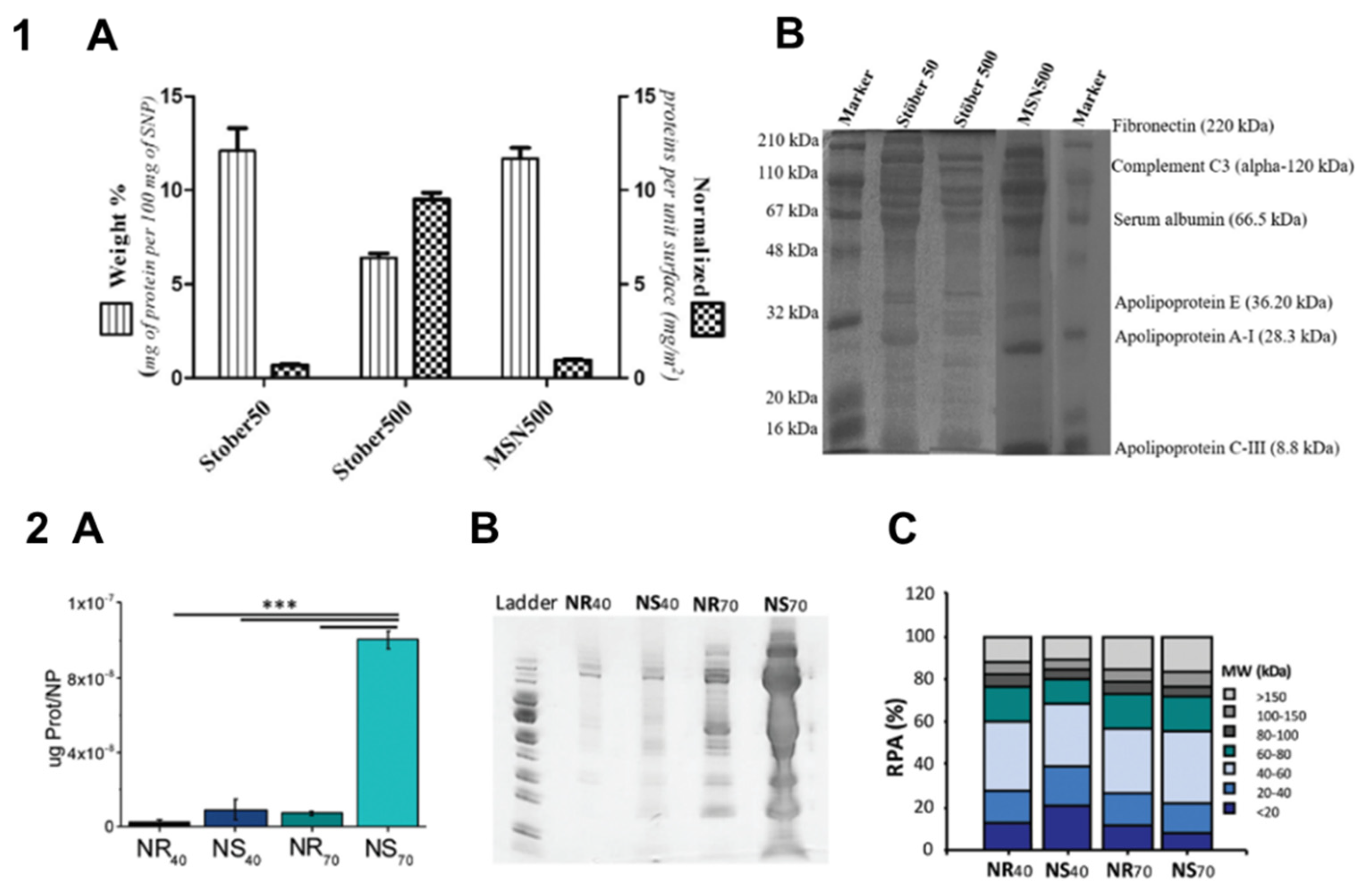
Figure 3.
Protein adsorption on particles with modified surfaces. Panel 1. Protein adsorption and cellular uptake of poly(ethyl ethylene phosphate) and polyethylene glycol nanocarriers. (A) Quantification of human plasma proteins adsorbed onto the nanocarrier surface (mg protein × m–2). The mean ± SD values of biological triplicates are shown; (B) flow cytometry analysis of RAW264.7 cells incubated with different nanocarriers for 2 h. The nanocarriers were incubated in human plasma or in water (–plasma) for 1 h at 37°C and separated from residual proteins by centrifugation before being added to cells. The mean ± SD values of biological triplicates are shown; (C) Classification of protein corona components identified by quantitative liquid chromatography–mass spectrometry. The average amount of each protein (in femtomoles) was calculated from three biological and two technical replicates. The identified proteins were grouped according to their function in biological processes, and the amounts of proteins were summed. Abbreviations: PS, polystyrene; PEG, poly(ethylene glycol); PEEP, poly(ethyl ethylene phosphate). Adapted with permission from Ref. [72]. Panel 2. Protein adsorption on, and interaction with cells of, zwitterion-coated microcapsules. (A) Fluorescence intensity of different microcapsules incubated with fluorescein isothiocyanate labelled bovine serum albumin (BSA–FITC); data are presented as the mean ± SD, n = 10; (B) the numbers of NIH/3T3 cells with 4′,6-diamidino-2-phenylindole (DAPI) staining adhered to different hydrogels; (C) the level of TNF-α secreted from macrophages after culturing in the presence of various microcapsules. (B, C) Data are presented as the mean ± SD, n = 3, p values were calculated using one-way ANOVA; **** p < 0.0001. Abbreviations: GelMA, gelatin methacrylate; CBMA, carboxybetaine methacrylate; MPC, phosphorylcholine methacrylate; SBMA, sulfobetaine methacrylate. Adapted with permission from Ref. [102].
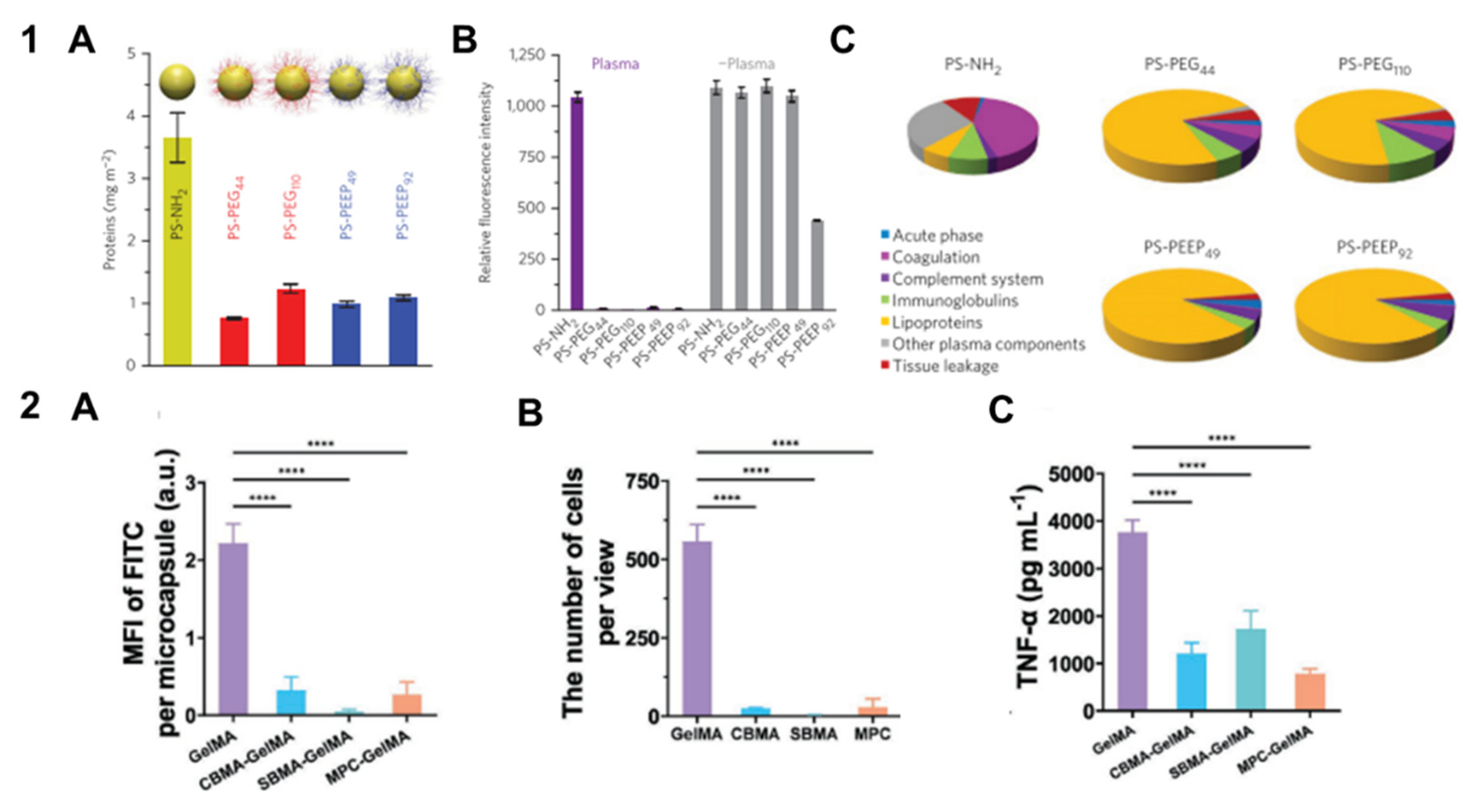
Figure 4.
Effect of protein precoating on the targeted delivery of metal–organic framework nanoparticles (PCN). (A) Gel electrophoresis of the PCN after 1-h incubation with 50% serum solutions and the resulting band intensities of PCN-adsorbed serum proteins; (B) flow cytometry analysis of the cellular uptake of DiI-loaded particles after incubation with SK-BR-3 and MDA-MB-468 cells in 50% serum media for 3 h; (C) changes in tumor volume for three weeks; ** p < 0.01, *** p < 0.001, Student’s t test; (D) Weight of tumors extracted from mice after treatment with different agents. Abbreviations: CPT, camptothecin; GSH, glutathione transferase; GST-H-PCN/GST-E-PCN, NPs coated with GSH-affibody (HER2/EGFR-targeting); PCN, metal–organic framework nanoparticles. Adapted with permission from Ref. [126].
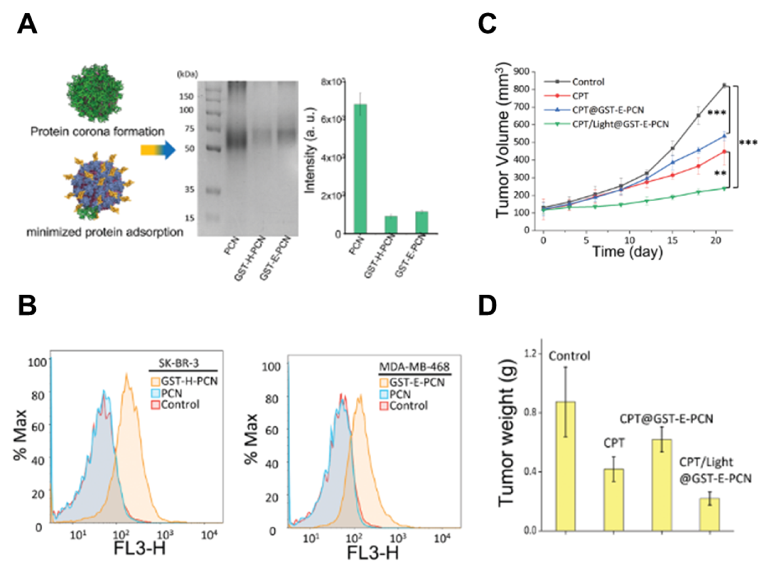

Table 1.
Effect of the morphological and physicochemical properties of nano- and microparticles on protein adsorption.
Table 1.
Effect of the morphological and physicochemical properties of nano- and microparticles on protein adsorption.
| Type of particles | Variable parameter(s) |
Incubation medium |
Main conclusions | Ref. |
|---|---|---|---|---|
| Nonporous silica particles, 50 and 500 nm; mesoporous silica particles, 500 nm | Size, porosity | FBS | The amount of adsorbed protein directly depends on the available surface area of particles. The protein profile is independent of surface area and porosity. | [12] |
| Silica particles with amidine surface modification, 30–1000 nm | Size | BSA and myoglobin solutions | Conformational changes of myoglobin and BSA upon adsorption are size-dependent. | [38] |
| Polystyrene particles, 50 and 100 nm; bare, carboxyl- and amine-modified | Size, surface chemistry | Human plasma | Size and surface chemistry significantly affect the composition of the protein corona. | [21] |
| Polystyrene microspheres (PS MSs), 1.33 and 4.46 μm, original and with etched surface | Size, surface morphology | Human plasma, BSA/fibrinogen solution | Protein adsorption is lower on etched PS MSs; the lowest adsorption is on PS MSs with the maximum number and minimum area of protuberances on the surface. This effect and total adsorption depend on size. MSs after etching (which changes the surface morphology) adsorb low-molecular-weight proteins; control MSs, a wider range of proteins. | [22] |
| Polystyrene particles, 100–500 nm | Size | HEWL solution | The particles form a protein corona of HEWL, mainly driven by hydrophobic interactions. 100-, 400-, and 500-nm particles promote HEWL to form amyloid fibrils. | [39] |
| Dense and mesoporous silica particles, 70–900 nm | Size, porosity | FBS | Both dense and porous particles with smaller size adsorb more protein; small dense particles adsorb proteins with lower Mw; all porous particles adsorb smaller proteins. | [34] |
| Elastin-like polypeptide (ELP)–based particles, 200 and 500 nm | Size, surface chemistry | Platelet-poor human plasma | All ELP constructs adsorb large amount of albumin, immunoglobulin G and activated complement factor 3; variations in the composition between different NPs are observed for plasminogen, fibronectin, activated fibrinogen, antithrombin and alpha2 macroglobulin. | [35] |
| LMW chitosan oligosaccharide (COS) NPs and HMW chitosan (CS) NPs, 130&150 and 95&110 nm, respectively | Size, composition | BSA and HSA solutions | CS NPs weakly interact with proteins because of the high structural rigidity of the polymer. The smaller CS NPs exhibit greater BSA binding. | [31] |
| Pyrolytic and colloidal silica particles, 20–120 and 50 nm, respectively | Size, surface chemistry | BSA solution | The difference in protein coverage appears to be related to differences in the distribution of surface silanols more than to differences in ζ-potential. | [56] |
| PS, PS-COOH, and PS-NH2 particles, 50 nm and 1 μm | Size, surface chemistry | Human saliva | Surface chemistry and size of the particles affect the adsorption of saliva proteins. Formation of the protein corona causes changes in the surface charge, aggregation and, hence, in vitro cytotoxicity. | [58] |
| PS particles, 50, 100, 200, and 500 nm and 1 μm; PS particles with the –COOH and –NH2 groups, 100 nm | Size, surface chemistry | Simulated salivary fluid, simulated gastric fluid, simulated intestinal fluid | The protein corona formed after in vitro digestion alters the macrophage uptake of uncharged 50- and 100-nm PS particles, but not charged 100-nm or larger particles. The presence of digestion proteins changes the adsorption of serum proteins from cell culture media. | [57] |
| Silica NPs, 8, 33, and 78 nm | Size | Yeast protein extract | Larger NPs adsorbed more protein per surface unit; most of the protein adsorbed on NPs with different size were identical. | [36] |
| PEGylated gold NPs, 20–150 nm | Size | HDL solution, human serum | Larger NPs exhibit a larger surface coverage with HDLs. This process is regulated by nonspecific interactions rather than adsorption of apolipoprotein A-I. | [37] |
| Solid lipid nanoparticles (SLNs), 120–480 nm | Size | BSA solution | BSA adsorption increases with the increasing size of the particles. | [25] |
| PEGylated plasmonic gold NPs (rods and stars), 40 and 70 nm | Size, shape | In vivo in CD-1 mice | Gold nanostars adsorb more protein than nanorods due to their larger surface area. The composition of the protein corona varies between NPs with different sizes and shapes. | [9] |
| Polystyrene NPs, 26, 80, and 200 nm | Size | Mouse serum | Protein corona formation is size-dependent. | [32] |
| Gold NPs modified with PEG, 21–109 nm | Size | FBS | Protein adsorption is greater on larger gold NPs. | [33] |
| Gold NPs (spheres, rods, stars, and cages) functionalized with R-PEG-SH (R= OCH3, COOH, or NH2), ∼50 nm | Shape, surface chemistry | Human serum | Cage-shaped NPs adsorb less protein and differ in the protein corona composition. | [10] |
| Mesoporous silica particles, spherical, faceted, and rod-shaped | Shape | Bovine serum | Considerable differences in the protein composition, surface coverage, and particle agglomeration of the protein corona-particle complex indicate specific protein adsorption profiles highly dependent on the number of exposed facets and aspect ratio. | [40] |
| Mesoporous silica particles: spherical, 270 nm; rod-shaped, 290×1100 nm | Shape | Serum, plasma | Rod-shaped particles adsorb more protein from serum and plasma. | [41] |
| Diethylaminoethyl-dextran cellulose microspheres, 100 μm; pore size, 165–282 nm | Pore size | BSA and γ-globulin solutions | The increasing-then-decreasing trends in the adsorption equilibria and uptake kinetics are observed as the pore size increases. A critical pore size at which the adsorption capacity and uptake are significantly enhanced has been determined. | [42] |
| Poly(methacrylic acid) (PMA) capsules and particles functionalized with mAbs | Structure | Human serum | The capsules and particles differ in the protein corona composition. The protein corona has little effect on the targeting ability of the mAb-functionalized capsules and particles. | [43] |
| Silica nanocapsules, 150–180 nm, with stiffnesses (Young moduli) of 704 kPa, 25 MPa, 459 MPa, and 9.7 GPa | Stiffness | FBS | The protein corona of the stiffest nanocapsules contains the largest amount of complement protein C3 and immunoglobulin proteins, which facilitates their macrophage uptake. | [50] |
| Hydrogel NPs with a lipid bilayer shell and unmodified or PEGylated surface, 100–150 nm; elasticity, 45 kPa–760 MPa | Stiffness | Mouse plasma | The protein corona composition varies nonmonotonically depending on the NP elasticity; the relative abundance of Apo-I is correlated with the blood clearance lifetime of the NPs. | [51] |
| Polymeric core/multishell, solid cationic and anionic NPs and polymeric soft nanogels (NGs), 75–175 nm | Surface chemistry | Human plasma | Solid anionic and cationic particles adsorb more protein than other particles; NPs with a protein corona more weakly interact with cells; NGs with a protein corona induce enhanced cytokine release | [20] |
| Core/shell and shell-only multilayered polyelectrolyte particles, 2 μm | Structure | Human serum, plasma | Differences in particle structure affect both the quantity of adsorbed proteins and the relative abundance of individual protein groups (apolipoproteins, complement proteins, and immunoglobulins). | [44] |
| PLGA-NPs, 152 nm; PLGA/cholesterol NPs, 237 nm; cholesterol NPs, 257 nm | Composition | Human plasma | Different protein corona compositions on particles with different matrix compositions in the presence or absence of targeting ligand has been shown. | [15] |
| MAA and PMMA polymeric beads | Composition, incubation medium | Serum, plasma | The two materials differ with respect to protein adsorption, which is associated with differences in complement activation. | [45] |
| Sulphated alginate, high G alginate, and poly-L-lysine-coated alginate microspheres, 565, 579, and 538 μm, respectively | Composition | Lepuridin-anticoagulated plasma | Differences in adsorption of complement and coagulation proteins can explain differences in the inflammatory and fibrotic responses to three types of alginate microspheres. | [46] |
| Lipid nanodiscoids, 47–63 nm | Composition | Mouse serum | The phospholipid composition affects the protein corona composition, which determines the particle stability, circulation, and biodistribution. | [26] |
| Elastomer (star-poly(D,L-lactide-co-e-caprolactone)) E2 and E5 microspheres, 184 and 370 μm | Composition | FBS, human plasma | Polymer chain flexibility affects protein adsorption in competitive adsorption environments. | [49] |
| Lipid NPs, 67–110 nm | Composition | Nude mice serum | Differences in lipid constituents altered composition of protein corona; NPs with apolipoprotein-rich corona showed higher delivery efficiency than NPs with vitronectin-rich corona. | [27] |
| PLGA and PCL particles, 416 and 559 nm | Composition | HSA, human serum | Serum proteins have a higher affinity to more hydrophobic (PCL) particles; the protein corona composition is material-specific. | [16] |
| Poly(methyl methacrylate-co-styrene) particles, ∼550 nm | Composition | BSA, IgG, and fibronectin solutions | Protein adsorption is enhanced on particles with a higher amount of styrene due to a higher hydrophobicity. | [19] |
| Albumin, cationic albumin, and ovalbumin cross-linked NPs, 165–206 nm | Composition | FBS | Protein-based NPs with various hydrophobicities and charges differ in the protein corona pattern; the protein corona affects in vitro macrophage uptake of BSA and cationic BSA NPs but not ovalbumin NPs. | [24] |
| Polystyrene NPs with –COOH, –NH2, –SO3, and –PO3 surface groups, ~100 nm | Surface chemistry | Human serum | High enrichment of apolipoproteins on NPs and preferential binding of apolipoproteins on NH2- and SO3- functionalized NPs has been found. | [23] |
| Poly(styrene) surfaces modified with –NH2, –COOH, and –PO3H2 | Surface chemistry | FBS | Protein adsorption varied between surfaces with different surface groups; this was related with differences in their binding by macrophages and inflammatory responses to them. | [52] |
| PLA and PLGA NPs, 188–224 nm | Surface chemistry, hydrophobicity, matrix polymer Mw, incubation medium | Human serum, plasma | The PLA matrix polymer Mw affects only the types of the bound proteins; adsorption of specific proteins depends on hydrophobicity; surface end group identity influences both the total amount and the profile of the adsorbed proteins. | [55] |
| Glycosylated polyhydroxy polymer-modified nanovesicles (CP-LVs), ~100 nm | Surface chemistry | Plasma, liver tissue interstitial fluid | CP-LVs with the highest amino-to-hydroxyl ratio are characterized by a decreased immunoglobulin adsorption and prolonged blood circulation; adsorption of tumor-specific proteins leads to effective cellular internalization. | [53] |
| Polyelectrolyte-doped block copolymer-stabilized NPs, 40–180 nm | Surface chemistry | BSA solution | NPs with an anionic (PAA) surface modification tend to adsorb less protein and are taken up only by macrophage-like cells, whereas cationic (DMAEMA) NPs are taken up by different types of cells. | [54] |
Abbreviations: BSA, bovine serum albumin; COS, chitosan oligosaccharide; CP-LVs, glycosylated polyhydroxy polymer-modified nanovesicles; CS, chitosan; DMAEMA, poly(N,N-dimethylaminoethyl methacrylate); ELP, elastin-like polypeptide; FBS, fetal bovine serum; HDL, high-density lipoprotein; HEWL, hen egg-white lysozyme; MAA, methacrylic acid co-methyl methacrylate; mAb, monoclonal antibody; NG, nanogel; NP, nanoparticle; PAA, poly(acrylic acid); PBS, phosphate-buffered saline; PCL, polycaprolactone; PEG, poly(ethylene glycol); PLA, poly(lactic acid); PLGA, poly(lactic-co-glycolic) acid; PMA, poly(methacrylic acid); PMMA, polymethyl methacrylate; PS, polystyrene; SLNs, solid lipid nanoparticles.
Table 2.
Surface modifications of nano- and microparticles and their effect on protein adsorption.
| Type of particles | Modification | Incubation medium |
Main conclusions | Ref. |
|---|---|---|---|---|
| Ovalbumin NCs, 250–300 nm | PEG | Human plasma | Conformation of PEG affects the adsorption of specific proteins (dysopsonins) on NCs and their uptake by phagocytic cells. | [71] |
| Polystyrene NPs, 90–100 nm | Lutensol AT50 (PEG-based surfactant), SDS | HSA solution | The NPs coated with Lutensol adsorb more protein than those coated with SDS; however, the loss of conformation is less pronounced in PEGylated (Lutensol-modified) NPs. | [69] |
| Phosphonic acid terminated poly(ethylene glycol)-conjugated layered double hydroxide (PEG-LDH) NPs, 100–170 nm | PEG | BSA solution | PEGylation of LDH particles enhances their stability, reduces BSA adsorption, and enhanced cellular uptake. | [60] |
| PEGylated gold NPs (GNPs), 125–214 nm | PEG (350, 550, 1000 Da) | Human plasma | GNPs modified with PEG 550 adsorb more albumin and transferrin, which correlates with higher uptake by, and cytotoxicity for, HepG2 cells and a higher antitumor effect in mice. | [74] |
| Mesoporous silica particles (MSNs), 980 nm | PEG (2000, 5000, 10000 Da) | BSA solution | The depth of BSA penetration into MSNs in higher than that of MSNs-PEG; the maximum penetration depth decreases with increasing PEG chain length. | [61] |
| Mesoporous silica (MS), MS-PEG, and PEG particles, 100, 450, and 800 nm | PEG | Human plasma | The rate of immune cell association of MS-PEG particles with protein coronas formed from plasmas of individual donors widely varies depending on the donor; the cell association of all PEG particles is minor irrespective of the protein corona source. | [76] |
| Mesoporous silica NPs (MSNs), 210–250 nm | PEG | 10% FCS in PBS | Method of PEGylation affects the NP pore size and the amount of adsorbed proteins. | [63] |
| Gold NPs (AuNPs), 18–78 nm | PEG (5000, 10000, 30000 Da) | BSA, GB3 (WT and K19C), and H1.5 peptide (with and without cysteine) solutions | The amount of adsorbed peptides/proteins decreases with decreasing PEG size; small molecules (glutathione, H1.5 peptide, GB3) can penetrate PEG layers: the presence of thiol groups enhances the adsorption on PEGylated AuNPs. | [73] |
| Strontium sulphite NPs (SSNs), 670–2100 nm | PEG | 10% mouse plasma | PEGylation of SSNs leads to reduction of protein adsorption from serum, reduction of cytotoxicity, and efficient tumor delivery. | [64] |
| PLGA and PLGA-PEG NPs, 100 and 200 nm | PEG | FBS | PEGylation significantly decreases the amount of bound proteins. | [59] |
| SiO2, SiO2-sulfobetaine silane (SBS), and SiO2-PEG2000 NPs, ~100 nm | Zwitterion, PEG | BSA and lysozyme solutions | Functionalization of SiO2 NPs with PEG and SBS prevents BSA adsorption but not lysozyme adsorption, although the latter is reduced, with no particle aggregation. | [62] |
| Polystyrene particles with –NH2, –PEG, or poly(ethyl ethylene phosphate) (PEEP) groups, 100–120 nm | PEG, PEEP | Human plasma | In addition to reducing protein adsorption, PEG and PEEP can affect the composition of the protein corona, and the presence of specific proteins is necessary to prevent nonspecific cellular uptake. | [72] |
| Alginate/chitosan/alginate (ACA) hydrogel microcapsules modified with chitosan-methoxy poly-(ethylene glycol) (CS-g-MPEG), 350 μm | MPEG | IgG solution | Adsorption of IgG is drastically reduced on microcapsules coated with CS-g-MPEG. The protein repulsion capacity depends on the degree of substitution and chain length of MPEG. | [70] |
| Hydroxyethyl starch NPs functionalized with PEG and modified with mannose | PEG, PEG–mannose | Human plasma | The PEG and PEG–mannose modifications of HES NPs reduces the protein adsorption but do not affect the protein corona composition. HES–PEG-mannose NPs are efficiently bound by dendritic cells. | [65] |
| Hydroxyethyl starch NPs functionalized with PEG | PEG | Human serum | PEGylation of HES NPs reduces the protein adsorption from serum by 37%. | [66] |
| Gold NPs functionalized with fluorescent Herceptin and backfilled with methoxy-terminated PEG (mPEG) , 73–97 nm | PEG (1000, 2000, 5000, 10000 Da) | Human serum | NPs with attached PEG groups adsorb less proteins in the presence of serum but retained the targeting specificity. | [67] |
| Iron oxide nanoparticles (IONPs) coated with polyethylene glycol-allyl glycidyl ether copolymer functionalized with RGD peptide or transferrin, 22–31 nm | PEG-b-AGE | FBS, human plasma | Coating of the NPs, with PEG-b-AGE leads to a decrease in protein adsorption and nonspecific uptake by macrophages of NPs, irrespective of functionalization. | [68] |
| PEGylated liposomal drug (Onyvide), 60 nm | PEG | Human plasma | PEGylation of liposomes does not prevent the formation of the protein corona; adsorbed proteins from plasma enhance binding to PANC-1 cells. | [77] |
| Gold NPs modified with PEG, 21–109 nm | PEG | FBS | PEG modification reduces plasma protein adsorption. | [33] |
| Liposomes functionalized with PEG or hyperbranched polyglycerol, 60–90 nm | PEG, PG | Human plasma | Protein adsorption on unmodified and PEG- and PG-modified liposomes is relatively low; macrophage uptake of liposomes is not affected by the protein corona; modification with PEG reduces cellular uptake, whereas modification with PG enhances it. | [82] |
| Nanodiamonds (NDs), 30, 50, and 100 nm; SPIONs, 22 nm | PEG, PG | FBS, human plasma | PG modification more efficiently reduces the protein adsorption and macrophage uptake than PEG modification. | [81] |
| Polystyrene NPs modified with poly(phosphoester) surfactants, ∼110 nm | PPE | HSA, plasma | Noncovalent modification of PS NPs with poly(phosphoester) surfactants reduces the protein adsorption and macrophage uptake after incubation in plasma. | [83] |
| Poly(organosiloxane) NPs functionalized with PEG or poly(2-ethyl-2-oxazoline), 22–26 nm | PEG, PEtOx | Bovine serum | Modification of the NPs with either PEG or PEtOx reduces the protein adsorption; modification with PEtOx reduces the cellular uptake in both serum and serum-free medium. | [84] |
| Biotin- and cysteine-coated silica NPs, 120 nm | Zwitterion (amino acid) | 10, 50, 100% human plasma | Zwitterionic (cysteine) coating interferes with the protein corona formation and improves the NP targeting ability. | [88] |
| Silica NPs functionalized with lysine/arginine/glycine/cysteine/phenylalanine, ∼60 nm | Zwitterions (amino acids) | BSA, 10% FBS | Functionalization of NPs with cysteine, lysine, and arginine reduces protein adsorption, the effect of lysine being the strongest. | [89] |
| Gold NPs conjugated with zwitterionic peptides or PEG, 50–100 nm | Zwitterions (peptides) | Human serum | Protein adsorption is reduced up to 60% compared with bare NPs; the protein adsorption profile depends on the charge motif and sequence but not amino acid composition. | [90] |
| Silica NPs (SiO2-organosiloxane), ∼120 nm | Zwitterion | BSA and lysozyme solutions, 50% FBS | Zwitterated silica NPs display antifouling properties in both BSA and lysozyme solutions, as well as in FBS. | [91] |
| Bi-functionalized silica NPs (SiO2- organosiloxane/COOH), ∼140 nm | Zwitterion | BSA and lysozyme solutions, 50% FBS | SiO2-organosiloxane/COOH5/1 particles absorb less protein while retaining the capacity for effectively binding target biomolecules. | [92] |
| Sulfobetaine-coated silica NPs, 250 nm | Zwitterion | BSA solution, FBS | Zwitterion-modification of silica NPs results in a 90% decrease in protein adsorption rate and a reduced NP uptake by macrophages and fibroblasts. | [95] |
| Polystyrene/Fe3O4/silica NPs functionalized with amino/alkene/betaine, 230–440 nm | Zwitterion | BSA and lysozyme solutions , FBS | Betaine functionalization reduces the protein adsorption from single-protein solutions and serum, with amino and alkene groups remaining accessible for interaction with biomolecules. | [93] |
| PEI-coated core/shell Fe3O4@SiO2 NPs functionalized with 2-methacryloyloxyethyl phosphorylcholine (MPC), ∼125 nm | Zwitterion | BSA solution, FBS | The protein corona formation in in 50-MPC- and 75-MPC-functionalized silica NPs is reduced in BSA solution and FBS. | [99] |
| Silica NPs functionalized with zwitterion and amino/mercapto/carboxylic groups, ∼100 nm | Zwitterion | BSA solution | Double-functionalized NPs exhibited a low protein adsorption level, with biologically active groups shielded from interactions. | [94] |
| Nanodiamonds (NDs) functionalized with zwitterion and tetraethylene glycol (TEG), ∼100 nm | Zwitterion | FBS | Combined surface functionalization of NDs with TEG chains and zwitterionic head groups ensures colloidal stability and prevents protein adsorption. | [108] |
| Silica NPs functionalized with carboxybetaine, 40 nm | Zwitterion | BSA solution | Protein binding to functionalized NPs is significantly reduced (by up to 94%). | [97] |
| Hollow mesoporous Prussian blue NPs (HMPBs) functionalized with sulfobetaine and peptide E5, 130 nm | Zwitterion | BSA solution | HMPBs@PEI-ZW and HMPBs@PEI-ZW-E5 NPs adsorb noticeably less BSA than the original NPs. | [96] |
| Poly(DMAEMA-co-carboxybetaine methacrylate)-modified silica NPs (MCB), 137–220 nm | Zwitterionic copolymer | BSA and lysozyme solutions | Modification of silica NPs using carboxybetaine copolymer with different quaternization degrees modulates the adsorption of basic and acidic proteins. | [103] |
| Poly(ε-caprolactone)-b-poly(N,N-diethylaminoethyl methacrylate)/(N-(3- sulfopropyl-N-methacryloxyethy-N,N-diethylammonium betaine)) (PCL-PDEAPS) and poly(ε-caprolactone)-b-poly(ethylene glycol) (PCL-PEG) micelles, 77–109 and 51 nm, respectively |
Zwitterionic polymers, PEG | BSA, lysozyme, and fibrinogen solurions; plasma | PCL-PDEAPS micelles adsorb more negatively charged proteins (BSA, fibrinogen) than PCL-PEG micelles due to electrostatic interactions; PCL-PDEAMS micelle uptake by macrophages after protein adsorption is low. | [107] |
| Magnetic gold NPs functionalized with poly(carboxybetaine methacrylate) and poly 2-carboxy- N,N-dimethyl-N-(2′-(methacryloyloxy)ethyl)ethanaminium), 123 nm | Zwitterion | HSA solution, serum | Magnetic gold NPs with CBMA coating adsorb significantly less protein than control NPs coated with PEG, PAH, or PEI. | [98] |
| Gold NPs coated with thiol-functionalized polymethacryloyloxyethyl phosphorylcholine polymers (pMPC), 230 nm | Zwitterion | Human serum, BALF | pMPC-coated gold NPs exhibit reduced protein adsorption and increased uptake by A549 cells after exposure to serum. | [100] |
| Core/shell GelMA microcapsules; zwitterionic coated CBMA-GelMA, SBMA-GelMA, and MPC-GelMA microcapsules, ~600 μm | Zwitterionic polymers | FITC-BSA in PBS | Zwitterionic coating effectively block nonspecific protein adsorption. | [102] |
| Poly(glycidyl methacrylate) (PGMA) microspheres modified with [3-(methacryloylamino)propyl]dimethyl(3-sulfopropyl)ammonium hydroxide (MPDSAH), 2 μm | Zwitterionic polymers | BSA solution | The amount of adsorbed BSA reduced up to half of that on the unmodified PGMA microspheres. | [101] |
| Silica particles with poly(2-methacryloyloxyethyl phosphorylcholine) coating | Zwitterionic polymers | HSA solution | Coating with zwitterion prevents the adsorption of tightly bound proteins, but a “soft” protein corona forms on the particles. | [106] |
| Mesoporous silica particles, poly(2-methacryloyloxyethylphosphorylcholine) (PMPC)-coated silica hybrid particles, PMPC replica particles, 164, 161, and 179 nm, respectively | Zwitterionic polymers | Human serum, human blood | Mesoporous silica particles adsorb more proteins of a wider range of molecular weights than PMPC-coated and PMPC replica particles. | [104] |
| Mesoporous silica particles and poly(2-methacryloyloxyethyl phosphorylcholine) (PMPC) particles, with or without coating with HSA, IgG, and C1q, ∼1 µm | Zwitterionic polymers, proteins | Human plasma, whole blood, washed blood | PMPC particles exhibit reduced adsorption of serum proteins and association with cells. The enrichment of the corona with some proteins facilitates association with specific cell types. | [105] |
| Poly(acrylic acid)-block-polystyrene (PAA22-b-PS144) polymersomes, 60 nm | BSA, IgG, lysozyme | – | The formation of protein coronas from all model proteins used reduces the cytotoxicity of the polymersomes | [110] |
| Poly-3-hydroxybutyrate-co-3-hydroxyhexanoate (PHBHHx) NPs, ~200 nm | BSA | Rat serum | Preformed albumin corona inhibits the plasma proteins adsorption, reduces the complement activation, prolongs the blood circulation time, and reduces the toxicity of the PHBHHx NPs. | [109] |
| PS particles with –COOH and –NH2 groups or without functionalization, 58–69 nm | Human plasma fractions (IgG, HSA, and low-abundant protein) | Human plasma | Pre-incubation with IgG-, HSA-, and low-abundant-protein-enriched plasma fractions alters cellular uptake and inhibits aggregation of PS and PS-NH2 particles in human plasma. | [130] |
| Supramolecular template NPs, metal–phenolic network (MPN)-coated template (core/shell) NPs, and MPN nanocapsules, ~100 nm | BSA, FBS, bovine serum | Human blood | Precoating using FBS effectively reduces the amount of adsorbed proteins and alters the composition of the protein corona, which reduces NP association with leukocytes. | [116] |
| PS particles with –COOH and –NH2 groups, 150–230 nm; hydroxyethyl starch (HES) NPs, 450 nm | Antibodies | Human serum or plasma | NPs with pre-adsorbed antibodies retain the targeting ability after the formation of the biomolecular corona. | [121] |
| PS particles with –COOH and –NH2 groups, 130–150 nm | IgG-depleted plasma | Cell culture medium containing human plasma | Precoating of NPs with Ig-depleted plasma reduces their cellular uptake; the preformed protein corona remains stable after the reintroducing to plasma. | [131] |
| Starch-coated poly(methyl methacrylate-co-acrylic acid) nanoparticles, 85 nm | Casein, soybean protein isolate, myoglobin, zein, gelatin | 30% FBS | NPs precoated with casein exhibit antifouling properties and excellent targeting in vitro and in vivo. | [120] |
| PS particles, 0.2 and 3 μm | BSA, myoglobin, β-lactoglobulin, lysozyme, fibrinogen | 10% FCS in cell culture medium | Precoating of particles strongly affects the protein corona formation; primary proteins still can be detected after the second incubation. Precoating affects particle–cell interactions, but not the particle toxicity. | [119] |
| Carboxylic acid-functionalized silica NPs, ~100 nm | γ-globulin, HSA | 10 or 55% human plasma in PBS | Precoating of the NPs with γ-globulins, but not HSA, leads to enrichment of the protein corona with immunoglobulins and complement factors, but the opsonin-rich corona does not enhance NP uptake by macrophages. | [118] |
| Polylactic acid (PLA)-PEG-poly(ε-caprolactone) (PCL) NPs, 118 and 189 nm | Antibody | Mouse serum or human serum | Complement proteins are adsorbed on the NP surface. NPs bearing the targeting antibody activate the classical immune pathway; untargeted NPs, the alternative pathway. | [122] |
| Layered double hydroxide (LDH) NPs, ~100 nm | BSA | Cell culture medium | BSA precoating stabilizes LDH suspensions in cell culture medium and enhances the NP uptake by CHO cells. | [112] |
| Gelatin–oleic nanoparticles (GONs) | BSA | Cell culture medium containing FBS | Uptake of GONs precoated with BSA by HEK293 cells is enhanced, but their uptake by A549 cells is suppressed in a FBS-containing cell medium | [111] |
| Chitosan-based nanocarriers (TsR NPs) loaded with siVEGF, 500–1500 nm | BSA-cRGD | 10% FBS | Adsorption of BSA-cRGD on TsR NPs results in a reduced serum protein adsorption and improves tumor targeting and therapeutic efficiency. | [113] |
| siVEGF-loaded chitosan-based NPs (CsR/siVEGF NPs), 150–1000 nm | BSA-cRGD | 10% mouse serum, 10% FBS | Precoating of CsR/siVEGF NPs with BSA-cRGD enhances their stability in serum and tumor targeting efficiency, which is related to reduced protein adsorption from serum and changes in the protein corona composition. | [114] |
| Polystyrene NPs coated with PEG, BSA, chitosan, and cell membranes from a human breast cancer cell line, 110–150 nm | PEG, BSA, chitosan, cell membranes | 10% FBS | All the tested coatings reduce protein adsorption, the effects of the BSA and cell membrane coatings being the strongest. | [117] |
| PLGA and chitosan–PLGA microparticles, ∼1.6 μm | HSA | Human plasma | HSA coating changes the protein profile of the corona and enhances binding to endothelial cells. | [115] |
| Zr6-based metal–organic framework NPs (PCN-224), 180–260 nm | GST-Afb | 50% serum | Precoating of PCN-224 with a protein layer reduces the protein adsorption on NPs and improved cancer cell and tumor targeting. | [126] |
| Metal–organic frameworks particles (MOF-808) loaded with camptothecin, 90–120 nm | GST-Afb and collagenase | 50% serum | Formation of a protein layer consisting of GST-Afb and collagenase reduces protein adsorption from serum and provides efficient tissue penetration and tumor targeting. | [127] |
| DOTAP/DNA lipid particles, ∼220 nm | Human plasma | – | Spontaneous coating with vitronectin promotes the uptake by cancer cells overexpressing vitronectin the ανβ3 integrin receptor. | [128] |
| Silica NPs, 110 nm | BSA, HSA, IgG, complement factor H, fibrinogen | Human serum | Coating with BSA, HSA, and fibrinogen reduces the adsorption of complement proteins on the NP surface; coating with complement factor H inhibits complement activation by the NPs. | [123] |
| Polystyrene NPs, 50 nm; HES NPs, 110 nm | Clusterin (apolipoprotein J) | IgG-enriched human plasma | Precoating of both types of NPs with clusterin reduces IgG adsorption from IgG-enriched plasma and suppresses cellular uptake. | [129] |
| Polystyrene NPs (PS-COOH), 100 nm | Apolipoproteins ApoA4, ApoC3, and ApoH; prothrombin; antithrombin III | – | Coating with ApoA4 and ApoC3 reduces the NP uptake by hMSCs, whereas coating with ApoH enhances the uptake. Prothrombin and antithrombin III do not affect the cellular uptake significantly. | [23] |
Abbreviations: BSA, bovine serum albumin; CBMA, carboxybetaine methacrylate; cRGD, cyclo(Arg-Gly-Asp-d-Tyr-Lys-) peptide; CS, chitosan; DMAEMA, poly(N,N-dimethylaminoethyl methacrylate); DOTAP, 1,2-dioleoyl-3- trimethylammonium propane; FBS, fetal bovine serum; FCS, fetal calf serum; FITC, fluorescein isothiocyanate; GB3, third IgG-binding domain of protein G; GNPs, gold nanoparticles; GONs, gelatin–oleic nanoparticles; GST-Afb, glutathione transferase–affibody; HES, hydroxyethyl starch; HMPBs, hollow mesoporous Prussian blue nanoparticles; HSA, human serum albumin; IONPs, iron oxide nanoparticles; LDH, layered double hydroxide; MPC, 2-methacryloyloxylethyl phosphorylcholine; mPEG, methoxy poly(ethylene glycol); MPN, metal–phenolic network; MSNs, mesoporous silica nanoparticles; NC, nanocarrier; ND, nanodiamond; NP, nanoparticle; PCL, polycaprolactone; PDEAPS, N-(3-sulfopropyl-N-methacryloxyethy-N,N-diethylammonium betaine); PEEP, poly(ethyl ethylene phosphate); PEG, poly(ethylene glycol); PEG-b-AGE, poly(ethylene glycol) and allyl glycidyl ether block copolymer; PEtOx, poly(2-ethyl-2-oxazoline); PG, poly(glycerol); PGMA, poly(glycidyl methacrylate); PHBHHx, poly-3-hydroxybutyrate-co-3-hydroxyhexanoate; PLA, poly(lactic acid); PLGA, poly(lactic-co-glycolic) acid; PMPDSAH, poly{ [3-(methacryloylamino)propyl]dimethyl(3-sulfopropyl)-ammonium hydroxide}; PPE, poly(phosphoester); PS, polystyrene; SBMA, sulfobetaine methacrylate; SBS, sulfobetaine silane; SDS, sodium dodecyl sulphate; SDS-PAGE, sodium dodecyl sulphate–polyacrylamide gel electrophoresis; siVEGF, small interfering RNA targeting vascular endothelial growth factor; SPIONs, superparamagnetic iron oxide nanoparticles; TEG, tetraethylene glycol; ZW, zwitterion.
Disclaimer/Publisher’s Note: The statements, opinions and data contained in all publications are solely those of the individual author(s) and contributor(s) and not of MDPI and/or the editor(s). MDPI and/or the editor(s) disclaim responsibility for any injury to people or property resulting from any ideas, methods, instructions or products referred to in the content. |
© 2025 by the authors. Licensee MDPI, Basel, Switzerland. This article is an open access article distributed under the terms and conditions of the Creative Commons Attribution (CC BY) license (http://creativecommons.org/licenses/by/4.0/).
Copyright: This open access article is published under a Creative Commons CC BY 4.0 license, which permit the free download, distribution, and reuse, provided that the author and preprint are cited in any reuse.


