Preprint
Article

This version is not peer-reviewed.

Re-Evaluation of ELISA for the Detection of Bovine Tuberculosis and a New Proposal for Its Use in Eradication Efforts on Outbreak Farms

A peer-reviewed article of this preprint also exists.

Submitted:

16 March 2025

Posted:

17 March 2025

You are already at the latest version

Abstract

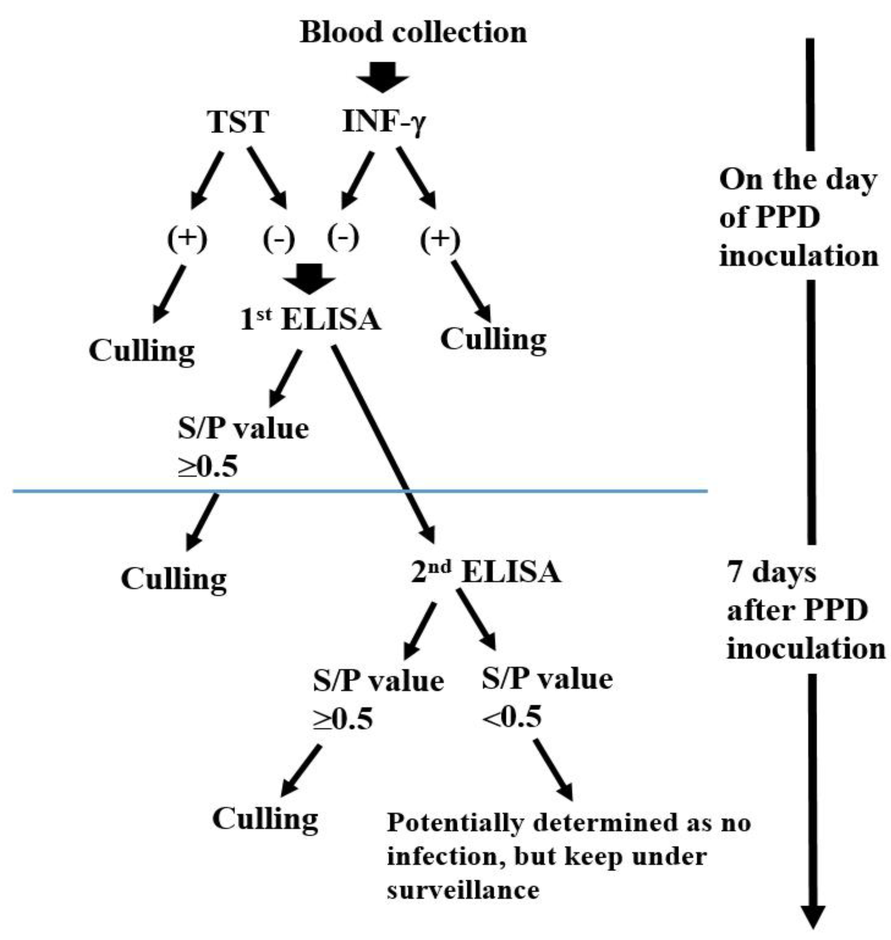
Bovine tuberculosis (bTB) is a zoonotic infectious disease and a chronic wasting illness. However, detecting and eradicating bTB remains a significant challenge in South Korea. This study evalu-ated the efficacy of a modified enzyme-linked immunosorbent assay (ELISA) protocol for de-tecting bTB in cattle. The protocol included two ELISA tests: one performed on the day of puri-fied protein derivative (PPD) inoculation and another seven days post-inoculation. Results showed a significant increase in ELISA detection rates, from 11% to 76%, particularly in cattle that tested positive for the tuberculin skin test (TST) and/or interferon-gamma (IFN-γ) assays (p < 0.0001). Notably, some cattle that were negative or had doubtful results in TST and IFN-γ as-says transitioned to ELISA-positive post-PPD inoculation. Additionally, some cattle identified as positive only by ELISA (S/P value ≥ 0.3) were confirmed to have bTB through gross examination or real-time reverse transcription polymerase chain reaction (rRT-PCR). The proposed protocol was validated in bTB outbreak farms using S/P thresholds of 0.3 (PPD day) and 0.5 (seven days post-PPD), enabling the detection of infected cattle missed by TST and IFN-γ assays. Implement-ing this approach successfully eradicated bTB in outbreak farms with minimal culling. These findings highlight the potential of incorporating sequential ELISA tests to enhance bTB detection and support eradication efforts.

Keywords: 
;  ;  ;  

1. Introduction

Bovine tuberculosis (bTB) is a zoonotic infectious disease caused by Mycobacterium bovis. Severe clinical symptoms of bTB include fever, weakness, weight loss, enlarged lymph nodes, anemia, and reduced milk yield. However, most bTB cases remain asymptomatic, necessitating diagnostic testing for confirmation. Farms experiencing bTB outbreaks have struggled with this disease for several decades. The most widely used diagnostic test for bTB detection in farms has been the tuberculin skin test (TST). In 2013, countries with advanced livestock management systems, such as the United Kingdom and Australia, introduced the interferon-gamma (IFN-γ) assay due to its high sensitivity and specificity. Both TST and IFN-γ assays detect bTB based on cellular immune responses, making them effective in identifying infections in early and middle stages [1,2].
However, their utility is limited in detecting late-stage bTB infections, where false-negative results can occur due to anergy induced by immunosuppressive conditions or drugs. Since 2013, South Korea has implemented the IFN-γ assay alongside the TST to enhance the efficiency of bTB testing. An epidemiological study conducted by the Korean Animal and Plant Quarantine Agency (QIA) analyzed 1,260 farms affected by tuberculosis outbreaks between 2012 and 2017. The study revealed that 24.4% (308/1,260) of these farms experienced three or more tuberculosis outbreaks, and 7.6% (96/1,260) of farms previously declared tuberculosis-free faced reinfection. Furthermore, cattle over 36 months old accounted for 49.1% of the total bTB-positive cases. These older cattle often display chronic disease symptoms, contributing to recurrent farm outbreaks [3]. As outlined above, bTB is a chronic disease, and despite detecting infected animals, outbreaks can recur years later due to latent, false-negative carriers. The enzyme-linked immunosorbent assay (ELISA) presents a promising alternative, as it is based on humoral immune response detection, making it particularly effective for late-stage bTB infections. Additionally, ELISA can detect anergic cattle suffering from immunosuppressive conditions, where TST and IFN-γ assays may fail [4,5,6]. While ELISA was temporarily implemented between 2009 and 2013, it was eventually replaced by IFN-γ assays due to low agreement with TST and poor detection rates [7,8,9,10].
World Organisation for Animal Health(WOAH) Manual of Diagnostic Tests and Vaccines for Terrestrial Animals, ELISA is particularly useful in diagnosing anergic T cells, and ELISA methods utilizing tuberculosis-specific antigens (e.g., MPB70 and MPB83) have demonstrated high specificity [11].
Furthermore, previous studies reported a significant increase in bTB detection rates when ELISA was performed post-TST in bTB-positive cattle [12,13,14]. Given the challenges associated with current bTB diagnostic methods, this study aims to comprehensively evaluate the role of ELISA in bTB detection and eradication. Specifically, we seek to investigate the underlying reasons for ELISA’s initially lower detection rates in comparison to TST and IFN-γ assays, as well as to assess its diagnostic agreement with these conventional tests. Furthermore, we aim to examine changes in ELISA results before and after purified protein derivative (PPD) inoculation in bTB-positive cattle to determine whether this approach enhances detection sensitivity.
Based on these findings, we propose a novel ELISA-based diagnostic protocol that can improve bTB eradication efforts in outbreak farms, facilitating more effective disease control with minimal culling.

2. Materials and Methods

2.1. Diagnostic Procedures Section

2.1.1. Tuberculin Skin Test (TST)

According to the product manual, the PPD solution (a purified protein derivative antigen, BoviShot®, CAVAC, South Korea) for TST was intradermally injected into the tail of the cattle. The skin-fold thickness at the inoculation site was measured between 48 and 72 hours after PPD inoculation. The difference in skin-fold thickness was interpreted as follows: The difference in skin-fold thickness was considered positive when it was 5 mm or more, doubtful if it was between 3 mm and 5 mm, and negative when it was less than 3 mm.

2.1.2. Interferon-Gamma Assay ( IFN-γ)

The whole blood was collected into heparinized tubes and stored at room temperature, with cultivation initiated within 12 hours. It was aliquoted into a 24-well cell culture plate, and PBS, PPD-A, and PPD-B antigen stimulants were added to each well. The plate was then incubated at 37°C for 16–24 hours.
After incubation, the samples were centrifuged, and plasma was collected by separating the supernatant. Following the manufacturer’s protocol (TB-Feron®, BIONOTE, South Korea), the plasma was mixed with sample diluent and aliquoted into an antibody adsorption plate. Positive and negative controls were included, and the plate was incubated at 37°C. Next, amplification and conjugate solutions were added, followed by washing and substrate addition. Finally, 100 µL of stop solution was added, and absorbance was measured at 450 nm. The results were interpreted based on the response to the stimulating antigens: A positive result was determined when the difference in absorbance between the PPD-B stimulated plasma and the negative control plasma was ≥0.1, and the difference between PPD-B and PPD-A was also ≥0.1. A negative result was indicated if the absorbance differences were below these thresholds.

2.1.3. ELISA

The BTB Ab ELISA 2.0 kit (BTB Ab ELISA 2.0®, BIONOTE, South Korea) was used to detect bovine tuberculosis (bTB) antibodies. The test was performed following the product manual. First, 50 µL of test samples (bovine serum), positive controls, and negative controls were added to each well. Then, 50 µL of enzyme conjugate was added to each well and incubated at 37°C for one hour. Each well was washed six times with 350 µL of diluted washing solution. After washing, 100 µL of substrate solution was added to each well and incubated at room temperature (15–25°C) for 15 minutes in the dark. Finally, 100 µL of stop solution was added, and absorbance was measured using a spectrophotometer at 450 nm with a reference wavelength of 620 nm.
The ELISA antibody results, expressed as the Sample/Positive ratio (S/P value), were interpreted as follows: values below 0.3 were considered negative, values between 0.3 and 0.5 were considered doubtful, and values above 0.5 were considered positive.

2.1.4. Tissue Sample Collection & rRT- PCR(Real-Time Reverse Transcription Polymerase Chain Reaction)

Tissue samples suspected of Mycobacterium bovis infection, including pharyngeal and pulmonary mediastinal lymph nodes, were collected postmortem. Five to ten grams of each tissue sample were placed into sterile stainless steel containers and cut into small pieces using sterile scissors. The tissue was then homogenized in 10 mL of phosphate-buffered saline (PBS) using a homogenizer. The homogenized tissue was transferred to centrifuge tubes, and an equal volume of 10% oxalic acid was added for decontamination. The mixture was incubated at room temperature for 10 minutes. Following incubation, the samples were filtered sequentially using 70 µm and 40 µm cell strainers. The filtered samples were centrifuged at 3,000 × g for 10 minutes. The supernatant was discarded, and the pellet was resuspended in 2 mL of PBS and vortexed vigorously.
DNA was extracted using the Viral Gene-spin™ Viral DNA/RNA Extraction Kit (iNtron, South Korea) according to the manufacturer’s protocol. The extracted DNA was used as the template for rRT-PCR. rRT-PCR was conducted using an Applied Biosystems™ 7500 Real-Time PCR device (Thermo Fisher®, USA) with the PowerChek™ Bovine Tuberculosis Real-Time PCR Kit (KogenBio®, South Korea). The PCR amplification conditions were as follows: The PCR amplification conditions were as follows: an initial cycle at 50°C for 2 minutes, followed by a denaturation step at 95°C for 10 minutes, and 45 cycles of 95°C for 15 seconds and 60°C for 1 minute.

2.1.5. Statistical Analyses

SAS (SAS Institute Inc®, United States) was used to compare the concordance rates of the three diagnostic methods and to assess the differences between groups before and after PPD vaccination. The agreement between the three diagnostic methods was measured using the Kappa statistic. The bTB-positive cattle detected by TST and IFN-γ assays were divided into three groups, and a paired t-test was conducted to analyze differences in antibody value changes over time between the PPD inoculation day and seven days later.

2.2. Experimental Design

2.2.1. Surveillance Period and Target Farms

From 2013 to 2017, three diagnostic tests (TST, IFN-γ, ELISA) were performed on 2,355 cattle from 32 farms experiencing bTB outbreak in Gangwon (Wonju, Hoengseong, Hongcheon). The detection rates and diagnostic agreement of the three tests were evaluated, and statistical analysis was performed to assess the correlation between diagnostic methods and the differences in ELISA results over time before and after PPD inoculation.

2.2.2. Selection of the ELISA-Only Positive Cattle in a bTB Outbreak Farm and PCR Confirmation

In 2013, three diagnostic methods—Tuberculin Skin Test (TST), IFN-γ assay, and ELISA—were performed on a farm in Ulju County, Gyeongsangnam-do, which housed 700 cattle (200 dairy cattle and 500 Hanwoo). This farm has reported a total of 200 cases of bovine tuberculosis over the past 10 years; however, it is not included among the 32 farms experiencing tuberculosis outbreaks in Gangwon-do as mentioned in section 2-1 above. Lesion tissue samples (pharyngeal lymph node, pulmonary mediastinal lymph node) were collected from the ELISA-positive cattle for further rRT-PCR testing.

2.2.3. Change of the ELISA Results 7 Days After PPD in TST and/or IFN-γ Positive Cattle

The study included 111 cattle selected from 165 cattle that had previously tested positive for tuberculosis using TST and/or the IFN-γ assay. ELISA results were obtained for these 111 cattle both before and after PPD inoculation to assess ELISA s/p value changes. ELISA test was performed on the day of PPD inoculation (first sample) and repeated seven days later (second sample). Blood samples were collected on both days to analyze changes in antibody levels, comparing the pre- and post-inoculation ELISA results.

2.2.4. Changes in the ELISA Antibody Level 7 Days After PPD Inoculation in the Cattle Which Were All Negative for TST, IFN-γ Assay, and ELISA

Twenty cattle, all of which were negative for TST, IFN-γ, and ELISA, were selected from outbreak farms in the southern regions of Gangwon Province, South Korea. These cattle had been housed in a cohabitation space with bTB-positive cattle. ELISA S/P values were measured on the day of PPD inoculation (first sample) and again seven days later (second sample) to assess changes over time. To further investigate bTB infection, bTB lesion samples (lymph nodes) were collected from the cattle, and rRT-PCR was performed to confirm bTB infection in cases where the ELISA result converted to positive (S/P > 0.5) seven days after PPD inoculation.

2.2.5. Application of ELISA for Eradication of bTB in the Chronic Outbreak Farms

To test the usefulness of ELISA to eradicate bTB in the outbreak farms, we chosen 12 farms with chronic and recurrent outbreaks at 5 different areas of Gangwon province of South Korea. For the purpose, we classified 3 categories depending on the application protocol of ELISA test for detection of bTB. A)Before November of 2013, ELISA was used for first monitoring followed by the second confirmation test, TST. B) From December 2013 to November 2015, ELISA test using the blood samples collected on the PPD inoculation day was performed together with TST and IFN-γ; for the duration, a cull recommendation was made for bTB-positive cattle with an S/P value of 0.5 or higher (S/P≥0.5). C) From December 2015 to June 2017, the ELISA test 7 days after PPD inoculation was secondly performed to both TST and IFN-γ negative cases for culling; culling was done in the cases that the ELISA value measured on the PPD inoculation day was higher than positive (S/P≥0.5) and The cattle were suspected of TB infection and housed in close contact with bTB-positive cattle.

Results

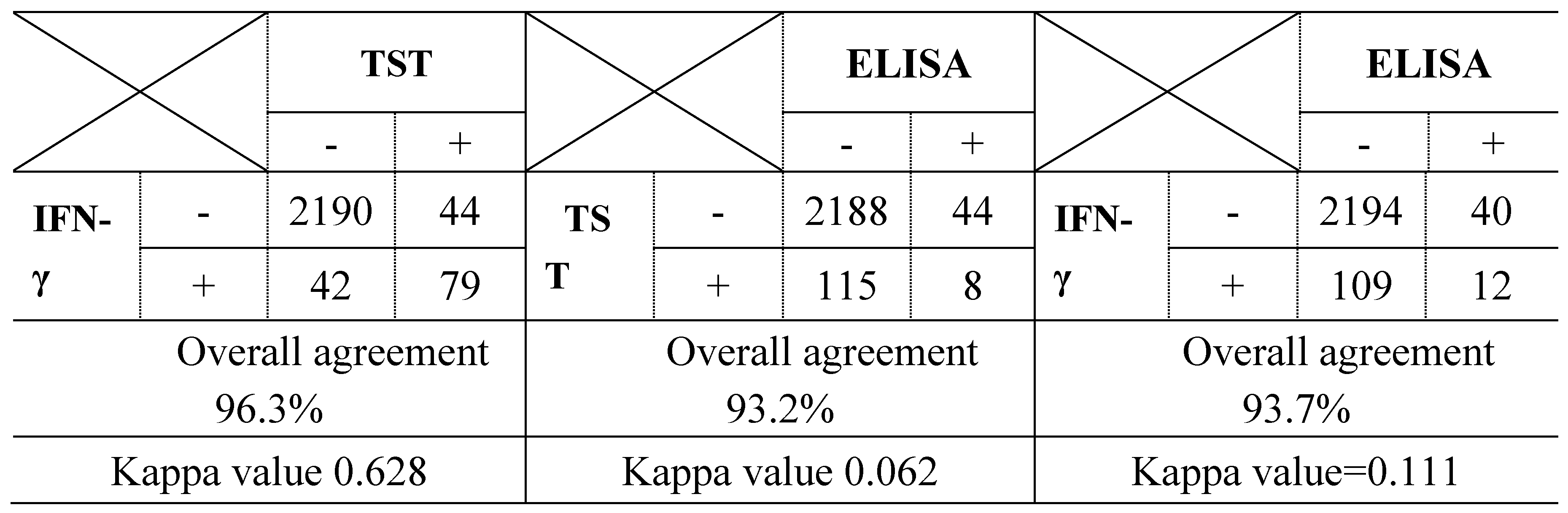
3.1. Detection Rate and Agreement of 3 Diagnostic Methods in 32 bTB Outbreak Farms

As shown in Table 1, from 2013 to 2017, a total of 205 cattle (8.7%) out of 2,355 cattle from 32 bTB outbreak farms were detected as bTB-positive by at least one of the three diagnostic methods. The detection rate of ELISA was relatively low at 52 cattle (2.2%), compared to TST (123 cattle, 5.2%) and IFN-γ (121 cattle, 5.1%). Additionally, the concordance of ELISA results with other methods was very low. ELISA results were concordant with only 12 cattle (0.51%) detected by IFN-γ and 8 cattle (0.34%) detected by TST. In contrast, the concordance rate between TST and IFN-γ was relatively high at 79 cattle (3.69%). Furthermore, the number of positive cattle detected by all three methods was only 8 cattle (Table 1).
The three diagnostic methods (TST, IFN-γ assay, and ELISA) were tested concurrently on 2,355 cattle from 32 bTB outbreak farms. The agreement between the three diagnostic methods was measured using the Kappa statistic.
The Kappa value for TST and IFN-γ assay was 0.628, indicating good agreement. In contrast, the Kappa value for TST and ELISA was 0.062, and for ELISA and IFN-γ assay, it was 0.111, both of which indicate poor concordance (Table 2).

3.2. The ELISA -Only Positive 12 Cattle in bTB Chronic Outbreak Farms

The three tests were performed three times, and a total of 17 bTB-positive cattle were ultimately detected. According to the results in Table 3, all cattle tested negative by the TST; however, five cattle were positive by the IFN-γ assay. Among the tested cattle, nine were strongly positive (S/P value > 0.5), and eight were weakly positive (S/P value between 0.3 and 0.5), resulting in a total of 12 cattle that tested only positive by ELISA.
Of the 17 cattle that tested positive for tuberculosis, 12 cattle were positive only by ELISA, despite negative results in TST and/or IFN-γ assays. The ELISA S/P values of these 12 cattle ranged from 0.3 to 1.62, with a mean S/P value of 0.71.
An autopsy was performed on the 12 cattle, and pulmonary mediastinal lymph nodes were collected. The rRT-PCR test confirmed the final positive results.
Our findings demonstrate that 12 cattle with an average S/P value of 0.3 or higher (doubtful or positive) in the ELISA test were confirmed as true bTB-positive cases, even when TST and IFN-γ assay results were negative in these chronic bTB outbreak farms.

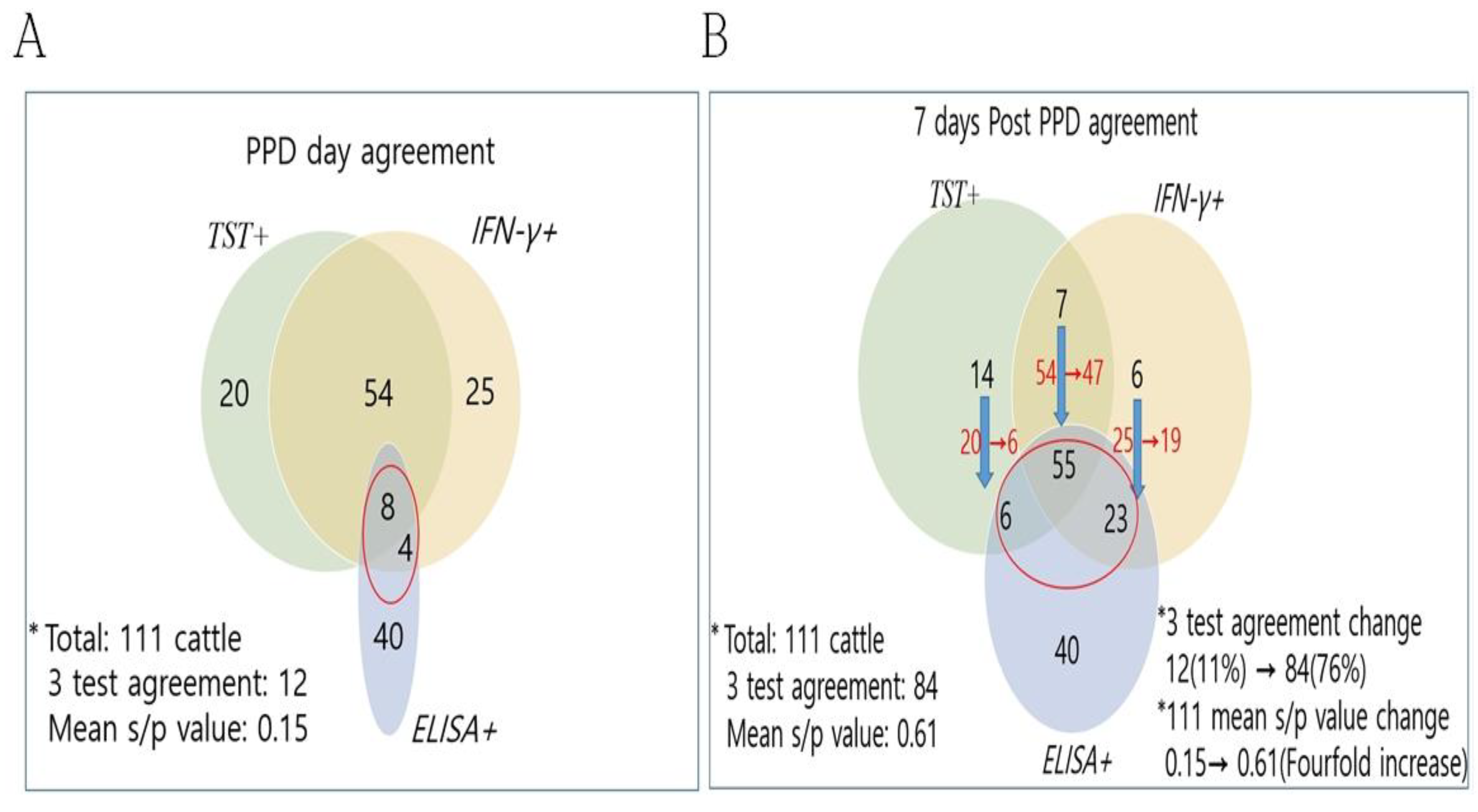
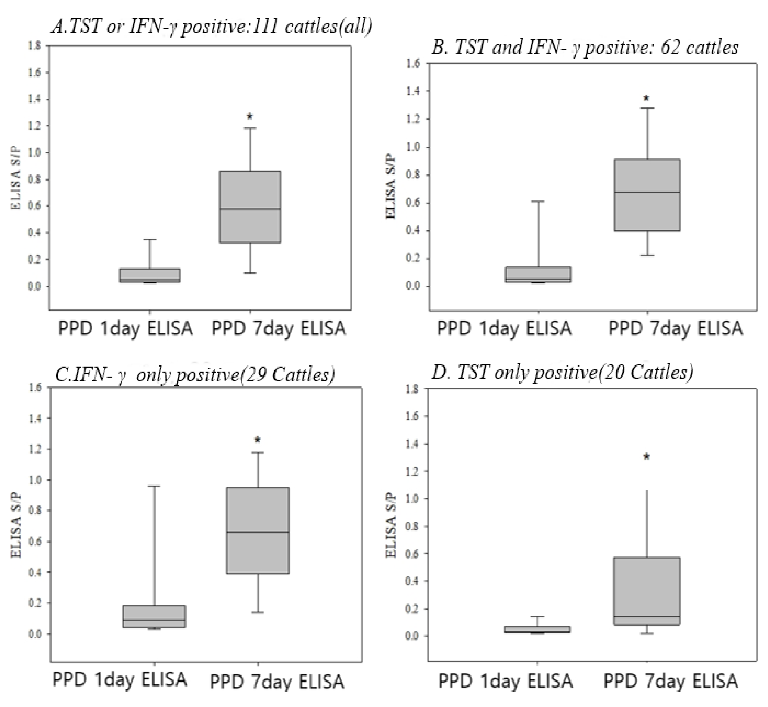
3.3. Change of the ELISA Results 7 Days After PPD in the TST and/or IFN-γ Positive Cattle

To investigate changes in ELISA results after PPD inoculation, ELISA was performed concurrently on 111 cattle among 165 cattle that tested positive by TST and/or IFN-γ from a total of 2,355 cattle across 32 farms. The number of ELISA-positive cattle markedly increased from 12 cattle (11%) to 84 cattle (76%). Additionally, ELISA S/P values showed a significant increase from 0.15 to 0.61, shifting to a strong positive response. These changes led to substantial differences in concordance with TST and IFN-γ results, as shown in Figure 1. As a result, many cases that were ELISA-negative converted to ELISA-positive seven days after PPD inoculation. The number of ELISA-positive cattle markedly increased from 12 cattle (11%) to 84 cattle (76%).
Additionally, ELISA S/P values showed a significant increase from 0.15 to 0.61, shifting to a strong positive response. These changes led to substantial differences in concordance with TST and IFN-γ results, as shown in Figure 1.
A total of 111 bTB-positive cattle, which were tested with ELISA before and after PPD inoculation, were classified into four groups based on the results: TST and IFN-γ both positive, IFN-γ only positive, TST only positive, and both TST and IFN-γ positive. Based on this, they were divided into the following four groups.: (A) Either TST- or IFN-γ-positive (111 cattle) (B) Both TST- and IFN-γ-positive (62 cattle), (C) IFN-γ-positive only (29 cattle), (D) TST-positive only (20 cattle)
A paired t-test using the SAS program was applied to statistically confirm the differences in ELISA S/P values between the day of PPD inoculation and seven days later in bTB-positive cattle.
The results showed that ELISA S/P values significantly increased in all groups after PPD inoculation (Figure 2). In particular, the increase was more pronounced in cattle that were positive for both TST and IFN-γ and in those that were only IFN-γ-positive (p < 0.0001), compared to cattle that were only TST-positive (p < 0.05).

3.4. Conversion to Positive in the ELISA Results 7 Days After PPD in All Negative Cattle for TST, IFN-γ, and ELISA

A total of 20 cattle from 10 farms, all of which tested negative for TST, IFN-γ, and ELISA in bTB outbreak farms, were selected. These cattle converted to ELISA-positive (S/P > 0.5) seven days after PPD inoculation.
The positivity of these cattle was further confirmed through rRT-PCR using lymph node tissue. Among the 20 ELISA-positive cattle, rRT-PCR tests were performed, and seven cattle from five farms were confirmed to be bTB-positive.
Table 4 presents the ELISA results before and seven days after PPD inoculation, along with PCR confirmation in the seven ELISA-positive cattle. As shown, the ELISA results demonstrated a significant increase in S/P values, rising above 0.5 seven days after PPD inoculation.

3.5. Eradication of bTB in the Outbreak Farms Using the ELISA Test 7 Days After PPD Inoculation

As shown in Table 5, with this modified ELISA application, farms experiencing a bTB outbreak were more rapidly converted to bTB-negative status, as indicated by the notably reduced number of recurrences before eradication.
Furthermore, recurrence was not observed to occur more quickly in outbreak farms that applied ELISA, TST, and IFN-γ simultaneously on the PPD inoculation day, compared to farms that used only TST and IFN-γ assays. Thus, ELISA performed on the PPD inoculation day together with TST and IFN-γ assays, followed by a second ELISA test seven days later, was confirmed to be the most effective detection method for bTB eradication. This approach enabled the detection and eradication of two to three times the average number of cases compared to other testing protocols.

3. Discussion

It has been a big challenge to detect or eradiate the bTB in South Korea, although three powerful methods, TST, IFN-γ , and ELISA have been applied for several decades[15,16]. The ELISA had been in use in South Korea from 2009 to 2013 and was excluded since 2013 because it had been assumed to generate false positive because of the low detection and agreement with TST results. As shown in Table 1 of this study, the detection rate of ELISA was just 2.2% on a sample base of 2,355 heads of cattle in the 32 farms with bTB outbreaks. This was much lower than that seen with TST (5.2%) and IFN-γ assay (5.1%).
Some research papers published in South Korea during this period showed a low ELISA detection rate and low agreement with TST, also demonstrated in this study that the Kappa value of ELISA and TST or IFN-γ assay indicated <0.12, comparable to the much higher Kappa value of 0.650 between TST and IFN-γ assay(Table 2) .The low detection rate of ELISA and agreement with the TST have generally been seen in the cattle with single caudal fold skin test for the detection of bTB[17]. Because of that, ELISA had not been used anymore and, instead, IFN-γ assay had been adopted as another bTB detection method together with TST since 2013 in Korea. Thereafter, TST and IFN-γ have been used simultaneously to detect bTB in the outbreak farms to cull in South Korea[18]. However, despite vigorous efforts for culling the bTB-positive cattle by using TST and IFN-γ assay, some farms have seen a recurrence several months or even years later, making it difficult to convert them to bTB-negative farms [9]
Although there have been previous reports showing the low detection rate of bTB and the low concordance rate with TST, the usefulness of ELISA, established on the basis of humoral immune response unlike the TST and IFN-γ assay, has not clarified yet. Our present study demonstrates the usefulness of ELISA and provide a critical example of eradication of bTB in the outbreak farms using the improved methodology of ELISA. First, our study showed that, although it was not always, ELISA positive cattle (≥S/P value 0.5) and doubtful cattle (0.3≤S/P value≤0.5), which were negative for TST and IFN-γ were confirmed to be infected with bTB (Table 3), clearly indicating that ELISA method could still be a useful method for detection of bTB, in particular in the cases which were undetectable in the TST and IFN-γ . As suggested by the previous reports, such discrepancy of the results between TST and ELISA has been postulated to be due to the detection time point of bTB between the two methods; ELISA detects the late stage of bTB infection, characterized by increased antibody level, while TST detects relatively early stage of bTB infection [1,2,3]. Our present study clearly showed that there must be the cattle which were positive only for ELISA test, but neither TST nor IFN-γ assay, in the bTB outbreak farms, also reported in some previous studies, indicating that ELISA could detect the bTB infected animals missed by the TST and IFN-γ assay. In our study, the S/P values of the bTB-infected cattle which were positive only for ELISA, but neither TST nor IFN-γ assay, ranged 0.3~1.62, taking an average of 0.63; here we need to pay an attention the meaningful low S/P value, 0.3, as the potential cases with bTB infection(Table 3),. Another important thing we found in the present study was that ELISA S/P value can be significantly changed after PPD inoculation, also strongly supported by the previous studies [4,13,15,19].
According to our results, ELISA S/P value was significantly increased 7 days after PPD inoculation, not only in the TST and/or IFN-γ positive 111 cases selected from the bTB outbreak farms, but also in the negative cases. The first ELISA value was measured on the day of PPD inoculation, and the second was evaluated 7 days later, thus indicating the change in antibody level during the interim time period. Of these 111 heads of cattle 72 cases (64%) showed a negative ELISA result, when tested on the PPD inoculation day, but these cattle were converted to a positive result, showing a fourfold increase in the average ELISA antibody values 7 days after PPD inoculation. Finally, the ELISA detection rates and agreements with other tests increased, thus negating the highlighted disadvantages of the ELISA test, as shown in Table 1.Those changes were particularly dramatic in the cattle showing positive for the IFN-γ assay including the Group A(Either TST- or IFN-γ-positive, p<0.0001 ), Group B(Both TST- and IFN-γ-positive, p<0.0001), and Group C(IFN-γ only, p<0.0001), compared with those in the cattle positive for only Group D (TST only, p<0.05) (Figure 2). The underlying mechanism why those changes in the ELISA values occurred after PPD inoculation was not clarified in the present study, but, based on the results shown in Figure 2, our results provide a clear evidence that ELISA measured 7 days after PPD inoculation must be a very effective method for bTB detection in the infected cattle which are undetectable by TST or IFN-γ assay or both [11,12,13].
In our study, the bTB infected cattle, which were undetectable for both TST and IFN-γ assay and represented negative or doubtful ELISA S/P values (≤ 0.3) when tested on the PPD inoculation day, dramatically increased to positive S/P values (≥0.5) 7 days after the PPD inoculation in Table 4. Notably, these seven cattle were housed together with truly negative cattle. These cattle, which were not detected by the three diagnostic methods in bTB outbreak farms, acted as vectors for transmission. It was an unexpected surprise, discovered in the present study, that the bTB infected cattle showing such a low S/P values of 0.01~0.03, which were also negative for TST and IFN-Γ γ assay, were detectable by ELISA, when measuring on 7 day after the PPD inoculation, as the S/P values were highly increased and changed into positive values (≥0.5). Those results may explain why it has been failed to eradicate bTB in Korea, especially in the outbreak farms where bTB occurred more than 3 times or more than one third of the cattle housed have been infected with bTB (Korean QIA reference). In addition, the results strongly support the usefulness of ELISA for eradication of bTB in such frustrating farms.
The increasing changes of ELISA S/P values after PPD inoculation had been also shown in the previous study (11, 12), in which the responses of antibodies specific against various antigens, especially MPB83, significantly increased 2 weeks after the skin test in the unvaccinated experimentally bTB infected cattle, depending on the disease severity and bacterial load. The boosting effect of TST in the bTB infected cattle was demonstrated again in this study, in the 111 cases positive for TST and/or INF in the bTB outbreak farms [4,12,13,16].
In addition, it was suggested that blood collection should be done 7 to 60 days after caudal-fold tuberculin (TST) inoculation, specifically between 7 and 14 days to diagnose latent bTB infection quickly. This method improved the sensitivity and accuracy of the specific ELISA (IDEXX M. bovis Ab test) as a method for bTB diagnosis. In other papers published globally, it was confirmed that the ELISA test performed 7 to 14 days after the PPD inoculation was better in detecting bTB-positive cattle. The best timing of the ELISA test after PPD inoculation seems to be dependent on the EILISA kit used and various factors such as the animal’s immune status. In our study, 7~10days post-tuberculin test was found to be efficient for the second ELISA test, when using the bTB 2.0®, BIONOTE [13] .
Taken together, our results could be summarized as the followings: Our findings suggest that bTB-positive cattle undetectable by TST and IFN-γ assays can be identified via ELISA seven days after PPD inoculation. In these cases, bTB infection was confirmed via rRT-PCR analysis of lesion samples. Finally, we propose a new ELISA-based protocol that facilitates bTB eradication with minimal culling and demonstrate its effectiveness in controlling outbreaks.
First, the cattle could be bTB-infected cases, showing ELISA S/P values more than 0.5, or highly suspicious showing more than S/P value ≥0.3, when measured on the day of TST, even they are all negative for both TST and IFN-γ assay; Second, for the cattle showing low negative S/P values (≤0.5) as well as all negative for TST and IFN-γ, if ELISA S/P values would highly increase and change to positive values (≥0.5) 7 days after the PPD inoculation, they could be bTB-infected cattle, being able to play a role for spreading bTB in the farms without culling. we propose a new ELISA-based protocol that facilitates bTB eradication with minimal culling and demonstrate its effectiveness in controlling outbreaks. Based on our results, we attempted to develop criteria for the use of ELISA to detect bTB, in particular in the bTB-infected cattle, which are undetectable by TST and/or IFN-γ assay, and tried to apply the criteria to eradicate bTB in the outbreak farms(Table 4).
We classified 3 subsequent steps for the application of ELISA for detection of bTB (Figure 3); As the first step, the cattle showing positive for either TST or IFN-Γ γ assay on the PPD inoculation day. As the second step, ELISA is performed on the day of PPD inoculation, and the cattle showing S/P value of 0.5 or higher and the TST and/or IFN-Γ γ positive cattle are suspected to be infected with bTB for culling. Cattle with an S/P value of ≥0.3 and ≤0.5 in the ELISA are exclude from culling at this stage. However, since these cattle have been housed in close proximity to bTB positive cattle, they could potentially play a role as latent infected individuals (Table 3). In this step, ELISA could be optionally performed only in the cattle which are negative for TST and IFN-Γ γ assay. As the third step, ELISA will be performed again 7 days after PPD inoculation for the cattle which are all negative for TST, IFN-Γ γ, and ELISA. A cull recommendation is then made for the bTB-positive cattle with an S/P value≥0.5 in the second ELISA application.
In the present study, we applied this critical application protocol of ELISA to the 12 bTB outbreak farms in Gangwon province of South Korea, to investigate how efficiently eradicate bTB. The farms chosen belonged to 5 different areas, having chronic and recurrent bTB outbreaks. Epidemiological investigations revealed that there were several risk factors for the spread of bTB in those farms such as purchase of cattle from outside, Transmission transmission from farms with tuberculosis outbreaks to neighboring farms, and so on. The HS-2 area was the largest bTB outbreak area in Gangwon province, with 200 bTB-positive heads of cattle. The HS-2 area had several additional risk factors, including common water sources, deer breeding, and the use of shared equipment. The risk factors associated with bovine tuberculosis (bTB) outbreaks in this study were identified through an epidemiological survey, with external purchase of cattle and transmission from neighboring farms being the main causes [20,21]. The farms targeted in this study for bovine tuberculosis consisted of a total of 33 farms, with 29 farms (88%) being Korean cattle (Hanwoo) farms and 4 farms (12%) being dairy farms. Since the majority of the farms were Hanwoo farms, further investigation into the dairy farms is necessarydue to genetic differences in susceptibility to bTB [22,23,24,25].
As shown in the Table 5, the simultaneous application of ELISA with the other two tests helped to convert bTB farms to bTB-negative farms. The farms that simultaneously apply ELISA, TST, and IFN-γ assay on the PPD inoculation day could eradicate bTB faster than the farms that used only two methods, TST and IFN-γ assay. In the farms that used ELISA with TST and IFN-γ assay simultaneously on PPD inoculation day and again 7 days after PPD inoculation, bovine tuberculosis recurrence dropped by an average of two to three times. Thus, the eradication of bTB was much faster in the outbreak farms when simultaneously applied ELISA as well as TST and IFN-Γ γ assay, compared to the farms that used only the TST and IFN-γ assay. In conclusion, our present study clearly demonstrated in the field farms that ELISA is a pivotal tool for fast and efficient eradication of bovine tuberculosis, by application of the newly developed protocol including the measurement of the secondary ELISA on 7 days after PPD inoculation.

Author Contributions

Conceptualization, S.M.W., C.H.P., J.M.K., and S.I.P.; methodology, C.H.P., J.M.K., and Y.H.J.; resources, J.H.K., K.W.K., and S. W. H.; formal analysis, C.H.P., B.I.Y., Y.H.J., and S.I.P.; investigation, C.H.P., J.M.K., and Y.H.J.; writing—original draft preparation, C.H.P. and B.I.Y.; writing—review and editing, S.W.R., B.Y.J., and B.I.Y.; supervision, B.I.Y. and S.I.P. All authors have read and agreed to the submitted version of the manuscript.

Funding

This study was conducted as part of the “Field Application Study of Antibody Testing Methods for Detecting False-Negative Cattle Infected with Tuberculosis,” supported by the Animal and Plant Quarantine Agency, with additional support from BIONOTE Inc.

Institutional Review Board Statement

Not applicable.

Informed Consent Statement

Informed consent was obtained from all participants in the study.

Data Availability Statement

The data presented in this study are contained within the article.

Acknowledgments

We would like to thank the researchers in the Bacterial Disease Division, Department of Animal & Plant Health Research, Animal and Plant Quarantine Agency for their advice on Bovine Tuberculosis.

Conflicts of Interest

The authors declare no conflict of interest.

References

  1. Pollock, J.M.; Neill, S.D. Pollock, J.M.; Neill, S.D. Mycobacterium bovis infection and tuberculosis in cattle. Vet. J. 2002, 163(2), 115-127. [CrossRef]
  2. World Organisation for Animal Health (OIE). Bovine tuberculosis. OIE Terrestrial Manual 2018, Chapter 3.4.6.
  3. Jang, Y.H.; Choi, J.S.; Kim, T.W.; Jeong, M.K.; Seo, Y.J.; Kim, Y.H.; Choi, E.J.; Yoon, S.S. Jang, Y.H.; Choi, J.S.; Kim, T.W.; Jeong, M.K.; Seo, Y.J.; Kim, Y.H.; Choi, E.J.; Yoon, S.S. Research on risk factors for transmission and bio-security measures associated with bovine tuberculosis breakdowns. QIA Research. 2019, 1442–1485.
  4. Waters, W.R.; Buddle, B.M.; Vordermeier, H.M.; Gormley, E.; Palmer, M.V.; Thacker, T.C.; Bannantine, J.P.; Stabel, J.R.; Linscott, R.; Martel, E.; Milian, F.; Foshaug, W.; Lawrence, J.C. Waters, W.R.; Buddle, B.M.; Vordermeier, H.M.; Gormley, E.; Palmer, M.V.; Thacker, T.C.; Bannantine, J.P.; Stabel, J.R.; Linscott, R.; Martel, E.; Milian, F.; Foshaug, W.; Lawrence, J.C. Development and evaluation of an enzyme-linked immunosorbent assay for use in the detection of bovine tuberculosis in cattle. Clin. Vaccine Immunol. 2011, 18(11), 1882-1888. [CrossRef]
  5. Plackett, P.; Ripper, J.; Corner, L.A.; Small, K.; de Witte, K.; Melville, L.; Hides, S.; Wood, P.R. Plackett, P.; Ripper, J.; Corner, L.A.; Small, K.; de Witte, K.; Melville, L.; Hides, S.; Wood, P.R. An ELISA for the detection of anergic tuberculous cattle. Aust Vet J. 1989, 66(1), 15-19.
  6. Welsh, M.D.; Cunningham, R.T.; Corbett, D.M.; Girvin, R.M.; McNair, J.; Skuce, R.A.; Bryson, D.G.; Pollock, J.M. Welsh, M.D.; Cunningham, R.T.; Corbett, D.M.; Girvin, R.M.; McNair, J.; Skuce, R.A.; Bryson, D.G.; Pollock, J.M. Influence of pathological progression on the balance between cellular and humoral immune responses in bovine tuberculosis. Immunology 2005, 114, 101–111. [CrossRef]
  7. Koni, A.; Juma, A.; Morini, M.; Nardelli, S.; Connor, R.; Koleci, X. Koni, A.; Juma, A.; Morini, M.; Nardelli, S.; Connor, R.; Koleci, X. Assessment of an ELISA method to support surveillance of bovine tuberculosis in Albania. Ir. Vet. J. 2016, 69, 11. [CrossRef]
  8. Choi KY, Choi ES. A comparison study of tuberculin, IFN-γ, and antibody ELISA assay for the diagnosis of bovintuberculosis. Graduation Thesis, College of Veterinary Medicine, Chonbuk National University.2014. http://www.riss.kr/link?id=T13576874&outLink=K.
  9. Jeong C, Yun GR, Ra DK, Kim KM, Lee JG, Kim KH, Lee SM. 2014. Case of bovine tuberculosis diagnosis in a slaugterhouse confirmed as PPD negative. Korean J Vet Serv, 2014, 37(2), 131-136 ISSN 1225-6552, eISSN 2287-7630. [CrossRef]
  10. Harboe, M.; Wiker, H.G.; Duncan, J.R.; Garcia, M.M.; Dukes, T.W.; Brooks, B.W.; Turcotte, C.; Nagai, S. Harboe, M.; Wiker, H.G.; Duncan, J.R.; Garcia, M.M.; Dukes, T.W.; Brooks, B.W.; Turcotte, C.; Nagai, S. Protein G-based enzyme-linked immunosorbent assay for anti-MPB70 antibodies in bovine tuberculosis. J. Clin. Microbiol. 1990, 28, 913–921. [CrossRef]
  11. Harboe, M.; Nagai, S.; Wiker, H.G.; Sletten, K.; Haga, S. Harboe, M.; Nagai, S.; Wiker, H.G.; Sletten, K.; Haga, S. Homology between the MPB70 and MPB83 proteins of Mycobacterium bovis BCG. Scand. J. Immunol. 1995, 42, 46–51. [CrossRef]
  12. Lyashchenko, K.; Whelan, A.O.; Greenwald, R.; Pollock, J.M.; Andersen, P.; Hewinson, R.G.; Vordermeier, H.M. Lyashchenko, K.; Whelan, A.O.; Greenwald, R.; Pollock, J.M.; Andersen, P.; Hewinson, R.G.; Vordermeier, H.M. Association of tuberculin-boosted antibody responses with pathology and cell-mediated immunity in cattle vaccinated with Mycobacterium bovis BCG and infected with M. bovis. Infect.Immun. 2004, 72, 2462–2467. [CrossRef]
  13. Waters, W.R.; Palmer, M.V.; Stafne, M.R.; Bass, K.E.; Maggioli, M.F.; Thacker, T.C.; Linscott, R.; Lawrence, J.C.; Nelson, J.T.; Esfandiari, J.; Greenwald, R.; Lyashchenko, K.P. Waters, W.R.; Palmer, M.V.; Stafne, M.R.; Bass, K.E.; Maggioli, M.F.; Thacker, T.C.; Linscott, R.; Lawrence, J.C.; Nelson, J.T.; Esfandiari, J.; Greenwald, R.; Lyashchenko, K.P. Effects of serial skin testing with purified protein derivative on the level and quality of antibodies to complex and defined antigens in Mycobacterium bovis-infected cattle. Clin. Vaccine Immunol. 2015, 22, 641–649. [CrossRef]
  14. Palmer MV, Waters WR, Thacker TC, et al. 2006. Effects of different tuberculin skin-testing regimens on gamma interferon and antibody responses in cattle experimentally infected with Mycobacterium bovis. Clin Vaccine Immunol. 13(3):377–384.
  15. Silva, E. Silva, E. Evaluation of an enzyme-linked immunosorbent assay in the diagnosis of bovine tuberculosis. Vet. Microbiol. 2001, 78, 111–117. [CrossRef]
  16. de la Rua-Domenech, R.; Goodchild, A.T.; Vordermeier, H.M.; Hewinson, R.G.; Christiansen, K.H.; Clifton-Hadley, R.S. de la Rua-Domenech, R.; Goodchild, A.T.; Vordermeier, H.M.; Hewinson, R.G.; Christiansen, K.H.; Clifton-Hadley, R.S. Ante mortem diagnosis of tuberculosis in cattle: a review of the tuberculin tests, gamma-interferon assay and other ancillary diagnostic techniques. Res. Vet. Sci. 2006, 81, 190–210. [CrossRef]
  17. Lee, J.J.; Kim, D.S.; Lee, J.H.; Lee, C.S. Lee, J.J.; Kim, D.S.; Lee, J.H.; Lee, C.S. A Comparative Study on the Diagnosis of ELISA Test and PPD Test of the Bovine Tuberculosis. Korean J. Vet. Serv. 2010, 33, 335–340.
  18. Jang, Y.H.; Kim, T.W.; Jeong, M.K.; Seo, Y.J.; Ryoo, S.; Park, C.H.; Kang, S.S.; Lee, Y.J.; Yoon, S.S.; Kim, J.M. Jang, Y.H.; Kim, T.W.; Jeong, M.K.; Seo, Y.J.; Ryoo, S.; Park, C.H.; Kang, S.S.; Lee, Y.J.; Yoon, S.S.; Kim, J.M. Introduction and application of the interferon-γ assay in the national bovine tuberculosis control program in South Korea. Front. Vet. Sci. 2020, 7, 222. [CrossRef]
  19. Klepp, L.I.; Blanco, F.C.; Bigi, M.M.; Vázquez, C.L.; García, E.A.; Sabio y García, J.; Bigi, F. Klepp, L.I.; Blanco, F.C.; Bigi, M.M.; Vázquez, C.L.; García, E.A.; Sabio y García, J.; Bigi, F. B Cell and Antibody Responses in Bovine Tuberculosis. Antibodies 2024, 13, 84. [CrossRef]
  20. Goodchild, A.V.; Clifton-Hadley, R.S. Goodchild, A.V.; Clifton-Hadley, R.S. Cattle-to-cattle transmission of Mycobacterium bovis. Tuberculosis. 2001, 81, 23–41. [CrossRef]
  21. Chu, G.S.; et al. Chu, G.S.; et al. Epidemiological study on bovine tuberculosis outbreaks in endemic areas. Korean J. Vet. Serv. 2009, 32, 119–124.
  22. Chai, J.; Wang, Q.; Qin, B.; Wang, S.; Wang, Y.; Shahid, M.; Liu, K.; Zhang, Y.; Qu, W. Chai, J.; Wang, Q.; Qin, B.; Wang, S.; Wang, Y.; Shahid, M.; Liu, K.; Zhang, Y.; Qu, W. Association of NOS2A gen polymorphisms with susceptibility to bovine tuberculosis in Chinese Holstein cattle. PLoS One 2021, 16(6), e0253339. [CrossRef]
  23. Kumar, R.; Gandham, S.; Rana, A.; Maity, H.K.; Sarkar, U.; Dey, B. Kumar, R.; Gandham, S.; Rana, A.; Maity, H.K.; Sarkar, U.; Dey, B. Divergent proinflammatory immune responses associated with the differential susceptibility of cattle breeds to tuberculosis. Front. Immunol. 2023, 14, 1199092. [CrossRef]
  24. Raphaka, K.; Matika, O.; Sánchez-Molano, E.; Mrode, R.; Coffey, M.P.; Riggio, V.; Glass, E.J.; Woolliams, J.A.; Bishop, S.C.; Banos, G. Raphaka, K.; Matika, O.; Sánchez-Molano, E.; Mrode, R.; Coffey, M.P.; Riggio, V.; Glass, E.J.; Woolliams, J.A.; Bishop, S.C.; Banos, G. Genomic regions underlying susceptibility to bovine tuberculosis in Holstein-Friesian cattle. BMC Genet. 2017, 18(1), 27. [CrossRef]
  25. Schiller, I.; Oesch, B.; Vordermeier, H.M.; Palmer, M.V.; Harris, B.N.; Orloski, K.A.; Buddle, B.M.; Thacker, T.C.; Lyashchenko, K.P.; Waters, W.R. Schiller, I.; Oesch, B.; Vordermeier, H.M.; Palmer, M.V.; Harris, B.N.; Orloski, K.A.; Buddle, B.M.; Thacker, T.C.; Lyashchenko, K.P.; Waters, W.R. Bovine tuberculosis: A review of current and emerging diagnostic techniques in view of their relevance for disease control and eradication. Transbound. Emerg. Dis. 2010, 57, 205–220. [CrossRef]
Figure 1. Change in the agreement of 3 test results 7 days after PPD inoculation in 111 cattle positive for TST and/or IFN-γ assay.
Preprints 152540 g001
Figure 2. Boosting changes in blood antibody levels 7 days after PPD inoculation in cattle showing positive for either TST, IFN-γ assay, or both (Paired t-test).
Preprints 152540 g002
Figure 3. Bovine Tuberculosis Eradication Strategy Using ELISA Testing.
Preprints 152540 g003
Table 1. Detection rate and concordance of each diagnostic methods in bTB outbreak farms.(n=2,355 cattle from 32 farms).
Group Positivity for bovine tuberculosis No. of cattle (%) No. of positive cattle for each method (detection rate, %)
TST IFN-γ ELISA TST IFN-γ ELISA
Negative - - - 2150(91.3) 0 0 0
TST only + - - 44(1.9) 44 0 0
IFN-γ only - + - 38(1.6) 0 38 0
ELISA only - - + 40(1.7) 0 0 40
TST & IFN-γ + + - 79(3.4) 79 79 -
TST & ELISA + - + 8(0.3) 8 - 8
IFN-γ & ELISA - + + 12(0.5) - 12 12
TST& IFN-γ& ELISA + + + 8(0.3) 8 8 8
Total positivity 205(8.7)* 2355(100)* 123(5.2)* 121(5.1)* 52(2.2)*
TST, Tuberculin Skin Test; INF-γ, Interferon-gamma; ELISA, Enzyme-linked Immunosorbent Assay. *, duplicate cattle were regarded as one case.
Table 2. Agreement between the 3 diagnostic methods in bTB outbreak farms.
Preprints 152540 i001
*; TST, Tuberculin Skin Test; IFN, Interferon-gamma; ELISA, Enzyme-linked Immunosorbent Assay.
Table 3. Confirmation of bovine tuberculosis infection in 12 cattle that tested positive only by ELISA, but negative by TST and/or IFN-γ assay, in chronic BTB outbreak farms.
Animal ID TST IFN-γ ELISA* Range of the ELISA S/P values ELISA Mean S/P value rRT-PCR
H2 - - + 0.68~1.62 1.17 +
H12 - - + 0.81~1.12 1.04 +
H5 - - + 0.53~1.27 0.95 +
H39 - - + 0.67~1.12 0.91 +
H20 - - + 0.74~0.98 0.88 +
H14 - - + 0.33~0.81 0.61 +
H1 - - + 0.49~0.70 0.60 +
H9 - - + 0.32~0.68 0.55 +
H38 - - + 0.35~0.71 0.54 +
H17 - - ± 0.36~0.67 0.42 +
H22 - - ± 0.31~0.66 0.41 +
H54 - - ± 0.3~0.74 0.39 +
Total - - 12 0.3~1.62 0.71 12 positive
rRT-PCR was performed using the pulmonary lymph node of each animal. *; -, negative(S/P value≤0.3); ±, doubtful (0.3≤ S/P value≤0.5); +, positive (S/P value≥0.5). TST: Tuberculin skin test; IFN-γ: Interferon-gamma; ELISA: Enzyme-linked immunosorbent assay.
Table 4. ELISA Results Before and Seven Days After PPD Inoculation in TST and IFN-γ Negative Cattle.
Farm ID Positive cattle Age TST result IFN-γ result ELISA S/P value (PPD inoculation day) ELISA S/P value (7 days after PPD) rRT-PCR
J○○ 89 month - - 0.47 1.11(+) +
L○○ 48 month - - 0.36 1.21(+) +
L○○ 15 month - - 0.49 0.77(+) +
Y○○ 35 month - - 0.29 0.51(+) +
U○○ 69 month - - 0.02 1.33(+) +
U○○ 65 month - - 0.01 0.67(+) +
K○○ 54 month - - 0.03 0.97(+) +
*; TST, Tuberculin Skin Test; IFN-γ, Interferon-gamma; ELISA, Enzyme-linked Immunosorbent Assay.
Table 5. Comparison of bTB Recurrence in Farms Using the Modified ELISA Protocol Versus Farms Using Traditional Methods.
Study period Area Farm ID Breed Herd size No. of bTB-cattle Application protocol No. of bTB recurrences
~2013.11 HS-1 U** Hanwoo 71 38 TST only confirm
(ELISA monitoring)
8
P** 69 18 5
2013.12
~
2015. 11
HC J* 74 3 3
HC J* 16 7 Simultaneous 3 Test
TST, IFN-γ, ELISA (7days post-PPD)
4
J** 34 22 4
HS-2 S** Dairy cow 42 8 4
SUM 306 96 MEAN 4.7
2015.12
~
2017. 06
HC J** Hanwoo 124 8 Simultaneous 3 Test
TST, IFN-γ, ELISA (7days post-PPD)
2
HS-2 B** 75 6 1
Y** 66 4 1
U** 157 19 2
HS-3 L** 142 43 2
HS-4 K** 47 34 2
SUM 611 114 MEAN 1.7
Disclaimer/Publisher’s Note: The statements, opinions and data contained in all publications are solely those of the individual author(s) and contributor(s) and not of MDPI and/or the editor(s). MDPI and/or the editor(s) disclaim responsibility for any injury to people or property resulting from any ideas, methods, instructions or products referred to in the content.
Copyright: This open access article is published under a Creative Commons CC BY 4.0 license, which permit the free download, distribution, and reuse, provided that the author and preprint are cited in any reuse.
Prerpints.org logo

Preprints.org is a free preprint server supported by MDPI in Basel, Switzerland.

Subscribe

Disclaimer

Terms of Use

Privacy Policy

Privacy Settings

© 2025 MDPI (Basel, Switzerland) unless otherwise stated