Preprint
Review

This version is not peer-reviewed.

Neovaginal Perforation in Sigmoid Vaginoplasty: An Underrecognized Complication– A Literature Review

A peer-reviewed article of this preprint also exists.

Submitted:

16 March 2025

Posted:

17 March 2025

You are already at the latest version

Abstract

Background: Sigmoid vaginoplasty has become a preferred technique for gender affirmation surgery in transgender women due to its ability to provide adequate vaginal width and depth, intrinsic mucosal lubrication, and resistance to postoperative shrinkage. However, despite its advantages, this technique is associated with rare but potentially severe complications, including neovaginal perforation. Objective: This review aims to comprehensively analyze neovaginal perforation following sigmoid vaginoplasty, highlighting its incidence, underlying mechanisms, clinical presentation, management strategies, and preventive measures. Methods: A systematic review of existing literature was conducted to evaluate reported cases of neovaginal perforation, its etiology, and treatment outcomes. Additionally, we present a representative case from our institution, providing further insights into the clinical course and management of this complication. Results: Neovaginal perforation, though uncommon, can lead to serious consequences such as abscess formation, peritonitis, and sepsis if not promptly diagnosed and treated. The primary contributing factors include improper postoperative vaginal dilation techniques, vascular compromise due to stenosis or ischemia, and tissue fragility of the sigmoid neovagina. Management strategies vary from conservative treatment with antibiotics and drainage to surgical intervention in severe cases. Conclusion: While sigmoid vaginoplasty remains a valuable technique for gender affirmation surgery, awareness of its potential complications—particularly neovaginal perforation—is crucial. Early recognition, appropriate management, and adherence to postoperative care protocols, including proper dilation techniques, are essential in preventing this rare but serious complication. A multidisciplinary approach involving plastic surgeons, colorectal surgeons, and gynecologists is key to optimizing patient outcomes and minimizing risks.

Keywords: 
;  

Introduction

Gender affirmation surgery is a critical component of the transition process for transgender women, significantly enhancing their quality of life and psychological well-being. As surgical expertise continues to expand, a variety of techniques have been developed, with penile inversion, peritoneal vaginoplasty, and sigmoid vaginoplasty being the most commonly performed procedures [1]. The choice among these techniques depends on multiple factors, including surgical invasiveness, available penile skin, vaginal depth, aesthetic outcomes, and functional properties.
Sigmoid vaginoplasty, also known as rectosigmoid neocolporrhaphy or neocolpopoiesis, was first conceptualized by Baldwin in 1904, utilizing an intestinal segment for vaginal reconstruction [2,3]. Building on this approach, Wallace successfully employed the sigmoid colon in 1911 [4]. The technique was later introduced for gender affirmation surgery by Markland and Hastings and has since gained widespread adoption [5]. With advancements in open, laparoscopic, and robotic-assisted approaches, modern sigmoid vaginoplasty now offers enhanced visualization, minimal invasiveness, superior cosmetic outcomes, and faster recovery times [1,6,7].
One of the key advantages of sigmoid colon vaginoplasty is its ability to provide sufficient vaginal depth and width, along with mucosal secretion that facilitates lubrication and reduces postoperative shrinkage [3,8,9]. However, despite these benefits, it remains a technically complex procedure requiring multidisciplinary collaboration and carries a risk of postoperative complications. The most frequently reported issues include introital stenosis and mucosal prolapse, while rarer but more serious complications, such as neovaginal perforation, rectovaginal fistula, and colonic necrosis, have also been documented [1,10,11,12,13,14]. Among these, neovaginal perforation is particularly concerning due to its potential to cause peritonitis, sepsis, and other life-threatening sequelae.
Given its low incidence but high clinical significance, a thorough understanding of the mechanisms, risk factors, and management strategies for neovaginal perforation is essential. This review provides a comprehensive analysis of this underrecognized complication, summarizing existing literature on its incidence, pathophysiology, and treatment. Additionally, we present a representative case from our institution, offering further insights into the diagnostic and therapeutic challenges associated with this condition.

Etiology and Pathophysiology

The pathophysiology of neovaginal perforation is complex and multifactorial, involving a combination of mechanical trauma, ischemic injury, infection, and structural weaknesses inherent to the sigmoid colon tissue.
Mechanical Trauma: One of the most common causes of neovaginal perforation is mechanical trauma, often resulting from improper dilation techniques or vigorous sexual activity. Postoperative dilation is essential for maintaining vaginal patency, but if performed incorrectly, it can create mucosal tears that compromise the neovaginal wall. These microtraumas may progressively weaken the tissue, leading to deeper submucosal injuries and eventual perforation. Similarly, excessive force during sexual intercourse, particularly in the early postoperative period, may contribute to structural failure [15].
Ischemia and Vascular Compromise: Vascular integrity plays a crucial role in neovaginal viability. If the sigmoid segment used for neovaginal construction experiences inadequate perfusion due to anastomotic disruption, arterial insufficiency, or venous congestion, ischemic necrosis can develop. This can lead to ulceration and thinning of the neovaginal wall, predisposing it to perforation. Introital stenosis may also exacerbate this issue by restricting blood flow, further increasing the risk of tissue breakdown [1].
Infection and Abscess Formation: Neovaginal perforation can be further complicated by bacterial infections, particularly when colonic bacteria invade the surrounding pelvic or peritoneal structures. Infections may originate from minor mucosal defects, which serve as entry points for bacterial translocation. If untreated, these infections may progress to abscess formation or sepsis, necessitating urgent surgical intervention [15].
Postoperative complications: Ischemia or inadequate healing of the bowel anastomosis, may also predispose patients to neovaginal perforation. In cases where compromised blood supply leads to ischemic necrosis of the neovaginal wall, the structural integrity of the tissue is weakened, rendering it more vulnerable to rupture. Infection, another well-recognized complication following intestinal-based neovaginoplasty, can further contribute to tissue degradation and increase the risk of perforation [1].
Introital stenosis: Introital stenosis is a common postoperative complication in neovaginal reconstructions. In patients with neovaginal stenosis, increased intravaginal pressure during dilation or intercourse may exert excessive force on the weakened neovaginal wall, increasing the likelihood of perforation. Additionally, inadequate postoperative care and failure to adhere to dilation protocols may contribute to the progressive narrowing of the neovaginal introitus, thereby exacerbating the risk [15,16].

Clinical Presentation and Case Reports

Neovaginal perforation following sigmoid vaginoplasty can present with a wide range of clinical manifestations, depending on the severity and timing of the perforation.
In the early stages, symptoms may be mild, including vague pelvic discomfort, localized tenderness, or abnormal vaginal discharge. As the perforation progresses, more severe signs of infection, such as fever, nausea, vomiting, and systemic inflammatory response syndrome (SIRS), may develop. If peritonitis occurs, patients may exhibit severe abdominal pain, rebound tenderness, and signs of sepsis, including hypotension and tachycardia.
A significant proportion of reported cases highlight that introital stenosis plays a critical role in neovaginal perforation. In these cases, patients often experience increasing difficulty with dilation, followed by a sudden inability to insert the dilator fully. This may be accompanied by deep-seated pain within the neovagina, which is often mistaken for simple stenosis. However, continued forceful dilation in the presence of underlying ischemia or tissue fragility can lead to catastrophic rupture.

Reported Cases

Several case reports illustrate the diverse presentations and management strategies for neovaginal perforation (Table 1):
  • Liguori et al. (2001) documented a case of acute peritonitis secondary to introital stenosis, leading to perforation of a neovagina constructed from a bowel segment [16].
  • Amirian et al. (2011) reported a patient who presented with lower abdominal pain and fever. CT imaging revealed free air in the retroperitoneum, and a leak through the vaginal apex was confirmed via vaginal contrast examination. This patient was successfully managed with conservative antibiotic therapy [17].
  • Shimamura et al. (2015) described a case of neovaginal perforation complicated by an intra-abdominal abscess, where clinical symptoms and radiologic findings were incongruent. Surgical intraperitoneal drainage was performed due to concerns that the abscess might not resolve with antibiotics alone [15].
  • Matthew et al. reported two cases involving diffuse stenosis of unknown etiology, leading to ischemia and subsequent perforation of the sigmoid conduit. One patient underwent midline laparotomy and was found to have multiple interloop abscesses, requiring prolonged intravenous antibiotic therapy. The second patient, who developed vaginal stenosis secondary to a high-riding perineum, required laparoscopic sigmoid conduit resection, followed by a midline incision and internal suturing of the colon flap one month postoperatively [1].
In addition to these reports, we present a case from our institution, where a 59-year-old transgender woman developed neovaginal perforation following improper dilation, The patient exhibited progressive lower abdominal pain and was found to have an abscess formation above the neovagina(Figure 1). Imaging confirmed gaseous distension and proximal small bowel dilatation. Colonoscopy revealed a mucosal defect at 10 cm above the vaginal introitus. The patient underwent colonoscopy open drainage, antibiotic therapy, and a structured rehabilitation program, leading to a full recovery. Three months post-discharge, colonoscopy confirmed complete perforation healing. A six-month follow-up CT scan showed a normal neovagina without residual abscess(Figure 2).
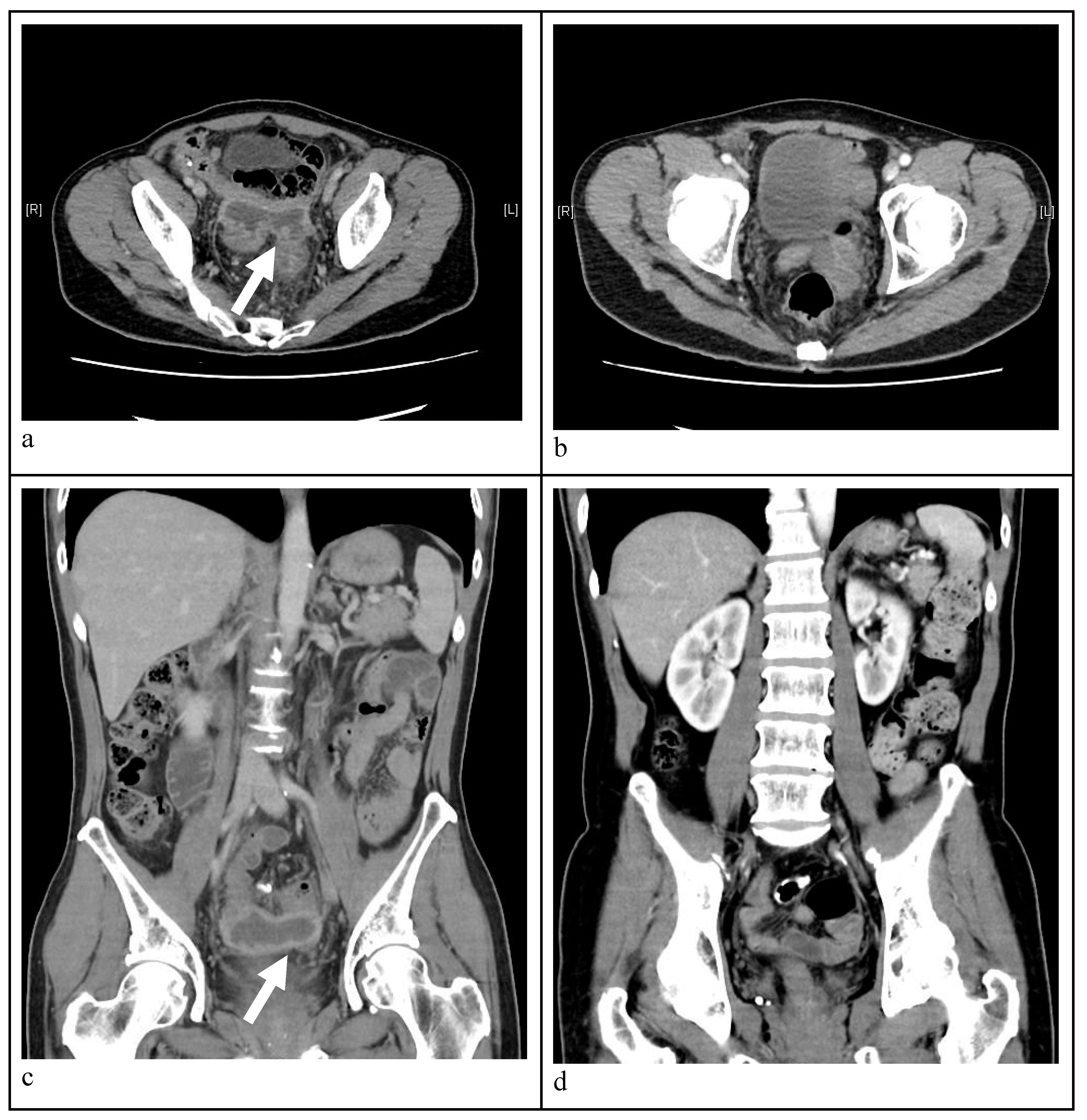
  • Axial view CT image indicating a massive abscess occupying a significant portion of the intra-abdominal cavity. A well-circumscribed abscess fluid collection with enhanced walls is seen as pointed by the white arrows.
  • Follow-up CT image disclosing no abscess and patent neovagina without perforation six months post-treatment.
  • Sagittal view abdominal CT image demonstrating a intra-abdominal abscess. A well-circumscribed abscess fluid collection with enhanced walls is seen as pointed by the white arrow.
  • Follow-up CT image disclosing no abscess and patent neovagina without perforation six months post-treatment.
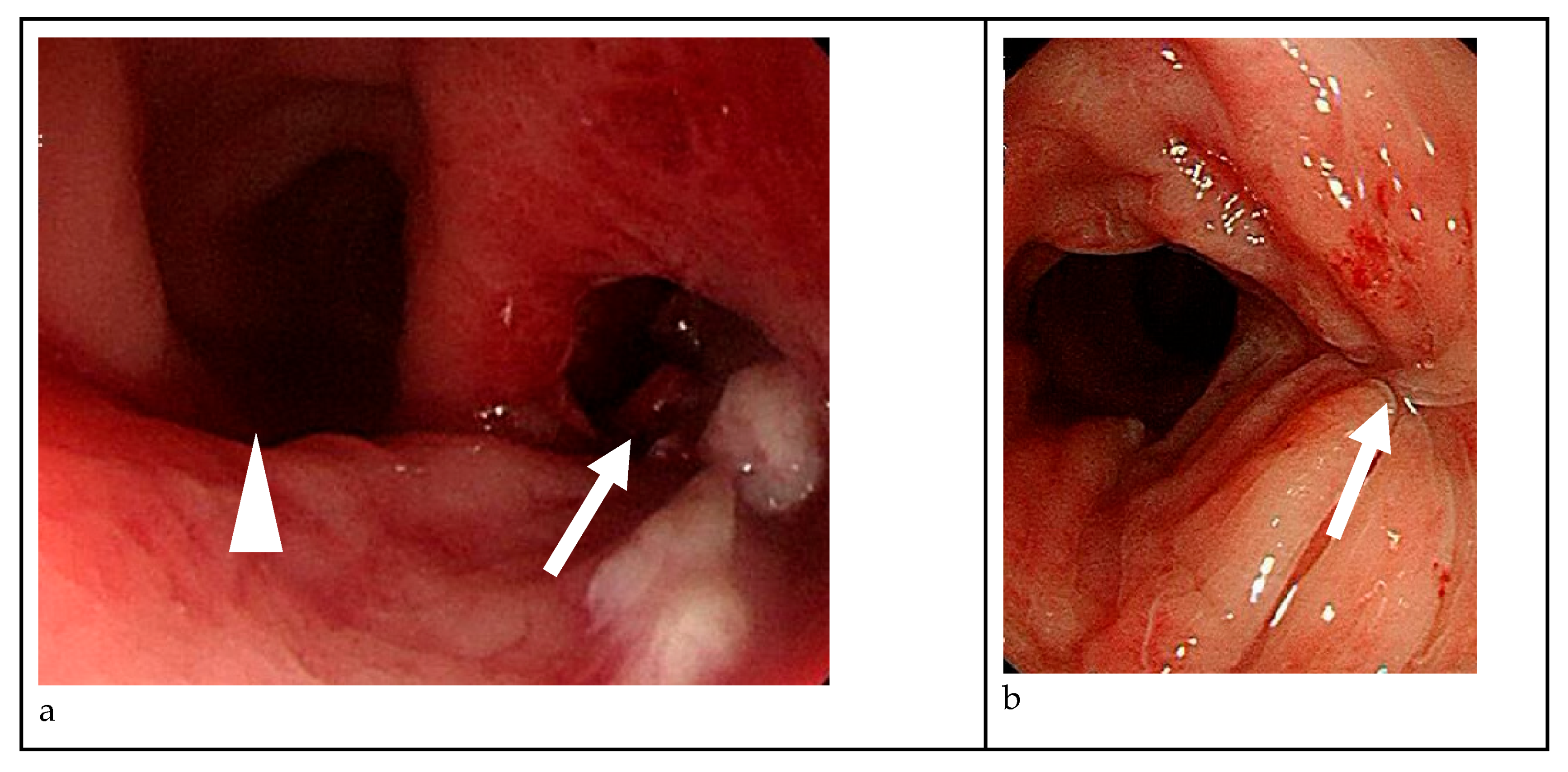
  • On colonoscopy, a perforation was found at 10 cm level above vaginal orifice and in the 4-5 o'clock direction(white arrow), whereas the left-hand side lies the neovagina, as pointed by the white arrow head.
  • The previous perforation site on the right-hand side was healed well and no further perforation was found after three months post-treatment, as pointed by the white arrows.

Diagnosis

Due to the nonspecific nature of symptoms, a high index of suspicion is required to diagnose neovaginal perforation. Computed tomography (CT) imaging is the gold standard diagnostic modality, as it can identify free air in the peritoneal cavity, abscess formation, or other signs of intra-abdominal infection. In some cases, contrast-enhanced imaging may help delineate the exact site of perforation [17].
For patients presenting with vague abdominal pain and a history of sigmoid vaginoplasty, pelvic examination should be performed with caution. In cases where perforation is suspected, a digital rectal or vaginal exam may reveal areas of tenderness, abnormal mucosal defects, or, in rare cases, direct visualization of a perforation [1]. However, forceful probing should be avoided to prevent exacerbating the injury.

Management Strategies

The management of neovaginal perforation depends on the severity of the condition and the extent of intra-abdominal contamination. In cases where the perforation is small and localized, conservative management with broad-spectrum intravenous antibiotics and bowel rest may be sufficient. Close monitoring for signs of worsening infection is essential, and patients should be advised to avoid any activities that could increase intra-abdominal pressure, such as dilation or sexual activity.
For patients with larger perforations, intra-abdominal abscess formation, or signs of peritonitis, surgical intervention is often required. The choice of surgical approach depends on the extent of the damage and the patient’s overall condition. In some cases, laparoscopic or open surgical repair may be performed to close the defect and drain any associated abscesses. If the neovagina is severely compromised, partial or complete reconstruction may be necessary, utilizing additional bowel segments or alternative techniques such as peritoneal or skin graft-based vaginoplasty.
Postoperative care is crucial in preventing recurrence, and patients should be closely followed to ensure proper healing. Long-term surveillance should include regular gynecologic evaluations, imaging if needed, and patient education regarding safe dilation and sexual practices to minimize the risk of re-injury.

Future Directions

Given the rarity of neovaginal perforation, there is a clear need for more comprehensive research to better understand its risk factors, prevention strategies, and optimal management approaches. One promising avenue for future studies is tissue engineering and regenerative medicine, which may offer alternative solutions for neovaginal reconstruction that reduce the risk of perforation. Research into bioengineered vaginal grafts, composed of autologous stem cell-derived tissues, may provide a more durable and structurally resilient neovaginal lining compared to traditional bowel-derived techniques.
Additionally, more data are needed regarding the long-term outcomes of sigmoid vaginoplasty and its associated complications. Large-scale, multicenter studies focusing on postoperative complications, including neovaginal perforation, could provide valuable insights into risk reduction strategies. Establishing standardized postoperative care protocols, including evidence-based dilation schedules and surveillance imaging for high-risk patients, may also contribute to early detection and prevention.
Finally, patient education and shared decision-making should be emphasized in future clinical practice. Many transgender women undergoing vaginoplasty may not be fully informed of the risks associated with intestinal-based neovaginal reconstructions, including the potential for perforation. Developing comprehensive educational materials and support programs can empower patients to make informed decisions about their surgical options and postoperative care.

Conclusion

Neovaginal perforation remains an underreported but significant complication of sigmoid vaginoplasty. While rare, its potential to cause severe morbidity underscores the importance of early recognition and appropriate management. Current evidence suggests that mechanical trauma, introital stenosis, and postoperative complications contribute to the development of neovaginal perforation, yet much remains unknown about its true incidence and optimal prevention strategies. As gender-affirming surgical techniques continue to evolve, further research is necessary to refine surgical approaches, improve postoperative care, and ultimately enhance the safety and quality of life for transgender women undergoing neovaginal reconstruction.Multidisciplinary care is essential for early detection and optimal management.Despite these risks, sigmoid vaginoplasty remains a safe and effective option for gender affirmation surgery, offering adequate depth, intrinsic lubrication, and minimal long-term complications when combined with proper surgical technique and postoperative care.

References

  1. Meece MS, Weber LE, Hernandez AE, Danker SJ, Paluvoi NV. Major complications of sigmoid vaginoplasty: a case series. J Surg Case Rep 2023;2023:rjad333. [CrossRef]
  2. Baldwin JF. XIV. The Formation of an Artificial Vagina by Intestinal Trransplantation. Ann Surg 1904;40:398–403.
  3. Salgado CJ, Nugent A, Kuhn J, Janette M, Bahna, H. Primary Sigmoid Vaginoplasty in Transwomen: Technique and Outcomes. Biomed Res Int 2018;2018:4907208. [CrossRef]
  4. Hendren WH, Atala, A. Use of bowel for vaginal reconstruction. J Urol 1994;152:752–5; discussion 756–7. [CrossRef]
  5. Markland, C., Hastings D. Vaginal reconstruction using cecal and sigmoid bowel segments in transsexual patients. J Urol 1974;111:217–9. [CrossRef]
  6. Wedler V, Meuli-Simmen C, Guggenheim M, Schneller-Gustafsson, M., Künzi, W. Laparoscopic technique for secondary vaginoplasty in male to female transsexuals using a modified vascularized pedicled sigmoid. Gynecol Obstet Invest 2004;57:181–5. [CrossRef]
  7. Kim C, Campbell B, Ferrer, F. Robotic sigmoid vaginoplasty: a novel technique. Urology 2008;72:847–9. [CrossRef]
  8. Kaushik N, Jindal O, Bhardwaj DK. Sigma-lead male-to-female gender affirmation surgery: Blending cosmesis with functionality. Plast Reconstr Surg Glob Open 2019;7:e2169. [CrossRef]
  9. Nowier, A., Esmat M., Hamza RT. Surgical and functional outcomes of sigmoid vaginoplasty among patients with variants of disorders of sex development. Int Braz J Urol 2012;38:380–6;discussions387–8. [CrossRef]
  10. Kwun Kim S, Hoon Park J, Cheol Lee K, Min Park J, Tae Kim, J., Chan Kim, M. Long-term results in patients after rectosigmoid vaginoplasty. Plast Reconstr Surg 2003;112:143–51. [CrossRef]
  11. Parsons JK, Gearhart SL, Gearhart JP. Vaginal reconstruction utilizing sigmoid colon: Complications and long-term results. J Pediatr Surg 2002;37:629–33. [CrossRef]
  12. Tran S, Guillot-Tantay C, Sabbagh P, Vidart A, Bosset P-O., Lebret, T., et al. Systematic review of neovaginal prolapse after vaginoplasty in trans women. Eur Urol Open Sci 2024;66:101–11. [CrossRef]
  13. Bouman M-B, van Zeijl MCT, Buncamper ME, Meijerink WJHJ, van Bodegraven AA, Mullender MG. Intestinal vaginoplasty revisited: a review of surgical techniques, complications, and sexual function. J Sex Med 2014;11:1835–47. [CrossRef]
  14. Manrique OJ, Adabi K, Martinez-Jorge J, Ciudad P, Nicoli F, Kiranantawat, K. Complications and patient-reported outcomes in male-to-female vaginoplasty-where we are today: A Systematic Review and Meta-Analysis. Ann Plast Surg 2018;80:684–91. [CrossRef]
  15. Shimamura Y, Fujikawa A, Kubota K, Ishii N, Fujita Y, Ohta, K. Perforation of the neovagina in a male-to-female transsexual: a case report. J Med Case Rep 2015;9:24. [CrossRef]
  16. Liguori G, Trombetta C, Buttazzi, L., Belgrano, E. Acute peritonitis due to introital stenosis and perforation of a bowel neovagina in a transsexual. Obstet Gynecol 2001;97:828–9. [CrossRef]
  17. Amirian I, Gögenur I, Rosenberg, J. Conservatively treated perforation of the neovagina in a male to female transsexual patient. BMJ Case Rep 2011;2011:bcr0820103241–bcr0820103241. [CrossRef]
Figure 1. Abdominal CT image.
Preprints 152543 g001
Figure 2. Colonoscopy.
Preprints 152543 g002
Table 1.
2001
Liguori et al.
2011
Amirian et al.
2015
Shimamura et al.
2023
Matthew et al
2023
Matthew et al.
Early complication
after surgery
Total introital stenosis of the neovagina no Mild stenosis of the neovagina Cellulitis and prolonged urinary
retention on post-operative Days 19 and 20
Vaginal stenosis secondary to a high riding perineum
Symptom Colicky abdominal pain, abdominal distension, and vomiting Lower abdominal pain and fever Persistent abdominal pain, nausea and vomiting Abdominal pain, vomiting, fever,
, large volume mucinous discharge, and an inability to dilate
Abdominal pain, fever, nausea, vomiting
Onset 1 year post-operatively Unknown Unknown 1 year post-operatively 3 years post-operatively
Image findings a large amount of fetid mucus in the abdominal cavity via laparoscopy
  • free air in the retroperitoneum by CT
  • a leak through the vaginal top via vaginal contrast examination
CT: a massive abscess occupying a significant portion of the intra-abdominal cavity
  • a significant vaginal stricture
  • a 2 cm perforation of the sigmoid conduit (necrotic)
  • multiple dense adhesions of the small bowel and right colon in the pelvis
  • completely occluded neovagina at the phallo-collic anastomosis
  • sigmoid conduit severely dilated, ischemic, with dense small bowel adhesions at the proximal portion
  • perforation at the most proximal aspect of the conduit
Management Exploratory laparotomy with primary repair Intravenous antibiotics only Exploratory laparotomy with primary repair Midline laparotomy and resection of the necrotic sigmoid conduit Laparoscopic resection of the sigmoid conduit
Prognosis Recurrent total stenosis of the neovaginal introitus Fair and no complications noted No complications related to the surgery Without further complication Without further complication
Disclaimer/Publisher’s Note: The statements, opinions and data contained in all publications are solely those of the individual author(s) and contributor(s) and not of MDPI and/or the editor(s). MDPI and/or the editor(s) disclaim responsibility for any injury to people or property resulting from any ideas, methods, instructions or products referred to in the content.
Copyright: This open access article is published under a Creative Commons CC BY 4.0 license, which permit the free download, distribution, and reuse, provided that the author and preprint are cited in any reuse.
Prerpints.org logo

Preprints.org is a free preprint server supported by MDPI in Basel, Switzerland.

Subscribe

Disclaimer

Terms of Use

Privacy Policy

Privacy Settings

© 2025 MDPI (Basel, Switzerland) unless otherwise stated