Preprint
Article

This version is not peer-reviewed.

First Description of a Carnivore Protoparvovirus Associated to a Clinical Case in the Iberian Lynx (Lynx pardinus)

A peer-reviewed article of this preprint also exists.

Submitted:

09 March 2025

Posted:

10 March 2025

You are already at the latest version

Abstract

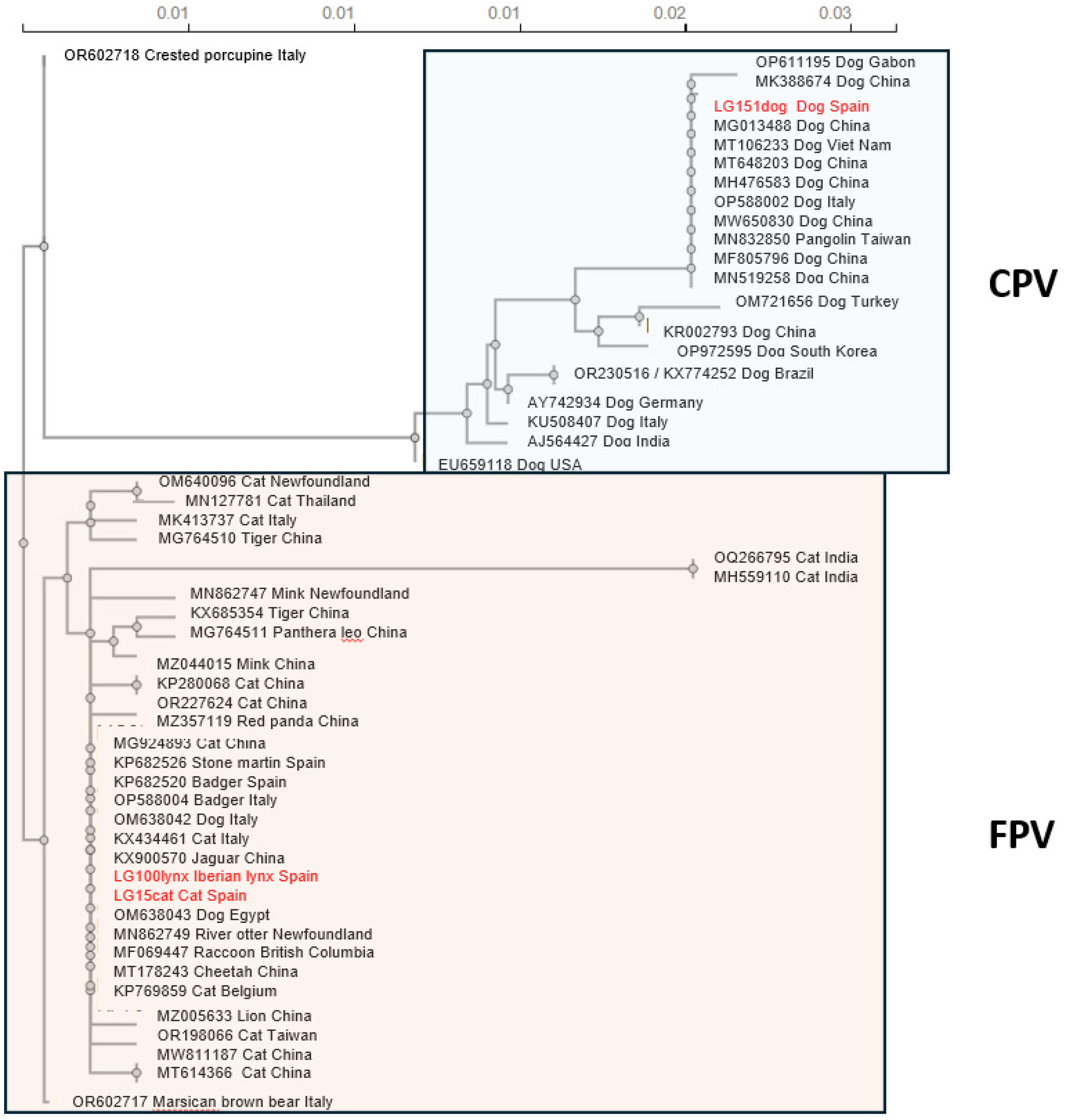
One of the main threats for the survival of the Iberian lynx are infectious diseases. Feline parvo-viruses cause an often fatal disease in cats and have been isolated from different species of Felidae and other carnivores. The present study is the first description of a parvoviral sequence isolated from the brain of an Iberian lynx which had died four weeks after been transferred to a quarantine centre from a hunting estate in Castilla-La-Mancha (southern border of the Iberian plateau). Four days prior to death he had developed anorexia and muscle weakness. The nucleotide sequence, 4,589 nt long (GenBank PP781551), was most proximal to that isolated from a Eurasian badger in Italy but showed also great homology with others from cats and other carnivores isolated in Spain and Italy, including that from a cat sequenced by us to elucidate the origin of the infection, which has not been clarified. The phylogenetic analysis of the capsid protein, VP2, which determines tropism and host range, confirmed that the lynx sequence was most proximal to feline than to canine parvoviruses, and was thus classified as Protoparvovirus carnivoran 1. More studies, in-cluding serology, are needed to understand the pathogenesis of this infection.

Keywords: 
;  ;  ;  ;  ;  

1. Introduction

The Iberian Lynx (Lynx pardinus) is one of the most threatened feline species worldwide. It is currently classified as “vulnerable” by the IUCN (International Union for Conservation of Nature) [1]. After 20 years of implementation of various in situ and ex situ programmes, a population of 2,021 individuals has been reached in the Iberian Peninsula in 2023, mainly located in the south-western regions of Spain (Andalusia, Castilla-La- Mancha and Extremadura) [2].
The reasons for the dramatic situation that the Iberian lynx has reached are various, including a drastic decrease in the population of rabbits, the main food source for lynxes, collisions with vehicles, and infectious diseases [3,4,5,6]. Between 2003 and 2011 infectious diseases such as pasteurellosis, tuberculosis, feline leukaemia virus and feline parvovirus infections, among others, were reported as the cause of 38.5% of recorded deaths of lynxes in the Andalusia region [7]. More recent studies also reported the detection of feline leukaemia virus, feline coronavirus, feline calicivirus and feline parvovirus in the Iberian lynx in the Extremadura region [8].
It has been reported that the Iberian lynx has a lack of acquired and innate immunity and immunocompetence, maybe due to inbreeding [9]. Susceptibility is enhanced by sharing the habitat with other carnivore species which may harbour pathogens, but which may pass unnoticed in the management of the area [5,6,8]. Consequently, infectious diseases may pose a serious risk for lynx conservation [5,6]. As feline parvovirus represents a threat to the Iberian lynx [8,10], screening tests are routinely carried out.
In January 2023 in Castilla-La-Mancha (central Spain), three weeks following translocation from a hunting estate for repopulation reasons, a subadult lynx was observed to have poor body condition, anorexia, diarrhoea and muscle weakness. Consequently, a veterinary examination was performed at the wildlife recovery centre “El Chaparrillo” in Castilla-La-Mancha. Despite stabilization attempts, the animal died four days later. A commercial qPCR for feline parvovirus diagnosis was performed with positive result, confirming for the first time the presence of the virus in this Spanish region.
Parvoviral infections, which may affect different species, are usually acute and frequently lethal. In cats, feline parvovirus (FPV) infection causes feline panleukopenia (FPL), a highly contagious disease, characterized by acute severe enteritis, dehydration and sepsis due to lymphoid depletion and pancytopenia, which may be fatal [11]. Parvoviruses are transmitted by the faecal-oral route, and are spread through contact with faeces and body fluids from infected animals, and fomites. Incubation time until clinical signs appear is usually 2 to 10 days [12].
Both FPV and its close relative, canine parvovirus (CPV), belong to genus Protoparvovirus, species Protoparvovirus carnivoran 1 within the family Parvoviridae, subfamily Parvovirinae. They are small non-enveloped icosahedral viruses with single-stranded DNA genome of approximately 5.2 Kb. FPV and CPV exhibit 98% genome homology [13,14]. The coding region of the genome contains two major open reading frames (ORFs); the 5’ ORF (ORF1) gives rise to non-structural (NS1 and NS2) proteins, whereas ORF2 translates into structural proteins (VP1 and VP2) [13]. NS proteins are essential for replication and DNA packaging, and VPs form the capsid and determine virus entry processes and host immunity [15]. The ends of the genome usually consist of untranslated regions (UTR) that form hairpin structures necessary for the initiation of replication [16,17].
Parvovirus lack the ability to induce cell multiplication and for the replication process to occur, they must infect cells that are in the S-phase of division, thus exhibiting tropism for tissues with high mitotic activity, such as the epithelial cells of the intestinal crypts, bone marrow and lymphoid tissues, and foetal cells [18,19]. Viral replication begins in the lymphoid tissue associated with the oropharynx, followed by a viraemia lasting 2-7 days during which it is distributed to other lymphoid organs such as the spleen or bone marrow, producing leukopenia and transient immunosuppression. It is finally spread to virtually all tissues and in pregnant females the virus crosses the placenta and infects the fetus. The ensuing shortening of the intestinal villi causes a malabsorption syndrome that triggers diarrhoea, which may be haemorrhagic or non-haemorrhagic, along with other associated clinical signs such as anorexia, dehydration, vomiting and generalized weakness. This clinical outcome makes parvoviral infection one of the most severe diseases in cats. Depending on the viral strain, immune status and age of the affected animal, FPV may produce its death [20,21]. Infection by FPV can be also subclinical, mainly in adult and subadult cats, which shed virus in their feces. This is supported by the high seroprevalence rates in some populations of unvaccinated adult cats [11], and by the probable persistence of the virus in the tissues of animals that have recovered from the infection [22].
Veterinary practitioners usually suspect of the disease based on the clinical signs, but confirmation by laboratory tests is recommended, of which PCR, both end-point or conventional (PCR) and quantitative or real-time (qPCR), are considered definitive.
Parvovirus have a large host spectrum [20]. The infection by FPV occurs worldwide and all members of the family Felidae are probably susceptible to infection with this virus [20]. Other carnivores of the families Viverridae, Procyonidae, Canidae and Mustelidae have been reported to be also susceptible to infection with these viruses [20]. Cases of parvovirus infection and/or disease have been described in lion, Eurasian lynx, tiger, leopard, cheetah and panther, besides Iberian lynx [4,18,23,24,25]. Clinical disease has been observed in raccoon (Procyon lotor), mink (genera Mustela and Neovison), and coatimundi (genus Nasua) [26] and leopard (Panthera pardus) [27]. Wild and domestic carnivore species sharing territory with the lynxes can act as reservoirs of FPV, favouring its transmission [10,21,23,28,29,30]. Pathogenesis and disease outcome of this viral disease has been mainly studied in domestic cats, and the infectivity of parvoviruses in the Iberian lynx is mostly unknown though both the virus (a sign of active infection) [3,28,31] and antibodies (a sign of past or present exposure to the virus) [5,6,31] have been detected in this threatened species.
The aim of the present study was to describe for the first time the complete sequence of a parvovirus genome from an Iberian lynx from the southern part of the Spanish plateau, which may be associated with its cause of death. As the possibility existed that it might have gotten infected from sympatric domestic animals and to determine if this sequence was related to other circulating parvoviruses in Spain, the parvoviral sequence of a cat and a dog from central Spain (though not exactly from the same area as the lynx) were also sequenced. All three of them were assigned to species Protoparvovirus carnivoran 1. According to the VP2 amino acid sequence, the parvoviral sequence of the lynx was classified as feline parvovirus.

2. Materials and Methods
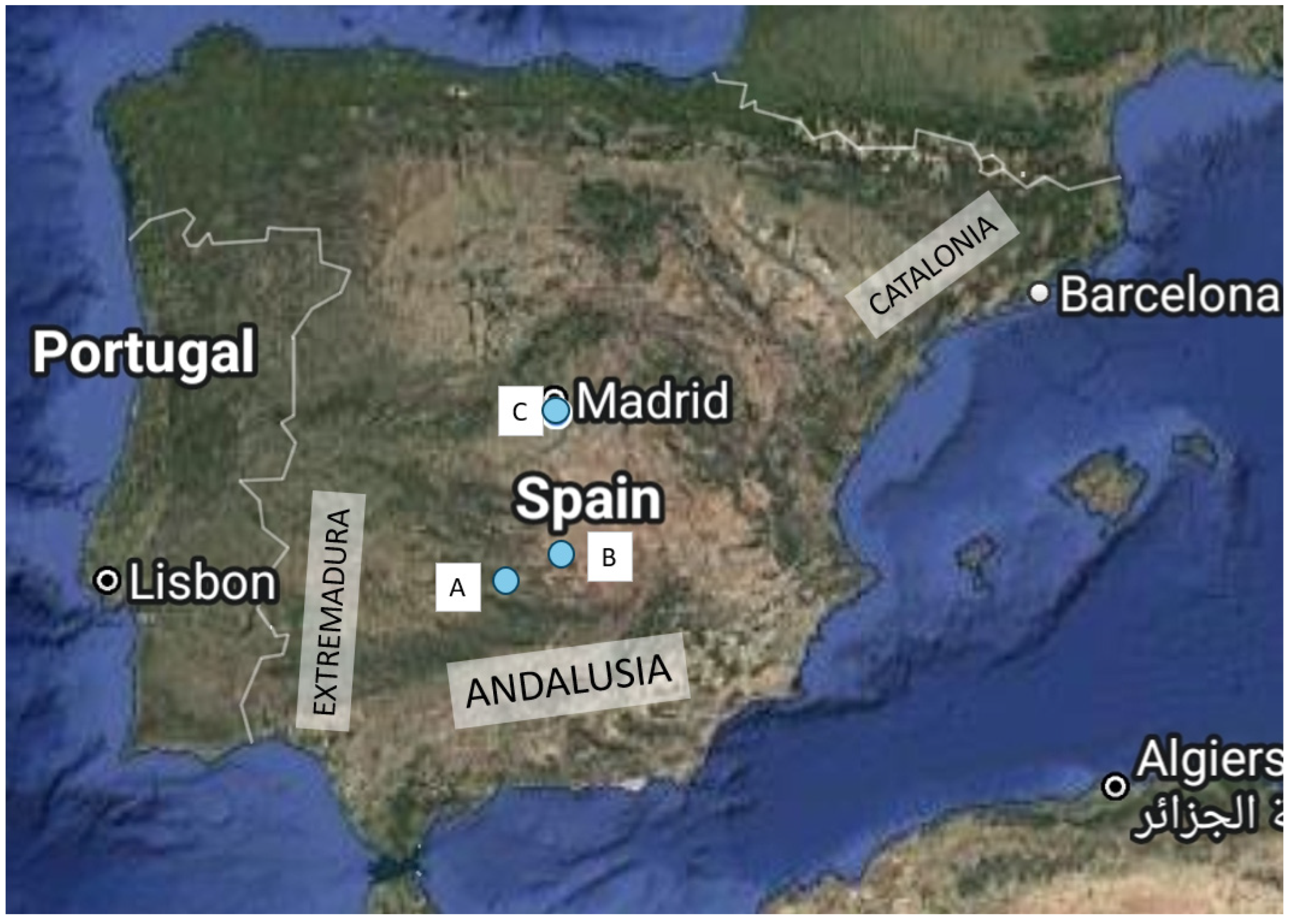
2.1. Case Report

A subadult (as judged by the dentition) male Iberian lynx was captured in a hunting estate located in Ciudad Real (a province of the region Castilla-La-Mancha, in the Southern limit of the Spanish plateau) for translocation and repopulation reasons in December 2022 in Castilla-La-Mancha (Figure 1). The lynx population in this hunting estate is estimated to be between 150 and 200 individuals. It is fenced all around, but lynxes have been observed to jump the fence in both directions. The lynx was transferred to the Recovery Centre “El Chaparrillo” in Ciudad Real, 93 km from the capture area, for quarantine. Upon arrival it was subjected to routine testing with no evident alterations in any of the parameters and no visible signs of illness. A rectal swab was sent to an external laboratory (Centre for the Analyses and Diagnosis of Wildlife in Andalusia, CAD, the reference laboratory for the Iberian lynx) for qPCR for FPV, giving a borderline result (Ct 33). The lynx was kept in a species-appropriate quarantine facility with a 9m2 box and a 400m2 outdoor area for three weeks, after which he developed acute disease, including severe diarrhoea, anorexia and dehydration. Despite stabilization attempts (fluid administration, antibiotic therapy and analgesia), he died four days later and a complete necropsy was performed. A rectal swab obtained when the lynx was sick and a sample of a mesenteric lymph node taken at necropsy were analysed by qPCR by the same external laboratory, giving positive results of Ct 17.93 and 20.18, respectively.

2.2. Samples

Sampling was carried out between March 2023 and March 2024 in the same hunting estate as described above. The sample set included n=51 stool samples; n=15 rectal swabs and n=15 serum samples from live Iberian lynxes; and n=12 stool samples from dogs, which were clinically healthy, from the same hunting estate as the lynxes. The 15 Iberian lynxes were captured for a sanitary survey and released after. All samples obtained from them were extracted under strict compliance of the protocols established for the health monitoring of the species by the Iberian Lynx Health Advisory Group (GAAS) [32]. To determine the similarities with other parvoviral strains circulating in Spain stools from a diarrheic cat (LG15) and dog (LG151), both from Madrid, were also analysed. They had been confirmed as positive to parvovirus by an in-practice immunochromatography test and were used as positive controls. Pet owners had been informed about the study and had agreed to participate in it. Faeces samples were suspended in RNAlater (Thermo Fisher).
In addition, we analysed samples of brain and heart, obtained from the necropsy of the dead lynx mentioned above, following a standard necropsy procedure. All samples were stored at -20˚C until DNA isolation was carried out.

2.3. DNA Extraction

Rectal swabs, faeces and necropsy tissues were resuspended in 1ml of phosphate-buffered saline (PBS). After mechanical maceration, samples were centrifuged at 4,000 rpm for 2 minutes; this facilitated the proper dilution of the sample in the added PBS and the sedimentation of solid matter at the bottom of the Eppendorf tubes, preventing obstruction of the extraction kit membrane. DNA was extracted from the supernatant using High Pure Viral Nucleic Acid Kit (Roche) following the manufacturer’s instructions and stored at -20 ˚C.

2.4. Commercial End-Point PCR

All DNA extracts from lynx, cat and dog samples were analysed by a commercial end-point PCR for canine and feline parvovirus (CPV-2-FLPV-EPPCR, Genetic PCR solutions™), following the instructions of the manufacturer. Amplified products were visualized in 0.8% agarose gels. A band of the expected size (around 300 bp) in the electrophoresis gel was considered positive.

2.5. Inhouse Nested PCR

As before, all DNA extracts from lynx, cat and dog samples (faeces, rectal swabs and necropsy samples) were screened by a nested PCR designed by us targeting the NS1 region of the parvoviral genome, using an external PCR, which amplified a 1671 nucleotide (nt) segment performed using the primers FPL28Fw and FPL1699Rev (Supplementary Table S1), followed by an internal PCR, giving a product of expected size of 959 nt, using the primers FPL438Fw and FPL1397Rev (Supplementary Table S1) of a 1:10 dilution of the first PCR product. Primers were designed by comparing 21 parvoviral genomes deposited in GenBank (Supplementary Table S2). This comparison included sequences from cats, dogs and wild animals from different areas of the world, which were aligned and analysed using MEGA software version 11 (MEGA11), selecting the most conserved areas. The primers were analysed using Vector NTI software (Thermo Fisher Scientific). The DNA extracted from cat LG15 was used as positive control, and non-template reaction was included as negative control.
Positive samples by the commercial PCR were subjected to complete genome amplification using a total of 17 sets of primers designed by us (Supplementary Table S1), following a similar strategy as described above. The most conserved regions of 18-23 nt separated 500–1000 nt from each other were used for the design of primers covering the entire parvovirus genome (detailed in Supplementary Tables S1 and S3, and Figure 2). Nested PCRs included external PCRs which could be up to 2500 nt in size, while internal PCRs were 900-1100 nt in size (Supplementary Table S3). For sequencing the dog parvovirus, primers published by Pérez et al. [16] were used to complete difficult to sequence areas.
Nested PCRs were performed using DNA Polymerase (1U/µl; Biotools) in a final volume of 25 µl comprising 16 or 14 μl of sterile distilled water (external or internal PCR, respectively), 2.5 μl of Buffer 10x with MgCl2 (Biotools), 0.5 μl dNTPs (10 mM Biotools), 1 μl of each primer (10 μM), 1 μl DNA Polymerase (1U/µl) and 3 or 5 μl of DNA (external or internal PCR, respectively). The first PCR product was diluted 1:10 and used as template for the internal PCR. Extreme care was taken to avoid PCR cross-contamination, and DNA extraction, external PCR, internal PCR and separation in gel were handled in separate work areas. Non-template controls were included in each assay to ensure that there was no sample contamination, which was also confirmed by the consistency of the results. The PCR amplification was performed with the following cycle conditions: initial denaturation at 95 ˚C for 5 min; followed by 30 cycles of denaturation at 95˚C for 1 min, annealing at 5 ˚C below the lowest Tm of the pair of primers for 1 min, extension at 72 ˚C for 2 or 1 min (external and internal PCRs, respectively); and final extension at 72 ˚C for 5 min.

2.6. Sequencing and Further Analyses

The amplified PCR products were purified using QIAquick PCR Purification Kit (Qiagen) and Sanger’s dideoxy based sequencing was performed using both forward and reverse primers at Macrogen, Inc. (South Korea). The nucleotide sequence data were checked with Chromas (Technelysium DNA Sequencing Software), subjected to Blast analysis, assembled and analysed using MEGA11 by the ClustalW method. The amino acid sequence, predicted using ORF Finder (https://www.bioinformatics.org/sms2/orf_find.html), was also subjected to Blast search and analysed with MEGA11. Assembled sequences were deposited in GenBank with Accession Numbers PP781551 (lynx parvovirus LG100), PQ436979 (feline parvovirus LG15), and PQ436980 (canine parvovirus LG151).
Phylogenetic analysis of genomes obtained was performed using the Clustal Omega core alignment engine which uses the HHalign algorithm and its default settings. The algorithm is described in [33]. The GenBank Accession Number of the 51 sequences of Protoparvovirus included for constructing the phylogenetic tree of the VP2 protein are shown in Supplementary Table S4. They were selected to cover a large spectrum of mammalian species hosts from different parts of the world.

2.7. Serological Analysis

Sera of the 15 Iberian lynxes captured for sanitary check-ups were analysed by ELISA for the detection of antibodies against FPV (IgG and IgM) (Feline Parvovirus ab EIA, Euroveterinaria), following the manufacturer’s instructions.

3. Results

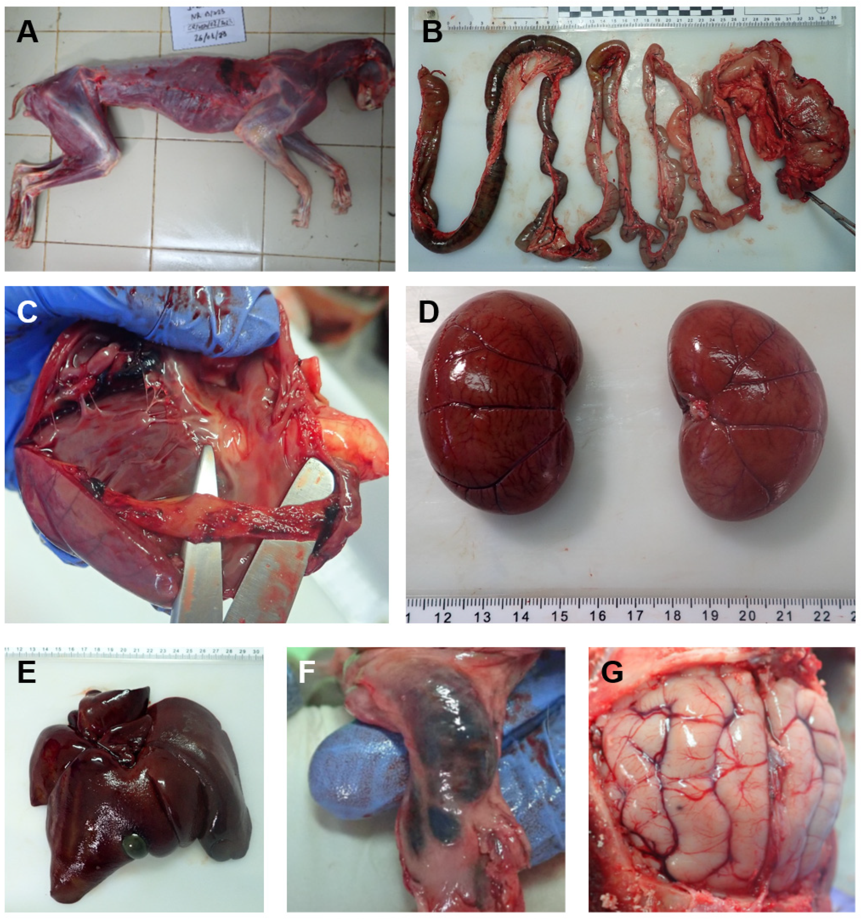
3.1. Necropsy Findings of the Affected Iberian Lynx

An Iberian lynx was found to have severe diarrhoea and dehydration three weeks after translocation from the hunting estate to the recovery centre “El Chaparrillo”. Despite intense attempts to stabilize him, he died four days after the beginning of the illness. Upon death, the lynx was found to be severely emaciated, with a body condition of 1.5 (scale 1-5) (Figure 3A). The necropsy (Figure 3) revealed the presence of haemorrhagic gastroenteritis with congestion of several organs, including the subcutaneous and submucosal vessels, the mesenteric lymph nodes and ileocecal papilla, the brain vessels, spleen, liver congestion and hepatomegaly, lung congestion, oedema and emphysema, among other lesions, all compatible with parvovirus infection or secondary bacterial infections due to increased intestinal permeability caused by the virus. Cardiomegaly and myocarditis were also observed but were not confirmed by histopathology. The wall of the bladder was engrossed. A commercial qPCR for feline parvovirus diagnosis was performed resulting in a Ct of 17.93 in a rectal swab and Ct 20.18 in a mesenteric lymph node sample of the lynx confirming for the first time the presence of the virus in this Spanish region.
The results of the histopathological study revealed encephalitis with multifocal satellitosis, centrilobular atrophy and hepatic congestion, multifocal interstitial nephritis, congestion and pulmonary oedema with diffuse interstitial pneumonia, and reactive lymphoid population in mesenteric lymph node. The intestinal fragments sent presented a high degree of autolysis, which hampered the correct assessment of the mucosa.
Samples from both the brain (LG100) and the heart (LG101) obtained in the necropsy of this lynx yielded a positive result by conventional PCR, both commercial and inhouse designed by us targeting the NS1 gene with 982 bp product (external primers FPL28Fw/FPL1699Rev; internal primers FPL438Fw/FPL1397Rev). DNA extracted from the brain was subjected to complete genome sequencing and was submitted to GenBank where it received the accession number PQ436979. Several PCR reactions with other primers were done also with the other positive sample from the same lynx (LG101) and as sequencing results were identical to LG100 it was not sequenced any further. From here on the complete genome sequence of the lynx virus will be referred to as LG100lynx. It shares 99.76% nucleotide identity with the Protoparvovirus carnivoran 1 OP588004 FPV variant, isolated from a Eurasian badger (Meles meles) in Italy, the closest relative so far in GenBank. The genomic sequence of LG100lynx comprises 4,589 nucleotides encoding two non-structural proteins, NS1 and NS2 (nt 64-2070) and two structural proteins, VP1 and VP2 (nt 2077-4333) (Table 1). According to the VP2 amino acid sequence LG100lynx is classified as feline parvovirus.

3.2. Prevalence of Parvovirus in the Hunting Estate

To determine the prevalence of the infection in the hunting estate we screened both by a commercial PCR and with our inhouse nested PCR, DNA extracted from faeces of local lynxes. Rectal swabs from the 15 individuals captured for sanitary survey reasons were also analysed. None of these 66 faeces and rectal swab samples were found to be positive. To determine the origin of the infection of the affected lynx, the faeces of 12 local dogs were also analysed, which turned out to be negative.
In addition, all sera from the 15 live lynxes captured for sanitary screening, were negative by ELISA to antibodies against feline parvovirus.

3.3. Parvoviral Sequences in a Cat and a Dog in Spain

In an attempt to determine the origin of the infection in the lynx by comparison with sequences of parvoviruses from other animals and in the absence of positive samples in other species in the hunting estate, stools from a parvovirus infected symptomatic cat (LG15) and dog (LG151) from Madrid were analysed. Both tested positive by the commercial and inhouse conventional PCRs. They were also subjected to complete genome sequencing, and deposited in GenBank with accession numbers PP781551 and PQ436980, respectively. They will be referred to as LG15cat and LG151dog, respectively. LG15cat shares 99.87% and 99.82% nucleotide identity with OP588004 and OM638042 FPV variants, also from Italy, isolated from the Eurasian badger and a dog, respectively. LG151dog shares 99.93% nucleotide identity with MG013488 FPV variant, isolated from a dog in China. LG100lynx shares 99.87% nucleotide identity with LG15cat and 98.52% with LG151dog. The genomic data of LG15cat and LG151dog are shown in Table 1. According to the deduced amino acid sequence of VP2 LG15cat is classified as FPV, while LG151dog as CPV-2c. The comparison of the deduced amino acid sequences of VP2 from the strains isolated in this study with other FPV and CPV-2 strains from different countries is shown in Table 2, in which sequences exactly like LG100lynx and LG151dog are omitted.

3.4. Phylogenetic Analysis of VP2

A phylogenetic tree based on full-length VP2 gene was constructed using 51 FPV and CPV-2 sequences (Supplementary Table S4), 48 of them obtained from GenBank and the three isolated in this study (LG100lynx, LG15cat, LG151dog) (Figure 3). As can be seen, VP2 is crucial for the classification of strains into CPV and FPV. Within FPV it is worth mentioning that isolates from two dogs in Italy, and one dog from Egypt fell in this species; and that the Spanish sequences were most similar to other strains from Spain and Italy.

4. Discussion

The Iberian lynx just passed from the IUCN list of “critically endangered species” to that of “vulnerable species” as a result of conservation efforts. The population has significantly increased thanks to captive breeding projects and reintroduction. While the population was 62 mature individuals in 2001, it increased to 648 in 2022. Today, the total population, including young and mature ones, is estimated to exceed 2000 individuals [1]. However, the situation is far from ideal, and lynxes are exposed to a multitude of dangers, being infectious diseases one of the most prominent threats. In this context it is especially important that lynxes which are being translocated to repopulate other areas are absolutely free of known pathogens. The lynx of this report was found to have a borderline result with a commercial qPCR for feline parvovirus (Ct 33) in a rectal swab at his entrance to the Recovery Centre “El Chaparrillo” where he had been taken for quarantine as an intermediate stage in the translocation process. Almost four weeks later he died from acute disease and qPCR showed an evident increase in the viral load (Ct 17.93 in rectal swab). Several questions were raised, including whether other lynxes were infected, where the infection came from, and whether the lynx was already infected before its capture in the hunting estate.
To determine if other lynxes were infected, faeces were screened to ascertain the presence of parvovirus in these samples. Stool samples were chosen as a non-invasive and adequate way to determine parvoviral infection in carnivores [3]. When analysing results, it must be taken into account that two or more faecal samples could proceed from the same animal so the number of stool samples may be higher than that of lynxes scrutinised. During the autumn health monitoring campaign (10 months after the detection of the positive case), rectal swabs and blood samples from 15 Iberian lynxes, belonging to the same subpopulation as the dead lynx and captured for a sanitary survey, were also analysed. All stool samples and rectal swabs turned out to be negative to FPV (0/66), as well as for antibodies in the sera from the same animals as the rectal swabs (0/15), suggesting that the prevalence of the parvoviral infection in the hunting estate could be null or minimal.
This low prevalence of parvovirus in the Iberian lynx in the southern part of the Spanish central plateau is consistent with other studies reporting low exposure to this pathogen in this species. Najera et al. [31] detected a prevalence of 1.5% (1 lynx out of 67 sampled); the infected lynx died because of vehicle collision and no signs associated to FPV were observed at necropsy. López et al. (2014) analysed 22 fresh carcasses of Iberian lynx in southern Spain (Andalusia) and identified only one in which death could be due to parvoviral infection [34]. This coincided with data from Meli et al. (2009), who studied different pathogens in two areas of Andalusia and found 2/75 (2.7%) free-ranging lynxes positive by PCR to FPV, both of them distant from the site of our study. A similar low prevalence has been observed in other lynx species worldwide [3,4]. The solitary and territorial nature of lynxes may account for this low prevalence, as contagion between individuals would be more difficult. However, serological surveys have detected the presence of anti-FPV antibodies in from ≤11% non-vaccinated Iberian lynxes [6,31], Eurasian lynx [35], and up to 30% [36]. In a serological survey performed in 2023 in Castilla-La-Mancha, three out of 64 (4.7%, co-author Elena Crespo, personal communication) of the lynxes screened were positive to parvoviral antibodies, confirming a previous but infrequent contact with the virus. In the present study and due to its non-invasive design, serology was performed only in the lynxes from which the rectal swabs were obtained. As mentioned, all of them were negative. However, due to the limited sample size, more studies about serology would be advisable, especially taking into account that in lynxes the infection may be subclinical, as highlighted by the finding of parvoviruses in the carcass and vehicle runovers mentioned above [31,34] and would be fundamental for understanding the pathogenesis of this infection in the Iberian lynx.
To trace the origin of the infection in the lynx we resorted to sequencing the genome of the virus. According to the genomic organization and phylogenetic inference of LG100lynx it was classified as a Protoparvovirus carnivoran 1. To the best of our knowledge, this is the first time that the complete sequence of a Protoparvovirus genome has been described in the Iberian lynx (Lynx pardinus), and that it may be associated with a clinical case in the Castilla-La-Mancha region in Spain, as previous findings corresponded to dead animals and the presence of parvovirus could not be associated to the death of the lynxes (they were found in a decomposed carcass and in runovers) [31,34].
The 12 dogs sampled from this location turned out to be negative. In the absence of finding parvoviruses in the hunting estate, we sequenced a FPV and a CPV from the central area of the country. LG100lynx had high similarity with LG15cat (99.87%). Blast analysis showed that both LG100lynx and LG15cat were very closely related to others previously found in different domestic carnivores from distant countries. This further supports that cross-species transmission takes place [21]. However, our results do not allow us to ascertain where and from which species the Iberian lynx might have gotten the parvoviral infection. In Spain parvoviruses have been found in different species of wild carnivores, including European wildcat, Eurasian badgers, a genet, and wolves (CPV-2c); wolves, red fox and stone marten (CPV-2b); and badger, genet and stone marten (FPV) [21,28]. Studies about the presence of FPV in areas where there is a population of Iberian lynx suggest FPV circulation and that it may have a reservoir in free-roaming domestic cats [5]. The well-known environmental resistance of parvoviruses would facilitate transmission.
To further compare LG100lynx with other parvoviruses, the amino acid sequence of VP2 was characterized. This is the most abundant protein of the capsid of parvoviruses [37] and determines host range, hemagglutination activity and antigenicity, and variations in the VP2 gene are closely associated with the virulence and transmissibility of the virus. Specific amino acid positions [34,38] in the VP2 protein differentiate FPV from CPV-2 and residue 426 allows differentiation between CPV-2a, -2b and -2c variants [39,40], and FPV from CPV [21]. The similarity with the sequence we obtained from the symptomatic cat in central Spain (LG15), and with the VP2 sequences of the stone martin (Martes foina) and the Eurasian badger obtained in Catalonia, in north eastern Spain [21] suggests LG100lynx might be derived from current circulating FPVs in the Spanish cat population, as observed in other studies in which the transmission of FPV from domestic to wild animals is described [4,10,29,30]. Further research is needed to understand the dynamics of FPV transmission between wild and domestic environments in this particular case, as well as the origin of the infection in the Iberian lynx. As expected, LG151dog exhibits similar amino acid variations as the other sequences classified as CPV-2c [21].
As to whether the lynx had been infected before it was quarantined, evidence points to a subclinical infection, as qPCR at entrance in the Recovery Centre was borderline though present (Ct 33). The onset of the infection could be attributed to captive and transport stress. Whether certain conditions such as immunosuppression could induce re-shedding of FPV or CPV in cats has not been investigated (in [11]), but other similar cases are reported in wild animals [27]. It has been suggested that Protoparvovirus carnivoran 1 DNA may persist for long periods in the tissues of animals that have recovered from infection without an active infection [22]. Subclinical infections, common in young adult cats, are difficult to detect, and may become apparent only when the animal is subjected to stress [11].
The fact that both the brain and the heart of the affected lynx were positive to parvovirus by PCR is intriguing. These are sites that may be affected in foetal kittens but not in adult cats, though it has been reported that parvoviruses may replicate in cerebral neurons of cats [41]. A specific mutation in the protein NS1, L582S, that may be related to neuronal tropism of FPV [42], even in adult cats, was not detected in the LG100lynx sequence. In our study congestion of encephalic vessels was evident, and the positive detection of parvovirus by PCR and its subsequent classification as FPV constitutes the first report of infection by this virus in the brain of an Iberian Lynx. This data may be relevant for the epidemiology of FPV in lynx, as the animal, a subadult, could be suffering an asymptomatic infection with presence of the virus in brain areas, without neurological signs, since anorexia and diarrhoea were the only clinical manifestations observed. It is worth mentioning that pathogenesis studies stem from data in cats and the progress might be different in other Felidae. The absence of the specific mutation in NS1 [42] also broadens the spectrum of strains that have tropism for brain tissues. As regards myocarditis it is a recognized complication of CPV infection in puppies, but it has not been convincingly associated with FPV in cats [11]. Unluckily other tissues were not made available to us, and would have helped to clarify the extension of the infection in this lynx, though histopathological analysis provided data as to the pathogenesis of the infection.

5. Conclusions

Infectious diseases can be a threat to the survival of endangered species, so the healthcare of populations is of utmost importance. Specifically in the Iberian lynx the lack of immunocompetence associated with consanguinity derived from the limited number of animals makes it especially vulnerable. On the other hand, the comeback of this species implies the occupation of suboptimal territories, closer to urban centres that involve a greater probability of contact with domestic cats. The naivety of the immune system to parvoviruses due to little contact with these pathogens, as shown by serological analyses, makes these viruses a potential threat to populations of Iberian lynx. Since this virus may cause high mortality in felines including the Iberian lynx [7] and other lynx species such as the Eurasian lynx [43] and bobcat [44], periodic screening should be conducted in Iberian lynxes to prevent and monitor possible cross-transmission from domestic to wild and susceptible species.
Based on the healthy condition of the lynx at the time of admission and the time it took to develop the disease, our hypothesis regarding the described case is that it had a subclinical FPV infection which become apparent when it was subjected to stress. More studies, including serology to determine previous exposures to the virus, and body locations where the virus may persist due to its resistance, are needed to determine the pathogenicity of FPV in the Iberian lynx and to prevent its spread, endangering even further the survival of the species.

Supplementary Materials

The following supporting information can be downloaded at the website of this paper posted on Preprints.org, Supplementary Figure S1. Phylogenetic tree of the 32 genome sequences deposited in GenBank with which LG100lynx, LG15cat, and LG151dog (marked with red stars) were compared, including the species from which they were isolated and the geographic location; Supplementary Table S1. Primers used in the study. Nucleotide positions in the genome of Protoparvovirus carnivoran 1 sequenced from a badger in Italy (GenBank Acc. No. OP588004). Primers were named according to the position of the first nucleotide in the + strand. UTR, untranslated region. Fw, forward primer. Rev, reverse primer. All primers were designed by us, except those marked with **, which were published before (Pérez et al. 2014). Supplementary Table S2. GenBank Accession Numbers of the 21 parvoviral genomes used for designing the primers used in this study, including the species in which they have been classified by their researchers. Supplementary Table S3. Protocols of nested PCRs used for sequencing the complete genome of Protoparvovirus carnivoran 1 from lynx (LG100), cat (LG15) and dog (LG151). The number in the name of the primer, where present, indicates the position in the genome of Protoparvovirus carnivoran 1 sequenced from a badger in Italy (GenBank Acc. No. OP588004). Supplementary Table S4. GenBank Accession Numbers of the sequences used for constructing the phylogenetic tree of the VP2 protein, including the species in which they have been classified by their researchers.

Author Contributions

Conceptualization, Esperanza Gomez-Lucia and Ana Domenech; Data curation, Almudena Campoy, Esperanza Gomez-Lucia and Sergio Fandiño; Formal analysis, Almudena Campoy, Esperanza Gomez-Lucia, Sergio Fandiño, Elena Crespo and Ana Domenech; Funding acquisition, Sonia Olmeda; Investigation, Almudena Campoy, Esperanza Gomez-Lucia, Tania Garcia, Sonia Olmeda, Felix Valcarcel; Methodology, Almudena Campoy, Tania Garcia, Felix Valcarcel, Elena Crespo and Ana Domenech; Project administration, Ana Domenech; Resources, Sonia Olmeda, Felix Valcarcel, and Ana Domenech; Supervision, Esperanza Gomez-Lucia; Validation, Almudena Campoy and Tania Garcia; Writing – original draft, Almudena Campoy and Esperanza Gomez-Lucia; Writing – review & editing, Almudena Campoy, Esperanza Gomez-Lucia, Sonia Olmeda, Felix Valcarcel, Elena Crespo and Ana Domenech.

Funding

This research received financial support from Finca La Garganta, who promoted the study. Financial support was used solely for the acquisition of reagents.

Institutional Review Board Statement

This study used feces for which no ethical approval was needed. Samples from live lynxes were obtained by veterinarians following the official protocol approved by the Institutional Review Board of the Wildlife Recovery Center “El Chaparrillo” for studies involving animals and described in the Manual sanitario del lince ibérico. https://www.lynxexsitu.es/ficheros/documentos_pdf/85/Manual_Sanitario_Lince_Ib_2014.pdf.

Informed Consent Statement

Owners of animals (cat and dog) from which feces were obtained were informed orally and given a written document to sign.

Data Availability Statement

Parvoviral sequences obtained in this study have been deposited in GenBank under Accession Numbers PP781551 (Iberian lynx LG100), PQ436979 (cat LG15) and PQ436980 (dog LG151).

Acknowledgments

The authors are greatly indebted to the hunting estate “La Garganta” (Villamagna, S.A. – Brazatortas, Ciudad Real) for promoting and inspiring this work, especially the head person His Grace the Duke of Westminster, the managers Ignacio Landaluce and José María Tercero, as well as the assistance of their employees for providing the fecal samples of the lynx. The assistance of the Veterinary Clinic “La Dehesa” (San Sebastian de los Reyes, Madrid, Spain), and the Veterinary Hospital of the Complutense University of Madrid (HCVC) for kindly supplying feline and canine feces is gratefully acknowledged. The altruistic assistance of Pere Busquets from Urano Diagnostics with performing the histopathological analysis is gratefully acknowledged.

Conflicts of Interest

The authors declare no conflicts of interest. The management team of the hunting estate had no role in the design of the study; in the analyses, or interpretation of data; or in the decision to publish the results.

References

  1. IUCN Recovery of the Iberian Lynx: A Conservation Success in Spain 2024.
  2. Spanish MInistry of Ecological Transition and Demographic Challenge Fauna de Vertebrados: Mamíferos) (Fauna of Vertebrates: Mammals).
  3. Duarte, A.; Fernandes, M.; Santos, N.; Tavares, L. Virological Survey in Free-Ranging Wildcats (Felis Silvestris) and Feral Domestic Cats in Portugal. Vet. Microbiol. 2012, 158, 400–404. [Google Scholar] [CrossRef] [PubMed]
  4. Wasieri, J.; Schmiedeknecht, G.; Förster, C.; König, M.; Reinacher, M. Parvovirus Infection in a Eurasian Lynx (Lynx Lynx) and in a European Wildcat (Felis Silvestris Silvestris). J. Comp. Pathol. 2009, 140, 203–207. [Google Scholar] [CrossRef] [PubMed]
  5. Millán, J.; Candela, M.G.; Palomares, F.; Cubero, M.J.; Rodríguez, A.; Barral, M.; de la Fuente, J.; Almería, S.; León-Vizcaíno, L. Disease Threats to the Endangered Iberian Lynx (Lynx Pardinus). Vet. J. 2009, 182, 114–124. [Google Scholar] [CrossRef] [PubMed]
  6. Roelke, M.E.; Johnson, W.E.; Millán, J.; Palomares, F.; Revilla, E.; Rodríguez, A.; Calzada, J.; Ferreras, P.; León-Vizcaíno, L.; Delibes, M.; et al. Exposure to Disease Agents in the Endangered Iberian Lynx (Lynx Pardinus). Eur. J. Wildl. Res. 2008, 54, 171–178. [Google Scholar] [CrossRef] [PubMed]
  7. López, G.; López-Parra, M.; Garrote, G.; Fernández, L.; Del Rey-Wamba, T.; Arenas-Rojas, R.; García-Tardío, M.; Ruiz, G.; Zorrilla, I.; Moral, M.; et al. Evaluating Mortality Rates and Causalities in a Critically Endangered Felid across Its Whole Distribution Range. Eur. J. Wildl. Res. 2014, 60, 359–366. [Google Scholar] [CrossRef]
  8. Nájera, F.; Grande-Gómez, R.; Peña, J.; Vázquez, A.; Palacios, M.J.; Rueda, C.; Corona-Bravo, A.I.; Zorrilla, I.; Revuelta, L.; Gil-Molino, M.; et al. Disease Surveillance during the Reintroduction of the Iberian Lynx (Lynx Pardinus) in Southwestern Spain. Animals 2021, 11, 547. [Google Scholar] [CrossRef] [PubMed]
  9. Peña, L.; Garcia, P.; Jiménez, M.Á.; Benito, A.; Alenza, M.D.P.; Sánchez, B. Histopathological and Immunohistochemical Findings in Lymphoid Tissues of the Endangered Iberian Lynx (Lynx Pardinus). Comp. Immunol. Microbiol. Infect. Dis. 2006, 29, 114–126. [Google Scholar] [CrossRef]
  10. Millán, J.; Candela, M.G.; Palomares, F.; Cubero, M.J.; Rodríguez, A.; Barral, M.; de la Fuente, J.; Almería, S.; León-Vizcaíno, L. Disease Threats to the Endangered Iberian Lynx (Lynx Pardinus). Vet. J. 2009, 182, 114–124. [Google Scholar] [CrossRef]
  11. Barrs, V.R. Feline Panleukopenia: A Re-Emergent Disease. Vet. Clin. N. Am. Small Anim. Pract. 2019, 49, 651–670. [Google Scholar] [CrossRef]
  12. Csiza, C.K.; De Lahunta, A.; Scott, F.W.; Gillespie, J.H. Pathogenesis of Feline Panleukopenia Virus in Susceptible Newborn Kittens II. Pathology and Immunofluorescence. Infect. Immun. 1971, 3, 838–846. [Google Scholar] [CrossRef]
  13. Cotmore, S.F.; Agbandje-McKenna, M.; Canuti, M.; Chiorini, J.A.; Eis-Hubinger, A.-M.; Hughes, J.; Mietzsch, M.; Modha, S.; Ogliastro, M.; Pénzes, J.J.; et al. ICTV Virus Taxonomy Profile: Parvoviridae. J. Gen. Virol. 2019, 100, 367–368. [Google Scholar] [CrossRef] [PubMed]
  14. Chung, H.-C.; Kim, S.-J.; Nguyen, V.G.; Shin, S.; Kim, J.Y.; Lim, S.-K.; Park, Y.H.; Park, B. New Genotype Classification and Molecular Characterization of Canine and Feline Parvoviruses. J. Vet. Sci. 2020, 21, e43. [Google Scholar] [CrossRef] [PubMed]
  15. Cotmore, S.F.; Agbandje-McKenna, M.; Chiorini, J.A.; Mukha, D.V.; Pintel, D.J.; Qiu, J.; Soderlund-Venermo, M.; Tattersall, P.; Tijssen, P.; Gatherer, D.; et al. The Family Parvoviridae. Arch. Virol. 2014, 159, 1239–1247. [Google Scholar] [CrossRef] [PubMed]
  16. Pérez, R.; Calleros, L.; Marandino, A.; Sarute, N.; Iraola, G.; Grecco, S.; Blanc, H.; Vignuzzi, M.; Isakov, O.; Shomron, N.; et al. Phylogenetic and Genome-Wide Deep-Sequencing Analyses of Canine Parvovirus Reveal Co-Infection with Field Variants and Emergence of a Recent Recombinant Strain. PLoS ONE 2014, 9, e111779. [Google Scholar] [CrossRef]
  17. Sánchez, C.; Doménech, A.; Gomez-Lucia, E.; Méndez, J.L.; Ortiz, J.C.; Benítez, L. A Novel Dependoparvovirus Identified in Cloacal Swabs of Monk Parakeet (Myiopsitta Monachus) from Urban Areas of Spain. Viruses 2023, 15, 850. [Google Scholar] [CrossRef]
  18. Steinel, A.; Parrish, C.R.; Bloom, M.E.; Truyen, U. Parvovirus Infections in Wild Carnivores. J. Wildl. Dis. 2001, 37, 594–607. [Google Scholar] [CrossRef]
  19. Stuetzer, B.; Hartmann, K. Feline Parvovirus Infection and Associated Diseases. Vet. J. 2014, 201, 150–155. [Google Scholar] [CrossRef] [PubMed]
  20. Capozza, P.; Martella, V.; Buonavoglia, C.; Decaro, N. Emerging Parvoviruses in Domestic Cats. Viruses 2021, 13, 1077. [Google Scholar] [CrossRef] [PubMed]
  21. Calatayud, O.; Esperón, F.; Velarde, R.; Oleaga, Á.; Llaneza, L.; Ribas, A.; Negre, N.; de la Torre, A.; Rodríguez, A.; Millán, J. Genetic Characterization of Carnivore Parvoviruses in Spanish Wildlife Reveals Domestic Dog and Cat-Related Sequences. Transbound. Emerg. Dis. 2020, 67, 626–634. [Google Scholar] [CrossRef] [PubMed]
  22. Allison, A.B.; Kohler, D.J.; Fox, K.A.; Brown, J.D.; Gerhold, R.W.; Shearn-Bochsler, V.I.; Dubovi, E.J.; Parrish, C.R.; Holmes, E.C. Frequent Cross-Species Transmission of Parvoviruses among Diverse Carnivore Hosts. J. Virol. 2013, 87, 2342–2347. [Google Scholar] [CrossRef]
  23. Leopardi, S.; Milani, A.; Cocchi, M.; Bregoli, M.; Schivo, A.; Leardini, S.; Festa, F.; Pastori, A.; de Zan, G.; Gobbo, F.; et al. Carnivore Protoparvovirus 1 (CPV-2 and FPV) Circulating in Wild Carnivores and in Puppies Illegally Imported into North-Eastern Italy. Viruses 2022, 14, 2612. [Google Scholar] [CrossRef] [PubMed]
  24. Yeo, Y.-G.; Kim, H.-R.; Park, J.; Kim, J.-M.; Shin, Y.-K.; Lee, K.-K.; Kwon, O.-K.; Jeoung, H.-Y.; Kang, H.-E.; Ku, B.-K.; et al. Epidemiological and Molecular Approaches for a Fatal Feline Panleukopenia Virus Infection of Captive Siberian Tigers (Panthera Tigris Altaica) in the Republic of Korea. Animals 2023, 13, 2991. [Google Scholar] [CrossRef] [PubMed]
  25. Sacristán, I.; Esperón, F.; Pérez, R.; Acuña, F.; Aguilar, E.; García, S.; López, M.J.; Neves, E.; Cabello, J.; Hidalgo-Hermoso, E.; et al. Epidemiology and Molecular Characterization of Carnivore Protoparvovirus-1 Infection in the Wild Felid Leopardus Guigna in Chile. Transbound. Emerg. Dis. 2021, 68, 3335–3348. [Google Scholar] [CrossRef]
  26. Parrish, C.R. Emergence, Natural History, and Variation of Canine, Mink, and Feline Parvoviruses. Adv. Virus Res. 1990, 38, 403–450. [Google Scholar] [CrossRef]
  27. Kolangath, S.M.; Upadhye, S.V.; Dhoot, V.M.; Pawshe, M.D.; Bhadane, B.K.; Gawande, A.P.; Kolangath, R.M. Molecular Investigation of Feline Panleukopenia in an Endangered Leopard (Panthera Pardus) – a Case Report. BMC Vet. Res. 2023, 19, 56. [Google Scholar] [CrossRef]
  28. León, C.I.; García-Bocanegra, I.; McCain, E.; Rodríguez, E.; Zorrilla, I.; Gómez, A.M.; Ruiz, C.; Molina, I.; Gómez-Guillamón, F. Prevalence of Selected Pathogens in Small Carnivores in Reintroduction Areas of the Iberian Lynx ( Lynx Pardinus ). Vet. Rec. 2017, 180, 252. [Google Scholar] [CrossRef]
  29. Miranda, C.; Santos, N.; Parrish, C.; Thompson, G. Genetic Characterization of Canine Parvovirus in Sympatric Free-Ranging Wild Carnivores in Portugal. J. Wildl. Dis. 2017, 53, 824–831. [Google Scholar] [CrossRef]
  30. Duarte, M.D.; Henriques, A.M.; Barros, S.C.; Fagulha, T.; Mendonça, P.; Carvalho, P.; Monteiro, M.; Fevereiro, M.; Basto, M.P.; Rosalino, L.M.; et al. Snapshot of Viral Infections in Wild Carnivores Reveals Ubiquity of Parvovirus and Susceptibility of Egyptian Mongoose to Feline Panleukopenia Virus. PLoS ONE 2013, 8, e59399. [Google Scholar] [CrossRef]
  31. Nájera, F.; Grande-Gómez, R.; Peña, J.; Vázquez, A.; Palacios, M.J.; Rueda, C.; Corona-Bravo, A.I.; Zorrilla, I.; Revuelta, L.; Gil-Molino, M.; et al. Disease Surveillance during the Reintroduction of the Iberian Lynx (Lynx Pardinus) in Southwestern Spain. Animals 2021, 11, 547. [Google Scholar] [CrossRef]
  32. Grupo de Manejo Sanitario del Lince Ibérico Manual Sanitario Del Lince Ibérico. 2014. Available online: https://Www.Lynxexsitu.Es/Ficheros/Documentos_pdf/85/Manual_Sanitario_Lince_Ib_2014.Pdf.
  33. Söding, J. Protein Homology Detection by HMM–HMM Comparison. Bioinformatics 2005, 21, 951–960. [Google Scholar] [CrossRef] [PubMed]
  34. López, G.; López-Parra, M.; Garrote, G.; Fernández, L.; Del Rey-Wamba, T.; Arenas-Rojas, R.; García-Tardío, M.; Ruiz, G.; Zorrilla, I.; Moral, M.; et al. Evaluating Mortality Rates and Causalities in a Critically Endangered Felid across Its Whole Distribution Range. Eur. J. Wildl. Res. 2014, 60, 359–366. [Google Scholar] [CrossRef]
  35. Ryser-Degiorgis, M.-P.; Hofmann-Lehmann, R.; Leutenegger, C.M.; af Segerstad, C.H.; Mörner, T.; Mattsson, R.; Lutz, H. Epizootiologic Investigations of Selected Infectious Disease Agents in Free-Ranging Eurasian Lynx from Sweden. J. Wildl. Dis. 2005, 41, 58–66. [Google Scholar] [CrossRef] [PubMed]
  36. Meli, M.L.; Cattori, V.; Martínez, F.; López, G.; Vargas, A.; Simón, M.A.; Zorrilla, I.; Muñoz, A.; Palomares, F.; López-Bao, J.V.; et al. Feline Leukemia Virus and Other Pathogens as Important Threats to the Survival of the Critically Endangered Iberian Lynx (Lynx Pardinus). PLoS ONE 2009, 4, e4744. [Google Scholar] [CrossRef] [PubMed]
  37. Parrish, C.R. Chapter 12. Parvoviridae. In Fenner’s Veterinary Virology; 2016; ISBN 978-0-12-800946-8. [Google Scholar]
  38. Steinel, A.; Parrish, C.R.; Bloom, M.E.; Truyen, U. Parvovirus Infections in Wild Carnivores. J. Wildl. Dis. 2001, 37, 594–607. [Google Scholar] [CrossRef]
  39. Decaro, N.; Martella, V.; Elia, G.; Desario, C.; Campolo, M.; Lorusso, E.; Colaianni, M.L.; Lorusso, A.; Buonavoglia, C. Tissue Distribution of the Antigenic Variants of Canine Parvovirus Type 2 in Dogs. Vet. Microbiol. 2007, 121, 39–44. [Google Scholar] [CrossRef]
  40. Parrish, C.R.; Aquadro, C.F.; Strassheim, M.L.; Evermann, J.F.; Sgro, J.Y.; Mohammed, H.O. Rapid Antigenic-Type Replacement and DNA Sequence Evolution of Canine Parvovirus. J. Virol. 1991, 65, 6544–6552. [Google Scholar] [CrossRef]
  41. Url, A.; Truyen, U.; Rebel-Bauder, B.; Weissenböck, H.; Schmidt, P. Evidence of Parvovirus Replication in Cerebral Neurons of Cats. J. Clin. Microbiol. 2003, 41, 3801–3805. [Google Scholar] [CrossRef]
  42. Garigliany, M.; Jolly, S.; Dive, M.; Bayrou, C.; Berthemin, S.; Robin, P.; Godenir, R.; Petry, J.; Dahout, S.; Cassart, D.; et al. Risk Factors and Effect of Selective Removal on Retroviral Infections Prevalence in Belgian Stray Cats. Vet. Rec. 2016, 178, 45. [Google Scholar] [CrossRef]
  43. Schmidt-Posthaus, H.; Breitenmoser-Wörsten, C.; Posthaus, H.; Bacciarini, L.; Breitenmoser, U. Causes of mortality in reintroduced Eurasian lynx in Switzerland. J. Wildl. Dis. 2002, 38, 84–92. [Google Scholar] [CrossRef]
  44. Wassmer, D.; Guenther, D.; Layne, J. Ecology of the Bobcat in South-Central Florida. Bull. Fla. Mus. Nat. Hist. 1988, 33, 159–228. [Google Scholar] [CrossRef]
Figure 1. Geographic position of the hunting estate (A), Recovery Centre “El Chaparrillo”, Ciudad Real (B) and Madrid (C) (http://googl.com/maps). The regions of Andalusia, Extremadura and Catalonia, mentioned in the text, are also shown.
Preprints 151764 g001
Figure 2. Strategy of nested PCRs used for sequencing the complete genome of Protoparvovirus carnivoran 1 from lynx (LG100), cat (LG15) and dog (LG151). Thin line on top represents parvoviral genome. Thick lines represent external PCRs, while thinner ones with shadow represent internal PCRs. The exact position of each primer is shown in Supplementary Table S2.
Preprints 151764 g002
Figure 3. Necropsy findings of the affected Iberian lynx. A. Body condition of the lynx was judged as 1.5 in a scale 1-5. B. Congestion of submucosal vessels and haemorrhagic enteritis. C. Presence of petechiae in the heart. D. Kidney congestion E. Hepatomegaly and hepatic congestion F. Congestion and enlargement of mesenteric lymph node. G. Congestion of encephalic vessels and petechiae.
Preprints 151764 g003
Figure 3. Phylogenetic tree based on the amino acid sequences of the full-length VP2 protein from Spanish strains sequenced in this study (indicated in red) and strains (shown by GenBank accession number, infected animal and country of isolation). Blue box, strains classified as CPV. Pink box, strains classified as FPV.
Preprints 151764 g004
Table 1. Genomic characteristics of the three sequences obtained in this study.
GenBank Acc. No. Host Sample Total nt sequenced ORF1 NS1 and NS2 ORF2 VP1 and VP2
PP781551 Lynx pardinus LG100 4589 nt 64-2070 nt 2077-4333
PQ436979 Felis catus LG15 4543 nt 52-2058 nt 2065-4320
PQ436980 Canis lupus domesticus LG151 4478 nt 23-2029 nt 2036-4291
Table 2. Amino acid comparison of VP2 region between strains from this study with other FPV and CPV-2 strains from NCBI. Sequences obtained in this study are shown in bold. VP2 amino acid sequences of stone marten (KP682526), Eurasian badger (KP682520) isolated in Spain [21], dog (OM638042), cat (KX434461) and Eurasian badger (OP588004) isolated in Italy, cat (MG924893) and jaguar (KX900570) isolated in China, are exactly like LG100lynx and are not included in the table. Likewise, VP2 amino acid sequences of dogs (MG013488, MT648203, MH476583, MW650830, MF805796, MN519258) isolated in China, in Vietnam (MT106233), and in Italy (OP588002), and in a Pangolin (MN832850) in Taiwan are exactly like LG151dog and are not included in the table.
AMINO ACID
POSITION (VP2)
5 6 7 9 13 16 19 21 29 37 52 58 66 67 70
LG100LYNX A V Q D P R R T G G K W S R H
LG15CAT . . . . . . . . . . . . . . .
LG151dog G . . . . . . . . . . . . . .
MH559110 P F H N . K K . . . N G T K L
OQ266795 P F H N . K K . . . N G T K L
MW811187 . . . . . . . . . . . . . . .
KP280068 . . . . . . . . . . . . . . .
OR227624 . . . . . . . . . . . . . . .
KX685354 . . . . . . . A . D . . . . .
OR198066 . . . . . . . . . . . . . . .
MG764510 . . . . . . . . . . . . . . .
MZ044015 . . . . . . . . . . . . . . .
KR002793 . . . . . . . . . . . . . . .
OM721656 G . . . S . . . . . . . . . .
OP972595 . . . . . . . . . . . . . . .
OR230516 . . . . . . . . . . . . . . .
OP611195 G . . . . . . . . . . . . . .
MK388674 G . . . . . . . S . . . . . .
AMINO ACID
POSITION (VP2)
80 85 87 91 93 103 224 232 234 267 297 300 305 322 323
LG100lynx K N M A K V G V H F S A D T D
LG15CAT . . . . . . . . . . . . . . .
LG151dog R . L . N A . I . Y A G Y . N
MH559110 . . . . . . . . . . . . . . .
OQ266795 . . . . . . . . . . . . . . .
MW811187 . . . S . . . . . . . . . . .
KP280068 . . . . . . . . . . . . . . .
OR227624 . . . . . . . . . . . . . . .
KX685354 . . . . . . . . . . . . . . .
OR198066 . . . . . . E . . . . . . . .
MG764510 . I . . . . . I . . . . . . .
MZ044015 . . . . . . . . Y . . . . . .
KR002793 R . L . N A . I . Y A G Y . N
OM721656 R . L . N A . I . Y A G Y . N
OP972595 R . L . N A . I . Y A G Y A N
OR230516 R . L . N A . I . . A G Y . N
OP611195 R . L . N A . I . Y A G Y . N
MK388674 R . L . N A . I . Y A G Y . N
AMINO ACID
POSITION (VP2)
324 370 373 390 412 426 440 447 564 568
LG100lynx Y Q D T G N T I N A
LG15cat . . . . . . . . . .
LG151dog I R . . . E . . S G
MH559110 . . N A R . . . . .
OQ266795 . . N A R . . . . .
MW811187 . . . . . . . . . .
KP280068 . . . . . . . . . .
OR227624 . . . . . . . . . .
KX685354 . . . . . . . . . .
OR198066 . . . . . . . . . .
MG764510 . . . . . . . . . .
MZ044015 . . . . . . . . . .
KR002793 I . . . . D A . S G
OM721656 I . . . . D A . S G
OP972595 I . . . . . A . S G
OR230516 L . . . . D . . S G
OP611195 I R . . . E . M S G
MK388674 I R . . . E . . S G
Disclaimer/Publisher’s Note: The statements, opinions and data contained in all publications are solely those of the individual author(s) and contributor(s) and not of MDPI and/or the editor(s). MDPI and/or the editor(s) disclaim responsibility for any injury to people or property resulting from any ideas, methods, instructions or products referred to in the content.
Copyright: This open access article is published under a Creative Commons CC BY 4.0 license, which permit the free download, distribution, and reuse, provided that the author and preprint are cited in any reuse.
Prerpints.org logo

Preprints.org is a free preprint server supported by MDPI in Basel, Switzerland.

Subscribe

Disclaimer

Terms of Use

Privacy Policy

Privacy Settings

© 2025 MDPI (Basel, Switzerland) unless otherwise stated