
Submitted:
08 October 2025
Posted:
09 October 2025
You are already at the latest version
Abstract
Wildlife can act as both a reservoir and a sentinel for emerging pathogens, but surveil-lance is often constrained by difficulties in obtaining samples without disturbing animals. This study explored the viral diversity of wild mammals inhabiting the Foreste Casen-tinesi National Park (Central Italy) using non-invasive fecal sampling. From 2021 to 2022, 99 fecal samples from several species were collected and analyzed by PCR and meta-genomic next-generation sequencing. Of 26 pools examined, 10 (38.5%) tested positive for at least one viral target. Astroviruses were the most frequently detected, found in deer, foxes, wolves, small mustelids, and porcupines. Foxes carried the widest range of viruses, including astrovirus, parvovirus, bocavirus, kobuvirus, adenovirus, and coronavirus. Several sequences showed low similarity to known strains, suggesting divergent or novel viral lineages. Metagenomic analysis also identified members of Circoviridae, Anelloviridae, and Picobirnaviridae. These results provide new insights into the virome of European wild-life, including the first reports of some viruses in certain species. Overall, our study demonstrates that non-invasive surveillance is a valuable tool for monitoring ecosystem health and supports a One Health approach to early detection of viral threats.
Keywords:
wildlife virome
; non-invasive surveillance
; next-generation sequencing
1. Introduction
Wildlife in Europe comprises more than 1,300 protected animal species, yet recent assessments indicate that 63% are unable to maintain a viable long-term presence in their natural habitats, while 81% of Europe’s natural habitats remain in unfavorable conservation status due to the loss of ecological, structural, and functional characteristics required to support native species. In this context, biodiversity conservation represents one of the most pressing global challenges, and the control of infectious diseases that may influence these dynamics plays a pivotal role [1]. Although the health impact of many pathogens identified in wildlife is still poorly understood, their ecological consequences, including infection spread and amplification in free-ranging species, must be considered. Unlike domestic animals, wildlife moves freely across landscapes and interacts with multiple species and environmental components, thereby increasing the risk of pathogen transmission, cross-species spillover (including predator–prey interactions), and opportunities for viral mutation and recombination. These processes are further amplified by anthropogenic drivers such as land-use change, habitat fragmentation, unintentional species translocations, and transformations in agricultural and livestock systems, which create conditions for bidirectional pathogen flow between wild and domestic hosts [2]. Wildlife species can therefore act both as reservoirs of emerging pathogens and as valuable sentinels for their early identification [1,3]. Monitoring wild animals offers crucial insights into ecosystem health and environmental changes, including pollution, climate change, antimicrobial resistance, and emerging infectious diseases [4]. Consequently, wildlife health surveillance has become a growing priority at national and international levels, with efforts to establish harmonized and integrated systems across Europe [5]. While its objectives are comparable to those in domestic animals, surveillance in wildlife is hindered by ecological and behavioral traits that complicate sampling. Available approaches—including live capture, carcass collection, or opportunistic sampling from hunting and culling—often restrict species coverage and may introduce bias by over- or underestimating pathogen prevalence when relying on symptomatic or dead individuals [5,6]. Non-invasive environmental sampling reduces animal impact but is prone to degradation and contamination, limiting diagnostic accuracy. Traditional molecular tools such as PCR can detect known pathogens but often fail with genetically divergent or previously undescribed agents. Next-generation sequencing (NGS) has expanded diagnostic capacity, enabling the discovery of novel viruses and host–pathogen associations [7]. Metagenomic approaches have revealed diverse viral agents, including theilovirus, phlebovirus, amdovirus, kobuvirus, and picobirnavirus in wild carnivores in Spain [8], supported zoonotic surveillance in bats in China [9], and detected Porcine Parvovirus 4 (PPV4) and novel Torque Teno Sus Virus (TTSuV) variants in wild suids [10]. In red foxes, viral sequences related to parvoviruses, astroviruses, and hepeviruses showed low homology with known strains, suggesting novel viral species [11]. Similarly, astrovirus studies in Hungarian wild boars identified domestic pigs as viral reservoir [12]. Based on this framework, our study aims to develop a model for wildlife health monitoring capable of detecting viral infectious agents circulating among populations of wild animals sharing the same ecosystem, using non-invasive sampling methods with minimal impact on both the environment and animal welfare. Enhancing wildlife health surveillance through such approaches may significantly improve early warning capacities for future health threats [4].
2. Materials and Methods
2.1. Foreste Casentinesi National Park (PNFC)
The Foreste Casentinesi National Park (PNFC) covers an area of 368 km² across two regions, Emilia-Romagna and Tuscany, with elevations ranging from 400 to 1658 meters above sea level. Forests account for over 80% of the Park’s total surface area. The extensive woodland coverage, the presence of diverse habitats and vegetation types, along with low human population density, make the Park an ideal environment for the presence and spread of wildlife, which is notable for its richness and diversity of species (https://www.parcoforestecasentinesi.it/). For the purposes of this study, only the portion of the Park located within the Province of Arezzo (138 km²) was considered (Figure 1).
2.2. Sampling
In our study fecal sample have been chooses due to their suitability for application in large and complex ecosystems, where direct contact with wild animals is often difficult or unfeasible. Fecal samples can be collected non-invasively from a wide range of species, without the need for animal capture or handling. Moreover, fecal samples allow the detection not only of pathogens carried by individual animals, but also, in case of predator/prey interaction, of those circulating within the broader community inhabiting the same environment. Sampling activities were carried out following an agreement between the Department of Veterinary Sciences of the University of Pisa and the Foreste Casentinesi, Monte Falterona and Campigna National Park Authority (26/02/2021). The study area, corresponding to the portion of the Park located within the Province of Arezzo, was divided into 11 cells measuring 5 × 5 km. Each transect was surveyed simultaneously by 2–3 operators, who followed and examined both the main trail/path and the surrounding area, considering a buffer zone of approximately 10 meters on each side. Given the aims of the study, only fresh fecal samples from wild mammals were collected. The sampling sessions were conducted between March 2021 and June 2022. Fecal samples were attributed to species based on morphological characteristics (appearance, size), deposition pattern, and content. Freshness was assessed based on consistency, color, moisture, mucus, and odor [89,90]. All samples were collected using sterile gloves (EN455), aliquoted into Eppendorf tubes, and stored at –80 °C within six hours of collection.
2.3. Sample Processing and PCR
In this study, samples were primarily analyzed in pools for conventional PCR screening and subsequent NGS analysis. Pools consisting of 3 to 4 fecal samples were created by grouping individuals belonging to the same species from the same or neighboring transects. Nucleic acid extraction was performed using the AllPrep PowerFecal DNA/RNA Kit (Qiagen, Hilden, Germany) according to the manufacturer’s instructions, including optional on-column DNase and RNase digestion. After lysis and clarification, an additional filtration step was performed using 0.45 μm filters (Euroclone, Milan, Italy). Extracted nucleic acids were quantified using a Nanodrop spectrophotometer (Thermofisher scientific, Waltam, MA, USA), aliquoted, and stored at –80 °C until further analysis.
2.3.1. PCR Screening
Conventional PRC screening has been performed on each pooled sample targeting the major fecal-shed viruses. Based on an extensive literature review, species-specific viral target panels were developed, while for less-studied species such as the porcupine (Hystrix cristata), badger (Meles meles), and small mustelids, broader panels were designed to include viruses from phylogenetically related taxa as well (supplementary data).
When possible—particularly for species with limited or no virological data—broad-range PCR protocols were also employed [2,13,14,15,16,17,18,19,20,21]. This strategy maximized the information obtained from each sample and provided an indirect means of investigating elusive species that are difficult or impossible to sample directly. Primer sequences used in this study, along reference details are provided in Table 1.
For the detection of viral DNA targets, conventional PCR was performed using the Wonder Taq Hot Start Kit (Euroclone, Milan, Italy), while RNA viruses were analyzed via RT-PCR using the OneStep RT-PCR Kit (Qiagen, Hilden, Germany). For the second round of nested or seminested protocols, Wonder Taq Hot Start was again employed. All reactions were set up following the manufacturers' protocols.
PCR products were subjected to agarose gel electrophoresis to verify the presence of the amplicons of expected size. Samples showing bands of the correct size were cut from gel and purified using the EuroSAP PCR Enzymatic Clean-up Kit (Euroclone, Milan, Italy) prior to sequencing.
Sanger sequencing was performed by BMR Genomics (Padua, Italy). Resulting sequences were analyzed using the online BLAST tool to confirm target identity. High-quality sequences were further processed using BioEdit and submitted to GenBank.
2.4. Metagenomics Analysis
Although PCR using consensus primers is a useful tool for characterizing viral populations, enabling the detection of viruses even distantly related to known ones, this approach remains limited by the need for primer design and the selection of targets by the operator. To overcome these constraints and reduce operator bias, we employed a next-generation sequencing (NGS) approach, which does not rely on prior knowledge of viral sequences. Metagenomic analysis is a well-established methodology with broad detection capabilities and applicability to various biological matrices, making it a valuable tool for exploring samples potentially containing unexpected or novel viruses [37].
Four pooled fecal samples from red foxes (Vulpes vulpes) were subjected to metagenomic analysis to characterize the fecal virome. To enable unbiased amplification of viral nucleic acids, both RNA and DNA extracted from each pool underwent Sequence-Independent Single-Primer Amplification (SISPA) [38,39].
Following SISPA and quantification using a Qubit fluorometer and the dsDNA Assay Kit (Thermo Fisher Scientific, Waltam, MA, USA), RNA- and DNA-derived products were combined in a 1:1 ratio and submitted to IGA Technology Services Srl (Udine, Italy) for shotgun metagenomic sequencing. Libraries were prepared and sequenced on the Illumina NovaSeq 6000 platform (Illumina, Madison, USA), generating approximately 30 million 150 bp paired-end reads per sample
2.5. Bioinformatics
A preliminary taxonomic classification of the sequencing reads was performed by IGA Technology Services using Kraken, a high-speed and accurate program for taxonomic assignment of DNA sequences.
Subsequently, a more detailed bioinformatic analysis was conducted using Geneious Prime® 2022.2.2 (www.geneious.com), as indicated in Pacini et al. [40] supported by computing resources provided by the University of Pisa Data Center. Analyses were carried out on a 64-bit Windows-based virtual machine equipped with dual Intel Xeon Gold 5120 CPUs (2.20 GHz) and 128 GB RAM.
3. Results
3.1. Sample Collection and Pool Preparation
During sampling session, a total of 99 fecal samples were collected along 21 transects. Fifteen samples were excluded during processing due to advanced degradation or uncertain species attribution.
Samples were grouped into 26 pools: 2 pools of roe deer, 4 of fallow deer, 3 of red deer, 4 of red foxes, 4 of wolves, 4 of badgers, 4 of small mustelids, and 1 of porcupines.
3.2. PCR Results
PCR performed on the nucleic acids extracted from fecal samples pool returned with the following results: out of the 26 fecal pools tested, 10 pools (38.5%) were positive for at least one viral target. Viral RNA or DNA were detected in multiple species, including red deer (2/3 pools), red foxes (3/4 pools), wolves (2/4 pools), small mustelids (2/4 pools), and porcupine (1/1 pool). In contrast, all pools from roe deer, fallow deer, and badgers tested negative for the entire panel of viral targets.
Among the viruses investigated, Astrovirus spp was the most frequently detected, found in red deer (2/3 pools), red foxes (3/4 pools), wolves (1/4 pools), small mustelids (1/4 pools), and porcupine (1/1 pool). Several additional viruses were identified in red foxes, including bocavirus, canine parvovirus (CPV), kobuvirus, canine adenovirus type 1 (CAdV-1), and coronavirus, each detected in one pool. In wolves, kobuvirus was also found in one pool, while adenovirus was the only additional virus detected in small mustelids (2/4 pools).
No pools tested positive for canine distemper virus (CDV), bovine viral diarrhea virus (BVDV), bovine papillomavirus (BopV), canine circovirus, or torque teno viruses (TTV1 and TTV2).
A detailed analysis of the positive pools revealed specific associations between viral targets and host species. Astrovirus was the most frequently detected virus, observed in multiple species. Several other viral agents were identified in red foxes, wolves, and small mustelids.
3.2.1. GenBank Submission and BLAST Analysis
All confirmed positive PCR sequences were submitted to GenBank. Table 2 lists accession numbers alongside BLAST results, including the closest match, nucleotide identity percentage, and E-value.
3.3. Metagenomic Analysis
Metagenomic analysis was performed on 4 red fox pools. Pools were selected based on PCR results, with red foxes chosen due to their high positivity rate and limited virological data in the literature. IGA Technology Services performed a preliminary taxonomic classification using Kraken 2 [40]. In Table 3 are presented the total reads, the percentage of classified vs. unclassified reads, and the taxonomic breakdown. Among classified reads, the vast majority (76–98%) belonged to Bacteria (taxid: 2), While viral reads (Viruses, taxid: 10239) represented 0.4–2%.
3.3.1. In-depth Bioinformatic Analysis
Further analyses were conducted using Geneious Prime®, aiming to refine viral identification from the metagenomic datasets. Sequencing reads were compared against two custom viral databases constructed through the NCBI Virus portal: one comprising all known viral sequences isolated from mammals (TaxID: 40674, excluding Homo sapiens, as of October 21, 2022), and another including viruses known to infect birds (TaxID: 8782). The mammalian virus database contained 470,053 sequences and 1,033 complete reference genomes, while the avian virus database included 297,656 sequences and 212 complete reference genomes.
Reads were aligned to the reference genomes within these databases to generate consensus sequences, each representing a potential viral fragment identified in the sample. For each consensus sequence, the following parameters were recorded: total consensus length, percentage of reference genome coverage, number of identical sites, pairwise identity, and BLAST E-value.
To ensure reliability and reduce background noise, only consensus sequences meeting strict filtering criteria were retained for further interpretation. Specifically, retained sequences were required to be longer than 150 base pairs, to present more than 75% identical sites, a pairwise identity above 80%, and a BLAST E-value less than or equal to 10⁻¹⁰⁰. [40]. These thresholds were set to minimize false positives and to focus the analysis on highly confident viral identifications. Table 4 presents these results in detail.
4. Discussion
Tuscany hosts a high density of wild mammals and harbors the largest population of wild ungulates in peninsular Italy, estimated at approximately 400,000 individuals, with steadily increasing numbers. The region accounts for about 40% of the national roe deer population, 45% of fallow deer, and 30% of wild boar. The PNFC constitutes a well-preserved ecosystem with high biodiversity and ecological continuity making it particularly suitable for wildlife virological surveillance.
Fecal samples are considered among the most practical and informative matrices for virological studies due to their suitability for non-invasive sampling across a wide range of host species and their capacity to reveal both host-specific enteric viruses and environmentally acquired pathogens [41,42]. They enable the collection of data from animals that cannot be captured or directly handled and are widely used in studies on both domestic and wild animals. Numerous investigations have analyzed viruses from feces or rectal swabs collected from wild animals that were either live-captured, found dead, or culled through management programs [9,11,43,44,45,46,47,48,49,50,51], using either targeted molecular assays [48,49,51,52,53] or broader metagenomic approaches for virome characterization [43,45,46,50,54,55,56].
Recently, there has been increasing interest in non-invasive environmental sampling, in response to growing awareness of the physiological stress induced by animal capture [29,57,58]. These studies include both targeted viral detection [57,58] and ecosystem-level virome characterization from feces collected in the environment [29,59]. However, only a limited number of studies have applied a multi-species sampling strategy to explore the virome at the ecosystem level.
In this study, a systematic sampling design was adopted based on predefined transects evenly distributed across the study area. The park's biodiversity, ecological continuity, varied habitats, and low levels of anthropogenic disturbance made it particularly suited for this approach. The main limitations concerned the quality and freshness of fecal samples, which were affected by environmental conditions such as temperature, precipitation, snow cover, and leaf litter. Deposition sites also influenced sample integrity, especially regarding potential environmental contamination. However, such contamination was not considered a limiting factor in this context, as the study aimed to characterize the ecosystem virome rather than assess the health status of individual animals.
Samples were processed as pooled specimens to optimize laboratory workflow and because the study area, due to its ecological continuity and limited spatial scale, could be considered a single epidemiological unit. Given the environmental origin of the samples, it cannot be excluded that some originated from the same individual or from members of the same social group in gregarious species.
The positive PCR results support the validity of the viral target selection performed during the study design phase, as most of the selected viruses were detected in at least one sample.
Furthermore, PCR screening revealed that foxes harbored the highest number of detected pathogens among the sampled species. Due to methodological constraints a limited number of samples were selected for metagenomic investigation. Fox sample pools were prioritized, as this species is one of the most widespread wild mammals, with opportunistic feeding habits that include scavenging and consumption of human waste. In recent years, fox populations have increased notably, particularly in urban areas [13,55,60]. Owing to these ecological traits, the red fox has been proposed in several studies as a sentinel species for monitoring ecosystem health, including pollution, climate change, antimicrobial resistance, and infectious diseases [61,62,63,64,65,66].
The absence of CDV was not unexpected. CDV has been reported in Italy in various carnivores, including canids, foxes, wolves, and mustelids, predominantly in animals found dead with suspected neurological symptoms [67,68,69,70,71,72,73]. Conversely, environmental monitoring based on fecal samples has consistently returned negative results [74,75].
Canine circovirus has only recently been identified in Italian foxes, with two independent studies reporting prevalence rates between 2% and 5%, lower than those documented in other European countries [76,77]. In a separate study on wild carnivores in Italy, the virus was not detected in foxes but was identified in nine wolves and one badger [78].
Regarding BVDV, its presence has been documented in red deer, fallow deer, and Apennine chamois in the central Apennines, but not in roe deer [79]. Although molecular evidence from the Alpine region is lacking, serological surveys suggest the potential circulation of the virus among wild ruminant populations in these areas as well [80,81,82].
In our study Astrovirus was the most frequently detected across almost all tested species, including deer, foxes, wolves, small mustelids, and porcupines. This result aligns with its known broad host. Astroviruses have previously been identified in foxes in the Netherlands and Australia through metagenomic approaches [55,65]. In the Netherlands, two novel species—Fox Astrovirus F4 and F5—were described, while Australian sequences showed high similarity to Feline Astrovirus. In our study, sequences obtained from fox pools in the PNFC (OQ079558) showed high homology with Bovine Astrovirus (Accession Number MW373720), suggesting the possibility of alimentary origin rather than active infection, further supporting the circulation of the virus in the study area.
Research on astroviruses in cervids is still limited. However, the virus has been identified in roe deer in Denmark (2010), in apparently healthy individuals hunted in Slovenia (2013), and in diarrheic red deer in the UK [83,84,85]. Additionally, in the U.S., Astrovirus was detected in five white-tailed deer (Odocoileus virginianus) with respiratory signs [86]. Two strains identified in roe deer have been classified as a new species—Capreolus capreolus astrovirus (CcAstV strains 1 and 2)—phylogenetically related to strains found in cattle, pigs, yak, porcupines, and dromedaries [84]. The sequences obtained from our deer samples (OQ079560 and OQ079562) showed relatively low nucleotide identity with both, CcAstV and White-tailed Deer Astrovirus (74% and 75%, respectively), but high similarity to Murine Astrovirus (88,49%), indicating the need for further phylogenetic analyses using more specific primers to refine the identification and contribute to the growing knowledge of astrovirus diversity in wild ruminants.
To date, there are no published reports of astrovirus infection in wolves, European porcupines (Hystrix cristata), or the small mustelid species sampled in this study, including weasel (Mustela nivalis), beech marten (Martes foina), pine marten (Martes martes), and polecat (Mustela putorius), making our findings the first documented detections in these species.
In a previous study Sarchese et al. reported Porcine Astrovirus in a wolf fecal sample, but the authors attributed this to dietary origin [41]. In contrast, the sequence identified in this study in wolf sample (OQ079564) showed homology with Canine Astrovirus suggesting a likely host-specific infection.
Although Porcupine Astrovirus has not previously been identified in Hystrix cristata, it has been reported in the Himalayan porcupine (Hystrix branchyura) in China. Phylogenetic analysis by Hu et al. demonstrated high similarity to Porcine Astrovirus 2 [87]. Our sequence (OQ079559) showed strong homology with both Porcupine Astrovirus (nucleotide identity 86.97%; E-value 2e-113) and Porcine Astrovirus Mamastrovirus 3 (nucleotide identity 91.27 %; E-value 2e-156), supporting the presence of closely related astroviruses in European porcupines.
Within mustelids, astroviruses have been detected in the European mink (Mustela lutreola), a species not present in the wild in Italy, as well as in domestic ferrets (Mustela putorius furo) [88,89]. Mink Astrovirus causes various clinical syndromes, including Shaking Mink Syndrome (SMS), pre-weaning diarrhea, and Wet Mink Syndrome (WMS) [89,90,91]. In ferrets, Murine Astrovirus has been identified. Similarly, sequence from the small mustelid pool (OQ079561) showed high similarity to Murine Astrovirus (88.4% nucleotide identity; E-value 8e-77) and to Rodent Astrovirus at percentage of identity 89.16% with E-value 5e-24. Since rodents are part of the mustelid diet, alimentary origin is plausible. However, given the detection of similar sequences in farmed ferrets fed poultry-based diets, further studies are needed to clarify transmission routes and evaluate the potential for true infection in wild mustelids [88].
Kobuviruses were also detected in foxes and wolves. Belonging to the Picornaviridae family, the Kobuvirus genus includes three species: Aichivirus A (Aichi virus), Aichivirus B (bovine kobuvirus), and Aichivirus C (porcine kobuvirus). In recent years, kobuviruses have been identified in an increasing number of host species worldwide, including domestic and wild carnivores, ruminants, suids, mustelids, rodents, and bats, indicating a continuously expanding host range [45,58,88,92,93,93,94,95].
In Italy, kobuviruses have been reported in dogs, foxes, wolves, roe deer, goats, pigs, cats, and cattle [92,93,96,97,98,99]. In our study, sequences obtained in foxes (OQ079566) by positive PCR analysis showed high nucleotide identity with Canine Kobuvirus (CaKoV), particularly with those previously identified in Italian foxes (KF781172; nucleotide identity 92.49%; E-value 2e-61). CaKoV is often found in co-infection with other viruses, likely acting as a secondary pathogen following primary infections by immunosuppressive agents such as canine distemper virus (CDV) or canine parvovirus [98,100,101]. Di Martino et al. reported CaKoV and Canine Coronavirus (CCoV) co-infection in both a fox and several dogs [92,102].
CaKoV detection in wolves has already been documented in Italy by Melegari et al. (2018), with a reported prevalence of 4.9% [93]. However, in our case, the wolf sequence identified (OQ079565) showed greater similarity to Porcine Kobuvirus (OQ129479.), suggesting a possible dietary origin. This finding is particularly relevant given that no wild suid samples were included in this study, as none were available at the time of sampling. Nevertheless, the detection in a wolf may indirectly reflect the circulation of kobuvirus in the local wild boar population.
By metagenomic analyses and PCR assays using Adenovirus spp generic primers, sequences related to Squirrel adenovirus (PX314121; PX314122; PX314123) and rodent Mastadenovirus (MAdV) (OQ079556; OQ079557), were detected.
In recent years, SqAdV has gained increasing relevance due to the expansion of its geographical range across several European regions and its impact on the health of the Eurasian red squirrel (Sciurus vulgaris), with significant implications for conservation and reintroduction programs targeting this protected species [103,104,105,106,107,108]. Similar to what has been documented for Squirrel poxvirus, SqAdV was introduced into Europe by the invasive Eastern grey squirrel (Sciurus carolinensis), which carries the virus asymptomatically. The pathogen subsequently spread among local populations, both free-living and captive, causing frequent fatal infections [104]. SqAdV infection was first reported in the United Kingdom in 1997 and has since been documented in several countries, including Germany, Portugal, South Korea, and Italy (Piedmont and Lombardy, 2014) [103,105,106,107,108]. The detection of SqAdV-1 by shotgun assay in fox faeces (PX314121; PX314122; PX314123), likely following predation events, represents a potential warning sign for the red squirrel population inhabiting the study area. Such detection may highlight a health issue that is difficult to identify by other means, as the infection does not typically produce easily recognisable clinical signs and, in natural environments, carcasses of deceased animals are rapidly removed by predators or scavengers. Metagenomic analysis performed on fox pool 2 gained a sequence (PX314124) related to Aviadenovirus (E-value: 3e-107). In this case as well, the finding is likely attributable to fox predatory activity, further supporting the role of this skilled predator as a sentinel species within the ecosystem.
In this research positivity for Adenovirus was also confirmed by PCR in two samples from small mustelids (OQ079556, OQ079557). In England, previous studies identified novel adenoviruses from liver samples of martens and otters: Marten adenovirus 1 and 2 (MAdV-1, MAdV-2) and Lutrine adenovirus (LAdV-1) [109]. While MAdV-2 and LAdV-1 are genetically similar to bat Mastadenoviruses (Vespertilionid adenovirus type 1 and Indian flying fox adenovirus type 5) and may have evolved through co-speciation events with their hosts, MAdV-1 is closely related to Aviadenoviruses and has been hypothesised to result from an historic prey-to-predator species jump, given the bird-rich diet of martens. In the present study, the Adenovirus sequences detected in mustelids (OQ079556: OQ079557) showed the highest similarity to rodent Mastadenoviruses, specifically Rodent adenovirus (KY369960, nucleotide identity 74.7–75.9%; E-value 1e-42–1e-49) and Rattus norvegicus adenovirus KU258174 (69.7–70.4% identity; E-value 1e-17–4e-17). It is plausible that these viruses have an alimentary origin, a hypothesis supported by the detection of a rodent-specific astrovirus in one of the two positive pools. Nevertheless, the possibility that these adenoviruses can infect small mustelids cannot be ruled out, as suggested by the precedent of MAdV-1 and the relatively low nucleotide identity observed, leaving open the hypothesis of a prey-to-predator host-switching event.
Canine coronavirus (CCoV), Canine Parvovirus (CPV) and Canine bocavirus (CBoV) were detected by PCR in foxes from the study area. These viruses are widespread among companion animals in Italy and are primarily associated with gastrointestinal disease, though extra-intestinal manifestations are also reported, affecting domestic carnivores and, less frequently, wildlife.
Canine coronavirus, has been reported in several wild canids, including coyotes, golden jackals — a species recently expanding its range into northern Italy — wolves, and foxes, although its pathogenic role in these species remains unclear [17,110,111,112]. In addition, CCoV has been detected in other carnivores such as the common genet, mongoose, and Eurasian otter [111,113]. In foxes, both genotype 1 and genotype 2 have been reported in China, and CCoV has also been detected in Portugal and Italy [111,114,115]. In wolves, alongside the classical form, the highly pathogenic pantropic variant (pCCoV) has been identified in Italy; this hyper-virulent strain is associated with leukopenia and high mortality rates [110,112]. In the present study, the fox CCoV sequence (OQ079555) showed the highest identity with a genotype 2 strain previously obtained from a in Italy and from a wolf (ON834692: 100% nucleotide identity; E-value = 0.0).
The same fox pool (number 2) also tested positive for CPV. In Europe, CPV is widely documented in numerous wild carnivore species, particularly canids and mustelids, but also ursids, procyonids, and viverrids [111,113,116,117,118,119]. A recent survey by Ndiana et al. (2021) [25] on the presence of Carnivore protoparvoviruses (CPV and FPV) in Italian wildlife reported an overall prevalence of 11.4%. CPV was found at high prevalence in wolves (53.4%) and badgers (60%), but appeared absent in foxes, where only FPV was detected (2.8% prevalence) and absent in wolves. These results align with literature indicating frequent detection of FPV-like viruses in foxes, leading some authors to hypothesise that foxes may have acted as an intermediate host species in the adaptation of FPV to dogs and its evolution into CPV. In contrast, CPV prevalence in foxes is usually reported as low, suggesting greater resistance compared to other wild carnivores such as wolves [113,116,117,120,121]. Our findings contrast with these patterns, as the only Carnivore protoparvovirus-1 positivity was recorded in a fox pool, while all wolf and mustelid samples — pooled or individual — tested negative. Moreover, the sequence obtained was more closely related to CPV than FPV. This is not the first detection of CPV in foxes in Italy, as it was previously reported by Zaccaria et al. [78]. The sequence in our study (OQ079554) showed the highest similarity to CPV-2c strains. Expanding sequence analysis to larger and more informative genomic regions would be valuable for more reliable phylogenetic inferences, particularly considering the evolutionary dynamics of CPV. Indeed, CPV appears to mutate more rapidly than FPV and most DNA viruses, as supported by phylogenetic analyses from Allison et al., which revealed substantial genetic diversity among parvoviruses in wildlife [18,122,123,124].
In recent years, a novel parvovirus, designated fox parvovirus, has been identified in foxes. Classified as Carnivore protoparvovirus-4, it is genetically distant from both CPV and FPV, as well as from Blue fox parvovirus and Gray fox Amdovirus [55,125].
Regarding other species analysed in this study, the complete absence of CPV detection in wolves was unexpected, given previous reports from Italy, including in the same study population [57,75,118,126]. For instance, Martinello et al. detected CPV in 4/49 wolf faecal samples collected in the PNFC, while all samples from three other areas — the Orecchiella Nature Park, the Alto Appennino Reggiano Regional Park, and the Alta Val Parma Reserve — tested negative [57].
Within the Parvoviridae family, a sequence corresponding to Fox adeno-associated virus, a novel adeno-associated virus (AAV) was identified in a fox fecal sample by NGS methodology (PX314120) [55]. AAVs belong to the genus Dependoparvovirus, so named because they are unable to replicate autonomously and require a helper virus, typically an adenovirus [127]. In our study, Fox AAV was detected in a pool in which two adenoviruses (Squirrel adenovirus and an Aviadenovirus) were also present; it would therefore be of interest to determine whether these viruses occurred in the same individual and whether Fox AAV was actively replicating. Several AAV serotypes have been isolated from birds and a wide range of mammalian species, including humans, but they have not been associated with clinical disease [127]. Although extensively studied for their potential as vectors in gene therapy and vaccine development, information on their circulation in domestic and wild animal populations remains limited.
Bocaparvovirus is a member of the Parvovirinae subfamily and has been reported in humans as well as in a wide range of domestic and wild species [29,42,56,128,129,130,131]. In both humans and animals, bocaviruses are mainly associated with gastrointestinal and respiratory disorders, although neurological manifestations have also been described, for instance in piglets infected with Porcine bocaparvovirus and in dogs infected with Canine bocaparvovirus 2 [130,132]. In wildlife, several novel bocaparvoviruses have been identified in recent years, including Pine marten bocavirus [56], Lupine bocavirus [29,55,133], and Fox bocavirus [55], all of which show genetic relationships with Porcine bocaparvovirus. In our study, the sequence obtained (OQ079563) displayed significant homology with both Feline bocaparvovirus 3 and Lupine bocavirus, suggesting the circulation of divergent strains in wild carnivores and warranting further investigations for accurate classification.
Picobirnavirus (PBV) is the only genus within the Picobirnaviridae family and has been identified in humans as well as in a wide range of animals, including mammals, reptiles, and birds, both domestic and wild [134]. The clinical relevance of these viruses remains controversial. In humans, PBVs have been detected in patients with gastroenteritis, either as sole agents or in coinfection with other enteric pathogens. In animals, they have been reported in fecal samples from both symptomatic and asymptomatic hosts, making it difficult to confirm or exclude a direct etiological role in enteric diseases, typically characterized by profuse diarrhea [134,135,136]. Currently, PBVs are mostly regarded as “opportunistic diarrhoeagenic pathogens,” excreted under conditions of immunosuppression or stress, such as in captive or farmed animals [134,135]. In red foxes, PBV sequences have already been described. In 2013, Bodewes et al., during a metagenomic analysis of the fecal virome of foxes, identified numerous sequences related to Picobirnavirus, including some clustering with the already known Microtus picobirnavirus and others assigned to a novel species, designated Fox picobirnavirus [55]. In our study, the sequence obtained (PX314117) showed high homology with a Porcine picobirnavirus strain detected in pig fecal samples. Porcine picobirnavirus was among the first PBVs identified in livestock and has since been reported in pig farms across different geographic regions, as well as in fecal samples from wild boars [134,136,137,138]. Considering that the main transmission route is fecal–oral and that wild boars are widespread within the PNFC, it is plausible that the presence of PBV in fox feces reflects ingestion of water or food contaminated with fecal material from infected wild boars.
Sequences belonging to the family Circoviridae (PX314128; PX314129) were also identified, most likely as a result of predation or contamination of food material. These sequences were assigned to Rodent circovirus and to a Porcine circovirus-like. The latter belongs to a heterogeneous group of viruses classified among the unclassified Circoviridae but genetically related to Porcine circovirus (PCV). Some members of this group have been isolated from pigs affected by postweaning multisystemic wasting syndrome (PMWS), a condition typically associated with PCV2 [139,140,141]. Since sequences related to this virus were detected in a pool of fox fecal samples, it is plausible that their origin lies in wild suids rather than domestic pigs, through fecal shedding and subsequent environmental contamination. This hypothesis is supported by the fact that, although Porcine circovirus-like has not yet been described in wild boars, these animals are known to be susceptible to PCV2, PCV3, and PCV4 [142,143,144,145]. In Europe, PCV2 seroprevalence in wild boars ranges from 23% to 65%, while in Italy it is estimated at around 39% [142,146,147]. Furthermore, as in domestic pigs, PCV2 has been demonstrated to cause PMWS in wild boars and to be vertically transmitted from sows to fetuses, potentially leading to reproductive disorders [142,143].
The sequence attributed to Rodent circovirus (PX314118) showed high similarity with sequences reported by Wu et al. in a study characterizing the fecal virome of rodents and other small mammals [148]. To date, however, reports of circoviruses in rodents remain scarce.
Finally, sequences belonging to the family Anelloviridae were also identified. Members of this family are widely distributed in humans and animals, although their impact on host health is still unclear. The sequence obtained in our study (PX314119) showed homology with Torque teno viruses (TTV) described in rodents, with an unclassified anelloviridae and with Rodent Torque teno virus 1. Unexpectedly, a TTV felis was also detected in feces attributed to foxes (PX314125; PX314126; PX34127). This virus has so far been reported only in felids, both domestic and wild, including the ocelot (Leopardus pardalis), puma (Puma concolor), bobcat (Lynx rufus), Canada lynx (Lynx canadensis), and caracal (Caracal caracal) [149,150]. Considering that the wildcat (Felis silvestris) is present in the PNFC area from which the fecal pool originated, this finding may reflect either contamination or misattribution of feces. Indeed, although the assignment of samples was carried out carefully and following established criteria, it cannot be ruled out that the pool included feces from Felis silvestris rather than from foxes, given the morphological and compositional similarities between the feces of these two species.
Our results highlight some interesting differences compared with recent investigations carried out on wildlife admitted to the Veterinary Teaching Hospital of the University of Pisa, following recovery from the surrounding province [40,151]. In the Pisa area, a lower number of viral taxa were detected, but with higher prevalence rates, whereas in the PNFC we identified a broader viral diversity, although at lower prevalence. These contrasting patterns can be plausibly explained by the ecological features of the two study areas. The province of Pisa is characterized by lower biodiversity and highly fragmented habitats, where wild animals live at higher population densities; this scenario may favor the circulation and high prevalence of specific pathogens. In contrast, the PNFC harbors greater biodiversity and large, continuous habitats, which likely support the coexistence of multiple viral species but limit their widespread circulation [40,151].
Another point deserving attention is the discrepancy observed between conventional PCR assays and metagenomic sequencing performed on the same samples. Several viral targets identified by PCR were not subsequently detected through metagenomics. This discrepancy largely reflects the intrinsic properties of the two methods. PCR remains highly reliable for specific targets, offering high sensitivity and specificity, particularly when nested protocols are applied and amplification products can be re-amplified to obtain sequences suitable for downstream analyses. Conversely, metagenomic workflows rely on random amplification strategies such as SISPA, which, although valuable in contexts where viral loads are expected to be low, inevitably introduce biases. SISPA may distort genome coverage by over-representing some regions while leaving others underrepresented, thereby limiting the recovery of complete genomes. It also tends to favor the amplification of more abundant viral genomes at the expense of those less represented, and contributes to an increase in unclassified sequences [152].
Despite these limitations, metagenomics proved to be a powerful complementary tool, enabling the detection of unexpected or previously unreported viruses that would not have been included in targeted PCR screening. The combined use of conventional and next-generation molecular approaches thus broadened the spectrum of detectable viruses, allowing the identification of both known pathogens—sometimes underrepresented in the population—and novel or unexpected viruses.
Interestingly, most viral sequences were recovered from carnivorous or scavenging species, reflecting not only their own pathogens but also those of their prey or other sympatric species. This emphasizes the ecological value of the chosen sample type, which provided insight into both the pathogens affecting the sampled species and those circulating in the broader ecosystem.
Overall, our findings document the circulation of pathogens typically associated with domestic species within wildlife populations, supporting the role of wild animals as potential reservoirs, shedders, or propagators. Moreover, we identified viral agents not previously reported in the host species or in Italy, underscoring the limited knowledge of the wildlife virome and its dynamic nature. These viruses may circulate silently, cross species barriers, and occasionally expand their host range. The detection of viruses considered non-pathogenic, or for which the impact on host health remains unknown, is also noteworthy. Microorganisms, including viruses, are integral components of ecosystems and often establish balanced interactions with their hosts. However, this equilibrium is fragile and increasingly threatened by global changes such as climate change, pollution, deforestation, and habitat degradation, all of which may disrupt host–pathogen dynamics and favor the emergence of disease outbreaks—even from microorganisms traditionally regarded as harmless.
In this perspective, our study confirms the importance of continued research on infectious agents in wildlife within a One Health framework, as a fundamental step toward safeguarding both animal and human health.
5. Conclusions
This study demonstrated the effectiveness of a non-invasive approach based on environmental fecal sampling for virological surveillance of wildlife in a high-biodiversity ecosystem such as the PNFC. The integration of conventional molecular techniques and metagenomic analyses enabled the detection of a wide range of viruses, including known pathogens, emerging viruses, and agents not previously reported in certain host species or in Italy. The findings confirm the central role of carnivorous and scavenging species, such as the red fox, as ecological sentinels capable of reflecting viral circulation at the community level. The presence of viruses typically associated with domestic species suggests potential interactions between wildlife and anthropogenic environments, with implications for both animal and public health. Moreover, the detection of viruses considered non-pathogenic or of unknown impact highlights the need to deepen our understanding of host–virus dynamics in natural settings. From a One Health perspective, ongoing monitoring of the wildlife virome represents a crucial tool for anticipating and mitigating the risks associated with the emergence of new infectious diseases.
Author Contributions
Conceptualization, M.I.P, M.F and M.M; methodology, M.I.P, M.F and M.M; investigation, M.I.P, M.F and M.M; resources, M.F and M.M; writing—original draft preparation, M.I.P; writing—review and editing, M.I.P, M.F and M.M; All authors have read and agreed to the published version of the manuscript.
Funding
This research was funded by Fondi Ateneo Università di Pisa.
Institutional Review Board Statement
Not applicable.
Acknowledgments
This study was conducted with the collaboration of the Parco Nazionale delle Foreste Casentinesi, based on the scientific cooperation agreement between the Department of Veterinary Science at the University of Pisa and the Parco Nazionale delle Foreste Casentinesi, Monte Falterona e Campigna (Framework Agreement for Scientific Collaboration, Rep. 728 dated 26/02/2021 - Accordo Quadro collaborazione scientifica tra il Dipartimento di Scienze Veterinarie dell’Università di Pisa e l’Ente Parco Nazionale delle Foreste Casentinesi.
Conflicts of Interest
The authors declare no conflicts of interest.
Abbreviations
| Canine parvovirus (CPV) |
| Canine adenovirus type 1 (CAdV-1) |
| Canine distemper virus (CDV) |
| Bovine viral diarrhea virus (BVDV) |
| Bovine papillomavirus (BopV) |
| Canine circovirus (CCoV) |
| Torque teno viruses (TTV1 and TTV2) |
| Adenovirus (AdV) |
| Rodent Adenovirus (RAdV) |
| Mastadenovirus (MAdV) |
| Postweaning multisystemic wasting syndrome (PMWS) |
| Rodent Mastadenovirus (RMAdV) |
| Squirrel adenovirus (SqAdv) |
| Canine astrovirus (CAsV) |
| Bokaviru (BoV) |
| Kobuvirus (KoV) |
| Canine Kobuvirus (CaKoV) |
| Capreolus capreolus astrovirus (CcAstV) |
| Wet Mink Syndrome (WMS) |
| Lutrine adenovirus (LAdV-1) |
| Picobirnavirus (PBV) |
| Porcine circovirus (PCV) |
| Parco Nazionale Foreste Casentinesi (PNFC) |
Appendix A
| [2,3,4,5,6,7,8,9,10,11,12,13,14] | AdV | PV | BoV | CV | TTV | V | AstV | CoV | PeV | BoPV | MoV | References |
| Roe deer | AdV spp. * | - | BoV spp. * | - | - | KoV spp. * | AstV spp. * | CoV spp. * | BVDV | BopV spp. * | - | [48,83,84,153,154,155,156,157,158,159,160,161] |
| Fallow deer | AdV spp. * | - | BoV spp. * | - | - | KoV spp. * | AstV spp. * | CoV spp. * | BVDV | BopV spp. * | - | |
| Red deer | AdV spp. * | - | BoV spp. * | - | - | KoV spp. * | AstV spp. * | CoV spp. * | BVDV | BopV spp. * | - | |
| Red fox |
CAdV 1,2 AdV spp. * |
CPV FPV |
CBoV 1,2,3 BoV spp. * |
CCV | - | KoV spp. * | AstV spp. * | CoV spp. * | - | - | CDV | [54,55,67,87,114,162,163,164,165,166,167,168,169,170,171] |
| Wolf |
CAdV 1,2 AdV spp. * |
CPV FPV |
LBoV CBoV 1,2,3 BoV spp. * |
CCV | - | KoV spp. * | AstV spp. * | CoV spp. * | - | - | CDV | [57,153,162,163,164,166,170,171,172,173] |
| Badger |
CAdV 1,2 AdV spp. * |
CPV FPV |
BoV spp. * | - | TTV 1,2 | KoV spp. * | AstV spp. * | CoV spp. * | - | - | CDV | [54,162,166,167,174,175,176,177,178,179,180,181,182,183] |
| Small mustelids |
CAdV 1,2 AdV spp. * |
CPV FPV |
BoV spp. * | - | TTV 1,2 | KoV spp. * | AstV spp. * | CoV spp. * | - | - | CDV | |
| Porcupine | AdV spp. * | - | BoV spp. * | - | - | KoV spp. * | AstV spp* | CoV spp. * | - | - | - | [45,87,177,184,185,186,187] |
References
- Daszak, P.; Cunningham, A.A.; Hyatt, A.D. Emerging Infectious Diseases of Wildlife-- Threats to Biodiversity and Human Health. Science 2000, 287, 443–449. [Google Scholar] [CrossRef]
- Kock, R. Drivers of Disease Emergence and Spread: Is Wildlife to Blame? Onderstepoort J Vet Res 2014, 81. [Google Scholar] [CrossRef]
- Shaheen, M.N.F. The Concept of One Health Applied to the Problem of Zoonotic Diseases. Reviews in Medical Virology 2022, 32, e2326. [Google Scholar] [CrossRef]
- Garcês, A.; Pires, I. Secrets of the Astute Red Fox (Vulpes Vulpes, Linnaeus, 1758): An Inside-Ecosystem Secret Agent Serving One Health. Environments 2021, 8, 103. [Google Scholar] [CrossRef]
- Wobeser, G.A. Disease in Wild Animals; Springer Berlin Heidelberg: Berlin, Heidelberg, 2007; Vol. 315, pp. 445–461. ISBN 978-3-540-48974-0. [Google Scholar]
- Breed, D.; Meyer, L.C.R.; Steyl, J.C.A.; Goddard, A.; Burroughs, R.; Kohn, T.A. Conserving Wildlife in a Changing World: Understanding Capture Myopathy—a Malignant Outcome of Stress during Capture and Translocation. Conservation Physiology 2019, 7, coz027. [Google Scholar] [CrossRef] [PubMed]
- Arumugam, R.; Uli, J.E.; Annavi, G. A Review of the Application of Next Generation Sequencing (NGS) in Wild Terrestrial Vertebrate Research. ARRB 2019, 1–9. [Google Scholar] [CrossRef]
- Bodewes, R.; Ruiz-Gonzalez, A.; Schapendonk, C.M.E.; Van Den Brand, J.M.A.; Osterhaus, A.D.M.E.; Smits, S.L. Viral Metagenomic Analysis of Feces of Wild Small Carnivores. Virology Journal 2014, 11, 1–13. [Google Scholar] [CrossRef] [PubMed]
- Yang, F.; Wang, Y.; Zheng, W.; He, B.; Jiang, T.; Li, Y.; Xia, L.; Feng, Y.; Fan, Q.; Tu, C. [Metagenomic analysis of bat virome in several Chinese regions]. Sheng Wu Gong Cheng Xue Bao 2013, 29, 586–600. [Google Scholar]
- Blomström, A.-L.; Ståhl, K.; Masembe, C.; Okoth, E.; Okurut, A.R.; Atmnedi, P.; Kemp, S.; Bishop, R.; Belák, S.; Berg, M. Viral Metagenomic Analysis of Bushpigs (Potamochoerus Larvatus) in Uganda Identifies Novel Variants of Porcine Parvovirus 4 and Torque Teno Sus Virus 1 and 2. Virol J 2012, 9, 192. [Google Scholar] [CrossRef]
- Bodewes, R.; van der Giessen, J.; Haagmans, B.L.; Osterhaus, A.D.M.E.; Smits, S.L. Identification of Multiple Novel Viruses, Including a Parvovirus and a Hepevirus, in Feces of Red Foxes. Journal of Virology 2013, 87, 7758–7764. [Google Scholar] [CrossRef]
- Reuter, G.; Nemes, C.; Boros, Á.; Kapusinszky, B.; Delwart, E.; Pankovics, P. Astrovirus in Wild Boars (Sus Scrofa) in Hungary. Arch Virol 2012, 157, 1143–1147. [Google Scholar] [CrossRef] [PubMed]
- Bradley, C.A.; Altizer, S. Urbanization and the Ecology of Wildlife Diseases. Trends in Ecology & Evolution 2007, 22, 95–102. [Google Scholar] [CrossRef]
- Abade Dos Santos, F.A.; Pinto, A.; Burgoyne, T.; Dalton, K.P.; Carvalho, C.L.; Ramilo, D.W.; Carneiro, C.; Carvalho, T.; Peleteiro, M.C.; Parra, F.; et al. Spillover Events of Rabbit Haemorrhagic Disease Virus 2 (Recombinant GI.4P-GI.2) from Lagomorpha to Eurasian Badger. Transbounding Emerging Dis 2022, 69, 1030–1045. [Google Scholar] [CrossRef] [PubMed]
- Parrish, C.R.; Holmes, E.C.; Morens, D.M.; Park, E.-C.; Burke, D.S.; Calisher, C.H.; Laughlin, C.A.; Saif, L.J.; Daszak, P. Cross-Species Virus Transmission and the Emergence of New Epidemic Diseases. Microbiol Mol Biol Rev 2008, 72, 457–470. [Google Scholar] [CrossRef]
- Cohen, J.M.; Sauer, E.L.; Santiago, O.; Spencer, S.; Rohr, J.R. Divergent Impacts of Warming Weather on Wildlife Disease Risk across Climates. Science 2020, 370, eabb1702. [Google Scholar] [CrossRef]
- Whittaker, G.; Stout, A. Coronaviruses in Wild Canids: A Review of the Literature. Qeios 2022. [Google Scholar] [CrossRef]
- Allison, A.B.; Kohler, D.J.; Fox, K.A.; Brown, J.D.; Gerhold, R.W.; Shearn-Bochsler, V.I.; Dubovi, E.J.; Parrish, C.R.; Holmes, E.C. Frequent Cross-Species Transmission of Parvoviruses among Diverse Carnivore Hosts. J Virol 2013, 87, 2342–2347. [Google Scholar] [CrossRef]
- Stallknecht, D.E. Impediments to Wildlife Disease Surveillance, Research, and Diagnostics. In Wildlife and Emerging Zoonotic Diseases: The Biology, Circumstances and Consequences of Cross-Species Transmission; Childs, J.E., Mackenzie, J.S., Richt, J.A., Eds.; Current Topics in Microbiology and Immunology; Springer Berlin Heidelberg: Berlin, Heidelberg, 2007; ISBN 978-3-540-70961-9. [Google Scholar]
- Ochola, G.O.; Li, B.; Obanda, V.; Ommeh, S.; Ochieng, H.; Yang, X.-L.; Onyuok, S.O.; Shi, Z.-L.; Agwanda, B.; Hu, B. Discovery of Novel DNA Viruses in Small Mammals from Kenya. Virologica Sinica 2022, 37, 491–502. [Google Scholar] [CrossRef]
- Dennehy, J.J. Evolutionary Ecology of Virus Emergence. Annals of the New York Academy of Sciences 2017, 1389, 124–146. [Google Scholar] [CrossRef] [PubMed]
- Wellehan, J.F.X.; Johnson, A.J.; Harrach, B.; Benkö, M.; Pessier, A.P.; Johnson, C.M.; Garner, M.M.; Childress, A.; Jacobson, E.R. Detection and Analysis of Six Lizard Adenoviruses by Consensus Primer PCR Provides Further Evidence of a Reptilian Origin for the Atadenoviruses. Journal of Virology 2004, 78, 13366–13369. [Google Scholar] [CrossRef]
- Hu, R.L.; Huang, G.; Qiu, W.; Zhong, Z.H.; Xia, X.Z.; Yin, Z. Detection and Differentiation of CAV-1 and CAV-2 by Polymerase Chain Reaction. Veterinary research communications 2001, 25, 77–84. [Google Scholar] [CrossRef]
- Schatzberg, S.J.; Haley, N.J.; Barr, S.C.; Parrish, C.; Steingold, S.; Summers, B.A.; Delahunta, A.; Kornegay, J.N.; Sharp, N.J.H. Polymerase Chain Reaction (PCR) Amplification of Parvoviral DNA from the Brains of Dogs and Cats with Cerebellar Hypoplasia. Wiley Online Library 2003, 17, 538–544. [Google Scholar] [CrossRef]
- Ndiana, L.A.; Lanave, G.; Desario, C.; Berjaoui, S.; Alfano, F.; Puglia, I.; Fusco, G.; Colaianni, M.L.; Vincifori, G.; Camarda, A.; et al. Circulation of Diverse Protoparvoviruses in Wild Carnivores, Italy. Transboundary and Emerging Diseases 2021, 68, 2489–2502. [Google Scholar] [CrossRef]
- Lau, S.K.P.; Woo, P.C.Y.; Yeung, H.C.; Teng, J.L.L.; Wu, Y.; Bai, R.; Fan, R.Y.Y.; Chan, K.H.; Yuen, K.Y. Identification and Characterization of Bocaviruses in Cats and Dogs Reveals a Novel Feline Bocavirus and a Novel Genetic Group of Canine Bocavirus. Journal of General Virology 2012, 93, 1573–1582. [Google Scholar] [CrossRef] [PubMed]
- Kapoor, A.; Mehta, N.; Dubovi, E.J.; Simmonds, P.; Govindasamy, L.; Medina, J.L.; Street, C.; Shields, S.; Ian Lipkin, W. Characterization of Novel Canine Bocaviruses and Their Association with Respiratory Disease. The Journal of General Virology 2012, 93, 341. [Google Scholar] [CrossRef] [PubMed]
- Li, L.; Pesavento, P.A.; Leutenegger, C.M.; Estrada, M.; Coffey, L.L.; Naccache, S.N.; Samayoa, E.; Chiu, C.; Qiu, J.; Wang, C.; et al. A Novel Bocavirus in Canine Liver. Virology Journal 2013, 10, 1–4. [Google Scholar] [CrossRef]
- Conceição-Neto, N.; Godinho, R.; Álvares, F.; Yinda, C.K.; Deboutte, W.; Zeller, M.; Laenen, L.; Heylen, E.; Roque, S.; Petrucci-Fonseca, F.; et al. Viral Gut Metagenomics of Sympatric Wild and Domestic Canids, and Monitoring of Viruses: Insights from an Endangered Wolf Population. Ecology and Evolution 2017, 7, 4135–4146. [Google Scholar] [CrossRef]
- Balboni, A.; Urbani, L.; Delogu, M.; Musto, C.; Fontana, M.C.; Merialdi, G.; Lucifora, G.; Terrusi, A.; Dondi, F.; Battilani, M. Integrated Use of Molecular Techniques to Detect and Genetically Characterise Dna Viruses in Italian Wolves (Canis Lupus Italicus). Animals 2021, 11, 2198. [Google Scholar] [CrossRef]
- Segalés, J.; Martínez-Guinó, L.; Cortey, M.; Navarro, N.; Huerta, E.; Sibila, M.; Pujols, J.; Kekarainen, T. Retrospective Study on Swine Torque Teno Virus Genogroups 1 and 2 Infection from 1985 to 2005 in Spain. Veterinary Microbiology 2009, 134, 199–207. [Google Scholar] [CrossRef] [PubMed]
- Reuter, G.; Boldizsár, Á.; Pankovics, P. Complete Nucleotide and Amino Acid Sequences and Genetic Organization of Porcine Kobuvirus, a Member of a New Species in the Genus Kobuvirus, Family Picornaviridae. Archives of Virology 2009, 154, 101–108. [Google Scholar] [CrossRef]
- Chu, D.K.W.; Poon, L.L.M.; Guan, Y.; Peiris, J.S.M. Novel Astroviruses in Insectivorous Bats. Journal of Virology 2008, 82, 9107. [Google Scholar] [CrossRef]
- Chu, D.K.W.; Leung, C.Y.H.; Gilbert, M.; Joyner, P.H.; Ng, E.M.; Tse, T.M.; Guan, Y.; Peiris, J.S.M.; Poon, L.L.M. Avian Coronavirus in Wild Aquatic Birds. Journal of Virology 2011, 85, 12815–12820. [Google Scholar] [CrossRef] [PubMed]
- Vilček, S.; Herring, A.J.; Herring, J.A.; Nettleton, P.F.; Lowings, J.P.; Paton, D.J. Pestiviruses Isolated from Pigs, Cattle and Sheep Can Be Allocated into at Least Three Genogroups Using Polymerase Chain Reaction and Restriction Endonuclease Analysis. Archives of Virology 1994, 136, 309–323. [Google Scholar] [CrossRef] [PubMed]
- László, Z.; Pankovics, P.; Reuter, G.; Cságola, A.; Bálint, Á.; Albert, M.; Boros, Á. Multiple Types of Novel Enteric Bopiviruses (Picornaviridae) with the Possibility of Interspecies Transmission Identified from Cloven-Hoofed Domestic Livestock (Ovine, Caprine and Bovine) in Hungary. Viruses 2021, 13. [Google Scholar] [CrossRef]
- Mokili, J.L.; Rohwer, F.; Dutilh, B.E. Metagenomics and Future Perspectives in Virus Discovery. Current Opinion in Virology 2012, 2, 63–77. [Google Scholar] [CrossRef] [PubMed]
- Reyes, G.R.; Kim, J.P. Sequence-Independent, Single-Primer Amplification (SISPA) of Complex DNA Populations. Molecular and Cellular Probes 1991, 5, 473–481. [Google Scholar] [CrossRef]
- Chrzastek, K.; Lee, D.; Smith, D.; Sharma, P.; Suarez, D.L.; Pantin-Jackwood, M.; Kapczynski, D.R. Use of Sequence-Independent, Single-Primer-Amplification (SISPA) for Rapid Detection, Identification, and Characterization of Avian RNA Viruses. Virology 2017, 509, 159–166. [Google Scholar] [CrossRef]
- Pacini, M.I.; Forzan, M.; Sgorbini, M.; Cingottini, D.; Mazzei, M. Metagenomic Analysis of the Fecal Virome in Wild Mammals Hospitalized in Pisa, Italy. Veterinary Sciences 2025, 12, 820. [Google Scholar] [CrossRef]
- Sarchese, V.; Fruci, P.; Palombieri, A.; Di Profio, F.; Robetto, S.; Ercolini, C.; Orusa, R.; Marsilio, F.; Martella, V.; Di Martino, B. Molecular Identification and Characterization of a Genotype 3 Hepatitis E Virus (HEV) Strain Detected in a Wolf Faecal Sample, Italy. Animals 2021, 11, 3465. [Google Scholar] [CrossRef]
- Li, L.; Shan, T.; Wang, C.; Côté, C.; Kolman, J.; Onions, D.; Gulland, F.M.D.; Delwart, E. The Fecal Viral Flora of California Sea Lions. J Virol 2011, 85, 9909–9917. [Google Scholar] [CrossRef]
- Bergner, L.M.; Orton, R.J.; Da Silva Filipe, A.; Shaw, A.E.; Becker, D.J.; Tello, C.; Biek, R.; Streicker, D.G. Using Noninvasive Metagenomics to Characterize Viral Communities from Wildlife. Molecular Ecology Resources 2019, 19, 128–143. [Google Scholar] [CrossRef]
- van den Brand, J.M.A.; van Leeuwen, M.; Schapendonk, C.M.; Simon, J.H.; Haagmans, B.L.; Osterhaus, A.D.M.E.; Smits, S.L. Metagenomic Analysis of the Viral Flora of Pine Marten and European Badger Feces. Journal of Virology 2012, 86, 2360–2365. [Google Scholar] [CrossRef]
- Phan, T.G.; Kapusinszky, B.; Wang, C.; Rose, R.K.; Lipton, H.L.; Delwart, E.L. The Fecal Viral Flora of Wild Rodents. PLoS Pathog 2011, 7, e1002218. [Google Scholar] [CrossRef]
- Ramírez-Martínez, L.A.; Loza-Rubio, E.; Mosqueda, J.; González-Garay, M.L.; García-Espinosa, G. Fecal Virome Composition of Migratory Wild Duck Species. PLoS ONE 2018, 13, e0206970. [Google Scholar] [CrossRef]
- Ao, Y.; Xu, J.; Duan, Z. A Novel Cardiovirus Species Identified in Feces of Wild Himalayan Marmots. Infection, Genetics and Evolution 2022, 103, 105347. [Google Scholar] [CrossRef]
- Huaman, J.L.; Pacioni, C.; Sarker, S.; Doyle, M.; Forsyth, D.M.; Pople, A.; Carvalho, T.G.; Helbig, K.J. Novel Picornavirus Detected in Wild Deer: Identification, Genomic Characterisation, and Prevalence in Australia. Viruses 2021, 13, 2412. [Google Scholar] [CrossRef]
- Rutjes, S.A.; Lodder-Verschoor, F.; Lodder, W.J.; Van Der Giessen, J.; Reesink, H.; Bouwknegt, M.; De Roda Husman, A.M. Seroprevalence and Molecular Detection of Hepatitis E Virus in Wild Boar and Red Deer in The Netherlands. Journal of Virological Methods 2010, 168, 197–206. [Google Scholar] [CrossRef]
- Vibin, J.; Chamings, A.; Collier, F.; Klaassen, M.; Nelson, T.M.; Alexandersen, S. Metagenomics Detection and Characterisation of Viruses in Faecal Samples from Australian Wild Birds. Sci Rep 2018, 8. [Google Scholar] [CrossRef] [PubMed]
- Yang, S.; Liu, D.; Wang, Y.; Qu, F.; He, Y.; Sun, Z.; Shen, Q.; Li, W.; Fu, X.; Deng, X.; et al. Bufavirus Protoparvovirus in Feces of Wild Rats in China. Virus Genes 2016, 52, 130–133. [Google Scholar] [CrossRef] [PubMed]
- Ao, Y.; Xu, J.; Duan, Z. A Novel Cardiovirus Species Identified in Feces of Wild Himalayan Marmots. Infection, Genetics and Evolution 2022, 103, 105347. [Google Scholar] [CrossRef] [PubMed]
- Reuter, G.; Boros, Á.; Földvári, G.; Szekeres, S.; Mátics, R.; Kapusinszky, B.; Delwart, E.; Pankovics, P. Dicipivirus (Family Picornaviridae) in Wild Northern White-Breasted Hedgehog (Erinaceus Roumanicus). Arch Virol 2018, 163, 175–181. [Google Scholar] [CrossRef] [PubMed]
- Bodewes, R.; Ruiz-Gonzalez, A.; Schapendonk, C.M.; Van Den Brand, J.M.; Osterhaus, A.D.; Smits, S.L. Viral Metagenomic Analysis of Feces of Wild Small Carnivores. Virol J 2014, 11. [Google Scholar] [CrossRef] [PubMed]
- Bodewes, R.; Van Der Giessen, J.; Haagmans, B.L.; Osterhaus, A.D.M.E.; Smits, S.L. Identification of Multiple Novel Viruses, Including a Parvovirus and a Hepevirus, in Feces of Red Foxes. J Virol 2013, 87, 7758–7764. [Google Scholar] [CrossRef]
- Van Den Brand, J.M.A.; Van Leeuwen, M.; Schapendonk, C.M.; Simon, J.H.; Haagmans, B.L.; Osterhaus, A.D.M.E.; Smits, S.L. Metagenomic Analysis of the Viral Flora of Pine Marten and European Badger Feces. J Virol 2012, 86, 2360–2365. [Google Scholar] [CrossRef]
- Martinello, F.; Galuppo, F.; Ostanello, F.; Guberti, V.; Prosperi, S. Detection of Canine Parvovirus in Wolves from Italy. Journal of Wildlife Diseases 1997, 33, 628–631. [Google Scholar] [CrossRef]
- Olarte-Castillo, X.A.; Heeger, F.; Mazzoni, C.J.; Greenwood, A.D.; Fyumagwa, R.; Moehlman, P.D.; Hofer, H.; East, M.L. Molecular Characterization of Canine Kobuvirus in Wild Carnivores and the Domestic Dog in Africa. Virology 2015, 477, 89–97. [Google Scholar] [CrossRef]
- Pannwitz, G.; Wolf, C.; Harder, T. Active Surveillance for Avian Influenza Virus Infection in Wild Birds by Analysis of Avian Fecal Samples from the Environment. Journal of Wildlife Diseases 2009, 45, 512–518. [Google Scholar] [CrossRef]
- Bateman, P.W.; Fleming, P.A. Big City Life: Carnivores in Urban Environments. Journal of Zoology 2012, 287, 1–23. [Google Scholar] [CrossRef]
- Garcês, A.; Pires, I. Secrets of the Astute Red Fox (Vulpes Vulpes, Linnaeus, 1758): An Inside-Ecosystem Secret Agent Serving One Health. Environments 2021, 8, 103. [Google Scholar] [CrossRef]
- Corsolini, S.; Burrini, L.; Focardi, S.; Lovari, S. How Can We Use the Red Fox as a Bioindicator of Organochlorines? Arch. Environ. Contam. Toxicol. 2000, 39, 547–556. [Google Scholar] [CrossRef]
- Kalisińska, E.; Palczewska-Komsa, M. Teeth of the Red Fox Vulpes Vulpes (L., 1758) as a Bioindicator in Studies on Fluoride Pollution. Acta Theriol 2011, 56. [Google Scholar] [CrossRef]
- Mo, S.S.; Urdahl, A.M.; Madslien, K.; Sunde, M.; Nesse, L.L.; Slettemeås, J.S.; Norström, M. What Does the Fox Say? Monitoring Antimicrobial Resistance in the Environment Using Wild Red Foxes as an Indicator. PLoS ONE 2018, 13, e0198019. [Google Scholar] [CrossRef]
- Campbell, S.J.; Ashley, W.; Gil-Fernandez, M.; Newsome, T.M.; Di Giallonardo, F.; Ortiz-Baez, A.S.; Mahar, J.E.; Towerton, A.L.; Gillings, M.; Holmes, E.C.; et al. Red Fox Viromes in Urban and Rural Landscapes. Virus Evolution 2020, 6. [Google Scholar] [CrossRef]
- Kim, B.I.; Blanton, J.D.; Gilbert, A.; Castrodale, L.; Hueffer, K.; Slate, D.; Rupprecht, C.E. A Conceptual Model for the Impact of Climate Change on Fox Rabies in Alaska, 1980–2010. Zoonoses and Public Health 2014, 61, 72–80. [Google Scholar] [CrossRef] [PubMed]
- Martella, V.; Pratelli, A.; Cirone, F.; Zizzo, N.; Decaro, N.; Tinelli, A.; Foti, M.; Buonavoglia, C. Detection and Genetic Characterization of Canine Distemper Virus (CDV) from Free-Ranging Red Foxes in Italy. Molecular and Cellular Probes 2002, 16, 77–83. [Google Scholar] [CrossRef]
- Martella, V.; Bianchi, A.; Bertoletti, I.; Pedrotti, L.; Gugiatti, A.; Catella, A.; Cordioli, P.; Lucente, M.S.; Elia, G.; Buonavoglia, C. Canine Distemper Epizootic among Red Foxes, Italy, 2009. Emerg. Infect. Dis. 2010, 16, 2007–2009. [Google Scholar] [CrossRef] [PubMed]
- Bianco, A.; Zecchin, B.; Fusaro, A.; Schivo, A.; Ormelli, S.; Bregoli, M.; Citterio, C.V.; Obber, F.; Dellamaria, D.; Trevisiol, K.; et al. Two Waves of Canine Distemper Virus Showing Different Spatio-Temporal Dynamics in Alpine Wildlife (2006–2018). Infection, Genetics and Evolution 2020, 84, 104359. [Google Scholar] [CrossRef]
- Blasio, A.D.; Irico, L.; Caruso, C.; Miceli, I.; Robetto, S.; Peletto, S.; Varello, K.; Giorda, F.; Mignone, W.; Rubinetti, F.; et al. CANINE DISTEMPER VIRUS AS AN EMERGING MULTIHOST PATHOGEN IN WILD CARNIVORES IN NORTHWEST ITALY. Journal of Wildlife Diseases 2019, 55, 844. [Google Scholar] [CrossRef] [PubMed]
- Trogu, T.; Canziani, S.; Salvato, S.; Bianchi, A.; Bertoletti, I.; Gibelli, L.R.; Alborali, G.L.; Barbieri, I.; Gaffuri, A.; Sala, G.; et al. Canine Distemper Outbreaks in Wild Carnivores in Northern Italy. Viruses 2021, 13, 99. [Google Scholar] [CrossRef]
- Nouvellet, P.; Donnelly, C.A.; De Nardi, M.; Rhodes, C.J.; De Benedictis, P.; Citterio, C.; Obber, F.; Lorenzetto, M.; Pozza, M.D.; Cauchemez, S.; et al. Rabies and Canine Distemper Virus Epidemics in the Red Fox Population of Northern Italy (2006–2010). PLoS ONE 2013, 8, e61588. [Google Scholar] [CrossRef]
- Monne, I.; Fusaro, A.; Valastro, V.; Citterio, C.; Pozza, M.D.; Obber, F.; Trevisiol, K.; Cova, M.; De Benedictis, P.; Bregoli, M.; et al. A Distinct CDV Genotype Causing a Major Epidemic in Alpine Wildlife. Veterinary Microbiology 2011, 150, 63–69. [Google Scholar] [CrossRef]
- Ambrogi, C.; Ragagli, C.; Decaro, N.; Ferroglio, E.; Mencucci, M.; Apollonio, M.; Mannelli, A. Health Survey on the Wolf Population in Tuscany, Italy. Hystrix 2019, 30, 19–23. [Google Scholar] [CrossRef]
- Di Francesco, C.E.; Smoglica, C.; Paoletti, B.; Angelucci, S.; Innocenti, M.; Antonucci, A.; Di Domenico, G.; Marsilio, F. Detection of Selected Pathogens in Apennine Wolf (Canis Lupus Italicus) by a Non-Invasive GPS-Based Telemetry Sampling of Two Packs from Majella National Park, Italy. Eur J Wildl Res 2019, 65, 84. [Google Scholar] [CrossRef] [PubMed]
- De Arcangeli, S.; Balboni, A.; Kaehler, E.; Urbani, L.; Verin, R.; Battilani, M. Genomic Characterization of Canine Circovirus Detected in Red Foxes (Vulpes Vulpes) from Italy Using a New Real-Time PCR Assay. Journal of Wildlife Diseases 2020, 56, 239. [Google Scholar] [CrossRef]
- Franzo, G.; Menandro, M.L.; Tucciarone, C.M.; Barbierato, G.; Crovato, L.; Mondin, A.; Libanora, M.; Obber, F.; Orusa, R.; Robetto, S.; et al. Canine Circovirus in Foxes from Northern Italy: Where Did It All Begin? Pathogens 2021, 10, 1002. [Google Scholar] [CrossRef]
- Zaccaria, G.; Malatesta, D.; Scipioni, G.; Di Felice, E.; Campolo, M.; Casaccia, C.; Savini, G.; Di Sabatino, D.; Lorusso, A. Circovirus in Domestic and Wild Carnivores: An Important Opportunistic Agent? Virology 2016, 490, 69–74. [Google Scholar] [CrossRef] [PubMed]
- Ricci, S.; Bartolini, S.; Morandi, F.; Cuteri, V.; Preziuso, S. Genotyping of Pestivirus A (Bovine Viral Diarrhea Virus 1) Detected in Faeces and in Other Specimens of Domestic and Wild Ruminants at the Wildlife-Livestock Interface. Veterinary Microbiology 2019, 235, 180–187. [Google Scholar] [CrossRef] [PubMed]
- Olde Riekerink, R.G.M.; Dominici, A.; Barkema, H.W.; De Smit, A.J. Seroprevalence of Pestivirus in Four Species of Alpine Wild Ungulates in the High Valley of Susa, Italy. Veterinary Microbiology 2005, 108, 297–303. [Google Scholar] [CrossRef]
- Citterio, C.V.; Luzzago, C.; Sala, M.; Sironi, G.; Gatti, P.; Gaffuri, A.; Lanfranchi, P. Serological Study of a Population of Alpine Chamois ( Rupkapra Rrupkapra ) Affected by an Outbreak of Respiratory Disease. Veterinary Record 2003, 153, 592–596. [Google Scholar] [CrossRef]
- Gaffuri, A.; Giacometti, M.; Tranquillo, V.M.; Magnino, S.; Cordioli, P.; Lanfranchi, P. Serosurvey of Roe Deer, Chamois and Domestic Sheep in the Central Italian Alps. Journal of Wildlife Diseases 2006, 42, 685–690. [Google Scholar] [CrossRef]
- Smits, S.L.; Van Leeuwen, M.; Kuiken, T.; Hammer, A.S.; Simon, J.H.; Osterhaus, A.D.M.E. Identification and Characterization of Deer Astroviruses. Journal of General Virology 2010, 91, 2719–2722. [Google Scholar] [CrossRef] [PubMed]
- Jamnikar-Ciglenecki, U.; Civnik, V.; Kirbis, A.; Kuhar, U. A Molecular Survey, Whole Genome Sequencing and Phylogenetic Analysis of Astroviruses from Roe Deer. BMC Vet Res 2020, 16, 68. [Google Scholar] [CrossRef]
- Tzipori, S.; Menzies, J.; Gray, E. Detection of Astrovirus in the Faeces of Red Deer. Veterinary Record 1981, 108, 286–286. [Google Scholar] [CrossRef]
- Wang, L.; Shen, H.; Zheng, Y.; Schumacher, L.; Li, G. Astrovirus in White-Tailed Deer, United States, 2018. Emerg. Infect. Dis. 2020, 26, 374–376. [Google Scholar] [CrossRef]
- Hu, B.; Chmura, A.A.; Li, J.; Zhu, G.; Desmond, J.S.; Zhang, Y.; Zhang, W.; Epstein, J.H.; Daszak, P.; Shi, Z. Detection of Diverse Novel Astroviruses from Small Mammals in China. Journal of General Virology 2014, 95, 2442–2449. [Google Scholar] [CrossRef]
- Smits, S.L.; Raj, V.S.; Oduber, M.D.; Schapendonk, C.M.E.; Bodewes, R.; Provacia, L.; Stittelaar, K.J.; Osterhaus, A.D.M.E.; Haagmans, B.L. Metagenomic Analysis of the Ferret Fecal Viral Flora. PLOS ONE 2013, 8, e71595. [Google Scholar] [CrossRef] [PubMed]
- Jakubczak, A.; Kowalczyk, M.; Mazurkiewicz, I.; Kondracki, M. Detection of Mink Astrovirus in Poland and Further Phylogenetic Comparison with Other European and Canadian Astroviruses. Virus Genes 2021, 57, 258–265. [Google Scholar] [CrossRef] [PubMed]
- Dolores, G.-W.; Caroline, B.; Hans, H.D.; Englund, L.; Anne, S.H.; Hedlund, K.-O.; Carl Hård Af, S.; Nilsson, K.; Nowotny, N.; Puurula, V.; et al. Investigations into Shaking Mink Syndrome: An Encephalomyelitis of Unknown Cause in Farmed Mink ( Mustela Vison ) Kits in Scandinavia. J VET Diagn Invest 2004, 16, 305–312. [Google Scholar] [CrossRef]
- Blomström, A.-L.; Widén, F.; Hammer, A.-S.; Belák, S.; Berg, M. Detection of a Novel Astrovirus in Brain Tissue of Mink Suffering from Shaking Mink Syndrome by Use of Viral Metagenomics. J Clin Microbiol 2010, 48, 4392–4396. [Google Scholar] [CrossRef]
- Di Martino, B.; Di Profio, F.; Melegari, I.; Robetto, S.; Di Felice, E.; Orusa, R.; Marsilio, F. Molecular Evidence of Kobuviruses in Free-Ranging Red Foxes (Vulpes Vulpes). Archives of Virology 2014, 159, 1803–1806. [Google Scholar] [CrossRef]
- Melegari, I.; Sarchese, V.; Di Profio, F.; Robetto, S.; Carella, E.; Bermudez Sanchez, S.; Orusa, R.; Martella, V.; Marsilio, F.; Di Martino, B. First Molecular Identification of Kobuviruses in Wolves (Canis Lupus) in Italy. Archives of Virology 2018, 163, 509–513. [Google Scholar] [CrossRef]
- Reuter, G.; Egyed, L. Bovine Kobuvirus in Europe. Emerg. Infect. Dis. 2009, 15, 822–823. [Google Scholar] [CrossRef]
- Reuter, G.; Boros, Á.; Pankovics, P. Kobuviruses – a Comprehensive Review. Reviews in Medical Virology 2011, 21, 32–41. [Google Scholar] [CrossRef]
- Di Martino, B.; Di Profio, F.; Melegari, I.; Di Felice, E.; Robetto, S.; Guidetti, C.; Orusa, R.; Martella, V.; Marsilio, F. Molecular Detection of Kobuviruses in European Roe Deer (Capreolus Capreolus) in Italy. Arch Virol 2015, 160, 2083–2086. [Google Scholar] [CrossRef]
- Melegari, I.; Di Profio, F.; Sarchese, V.; Martella, V.; Marsilio, F.; Di Martino, B. First Molecular Evidence of Kobuviruses in Goats in Italy. Arch Virol 2016, 161, 3245–3248. [Google Scholar] [CrossRef]
- Di Bartolo, I.; Angeloni, G.; Tofani, S.; Monini, M.; Ruggeri, F.M. Infection of Farmed Pigs with Porcine Kobuviruses in Italy. Arch Virol 2015, 160, 1533–1536. [Google Scholar] [CrossRef]
- Di Martino, B.; Di Profio, F.; Di Felice, E.; Ceci, C.; Pistilli, M.G.; Marsilio, F. Molecular Detection of Bovine Kobuviruses in Italy. Arch Virol 2012, 157, 2393–2396. [Google Scholar] [CrossRef] [PubMed]
- Abi, K.; Zhang, Q.; Jing, Z.Z.; Tang, C. First Detection and Molecular Characteristics of Caprine Kobuvirus in Goats in China. Infection, Genetics and Evolution 2020, 85, 104566. [Google Scholar] [CrossRef] [PubMed]
- Yamashita, T.; Ito, M.; Kabashima, Y.; Tsuzuki, H.; Fujiura, A.; Sakae, K. Isolation and Characterization of a New Species of Kobuvirus Associated with Cattle. Journal of General Virology 2003, 84, 3069–3077. [Google Scholar] [CrossRef] [PubMed]
- Di Martino, B.; Di Felice, E.; Ceci, C.; Di Profio, F.; Marsilio, F. Canine Kobuviruses in Diarrhoeic Dogs in Italy. Veterinary Microbiology 2013, 166, 246–249. [Google Scholar] [CrossRef]
- Wernike, K.; Wylezich, C.; Höper, D.; Schneider, J.; Lurz, P.W.W.; Meredith, A.; Milne, E.; Beer, M.; Ulrich, R.G. Widespread Occurrence of Squirrel Adenovirus 1 in Red and Grey Squirrels in Scotland Detected by a Novel Real-Time PCR Assay. Virus Research 2018, 257, 113–118. [Google Scholar] [CrossRef] [PubMed]
- Everest, D.J.; Shuttleworth, C.M.; Stidworthy, M.F.; Grierson, S.S.; Duff, J.P.; Kenward, R.E. Adenovirus: An Emerging Factor in Red Squirrel S Ciurus Vulgaris Conservation. Mammal Review 2014, 44, 225–233. [Google Scholar] [CrossRef]
- Martínez-Jiménez, D.; Graham, D.; Couper, D.; Benkö, M.; Schöniger, S.; Gurnell, J.; Sainsbury, A.W. EPIZOOTIOLOGY AND PATHOLOGIC FINDINGS ASSOCIATED WITH A NEWLY DESCRIBED ADENOVIRUS IN THE RED SQUIRREL, SCIURUS VULGARIS. Journal of Wildlife Diseases 2011, 47, 442–454. [Google Scholar] [CrossRef] [PubMed]
- Romeo, C.; Ferrari, N.; Rossi, C.; Everest, D.J.; Grierson, S.S.; Lanfranchi, P.; Martinoli, A.; Saino, N.; Wauters, L.A.; Hauffe, H.C. Ljungan Virus and an Adenovirus in Italian Squirrel Populations. Journal of Wildlife Diseases 2014, 50, 409–411. [Google Scholar] [CrossRef]
- Côrte-Real, J.V.; Lopes, A.M.; Rebelo, H.; Paulo Lopes, J.; Amorim, F.; Pita, R.; Correia, J.; Melo, P.; Beja, P.; José Esteves, P.; et al. Adenovirus Emergence in a Red Squirrel ( Sciurus Vulgaris ) in Iberian Peninsula. Transbound. Emerg. Dis. 2020, 67, 2300–2306. [Google Scholar] [CrossRef]
- Everest, D.J.; Shuttleworth, C.M.; Grierson, S.S.; Dastjerdi, A.; Stidworthy, M.F.; Duff, J.P.; Higgins, R.J.; Mill, A.; Chantrey, J. The Implications of Significant Adenovirus Infection in UK Captive Red Squirrel (Sciurus Vulgaris) Collections: How Histological Screening Can Aid Applied Conservation Management. Mammalian Biology 2018, 88, 123–129. [Google Scholar] [CrossRef]
- Walker, D.; Gregory, W.F.; Turnbull, D.; Rocchi, M.; Meredith, A.L.; Philbey, A.W.; Sharp, C.P. Novel Adenoviruses Detected in British Mustelids, Including a Unique Aviadenovirus in the Tissues of Pine Martens (Martes Martes). Journal of Medical Microbiology 2017, 66, 1177. [Google Scholar] [CrossRef]
- Molnar, B.; Duchamp, C.; Möstl, K.; Diehl, P.A.; Betschart, B. Comparative Survey of Canine Parvovirus, Canine Distemper Virus and Canine Enteric Coronavirus Infection in Free-Ranging Wolves of Central Italy and South-Eastern France. European Journal of Wildlife Research 2014, 60, 613–624. [Google Scholar] [CrossRef]
- Rosa, G.M.; Santos, N.; Grøndahl-Rosado, R.; Fonseca, F.P.; Tavares, L.; Neto, I.; Cartaxeiro, C.; Duarte, A. Unveiling Patterns of Viral Pathogen Infection in Free-Ranging Carnivores of Northern Portugal Using a Complementary Methodological Approach. Comparative Immunology, Microbiology and Infectious Diseases 2020, 69, 101432. [Google Scholar] [CrossRef]
- Alfano, F.; Dowgier, G.; Valentino, M.P.; Galiero, G.; Tinelli, A.; Decaro, N.; Fusco, G. Identification of Pantropic Canine Coronavirus in a Wolf (Canis Lupus Italicus) in Italy. Journal of Wildlife Diseases 2019, 55, 504–508. [Google Scholar] [CrossRef]
- Duarte, M.D.; Henriques, A.M.; Barros, S.C.; Fagulha, T.; Mendonça, P.; Carvalho, P.; Monteiro, M.; Fevereiro, M.; Basto, M.P.; Rosalino, L.M.; et al. Snapshot of Viral Infections in Wild Carnivores Reveals Ubiquity of Parvovirus and Susceptibility of Egyptian Mongoose to Feline Panleukopenia Virus. PLoS ONE 2013, 8, e59399. [Google Scholar] [CrossRef]
- Di Martino, B.; Di Profio, F.; Melegari, I.; Robetto, S.; Di Felice, E.; Orusa, R.; Marsilio, F. Molecular Evidence of Kobuviruses in Free-Ranging Red Foxes (Vulpes Vulpes). Arch Virol 2014, 159, 1803–1806. [Google Scholar] [CrossRef]
- Wang, Y.; Ma, G.; Lu, C.; Wen, H. Detection of Canine Coronaviruses Genotype I and II in Raised Canidae Animals in China. Berliner und Munchener Tierarztliche Wochenschrift 2006, 119, 35–39. [Google Scholar]
- Calatayud, O.; Esperón, F.; Velarde, R.; Oleaga, Á.; Llaneza, L.; Ribas, A.; Negre, N.; de la Torre, A.; Rodríguez, A.; Millán, J. Genetic Characterization of Carnivore Parvoviruses in Spanish Wildlife Reveals Domestic Dog and Cat-Related Sequences. Transboundary and Emerging Diseases 2020, 67, 626–634. [Google Scholar] [CrossRef] [PubMed]
- Truyen, U.; Müller, T.; Heidrich, R.; Tackmann, K.; Carmichael, L.E. Survey on Viral Pathogens in Wild Red Foxes ( Vulpes Vulpes ) in Germany with Emphasis on Parvoviruses and Analysis of a DNA Sequence from a Red Fox Parvovirus. Epidemiol. Infect. 1998, 121, 433–440. [Google Scholar] [CrossRef] [PubMed]
- Amoroso, M.G.; Di Concilio, D.; D’Alessio, N.; Veneziano, V.; Galiero, G.; Fusco, G. Canine Parvovirus and Pseudorabies Virus Coinfection as a Cause of Death in a Wolf ( Canis Lupus ) from Southern Italy. Veterinary Medicine & Sci 2020, 6, 600–605. [Google Scholar] [CrossRef]
- Canuti, M.; Todd, M.; Monteiro, P.; Van Osch, K.; Weir, R.; Schwantje, H.; Britton, A.P.; Lang, A.S. Ecology and Infection Dynamics of Multi-Host Amdoparvoviral and Protoparvoviral Carnivore Pathogens. Pathogens 2020, 9, 124. [Google Scholar] [CrossRef] [PubMed]
- Hoelzer, K.; Parrish, C.R. The Emergence of Parvoviruses of Carnivores. Vet. Res. 2010, 41, 39. [Google Scholar] [CrossRef]
- Sobrino, R.; Arnal, M.C.; Luco, D.F.; Gortázar, C. Prevalence of Antibodies against Canine Distemper Virus and Canine Parvovirus among Foxes and Wolves from Spain. Veterinary Microbiology 2008, 126, 251–256. [Google Scholar] [CrossRef]
- Decaro, N.; Desario, C.; Parisi, A.; Martella, V.; Lorusso, A.; Miccolupo, A.; Mari, V.; Colaianni, M.L.; Cavalli, A.; Di Trani, L.; et al. Genetic Analysis of Canine Parvovirus Type 2c. Virology 2009, 385, 5–10. [Google Scholar] [CrossRef]
- Decaro, N.; Desario, C.; Miccolupo, A.; Campolo, M.; Parisi, A.; Martella, V.; Amorisco, F.; Lucente, M.S.; Lavazza, A.; Buonavoglia, C. Genetic Analysis of Feline Panleukopenia Viruses from Cats with Gastroenteritis. Journal of General Virology 2008, 89, 2290–2298. [Google Scholar] [CrossRef]
- Shackelton, L.A.; Parrish, C.R.; Truyen, U.; Holmes, E.C. High Rate of Viral Evolution Associated with the Emergence of Carnivore Parvovirus. Proc. Natl. Acad. Sci. U.S.A. 2005, 102, 379–384. [Google Scholar] [CrossRef]
- Lojkić, I.; Biđin, M.; Prpić, J.; Šimić, I.; Krešić, N.; Bedeković, T. Faecal Virome of Red Foxes from Peri-Urban Areas. Comparative Immunology, Microbiology and Infectious Diseases 2016, 45, 10–15. [Google Scholar] [CrossRef] [PubMed]
- Battilani, M.; Scagliarini, A.; Tisato, E.; Turilli, C.; Jacoboni, I.; Casadio, R.; Prosperi, S. Analysis of Canine Parvovirus Sequences from Wolves and Dogs Isolated in Italy. Journal of General Virology 2001, 82, 1555–1560. [Google Scholar] [CrossRef] [PubMed]
- Berns, K.I.; Giraud, C. Biology of Adeno-Associated Virus. In Adeno-Associated Virus. In Adeno-Associated Virus (AAV) Vectors in Gene Therapy; Berns, K.I., Giraud, C., Eds.; Current Topics in Microbiology and Immunology; Springer Berlin Heidelberg: Berlin, Heidelberg, 1996; Vol. 218, pp. 1–23. ISBN 978-3-642-80209-6. [Google Scholar]
- Zhou, F.; Sun, H.; Wang, Y. Porcine Bocavirus: Achievements in the Past Five Years. Viruses 2014, 6, 4946–4960. [Google Scholar] [CrossRef]
- Yang, S.; Wang, Y.; Li, W.; Fan, Z.; Jiang, L.; Lin, Y.; Fu, X.; Shen, Q.; Sun, Z.; Wang, X.; et al. A Novel Bocavirus from Domestic Mink, China. Virus Genes 2016, 52, 887–890. [Google Scholar] [CrossRef]
- Manteufel, J.; Truyen, U. Animal Bocaviruses: A Brief Review. Intervirology 2008, 51, 328–334. [Google Scholar] [CrossRef] [PubMed]
- Niu, J.; Yi, S.; Wang, H.; Dong, G.; Zhao, Y.; Guo, Y.; Dong, H.; Wang, K.; Hu, G. Complete Genome Sequence Analysis of Canine Bocavirus 1 Identified for the First Time in Domestic Cats. Arch Virol 2019, 164, 601–605. [Google Scholar] [CrossRef]
- Pfankuche, V.M.; Bodewes, R.; Hahn, K.; Puff, C.; Beineke, A.; Habierski, A.; Osterhaus, A.D.M.E.; Baumgärtner, W. Porcine Bocavirus Infection Associated with Encephalomyelitis in a Pig, Germany1. Emerg. Infect. Dis. 2016, 22, 1310–1312. [Google Scholar] [CrossRef]
- Canuti, M.; Fry, K.; Cluff, H.D.; Mira, F.; Fenton, H.; Lang, A.S. Co-circulation of Five Species of Dog Parvoviruses and Canine Adenovirus Type 1 among Gray Wolves ( Canis Lupus ) in Northern Canada. Transbounding Emerging Dis 2022, 69. [Google Scholar] [CrossRef]
- Ganesh, B.; Masachessi, G.; Mladenova, Z. Animal Picobirnavirus. VirusDis. 2014, 25, 223–238. [Google Scholar] [CrossRef]
- Masachessi, G.; Martínez, L.C.; Giordano, M.O.; Barril, P.A.; Isa, B.M.; Ferreyra, L.; Villareal, D.; Carello, M.; Asis, C.; Nates, S.V. Picobirnavirus (PBV) Natural Hosts in Captivity and Virus Excretion Pattern in Infected Animals. Arch Virol 2007, 152, 989–998. [Google Scholar] [CrossRef]
- Martínez, L.C.; Masachessi, G.; Carruyo, G.; Ferreyra, L.J.; Barril, P.A.; Isa, M.B.; Giordano, M.O.; Ludert, J.E.; Nates, S.V. Picobirnavirus Causes Persistent Infection in Pigs. Infection, Genetics and Evolution 2010, 10, 984–988. [Google Scholar] [CrossRef] [PubMed]
- Smits, S.L.; Poon, L.L.M.; Van Leeuwen, M.; Lau, P.-N.; Perera, H.K.K.; Peiris, J.S.M.; Simon, J.H.; Osterhaus, A.D.M.E. Genogroup I and II Picobirnaviruses in Respiratory Tracts of Pigs. Emerg. Infect. Dis. 2011, 17, 2328–2330. [Google Scholar] [CrossRef] [PubMed]
- Zhang, W.; Kataoka, M.; Doan, Y.H.; Oi, T.; Furuya, T.; Oba, M.; Mizutani, T.; Oka, T.; Li, T.-C.; Nagai, M. Isolation and Characterization of Mammalian Orthoreovirus Type 3 from a Fecal Sample from a Wild Boar in Japan. Arch Virol 2021, 166, 1671–1680. [Google Scholar] [CrossRef]
- Allan, G.M.; McNeilly, F.; Kennedy, S.; Daft, B.; Ellis, J.A.; Haines, D.M.; Meehan, B.M.; Adair, B.M. Isolation of Porcine Circovirus-like Viruses from Pigs with a Wasting Disease in the USA and Europe. J VET Diagn Invest 1998, 10, 3–10. [Google Scholar] [CrossRef] [PubMed]
- Wen, L.; He, K.; Yu, Z.; Mao, A.; Ni, Y.; Zhang, X.; Guo, R.; Li, B.; Wang, X.; Zhou, J.; et al. Complete Genome Sequence of a Novel Porcine Circovirus-Like Agent. J Virol 2012, 86, 639–639. [Google Scholar] [CrossRef]
- Shan, T.; Li, L.; Simmonds, P.; Wang, C.; Moeser, A.; Delwart, E. The Fecal Virome of Pigs on a High-Density Farm. J Virol 2011, 85, 11697–11708. [Google Scholar] [CrossRef]
- Pacini, M.I.; Forzan, M.; Cilia, G.; Bertelloni, F.; Fratini, F.; Mazzei, M. Detection and Characterization of Viral Pathogens Associated with Reproductive Failure in Wild Boars in Central Italy. Animals 2021, 11, 304. [Google Scholar] [CrossRef]
- Corrêa, A.M.R.; Zlotowski, P.; Rozza, D.B.; Borba, M.R.; Leal, J.D.S.; Cruz, C.E.F.D.; Driemeier, D. Postweaning Multisystemic Wasting Syndrome in Farmed Wild Boars (Sus Scrofa) in Rio Grande Do Sul. Pesq. Vet. Bras. 2006, 26, 154–156. [Google Scholar] [CrossRef]
- Wu, H.; Hou, C.; Wang, Z.; Meng, P.; Chen, H.; Cao, H. First Complete Genomic Sequence Analysis of Porcine Circovirus Type 4 (PCV4) in Wild Boars. Veterinary Microbiology 2022, 273, 109547. [Google Scholar] [CrossRef]
- Franzo, G.; Tucciarone, C.M.; Drigo, M.; Cecchinato, M.; Martini, M.; Mondin, A.; Menandro, M.L. First Report of Wild Boar Susceptibility to Porcine Circovirus Type 3: High Prevalence in the Colli Euganei Regional Park (Italy) in the Absence of Clinical Signs. Transbound Emerg Dis 2018, 65, 957–962. [Google Scholar] [CrossRef]
- Vicente, J.; Segalés, J.; Höfle, U.; Balasch, M.; Plana-Durán, J.; Domingo, M.; Gortázar, C. Epidemiological Study on Porcine Circovirus Type 2 (PCV2) Infection in the European Wild Boar ( Sus Scrofa ). Vet. Res. 2004, 35, 243–253. [Google Scholar] [CrossRef]
- Cságola, A.; Kecskeméti, S.; Kardos, G.; Kiss, I.; Tuboly, T. Genetic Characterization of Type 2 Porcine Circoviruses Detected in Hungarian Wild Boars. Arch Virol 2006, 151, 495–507. [Google Scholar] [CrossRef]
- Wu, Z.; Lu, L.; Du, J.; Yang, L.; Ren, X.; Liu, B.; Jiang, J.; Yang, J.; Dong, J.; Sun, L.; et al. Comparative Analysis of Rodent and Small Mammal Viromes to Better Understand the Wildlife Origin of Emerging Infectious Diseases. Microbiome 2018, 6, 178. [Google Scholar] [CrossRef] [PubMed]
- Jarošová, V.; Hrazdilová, K.; Filipejová, Z.; Schánilec, P.; Celer, V. Whole Genome Sequencing and Phylogenetic Analysis of Feline Anelloviruses. Infection, Genetics and Evolution 2015, 32, 130–134. [Google Scholar] [CrossRef]
- Kraberger, S.; Serieys, L.Ek.; Richet, C.; Fountain-Jones, N.M.; Baele, G.; Bishop, J.M.; Nehring, M.; Ivan, J.S.; Newkirk, E.S.; Squires, J.R.; et al. Complex Evolutionary History of Felid Anelloviruses. Virology 2021, 562, 176–189. [Google Scholar] [CrossRef]
- Pacini, M.I.; Mazzei, M.; Sgorbini, M.; D’Alfonso, R.; Papini, R.A. A One-Year Retrospective Analysis of Viral and Parasitological Agents in Wildlife Animals Admitted to a First Aid Hospital. Animals 2023, 13, 931. [Google Scholar] [CrossRef] [PubMed]
- Karlsson, O.E.; Belák, S.; Granberg, F. The Effect of Preprocessing by Sequence-Independent, Single-Primer Amplification (SISPA) on Metagenomic Detection of Viruses. Biosecurity and Bioterrorism: Biodefense Strategy, Practice, and Science 2013, 11, S227–S234. [Google Scholar] [CrossRef] [PubMed]
- Melegari, I.; Sarchese, V.; Di Profio, F.; Robetto, S.; Carella, E.; Bermudez Sanchez, S.; Orusa, R.; Martella, V.; Marsilio, F.; Di Martino, B. First Molecular Identification of Kobuviruses in Wolves (Canis Lupus) in Italy. Arch Virol 2018, 163, 509–513. [Google Scholar] [CrossRef]
- Tsunemitsu, H.; el-Kanawati, Z.R.; Smith, D.R.; Reed, H.H.; Saif, L.J. Isolation of Coronaviruses Antigenically Indistinguishable from Bovine Coronavirus from Wild Ruminants with Diarrhea. J Clin Microbiol 1995, 33, 3264–3269. [Google Scholar] [CrossRef]
- Olde Riekerink, R.G.M.; Dominici, A.; Barkema, H.W.; De Smit, A.J. Seroprevalence of Pestivirus in Four Species of Alpine Wild Ungulates in the High Valley of Susa, Italy. Veterinary Microbiology 2005, 108, 297–303. [Google Scholar] [CrossRef]
- Palombieri, A.; Fruci, P.; Di Profio, F.; Sarchese, V.; Robetto, S.; Martella, V.; Di Martino, B. Detection and Characterization of Bopiviruses in Domestic and Wild Ruminants. Transbounding Emerging Dis 2022, 69, 3972–3978. [Google Scholar] [CrossRef]
- Woods, L.W.; Hanley, R.S.; Chiu, P.H.; Lehmkuhl, H.D.; Nordhausen, R.W.; Stillian, M.H.; Swift, P.K. Lesions and Transmission of Experimental Adenovirus Hemorrhagic Disease in Black-Tailed Deer Fawns. Vet Pathol 1999, 36, 100–110. [Google Scholar] [CrossRef]
- Ridpath, J.F.; Neill, J.D.; Palmer, M.V.; Bauermann, F.V.; Falkenberg, S.M.; Wolff, P.L. Isolation and Characterization of a Novel Cervid Adenovirus from White-Tailed Deer ( Odocoileus Virginianus ) Fawns in a Captive Herd. Virus Research 2017, 238, 198–203. [Google Scholar] [CrossRef]
- Dastjerdi, A.; Jeckel, S.; Davies, H.; Irving, J.; Longue, C.; Plummer, C.; Vidovszky, M.Z.; Harrach, B.; Chantrey, J.; Martineau, H.; et al. Novel Adenovirus Associated with Necrotizing Bronchiolitis in a Captive Reindeer ( Rangifer Tarandus). Transbounding Emerging Dis 2022, 69, 3097–3102. [Google Scholar] [CrossRef]
- Dai, Z.; Wang, H.; Yang, S.; Shen, Q.; Wang, X.; Zhou, T.; Feng, Z.; Zhang, W. Identification and Characterization of a Novel Bocaparvovirus in Tufted Deer (Elaphodus Cephalophus) in China. Arch Virol 2022, 167, 201–206. [Google Scholar] [CrossRef] [PubMed]
- Casaubon, J.; Vogt, H.-R.; Stalder, H.; Hug, C.; Ryser-Degiorgis, M.-P. Bovine Viral Diarrhea Virus in Free-Ranging Wild Ruminants in Switzerland: Low Prevalence of Infection despite Regular Interactions with Domestic Livestock. BMC Vet Res 2012, 8, 204. [Google Scholar] [CrossRef]
- Ndiana, L.A.; Lanave, G.; Desario, C.; Berjaoui, S.; Alfano, F.; Puglia, I.; Fusco, G.; Colaianni, M.L.; Vincifori, G.; Camarda, A.; et al. Circulation of Diverse Protoparvoviruses in Wild Carnivores, Italy. Transbound Emerg Dis 2021, 68, 2489–2502. [Google Scholar] [CrossRef] [PubMed]
- Conceição-Neto, N.; Godinho, R.; Álvares, F.; Yinda, C.K.; Deboutte, W.; Zeller, M.; Laenen, L.; Heylen, E.; Roque, S.; Petrucci-Fonseca, F.; et al. Viral Gut Metagenomics of Sympatric Wild and Domestic Canids, and Monitoring of Viruses: Insights from an Endangered Wolf Population. Ecology and Evolution 2017, 7, 4135–4146. [Google Scholar] [CrossRef] [PubMed]
- Olarte-Castillo, X.A.; Heeger, F.; Mazzoni, C.J.; Greenwood, A.D.; Fyumagwa, R.; Moehlman, P.D.; Hofer, H.; East, M.L. Molecular Characterization of Canine Kobuvirus in Wild Carnivores and the Domestic Dog in Africa. Virology 2015, 477, 89–97. [Google Scholar] [CrossRef] [PubMed]
- Balboni, A.; Verin, R.; Morandi, F.; Poli, A.; Prosperi, S.; Battilani, M. Molecular Epidemiology of Canine Adenovirus Type 1 and Type 2 in Free-Ranging Red Foxes (Vulpes Vulpes) in Italy. Veterinary Microbiology 2013, 162, 551–557. [Google Scholar] [CrossRef] [PubMed]
- Calatayud, O.; Esperón, F.; Velarde, R.; Oleaga, Á.; Llaneza, L.; Ribas, A.; Negre, N.; Torre, A.; Rodríguez, A.; Millán, J. Genetic Characterization of Carnivore Parvoviruses in Spanish Wildlife Reveals Domestic Dog and Cat-related Sequences. Transbound Emerg Dis 2020, 67, 626–634. [Google Scholar] [CrossRef] [PubMed]
- Rosa, G.M.; Santos, N.; Grøndahl-Rosado, R.; Fonseca, F.P.; Tavares, L.; Neto, I.; Cartaxeiro, C.; Duarte, A. Unveiling Patterns of Viral Pathogen Infection in Free-Ranging Carnivores of Northern Portugal Using a Complementary Methodological Approach. Comparative Immunology, Microbiology and Infectious Diseases 2020, 69, 101432. [Google Scholar] [CrossRef]
- Walker, D.; Abbondati, E.; Cox, A.L.; Mitchell, G.B.B.; Pizzi, R.; Sharp, C.P.; Philbey, A.W. Infectious Canine Hepatitis in Red Foxes ( Vulpes Vulpes ) in Wildlife Rescue Centres in the UK. Veterinary Record 2016, 178, 421–421. [Google Scholar] [CrossRef]
- Verin, R.; Forzan, M.; Schulze, C.; Rocchigiani, G.; Balboni, A.; Poli, A.; Mazzei, M. Multicentric Molecular and Pathologic Study on Canine Adenovirus Type 1 in Red Foxes (Vulpes Vulpes) in Three European Countries. Journal of Wildlife Diseases 2019, 55, 935. [Google Scholar] [CrossRef]
- Åkerstedt, J.; Lillehaug, A.; Larsen, I.-L.; Eide, N.E.; Arnemo, J.M.; Handeland, K. SEROSURVEY FOR CANINE DISTEMPER VIRUS, CANINE ADENOVIRUS, LEPTOSPIRA INTERROGANS, AND TOXOPLASMA GONDII IN FREE-RANGING CANIDS IN SCANDINAVIA AND SVALBARD. Journal of Wildlife Diseases 2010, 46, 474–480. [Google Scholar] [CrossRef]
- Whittaker, G.; Stout, A. Coronaviruses in Wild Canids: A Review of the Literature 2022.
- Molnar, B.; Duchamp, C.; Möstl, K.; Diehl, P.-A.; Betschart, B. Comparative Survey of Canine Parvovirus, Canine Distemper Virus and Canine Enteric Coronavirus Infection in Free-Ranging Wolves of Central Italy and South-Eastern France. Eur J Wildl Res 2014, 60, 613–624. [Google Scholar] [CrossRef]
- Millán, J.; López-Bao, J.V.; García, E.J.; Oleaga, Á.; Llaneza, L.; Palacios, V.; De La Torre, A.; Rodríguez, A.; Dubovi, E.J.; Esperón, F. Patterns of Exposure of Iberian Wolves (Canis Lupus) to Canine Viruses in Human-Dominated Landscapes. EcoHealth 2016, 13, 123–134. [Google Scholar] [CrossRef]
- Van Den Brand, J.M.A.; Van Leeuwen, M.; Schapendonk, C.M.; Simon, J.H.; Haagmans, B.L.; Osterhaus, A.D.M.E.; Smits, S.L. Metagenomic Analysis of the Viral Flora of Pine Marten and European Badger Feces. J Virol 2012, 86, 2360–2365. [Google Scholar] [CrossRef]
- Smits, S.L.; Raj, V.S.; Oduber, M.D.; Schapendonk, C.M.E.; Bodewes, R.; Provacia, L.; Stittelaar, K.J.; Osterhaus, A.D.M.E.; Haagmans, B.L. Metagenomic Analysis of the Ferret Fecal Viral Flora. PLoS ONE 2013, 8, e71595. [Google Scholar] [CrossRef] [PubMed]
- Stout, A.E.; Guo, Q.; Millet, J.K.; De Matos, R.; Whittaker, G.R. Coronaviruses Associated with the Superfamily Musteloidea. mBio 2021, 12, e02873–20. [Google Scholar] [CrossRef]
- Bourque, L.; Nielsen, O.; Jager, M.; Ojkic, D.; Provost, C.; Gagnon, C.A.; Lockerbie, B.; Snyman, H.; Stevens, B.; Needle, D.; et al. IN SITU HYBRIDIZATION AND VIRUS CHARACTERIZATION OF SKUNK ADENOVIRUS IN NORTH AMERICAN WILDLIFE REVEALS MULTISYSTEMIC INFECTIONS IN A BROAD RANGE OF HOSTS. Journal of Wildlife Diseases 2022, 58. [Google Scholar] [CrossRef]
- Di Sabatino, D.; Di Francesco, G.; Zaccaria, G.; Malatesta, D.; Brugnola, L.; Marcacci, M.; Portanti, O.; De Massis, F.; Savini, G.; Teodori, L.; et al. Lethal Distemper in Badgers ( Meles Meles ) Following Epidemic in Dogs and Wolves. Infection, Genetics and Evolution 2016, 46, 130–137. [Google Scholar] [CrossRef]
- Birch, J.M.; Ullman, K.; Struve, T.; Agger, J.F.; Hammer, A.S.; Leijon, M.; Jensen, H.E. Investigation of the Viral and Bacterial Microbiota in Intestinal Samples from Mink (Neovison Vison) with Pre-Weaning Diarrhea Syndrome Using next Generation Sequencing. PLoS ONE 2018, 13, e0205890. [Google Scholar] [CrossRef]
- Williams, B.H.; Burek Huntington, K.; Miller, M. Mustelids. In Pathology of Wildlife and Zoo Animals; Elsevier, 2018; pp. 287–304. ISBN 978-0-12-805306-5. [Google Scholar]
- Lempp, C.; Jungwirth, N.; Grilo, M.L.; Reckendorf, A.; Ulrich, A.; Van Neer, A.; Bodewes, R.; Pfankuche, V.M.; Bauer, C.; Osterhaus, A.D.M.E.; et al. Pathological Findings in the Red Fox (Vulpes Vulpes), Stone Marten (Martes Foina) and Raccoon Dog (Nyctereutes Procyonoides), with Special Emphasis on Infectious and Zoonotic Agents in Northern Germany. PLoS ONE 2017, 12, e0175469. [Google Scholar] [CrossRef]
- Walker, D.; Gregory, W.F.; Turnbull, D.; Rocchi, M.; Meredith, A.L.; Philbey, A.W.; Sharp, C.P. Novel Adenoviruses Detected in British Mustelids, Including a Unique Aviadenovirus in the Tissues of Pine Martens (Martes Martes). Journal of Medical Microbiology 2017, 66, 1177–1182. [Google Scholar] [CrossRef]
- Park, N.Y.; Lee, M.C.; Kurkure, N.V.; Cho, H.S. Canine Adenovirus Type 1 Infection of a Eurasian River Otter ( Lutra Lutra ). Vet Pathol 2007, 44, 536–539. [Google Scholar] [CrossRef]
- Gao, Y.; He, W.; Fu, J.; Li, Y.; He, H.; Chen, Q. Epidemiological Evidence for Fecal-Oral Transmission of Murine Kobuvirus. Front. Public Health 2022, 10, 865605. [Google Scholar] [CrossRef] [PubMed]
- Wernike, K.; Wylezich, C.; Höper, D.; Schneider, J.; Lurz, P.W.W.; Meredith, A.; Milne, E.; Beer, M.; Ulrich, R.G. Widespread Occurrence of Squirrel Adenovirus 1 in Red and Grey Squirrels in Scotland Detected by a Novel Real-Time PCR Assay. Virus Research 2018, 257, 113–118. [Google Scholar] [CrossRef] [PubMed]
- Balik, S.; Bunting, E.; Dubovi, E.; Renshaw, R.; Childs-Sanford, S. DETECTION OF SKUNK ADENOVIRUS 1 IN TWO NORTH AMERICAN PORCUPINES (ERETHIZON DORSATUM) WITH RESPIRATORY DISEASE. Journal of Zoo and Wildlife Medicine 2020, 50, 1012. [Google Scholar] [CrossRef] [PubMed]
- He, W.-T.; Hou, X.; Zhao, J.; Sun, J.; He, H.; Si, W.; Wang, J.; Jiang, Z.; Yan, Z.; Xing, G.; et al. Virome Characterization of Game Animals in China Reveals a Spectrum of Emerging Pathogens. Cell 2022, 185, 1117–1129.e8. [Google Scholar] [CrossRef] [PubMed]
Figure 1.
The PNFC sampling area, located in Tuscany and Emilia-Romagna, is shown with a green circle.
Figure 1.
The PNFC sampling area, located in Tuscany and Emilia-Romagna, is shown with a green circle.
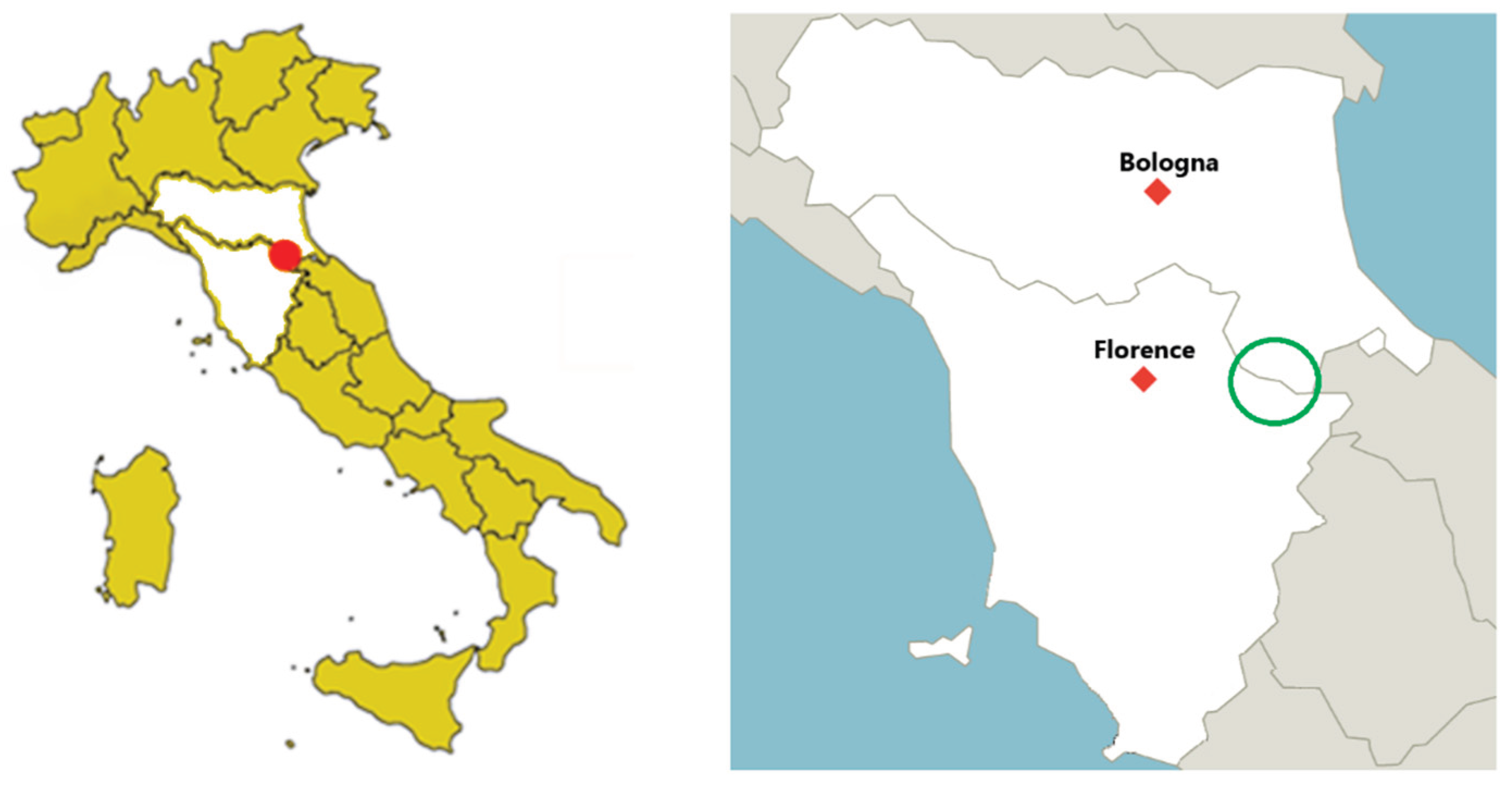

Table 1.
5’-3’ primer sequences used for PCR analysis.
| 5’-3’PRIMER FORWARD | 5’-3’ PRIMER REVERSE | REFERENCE | |
|---|---|---|---|
| Adenovirus spp* | 1° round: TNMGNGGNGGNMGNTGYTAYCC 2° round: GTDGCRAANSHNCCRTABARNGMRT |
1° round: GTDGCRAANSHNCCRTABARNGMRTT 2° round: CCANCCBCDRTTRTGNARNGTRA |
[22] |
| CAdV 1,2 | CGCGCTGAACATTACTACCTTGTC | CCTAGAGCACTTCGTGTCCGCTT | [23] |
| CPV | ACAAGATAAAAGACGTGGTGTAACTCAA | CAACCTCAGCTGGTCTCATAATAGT | [24] |
| FPV/CPV | ACAAGATAAAAGACGTGGTGTAACTCAA | CAACCTCAGCTGGTCTCATAATAGT | [25] |
| Bocavirus spp* | GCCAGCACNGGNAARACMAA | CATNAGNCAYTCYTCCCACCA | [26] |
| CBov 1 | 1° round: CARTGGTAYGCTCCMATYTTTAA 2° round: TGGTAYGCTCCMATYTTTAAYGG |
1° round: TGGCTCCCGTCACAAAAKATRTG 2° round: GCTCCCGTCACAAAAKATRTGAAC |
[27] |
| CBoV 2 | AGGTCGGCCACTGGCTGT | CAGCTTAACGGCATTCACTA | [26] |
| CBoV 3 | 1° round: CAGATTTGGGGGTCCTGCAT 2° round: ATGCCGTCACCAATCCACAT |
1° round: GCACTGTCTGCGCTGAAAAA 2° round: AGCTTGTGGTGGACAGTAGC |
[28] |
| LBoV | AGACCAGATGCTCCACATGG | TGCCTGCCACGGATTGTACC | [29] |
| Canine CV | CTGAAAGATAAAGGCCTCTCGCT | AGGGGGGTGAACAGGTAAACG | [30] |
| TTV1 | CGGGTTCAGGAGGCTCAAT | GCCATTCGGAACTGCACTTACT | [31] |
| TTV2 | TCATGACAGGGTTCACCGGA | CGTCTGCGCACTTACTTATATACTCTA | [31] |
| Kobuvirus spp* | TGGAYTACAAGTGTTTTGATGC | ATGTTGTTRATGATGGTGTTGA | [32] |
| Astrovirus spp* | 1° round a: GARTTYGATTGGRCKCGKTAYGA 1° round b: GARTTYGATTGGRCKAGGTAYGA 2° round a: CGKTAYGATGGKACKATHCC 2° round b: AGGTAYGATGGKACKATHCC |
GGYTTKACCCACATNCCRAA | [33] |
| Coronavirus spp.* | 1° round: GGKTGGGAYTAYCCKAARTG *--2° round: GGTTGGGACTATCCTAAGTGTGA |
1° round: TGYTGTSWRCARAAYTCRTG 2° round: CCATCATCAGATAGAATCATCAT |
[34] |
| BVDV | ATGCCCWTAGTAGGACTAGCA | TCAACTCCATGTGCCATGTAC | [35] |
| Bopivirus spp* | CTGRGCAAGTTCACCAACAA | GTCCATGACAGGGTGAATCA | [36] |
| CDV | ACTTCCGCGATCTCCACTGG | GCTCCACTGCATCTGTATGG | [29] |
| Virus | Host | Genbank | Reference sequence |
Per. Ident | E value |
|---|---|---|---|---|---|
| CPV | Red Fox | CPV/fox/169/IT OQ079554 |
Canine parvovirus 2c MF177262.1 |
84,83% | 6e-31 |
| CCoV | Red Fox | CCoV/fox/208/IT OQ079555 |
Canine coronavirus ON834692.1 |
100.00% | 0.0 |
| AdV | Small mustelids |
RAdV/mustelidae/878/IT OQ079556 |
Rodent adenovirus KY369960.1 |
75.91% | 1e-49 |
| Small mustelids |
RAdV/mustelidae/888/IT OQ079557 |
Rodent adenovirus KY369960.1 |
74.74% | 1e-42 | |
| AstV | Red Fox | Mastv/fox/46/IT OQ079558 |
Bovine astrovirus MW373720.1 |
90.82% | 7e-155 |
| Porcupine | Mastv/porcupine/484/IT OQ079559 |
Mamastrovirus 3 MH399894.1 |
91,27% | 2e-156 | |
| Cervo | Mastv/reddeer/707/IT OQ079560 |
Murine astrovirus JQ408746.1 |
88.49% | 9e-128 | |
| Small mustelids |
Mastv/mustelidae/883/IT OQ079561 |
Murine astrovirus JQ408746.1 |
87.95% | 3e-81 | |
| Red Deer | Mastv/redeer/885/IT OQ079562 |
Murine astrovirus JQ408746.1 |
88.56% | 7e-129 | |
| Wolf | CAsV/wolf/481/IT OQ079564 |
Canine astrovirus OQ198039 |
88.80% | 3e-133 | |
| BoV | Red Fox | BoV/fox/171/IT OQ079563 |
Bocaparvovirus carnivoran5 PP592987 |
88.55% | 3-e83 |
| KoV | Wolf | Kov/wolf/525/IT OQ079565 |
Porcine kobuvirus MH184668.1 |
93.30% | 8e-88 |
| Red Fox | Kov/fox/88/IT OQ079566 |
Fox kobuvirus KF781172.1 |
92.49% | 5e-63 |
Table 3.
Results of the taxonomic classification using Kraken 2.
| Pool Number | N° of sequences |
Classified sequences * (%) |
Unclassified sequences * (%) |
Bacteria sequences * (%) |
Viral sequences * (%) |
|---|---|---|---|---|---|
| 1 | 1.86 x 10⁷ | 21 | 79 | 76 | 2 |
| 2 | 1.79 x 10⁷ | 29 | 72 | 92 | 1 |
| 3 | 2.17 x 10⁷ | 26 | 74 | 89 | 0.7 |
| 4 | 2.18 x 10⁷ | 59 | 41 | 98 | 0.4 |
The percentage of sequences assigned to Bacteria and Viruses is calculated over the number of classified sequences (*).
Table 4.
Post data analysis of NGS results.
| Pool number | Genbank | Ref- Seq | n° Reads | n° nucleotide |
Pairwise Identity | e Value |
|---|---|---|---|---|---|---|
| 1 | PX314117 |
Porcine picobirnavirus MW978536.1 |
213 | 467 | 100% | 0.0 |
| PX314118 |
Rodent circovirus KY370041.1 |
7 | 480 | 89.97% | 1e-145 | |
| PX314119 |
Anelloviridae sp. MF346362.1 |
2 | 427 | 99,77% | 0.0 | |
| 2 | PX314120 |
Fox adeno-associated virus KC878874 |
28 | 1288 | 100% | 0.0 |
| PX314121 PX314122 PX314123 |
Red squirrel adenovirus -1 NC_035207 |
27 | 2129 | 69.66% 88.58% 100 |
1e-24 1e-1770 |
|
| PX314124 |
Pigeon adenovirus NC024474 |
17 | 319 | 100% | 2e-104 | |
| PX314125 PX314126 PX314127 |
Torqueteno felis virus MT538010 |
25 | 779 | 92.21% 98.06% 70.71% |
4e-51 7e-67 2e-30 |
|
| 3 | PX314128 PX314129 |
Porcine circo-like virus JF713717 |
31 | 1759 | 90.90% 98.15% |
0.0 0.0 |
Disclaimer/Publisher’s Note: The statements, opinions and data contained in all publications are solely those of the individual author(s) and contributor(s) and not of MDPI and/or the editor(s). MDPI and/or the editor(s) disclaim responsibility for any injury to people or property resulting from any ideas, methods, instructions or products referred to in the content. |
© 2025 by the authors. Licensee MDPI, Basel, Switzerland. This article is an open access article distributed under the terms and conditions of the Creative Commons Attribution (CC BY) license (http://creativecommons.org/licenses/by/4.0/).
Copyright: This open access article is published under a Creative Commons CC BY 4.0 license, which permit the free download, distribution, and reuse, provided that the author and preprint are cited in any reuse.


