Preprint
Article

This version is not peer-reviewed.

Saponin Improves Detachment and Recovery of Bacteria from Orthopaedic Implants—An In Vitro Study

A peer-reviewed article of this preprint also exists.

Submitted:

08 March 2025

Posted:

10 March 2025

You are already at the latest version

Abstract

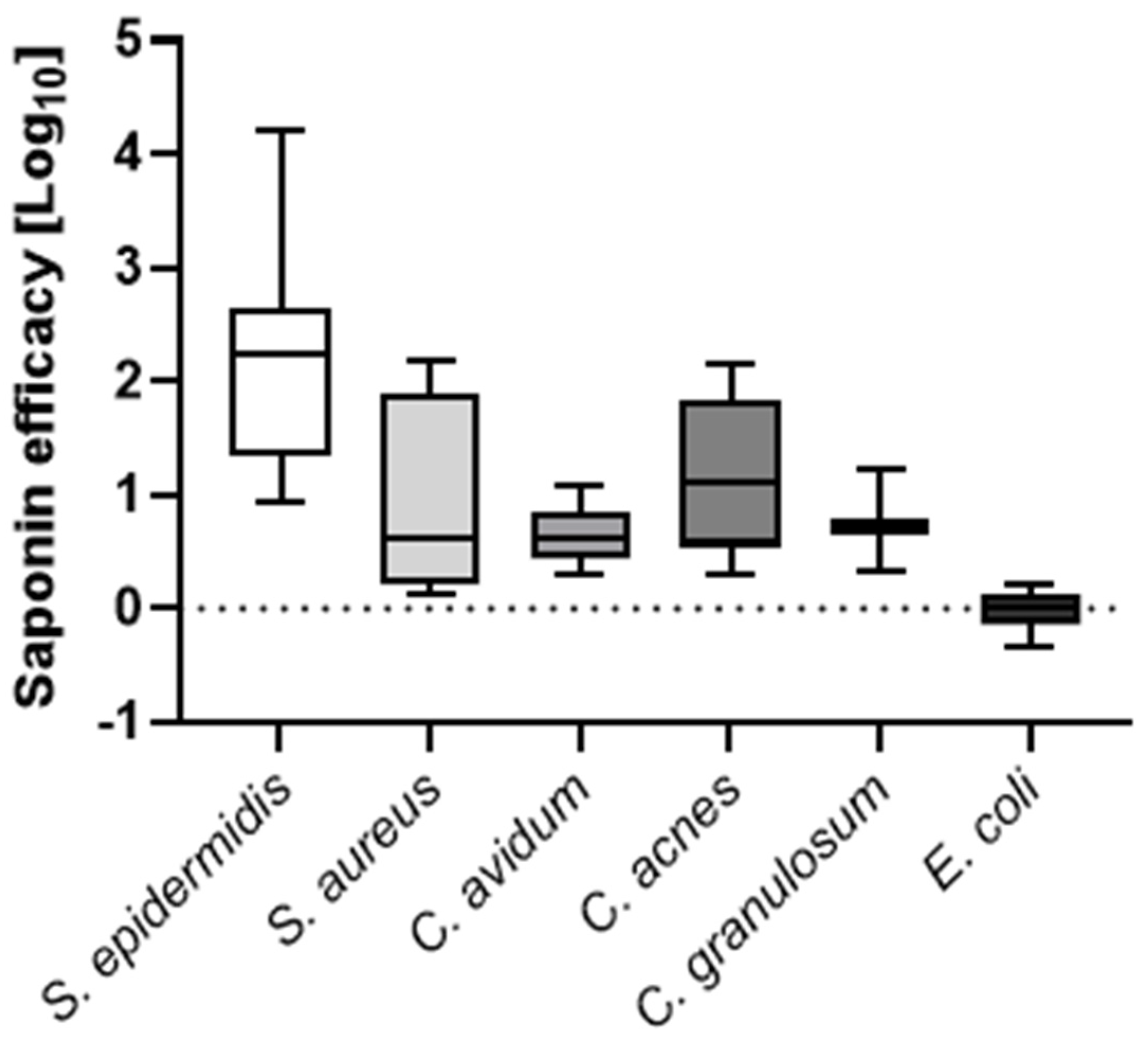
Biofilm formation on orthopedic joint implants complicates treatment and diagnosis of periprosthetic joint infections (PJIs). Sonication of explanted orthopedic implants enhances pathogen detection, but it shows limitations in sensitivity and handling. We investigated whether the biosurfactant saponin could improve bacterial recovery from explanted implants. Orthopaedic material discs of 1 cm diameter were contaminated with different clinical bacterial PJI isolates. Biofilms of Staphylococcus epidermidis, Staphylococcus aureus, Escherichia coli, Cutibacterium avidum, Cutibacterium acnes, and Cutibacterium granulosum were grown on the discs, which were then treated with either saline solution or various concentrations of saponin. Next, disks were vortexed or sonicated. prior to vortexing or sonication. Colony forming units (CFUs) enumeration and time-to-positivity of liquid cultures were determined. Additionally, a novel 3D PJI soft-tissue in vitro model was established to validate these findings in a more representative scenario. Median CFU enumeration showed that 0.001% (w/v) saponin as compared to saline solution increased CFUs recovery by 2.2 log10 for S. epidermidis, 0.6 log10 for S. aureus, 0.6 log10 for C. avidum, 1.1 log10 for C. acnes, 0.7 log10 for C. granulosum, and 0.01 log10 for E. coli. Further, saponin treatment resulted in a >1 log10 increase in S. epidermidis CFU recovery from implants in the 3D tissue model compared to standard saline sonication. With that, we propose a novel two-component kit, consisting of a saponin solution and a specialized transportation box, for the efficient collection, transportation, and processing of potentially infected implants. Our data suggests that biosurfactants can enhance bacterial recovery from artificially contaminated orthopedic implants, potentially improving the diagnosis of PJIs.

Keywords: 
;  ;  ;  ;  ;  

1. Introduction

Periprosthetic joint infections (PJIs) is a serious complication following the implantation of an orthopedic device. Despite enhanced efforts to prevent infections, the incidence of infections in hip and knee arthroplasties is rising. PJIs affect up to 1-2% of patients with such a procedure [1], even with standard preoperative skin antisepsis and perioperative antibiotic prophylaxis [2,3]. PJIs are associated with increased morbidity, prolonged antibiotic therapy, and repeated surgical interventions. Furthermore, the cost of treating a single episode of PJI exceeds $45’000 [1].
The most frequently isolated microorganisms in PJIs include staphylococci, streptococci, and anaerobic bacteria such as Cutibacterium acnes and Cutibacterium avidum [4]. These bacteria are known to form biofilms, which is a major factor to the challenges in treating PJIs [5,6]. Despite the many therapeutic challenges, the diagnostic of PJI is also not straight forward for most microbiological laboratories. Often patients were pre-treated with antibiotics, which results in slow or no growth. Some bacterial species require specific growth conditions, such as C. acnes. In addition, intraoperative tissue cultures often show low sensitivity and specificity for pathogen identification [7,8,9], requiring multiple positive samples to confirm infection [10]. To improve pathogen detection, sonication of explanted implants, which dislodges bacterial biofilms, is used for over a decade [11,12,13,14]. While sonication generally provides better sensitivity over conventional tissue culture [15,16], this method still has its limitations. First, there are no standardized protocols or equipment for sonication [11,13,14,17], making it a laboratory-developed test in most cases and resulting in variability when comparing studies. Second, sonication fails to achieve high sensitivity. Partly because bacteria are less affected by ultrasound [18,19]. Third, detached biofilm bacteria can persist as aggregates in the sonication solution [20,21]. This results in reduced colony-forming unit (CFU) counts, potentially missing the cut-off for diagnosing PJI based on CFUs [22]. As a consequence of these diagnostic challenges, the long time-to-positivity results (TTP) of bacteria in biofilm aggregates results in delayed tailored antibiotic treatments, which affects patient outcomes [8]. Recently, it has been shown that chemical or enzymatic agents have been explored to detach biofilms from orthopedic implants and improve bacterial recovery [17,23]. Dithiothreitol (DTT) has shown promise in vitro and in initial clinical trials [24,25], but its limited stability, efficiency, and toxicity profile have restricted its clinical application[26,27].
Surfactants, which can reduce surface tension and promote detachment of biofilm, might offer a solution. [28,29]. However, most surfactants also have cell-permeabilizing effects, limiting their applicability [30,31]. The biosurfactant saponin, derived from Quillaja species, stands out due to its soap-like properties and potential for biofilm detachment [32,33].
As mentioned, sonication lacks standardized equipment and protocols. The devices used for implant processing – simple sterile plastic boxes—require complex and manual manipulations, increasing the risk of contamination and false positive results [13,14,24]. Variable volumes of sonication solution further complicate PJI diagnosis based on CFU cut-off values [22] as there is no correction to the volume of fluid used.
To address these challenges, we investigated whether saponin could enhance bacterial CFUs recovery from biofilm grown on orthopedic material in vitro. We used saponin and in combination with sonication or alone. We also explored if this procedure reduced the time-to-results in a 3D soft tissue PJI model.

2. Materials and Methods

2.1. Bacterial Strains

The following clinical isolates from patients suffering from PJI were used for PDT experiments: Staphylococcus epidermidis BCI112, BCI135 and BCI195, Staphylococcus aureus BCI175 and BCI187, Cutibacterium avidum BCI100, Cutibacterium granulosum BCI918, Cutibacterium acnes BCI104 and BCI105 and Escherichia coli 1 and 2. The bacterial biobank was approved by the institutional review board in Zurich, Switzerland (KEK Nr 2016-00145, KEK Nr 2017-01458). Fresh cultures were prepared from frozen cultures and were put on Brain Heart Infusion (BHI, Becton Dickinson, Heidelberg, Germany) agar plates at 37°C for 24h under aerobic conditions for S. aureus, S. epidermidis, and E. coli or under anaerobic conditions for 3 days in the case of C. acnes, C. avidum, and C. granulosum (GENbags, bioMérieux, Mary-l’Etoile, France). Liquid cultures were made with colonies from the agar plates in BHI broth in a 37°C incubator with shaking overnight under aerobic conditions for S. aureus, S. epidermidis as well as E. coli and shaking for three days under anaerobic conditions for C. acnes, C. avidum and C. granulosum. Then, 1:1000 dilutions were made in fresh BHI medium. Samples were vortexed and used as start inoculum for biofilm assays right away.

2.2. In Vitro PJI Biofilm

Biofilms were grown as described previously with minor adaptations [34]. In brief, bacterial liquid cultures in RPMI-1640 + 5% fetal calf serum (FCS, Gibco, ThermoFisher Scientific, Massachusetts, US) were used to inoculate sterilized polyethylene (PE), titanium alloy Ti-6AI-4V (TAV), cobalt-chromium-molybdenum (CCM) all provided by Synthes GmbH (Zuchwil, Switzerland) and polymethyl methacrylate (PMMA)-based bone cement (PMMA) provided by Heraeus Group (Hanau, Germany) of 1 cm diameter in 24-well plates. The plates were incubated for 3 days at 37°C for S. epidermidis, S. aureus and E. coli and for 4 days at 37°C under anaerobic conditions for C. acnes, C. avidum and C. granulosum.

2.3. Biofilm Detachment

After the incubation period, the supernatant was removed and the discs containing biofilms were transferred to fresh wells. The discs were then washed 3 times with phosphate-buffered saline (PBS, Sigma Aldrich, Missouri, US) to remove planktonic bacteria and transferred to 15 ml tubes containing 2 ml of either PBS alone or PBS containing saponin from Quillaja sp. (Sigma Aldrich, Missouri, US) at various concentrations. The tubes with the discs were then assigned to either the Vortex or the Sonication group. The Vortex group tubes were vortexed for 1 minute, while the Sonication group tubes were vortexed for 30 sec, followed by sonication for 1 min at 40 Hz and a second round of vortexing for 30 sec. The resulting vortex- or sonication-fluids were then serially ten-fold diluted and drop-platted on agar BHI plates to enumerate CFUs as well as diluted 1:10 in fresh BHI medium to visually determine time-to-positivity after 24, 48, 72, and 96 hours. Agar plates were incubated at 37°C for 1 day for S. epidermidis, S. aureus, and E. coli as well as for 3 days at 37°C under anaerobic conditions for C. acnes, C. avidum, and C. granulosum.

2.4. Novel In Vitro 3D PJI Soft Tissue Model

To build the model, 24-wells were covered with a 0.5 mg/ml collagen type I solution (Rat Tail, Sigma Aldrich, Missouri, US) in DMEM/F12 (Gibco, ThermoFisher Scientific, Massachusetts, US) and allowed to solidify for 20 min at 37°C. Then, a TAV disc was placed in the center of the well. Next, a fibroblast containing collagen solution was prepared. To do so, the human skin fibroblast cell line BJ (ATCC, Virginia, US) was grown in DMEM/F12 + 5% FCS until confluent and then harvested. A 2 mg/ml collagen solution containing 5x10⁴ fibroblasts per 300 µl was prepared in DMEM/F12 + 5% FCS and added to the well plate containing the implant. After an incubation period of 20 min at 37°C + 5% CO2, 200 µl DMEM/F12 + 5 % FCS were added to each well. The models were incubated for 3 days at 37°C + 5% CO2. The medium was replaced on day 4 and models were allowed to grow for another 2 days until infection.

2.5. Infection and Processing of the Novel 3D PJI Soft Tissue Model

Bacterial overnight cultures in BHI (S. epidermidis) were centrifuged and resuspended in PBS. They were then set to an OD 0.5 in PBS and diluted 1:106 in RPMI + 5% FCS to reach an inoculum between 50 and 200 CFUs. Models were then infected with 50 µl of the prepared bacterial suspensions and topped with 150 µl RPMI + 5% FCS. Models were incubated for 3 days at 37C + 5% CO2. Prior to processing, the supernatant was removed. The implant discs were then separated from the tissue and placed in 15 ml canonical tubes, while the tissues were placed in 2 ml tubes. To each sample, either 2 ml of PBS or saponin 0.001% were added. PBS tissue samples were homogenized in a bead beater with sterile metal beads for 10 min at 30 kHz, while the PBS implants were sonicated for 1 min. Saponin treated discs and tissues were vortexted for 1 min. Resulting solutions were then 10-fold diluted in PBS and drop platted on BHI agar plates.

2.6. Confocal Laser Scanning Microscopy

In order to visualize S. epidermidis biofilm aggregates on the TAV discs, we imaged biofilms surrounded by human cells and host tissue (Collagen) and single S. epidermidis colonies within the host tissue matrix colonized by human fibroblasts. Models were fixed with 4% PFA for 20 min and then stored at 4°C until use. They were stained with picrosirius red (PSR) staining solution [0.5 g of Direct Red 80 (Sigma Aldrich, Missouri, US) in 500 mL of saturated picric acid (Sigma Aldrich, Missouri, US) as described previously [35]. Next, they were washed three times with 0.5% acetic acid prior to one wash with PBS. Then, they were stained with 1:1000 dilution of both Wheat Germ Agglutinin Alexa FluorTM 488 (ThermoFisher Scientific, Massachusetts, US) and Hoechst 33342 (ThermoFisher Scientific, Massachusetts, US ) in PBS for 15 min. Samples were visualized and acquired by confocal laser scanning microscopy (CLSM) with a Leica TCS SP8 inverted microscope (Leica, Hessen, Germany) under a 63×/1.4 NA oil immersion objective. The obtained images were processed to 3D images using Imaris 9.2.0 (Bitplane).

2.7. Moleculight i:XTM Imaging

One day prior to harvesting, models were supplemented with 5 mM hexyl-aminolevulinate and incubated for 24 h. The supernatant was removed, models washed once gently with PBS and pictures were taken with the Moleculight i:XTM (MolecuLight Inc, Toronto, CA) device according to the manufacturer’s instructions. The Moleculight i:XTM) device allows to image autofluorescence produced by collagenous tissue and bacterial biofilms [36,37,38].
Images were processed with ImageJ.

2.8. Statistical Analysis

Data visualization and quantitative analyses were performed using Prism 9.2.0 (GraphPad Software, San Diego, USA).

3. Results

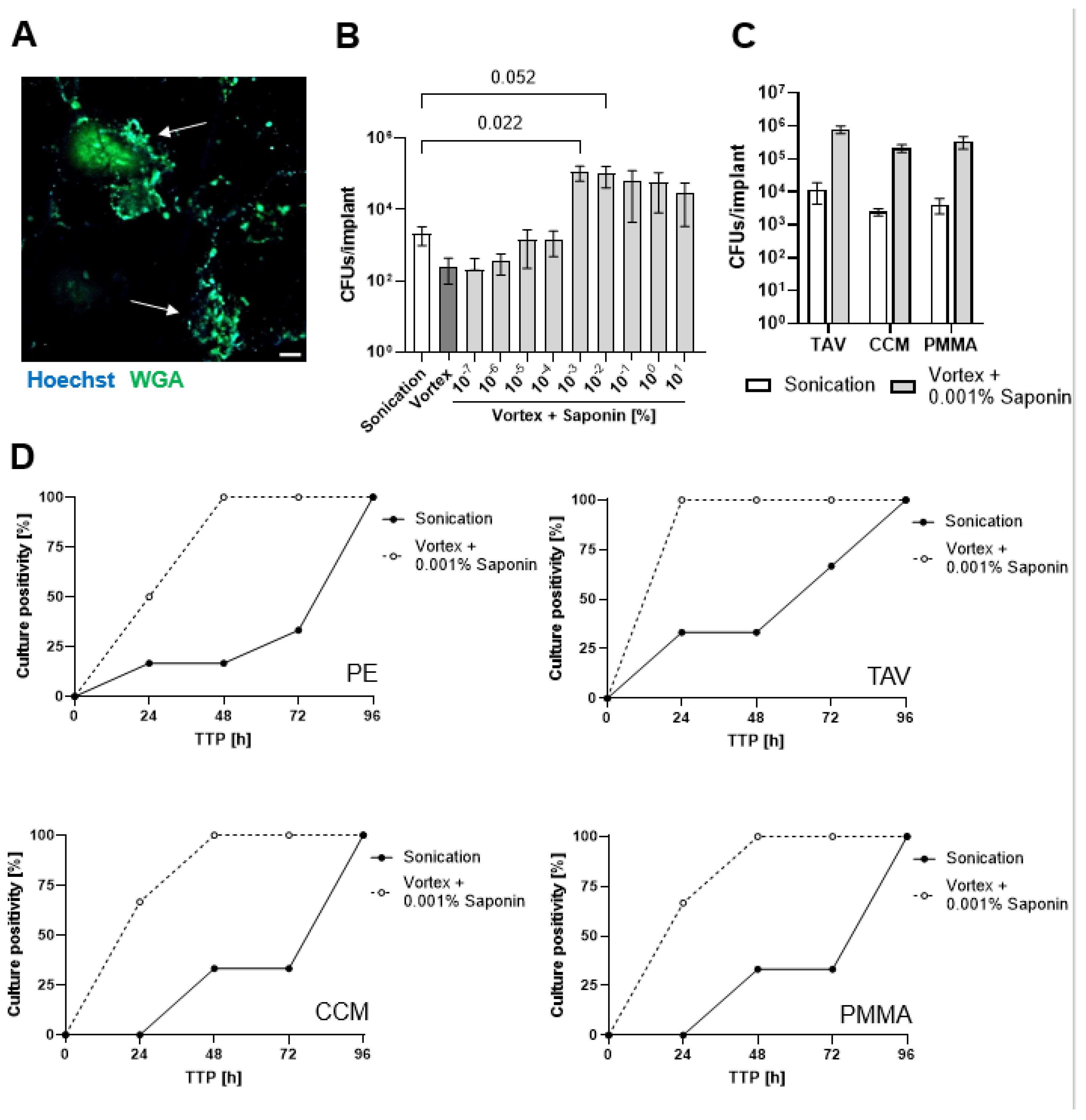
3.1. Finding the Optimum Saponin Concentration for Bacterial Recovery from Biofilms on Implant Discs

To assess the effect of saponin on bacterial recovery, implant discs were colonized with S. epidermidis for three days in physiological medium, resulting in the formation of biofilm-like aggregates on the implant disc surface, representing low-grade infections (Figure 1A). We first performed a concentration titration of saponin to determine its effect on bacterial recovery from PE implant discs. Sonication with saline solution resulted in a >1 log10 increase in CFU recovery compared to vortexing with saline solution Figure 1B). Increasing the sonication time from 1 to 5 minutes did not significantly enhance bacterial (Supplementary Figure S1). While vortexing with 10-7 and 10-6 % (w/v) saponin showed no difference compared to saline vortexing, 10-5 and 10-4 % (w/v) saponin achieved recovery levels comparable to saline sonication. Concentration ≥0.001% (w/v) saponin resulted in a remarkable increase of >2 log10 CFUs recovery compared to saline sonication. However, 0.1% (w/v) saponin showed a trend toward decreased CFU recovery. No additional benefit was observed when combining saponin with sonication (Supplementary Figure S2), therefore we chose 0.001% (w/v) saponin with vortexing for all subsequent experiments. Saponin vortexing also resulted in >2 log10 CFU recovery of bacteria from TAV, CCM, and bone cement PMMA discs compared to saline sonication (Figure 1C).

3.2. Saponin Effectively Reduces Time-to-Positivity (TTP)

We then assessed the liquid culture time-to-positivity (TTP) for low-grade infections. Solutions obtained from either saline sonication or saponin vortexing of S. epidermidis biofilms described above were diluted 1:10 and incubated in fresh liquid medium to monitor TTP. All saponin-treated samples showed 100% culture growth after 48 hours, while saline sonication samples required four days to reach 100% culture growth (Figure 1D).

3.3. Saponin Enhances Recovery of the Most Prevalent PJI-Causing Bacteria from Orthopedic Material

Since strain-dependent effects might occur, we chose to assess the effect of 0.001% (w/v) saponin vortexing as compared to saline sonication for further clinical PJI isolates, representing the major causative species of PJI. For S. epidermidis (total 3 PJI isolates), a median 2.2 log10 CFUs increase was observed with saponin, while for S. aureus (2 PJI isolates), a 0.6 log10 CFU increase was observed with saponin (Figure 2). Concerning Cutibacteria, we observed a median 0.6 log10 CFUs increase for C. avidum (2 PJI isolates), a median 1.1 log10 CFUs increase for C. acnes (2 PJI isolates) and a median 0.7 log10 CFUs increase for C. granulosum (1 PJI isolate) with saponin (Figure 2). For E. coli (2 PJI isolates), we only observed a negligible median 0.01 log10 CFUs increase (Figure 2).

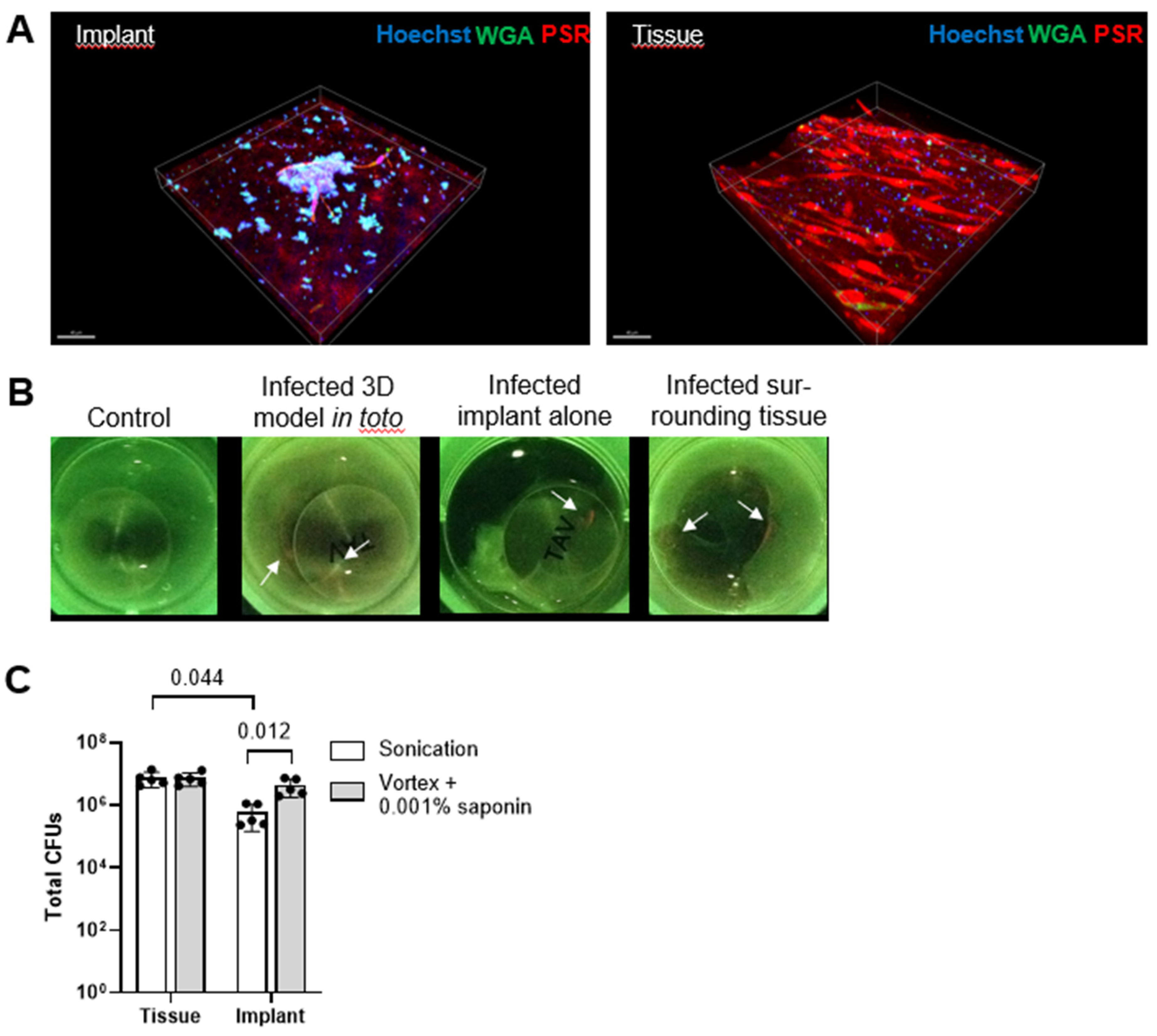
3.4. Saponin Enhances Recovery of S. epidermidis from Orthopedic Implant Material in a Novel 3D PJI Soft Tissue Model

To evaluate whether saponin would maintain its effect on bacterial recovery in a more physiological context- we developed a simple 3D model. Infection of the model with S. epidermidis for three days resulted in the formation of small bacterial biofilm aggregates on the implant disc in contact with host tissue and cells (Figure 3A, left) as well as single colonies distributed throughout the tissue surrounding the implant (Figure 3A, right). Macroscopic evidence of bacterial infection and biofilm formation was also observed using the Moleculight i:XTM device (Figure 3B), which exploits bacterial autofluorescence (red) caused by porphyrin production [36]; we observed a significant increase of red signal over green (collagen) signal, for infected models as compared to uninfected models. Upon separating the implant disc from the cell layers, bacterial biofilm formation was primarily evident at the tissue-implant interface as well as on the implant itself. Tissue and implant were separated and then treated either with saline sonication or with saponin vortexing. Saponin-vortex-treated implants showed significantly higher CFU recoveries compared to saline sonicated implants, while no difference was observed in tissue samples (Figure 3C).

4. Discussion

Biofilm formation on orthopedic joint implants is a significant challenge to identify the causative pathogens of PJI. Current guidelines recommend ≥ 2 culture positive samples (i.e., tissue, fluid etc.) with the same microorganism or ≥ 50 CFUs/ml of any organism from sonication [16]. Sonication of explanted implants has significantly improved the sensitivity of microbiologic diagnostics for PJIs by dislodging bacteria from biofilms adhered to prosthetic surfaces [11,13,14,15,22]. However, on the one hand, bacterial recovery from biofilms is still suboptimal and on the other hand, the currently used sonication process with simple plastic boxes requires multiple manipulations that increase the risk of contamination and false positive results. Therefore, we investigated whether surfactants, specifically the biosurfactant saponin, could increase bacterial recovery from orthopedic material in vitro. We demonstrated that saponin strongly enhances recovery of bacteria causing PJI particularly S. epidermidis, which is often difficult to diagnose. Importantly, saponin can be used in combination with or independent of sonication, resulting in significantly higher recovery as compared to standard saline sonication. To address contamination issues, we propose a novel two-component kit to collect, transport and process potentially infected implants with a saponin solution in an efficient and sterile manner.
We found that saponin’s effect was concentration dependent. At 0.001% (w/v) saponin, vortexing with saponin was significantly more effective than saline sonication. While concentrations >0.001% (w/v) led to a trend of decreasing CFU recovery, it still was higher than with sonication, even at 10% (w/v) saponin. Saponin is known to exhibit potential toxicity towards pro- and eukaryotic cells [58], however, no toxicity was observed at 0.001% in our experiments. This is not the first study showing increased CFU recovery from implants with a substance compared to standard sonication. DTT, for instance, has been extensively studied and shown superiority in both in vitro and early clinical studies for CFU recovery and sensitivity [17,24]. However, follow-up clinical studies demonstrated inferior sensitivity of DTT as compared to sonication, possible due to stability issues of DTT [26,27]. Enzymes and other substances may face similar stability and cost challenges, while saponin is known for its excellent stability in solution.
Among the tested pathogens causing PJIs, saponin-vortexing demonstrated the highest efficacy in recovering S. epidermidis from implants. This is an important finding, since S. epidermidis is a major cause of PJIs [39,40] in particularly chronic PJIs [8,41]. Chronic PJIs are more challenging than acute infections because they often present with only pain, making them harder to identify and treat [42,43]. In sonication studies, S. epidermidis is one of the bacteria known to withstand dislodgment from surfaces [19], which may explain some of the difficulties in diagnosing S. epidermidis PJIs. Furthermore, we chose an incubation time that would promote the formation of biofilm clusters of S. epidermidis, which also result in relatively low CFU recovery by sonication, a result similar to what is typically observed with clinical orthopedic implants [44,45].
For all other tested bacteria (S. aureus, C. avidum, C. acnes and C. granulosum, E. coli), we observed an effect of saponin-vortexing in enhancing bacterial recovery from implant surfaces, except for E. coli. E. coli already showed relatively high recovery with sonication compared to the other isolates. Although the exact reason for the lack of increased recovery with saponin is unclear, it is possible that the attachment mode and biofilm matrix composition of E. coli [46,47] make it more susceptible to sonication than other bacteria. The increased recovery of S. aureus is noteworthy, although, it may not provide significant benefits for diagnosis, as S. aureus typically causes acute, highly virulent infections [48]. However, in cases with low bacterial loads, such as in persistent or relapsing infections [49] saponin could be valuable. For Cutibacteria, which, alongside S. epidermidis, are major causes of difficult-to-diagnose PJIs [50,51], we observed an overall 1 log10 increase in CFU recovery. Considering the EBJIS PJI definition guidelines (i.e., ≥50 CFUs/ml) for sonication diagnosis [16], a 1 log10 increase in recovery could turn previously negative samples, which were considered contaminants due to low CFU counts, into positive PJI diagnoses. However, these guidelines should be carefully evaluated, as there are no standardized sonication protocols, particularly regarding the volume of sonication solution used [52].
We showed that vortexing with saponin at 0.001% (w/v) as compared to sonication demonstrated consistent higher bacterial recovery from implants made of various materials used in orthopedic surgeries, i.e., PE, TAV, CCM, and PMMA. This superiority was also reflected in the time-to-positivity (TTP), where saponin-treated samples showed markedly faster TTP. TTP is an important factor to consider in chronic infections [8]. Although no studies have assessed the impact of reduced TTP on treatment duration or associated financial costs [53], it can be assumed that each additional day without a positive culture - delaying antibiotic resistance testing—may prolong treatment and potentially increase morbidity [54]. This could also translate into prolonged hospital stays, resulting in increases healthcare costs. Therefore, reducing TTP could significantly improve patient outcomes and reduce costs.
We developed a simple 3D model mimicking a soft/connective-tissue PJI scenario, which more accurately reflects the clinical situation where bacterial localization often occurs at the implant-interface membrane [55,56]. Infection with S. epidermidis resulted in biofilm-like aggregates on the implant and in the tissue, similar to clinical PJI samples [57]. CFU recovery from homogenized tissue was significantly higher than with saline sonication, highlighting tissue as a potential infection reservoir [40,58,59]. Identifying infected tissue intraoperatively is challenging, as bacteria are scattered around the implant. Biofilm on implants cannot be directly seen but can be cultured to pinpoint infection locations [60,61,62]. Treatment with saponin-vortexing yielded similar CFU recovery to homogenization, offering a quicker and cheaper alternative. While the model does not fully mimic thick tissue in the clinical PJI environment, it confirmed that vortexing with saponin solution improves CFU recovery compared to saline sonication. In comparison to in vitro models, the 3D model better represents the clinical PJI environment. Saponin’s effect on bacterial recovery was reduced in the 3D model, but even a 1-log increase in CFUs could have significant clinical implications.
As a proof-of-concept, we propose a novel two-component kit consisting of a saponin solution and a specialized transportation box for the efficient collection, transport, and processing of potentially infected implants (Supplementary Figure 3). Once the implant is placed in the collection container, the container remains sealed. The lid of the container is equipped with a self-sealing access port that allows connecting to the squeeze bag in order to add the bacteria detachment solution to the implant. After processing (vortexing or sonication), a collection syringe can be attached to the container to withdraw the solution containing the detached bacteria. Following this step, routine diagnostic procedures can be followed (Video S1). This kit could enable more efficient and sterile processing of implants compared to current practices. Most laboratories use solid boxes that require multiple openings by different personnel, increasing the risk of contamination with skin bacteria. Using bags for collection also raises contamination risks [13,14]. Our two-component system eliminates the need to open the box once the implant is placed inside. Additionally, employing such a system would establish a clear protocol, particularly regarding the volume of solution to be added, as it is defined by the volume of the squeeze bag. This could standardize PJI infection definitions and make results across multiple centers more comparable. Furthermore, any solution other than saponin could be used in the squeeze bag to increase the chances of bacterial recovery. This box is currently being developed to marketability and produced by a service provider, and a patent has been filed for the design.
Our study has several limitations. First, we focused solely on saponin; other surfactants like Tween 20 or 80 may yield similar results. Second, we did not assess different biofilm incubation timepoints, as biofilm maturity might affect susceptibility to saponin. However, replicating real biofilm maturity in vitro is challenging, so we chose a timepoint that reflects CFU levels in clinically infected implants. Third, we tested saponin on a limited number of strains; given saponin’s soap-like mechanism, we do not anticipate strain resistance. Lastly, while promising, saponin should undergo further clinical studies to validate its effectiveness and handling in the clinical setting, and the transportation box is currently under development for future use in PJI diagnosis.
Our findings show that vortexing with the biosurfactant saponin as compared to standard sonication improves bacterial recovery from infected orthopedic materials in vitro. We also proposed a novel collection and transportation system for more efficient processing of potentially infected implants. Pending clinical validation, these approaches could enhance PJI diagnosis, improve treatment outcomes, and reduce associated costs.

Supplementary Materials

The following supporting information can be downloaded at the website of this paper posted on Preprints.org, Figure S1: title; Table S1: title; Video S1: title.

Author Contributions

Tiziano A. Schweizer: Conceptualization, Methodology, Validation, Formal analysis, Investigation, Data Curation, Writing - Original Draft, Visualization, Funding acquisition. Adrian Egli: Methodology, Resources, Writing - Review & Editing. Philipp P. Bosshard: Conceptualization, Methodology, Writing - Review & Editing. Yvonne Achermann: Conceptualization, Methodology, Validation, Resources, Supervision, Project administration, Funding acquisition, Writing - Review & Editing.

Funding

This work was supported by the UZH Innovation Grant (MEDIG23-013).

Data Availability Statement

The data presented in this study are available in the Supplementary Material.

Acknowledgments

Imaging was performed with equipment maintained by the Center for Microscopy and Image Analysis, University of Zurich. We would like to thank Oliver Nolte and Sonia Moser for support with sonication solutions. We thank Beat Lechmann from Synthes GmbH for manufacturing the TAV, CCM and PE implant material discs for biofilm assays as well as Heraeus Group for manufacturing the PMMA implant material discs. We thank MolecuLight Inc. for providing the Moleculight i:XTM device.

Conflicts of Interest

TAS, PPB and YA are listed as inventors on a patent application filed with the European Patent Office.

References

  1. Patel, R. “Periprosthetic joint infection.” N Engl J Med 388 (2023): 251-62. https://www.ncbi.nlm.nih.gov/pubmed/36652356. [CrossRef]
  2. Kusejko, K., A. Aunon, B. Jost, B. Natividad, C. Strahm, C. Thurnheer, D. Pablo-Marcos, D. Slama, G. Scanferla, I. Uckay, et al. “The impact of surgical strategy and rifampin on treatment outcome in cutibacterium periprosthetic joint infections.” Clin Infect Dis 72 (2021): e1064-e73. https://www.ncbi.nlm.nih.gov/pubmed/33300545. [CrossRef]
  3. Maurer, S. M., L. Kursawe, S. Rahm, J. Prinz, A. S. Zinkernagel, A. Moter, S. P. Kuster, R. Zbinden, P. O. Zingg and Y. Achermann. “Cutibacterium avidum resists surgical skin antisepsis in the groin-a potential risk factor for periprosthetic joint infection: A quality control study.” Antimicrob Resist Infect Control 10 (2021): 27. [CrossRef]
  4. McCulloch, R. A., A. Adlan, N. Jenkins, M. Parry, J. D. Stevenson and L. Jeys. “A comparison of the microbiology profile for periprosthetic joint infection of knee arthroplasty and lower-limb endoprostheses in tumour surgery.” J Bone Jt Infect 7 (2022): 177-82. https://www.ncbi.nlm.nih.gov/pubmed/36032799. [CrossRef]
  5. Karachalios, T. and G. A. Komnos. “Management strategies for prosthetic joint infection: Long-term infection control rates, overall survival rates, functional and quality of life outcomes.” EFORT Open Rev 6 (2021): 727-34. https://www.ncbi.nlm.nih.gov/pubmed/34667643. [CrossRef]
  6. Ricciardi, B. F., G. Muthukrishnan, E. A. Masters, N. Kaplan, J. L. Daiss and E. M. Schwarz. “New developments and future challenges in prevention, diagnosis, and treatment of prosthetic joint infection.” J Orthop Res 38 (2020): 1423-35. https://www.ncbi.nlm.nih.gov/pubmed/31965585. [CrossRef]
  7. Ryu, S. Y., K. E. Greenwood-Quaintance, A. D. Hanssen, J. N. Mandrekar and R. Patel. “Low sensitivity of periprosthetic tissue pcr for prosthetic knee infection diagnosis.” Diagn Microbiol Infect Dis 79 (2014): 448-53. https://www.ncbi.nlm.nih.gov/pubmed/24972853. [CrossRef]
  8. Talsma, D. T., J. J. W. Ploegmakers, P. C. Jutte, G. Kampinga and M. Wouthuyzen-Bakker. “Time to positivity of acute and chronic periprosthetic joint infection cultures.” Diagn Microbiol Infect Dis 99 (2021): 115178. https://www.ncbi.nlm.nih.gov/pubmed/33017799. [CrossRef]
  9. Yan, Q., M. J. Karau, K. E. Greenwood-Quaintance, J. N. Mandrekar, D. R. Osmon, M. P. Abdel and R. Patel. “Comparison of diagnostic accuracy of periprosthetic tissue culture in blood culture bottles to that of prosthesis sonication fluid culture for diagnosis of prosthetic joint infection (pji) by use of bayesian latent class modeling and idsa pji criteria for classification.” J Clin Microbiol 56 (2018). https://www.ncbi.nlm.nih.gov/pubmed/29643202. [CrossRef]
  10. Peel, T. N., T. Spelman, B. L. Dylla, J. G. Hughes, K. E. Greenwood-Quaintance, A. C. Cheng, J. N. Mandrekar and R. Patel. “Optimal periprosthetic tissue specimen number for diagnosis of prosthetic joint infection.” J Clin Microbiol 55 (2017): 234-43. https://www.ncbi.nlm.nih.gov/pubmed/27807152. [CrossRef]
  11. Achermann, Y., M. Vogt, M. Leunig, J. Wust and A. Trampuz. “Improved diagnosis of periprosthetic joint infection by multiplex pcr of sonication fluid from removed implants.” J Clin Microbiol 48 (2010): 1208-14. https://www.ncbi.nlm.nih.gov/pubmed/20164283. NOT IN FILE. [CrossRef]
  12. Rieger, U. M., G. Pierer, N. J. Luscher and A. Trampuz. “Sonication of removed breast implants for improved detection of subclinical infection.” Aesthetic Plast Surg 33 (2009): 404-8. https://www.ncbi.nlm.nih.gov/pubmed/19322605. [CrossRef]
  13. Trampuz, A., K. E. Piper, A. D. Hanssen, D. R. Osmon, F. R. Cockerill, J. M. Steckelberg and R. Patel. “Sonication of explanted prosthetic components in bags for diagnosis of prosthetic joint infection is associated with risk of contamination.” J Clin Microbiol 44 (2006): 628-31. https://www.ncbi.nlm.nih.gov/pubmed/16455930. [CrossRef]
  14. Trampuz, A., K. E. Piper, M. J. Jacobson, A. D. Hanssen, K. K. Unni, D. R. Osmon, J. N. Mandrekar, F. R. Cockerill, J. M. Steckelberg, J. F. Greenleaf and R. Patel. “Sonication of removed hip and knee prostheses for diagnosis of infection.” N Engl J Med 357 (2007): 654-63. https://www.ncbi.nlm.nih.gov/pubmed/17699815. NOT IN FILE. [CrossRef]
  15. Dudareva, M., L. Barrett, M. Figtree, M. Scarborough, M. Watanabe, R. Newnham, R. Wallis, S. Oakley, B. Kendrick, D. Stubbs, et al. “Sonication versus tissue sampling for diagnosis of prosthetic joint and other orthopedic device-related infections.” J Clin Microbiol 56 (2018). https://www.ncbi.nlm.nih.gov/pubmed/30209185. [CrossRef]
  16. Ribeiro, T. C., E. K. Honda, D. Daniachi, R. P. L. Cury, C. B. da Silva, G. B. Klautau and M. J. Salles. “The impact of sonication cultures when the diagnosis of prosthetic joint infection is inconclusive.” PLoS One 16 (2021): e0252322. https://www.ncbi.nlm.nih.gov/pubmed/34255768. [CrossRef]
  17. Drago, L., V. Signori, E. De Vecchi, C. Vassena, E. Palazzi, L. Cappelletti, D. Romano and C. L. Romano. “Use of dithiothreitol to improve the diagnosis of prosthetic joint infections.” J Orthop Res 31 (2013): 1694-9. https://www.ncbi.nlm.nih.gov/pubmed/23817975. [CrossRef]
  18. Sandbakken, E. T., E. Hoyer, E. Witso, C. K. Sogaard, A. Diez-Sanchez, L. Hoang, T. S. Wik and K. Bergh. “Biofilm and the effect of sonication in a chronic staphylococcus epidermidis orthopedic in vivo implant infection model.” J Orthop Surg Res 19 (2024): 820. https://www.ncbi.nlm.nih.gov/pubmed/39633500. [CrossRef]
  19. Sandbakken, E. T., E. Witso, B. Sporsheim, K. W. Egeberg, O. A. Foss, L. Hoang, G. Bjerkan, K. Loseth and K. Bergh. “Highly variable effect of sonication to dislodge biofilm-embedded staphylococcus epidermidis directly quantified by epifluorescence microscopy: An in vitro model study.” J Orthop Surg Res 15 (2020): 522. https://www.ncbi.nlm.nih.gov/pubmed/33176843. [CrossRef]
  20. Drago, L., A. Fidanza, A. Giannetti, A. Ciuffoletti, G. Logroscino and C. L. Romano. “Bacteria living in biofilms in fluids: Could chemical antibiofilm pretreatment of culture represent a paradigm shift in diagnostics?” Microorganisms 12 (2024). https://www.ncbi.nlm.nih.gov/pubmed/38399663. [CrossRef]
  21. Kragh, K. N., T. Tolker-Nielsen and M. Lichtenberg. “The non-attached biofilm aggregate.” Commun Biol 6 (2023): 898. https://www.ncbi.nlm.nih.gov/pubmed/37658117. [CrossRef]
  22. Alvarez Otero, J., M. J. Karau, K. E. Greenwood-Quaintance, M. P. Abdel, J. Mandrekar and R. Patel. “Evaluation of sonicate fluid culture cutoff points for periprosthetic joint infection diagnosis.” Open Forum Infect Dis 11 (2024): ofae159. https://www.ncbi.nlm.nih.gov/pubmed/38715572. [CrossRef]
  23. Henriquez, L., C. Martin, M. Echeverz, I. Lasa, C. Ezpeleta and M. E. Portillo. “Evaluation of the use of sonication combined with enzymatic treatment for biofilm removal in the microbiological diagnosis of prosthetic joint infection.” Microbiol Spectr 12 (2024): e0002024. https://www.ncbi.nlm.nih.gov/pubmed/38916322. [CrossRef]
  24. Calori, G. M., M. Colombo, P. Navone, M. Nobile, F. Auxilia, M. Toscano and L. Drago. “Comparative evaluation of microdttect device and flocked swabs in the diagnosis of prosthetic and orthopaedic infections.” Injury 47 Suppl 4 (2016): S17-S21. https://www.ncbi.nlm.nih.gov/pubmed/27492065. [CrossRef]
  25. Kolenda, C., J. Josse, C. Batailler, A. Faure, A. Monteix, S. Lustig, T. Ferry, F. Laurent and C. Dupieux. “Experience with the use of the microdttect device for the diagnosis of low-grade chronic prosthetic joint infections in a routine setting.” Front Med (Lausanne) 8 (2021): 565555. https://www.ncbi.nlm.nih.gov/pubmed/33796542. [CrossRef]
  26. Karbysheva, S., S. Cabric, A. Koliszak, M. Bervar, S. Kirschbaum, S. Hardt, C. Perka and A. Trampuz. “Clinical evaluation of dithiothreitol in comparison with sonication for biofilm dislodgement in the microbiological diagnosis of periprosthetic joint infection.” Diagn Microbiol Infect Dis 103 (2022): 115679. https://www.ncbi.nlm.nih.gov/pubmed/35395437. [CrossRef]
  27. Karbysheva, S., M. Di Luca, M. E. Butini, T. Winkler, M. Schutz and A. Trampuz. “Comparison of sonication with chemical biofilm dislodgement methods using chelating and reducing agents: Implications for the microbiological diagnosis of implant associated infection.” PLoS One 15 (2020): e0231389. https://www.ncbi.nlm.nih.gov/pubmed/32267888. [CrossRef]
  28. Huang, H., Q. Yu, H. Ren, J. Geng, K. Xu, Y. Zhang and L. Ding. “Towards physicochemical and biological effects on detachment and activity recovery of aging biofilm by enzyme and surfactant treatments.” Bioresour Technol 247 (2018): 319-26. https://www.ncbi.nlm.nih.gov/pubmed/28950141. [CrossRef]
  29. Percival, S. L., D. Mayer, R. S. Kirsner, G. Schultz, D. Weir, S. Roy, A. Alavi and M. Romanelli. “Surfactants: Role in biofilm management and cellular behaviour.” Int Wound J 16 (2019): 753-60. https://www.ncbi.nlm.nih.gov/pubmed/30883044. [CrossRef]
  30. Koley, D. and A. J. Bard. “Triton x-100 concentration effects on membrane permeability of a single hela cell by scanning electrochemical microscopy (secm).” Proc Natl Acad Sci U S A 107 (2010): 16783-7. https://www.ncbi.nlm.nih.gov/pubmed/20837548. [CrossRef]
  31. Sutormin, O. S., E. M. Kolosova, I. G. Torgashina, V. A. Kratasyuk, N. S. Kudryasheva, J. S. Kinstler and D. I. Stom. “Toxicity of different types of surfactants via cellular and enzymatic assay systems.” Int J Mol Sci 24 (2022). https://www.ncbi.nlm.nih.gov/pubmed/36613956. [CrossRef]
  32. George, A. J. “Legal status and toxicity of saponins.” Food Cosmet Toxicol 3 (1965): 85-91. https://www.ncbi.nlm.nih.gov/pubmed/14342799. [CrossRef]
  33. Savarino, P., M. Demeyer, C. Decroo, E. Colson and P. Gerbaux. “Mass spectrometry analysis of saponins.” Mass Spectrom Rev 42 (2023): 954-83. https://www.ncbi.nlm.nih.gov/pubmed/34431118. [CrossRef]
  34. Prinz, J., M. Wink, S. Neuhaus, M. C. Grob, H. Walt, P. P. Bosshard and Y. Achermann. “Effective biofilm eradication on orthopedic implants with methylene blue based antimicrobial photodynamic therapy in vitro.” Antibiotics (Basel) 12 (2023). https://www.ncbi.nlm.nih.gov/pubmed/36671319. [CrossRef]
  35. Wegner, K. A., A. Keikhosravi, K. W. Eliceiri and C. M. Vezina. “Fluorescence of picrosirius red multiplexed with immunohistochemistry for the quantitative assessment of collagen in tissue sections.” J Histochem Cytochem 65 (2017): 479-90. https://www.ncbi.nlm.nih.gov/pubmed/28692327. [CrossRef]
  36. Jones, L. M., D. Dunham, M. Y. Rennie, J. Kirman, A. J. Lopez, K. C. Keim, W. Little, A. Gomez, J. Bourke, H. Ng, et al. “In vitro detection of porphyrin-producing wound bacteria with real-time fluorescence imaging.” Future Microbiol 15 (2020): 319-32. https://www.ncbi.nlm.nih.gov/pubmed/32101035. [CrossRef]
  37. Redmond, S., C. J. Lewis, S. Rowe, E. Raby and S. Rea. “The use of moleculight for early detection of colonisation in dermal templates.” Burns 45 (2019): 1940-42. https://www.ncbi.nlm.nih.gov/pubmed/31672470. [CrossRef]
  38. Raizman, R., D. Dunham, L. Lindvere-Teene, L. M. Jones, K. Tapang, R. Linden and M. Y. Rennie. “Use of a bacterial fluorescence imaging device: Wound measurement, bacterial detection and targeted debridement.” J Wound Care 28 (2019): 824-34. https://www.ncbi.nlm.nih.gov/pubmed/31825778. [CrossRef]
  39. Shabana, N. S., G. Seeber, A. Soriano, P. C. Jutte, S. Westermann, G. Mithoe, L. Pirii, T. Siebers, B. T. Have, W. Zijlstra, et al. “The clinical outcome of early periprosthetic joint infections caused by staphylococcus epidermidis and managed by surgical debridement in an era of increasing resistance.” Antibiotics (Basel) 12 (2022). https://www.ncbi.nlm.nih.gov/pubmed/36671241. [CrossRef]
  40. Widerstrom, M., M. Stegger, A. Johansson, B. K. Gurram, A. R. Larsen, L. Wallinder, H. Edebro and T. Monsen. “Heterogeneity of staphylococcus epidermidis in prosthetic joint infections: Time to reevaluate microbiological criteria?” Eur J Clin Microbiol Infect Dis 41 (2022): 87-97. https://www.ncbi.nlm.nih.gov/pubmed/34599708. [CrossRef]
  41. Fernandez-Rodriguez, D., C. A. Colin-Castro, M. Hernandez-Duran, L. E. Lopez-Jacome and R. Franco-Cendejas. “Staphylococcus epidermidis small colony variants, clinically significant quiescent threats for patients with prosthetic joint infection.” Microbes Infect 23 (2021): 104854. https://www.ncbi.nlm.nih.gov/pubmed/34214690. [CrossRef]
  42. Christopher, Z. K., K. S. McQuivey, D. G. Deckey, J. Haglin, M. J. Spangehl and J. S. Bingham. “Acute or chronic periprosthetic joint infection? Using the esr ∕ crp ratio to aid in determining the acuity of periprosthetic joint infections.” J Bone Jt Infect 6 (2021): 229-34. https://www.ncbi.nlm.nih.gov/pubmed/34159047. [CrossRef]
  43. Youssef, Y., E. Roschke, N. Dietze, A. J. Dahse, I. F. Chaberny, D. Ranft, C. Pempe, S. Goralski, M. Ghanem, R. Kluge, et al. “Early-outcome differences between acute and chronic periprosthetic joint infections-a retrospective single-center study.” Antibiotics (Basel) 13 (2024). https://www.ncbi.nlm.nih.gov/pubmed/38534633. [CrossRef]
  44. Esteban, J., E. Gomez-Barrena, J. Cordero, N. Z. Martin-de-Hijas, T. J. Kinnari and R. Fernandez-Roblas. “Evaluation of quantitative analysis of cultures from sonicated retrieved orthopedic implants in diagnosis of orthopedic infection.” J Clin Microbiol 46 (2008): 488-92. https://www.ncbi.nlm.nih.gov/pubmed/18077647. [CrossRef]
  45. Tunney, M. M., S. Patrick, S. P. Gorman, J. R. Nixon, N. Anderson, R. I. Davis, D. Hanna and G. Ramage. “Improved detection of infection in hip replacements. A currently underestimated problem.” J Bone Joint Surg Br 80 (1998): 568-72. https://www.ncbi.nlm.nih.gov/pubmed/9699813. NOT IN FILE.
  46. Beloin, C., A. Roux and J. M. Ghigo. “Escherichia coli biofilms.” Curr Top Microbiol Immunol 322 (2008): 249-89. https://www.ncbi.nlm.nih.gov/pubmed/18453280. [CrossRef]
  47. McCrate, O. A., X. Zhou, C. Reichhardt and L. Cegelski. “Sum of the parts: Composition and architecture of the bacterial extracellular matrix.” J Mol Biol 425 (2013): 4286-94. https://www.ncbi.nlm.nih.gov/pubmed/23827139. [CrossRef]
  48. de Buys, M., K. Moodley, J. N. Cakic and J. R. T. Pietrzak. “Staphylococcus aureus colonization and periprosthetic joint infection in patients undergoing elective total joint arthroplasty: A narrative review.” EFORT Open Rev 8 (2023): 680-89. https://www.ncbi.nlm.nih.gov/pubmed/37655845. [CrossRef]
  49. Munoz-Gallego, I., M. A. Melendez-Carmona, J. Lora-Tamayo, C. Garrido-Allepuz, F. Chaves, V. Sebastian and E. Viedma. “Microbiological and molecular features associated with persistent and relapsing staphylococcus aureus prosthetic joint infection.” Antibiotics (Basel) 11 (2022). https://www.ncbi.nlm.nih.gov/pubmed/36009988. [CrossRef]
  50. Boisrenoult, P. “Cutibacterium acnes prosthetic joint infection: Diagnosis and treatment.” Orthop Traumatol Surg Res 104 (2018): S19-S24. https://www.ncbi.nlm.nih.gov/pubmed/29203432. [CrossRef]
  51. Prinz, J., B. Schmid, R. Zbinden, P. O. Zingg, I. Uckay, Y. Achermann and P. P. Bosshard. “Fast and sensitive multiplex real-time quantitative pcr to detect cutibacterium periprosthetic joint infections.” J Mol Diagn 24 (2022): 666-73. https://www.ncbi.nlm.nih.gov/pubmed/35364323. [CrossRef]
  52. Silva, N. D. S., B. S. T. De Melo, A. Oliva and P. S. R. de Araujo. “Sonication protocols and their contributions to the microbiological diagnosis of implant-associated infections: A review of the current scenario.” Front Cell Infect Microbiol 14 (2024): 1398461. https://www.ncbi.nlm.nih.gov/pubmed/38803573. [CrossRef]
  53. Premkumar, A., D. A. Kolin, K. X. Farley, J. M. Wilson, A. S. McLawhorn, M. B. Cross and P. K. Sculco. “Projected economic burden of periprosthetic joint infection of the hip and knee in the united states.” J Arthroplasty 36 (2021): 1484-89 e3. https://www.ncbi.nlm.nih.gov/pubmed/33422392. [CrossRef]
  54. Szymski, D., N. Walter, K. Hierl, M. Rupp and V. Alt. “Direct hospital costs per case of periprosthetic hip and knee joint infections in europe - a systematic review.” J Arthroplasty 39 (2024): 1876-81. https://www.ncbi.nlm.nih.gov/pubmed/38266688. [CrossRef]
  55. Bori, G., E. Munoz-Mahamud, S. Garcia, C. Mallofre, X. Gallart, J. Bosch, E. Garcia, J. Riba, J. Mensa and A. Soriano. “Interface membrane is the best sample for histological study to diagnose prosthetic joint infection.” Mod Pathol 24 (2011): 579-84. https://www.ncbi.nlm.nih.gov/pubmed/21131917. [CrossRef]
  56. Muller, M., L. Morawietz, O. Hasart, P. Strube, C. Perka and S. Tohtz. “[histopathological diagnosis of periprosthetic joint infection following total hip arthroplasty : Use of a standardized classification system of the periprosthetic interface membrane].” Orthopade 38 (2009): 1087-96. https://www.ncbi.nlm.nih.gov/pubmed/19690832. Histopathologische Diagnose der periprothetischen Gelenkinfektion nach Huftgelenkersatz : Verwendung eines standardisierten Klassifikationssystems der periprothetischen Interface-Membranen. [CrossRef]
  57. Stoodley, P., L. Nistico, S. Johnson, L. A. Lasko, M. Baratz, V. Gahlot, G. D. Ehrlich and S. Kathju. “Direct demonstration of viable staphylococcus aureus biofilms in an infected total joint arthroplasty. A case report.” J Bone Joint Surg Am 90 (2008): 1751-8. https://www.ncbi.nlm.nih.gov/pubmed/18676908. [CrossRef]
  58. de Breij, A., M. Riool, P. H. Kwakman, L. de Boer, R. A. Cordfunke, J. W. Drijfhout, O. Cohen, N. Emanuel, S. A. Zaat, P. H. Nibbering and T. F. Moriarty. “Prevention of staphylococcus aureus biomaterial-associated infections using a polymer-lipid coating containing the antimicrobial peptide op-145.” J Control Release 222 (2016): 1-8. https://www.ncbi.nlm.nih.gov/pubmed/26658071. [CrossRef]
  59. Perez, K. and R. Patel. “Survival of staphylococcus epidermidis in fibroblasts and osteoblasts.” Infect Immun 86 (2018). https://www.ncbi.nlm.nih.gov/pubmed/30061380. [CrossRef]
  60. Brooks, J. R., D. J. Chonko, M. Pigott, A. C. Sullivan, K. Moore and P. Stoodley. “Mapping bacterial biofilm on explanted orthopedic hardware: An analysis of 14 consecutive cases.” APMIS 131 (2023): 170-79. https://www.ncbi.nlm.nih.gov/pubmed/36656746. [CrossRef]
  61. Moley, J. P., M. S. McGrath, J. F. Granger, A. C. Sullivan, P. Stoodley and D. H. Dusane. “Mapping bacterial biofilms on recovered orthopaedic implants by a novel agar candle dip method.” APMIS 127 (2019): 123-30. https://www.ncbi.nlm.nih.gov/pubmed/30687941. [CrossRef]
  62. Moore, K., N. Gupta, T. T. Gupta, K. Patel, J. R. Brooks, A. Sullivan, A. S. Litsky and P. Stoodley. “Mapping bacterial biofilm on features of orthopedic implants in vitro.” Microorganisms 10 (2022). https://www.ncbi.nlm.nih.gov/pubmed/35336161. [CrossRef]
Figure 1. Finding optimum saponin concentration for S. epidermidis CFU recovery. (A) CLSM image showing S. epidermidis biofilm aggregates formed on the PE disc after 3 days of incubation in physiological medium. The scale bar indicates 20 µm. (B) Enumeration of recovered S. epidermidis (CFUs/ml) from infected PE discs for the indicated treatment. Sonication was performed using PBS alone, whereas vortex treatment was conducted with PBS containing 0–10% (w/v) saponin. Statistical significance (p-value) was determined by One-Way ANOVA and is indicated above the brackets for the compared groups. (C) Enumeration of recovered S. epidermidis (CFUs/ml) from infected TAV, CCM and PMMA discs. Sonication was perfomred with PBS alone, while vortex treatment was done with 0.001% (w/v) saponin. Despite remarkable differences, none of the groups showed any statistical significance with Two-Way ANOVA. (D) Time to positivity (TTP) of diluted cultures from S. epidermidis infected PE, TAV, CCM and PMMA discs. Sonication was performed with PBS alone, while vortexing was done with PBS containing 0.001% (w/v) saponin. N = 3. Abbreviations: CFU, colony forming units; CLSM, confocal laser scanning microscopy; PE, polyethylene; PBS, phosphate-buffered saline; TAV, titanium alloy Ti-6AI-4V; CCM, cobalt-chromium-molybdenum; PMMA, polymethyl methacrylate (PMMA)-based bone cement.
Preprints 151758 g001
Figure 2. Relative CFUs recovery efficacy of vortexing in PBS containing 0.001% (w/v) saponin compared to saline sonication. Indicated are Log10 increased recovery of CFUs from infected PE discs of saponin (0.001%) vortexing relative to saline sonication. The median values for the following clinical isolates are shown: S. epidermidis, n = 3 different clinical isolates; S. aureus, n = 2 different clinical isolates; C. avidum, n = 2 different clinical isolates; C. acnes, n = 2 different clinical isolates; C. granulosum, n = 1 clinical isolate; E. coli, n = 2 different clinical isolates. N = 3-9. Abbreviations: CFU, colony-forming units.
Preprints 151758 g002
Figure 3. CFU recovery of vortexing in PBS containing 0.001% (w/v) saponin in a 3D soft tissue model of S. epidermidis PJI. (A) CLSM images demonstrating the presence of S. epidermidis biofilm aggregates (turquoise) on the TAV discs, surrounded by human cells and host tissue (red, collagen) and single S. epidermidis colonies (green) within the host tissue matrix colonized by human fibroblasts (red). Scale bars indicate 50 µm. (B) Fluorescent images obtained from the tissue model with the Moleculight i:XTM device showing the host tissue (green, collagen) surrounding the TAV discs. Red signal indicates S. epidermidis on the implant and in the tissue. (C) CFU recovery of infected tissue models. The implant disc and tissue were collected and treated separately either with 0.001% (w/v) saponin in PBS combined with vortexing or saline sonication, n = 5. Abbreviations: CFUs, colony forming units; CLSM, confocal laser scanning microscopy; TAV, titanium alloy Ti-6AI-4V.
Preprints 151758 g003
Disclaimer/Publisher’s Note: The statements, opinions and data contained in all publications are solely those of the individual author(s) and contributor(s) and not of MDPI and/or the editor(s). MDPI and/or the editor(s) disclaim responsibility for any injury to people or property resulting from any ideas, methods, instructions or products referred to in the content.
Copyright: This open access article is published under a Creative Commons CC BY 4.0 license, which permit the free download, distribution, and reuse, provided that the author and preprint are cited in any reuse.
Prerpints.org logo

Preprints.org is a free preprint server supported by MDPI in Basel, Switzerland.

Subscribe

Disclaimer

Terms of Use

Privacy Policy

Privacy Settings

© 2025 MDPI (Basel, Switzerland) unless otherwise stated