Preprint
Article

This version is not peer-reviewed.

Clinical Diagnostics and Disease Profiles of Helicobacter pylori Infected Inpatients Reveal Age- and Gender-Specificity and Aggravation in a Select Sequalae of Chronic Syndromes

Submitted:

07 March 2025

Posted:

07 March 2025

You are already at the latest version

Abstract

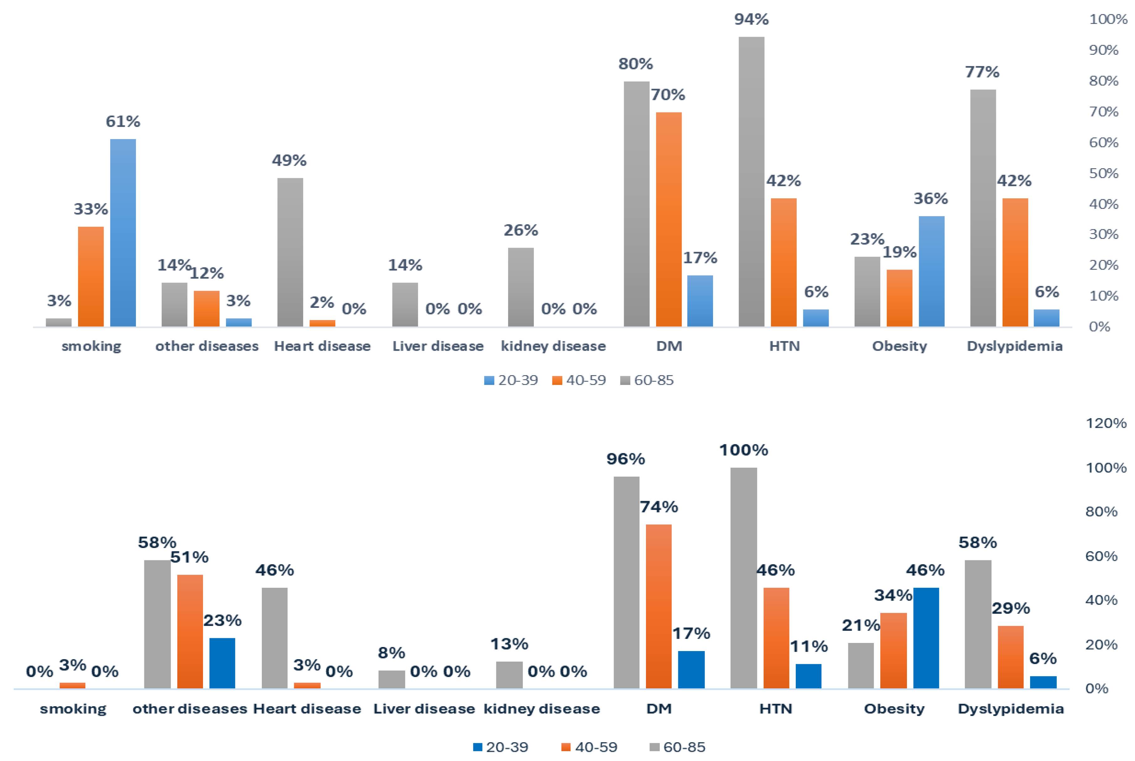
Background/Objectives: Non-communicable diseases like obesity, diabetes mellitus (DM), and hypertension (HTN) impose major global burdens. Helicobacter pylori infection may worsen these conditions. This study examined the clinical, age-, and gender-specific profiles of H. pylori-infected inpatients and its association with dyslipidemia, obesity, HTN, DM, and other chronic conditions. Methods: Between September 2024 and February 2025, patients were tested using a stool antigen assay (SIMA CHECK–ACON BIOTEC). Demographic data, smoking status, and clinical diagnoses (including kidney, liver, and heart disease) were obtained from hospital records and confirmed with standard criteria. Associations were analyzed using chi-square/Fisher’s tests and logistic regression (adjusted for age, gender, and smoking). Results: Overall, 30.7% (64/208) of inpatients were H. pylori–positive, highest in middle-aged females (42.9%) and lowest in middle-aged males (20.9%). Dyslipidemia rose sharply in males (5.6%→77.1%) versus females (5.7%→58.3%), with infection modestly elevating rates (OR=1.648). Obesity declined with age (males: 36.1%→22.9%; females: 45.7%→20.8%) yet strongly correlated with H. pylori (OR=19.217). HTN (5.694.3% in males; 11.4100% in females) and DM (16.780% vs. 17.195.8%) increased with age but showed no infection link reflecting inflammatory preference. Smoking peaked at 61.1% in younger males. Kidney and heart diseases appeared only in ages 60–85. Overall, the prevalence of comorbidities markedly increased with age. Conclusion: H. pylori infection is common among inpatients, particularly in middle-aged females, and is significantly linked to dyslipidemia and obesity. These findings support targeted screening, especially in females and individuals with metabolic abnormalities, while future studies should assess whether eradication therapy can mitigate chronic metabolic risks.

Keywords: 
;  ;  ;  ;  ;  

1. Introduction

Non-communicable diseases (NCDs) impose enormous health and economic burdens worldwide, accounting for an estimated 41 million deaths each year and significantly straining healthcare systems and national economies [1]. In many low- and middle-income regions, rising obesity rates, escalating diabetes incidence, and the increasing prevalence of associated metabolic disorders intensify these challenges (20). The Eastern Mediterranean Region, for example, has experienced a marked surge in overweight and obesity, with recent estimates surpassing 30% among adults, leading to billions of dollars in healthcare expenditures and lost productivity [2]. Such burdens underscore the critical need for research elucidating modifiable risk factors, including possible contributions from persistent infections such as Helicobacter pylori (H. pylori) [3].
H. pylori is a gram-negative microaerophilic bacillus that has been present for thousands of years in the gastric and duodenal environment of the human host [4]. In the majority of cases, the infection is asymptomatic; however, the chronic infection induces host–gastritis response by the gastric epithelium, causing various disorders of the gastrointestinal tract like peptic ulcer disease, gastritis, and gastric cancers [5]. Detection of this organism has revolutionized the knowledge about the etiology of peptic ulcers by bringing the emphasis from mere hyperacidity towards host–pathogen interaction and the impact of the microbe [6]. Up-to-date epidemiological data suggest nearly fifty percent of the global populace could be infected by this microbe; however, the prevalence is governed by geographical distribution, socioeconomic status, and ethnic groups [7,8].
Despite its main impact on the digestive system, mounting bodies of scientific work confirm the idea that infection by H. pylori can also influence extra-gastric diseases, including some metabolic and cardiovascular disorders [9]. Proposed mechanisms involve not only the organism's presence causing direct effects, but also indirect processes by the host. These processes can involve cytokine profile changes and modulation of the immune system, potentially affecting insulin resistance, lipid metabolism, and vascular tone [10]. Systemic inflammation caused by continuous stimulation of the immune system could theoretically induce pro-inflammatory environment predisposing the patient for metabolic disorders [2]. This theoretical concept has gained popularity, but the strength of the associations is not clear, and thus the need for well-characterised cohorts and stringent data analysis is emphasised [11].
Among the diseases caused by the alleged association of H. pylori infection, diabetes mellitus (DM) has drawn much attention. In some reports, the ongoing inflammation from the infection by H. pylori has the potential for exacerbating insulin resistance, thus compromising the control over blood sugars and potentially accelerating the diabetes-related sequelae [12,13]. Similarly, hypertension (HTN) has also been studied alongside the potential impact from infection by H. pylori, owing to the relationship between vascular endothelial dysfunction and systemic pro-inflammatory biomarkers [14]. Another essential component of the metabolic syndrome is the potential impact from ongoing infection through lipid metabolism changes and pro-atherogenic processes [15]. Yet the biological processes involved remain poorly elucidated, and thus the need for continued investigation into the potential for increased risk from the infection by H. pylori for the dysregulated lipid levels.
Obesity introduces one more consideration into this complex interaction, given its definition as complex chronic disorder subject to influences from genetics, lifestyle, and environment. In the past, obesity has also been linked historically with insulin resistance and dyslipidemia; however, one recent hypothesis is that infection by the organism H. pylori increases or decreases these processes through the influence upon the gut hormones [16]. To explore whether H. pylori influence the onset and progression of obesity alone or combined with other etiologies is one very significant opportunity for understanding the broader clinical implications for persistent infections. In addition to the metabolic disorder, the consequences of smoking were also studied through the epidemiological data for its possible influence on the infection susceptibility by H. pylori and the attendant clinical manifestations. Tobacco consumption increases the systemic level of inflammation substantially, thus compromising the protective functions of the gastric mucosa, potentially exacerbating the pathological impacts of H. pylori. In contrast, some groups may engage health-enhancing habits or lifestyle changes that remove one risk component while incidentally exacerbating the other [17]. These complexities highlight the need for undertaking an overall risk analysis incorporating demographic variables like the subject's age and gender, each potentially playing unique impacts upon the disease burden.
Despite the wealth of published data, inconsistencies in sample size, geographical context, and methodology continue to obscure definitive conclusions regarding the exact relationship of H. pylori to chronic conditions like DM, HTN, dyslipidemia, and obesity. Variability in diagnostic assays—such as serology, urea breath tests, and stool antigen tests—further complicates comparisons across studies [18]. Additionally, many investigations have been confined to specific geographic or ethnic groups, limiting the generalizability of their findings [19]. These gaps emphasize the importance of large-scale, population-based studies that employ standardized testing protocols for H. pylori and robust clinical assessments of coexisting chronic diseases [20].
In the context of this study, the present cross-sectional analysis was undertaken to explain the prevalence of infection by H. pylori in individuals segregated by multiple age groups and by gender differences, while simultaneously analyzing its relationship with the prevalence of associated chronic diseases. In this study, through the systematic evaluation of dyslipidemia, obesity, hypertension, diabetes mellitus, and other comorbidities relative to the presence of H. pylori infection, the objective is to improve the understanding of the widespread health implications caused by this omnipresent organism.

2. Materials and Methods

2.1. Work Environment: Ha’il Province, City, and all Socio-Economic Strata

The King Salman Specialist hospital (KSSH) is certified and accredited by the Saudi Central Board for Accreditation of Healthcare Institutions (CBAHI)-Ref.no. HAL/MOH/HO5/34213 and along with the Ha’il Health Regional Laboratory (HHRL) which is also certified and accredited by the CBAHI)-Code 2739 constitutes a major cluster for healthcare diagnostic centers that receive samples for testing. Since H.pylori is a major environmental carcinogenic pathogen, it is imperative to describe climate and environmental conditions of the Ha’il city. Ha’il lies in the north-central Saudi Arabia, bordering five provinces, namely, Madinah, Tabouk, Northern Border, Riyadh and Qassim. It has a population size of nearly one million mostly in the Ha’il city which is in the Waadi Ha’il with its attraction of magnificent prehistoric rock carving and archaeological excavations. The region has several major hospitals, among them the KSSH, a tertiary care center serving the whole region.
A retrospective cross-sectional study was conducted between September 2024 and February 2025, enrolling 208 in-patients who underwent clinical evaluation for H. pylori infection. All participants were tested for H. pylori and assessed for relevant gastrointestinal and metabolic conditions during their hospitalization. Demographic data (age and gender), smoking status, and the presence or absence of dyslipidemia, obesity, HTN, DM, kidney disease, liver disease, and heart disease were recorded at the time of admission or within the hospital stay.
All inpatients stool samples were tested using a qualitative immunochromatographic assay (SIMA CHECK–ACON BIOTEC) as described by Ozdemir and Baykan (2005), Antos et al. (2005), and Silva et al. (2010). Following the manufacturer’s protocol, each stool sample was first brought to room temperature and mixed thoroughly. Two drops (70–90 µL) of the homogenized specimen were then dispensed into the sample well of the test cassette. After a 10-minute incubation period, the appearance of distinct red lines was interpreted as follows: A single red line at the control (C) region indicated a negative result; Two red lines (one at the C region and one at the test [T] region) signified a positive result. Absence of a red line in the C region, regardless of whether a T line appeared, rendered the test invalid, and the assay was repeated with a new device. Excess specimen volume was avoided, since it could yield invalid results. All interpretations were made within 10–15 minutes of applying the sample to ensure proper reading.
Diagnosis of obesity followed a body mass index (BMI) threshold of ≥30 kg/m², and HTN and DM were determined through standard clinical criteria or documented medical histories. Chronic pathologies such as kidney, liver, and heart disease were confirmed through relevant clinical findings and laboratory or imaging tests. Smoking status was self-reported during clinical interviews, and patients were categorized as current smokers or non-smokers.
Data were analyzed using SPSS version 23.0 (SPSS Inc., Chicago, IL, USA). Categorical variables were expressed as frequencies and percentages. Associations between H. pylori status and dichotomous outcomes (e.g., presence vs. absence of specific comorbidities) were evaluated by chi-square or Fisher’s exact tests, as appropriate. Odds ratios (OR) with 95% confidence intervals (CI) were calculated to gauge the strength of each association. Where relevant, logistic regression models were employed to adjust for potential confounders, including age, gender, and smoking status. Statistical significance was defined by a two-sided p-value <0.05.

3. Results

Overall Demographics

A total of 208 participants were evaluated, consisting of 114 males (54.8%) and 94 females (45.2%), thereby allowing balanced assessment of H. pylori status and clinical comorbidities across both gender groups (Table 1). The data set permitted exploration of metabolic and organ-specific disorders in conjunction with demographics, aiding in understanding potential risk variations.

Age, Gender, and Chronic Syndromes

As shown in Table 1, dyslipidemia rose substantially among older males (60–85 years: 77.1%) and was also elevated in older females (60–85 years: 58.3%), compared to much lower proportions in younger and middle-aged participants. Obesity exceeded 45% in younger females (20–39 years: 45.7%) versus 36.1% in males of the same age and then decreased with age in both sexes, suggesting a notable gender divergence in early adulthood (Table 1). Hypertension (HTN) climbed from 5.6% (20–39 years) to 94.3% (60–85 years) in males and from 11.4% to 100.0% in females across comparable age ranges. Diabetes mellitus (DM) similarly increased with age, rising to 80.0% in older males and 95.8% in older females (Table 1a). Heart, kidney, and liver diseases predominantly affected individuals aged 60–85, with higher absolute frequencies in males. Smoking habits were reported in 61.1% of younger males but dropped to 2.9% in older males while females seldom smoked (Table 1).

Age-, and Gender-Specificity of H. pylori and Association to Obesity and Dyslipidemia

Stool antigen testing indicated an H. pylori positivity rate exceeding 30% overall, spanning from 20.9% to 42.9% among 40–59-year-olds and approximating 25–34% in both younger (20–39 years) and older (60–85 years) age groups (Table 1). The highest prevalence was detected in middle-aged females (42.9%), while middle-aged males exhibited the lowest infection frequency (20.9%) (Table 1). These differences suggest that behavioral and/or physiological factors may alter gender-specific H. pylori susceptibility in midlife (Figure 1). In logistic regression, the multivariate models revealed significant gender specificity on obesity and dyslipidemia. An odds ratio (OR) of 1.491 (95% CI: 1.006–2.209, p=0.041) was noted for dyslipidemia in males (Table 1b). Obesity, however, was strongly associated with H. pylori infection, as indicated by an OR of 19.217 (95% CI: 9.117–40.507, p<0.001) (Table 2). HTN, DM, and other chronic conditions did not show significant ORs with respect to H. pylori positivity in this dataset (Table 2).
Analysis of H. pylori Infection with dyslipidemia (borderline effect) indicated the latter disorder affected 73 of 208 participants (35.1%), with a higher rate (43.1%) in H. pylori-positive individuals than in those testing negative (31.5%) (Supplementary Table S2a). Although the Pearson chi-square test approached significance (p=0.104), it did not definitively confirm an association; the OR (1.648, 95% CI: 0.900–3.017) further suggested a borderline effect (Supplementary Table S2b, Supplementary Table S2c). Additional analyses by gender (Supplementary Table S2d–S2f) revealed that males accounted for 64.4% of dyslipidemia cases (p=0.041), underlining a notable predisposition in men (Supplementary Table S2f). Analysis of H. pylori Infection association with obesity has shown that among the 62 obese individuals (29.8%), 70.8% tested positive for H. pylori, whereas only 11.2% of non-obese participants were infected (Table 3). Statistical analyses confirmed a strong association (p<0.001), underscored by an OR of 19.217 (95% CI: 9.117–40.507), indicating that H. pylori infection was markedly linked with obesity (Table 3, Table 4 and Table 5). This finding emphasizes the potential impact of H. pylori on body weight regulation and warrants further investigation.

Association of H. pylori Infection with Hypertension, Diabetes Mellitus, Heart, Kidney, and Liver Disease

Although HTN prevalence increased from 5.6% in younger males to 94.3% in older males, and from 11.4% to 100.0% in females over the same age ranges (Table 1), the infection status itself did not correlate with higher HTN rates. H. pylori-positive individuals showed a slightly lower HTN prevalence (40.0%) than negative ones (49.7%) (Supplementary Table S4a), yielding p=0.196 and an OR of 0.676 (95% CI: 0.373–1.225) (Supplementary Table S4b–S4c). Similarly, DM was diagnosed in 119 participants (57.2%), with comparable frequencies between H. pylori-positive (53.8%) and H. pylori-negative (58.7%) groups (Supplementary Table S5a). However, the chi-square test (p=0.508) and OR of 0.819 (95% CI: 0.454–1.479) (Supplementary Table S5b–S5c) revealed no significant association, implying that there is no association between the infection and DM host conditions (Table 1). Heart disease, kidney disease, and liver disease were confined to seniors. Heart disease was observed in 15.4% of H. pylori-positive subjects versus 14.0% of negative ones (p=0.790) (Supplementary Table S8a–S8c). Kidney disease was detected in 7.7% of infected patients and 4.9% of non-infected ones (p=0.423) (Supplementary Table S6a–S6c), whereas liver disease affected 6.2% of those positive for H. pylori and 2.1% of those negative (p=0.133) (Supplementary Table S7a–S7c). None of these organ-specific conditions displayed a strong link to H. pylori infection.
In summarizing results, H. pylori prevalence exceeded 30% across the cohort and was highest in middle-aged females (42.9%) and lowest in middle-aged males (20.9%) (Table 1). Dyslipidemia, although mildly higher in infected individuals (p=0.104), was heavily influenced by male gender (64.4% of cases) (Supplementary Table S2a, Table S2d). Obesity showed the strongest correlation with H. pylori, with 70.8% of obese participants testing positive (p<0.001) (Table 3). Hypertension, diabetes, and organ-specific conditions were predominantly age-driven and did not exhibit statistically meaningful relationships with H. pylori status (Supplementary Tables S4–S8). Overall, these data highlight a potential role of H. pylori in exacerbating obesity and possibly contributing to dyslipidemia in certain subgroups, while hypertension, diabetes mellitus, and other pathologies seem more closely tied to age and gender. Graphical depictions of age and gender distributions, as well as infection rates, are presented (Figure 1).

4. Discussion

Among the diseases purportedly associated with H. pylori infection, DM continues to receive considerable attention. In some reports, ongoing inflammation triggered by H. pylori may exacerbate insulin resistance [21], compromise blood sugar control, and accelerate diabetes-related sequelae [22]. DM predisposes patients to cardiovascular disease (CVD) and impairs normal immune responses, thus highlighting the importance of exploring potential links with H. pylori, particularly in regions where diabetes prevalence is high [23]. Furthermore, there is a well-documented interplay between DM and HTN, wherein hyperglycemia fosters endothelial dysfunction and contributes to elevated blood pressure, thereby compounding the risk of CVD. Older individuals with diabetes are especially prone to developing HTN, magnifying their susceptibility to adverse cardiovascular events.
This study examined the prevalence of H. pylori infection in relation to chronic conditions, including dyslipidemia, obesity, HTN, DM, kidney disease, liver disease, and heart disease. Evidence suggests that H. pylori, commonly acquired early in life, can provoke persistent inflammatory responses influencing metabolic and cardiovascular health [24,25]. The current findings, which show that approximately one-third of participants tested positive for H. pylori across various age groups, are in line with epidemiological data indicating that up to two-thirds of the global population may carry the organism [24,26]. Although many studies reveal similar infection frequencies for males and females [27], we observed slight differences in certain age brackets, particularly an elevated rate among middle-aged females compared to their male counterparts. These variations may reflect diverse behavioral or hormonal factors, or differences in exposure that vary by gender [28]. Several studies have documented non-linear trends in H. pylori prevalence across age groups. For example, Khan et al [29] reported that while infection rates may rise during childhood and adolescence, they tend to plateau in adulthood. Other reports have sometimes pointed to higher male prevalence [30,31], underscoring the complexity of cultural, genetic, or socioeconomic factors that shape H. pylori acquisition [32,33].
In this cohort, H. pylori prevalence exceeded 30%, with middle-aged females (40–59 years) having a notably higher rate than their male counterparts. This observation aligns with literature proposing that variations in hygiene, diet, or lifestyle habits can influence infection status [32,33]. Some investigators have noted that the age at which H. pylori is acquired may determine the intensity of chronic inflammation and eventual pathology. Infection during childhood may be more likely to lead to severe gastric conditions, while later infection could show more association with duodenal ulceration or milder inflammatory phenomena [34,35]. Although the present study did not track exact age of acquisition, the finding that H. pylori prevalence varied by age group suggests a role for differing exposure patterns across the lifespan.
One of the more striking observations was the association between H. pylori infection and dyslipidemia. We found that men were more likely to have dyslipidemia, and when we considered H. pylori status, a statistically significant connection emerged. Research in other populations, such as in Jimma, Ethiopia, has similarly reported higher rates of abnormal lipid profiles among H. pylori-positive individuals, noting rises in total cholesterol, triglycerides, and LDL cholesterol [36]. Dyslipidemia itself is strongly implicated in cardiovascular disease risk [37]. It is likely that the chronic low-grade inflammation caused by H. pylori infection promotes lipid metabolism disturbances [38,39], partly via cytokines like tumor necrosis factor-alpha (TNF-α) and interleukin-6 (IL-6) [40,41]. These mediators can disrupt normal lipoprotein lipase activity and impair cholesterol transport [42]. H. pylori strains with virulence factors such as CagA may further contribute to metabolic disruptions by altering pathways responsible for cholesterol synthesis, including key enzymes such as squalene epoxidase [42]
Notable gender differences also emerged in obesity rates, with younger females displaying higher obesity percentages. Although no straightforward, one-way correlation to H. pylori was evident, the fact that obesity was frequent in certain female subgroups aligns with hypotheses linking chronic infection and metabolic alterations. Some studies show that H. pylori may contribute to weight gain by interfering with adipocyte hormones [43], whereas others find an inverse or nonexistent association, suggesting an interplay among genetics, diet, and hormonal factors [44]. The elevated obesity prevalence among younger females with H. pylori infection here points to the possible role of hormonal influences and demands further exploration.
Hypertension and DM were both strongly tied to older age ranges in this study. Participants with H. pylori infection exhibited higher frequencies of these metabolic conditions, supporting evidence that chronic H. pylori infection can promote insulin resistance through heightened production of TNF-α and IL-6 [22]. These pro-inflammatory signals compromise insulin receptor functionality and engender endothelial dysfunction, culminating in increased blood pressure [45]. Cohort studies in other settings have similarly implicated H. pylori in elevated risks of type 2 diabetes, especially among males [46]. When DM and HTN co-occur, as is common in older adults, the risk of cardiovascular events escalates considerably, emphasizing the need to consider H. pylori status in patients with unexplained metabolic or cardiovascular risk factors.
Kidney, liver, and heart diseases also increased with advancing age, mirroring the general trend that older individuals accumulate greater comorbidity. Although prevalence rates of these organ-specific pathologies were relatively modest compared to those of HTN or DM, H. pylori positivity correlated with higher frequencies in older age brackets. Some authors attribute these links to systemic inflammation and atherogenic processes accelerated by chronic infection [47]. Associations between H. pylori and nonalcoholic fatty liver disease, including elevated liver enzymes, have also been documented [48]. With respect to heart disease, certain investigations propose that H. pylori eradication could ameliorate endothelial function and reduce atherosclerotic risk [49]. In this study, older males with both H. pylori and dyslipidemia were more likely to manifest heart disease, although the cross-sectional design precludes definitive causal inferences.
Smoking emerged as a crucial determinant of metabolic and cardiovascular health, particularly prevalent among younger males in this study, while negligible in females. Tobacco use substantially exacerbates the inflammatory environment, which in turn may amplify any adverse metabolic effects from H. pylori [50]. Given that smoking rates declined in older males, potential harm to vascular and metabolic systems may have been exerted earlier in life, compounding later risks. Future investigations assessing H. pylori and metabolic diseases should therefore control for smoking behaviors to isolate the bacterium’s specific contributions [50].
Several pathways have been proposed to explain how H. pylori could disrupt metabolic homeostasis. Chronic infection is known to elicit immune responses that produce pro-inflammatory cytokines, which interfere with insulin signaling in peripheral tissues and the liver. H. pylori may also affect gut microbial composition [51,52]. The gut microbiota is well-recognized as a key regulator of energy extraction and storage [53]. When gut microbes shift toward communities that can harvest more energy from the diet, obesity and insulin resistance may become more likely [53,54]. Although this study did not characterize the gut microbiome, the interaction between H. pylori and other gut bacteria deserves further study, especially in populations with high rates of both H. pylori infection and obesity.
From a clinical perspective, these results highlight the importance of considering H. pylori infection in patients who present with a spectrum of metabolic or cardiovascular risks, including unexplained dyslipidemia, elevated blood pressure, and insulin resistance. If further research confirms that treating H. pylori not only resolves gastric problems but also improves metabolic markers, then routine screening for this organism might become more broadly recommended. Public health initiatives aimed at preventing H. pylori transmission, such as improving sanitation and public awareness, could yield benefits that go well beyond reducing gastric morbidity, potentially lowering the incidence of metabolic syndrome components as well. Still, it is crucial to acknowledge that many of the current data are cross-sectional. Longitudinal designs, along with randomized controlled trials, are needed to establish whether eradicating H. pylori yields sustained improvements in lipid profiles, glycemic control, or blood pressure. Standardized methods for diagnosing H. pylori, robust sample sizes, and multivariate models that adjust for factors like smoking, diet, and socioeconomic status will be particularly important. Gender differences should also receive closer scrutiny, given the patterns we observed in obesity, smoking, and H. pylori prevalence. Hormonal influences and culturally shaped behaviors may alter both the risk of acquiring H. pylori and the infection’s systemic effects. Future investigations that incorporate advanced molecular and microbiome analyses might explain precisely how H. pylori triggers systemic inflammation and influences pathways that govern metabolism.

5. Conclusions

In conclusion, the study revealed significant correlations between H. pylori infection and dyslipidemia, hypertension, and diabetes mellitus. Older participants, especially males, were more prone to dyslipidemia, whereas younger females showed higher obesity rates. Smoking was disproportionately common among younger males, potentially intensifying the inflammatory response associated with H. pylori. Although causality remains undetermined, the collective evidence implies that persistent H. pylori infection could compound metabolic processes leading to these conditions. Screening and possible eradication therapy could thus represent an avenue for mitigating cardiovascular risk. Beyond peptic ulcer disease and gastric cancer, H. pylori’s possible role in precipitating chronic metabolic disease warrants a broadened perspective in both clinical practice and public health policy. Strategies that concurrently address infection, lifestyle factors, and demographic differences may more comprehensively lessen the burden of chronic illnesses in diverse populations.

Author Contributions

Conceptualization—Conceptualization, Kamaleldin Said; Data curation, Kamaleldin Said, Khalid F Alshammari, Safia Moussa, Ruba Ahmed, Ahmed Aljadani, Najd Albalawi, Layan Al-Hujaili, Ruaa Alharbi, Arwa Alotaibi, Fahad Alshammary, Alfatih Alnajib, Fayez Alfouzan, Abuzar Osman, Zaid Albayih, Bader Alkharisi and Naif Altamimi; Formal analysis, Kamaleldin Said, Khalid F Alshammari, Safia Moussa, Ruba Ahmed, Ahmed Aljadani, Najd Albalawi, Layan Al-Hujaili, Ruaa Alharbi, Arwa Alotaibi, Fahad Alshammary, Alfatih Alnajib, Fayez Alfouzan, Abuzar Osman, Zaid Albayih, Bader Alkharisi and Naif Altamimi; Funding acquisition, Kamaleldin Said; Investigation, Kamaleldin Said; Methodology, Kamaleldin Said, Khalid F Alshammari, Safia Moussa, Ruba Ahmed, Ahmed Aljadani, Najd Albalawi, Layan Al-Hujaili, Ruaa Alharbi, Arwa Alotaibi, Fahad Alshammary, Alfatih Alnajib, Fayez Alfouzan, Abuzar Osman, Zaid Albayih, Bader Alkharisi and Naif Altamimi; Project administration, Kamaleldin Said; Resources, Kamaleldin Said, Khalid F Alshammari, Safia Moussa, Ruba Ahmed, Ahmed Aljadani, Najd Albalawi, Layan Al-Hujaili, Ruaa Alharbi, Arwa Alotaibi, Fahad Alshammary, Alfatih Alnajib, Fayez Alfouzan, Abuzar Osman, Zaid Albayih, Bader Alkharisi and Naif Altamimi; Software, Kamaleldin Said, Khalid F Alshammari, Safia Moussa, Ruba Ahmed, Ahmed Aljadani, Najd Albalawi, Layan Al-Hujaili, Ruaa Alharbi, Arwa Alotaibi, Fahad Alshammary, Alfatih Alnajib, Fayez Alfouzan, Abuzar Osman, Zaid Albayih, Bader Alkharisi and Naif Altamimi; Supervision, Kamaleldin Said; Validation, Kamaleldin Said, Khalid F Alshammari, Safia Moussa, Ruba Ahmed, Ahmed Aljadani, Najd Albalawi, Layan Al-Hujaili, Ruaa Alharbi, Arwa Alotaibi, Fahad Alshammary, Alfatih Alnajib, Fayez Alfouzan, Abuzar Osman, Zaid Albayih, Bader Alkharisi and Naif Altamimi; Visualization, Kamaleldin Said, Khalid F Alshammari, Safia Moussa, Ruba Ahmed, Ahmed Aljadani, Najd Albalawi, Layan Al-Hujaili, Ruaa Alharbi, Arwa Alotaibi, Fahad Alshammary, Alfatih Alnajib, Fayez Alfouzan, Abuzar Osman, Zaid Albayih, Bader Alkharisi and Naif Altamimi; Writing – original draft, Kamaleldin Said; Writing – review & editing, Kamaleldin Said, Khalid F Alshammari, Safia Moussa, Ruba Ahmed, Ahmed Aljadani, Najd Albalawi, Layan Al-Hujaili, Ruaa Alharbi, Arwa Alotaibi, Fahad Alshammary, Alfatih Alnajib, Fayez Alfouzan, Abuzar Osman, Zaid Albayih, Bader Alkharisi and Naif Altamimi.

Funding

This research received no funding.

Institutional Review Board Statement

The Research Ethical Committee (REC) of University of Ha’il, Saudi Arabia, has Approved this research by the number (H-2024-941), dated REC 4112024. In addition, IRB Approval (Log 2024-120, Dec 2024) was obtained from Ha’il Health Cluster, Ha’il to perform this work.

Informed Consent Statement

Informed consent was obtained from all subjects involved in the study.

Data Availability Statement

Supplementary Materials: The supplementary tables are enclosed with this manuscript.

Acknowledgments

We acknowledge the University of Ha’il’s Deanship for research, REC, and clinics for their support throughout this work.

Conflicts of Interest

The authors declare no conflict of interest.

References

  1. Kazibwe J, Tran PB, Annerstedt KS. The household financial burden of non-communicable diseases in low- and middle-income countries: a systematic review. Health Research Policy and Systems. 2021;19(1):96.
  2. Buzás, GM. Metabolic consequences of Helicobacter pylori infection and eradication. World J Gastroenterol. 2014;20(18):5226-34.
  3. Asmelash D, Nigatie M, Melak T, Alemayehu E, Ashagre A, Worede A. Metabolic syndrome and associated factors among H. pylori-infected and negative controls in Northeast Ethiopia: a comparative cross-sectional study. Frontiers in Endocrinology. 2024;15.
  4. Murray DM, DuPont HL, Cooperstock M, Corrado ML, Fekety R. Evaluation of new anti-infective drugs for the treatment of gastritis and peptic ulcer disease associated with infection by Helicobacter pylori. Infectious Diseases Society of America and the Food and Drug Administration. Clin Infect Dis. 1992;15 Suppl 1:S268-73.
  5. Ruggiero, P. Helicobacter pylori and inflammation. Curr Pharm Des. 2010;16(38):4225-36.
  6. Rebora A, Drago F, Parodi A. May Helicobacter pylori be important for dermatologists? Dermatology. 1995;191(1):6-8.
  7. Wands DIF, El-Omar EM, Hansen R. Helicobacter pylori: getting to grips with the guidance. Frontline Gastroenterol. 2021;12(7):650-5.
  8. Brown, LM. Helicobacter pylori: epidemiology and routes of transmission. Epidemiol Rev. 2000;22(2):283-97.
  9. Franceschi F, Gasbarrini A, Polyzos SA, Kountouras J. Extragastric Diseases and Helicobacter pylori. Helicobacter. 2015;20 Suppl 1:40-6.
  10. Gravina AG, Zagari RM, De Musis C, Romano L, Loguercio C, Romano M. Helicobacter pylori and extragastric diseases: A review. World J Gastroenterol. 2018;24(29):3204-21.
  11. Pellicano R, Ianiro G, Fagoonee S, Settanni CR, Gasbarrini A. Review: Extragastric diseases and Helicobacter pylori. Helicobacter. 2020;25 Suppl 1:e12741.
  12. Polyzos SA, Kountouras J, Zavos C, Deretzi G. The association between Helicobacter pylori infection and insulin resistance: a systematic review. Helicobacter. 2011;16(2):79-88.
  13. Zhou X, Liu W, Gu M, Zhou H, Zhang G. Helicobacter pylori infection causes hepatic insulin resistance by the c-Jun/miR-203/SOCS3 signaling pathway. J Gastroenterol. 2015;50(10):1027-40.
  14. Longo-Mbenza B, Nsenga JN, Mokondjimobe E, Gombet T, Assori IN, Ibara JR, et al. Helicobacter pylori infection is identified as a cardiovascular risk factor in Central Africans. Vasc Health Risk Manag. 2012;6:455-61.
  15. Fan N, Peng L, Xia Z, Zhang L, Wang Y, Peng Y. Helicobacter pylori Infection Is Not Associated with Non-alcoholic Fatty Liver Disease: A Cross-Sectional Study in China. Front Microbiol. 2018;9:73.
  16. Jeffery PL, McGuckin MA, Linden SK. Endocrine impact of Helicobacter pylori: focus on ghrelin and ghrelin o-acyltransferase. World J Gastroenterol. 2011;17(10):1249-60.
  17. Ferro A, Morais S, Pelucchi C, Aragonés N, Kogevinas M, López-Carrillo L, et al. Smoking and Helicobacter pylori infection: an individual participant pooled analysis (Stomach Cancer Pooling- StoP Project). Eur J Cancer Prev. 2019;28(5):390-6.
  18. Wang YK, Kuo FC, Liu CJ, Wu MC, Shih HY, Wang SS, et al. Diagnosis of Helicobacter pylori infection: Current options and developments. World J Gastroenterol. 2015;21(40):11221-35.
  19. Shah SC, Halvorson AE, Lee D, Bustamante R, McBay B, Gupta R, et al. Helicobacter pylori Burden in the United States According to Individual Demographics and Geography: A Nationwide Analysis of the Veterans Healthcare System. Clin Gastroenterol Hepatol. 2024;22(1):42-50.e26.
  20. Liu ZC, Li WQ. Large-scale cluster randomised trial reveals effectiveness of Helicobacter pylori eradication for gastric cancer prevention. Clin Transl Med. 2025;15(2):e70229.
  21. Azami M, Baradaran HR, Dehghanbanadaki H, Kohnepoushi P, Saed L, Moradkhani A, et al. Association of Helicobacter pylori infection with the risk of metabolic syndrome and insulin resistance: an updated systematic review and meta-analysis. Diabetology & Metabolic Syndrome. 2021;13(1):145.
  22. He C, Yang Z, Lu NH. Helicobacter pylori infection and diabetes: is it a myth or fact? World J Gastroenterol. 2014;20(16):4607-17.
  23. Chua W-K, Hong Y-K, Hu S-W, Fan H-C, Ting W-H. A Significant Association between Type 1 Diabetes and Helicobacter pylori Infection: A Meta-Analysis Study. Medicina [Internet]. 2024; 60(1).
  24. Zamani M, Ebrahimtabar F, Zamani V, Miller WH, Alizadeh-Navaei R, Shokri-Shirvani J, et al. Systematic review with meta-analysis: the worldwide prevalence of Helicobacter pylori infection. Aliment Pharmacol Ther. 2018;47(7):868-76.
  25. Hooi JKY, Lai WY, Ng WK, Suen MMY, Underwood FE, Tanyingoh D, et al. Global Prevalence of Helicobacter pylori Infection: Systematic Review and Meta-Analysis. Gastroenterology. 2017;153(2):420-9.
  26. Malfertheiner P, Camargo MC, El-Omar E, Liou JM, Peek R, Schulz C, et al. Helicobacter pylori infection. Nat Rev Dis Primers. 2023;9(1):19.
  27. Almashhadany DA, Mayas SM, Mohammed HI, Hassan AA, Khan IUH. Population- and Gender-Based Investigation for Prevalence of Helicobacter pylori in Dhamar, Yemen. Canadian Journal of Gastroenterology and Hepatology. 2023;2023(1):3800810.
  28. Dias Sara P, Brouwer Matthijs C, van de Beek D. Sex and Gender Differences in Bacterial Infections. Infection and Immunity. 2022;90(10):e00283-22.
  29. Khan, AR. An age- and gender-specific analysis of H. Pylori infection. Ann Saudi Med. 1998;18(1):6-8.
  30. Hong W, Tang H, Dong X, Hu S, Yan Y, Basharat Z, et al. Prevalence of Helicobacter pylori infection in a third-tier Chinese city: relationship with gender, age, birth-year and survey years. Microb Health Dis. 2019;1:e150.
  31. Rosu OM, Gimiga N, Stefanescu G, Anton C, Paduraru G, Tataranu E, et al. Helicobacter pylori Infection in a Pediatric Population from Romania: Risk Factors, Clinical and Endoscopic Features and Treatment Compliance. J Clin Med. 2022;11(9).
  32. Chen J, Bu XL, Wang QY, Hu PJ, Chen MH. Decreasing seroprevalence of Helicobacter pylori infection during 1993-2003 in Guangzhou, southern China. Helicobacter. 2007;12(2):164-9.
  33. den Hoed CM, Vila AJ, Holster IL, Perez-Perez GI, Blaser MJ, de Jongste JC, et al. Helicobacter pylori and the birth cohort effect: evidence for stabilized colonization rates in childhood. Helicobacter. 2011;16(5):405-9.
  34. Graham, DY. Helicobacter pylori: its epidemiology and its role in duodenal ulcer disease. J Gastroenterol Hepatol. 1991;6(2):105-13.
  35. Wroblewski LE, Peek RM, Jr., Wilson KT. Helicobacter pylori and gastric cancer: factors that modulate disease risk. Clin Microbiol Rev. 2010;23(4):713-39.
  36. Abdu A, Cheneke W, Adem M, Belete R, Getachew A. Dyslipidemia and Associated Factors Among Patients Suspected to Have Helicobacter pylori Infection at Jimma University Medical Center, Jimma, Ethiopia. Int J Gen Med. 2020;13:311-21.
  37. Grundy SM, Cleeman JI, Daniels SR, Donato KA, Eckel RH, Franklin BA, et al. Diagnosis and management of the metabolic syndrome: an American Heart Association/National Heart, Lung, and Blood Institute Scientific Statement. Circulation. 2005;112(17):2735-52.
  38. Hashim M, Mohammed O, T GE, Wolde M. The association of Helicobacter Pylori infection with dyslipidaemia and other atherogenic factors in dyspeptic patients at St. Paul's Hospital Millennium Medical College. Heliyon. 2022;8(5):e09430.
  39. Khera AV, Demler OV, Adelman SJ, Collins HL, Glynn RJ, Ridker PM, et al. Cholesterol Efflux Capacity, High-Density Lipoprotein Particle Number, and Incident Cardiovascular Events: An Analysis From the JUPITER Trial (Justification for the Use of Statins in Prevention: An Intervention Trial Evaluating Rosuvastatin). Circulation. 2017;135(25):2494-504.
  40. Vijayvergiya R, Vadivelu R. Role of Helicobacter pylori infection in pathogenesis of atherosclerosis. World J Cardiol. 2015;7(3):134-43.
  41. Morningstar-Wright L, Czinn SJ, Piazuelo MB, Banerjee A, Godlewska R, Blanchard TG. The TNF-Alpha Inducing Protein is Associated With Gastric Inflammation and Hyperplasia in a Murine Model of Helicobacter pylori Infection. Front Pharmacol. 2022;13:817237.
  42. Liu S, Zhang N, Ji X, Yang S, Zhao Z, Li P. Helicobacter pylori CagA promotes gastric cancer immune escape by upregulating SQLE. Cell Death & Disease. 2025;16(1):17.
  43. Chen L-W, Kuo S-F, Chen C-H, Chien C-H, Lin C-L, Chien R-N. A community-based study on the association between Helicobacter pylori Infection and obesity. Scientific Reports. 2018;8(1):10746.
  44. Kamarehei F, Mohammadi Y. The Effect of Helicobacter pylori Infection on Overweight: A Systematic Review and Meta-Analysis. Iran J Public Health. 2022;51(11):2417-24.
  45. Liu Y, Shuai P, Chen W, Liu Y, Li D. Association between Helicobacter pylori infection and metabolic syndrome and its components. Front Endocrinol (Lausanne). 2023;14:1188487.
  46. Jeon CY, Haan MN, Cheng C, Clayton ER, Mayeda ER, Miller JW, et al. Helicobacter pylori infection is associated with an increased rate of diabetes. Diabetes Care. 2012;35(3):520-5.
  47. Pan W, Zhang H, Wang L, Zhu T, Chen B, Fan J. Association between Helicobacter pylori infection and kidney damage in patients with peptic ulcer. Renal Failure. 2019;41(1):1028-34.
  48. Abo-Amer YE, Sabal A, Ahmed R, Hasan NFE, Refaie R, Mostafa SM, et al. Relationship Between Helicobacter pylori Infection and Nonalcoholic Fatty Liver Disease (NAFLD) in a Developing Country: A Cross-Sectional Study. Diabetes Metab Syndr Obes. 2020;13:619-25.
  49. Aramouni K, Assaf RK, Azar M, Jabbour K, Shaito A, Sahebkar A, et al. Infection with Helicobacter pylori may predispose to atherosclerosis: role of inflammation and thickening of intima-media of carotid arteries. Front Pharmacol. 2023;14:1285754.
  50. Addissouky TA, El Sayed IET, Ali MMA, Wang Y, El Baz A, Elarabany N, et al. Oxidative stress and inflammation: elucidating mechanisms of smoking-attributable pathology for therapeutic targeting. Bulletin of the National Research Centre. 2024;48(1):16.
  51. Elghannam MT, Hassanien MH, Ameen YA, Turky EA, Elattar GM, Elray AA, et al. Helicobacter pylori and oral–gut microbiome: clinical implications. Infection. 2024;52(2):289-300.
  52. Zhang L, Zhao M, Fu X. Gastric microbiota dysbiosis and Helicobacter pylori infection. Front Microbiol. 2023;14:1153269.
  53. Turnbaugh PJ, Ley RE, Mahowald MA, Magrini V, Mardis ER, Gordon JI. An obesity-associated gut microbiome with increased capacity for energy harvest. Nature. 2006;444(7122):1027-31.
  54. Bäckhed F, Ding H, Wang T, Hooper LV, Koh GY, Nagy A, et al. The gut microbiota as an environmental factor that regulates fat storage. Proceedings of the National Academy of Sciences. 2004;101(44):15718-23.
Figure 1. Prevalence of chronic conditions among study participants stratified by age and gender. The upper panel represents male participants, while the lower panel represents female participants across three age groups (20–39, 40–59, and 60–85 years).
Preprints 151573 g001
Table 1. Age-, and gender- stratified frequencies of H. pylori infections and chronic diseases .
Gender
Male
5(N=114; 54.8%)
Female
5(N=94; 45.2%)
Age Age
20-39 (n=36) 40-59 (n=43) 60-85 (n=35) 20-39 (n=35) 40-59 (n=35) 60-85 (n=24)
N N % N N % N N % N N % N N % N N %
Dyslypidemia (m/f: 64.4M, 35.6%F) no 34 94.4% 25 58.1%
5
8 22.9% 33 94.3% 25 71.4%
5
10 41.7%
yes 2 5.6% 18 41.9% 27 77.1% 2 5.7% 10 28.6% 14 58.3%
Obesity (BMI > 30 kg/m²)
5(m/f: 47%M, 53%F)
no 23 63.9% 35 81.4% 27 77.1% 19 54.3% 23 65.7% 19 79.2%
yes 13 36.1%
5
8 18.6%
5
8 22.9%
5
16 45.7%
5
12 34.3%
5
5 20.8%
5
HTN
5(Mf: 55%M; 45.4F)
no 34 94.4% 25 58.1% 2 5.7% 31 88.6% 19 54.3% 0 0.0%
yes 2 5.6% 18 41.9% 33 94.3% 4 11.4% 16 45.7% 24 100%
DM
5(m/f: 54%M; 46%F)
no 30 83.3% 13 30.2% 7 20.0% 29 82.9% 9 25.7% 1 4.2%
yes 6 16.7% 30 69.8% 28 80.0% 6 17.1% 26 74.3% 23 95.8%
Heart disease
5(m/f: 75%M; 25%F)
no 36 100% 42 97.7% 18 51.4% 35 100% 34 97.1% 13 54.2%
yes 0 0.0% 1 2.3% 17 48.6% 0 0.0% 1 2.9% 11 45.8%
kidney disease
5(m/f: 71.4%M; 29%F)
no 36 100% 43 100% 26 74.3% 35 100% 35 100.0% 21 87.5%
yes 0 0.0% 0 0.0% 9 25.7% 0 0.0% 0 0.0% 3 12.5%
Liver disease
5(m/f: 60%M; 40%F)
no 36 100% 43 100.0% 30 85.7% 35 100.% 35 100.0% 22 91.7%
yes 0 0.0% 0 0.0% 5 14.3% 0 0.0% 0 0.0% 2 8.3%
other diseases no 35 97.2% 38 88.4% 30 85.7% 27 77.1% 17 48.6% 10 41.7%
yes 1 2.8% 5 11.6% 5 14.3% 8 22.9% 18 51.4% 14 58.3%
Smoking (94%M) no 14 38.9% 29 67.4% 34 97.1% 35 100% 34 97.1% 24 100.0%
yes 22 61.1% 14 32.6% 1 2.9% 0 0.0% 1 2.9% 0 0.0%
Stool Ag test
5(31.25%; 65/208)
5(mf: 49%; 51%F)
negative 24 66.7% 34 79.1% 24 68.6% 23 65.7% 20 57.1% 18 75.0%
positive 12 33.3% 9 20.9% 11 31.4% 12 34.3% 15 42.9%
5
6 25.0%
Table 2. Logistic regression analysis for risk estimates of Helicobacter pylori infection and gender in chronic diseases.
Gender-Specific Associationto Chronic Diseases Risk of H.pyloriInfection and Association in Chronic Diseases
Odd’s Ratio 95% Confidence Interval pValue Odd’s Ratio 95% Confidence Interval pValue
Dyslipidemia 1.491 (1.006 2.209) 0.041 1.648 .900 3.017 .052
Obesity 1.586 .872 2.882 .036 19.217 9.117 40.507 .000
Hypertension 1.103 .586 1.751 .002 .676 .373 1.225 1.672
DM 1.102 .634 1.914 .118 .819 .454 1.479 .437
Kidney diseases .385 0.101 1.464 2.096 1.619 .494 5.307 .643
Liver diseases .474 .090 2.500 .808 3.060 .665 14.088 2.260
Heart diseases 0.780 0.355 1.716 0.382 1.118 .491 2.546 .071
Smoking 0.022 0.003 0.167 0.000
Other diseases 1.277 .654 2.490 .514a
Table 3. Prevalence of obesity among study participants stratified by H. pylori stool antigen test results.
Obesity Stool Ag test Total
negative positive
no Count 127 19 146
% within stool Ag test 88.8% 29.2% 70.2%
yes Count 16 46 62
% within stool Ag test 11.2% 70.8% 29.8%
Table 4. Chi-square test results for the association between obesity and H. pylori infection.
Chi-Square Tests Value Asymp. Sig. (2-sided) Exact Sig. (2-sided) Exact Sig. (1-sided)
Pearson Chi-Square 75.819a .000
Continuity Correctionb 72.998 .000
Likelihood Ratio 74.664 .000
Fisher's Exact Test .000 .000
Linear-by-Linear Association 75.454 .000
N of Valid Cases 208
Table 5. Risk estimates for obesity in relation to H. pylori infection.
Risk Estimate Value 95% Confidence Interval
Lower Upper
Odds Ratio for Obesity (no / yes) 19.217 9.117 40.507
For cohort stool Ag test = negative 3.371 2.200 5.165
For cohort stool Ag test = positive .175 .112 .274
Disclaimer/Publisher’s Note: The statements, opinions and data contained in all publications are solely those of the individual author(s) and contributor(s) and not of MDPI and/or the editor(s). MDPI and/or the editor(s) disclaim responsibility for any injury to people or property resulting from any ideas, methods, instructions or products referred to in the content.
Copyright: This open access article is published under a Creative Commons CC BY 4.0 license, which permit the free download, distribution, and reuse, provided that the author and preprint are cited in any reuse.
Prerpints.org logo

Preprints.org is a free preprint server supported by MDPI in Basel, Switzerland.

Subscribe

Disclaimer

Terms of Use

Privacy Policy

Privacy Settings

© 2025 MDPI (Basel, Switzerland) unless otherwise stated