Introduction
Glaucoma is a leading cause of irreversible blindness globally, characterized by progressive optic nerve damage and visual field loss [
1,
2,
3]. It poses a significant public health challenge, impacting individuals worldwide with its insidious progression and debilitating consequences on visual function. The management of glaucoma entails a multifaceted approach involving various treatment modalities, ranging from topical medications to laser procedures and surgical interventions [
4,
5,
6]. Among these treatment options, trabeculectomy with mitomycin C stands out as a cornerstone surgical procedure utilized in cases where conservative measures fail to adequately control intraocular pressure [
7,
8,
9]. By creating a new filtration pathway for aqueous humor drainage, trabeculectomy aims to alleviate the elevated intraocular pressure burden that contributes to disease progression in glaucomatous eyes [
10,
11]. Despite its proven efficacy in lowering intraocular pressure and preserving visual function, trabeculectomy outcomes can exhibit variability influenced by a multitude of preoperative, intraoperative, and postoperative factors.
The success of trabeculectomy is contingent not only on the surgical technique and postoperative management but also on the preoperative ocular characteristics that set the stage for the surgical procedure. The conjunctiva, a thin and transparent membrane covering the ocular surface, plays a pivotal role in trabeculectomy outcomes by influencing the formation and function of the filtration bleb, a critical component in maintaining long-term intraocular pressure control postoperatively [
12,
13]. Among the various preoperative factors that can impact trabeculectomy success, the vascularity of the conjunctiva emerges as a key determinant deserving closer examination. Conjunctival vascular characteristics, including the density, tortuosity, and caliber of vessels within the conjunctiva, are believed to have implications for bleb morphology, wound healing dynamics, and postsurgical outcomes following trabeculectomy with mitomycin C [
14,
15]. Therefore, understanding the influence of preoperative conjunctival vascular features on surgical results is essential in optimizing treatment strategies and enhancing visual outcomes for glaucoma patients undergoing trabeculectomy procedures.
In light of the intricate interplay between preoperative conjunctival vascularity and trabeculectomy outcomes, this study endeavors to delve deeper into the relationship between preoperative conjunctival vascular area and postoperative clinical parameters in patients undergoing trabeculectomy with mitomycin C for glaucoma. By comprehensively analyzing preoperative conjunctival vascular characteristics and their impact on surgical success, this research aims to provide valuable insights into personalized treatment approaches tailored to individual conjunctival profiles and optimize therapeutic outcomes in the management of glaucoma.
Material and Methods
Ethical Approval
This retrospective cohort study was approved by our institutions committee (Tohankai eye institutions Ethics Committee, approval number 0002, January 31st, 2023) and adhered to the regulations of clinical practice and the tenets of the Declaration of Helsinki. The authors had access to information that could identify individual participants during and after data collection. Informed consent has been obtained from all participants in the study. The data for our research purposes were accessed from February 1st, 2024. During the data collection process, the authors did not have access to any information that could identify individual participants. All data were anonymized to maintain confidentiality.
Study Design
This study is a retrospective cohort study.Glaucoma patients underwent trabeculectomy with 0.04% mitomycin C by Hayakawa Y at Urawa central eye institutions, Chiba central eye institution, Koshigaya central eye institution, Kumagaya central eye institution, Yorii central eye institution, Onohara eye institution, and Kisarazu central eye institution, between January 1st 2020 to December 31st 2023.
Inclusion and Exclusion Criteria
The study enrolled subjects with mild-to-severe glaucoma including primary open angle glaucoma (POAG), pseudoexfoliation glaucoma (PEG) and combined mechanism glaucoma (CMG) who were treated with one or more ophthalmic antiglaucoma agents. POAG was diagnosed with the characteristic glaucomatous optic neuropathy, two reliable visual field tests with repeatable glaucomatous defect, open-angle on gonioscopy and IOP more than 21 mmHg on two consecutive visits using a Goldman applanation tonometer. PEG was diagnosed by the deposition of white dandruff-like material in the trabecular meshwork confirmed by gonioscopy. Those that do not fit POAG and chronic angle closure glaucoma (CACG) were diagnosed as CMG. Secondary glaucoma, and neovascular glaucoma were excluded in this study.
Surgical Procedure
For fornix-based trabeculectomy, a limbal peritomy centered at the temporal upper position was created. The conjunctiva and Tenon’s capsule were dissected using Westcott scissors and a MMC-soaked cellulose sponge applied as determined by surgeon preference as above. A partial-thickness scleral flap was created followed by a sclerotomy and iridectomy in the same fashion as the limbus-based trabeculectomy. The scleral flap was closed using 10-0 nylon sutures, and conjunctiva and Tenon’s capsule were closed using two 10-0 nylon sutures at the limbus.
Measurement
A retrospective cohort of 72 eyes from 72 consecutive patients who underwent primary trabeculectomy with mitomycin C for glaucoma within a specified timeframe was meticulously evaluated in this study. The patient cohort represented a diverse population with varying demographics, underlying glaucoma subtypes, and preoperative ocular characteristics, all of which were meticulously documented and analyzed for their potential influence on surgical outcomes.
Prior to undergoing trabeculectomy, the conjunctival vascular density was measured using ImageJ software as follows: The photo of the anterior segment was taken using a slit lamp microscope (Haag-Streit AG, Baden, Switzerland) at a magnification of 6.3x, focusing on the area where a trabeculectomy was performed in the superior-temporal quadrant, converted to TIFF files, and then transformed into 8-bit black and white images using ImageJ software (NIH, Madison, WI) for measurement.
Postoperatively, patients were followed up for a minimum of 12 months to monitor their clinical progress and assess various parameters indicative of surgical success or potential complications. The key postoperative parameters evaluated in this study included intraocular pressure measurements at the 12-month mark, the rate of intraocular pressure reduction from baseline, the morphological characteristics of filtration blebs, the frequency of laser suture lysis (LSL) procedures, and alterations in glaucoma medication regimens post-trabeculectomy, and the occurrence and resolution of hyphema, occurrence of flat anterior chamber (FAC),
IOP was measured using Goldman applanation tonometer (GAT). The ratio of IOP reduction, calculated as (preoperative IOP - postoperative IOP / preoperative IOP) x 100, was also a critical parameter assessed in this study.
In classifying the post-trabeculectomy blebs, a simplified classification based on Migdal and Hitchings’ classification [
16] was used and customized as follows: Grade 1; thin and transconjunctival flow of aqueous humor resulting in good filtration (avascular sub-Tenon’s bleb), Grade 2; thin, diffuse, relatively avascular bleb with microcysts resulting in good filtration, Grade 3; flat, non-microcystic bleb with engorged vessels resulting in non-filtration bleb, Grade 4; localized, highly elevated, engorged vessels, cyst-like or hypertrophied Tenon’s resulting in encapsulated bleb. The blebs were classified under slit lamp examination at the subconjunctival lump at 12 months postoperatively. The MBGS (Moorfields Bleb Grading System) and IBAGS (Indiana Bleb Appearance Grading Scale) are commonly used for classifying blebs [
17,
18,
19]. However, in this study, we chose not to use these classifications as we felt that they were too detailed for the scope of our study, making it difficult to detect significant differences within the sample size.
The presence or absence of a FAC was confirmed by slit lamp examination, and FAC grading of 1-3 was classified as FAC + [
20,
21].
For cases receiving preoperative antithrombotic agents, administration was halted. In this study, all cases of hyphema were graded as less than grade 1, therefore classified as - for absence of hyphema and + for presence of hyphema.
Statistical Analysis
Statistical analyses, including logistic regression models, Pearson’s correlation coefficient, and Spearman’s rank correlation coefficient, were used to explore the relationship between preoperative conjunctival vascular area and post-trabeculectomy intraocular pressure, rate of intraocular pressure reduction, morphology classification of the bleb, implementation of LSL, postoperative number of eye drops. FAT, occurrence of FAT, and occurrence of hyphema. The statistical software used for these analyses was Stat Plus.
Results
Demographic
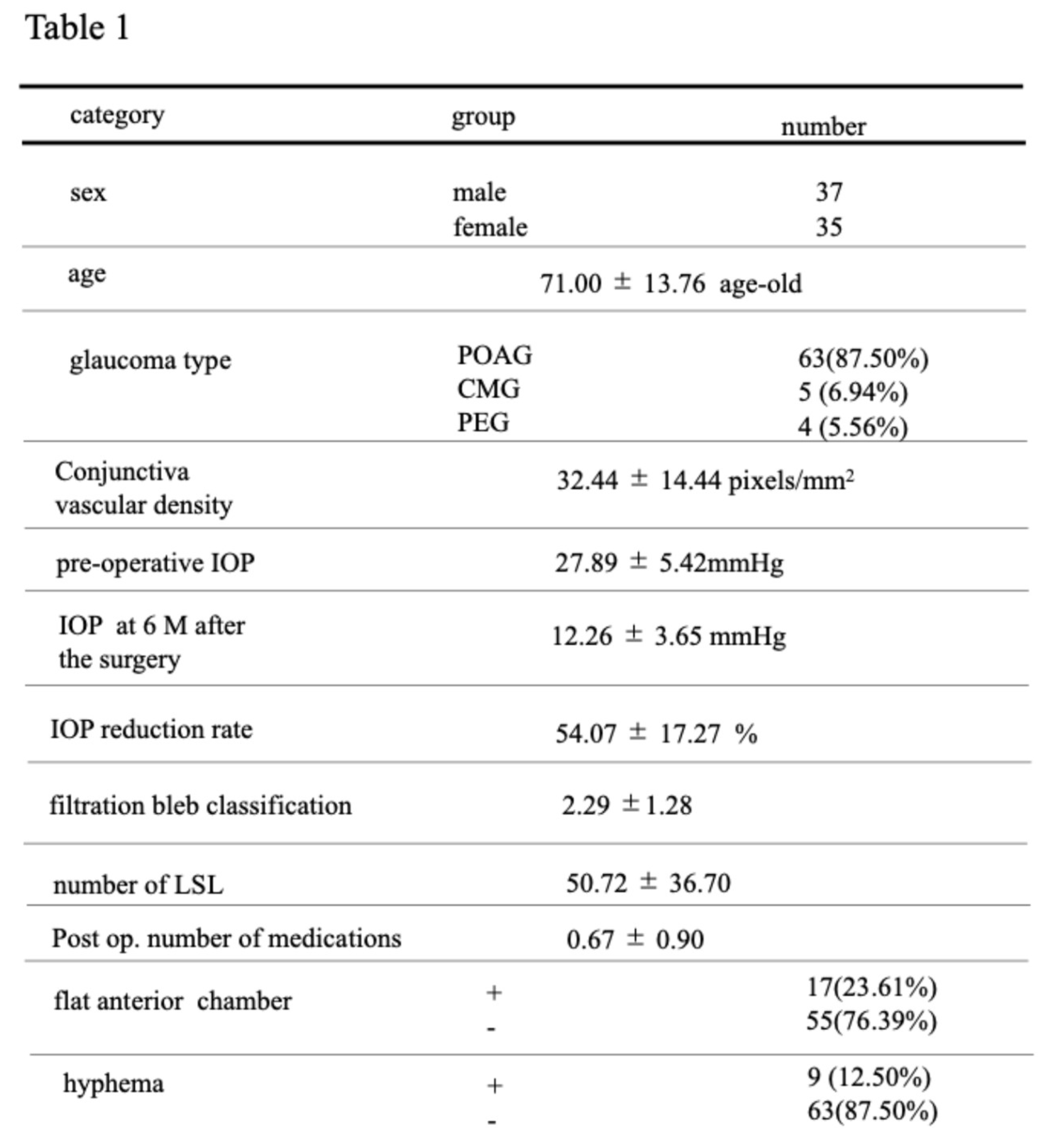
Seventy-two eyes with POAG, PEG, CMG underwent trabeculectomy. The summary of patient data and a compilation of measured values are presented in
Table 1.
The data analysis revealed a series of noteworthy associations between preoperative conjunctival vascular density and various postoperative parameters, shedding light on the intricate relationship between conjunctival vascularity and surgical outcomes in patients undergoing trabeculectomy with mitomycin C for glaucoma.
The Relationship Between Conjunctival Vascular Density, IOP, Bleb Morphology, LSL, and Glaucoma Eye-Drop
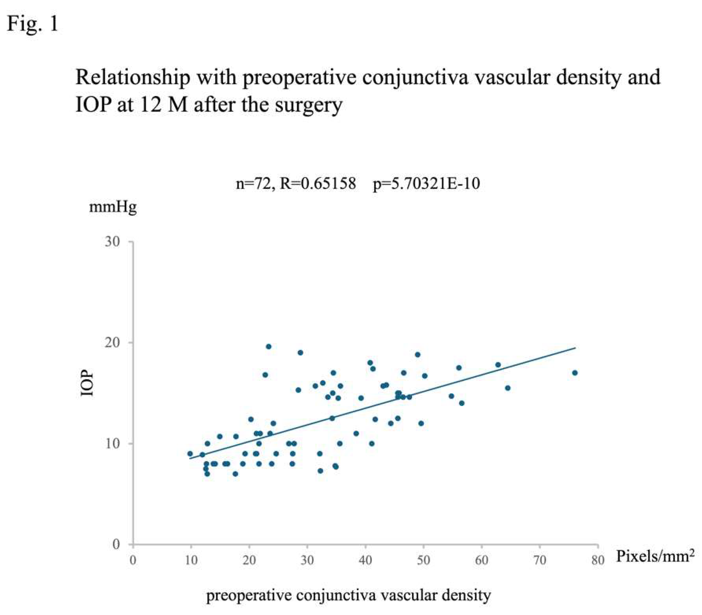
A positive correlation was observed between preoperative conjunctival vessel density and postoperative intraocular pressure (IOP) (n=72, Rho=-0.67446, p=8.29203E-11,
Figure 1).
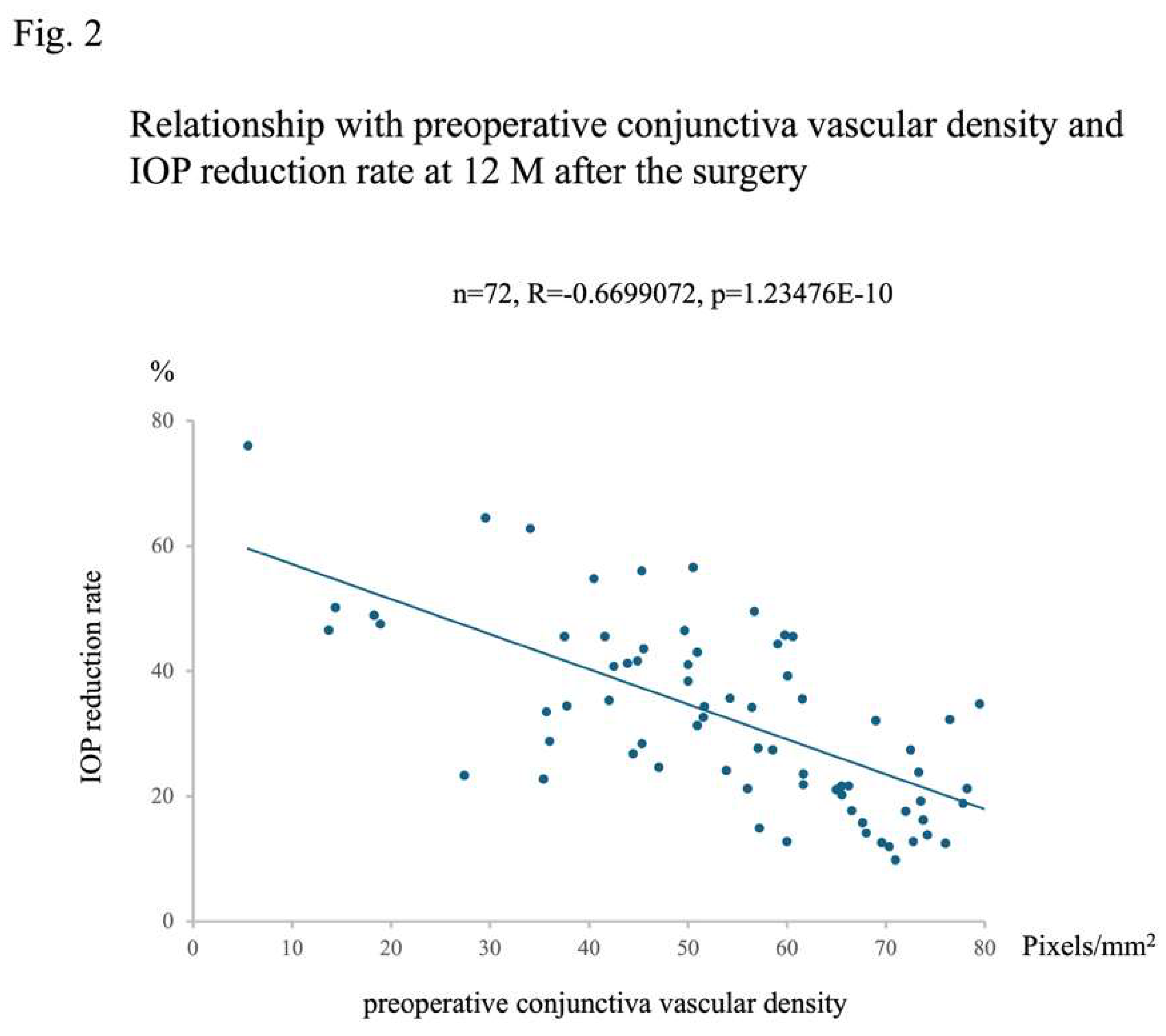
Specifically, higher preoperative conjunctival vessel density was associated with higher IOP values at 12 months postoperatively, while lower preoperative conjunctival vessel density was associated with lower IOP values. This relationship extended to the negative correlation between preoperative conjunctival vessel density and the rate of IOP decrease at 12 months postoperatively (n=72, R=-0.6699072, p=1.23476E-10,
Figure 2). In essence, higher preoperative conjunctival vessel density was linked to a lower rate of IOP decrease over 12 months, while lower preoperative conjunctival vessel density was associated with a higher rate of IOP decrease.
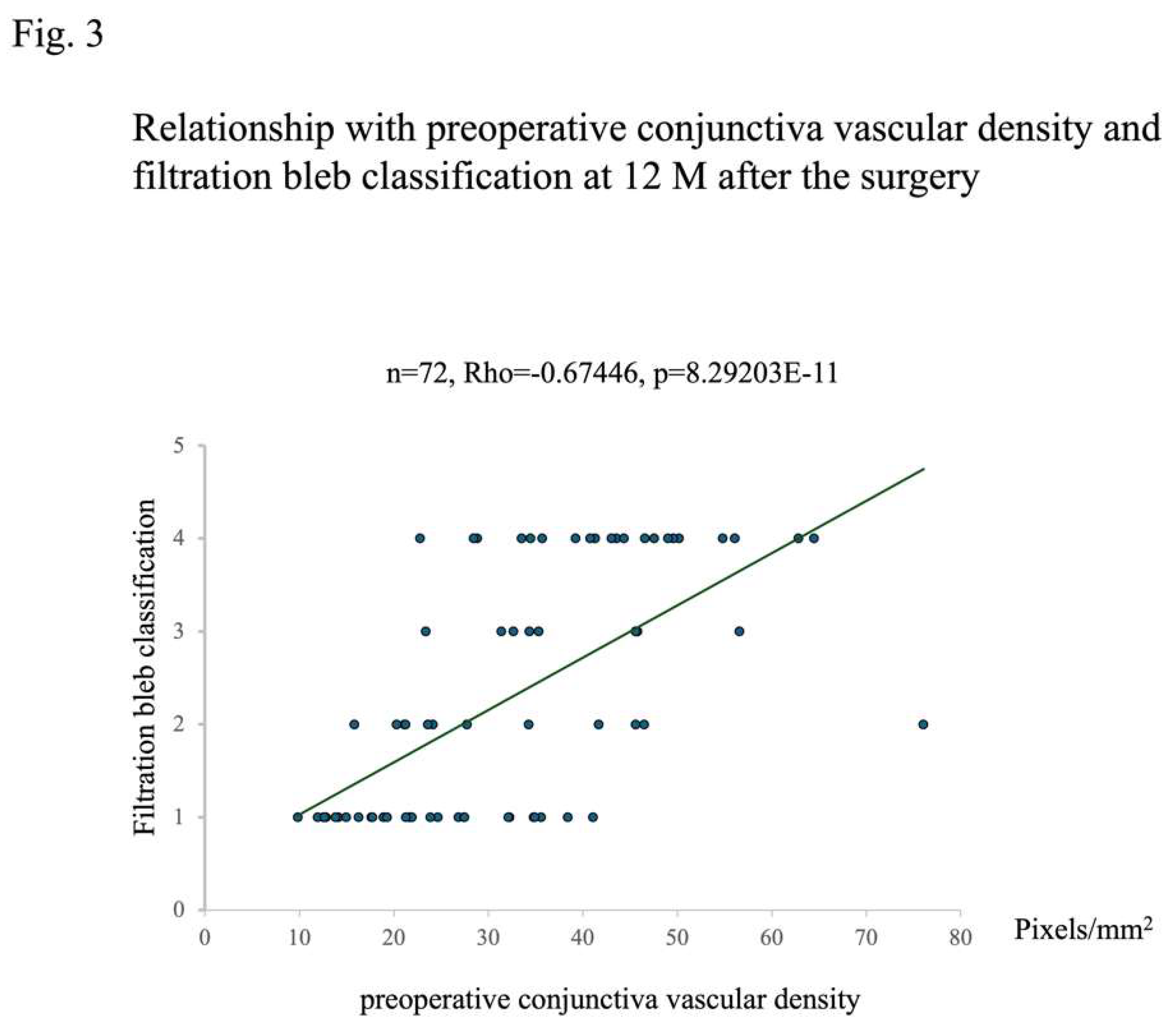
Regarding the relationship between preoperative conjunctival vessel density and bleb morphology at 12 months postoperatively, a positive correlation was observed (n=72, Rho=-0.67446, p=8.29203E-11,
Figure 3). This finding indicated that higher preoperative conjunctival vessel density led to higher rankings in bleb classification, while lower preoperative vessel density resulted in lower rankings. Given that lower rankings imply better filtration in bleb morphology, it was found that lower preoperative conjunctival vessel density could facilitate the formation of more favorable blebs.
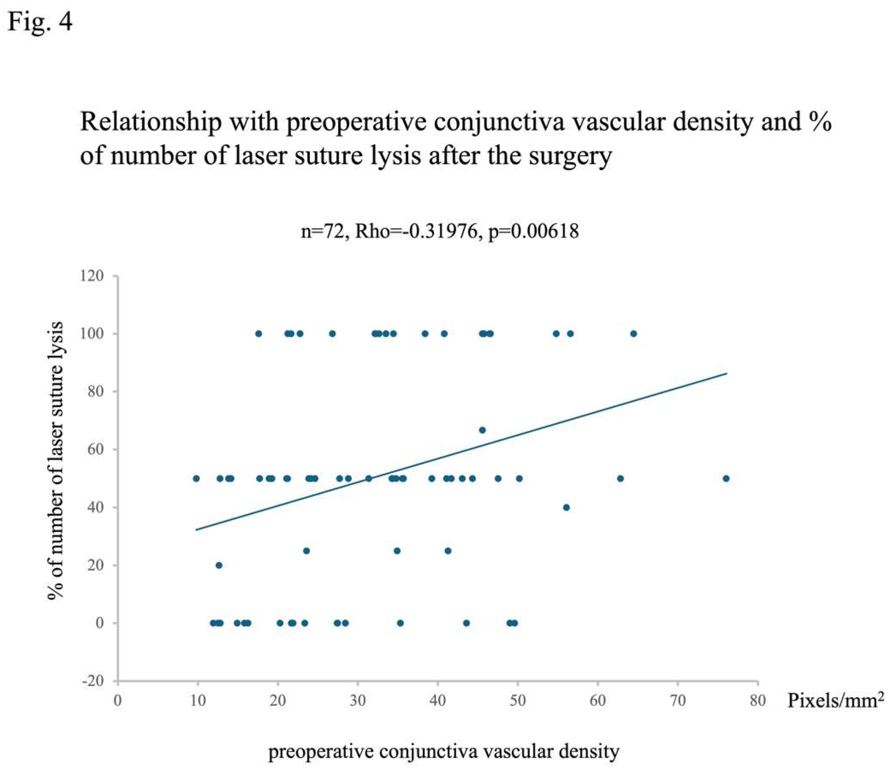
Furthermore, a positive correlation was noted between preoperative conjunctival vessel density and the frequency of undergoing LSL (n=72, Rho=-0.31976, p=0.00004,
Figure 4). This result aligned with the previous findings related to postoperative IOP and bleb classification, maintaining a consistent pattern across the relationships.
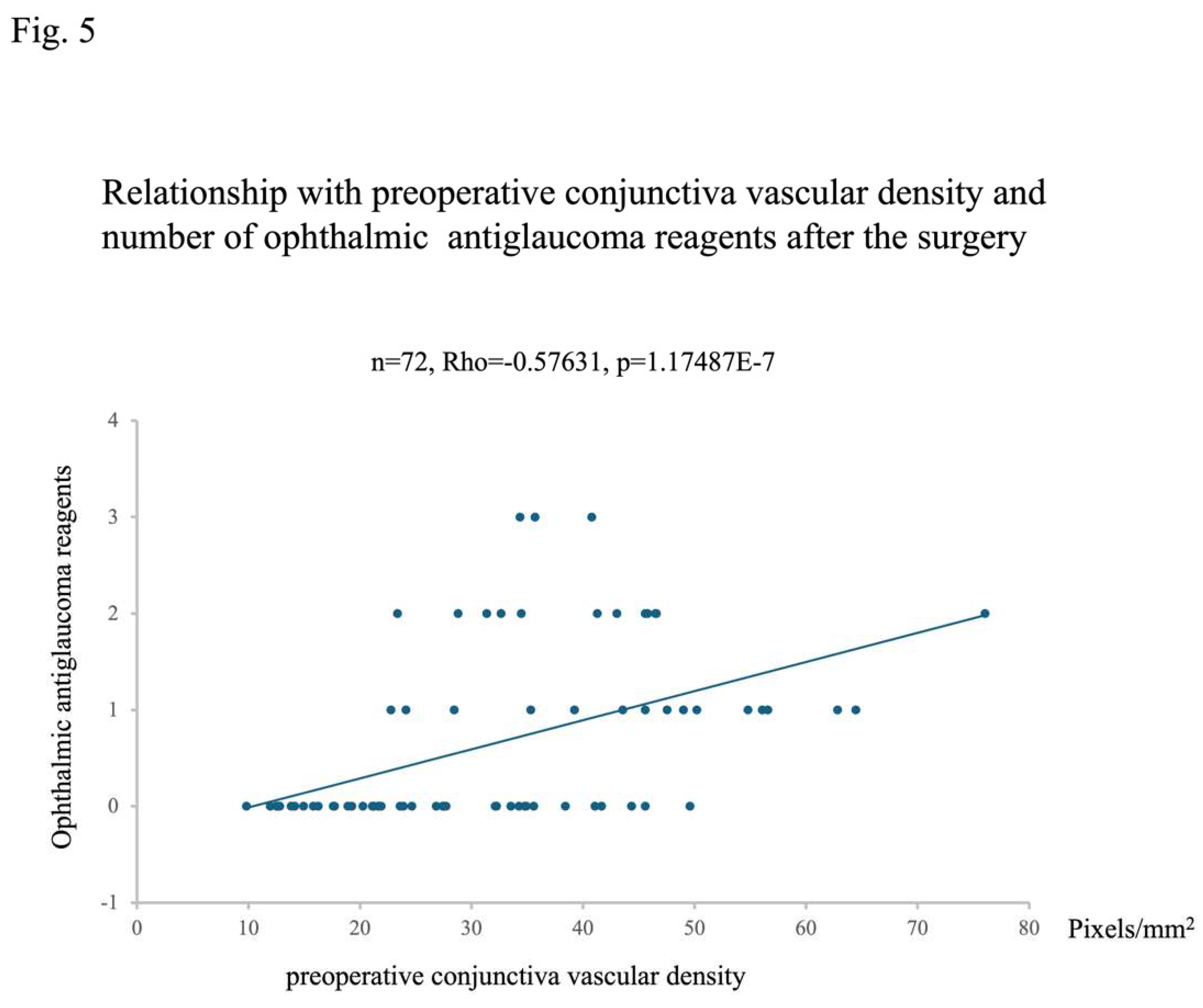
The relationship between the preoperative conjunctival vascular area and the number of postoperative glaucoma eye drops showed a positive correlation, indicating that a larger conjunctival vascular area was associated with a higher number of postoperative eye drops (n=72, Rho=-0.57631, p=1.17487E-7,
Figure 5). This result also reflects the relationship between the preoperative conjunctival vascular area and postoperative intraocular pressure.
The Relationship Between Conjunctival Vascular Density, Hyphema, and FAT
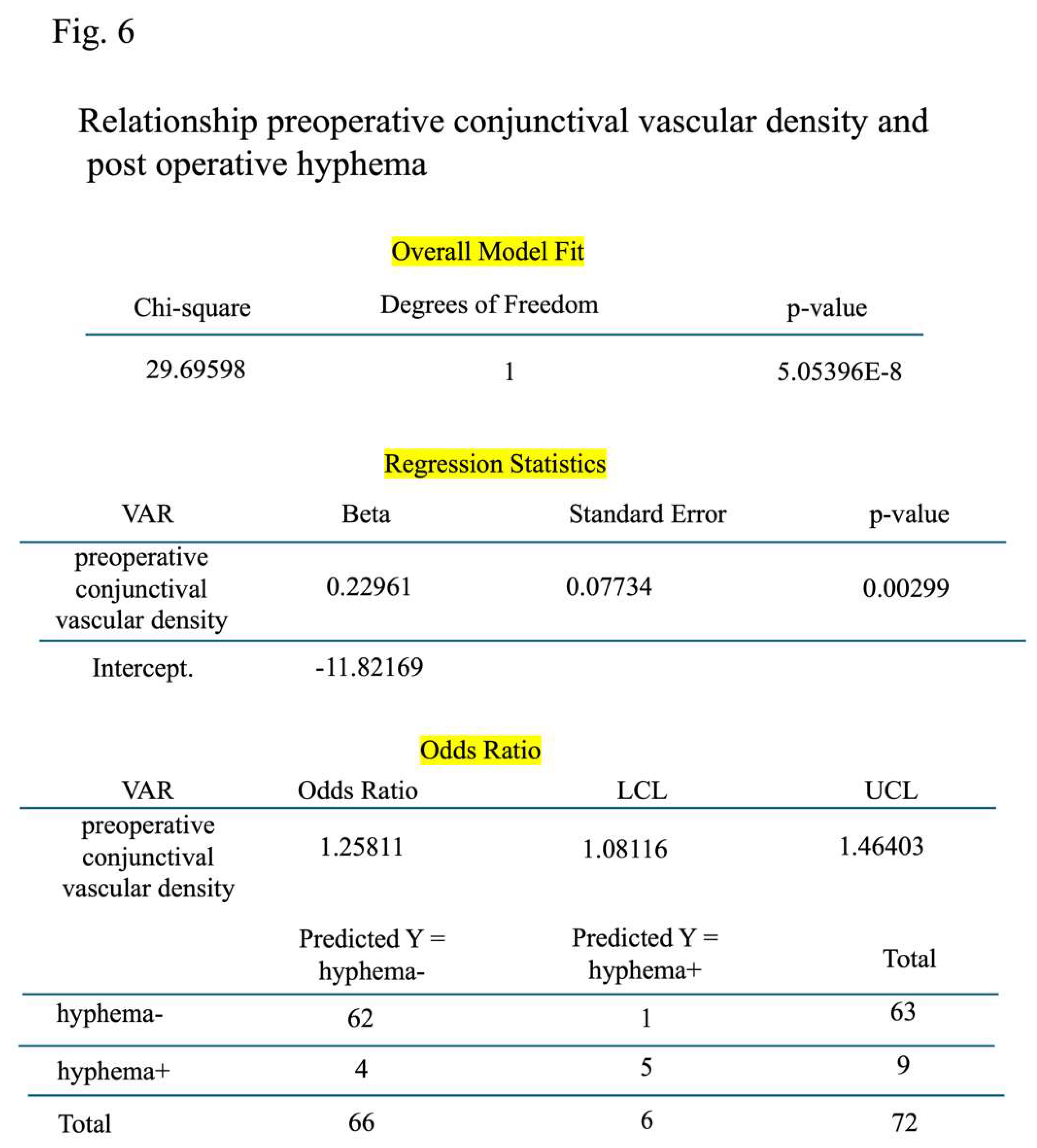
In addition, logistic regression analysis revealed an association between preoperative conjunctival vessel density and the postoperative occurrence of hyphema (n=72, p=0.0030,
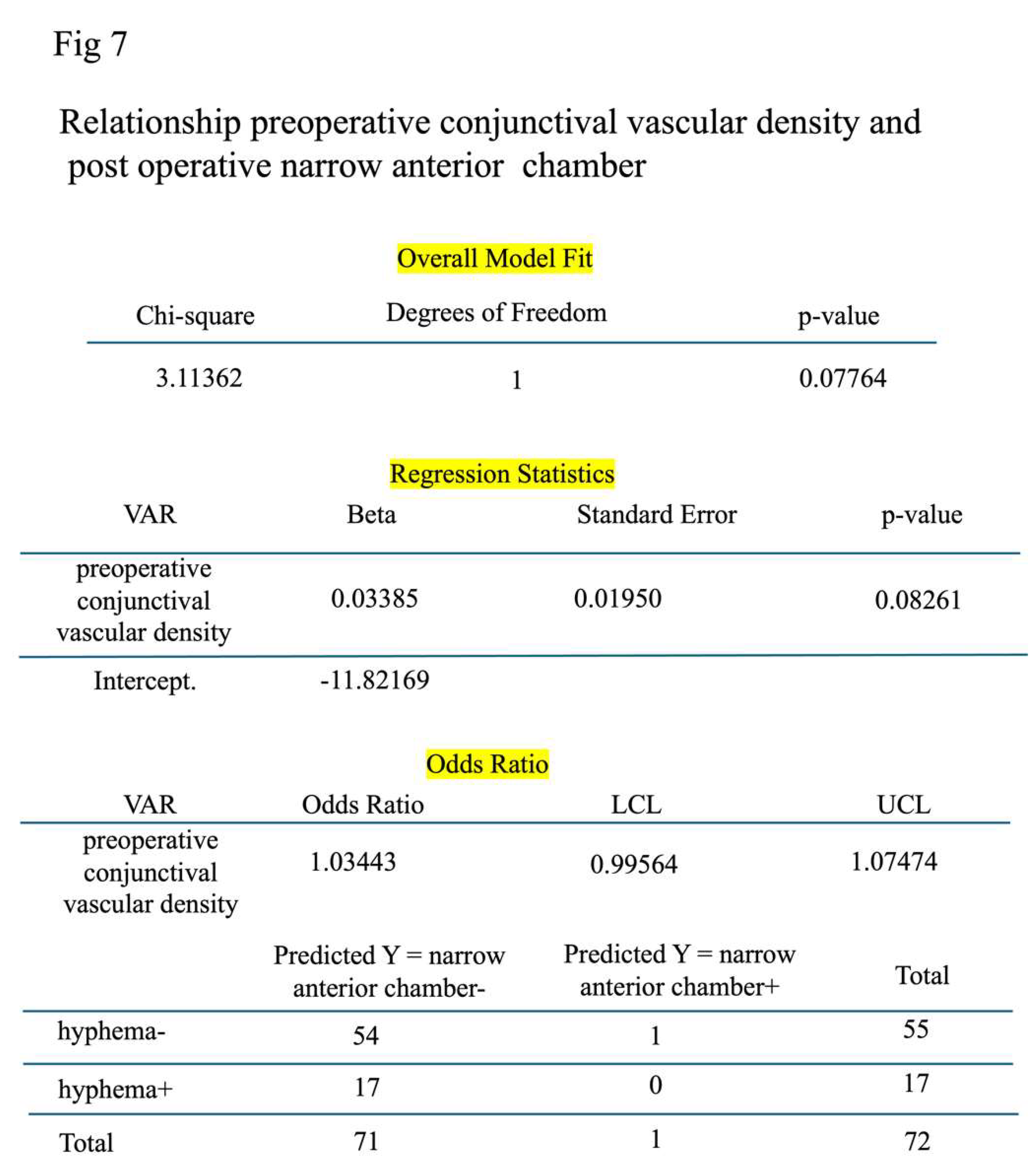
Figure 6). However, no significant association was found between preoperative conjunctival vessel density and FAT (n=72, p=0.083,
Figure 7).
Discussion
This study analyzed the correlation between preoperative conjunctival vessel density and various postoperative parameters. It was found that a lower preoperative conjunctival vessel density, indicating less congestion, resulted in the formation of a better bleb, reduced frequency of LSL, decreased intraocular pressure, and reduced reliance on glaucoma eye drops. Additionally, concerning common complications such as hyphema and FAC, it was observed that a higher preoperative conjunctival vessel density was associated with increased hyphema.
Previous studies similar to this research have utilized early post-Trabeculectomy and PreserFlo microshunto As-OCT angiography to investigate the relationship between postoperative conjunctival vessel density and future bleb formation and intraocular pressure [
22,
23,
24]. However, studies directly examining the correlation between preoperative conjunctival vessel density and various postoperative parameters, like ours, are quite rare and provide valuable data. Although our study did not directly assess conjunctival vessel density preoperatively, there have been reports suggesting that the preoperative deepening of the upper eyelid sulcus (DEUS) or Prostaglandin- Associated Periorbitopathy (PAP) in Trabeculectomy cases impacts postoperative outcomes [
25]. These findings echo our results, indicating that cases with severe preoperative DEUS may have higher preoperative eye drops, suggesting greater conjunctival congestion, aligning with our study’s outcomes.
One limitation of our study is that conjunctival vessel density measurements were not conducted using AS-OCT angiography. However, AS-OCT angiography is a costly instrument that may not be available in all facilities conducting glaucoma surgeries [
26,
27]. Instead, our methodology involved capturing photographs of the anterior chamber, converting them, and quantifying them using ImageJ, a more accessible and simplified method that can be used in various settings. While this method may not offer the same level of precision as AS-OCT angiography, it was deemed capable of providing a rough quantification.
In conclusion, our results suggest that a lower preoperative conjunctival vessel density leads to better outcomes in Trabeculectomy procedures. Therefore, preparing the conjunctival environment, reducing inflammation, and minimizing congestion before surgery may be key to achieving favorable postoperative results.
Author Contributions
Y.H. performed the surgery. Y. H. and T. I. conceived and designed the research and analyzed the data. Y. H. wrote the article. All authors have read and approved the final manuscript.
Statement of Ethics
This retrospective cohort study was approved by our institutions committee (Tohankai eye institutions Ethics Committee, approval number 0002, January 31,2023) and adhered to the regulations of clinical practice and the tenets of the Declaration of Helsinki.
Example statement
Informed consent was obtained from all individual participants included in the study.
Funding
This study was not supported by any sponsor or funder.
Conflicts of Interest Statement
The authors have no conflicts of interest to declare.
Data Availability Statement
The datasets generated and analyzed during the current study are available from the corresponding author on reasonable request.
Acknowledgments
The authors would like to thank Dr. Tamiya Saito for conducting the statistical analysis.
Competing interes
No competing interest.
References
- 1 Rabiolo, A. , Morales, E., Kim, J.H., Afifi, A.A., Yu, F., Nouri-Mahdavi, K., Caprioli J. Predictors of Long-Term Visual Field Fluctuation in Glaucoma Patients 2020, Ophthalmology, 127, 739-747. Epub 2019 Dec 5. [CrossRef] [PubMed]
- 2 Jonas, J.B. , Aung, T., Bourne, R.R., Bron, A.M., Ritch, R., Panda-Jonas, S. Glaucoma 2017, Lancet, 390, 2183-2193. Epub 2017 May 31. PMID: 28577860 Review. [CrossRef]
- 3 Weinreb, R.N. , Khaw, P.T. Primary open-angle glaucoma 2004, Lancet, 363, 1711-1720. PMID: 15158634 Review. [CrossRef]
- 4 Mehran, N.A. , Sinha, S., Razeghinejad, R. New glaucoma medications: latanoprostene bunod, netarsudil, and fixed combination netarsudil-latanoprost 2020, Eye (Lond), 34, 72-88. Epub 2019 Nov 6. [CrossRef] [PubMed]
- 5 Quigley, H.A. Glaucoma 2011, Lancet, 377, 1367-1377. Epub 2011 Mar 30. PMID: 21453963 Review. [CrossRef]
- 6 Lusthaus, J. , Goldberg, I. Current management of glaucoma 2019, Med. J. Aust., 210, 180-187. Epub 2019 Feb 14. [CrossRef] [PubMed]
- 7 King, A.J. , Hudson, J., Fernie, G., Kernohan, A., Azuara-Blanco, A., Burr, J., Homer, T., Shabaninejad, H., Sparrow, J.M., Garway-Heath, D., Barton, K., Norrie, J., McDonald, A., Vale, L., MacLennan, G.; TAGS Study Group.Primary trabeculectomy for advanced glaucoma: pragmatic multicentre randomised controlled trial (TAGS) 2021, B.M.J.,12, 373:n1014. PMID: 33980505. [CrossRef] [PubMed]
- 8 Nouri-Mahdavi, K. , Brigatti, L., Weitzman, M., Caprioli, J. Outcomes of trabeculectomy for primary open-angle glaucoma 1995, Ophthalmology, 102, 1760-1769. [CrossRef] [PubMed]
- 9 Hoffmann, E.M. , Hengerer, F. Glaucoma surgery today 2021, 118, 239–247. [Google Scholar] [CrossRef] [PubMed]
- 10 Wang, S.Y. , Singh, K. Management of the glaucoma patient progressing at low normal intraocular pressure 2020, 31, 107–113. [Google Scholar] [CrossRef] [PubMed]
- 11 Lazaro, C. , Garcia-Feijoo, J., Castillo, A., Perea, J., Martinez-Casa, J.M., Garcia-Sanchez, J. Impact of intraocular pressure after filtration surgery on visual field progression in primary open-angle glaucoma 2007, Eur. J. Ophthalmol., 17, 357-362. [CrossRef] [PubMed]
- 12 Broadway, D.C. , Chang, L.P. Trabeculectomy, risk factors for failure and the preoperative state of the conjunctiva 2001, J. Glaucoma, 10, 237-249. [CrossRef] [PubMed]
- 13 Broadway, D. , Grierson, I., Hitchings, R. Racial differences in the results of glaucoma filtration surgery: are racial differences in the conjunctival cell profile important? 1994, Br. J. Ophthalmol., 78, 466-475. [CrossRef] [PubMed]
- 14 Gwynn, D.R. , Stewart, W.C., Hennis, H.L., McMillan, T.A., Pitts, R.A. The influence of age upon inflammatory cell counts and structure of conjunctiva in chronic open-angle glaucoma 1993, Acta Ophthalmol. (Copenh), 71, 691-695. [CrossRef] [PubMed]
- 15 Arici, M.K. , Demircan, S. S. Effect of conjunctival structure and inflammatory cell counts on intraocular pressure after trabeculectomy 1999, 213, 371–375. [Google Scholar] [CrossRef] [PubMed]
- 16 Migdal, C. , Hitchings, R. The developing bleb: effect of topical antiprostaglandins on the outcome of glaucoma fistulising surgery 1983, Br. J. Ophthalmol., 67, 655-660. [CrossRef] [PubMed]
- 17 Cantor, L.B. , Mantravadi, A., WuDunn, D., Swamynathan, K., Cortes, A. Morphologic classification of filtering blebs after glaucoma filtration surgery: the Indiana Bleb Appearance Grading Scale. J. Glaucoma 2003, 12, 266–271. [Google Scholar]
- 18 Chen, T.C. , Wilensky, J.T., Viana, M.A. Long-term follow-up of initially successful trabeculectomy. Ophthalmology. 1997, 104, 1120–1125. [Google Scholar] [CrossRef] [PubMed]
- 19 Crowston, J.G. , Kirwan, J.F., Wells, A., Kennedy, C., Murdoch, I.E. Evaluating clinical signs in trabeculectomized eyes 2004, Eye (Lond), 18, 299-303. Baez KA, Martinez SM, Spaeth GL.
- 20 de Barros, D.S. , Navarro, J.B., Mantravadi, A.V., Siam, G.A., Gheith, M.E., Tittler, E.H. The early flat anterior chamber after trabeculectomy: a randomized, prospective study of 3 methods of management. J. Glaucoma 2009, 18, 13–20. [Google Scholar]
- 21 Tunç, Y. , Tetikoglu, M., Kara, N., Sagdık, H.M., Özarpaci, S., Elçioğlu, M.N. Management of hypotony and flat anterior chamber associated with glaucoma filtration surgery 2015, Int. J. Ophthalmol., 8, 950-953. eCollection 2015. [CrossRef] [PubMed]
- 22 Schneider, S. , Kallab, M., Murauer, O., Reisinger, A.S., Strohmaier, S., Huang, A.S., Bolz, M., Strohmaier, C.A. Bleb vessel density as a predictive factor for surgical revisions after Preserflo Microshunt implantation 2024, Acta Ophthalmol., 102, e797-e804. Epub 2024 Feb 2. [CrossRef] [PubMed]
- 23 Ibarz Barberá, M. , Morales Fernández, L., Tañá Rivero, P., Gómez de Liaño, R., Teus, M.A. Anterior-segment optical coherence tomography of filtering blebs in the early postoperative period of ab externo SIBS microshunt implantation with mitomycin C: Morphological analysis and correlation with intraocular pressure reduction 2022, Acta Ophthalmol., 100, e192-e203. Epub 2021 Apr 10. [CrossRef] [PubMed]
- 24 Luo, M. , Xiao, H., Huang, J., Jin, L., Li, Z., Tu, S., Huang, H., Zhu, Y., Li, Y., Zhuo, Y. Multi-Quantitative Assessment of AS-OCTA Complemented AS-OCT for Monitoring Filtering Bleb Function After Trabeculectomy 2023, Transl. Vis. Sci. Technol., 12, 18. [CrossRef] [PubMed]
- 25 Miki, T. , Naito, T., Fujiwara, M., Araki, R., Kiyoi, R., Shiode, Y., Fujiwara, A., Morizane, Y., Shiraga, F. Effects of pre-surgical administration of prostaglandin analogs on the outcome of trabeculectomy 2017, PLoS One, 12, e0181550. eCollection 2017. [CrossRef] [PubMed]
- 26 Yin, X. , Cai, Q., Song, R., He, X., Lu, P. Relationship between filtering bleb vascularization and surgical outcomes after trabeculectomy: an optical coherence tomography angiography study 2018, Graefes Arch. Clin. Exp Ophthalmol., 256, 2399-2405. Epub 2018 Sep 12. [CrossRef] [PubMed]
- 27 Mastropasqua, R. , Brescia, L., Di Antonio, L., Guarini, D., Giattini, D., Zuppardi, E., Agnifili, L. Angiographic biomarkers of filtering bleb function after XEN gel implantation for glaucoma: an optical coherence tomography-angiography study 2020, Acta Ophthalmol., 98: e761-e767. Epub 2020 Feb 5. [CrossRef] [PubMed]
|
Disclaimer/Publisher’s Note: The statements, opinions and data contained in all publications are solely those of the individual author(s) and contributor(s) and not of MDPI and/or the editor(s). MDPI and/or the editor(s) disclaim responsibility for any injury to people or property resulting from any ideas, methods, instructions or products referred to in the content. |
© 2025 by the authors. Licensee MDPI, Basel, Switzerland. This article is an open access article distributed under the terms and conditions of the Creative Commons Attribution (CC BY) license (http://creativecommons.org/licenses/by/4.0/).