Preprint
Article

This version is not peer-reviewed.

Fungistatic and Bactericidal Activity of Hydroalcoholic Extracts of Root of Jatropha dioica Sessé

A peer-reviewed article of this preprint also exists.

Submitted:

05 March 2025

Posted:

07 March 2025

You are already at the latest version

Abstract

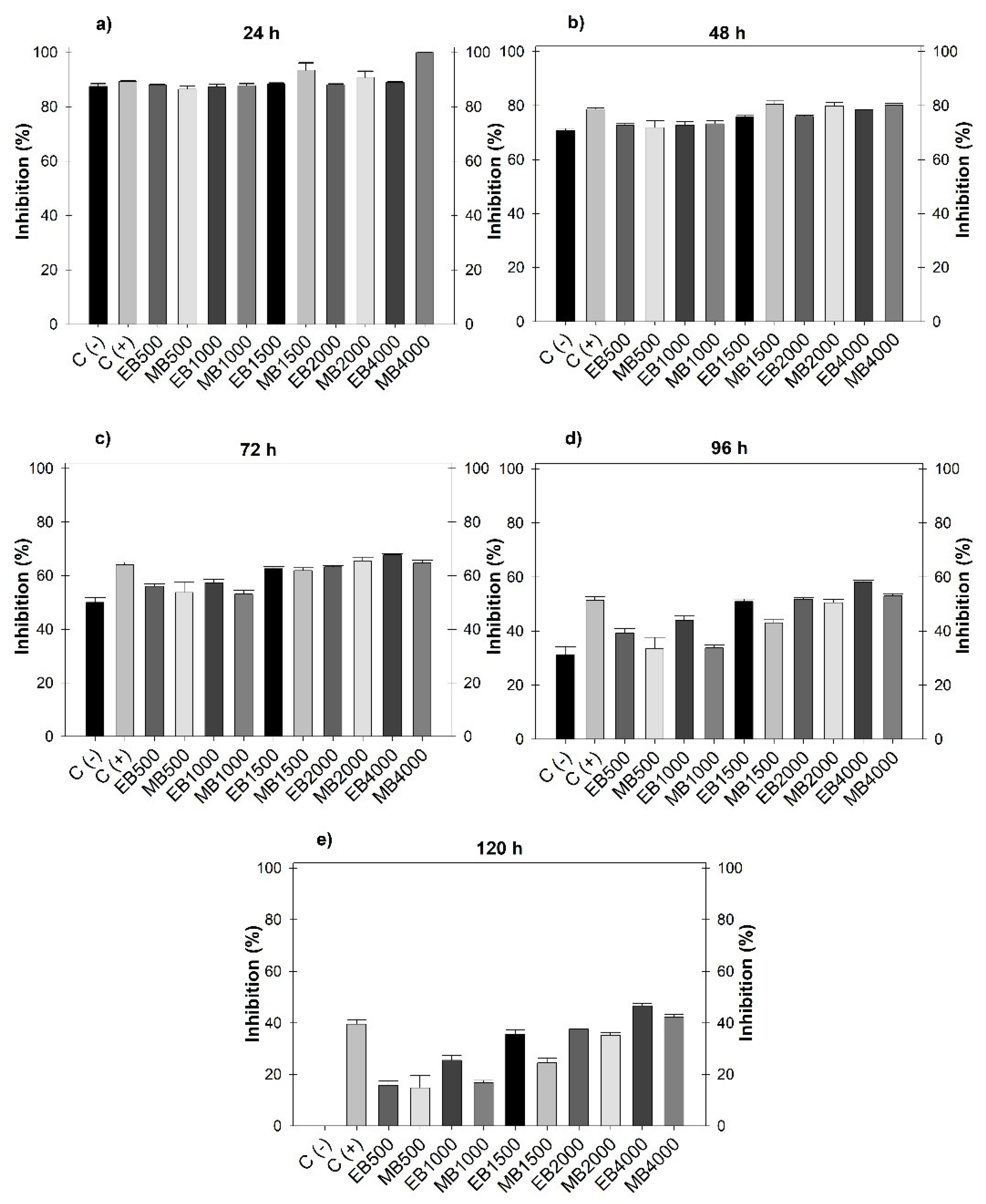
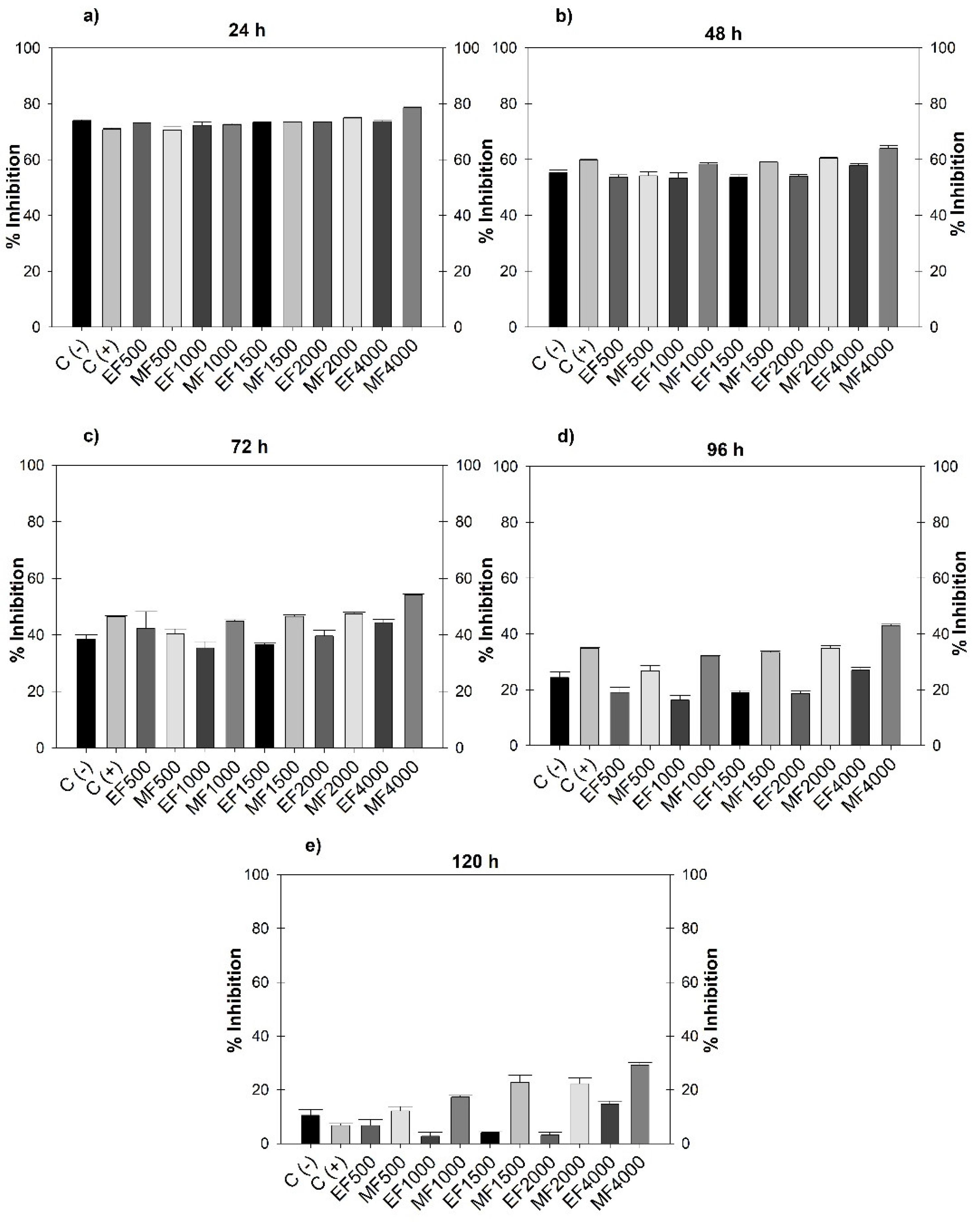
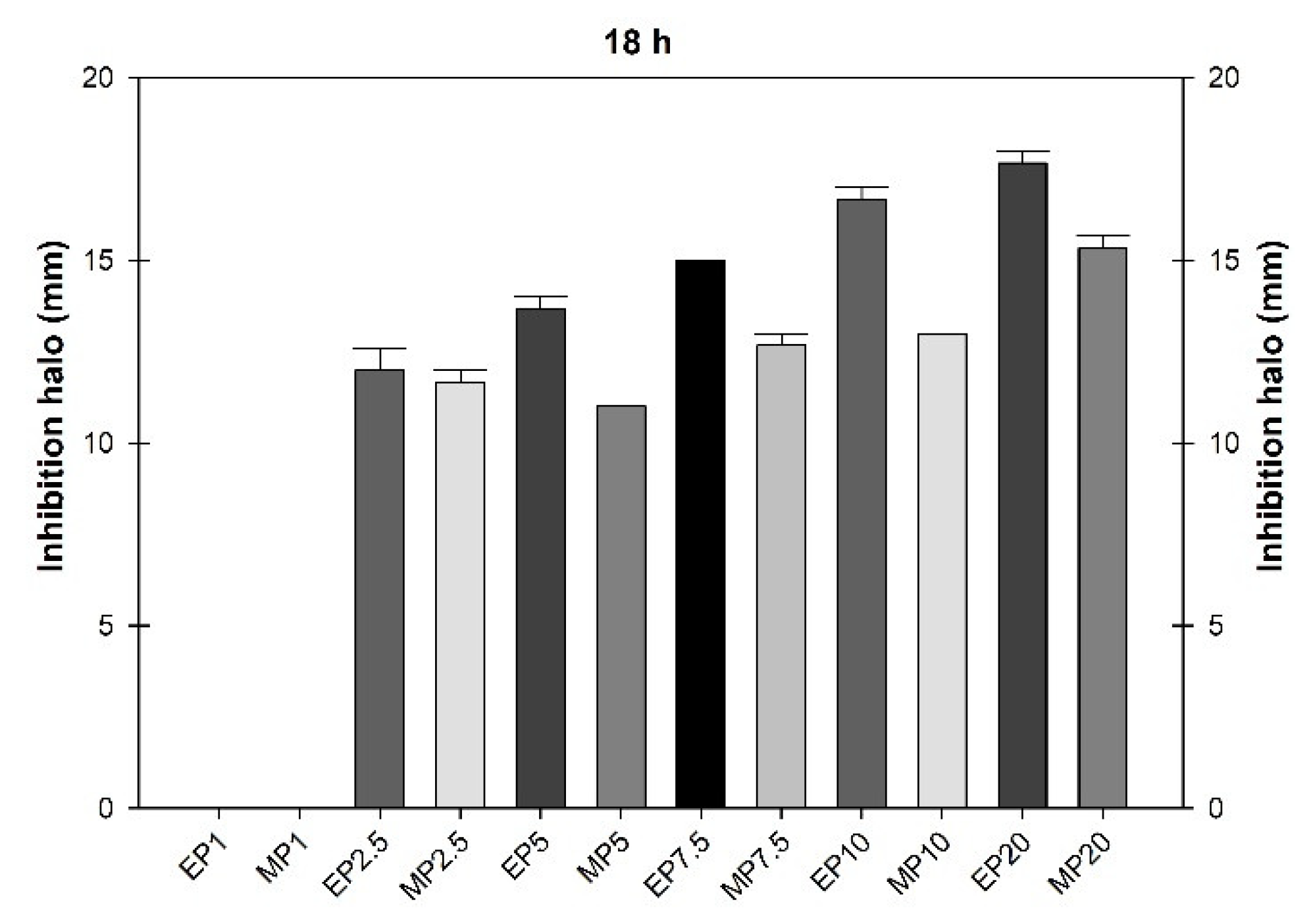
Jatropha dioica Sessé (JD) is a plant from arid and semiarid areas of Mexico, and has been linked to medicinal uses. Due to the current limitation of agrochemicals in agriculture and post-harvest due to environmental effects, health and resistance of phytopathogenic microorganisms, it is presumed that secondary metabolites of J. dioica can act as an alternative biological control. The bactericidal and fungistatic activity of hydroalcoholic extracts (ethanol and methanol) on Botrytis cinerea, Fusarium oxysporum and Pseudomonas syringae from J. dioica roots was evaluated in vitro, considering that the content of total phenols and flavonoids (Folin-Ciocalteu and aluminum chloride method) may have antimicrobial activity. The ethanolic and methanolic extracts have fungistatic biological activity on B. cinerea, with growth inhibition of 42.27 ±1.09 and 46.68 ±0.98% respectively and an IC50 of 5.04 mg mL-1 without significant differences in the use of solvents. In F. oxysporum, inhibition of 14.77 ±1.08 and 29.19 ±0.89% was obtained, where the methanolic extract was more efficient generating a stress response to the ethanolic extract. Regarding the bactericidal activity in P. syringae, the inhibition halo was 17.66 ±0.33 and 16.66 ±0.33 mm, showing slight difference when using ethanol. The phenolic content obtained was 8.92 ±0.25 and 12.10 ±0.34 mg EAG g-1 and flavonoids 20.49 ±0.33 and 28.21 ±0.73 mg QE g-1 of dry weight of the sample. The results highlight the importance of reorienting and revaluing J. dioica as an alternative source of metabolites for agricultural and post-harvest formulations.

Keywords: 
;  ;  ;  

1. Introduction

The arid, semiarid and hyperarid zones in Mexico constitute 63% of its surface [1], and its rural population depends on non-timber forest resources (NTFR) [2] [3]. Some are the nopal (Opuntia ficus-indica), yuca (Yucca sp.), gobernadora (Larrea tridentata), sangre de drago (Jatropha dioica Sessé), candelilla (Euphorbia antisyphilitica), sotol (Dasylirion lucidum), damiana (Turnera difusa) senna leaf (Flourensia cernua), ocotillo (Fouquieria splendes), maguey (Agave sp.) among others [4]; and as a response to environmental limitations, many of these species synthesize a wide range of secondary metabolites (Sm), which have recorded biological activity oriented to public health or antimicrobial applications [5]. Through bioprospective research, metabolites can be used for the development of products with economic value and generate rural projects in the medium term [6]. Such is the case of J. dioca (Euphorbiaceae), a RFNM of the microphyllous desert scrub vegetation growing in calcaric regosol soil, at altitudes ranging from 1851 to 2100 [7], from whose biomass flavonoids, lactones, quinones and sterols have been recorded [8] [9].
Currently, plant scientists have focused on designing applications for the control of pest organisms in both crops and harvested products. Normally, high-impact synthetic pesticides are applied that induce negative effects on the environment (water, air, soil), biodiversity, and cause health problems, such as cancer, diabetes, reproductive, respiratory and neurological disorders [10] [11]. An alternative is to derive low-cost economic and environmental benefits from plant extracts that reduce or eliminate pathogenic microorganisms [11,12,13]. Economic losses in agriculture and post-harvest life due to pest organisms [14] of 40% [15] [16] have been recorded worldwide, which put food security at risk [17] [18].
The fungus Fusarium oxysporum impairs the absorption of water and nutrients by altering the metabolism of the plant, inducing chlorosis, wilting, flower shedding and vascular necrosis [19,20,21]. Botrytis cinerea is the causal agent of gray mold [22], a necrotrophic pathogen that causes losses in more than 500 species of plants in the field and storage, attacking stems, leaves, flowers, fruits, seeds with impacts of up to USD $100,000 million [23] [24]. Similarly, the bacterium Pseudomona Syringae, causal agent of bacterial canker (aerobic Gram-negative) has associated losses in more than 40 plants annually, with up to 25% and survives as a saprophyte [25] [26].
To extend the shelf life of crops, FAO (2024) suggests having continuous monitoring and being able to respond quickly with environmentally sustainable preventive control strategies [18]. Chemical treatments are regulated and restricted due to their associated impacts [27] [28,29,30], and therefore, the use of formulations of biological origin can be an ally. Based on the above, hydroalcoholic extracts of J. dioica root were evaluated on the in vitro growth of Botrytis Cinerea, Fusarium Oxysporum and Pseudomonas Syringae, in order to design in the medium term formulations and compositions for agricultural use and post-harvest management.

2. Materials and Methods

2.1. Collection of Plant Material and Preparation of the Extract

Jatropha dioica (Euphorbiaceae) was collected during the dry season, January-April 2024 in Ejido Loma de la Carreta, Villa González Ortega, Zacatecas, Mexico (23° 11′ and 22° 27′ N and 101° 22′ and 101° 57′ W) with mean annual temperature and precipitation of 19 °C and 373.9 mm [31] respectively. 602 g of root were collected, chopped and dried at 20 °C ±2 and 60% relative humidity without an oven for 7d. The particle size was made uniform with a HC-700Y mill (Grinder, China) and a No. 20 sieve. To obtain the extracts, 50 g of dry sample was macerated with 500 mL of 80% ethanol for 7 d, making three solvent changes every 24 h. Similarly, 50 g of dry and ground root was macerated with 250 mL of 99.9% solvent for 7 d. They were then vacuum filtered with filter paper and concentrated in a rotary evaporator (RE100-PRO, DLAB, USA) at 120 rpm at 40 °C. They were placed in a 9023A drying oven (ECOSHEL, USA) at 40 °C for one week and stored in refrigeration until use [32] [33]. The yield of the hydroalcoholic extracts was calculated through the equation:
Yield (%) = (W1*100) /W2; where: W1 is the dry weight of the extract after rotary evaporation, and W2 is the dry weight of the plant used, recording 10.32% for ethanol and 1056% for methanol.

2.2. Inoculum Preparation

Inoculums were provided by the laboratories of the National Institute of Forestry, Agriculture and Livestock Research (INIFAP- Zacatecas) and by the Potosino Institute of Scientific and Technological Research (IPICYT). The fungal strains B. cinerea and F. oxysporum were reseeded after 4 and 7 d of growth. A 4.6 mm portion of mycelium was aseptically placed on potato dextrose agar (PDA) for evaluation. For the P. Syringae bacteria, after 24 h of incubation, a colony was taken with a bacteriological loop and was aseptically transferred in saline water (0.85%), for 5 min the suspension was diluted in a vortex followed by a suspension adjustment in a Genesys 10S Vis spectrophotometer (Thermo Fisher Scientific, USA) at 625 nm based on the turbidity standard of 0.5 McFarland (106 CFU mL-1) [33].

2.3. Preparation of Stock Solution and Fungistatic Activity

To obtain the stock solution with a concentration of 8666.66 µg mL-1 of the hydroalcoholic extracts, 0.13 g was weighed in 15 mL of distilled water in falcon tubes (50 mL) and placed in an ultrasonic bath for complete dilution. For the fungi, the agar dilution method was used in the potato dextrose agar (PDA) culture medium from DIBICO. In the case of the bacteria, the susceptibility method was the well diffusion method in Luria Bertani (LB) agar [34]. The PDA medium was added considering the concentrations, repetitions and type of extract to be used (5x5x1), the result was multiplied by the volume needed for a 90x15 mm Petri dish (20 mL) and divided by two to obtain a 50:50 ratio. In which, 50% was concentrated PDA medium, and 50% water and extract. When preparing the PDA medium, the result (g) was doubled, as was the extract concentration (mL). The mixture of the extract with the water of the culture medium was considered, since the concentration should not be affected by the water contained in the PDA medium. From the mother solution of the hydroalcoholic extracts, the concentrations were prepared with a final volume of 50 mL (Table 1).
The concentrations were added to the culture medium and sterilized in an autoclave (121 °C for 15 min), added to Petri dishes for solidification, and a 4.6 mm piece of mycelium incubated at 25 ±2 °C was placed in the center. Measurements were taken every 24 h for 5 days or until the control reached its full growth [28]. Finally, after the incubation time, the growth inhibition halo was measured. The results were obtained as an average ± standard deviation, and the percentage of inhibition was determined through the following formula:
% of mycelial growth inhibition = diameter (mm) of the negative control - Diameter (mm) of the phytopathogen growth in the extract/diameter (mm) of the negative control *100.
Through a linear regression in RStudio, the IC50 (minimum dose that achieves a 50% decrease in mycelial growth) of the hydroalcoholic extracts was estimated.
The percentage of growth inhibition of the hydroalcoholic extracts of J. dioica root was obtained through the measurement in millimeters (mm) for five days of the two phytopathogenic fungi (Botrytis cinerea and Fusarium oxysporum). A positive control was considered using the concentration provided by the manufacturer of a commercial fungicide called Ridomil Gold (metalaxyl-M: methyl N-(2,6-dimethyl phenyl)-N-(2-methoxy-acetyl)-D-alaninate at 4%, and 64% mancozeb: ethylene bisdithiocarbamate of manganese and zinc) and a negative control. A repeated measures ANOVA was performed in STATISTIC 7.0 with TUKEY tests (p ≤ 0.05).

2.4. Bactericidal Activity

The stock solutions of the hydroalcoholic extracts were prepared, 50 mg were weighed in 1000 µL of distilled water dissolved in a sonicator bath for 5 min. From which the following concentrations were obtained (Table 2)
A 10 µL aliquot of the adjusted suspension was applied to the LB medium. This was inoculated into the corresponding media with a uniformized driglasky loop and left to stand for 5 min. Then, wells were made with a sterile 6 mm punch, followed by the application of 100 µL of the different concentrations with five repetitions, left to stand for one hour and incubated at 37 °C for 18 h [32]. Then, the inhibition zone was measured in millimeters with a vernier. A one-way ANO-VA was performed and the Tukey Post-Hoc test was run (p ≤ 0.05) in STATISTIC 7.0.

2.5. Total Phenol Analysis

0.3 mL of each extract concentration (0.50-4 mg mL-1) was added to test tubes, followed by 1.5 mL of Folin’s reagent (10%) and 1.2 mL of Na2CO3 (7%). The mixtures were shaken and incubated for 30 min at 40 °C in a water bath. Finally, the absorbance at 760 nm was measured, using 1.5 mL of Folin’s reagent and 1.2 mL of Na2CO3 (7%) as a blank. The samples were prepared in triplicate and the averages were used to fit a calibration curve with gallic acid [35].

2.6. Preparation of Gallic Acid Standard and Analysis of Flavonoids

A standard solution of gallic acid was prepared by dissolving 10 mg in 10 mL of distilled water (1 mg mL-1), from which various concentrations were prepared in 10 mL volumetric flasks (9.4×10⁻³ - 1.5×10⁻¹ mg mL-1) [36]. Flavonoid content was determined by the colorimetric aluminum chloride assay. 0.5 mL of each extract concentration was added to a test tube containing 2 mL of distilled water. At the same time, 0.15 mL of 5% NaNO2 was added and after 5 min 0.15 mL of 10% AlCl3. After 6 min, 2 mL of 1 M NaOH was added to the mixture. The volume of the mixture was brought to 5 mL by immediately adding 1.2 mL of distilled water. The absorbance was measured in a spectrophotometer at 510 nm. Readings were taken in triplicate and the average absorbance value was used to calculate the total flavonoid content. The flavonoid content was expressed as quercetin equivalent (mg QE/g) using the linear equation based on the standard calibration curve [35]. Regarding the preparation of the quercetin standard, 20 mg of quercetin was weighed and diluted in 10 mL of distilled water, with which the concentrations for the calibration curve were prepared (0.0625-4 mg mL-1).

3. Results

3.1. Fungistatic Activity

During the five days evaluated, there were no significant differences between the negative control and the treatments. However, at 24 and 48 h (Figure 1.a. and 1.B) significant differences were recorded as the positive control was the same at all doses evaluated; and at 72 h (Figure 1.c) the positive control did not record differences with the doses of 500 and 1000 µg mL-1. For both extracts at 96 and 120 h (Figure 1.d. and 1e) significant differences were observed from 1500, 2000 and 4000 µg mL-1, the latter being the concentration that registered the highest inhibitory value with 42.27 ±1.09% and 46.68 ±0.98% and a calculated IC50 of 5482.21 and 5024.35 µg mL-1 for ethanol and methanol respectively. No significant difference was observed between the use of solvents for extraction, even though methanol is of greater polarity and metabolite drag.
At 120 h, Fusarium recorded an effect on the ethanol extract at 1000 and 2000 µg mL-1, generating greater growth compared to the negative control. Regarding the positive control, there were no significant differences with the ethanolic doses; however, with methanol differences were observed from 1000-4000 µg mL-1. The highest dose of ethanol 4000 µg mL-1 (14.77 ±1.08) showed greater inhibition, however, when compared to the positive control there was no statistical difference. When comparing the 4000 µg mL-1 dose of ethanol and methanol, significant differences were observed between solvents, with methanol achieving greater inhibition (29.19 ±0.89).

3.2. Bactericidal Activity

The mean growth inhibition halo of P. Syringae treated with hydroalcoholic extracts of J. dioica root was obtained at 18 h, and according to the analysis, the doses of ethanol and methanol at 1000 µg mL-1 were equal (Figure 3).
Figure 3 shows that the ethanolic extract limited the growth of P. Syringae to a greater extent; and in some cases the ethanol concentration of 2500 µg mL-1 was statistically similar to the methanol concentrations of 2500, 5000, 7500 and 1000 µg mL-1. This showed that the ethanolic extraction was more efficient (p≤0.05) in controlling the growth of the P. Syringae bacteria with a mean inhibition halo of 16.66 ±0.33 and 17.66 ±0.33.

3.3. Total Phenol and Flavonoid Content

The concentration of polyphenolic compounds (phenolic acids) and flavonoids obtained in the hydroalcoholic extracts is expressed in Table 3. The total phenol content was determined using the Folin Ciocalteu method. A calibration curve was made with the gallic acid standard, from which an equation was generated (y=9.5597x; R2=0.9956), expressed in milligrams equivalent of gallic acid in grams of dry weight of the sample (mg EAG g-1). Similarly, the total flavonoid content in the hydroalcoholic extracts was calculated using a calibration curve equation (y=0.3718x; R2=0.9993) and expressed in milligrams equivalent of gallic acid in grams of dry weight of the sample (mg EQE g-1).

4. Discussion

Previous studies evaluated the growth inhibition of B. cinerea through hexane, dichloromethane, methanol and water extracts of V. amigdalina. In which, the dichloromethane extract had the highest percentage of inhibition (74.85 - 75.7%) followed by methanol, aqueous and hexane. However, the doses evaluated were 100-500 mg mL-1, higher doses than those used in our study, observing that the 50% inhibition was obtained with 500 mg mL-1 with methanol, while 100 mg mL-1 registered only 30% [37]. Also, the extraction of polymeric proanthocyanidins from grape seeds on B. cinerea is reported, in which the inhibition of mycelial growth increased progressively as the concentration increased, the doses evaluated were 1 to 21 mg mL-1. An IC50 of 11.23 - 12.15 mg mL-1 was obtained and the effect started from 8 mg mL-1 [38]. When comparing these data with the present study, less inhibition was obtained (46.68 ±0.98 and 42.27 ±1.09), with an IC50 of 5.04 mg mL-1.
Other studies evaluated the inhibitory activity of laurel leaf extracts (Laurus nobilis) on B. cinerea isolates (2600-4200 µL L-1) recording that the highest dose did not have a fungistatic effect [39]. Other plants such as Lomicera japonica and Bacharis trimera had a growth inhibition percentage lower and equal to the present study with values of 10 and 48%. However, several solvents were used to obtain an acid extract, and the yields obtained were not efficient to inhibit the growth of phytopathogens in the field [40].
Depending on the climatic conditions where the plant is collected and even the type of extraction and solvent used, the presence and conservation of secondary metabolites can be altered [41]. In particular, Jatropha dioica is distributed in arid and semi-arid areas, where drought and salinity alter the metabolic and physiological processes of plants, which stimulates the production of terpenes, alkaloids, anthocyanins and others [42]. Also, the purity, structural stability and yield of secondary metabolites depend on the extraction method and solvent used.
Maceration is an economical and efficient extraction method to extract thermolabile compounds, such as polyphenols [43], and together, secondary metabolites are responsible for fungistatic activity since they interfere with the vital process of fungi, changing the physiological state of cells and weakening or destroying the permeability barrier of the cell membrane [44]. The above affects phytopathogens through enzymatic inhibition by damaging physiological activity and DNA alkylation, interfering with the signaling compounds of pathogenic cells and their reproductive system [45].
A purified flavonoid called gnaphalin A was obtained from Pseudognaphalium robustuma, which showed fungicidal activity on B. cinerea with an ED50 of 45.5 µg mL-1 [46]. Similarly, a 5% w/v concentration of ethyl acetate extract (EtOAc) of Curcuma aromatica generated 67.44% inhibition, or Garcinia indica, which registered 79%. Other plants such as Sechium compositum have shown inhibition of the growth of B. cinerea using the juice at low concentrations (1, 2.5 and 5%) with values of 83.15, 90 and 94.87% attributed to tetracyclic triterpenes [47].
Fungistatic activity has been recorded with hexane extract of J. dioica root on Alternaria alternata, Sclerotium rolfsii, Colletotrichum gloesporoides, Rhizoctonia solani and Fusarium oxysporum with inhibition percentages of 35.9, 45.2, 19.6, 3.1 and 12% respectively [48]. In the present study, growth inhibition values were obtained by both extracts (ethanol, methanol) higher than those reported, for example, 14.77 and 29.19% in Fusarium oxysporum. This may be due to the fact that the solvents used have greater polarity than hexane and the greater polarity optimizes the extraction yield, trapping more free radicals and secondary metabolites [49]. The concentrated ethanolic extract of J. dioica root was also reported on F. oxysporum, with inhibition values of 50 to 60% [50]. Here the inhibition percentages are higher. However, [50] did not concentrate the ethanolic extract, and the variations could be due to a synergistic effect of the solvent.
The fungistatic activity on F. oxysporum has been reported for several years in medicinal plants of oriental origin at concentrations of 10 mg disk-1 and only 20 plant species had moderate activity and 31 revealed low or no activity. Of these, the root bark extract of R. ondulatum and C. japonica root were also evaluated and showed inhibition diameters of 21-30 mm [51]. Also, thyme (Thymus vulgaris) and ginger (Zingiber officinale) have been recorded, which were evaluated on F. oxysporum at 4 mg mL-1, showing inhibition of 68.98 and 46.01% [52].
The biological activity of secondary metabolites is reported as antioxidant, antiproliferative, hepatoprotective, antibacterial, hemolytic, thrombolytic, anti-inflammatory, antidiabetic and neuroprotective activity due to the presence of compounds that demonstrate pharmacological and medicinal use [53,54,55]. For example, the aqueous extract of the pomegranate peel (Punica granatum) at 1% inhibited from 40 to 45% the mycelial growth of F. oxysporum attributed to the total content of phenols (542 mg GAE/g) and positively correlated with the antifungal activity [44]. Although plant extracts with antifungal activity are limited by the development of enzymatic resistance or by genetic substitution processes [56], the reported activity of J. dioica on the control in B. cinerea of the present study represents a first report.
Other previous studies focused mainly on J. dioica attribute to it hypoglycemic [8], chemoprotective [57], cytotoxic [33] and even antimicrobial biological activity against pathogens that cause oral caries, in which its extract has a significant effect in the control of Streptococcus mutans [58]. Likewise, bactericidal activity has been recorded with root extract fractions against Pseudomonas Syringae pv tomato and Clavibacter michiganensis subsp. Michiganensis with an IC50 of 0.5 and 1.7 mg mL-1 [59].
On the other hand, volumes of 10 to 50 µL of Carum copticum essential oil were evaluated, which obtained an inhibition halo in Pseudomonas syringae of 7.25 to 10 mm [60]. A dose of 1.34 mg of Allium sativum essential oil in 100 µL achieved a mean inhibition halo of 24.66 ±0.13, a higher value than that recorded with 1% copper sulphate with 20.66 ±0.1 [61,62,63].
According to the literature, climatic conditions, soil type, geographic location, collection time and even the method of extraction of essential oils or plant extracts are factors that intervene in the synthesis of bioactive compounds, biological activity, and how certain species are more efficient than others [64,65,66]. In this regard, several studies report the total phenol content of J. dioica root and stem with different extraction methods and solvents. In the first, the phenol values decreased depending on the organ of the plant, being higher the content in the hydroalcoholic extract of root compared to the stem, with 43.44 ±0.723 µg mL-1 [8].
Different concentrations of total phenols, flavonoids and terpenes are reported in the different organs of the plant (root and stem), with the highest content in the root regardless of the extraction method and solvent [48]. In the stem, the drying process was carried out through freeze-drying, sterilized and non-sterilized extracts were evaluated with values of 194.31 ±7.04 and 99.25 ±2.50 mg EAG g-1, and the dry weight of the sample was considered [58]. The values obtained are higher than those obtained in the present study, and could be due to the freeze-drying used since in some reports freeze-drying increases the amount of compounds [67], but it is a costly process that involves time and an adequate application of temperature so as not to interfere with the final properties of the product [68]. However, extraction with hydroalcoholic solvents shows that the compounds present in the plant are thermostable since they are maintained with exposure to high temperatures [41].
The phenol content obtained from the ethanol and methanol extract of J. dioica was 8.92 ±0.25 and 12.10 ±0.34 mg EAG g-1 (Table 3). Data that, when compared with the third and fourth study, double the amount of total phenols in the root. With a total of 46.06 ± 4.14 mg GAE g-1 of extract [69], in which maceration and hydroalcoholic solvent coincide as extraction methods. Similarly, [65] evaluated the presence of total phenols with two extraction methods: reflux with heat (2.34 ± 0.93 mg g-1) and microwave-assisted extraction (1.80 ± 0.48 mg g-1) per gram of biomass. These data are lower than those obtained in the present study, and the difference could be due to the extraction method and solvent used, since the effect of temperature, particle size, time and use of solvents influence the yield, stability and content of the bioactive compounds.
Currently, extraction methods are classified as conventional and modern, whose main function is to optimize the process by using less solvents and obtaining higher yield. But some have disadvantages, since some techniques that use heat decompose or degrade thermolabile secondary metabolites. However, it would be important to optimize the extraction processes of the plant tissue of interest, since each sieve is different. All possible variables should be controlled to obtain greater compounds with economic viability, less time and energy used, and better yields without affecting biological activity [70,71,72].
The flavonoid content obtained from the extract of J. dioica with ethanol and methanol was 20.49 ±0.33 and 28.21 ±0.73 mg QE g-1. According to the studies that report the flavonoid content in J. dioica, both data are different from those obtained in the present study. For example, 36.16 ±1.5 QE µg mL-1 by [70], and 2.25 ±0.10 mg QE g-1 by [69]. This may be due to various climatic factors, such as variation in altitude, soil type, climate, phenological stage of the species, part of the plant used and some genetic factors [73,74,75]. As observed in Table 3, the flavonoid content (20.49 ±0.33 and 28.21 ±0.73 mg QE g-1) is higher than the total phenols (8.92 ±0.25 and 12.10 ±0.34 mg EAG g-1). This may be due to an analytical limitation in the methodology used, since the absorption spectra and chemical structure of each molecule are different, their use cannot be considered selective and specific [76]. However, some studies report higher flavonoid quantification than total phenols, but this behavior is not discussed [77]. Also, the aqueous extract of Thalictrum foliolosum collected from different locations at different elevations obtained the same behavior with higher flavonoids than total phenols [74].
Dillenia pentagyna recorded 0.75 ±0.03 mgGAE g-1 of phenols in seeds and 24 mg RUE g-1 of flavonoids [78]. The root of Polyscias fruticosa, which obtained a value of total phenols of 8.57 µgGAE mg-1, in flavonoids 11.79 µgQE mg-1 [79]. Because there are antecedents of this behavior, it is considered that the experimental factors intervene in the absorbance measurements and their quantification, such is the case of the amount of solvent used in the preparation or dilution of the samples, the standard used, the concentration of the alkaline medium, absorbance, temperature and reaction time; methodological aspects that underestimate the quantification of the flavonoids [80]. Even some reagents used in these methodologies can react with other oxidizing molecules, or with inorganic, aromatic, aliphatic compounds, sugars, proteins or polysaccharides [81]. In conclusion, as previously mentioned, variations can be caused by the amount of sample and solvent used in the extraction of the compounds [79] and by climatic factors that contribute to the generation of different secondary metabolites.
The differences in the percentages of inhibition in the different phytopathogens are due to the type of plant and organ to be used since they synthesize different compounds in greater or lesser concentration, of which there are compounds that are only found in specific plants or may have a greater presence of other important secondary metabolites such as cyanogenic glucosides, glucosinolates or alkaloids [44,82,83]. It is highlighted that plant extracts are capable of generating different biological activities, with possible uses in agriculture, such as biological control, stimulation of growth of healthy plants and as inducers of resistance, decreasing the intensity of the disease and as a response increasing the amount of bioactive compounds [84] [85].
Secondary metabolites present in plant extracts achieve antimicrobial biological activity, quantified as the inhibition or elimination of pathogenic microorganisms; however, new methods have yet to be developed or adapted to further elucidate the mechanisms of action. The action has been identified as the part of the cell that interacts with secondary metabolites, such as intracellular proteins, enzymes, nucleic acids, membrane and cell wall, while the mode of action refers to the biochemical interaction that occurs to achieve such inhibition. Both actions are evaluated through methods related to the cell wall [86].
Some studies report phenolic acids responsible for interfering with DNA and RNA synthesis due to the hydroxyl group, which affects the function and structure of the membrane and inactivates metabolic enzymes such as proteases, histidine, decarboxylase and amylase [87]. Phenols also alter the permeability of cells, causing alterations in the structure, such that an altered membrane no longer performs its functions normally, such as absorption. de nutrientes, transporte de electrones, síntesis de proteínas y ácidos nucleicos [88]
The flavonoids Quercetagetin 7-O-glucoside and Quercetagetin 7-O-arbinosyl galactoside, evaluated on tested Gram-negative strains, were more sensitive and differences were detected in the action of the compounds on RecA (-) mutant strains of E. coli and RecA (+) strains with SOS repair system [89]. Similarly, aqueous, alcoholic and ether extracts of Simira cordifolia against Candida albicans and Escherichia coli revealed the presence of saponins, flavonoids, phenols/tannins, amino acids and alkaloids, with broader results from the aqueous extract.
The antimicrobial test showed significant inhibition zones for both microorganisms, with E. coli being more sensitive, exhibiting zones of up to 20 mm, compared to C. albicans with zones of up to 18 mm [90]. Extracts of chili and corn biomass obtained with deep eutectic solvents and a lactic fermentation with Lactobacillus plantarum were evaluated against phytopathogens of economic importance for agriculture in vitro and in vivo against Clavibacter michiganensis subsp. michiganensis, Xanthomonas vesicatoria, Ralstonia solanacearum, Fusarium oxysporum f. sp. licopersici, Colletotrichum gloeosporioides, Botrytis cinerea and Alternaria solani. The in vitro results demonstrated that the extracts obtained with DES were effective against bacteria, unlike in vivo tests that inhibited the growth of fungi [91]. A polyphenol-rich Mangifera indica leaf extract microencapsulated by spray drying to preserve its antifungal activity in vivo demonstrated in vivo antifungal activity against Penicillium digitatum in oranges and Botrytis cinerea in blueberries [92].

5. Conclusions

Ethanol and methanol extracts of Jatropha dioica Sessé root have fungistatic biological activity against Botrytis cinerea, since they inhibit mycelial growth (42.27 ±1.09 and 46.68 ±0.98, IC50 of 5.04 mg mL-1). For this fungus there are no significant differences in the use of extraction solvents. Fusarium oxysporum records growth inhibition (14.77 ±1.08 and 29.19 ±0.89), and the methanol extract was more efficient. The growth of the bacteria Pseudomonas syringae was inhibited (17.66 ±0.33 and 16.66 ±0.33 mm), with differences in the use of solvents, being ethanol slightly more efficient. Hydroalcoholic extracts from J. dioica roots are a local resource with potential for use in agriculture, so that reorienting their uses to biological control may be important for the conservation of the species. The solvents used are accessible and economical for obtaining extracts from farmers, and future studies can be directed towards evaluating the doses in open field and greenhouse crops.

Author Contributions

Conceptualization, JCI. and LGA.; methodology, DAGF and JCI.; validation, GLA. and MAOA.; formal analysis, LGA.; investigation, LGA. And DRE; resources, GLA.; data curation, MAOA.; writing—original draft preparation, LGA.; writing—review and editing, JCI.

Funding

This work was funded by the Colegio de Postgraduados, Campus San Luis Potosí. The first author received financial support from the Secretariat of Science, Humanities, Technology and Innovation (SECIHTI). With reference number 1261494.

Informed Consent Statement

Not applicable.

Data Availability Statement

Not applicable.

Conflicts of Interest

The authors declare no conflicts of interest.

References

  1. Díaz Padilla, G.; Sánchez Cohen, I.; Guajardo Panes, R. A.; Ángel Pérez, A. L.; Ruíz Corral, A.; Medina García, G.; Ibarra Castillo, D. Mapeo del índice de aridez y su distribución poblacional en México. Revista Chapingo, Ciencias Forestales y del Ambiente. 2011, 17, 267–275 . [Google Scholar]
  2. Guzzon, F.; Arandia Rios, L.W.; Caviedes Cepeda, G.M.; Céspedes Polo, M.; Chavez Cabrera, A.; Muriel Figueroa, J.; Medina Hoyos, A.E.; Jara Calvo, T.W.; Molnar, T.; Narro León, L.A.; Narro León, T.P.; Mejía Kerguelén, S.L.; Ospina Rojas, J.G.; Vázquez, G.; Preciado Ortiz, R.E.; Zambrano, J.L.; Palacios Rojas, N.; Pixley, K. Conservation and Use of Latin American Maize Diversity: Pillar of Nutrition Security and Cultural Heritage of Humanity. Agronomy. 2021, 11, 1, 172 . [Google Scholar]
  3. Villavicencio Gutiérrez, E. E.; Cano Pineda, A.; Castillo Quiroz, D.; Hernández Ramos, A.; Martínez Burciaga, O. U. Manejo forestal sustentable de los recursos no maderables en el semidesierto del norte de México. Revista Mexicana de Ciencias Forestales. 2021, 12, 31–63 [Google Scholar] [CroosRef]. [Google Scholar]
  4. Comisión Nacional Forestal. Catálogo de recursos forestales maderables y no maderables. Available online: https://www.conafor.gob.mx/biblioteca/Catalogo_de_recursos_forestales_M_y_N.pdf. (accessed on 3 march 2024).
  5. Dávila Rangel, I. E.; Charles Rodríguez, A. V.; López Romero, J. C.; Flores López, M. L. Plants from Arid and Semi-Arid Zones of Mexico Used to Treat Respiratory Diseases: A Review. Plants. 2024, 13, 6 . [Google Scholar]
  6. Llauradó Maury, G.; Méndez Rodríguez, D.; Hendrix, S.; Escalona Arranz, J. C.; Fung Boix, Y.; Ochoa Pacheco, A.; García Díaz, J.; Morris Quevedo, H. J.; Ferrer Dubois, A.; Isaac Aleman, I.; Beenaerts, N.; Méndez Santos, I. E.; Orberá Ratón, T.; Cos, P.; Cuypers, A. Antioxidants in Plants: A Valorization Potential Emphasizing the Need for the Conservation of Plant Biodiversity in Cuba. Antioxidants. 2020, 9, 11,1048 [CrossRef] [PubMed]. [Google Scholar]
  7. Martinez Calderas, J. M.; Palacio Núñez, J.; Sanchez Rojas, G.; Martinez Montoya, J.F.; Olmos Oropeza, G.; Clemente Sanchez, F. Distribución y abundancia de Jatropha dioica en el centro-norte de México. Agrociencia. 2019, 53, 3, 433–446 [Google Scholar]. [Google Scholar]
  8. Ramírez Moreno, A.; García Garza, R.; Pedroza Escobar, D.; Soto Domínguez, A.; Flores Loyola, E.; Castillo Maldonado, I.; Keita, H.; Núñez, I.; Delgadillo Guzmán, D. Antioxidant Effect of Jatropha dioica Extract on Immunoreactivity of Claudin 2 in the Kidney of Rats with Induced Diabetes. Natural Product Communications. 2023, 18, 3 . [Google Scholar] [CrossRef]
  9. Araujo Espino, D. I.; Zamora Perez, A. L.; Zúñiga González, G. M.; Gutiérrez Hernández, R.; Morales Velazquez, G.; Lazald Ramos, B. P. Genotoxic and cytotoxic evaluation of Jatropha dioica Sessé ex Cerv. by the micronucleus test in mouse peripheral blood. Regulatory Toxicology and Pharmacology. 2020, 86, 260–264 . [Google Scholar]
  10. Rani, L.; Thapa, K.; Kanojia, N.; Sharma, N.; Singh, S.; Grewal, A. S.; Srivastav, A. L.; Kaushal, J. An extensive review on the consequences of chemical pesticides on human health and environment. Journal of Cleaner Production. 2021, 283.
  11. Jabłońska Trypuć, A.; Wydro, U.; Wołejko, E.; Makuła, M.; Krętowski, R.; Naumowicz, M.; Sokołowska, G.; Serra Majem, L.; Cechowska Pasko, M.; Łozowicka, B.; Kaczyński, P.; Wiater, J. Selected Fungicides as Potential EDC Estrogenic Micropollutants in the Environment. Molecules. 2023, 28, 21, 7437 . [Google Scholar]
  12. Herrera González, J. A.; Bautista Baños, S.; Salazar García, S.; Gutiérrez Martínez, P. Manejo Postcosecha aguacate Situación actual del manejo poscosecha y de enfermedades fungosas del aguacate ‘Hass’ para exportación. Revista Mexicana de Ciencias Agrícolas. 2020, 11, 7, 1647–1660 . [Google Scholar]
  13. Castillo Henríquez, L.; Alfaro Aguilar, K.; Ugalde Álvarez, J.; Vega Fernández, L.; Montes de Oca Vásquez, G.; Vega Baudrit, J. R. Green Synthesis of gold and silver nanoparticles from plant extracts and their possible applications as antimicrobial agents in the Agricultural area. Nanomaterials. 2020, 10, 9, 1763 . [Google Scholar]
  14. López Sánchez, A.; Luque Badillo, A.C.; Orozco Nunnelly, D.; Alencastro Larios, N.S.; Ruiz Gómez, J.A.; García Cayuela, T.; Gradilla Hernández, M.S. Food loss in the agricultural sector of a developing country: Transitioning to a more sustainable approach. The case of Jalisco, Mexico. Environmental Challenges. 2021, 5, 100327 . [Google Scholar]
  15. Organización de las Naciones Unidas para la Alimentación y la Agricultura. Available online: https://www.fao.org/home/es (accessed on 3 August 2024).
  16. Chandrasekaran, I.; Geetha, P. Postharvest technology and value addition of tomatoes. Food Science Research Journal. 2020, 11, 2, 217–229 [Google Scholar]. [Google Scholar]
  17. Savary, S.; Ficke, A.; Aubertot, J.N.; Hollier, C. Crop losses due to diseases and their implications for global food production losses and food security. Food Security. 2012, 4, 519–537 . [Google Scholar]
  18. Różewicz, M.; Wyzińska, M.; Grabiński, J. The most important fungal diseases of cereals, problems and possible solutions. Agronomy. 2021, 11, 4 . [Google Scholar]
  19. Poletto, T.; Brião Muniz, M. F.; Spolaor Fantinel, V.; Harakava, R.; Mengue Rolim, J. Characterization and pathogenicity of Fusarium oxysporum associated with Carya illinoinensis seedlings. Floresta e Ambiente. 2020, 27, 2 [Google Scholar]. [Google Scholar]
  20. Velarde Félix, S.; Garzón Tiznado, J. A.; Hernández Verdugo, S.; López Orona, C. A.; Retes Manjarrez, J. E. Occurrence of Fusarium oxysporum causing wilt on pepper in Mexico. Canadian Journal of Plant Pathology. 2018, 40, 2, 238–247 . [Google Scholar]
  21. Nelson, P. E.; Toussoun, T. A.; Marasas, W. F. O. Fusarium Species: An Illustrated Manual for Identification, 1rd edition; Pennsylvania State University Press. 1983.
  22. Ahmed, F.; Sipes, B.; Alvarez, A. Postharvest diseases of tomato and natural products for disease management. African Journal of Agricultural. 2017, 12, 9, 684–691 [Google Scholar]. [Google Scholar]
  23. Romanazzi, G.; Feliziani, E. Botrytis cinerea (Gray Mold). In Postharvest Decay; Bautista Baños, S. Elsevier: Yautepec Morelos, México, 2014; pp. 131–146. [Google Scholar]
  24. Hua, L.; Yong, C.; Zhanquan, Z.; Boqiang, L.; Guozheng, Q.; Shiping, T. Pathogenic mechanisms and control strategies of Botrytis cinerea causing post-harvest decay in fruits and vegetables. Food Quality and Safety. 2018, 2, 3, 111–119 . [Google Scholar]
  25. Preston, G. M. Pseudomonas syringae pv. tomato: The right pathogen, of the right plant, at the right time. Molecular Plant Pathology. 2001, 1, 5, 263–275 . [Google Scholar]
  26. Lamichhane, J. R.; Messéan, A.; Morris, C. E. Insights into epidemiology and control of diseases of annual plants caused by the Pseudomonas syringae species complex. Journal of General Plant Pathology. 2015, 81, 5, 331–350 . [Google Scholar] [CrossRef]
  27. Williamson, B.; Tudzynski, B.; Tudzynski, P.; Van Kan, J. Botrytis cinerea: The cause of grey mould disease. Molecular Plant Pathology. 2007, 8, 5, 561–580 [CrossRef]. [Google Scholar]
  28. Goyal, A.; Manoharachary, C. Future challenges in crop protection against fungal pathogens; Springer: New York, NY, 2008. [Google Scholar]
  29. Jasso, D.; Salas Méndez, E.; Rodríguez García, R.; Hernández Castillo, F. D.; Díaz Jiménez, M. L. V.; Sáenz Galindo, A., González Morales, S.; Flores López, M. L.; Villarreal Quintanilla, J. A.; Peña Ramos, F. M.; Carrillo Lomelí, D. A. Antifungal activity in vitro of ethanol and aqueous extracts of leaves and branches of Flourensia spp. Against postharvest fungi. Industrial Crops and Products. 2017, 107, 499-508.
  30. Villa, P.; Alfonso, I.; Rivero, M.J.; González, G. Evaluación de cepas de Bacillus subtilis bioantagonistas de hongos fitopatógenos del género Fusarium. ICIDCA, sobre los derivados de la caña de azúcar. 2007, XLI, 1, 52-56.
  31. Comisión Nacional del Agua. Precipitación y temperatura actual diaria. Available online: https://www.gob.mx/conagua (accessed on 24 march 2023).
  32. Al Sa’ady, A.T. Antibacterial screening for five local medicinal plants against nosocomial pathogens: Klebsiella pneumoniae and Staphylococcus epidermidis. Eur Asian Journal of BioScienses. 2020, 14, 553–559 [Google Scholar]. [Google Scholar]
  33. Silva Belmares, Y.; Rivas Morales, C.; Viveros Valdez, E.; De la Cruz Galicia, M.; Carranza Rosales, P. Antimicrobial and citotoxic activities from Jatropha dioica roots. Pakistan Journal of Biological Sciences. 2020, 17, 5, 748–750 . [Google Scholar]
  34. Balouiri, M.; Sadiki, M.; Ibnsouda, S. K. Methods for in vitro evaluating antimicrobial activity: A review. Journal of Pharmaceutical Analysis. 2016, 6, 2, 71–79 . [Google Scholar] [CrossRef]
  35. Phuyal, N.; Kumar, P.; Raturi, P. P.; Rajbhandary, S. Total Phenolic, Flavonoid Contents, and Antioxidant Activities of Fruit, Seed, and Bark Extracts of Zanthoxylum armatum DC. The Scientific World Journal. 2020, 1.
  36. Ramírez Moreno, E.; Morales, P.; Cortes Sanchez, M.; Carvalho, A. M.; Ferreira, I. Nutritional and antioxidant properties of pulp and seeds of two xoconostle cultivars (Opuntia joconostle F. A.C. Weber ex Diguet and Opuntia matudae Scheinvar) of high consumption in Mexico. Food research International, 2012, 46, 1, 279–285 . [Google Scholar]
  37. Siti Fairuz, Y.; Farah Farhanah, H.; Mahmud Tengku, M.; Norhayu, A.; Siti Zaharah, S.; Faizab Abu, K.; Siti Izera, I. Antifungal Activity and phytochemical screening of Vernonia amygdalina extract against Botrytis cinerea causing gray mold disease on tomato fruits. Biology. 2020, 9, 286 . [Google Scholar]
  38. Nechita, A.; Filimon, R.; Filimon, R.; Colibaba, L.; Gherghel, D.; Damian, D.; Pașa, R.; Cotea, V. In vitro Antifungal Activity of a new bioproduct obtained from grape seed proanthocyanidins on Botrytis cinerea mycelium and spores. Notulae Botanicae Horti Agrobotanici Cluj-Napoca. 2019, 47, 2, 418–425 . [Google Scholar] [CrossRef]
  39. Dėnė, L.; Valiuškaitė, A. Sensitivity of Botrytis cinerea isolates complex to plant extracts. Molecules. 2021, 26, 15, 4595 . [Google Scholar]
  40. Dellavalle, P. D.; Cabrera, A.; Alem, D.; Larrañaga, P.; Ferreira, F.; Dalla Riza, M. Antifungal activity of medicinal plant extracts against phytopathogenic fungus Alternaria spp. Chilean Journal of Agricultural research. 2011, 71, 231–239 [Google Scholar]. [Google Scholar]
  41. Ramírez Moreno, A.; Serrano Gallardo, L. B.; Barragán Ledezma, L. E.; Quintanar Escorza, M. A.; Arellano Pérez, D.; Delgadillo Guzmán, D. Determinación de los compuestos polifenólicos en extractos de Jatropha dioica y su capacidad antioxidante. Revista Mexicana de Ciencias Farmacéuticas. 2016, 47, 4, 42–48 [Google Scholar]. [Google Scholar]
  42. Topolovec Pintaric, S. Plant Diseases: Current Threats and Management Trends; Intech Open: Zagreb, Croacia, 2020. [Google Scholar]
  43. Tarun Kumar, P.; Kamlesh, S.; Ramsingh, K.; Seema,U.; Rajendra, J.; Ravishankar, C. Phytochemical screening and determination of phenolics and flavonoids in Dillenia pentagyna using UV–vis and FTIR spectroscopy. Molecular and Biomolecular Spectroscopy.2020, 242, 10.
  44. Rongai, D.; Pulcini, P.; Pesce, B.; Milano, F. Antifungal activity of some botanical extracts on Fusarium oxysporum. Open Life Sciences. 2015, 10, 409–416 . [Google Scholar]
  45. Frérot, B.; Leppik, E.; Groot, A. T.; Unbehend, M.; Holopainen, J. K. Chemical signatures in plant insect interactions. In Advances in Botanical Research; Sauvion, N.; Thiéry, D. Calatayud. Academic Press; Italia, Francia, 2017; 81, pp. 139-177.
  46. Cotoras, M.; Mendoza, L.; Muñoz, A.; Yáñez, K.; Castro, P.; Aguirre, M. Fungitoxicity against Botrytis cinerea of a flavonoid isolated from Pseudognaphalium robustum. Molecules. 2011, 16, 5 . [Google Scholar]
  47. Gordillo Salinas, L. S. Actividad antifúngica de Sechium compositum contra Botrytis cinerea y Colletotrichum gloeosporioides en condiciones in vitro. 2019.
  48. Gutiérrez Tlahque, J.; Aguirre Mancilla, C. L.; López Palestina, C.; Sánchez Fernández, R. E.; Hernández Fuentes, A. D.; Torres Valencia, J.M. Constituents, antioxidant and antifungal properties of Jatropha dioica var. dioica. Natural product communications. 2019, 14, 5 . [Google Scholar] [CrossRef]
  49. Ghaffar, N.; Perveen, A. Solvent polarity effects on extraction yield, phenolic content, and antioxidant properties of Malvaceae family seeds: A comparative study. New Zealand Journal of Botany. 2024, 1-11.
  50. Tucuch Pérez, M. A.; Arredondo Valdés, R.; Hernández Castillo, F. D. Antifungal activity of phytochemical compounds of extracts from Mexican semi desert plants against Fusarium oxysporum from tomato by microdilution in plate method. Nova Scientia. 2020, 12, 25 [Google Scholar]. [Google Scholar]
  51. Jae Ki, Y., Kap Hee, R.; Jeong Hyun, K.; Sung Sunk, L.; Young Joon, A. Fungicidal activity of oriental medicinal plant extracts against plant pathogenic fungi. Applied Biological Chemistry. 1998, 41, 8, 600-604.
  52. Al Rahmah, A. N.; Mostafa, A. A.; Abdel Megeed, A.; Yakout, S. M.; Husssein, S. A. Fungicidal activities of certain methanolic plant extracts against tomato phytopathogenic fungi. African Journal of Microbiology Research. 2013, 7, 6, 517–524 [Google Scholar]. [Google Scholar]
  53. Arcusa, R.; Villaño, D.; Marhuenda, J.; Cano, M.; Cerdà, B.; Zafrilla, P. Potential role of ginger (Zingiber officinale Roscoe) in the prevention of neurodegenerative diseases. Frontiers in Nutrition. 2022, 9.
  54. Saleem, A.; Afzal, M.; Naveed, M.; Makhdoom, S. I.; Mazhar, M.; Aziz, T.; Khan, A. A.; Kamal, Z.; Shahzad, M.; Alharbi, M.; Alshammari, A. HPLC, FTIR and GC-MS analyses of Thymus vulgaris phytochemicals executing in vitro and in vivo biological activities and effects on COX-1, COX-2 and gastric cancer genes computationally. Molecules. 2022, 27, 23 . [Google Scholar]
  55. Sanches Silva, A.; Tewari, D.; Sureda, A.; Suntar, I.; Belwal, T.; Battino, M.; Nabavi, S. M.; Nabavi, S. F. The evidence of health benefits and food applications of Thymus vulgaris L. Trends In Food Science & Technology. 2021, 117, 218–227 . [Google Scholar]
  56. Villota Burbano, J. E.; Vazquez Ochoa, O. Y. Evaluación in vitro del extracto etanólico de hojas de borraja (Borago officinalis) contra la actividad fungistática. Agronomía Costarricense. 2021, 45, 2, 9–27 . [Google Scholar]
  57. Martínez, N.; Almaguer, G.; Vázquez Alvarado, P.; Figueroa, A.; Zúñiga, C.; Hernández Ceruelos, A. Análisis fitoquímico de Jatropha dioica y determinación de su efecto antioxidante y quimioprotector sobre el potencial genotóxico de ciclofosfamida, daunorrubicina y metilmetanosulfonato evaluado mediante el ensayo cometa. Boletín Latinoamericano y del Caribe de Plantas Medicinales y Aromáticas. 2014, 13, 5, 437–457 [Google Scholar]. [Google Scholar]
  58. Terrazas Hernández, J. A. Efecto de la esterilización sobre compuestos bioactivos de los extractos del fruto xoconostle ulapa (opuntia oligacantha) y de la planta sangre de drago (jatropha dioica sessé ex cerv.) con posible aplicación antimicrobiana en productos bucales. Doctoral thesis, Autonomous University of the State of Hidalgo, Pachuca, Hidalgo, 2019.
  59. Pérez Pérez, J. U.; Guerra Ramírez, D.; Reyes Trejo, B.; Cuevas Sánchez, J. A.; Guerra Ramírez, P. ; Actividad antimicrobiana in vitro de extractos de Jatropha dioica Seseé contra bacterias fitopatógenas de tomate. Polibotánica. 2020, 49, 125–133 [Google Scholar]. [Google Scholar] [CrossRef]
  60. Feizi, H.; Tahan, V.; Kariman, K. In vitro antibacterial activity of essential oils from Carum copticum and Ziziphora clinopodioides plants against the phytopathogen Pseudomonas syringae pv. Syringae. Plant Biosystems - An International Journal Dealing with all Aspects of Plant Biology. 2022, 157, 2, 487–492 . [Google Scholar]
  61. Mougou, I.; Boughalleb, N. Biocontrol of Pseudomonas syringae pv. Syringae affecting citrus orchards in Tunisia by using indigenous Bacillus spp. And garlic extract. Egyptian Journal of Biological Pest Control. 2018, 28, 60.
  62. Lidiková, J.; Čeryová, N.; Tomáš, T.; Musilová, J.; Vollmannová, A.; Kushvara, M. Garlic (Allium sativum L.): Characterization of bioactive compounds and related health benefits. On herbs and spices; Ivanišová, E.; Slovaquia, Europa, 2023, pp. 280.
  63. Lu, M.; Pan, J.; Hu, Y.; Ding, L.; Li, Y.; Cui, X.; Zhang, M.; Zhang, Z.; Li, C. Advances in the study of vascular related protective effect of garlic (Allium sativum) extract and compounds. The Journal of Nutritional Biochemistry. 2024, 124, 109531 . [Google Scholar] [CrossRef] [PubMed]
  64. Álvarez Martínez, F. J.; Barrajón Catalán, E.; Herranz López, M.; Micol, V. Antibacterial plant compounds, extracts and essential oils: An updated review on their effects and putative mechanisms of action. Phytomedicine: International Journal of Phytotherapy and Phytopharmacology. 2021, 90, 153626 . [Google Scholar] [CrossRef]
  65. Dotulong, V.; Wonggo, D.; Montolalau, L. Phytochemical content, total phenols, and antioxidant activity of mangrove sonneratia albaYoung leaf through different extraction methods and solvents. International Journal of ChemTech Research. 2018, 11, 11 [Google Scholar]. [Google Scholar] [CrossRef]
  66. Nahar, L.; Hesham, S.; Khalifa, S. A. M.; Mohammadhosseini, M.; Sarker, S. D. Ruta essential oils: Composition and bioactivities. Molecules. 2021, 26, 16 . [Google Scholar] [CrossRef]
  67. Wan Najiyah, H. W.N.; Narul Najiha, A.I.; Woon Kuo, H.; Azliana Abu, B. S.; Noor Soffalina, S. S.; Wan Aida, W. M.; Hafeedza Abdul, R. Effects of different drying methods and solvents on biological activities of Curcuma aeruginosa leaves extract. Sains Malaysiana. 2021, 50, 8, 2207–2218 [Google Scholar]. [Google Scholar]
  68. Martínez Las Heras, R.; Heredia, A.; Castelló, M. L.; Andrés, A. Influence of drying method and extraction variables on the antioxidant properties of persimmon leaves. Food Bioscience. 2014, 6, 1–8 . [Google Scholar] [CrossRef]
  69. González Machado, A. C.; Torres León, C.; Castillo Maldonado, I.; Delgadillo Guzmán, D.; Hernández Morales, C.; Flores Loyola, E.; Marszalek, J. E.; Balagurusamy, N.; Vega Menchaca, M.; Ascacio Valdés, J. A.; Pedroza Escobar, D.; Ramírez Moreno, A. Content of polyphenolic compounds, flavonoids, antioxidant activity and antibacterial activity of Jatropha dioica hydroalcoholic extracts against Streptococcus mutans. International Journal of Food Science and Technology. 2023, 58, 12 . [Google Scholar] [CrossRef]
  70. Aranda Ledesma, N. E.; Aguilar Zárate, P.; Bautista Hernández, I.; Rojas, R.; Robledo Jiménez, C. L.; Martínez Ávila, G. C. G. The optimization of ultrasound assisted extraction for bioactive compounds from Flourensia cernua and Jatropha dioica and the evaluation of their functional properties. Horticulturae. 2024, 10, 7 . [Google Scholar] [CrossRef]
  71. Chibuye, B.; Singh, S. I.; Chimuka, L.; Maseka, K. K. A review of modern and conventional extraction techniques and their applications for extracting phytochemicals from plants. Scientific African. 2023, 19.
  72. Osorio Tobón, J. F. Recent advances and comparisons of conventional and alternative extraction techniques of phenolic compounds. Journal of Food Science and Technology. 2020, 57, 12 . [Google Scholar]
  73. Chetoui, I.; Messaoud, C.; Boussaid, M.; Zaouali, Y. Antioxidant activity, total phenolic and flavonoid content variation among Tunisian natural populations of Rhus tripartita (Ucria) Grande and Rhus pentaphylla Desf. Industrial Crops and Products. 2013, 51.
  74. Pandey, G.; Khatoon, S.; Pandey, M. M.; Rawat, A. K. S. Altitudinal variation of berberine, total phenolics and flavonoid content in Thalictrum foliolosum and their correlation with antimicrobial and antioxidant activities. Journal of Ayurveda and Integrative Medicine. 2018, 9, 3 . [Google Scholar]
  75. Rieger, G.; Müller, M.; Guttenberger, H.; Bucar, F. Influence of altitudinal variation on the content of phenolic compounds in wild populations of Calluna vulgaris, Sambucus nigra, and Vaccinium myrtillus. Journal of Agricultural and Food Chemistry. 2008, 56, 19 . [Google Scholar]
  76. Papoti, V. T.; Xystouris, S.; Papagianni, G.; Tsimidou, M. Z. Total flavonoid content assesment via aluminum complexation reactions. What we really measure?. Italian Journal of Food Science. 2011, 23, 252–258 [Google Scholar]. [Google Scholar]
  77. Subedi, L.; Timalsena, S.; Duwadi, P.; Thapa, R.; Paudel, A.; Parajuli, K. Antioxidant activity and phenol and flavonoid contents of eight medicinal plants from Western Nepal. Journal of Traditional Chinese Medicine. 2014, 34, 584–590 . [Google Scholar] [PubMed]
  78. Kumar Patle, T.; Shrivas, K.; Kurrey, R.; Upadhyay, S.; Jangde, R.; Chauhan, R. Phytochemical screening and determination of phenolics and flavonoids in Dillenia pentagyna using UV–vis and FTIR spectroscopy. Molecular and Biomolecular Spectroscopy. 2020, 242, 10 . [Google Scholar]
  79. Nguyen, N. Q.; Nguyen, M. T.; Nguyen, V. T.; Le, V. M.; Trieu, L. H.; Le, X. T.; Khang, T. V.; Giang, N. T. L.; Thach, N. Q.; Hung, T. T. The effects of different extraction conditions on the polyphenol, flavonoids components and antioxidant activity of Polyscias fruticosa roots. Conference Series: Materials Science and Engineering. 2020, 736, 022067 [Google Scholar]. [Google Scholar]
  80. Amjad, M. S.; Talaat, A.A; Mizanur, R.; Yousef, H. Determination of total flavonoid content by aluminum chloride assay: A critical evaluation. Food Science and Technology. 2021, 150, 11 . [Google Scholar]
  81. Raposo, F.; Borja, R.; Gutiérrez González, J. A. A comprehensive and critical review of the unstandardized Folin-Ciocalteu assay to determine the total content of polyphenols: The conundrum of the experimental factors and method validation. Talanta. 2024, 272, 125771 . [Google Scholar]
  82. Anokwuru, C.P.; Anyasor, G.N.; Ajibaye, O.; Fakoya, O.; Okebugwu, P. Effect of extraction solvents on phenolic, flavonoid and antioxidant activities of three Nigerian medicinal plants. Nature and Science. 2011, 9, 7, 53–61 [Google Scholar]. [Google Scholar]
  83. Bennett, R. N.; Wallsgrove, R. M. Secondary metabolites in plant defence mechanisms. New Phytologist. 1994, 127, 4, 617–633 . [Google Scholar]
  84. Guerra Ramírez, P.; Guerra Ramírez, D.; Zavaleta Mejía, E.; Aranda Ocampo, S.; Nava Díaz, C.; Rojas Martínez, R. I. Extracts of Stevia rebaudiana against Fusarium oxysporum associated with tomato cultivation. Scientia Horticulturae. 2020, 259.
  85. Kahkashan, A.; Samir, K. B.; Mohd, R. Biochemical evidences of defence response in tomato against Fusarium wilt induced by plant extracts. Plant Pathology Journal. 2012, 11, 2 . [Google Scholar]
  86. Mani López, E.; Cortés Zavaleta, O.; López-Malo, A. A review of the methods used to determine the target site or the mechanism of action of essential oils and their components against fungi. SN Applied Sciences, 3, 44.
  87. Hafiz Abdul, R.; Abdur, R.; Aiman, K.; Fahad, A.; Haiying, C.; Lin, L. A comprehensive insight into plant-derived extracts/bioactives: Exploring their antimicrobial mechanisms and potential for high-performance food applications. Food Bioscience. 2024, 59.
  88. Bajpai, V. K.; Rahman, A.; Dung, N. T.; Huh, M. K.; Kang, S. C. In vitro inhibition of food spoilage and foodborne pathogenic bacteria by essential oil and leaf extracts of Magnolia liliflora Desr. Journal of Food Science. 2008, 73, 6 . [Google Scholar]
  89. Tereschuk, M.L.; Quarenghi, M.; González, M.; Baigorí, M.D. Antimicrobial activity of isolated flavonoids from argentine Tagetes. Boletín Latinoamericano y del Caribe de Plantas Medicinales y Aromáticas. 2007, 6, 6 [Google Scholar]. [Google Scholar]
  90. Dahua Gualinga, R. D.; Rivera Barreto, J. L.; Rodríguez Almeida, N. N.; Sancho Aguilera, D. Actividad antimicrobiana, antifúngica y tamizaje fitoquímico de Simira cordifolia. Código Científico Revista De Investigación. 2024, 5, 1 . [Google Scholar]
  91. Jiménez Ortega, L.A.; Valdez Baro, O.; Heredia Bátiz, J.M.; García Estrada, R.S.; Basilio, H.J. ; Control de fitopatógenos con extractos de biomasa de chile y de maíz. TIP Revista Especializada en Ciencias Químico Biológicas. 2023. 26, 1.
  92. Domínguez Ruvalcaba, J. E.; Calderón Santoyo, M.; González Gutiérrez, K. N.; Ragazzo Sanchez, J. A. Extracto polifenólico de hojas de mango (Mangifera indica L.): Microencapsulación, caracterización fisicoquímica y actividad antifúngica in vivo. Biotecnia. 2024, 26.
Figure 1. Percentage value of growth inhibition of Botrytis cinerea with hydroalcoholic extracts (ethanol and methanol) evaluated for five days, at concentrations of 500,1000,1500,2000,4000 µg mL-1. C (-). Negative control. C (+). Positive control with commercial fungicide Ridomil Gold. EB (Botrytis ethanol). MB (Botrytis methanol). Average values ± standard error.
Preprints 151411 g001
Figure 2. Growth inhibition value of Fusarium oxysporum with the hydroalcoholic extracts evaluated for five days, at concentrations of 500,1000,1500,2000,4000 µg mL-1. C (-). Negative control. C (+). Positive control with commercial fungicide Ridomil Gold. EF (Fusarium ethanol). MF (Fusarium methanol). Average values ± standard error.
Preprints 151411 g002
Figure 3. Average value of inhibition halo of Pseudomonas Syringae treated with hydroalcoholic extracts of Jatropha dioica Sessé root at doses of 1000 to 20000 µg mL-1. EP1 (ethanol 1000); MP1 (methanol 1000); EP2.5 (ethanol 2500); MP2.5 (methanol 2500); EP7.5 (ethanol 7500); MP7.5 (7500); EP10 (ethanol 10000); MP10 (methanol 10000); EP20 (ethanol 20000); MP20 (20000). Average values ± standard error.
Preprints 151411 g003
Table 1. Extract used for design of concentrations (µg mL-1) calibrated to 50 mL.
Concentration (µg mL-1) Extract (mL) H2O distilled (mL)
500 5.8 44.2
1000 11.5 38.5
1500 17.3 32.7
2000 23.1 26.9
4000 46.2 3.8
Table 2. Extract (µL) used to obtain the concentrations (µg mL-1) calibrated to 1 mL.
Concentration (µg mL-1) Extracts (µL) H2O distilled (µL)
1000 20 980
2500 50 950
5000 100 900
7500 150 850
10000 200 800
20000 400 600
Table 3. Total phenolic (TPC) and flavonoid (TF) content of the hydroalcoholic extracts of J. dioica; EAG: gallic acid equivalents; QE: quercetin equivalents. All values were expressed taking into account the dry weight of the sample.
Solvent Total phenol content
(TPC) mg EAG/g
Flavonoid content
(TFC) mg QE/g
Ethanol 8.92 ±0.25 20.49 ±0.33
Methanol 12.10 ±0.34 28.21 ±0.73
Disclaimer/Publisher’s Note: The statements, opinions and data contained in all publications are solely those of the individual author(s) and contributor(s) and not of MDPI and/or the editor(s). MDPI and/or the editor(s) disclaim responsibility for any injury to people or property resulting from any ideas, methods, instructions or products referred to in the content.
Copyright: This open access article is published under a Creative Commons CC BY 4.0 license, which permit the free download, distribution, and reuse, provided that the author and preprint are cited in any reuse.
Prerpints.org logo

Preprints.org is a free preprint server supported by MDPI in Basel, Switzerland.

Subscribe

Disclaimer

Terms of Use

Privacy Policy

Privacy Settings

© 2025 MDPI (Basel, Switzerland) unless otherwise stated