Preprint
Article

This version is not peer-reviewed.

First Report of Streptococcus agalactiae Meningitis in a Non-Pregnant Adult in Italy

A peer-reviewed article of this preprint also exists.

Submitted:

03 March 2025

Posted:

04 March 2025

You are already at the latest version

Abstract

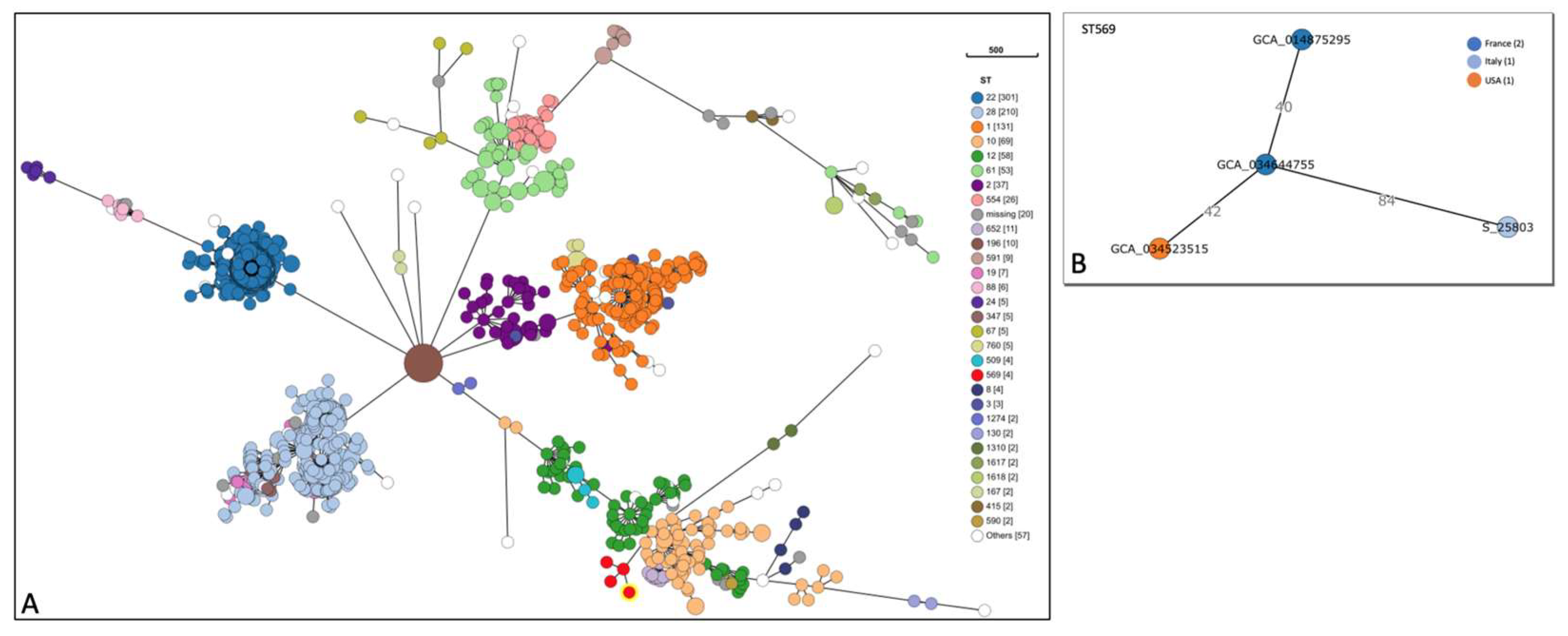
This study, for the first time in Italy, analyses by WGS a Streptococcus agalactiae strain isolated from a non-pregnant adult affected by meningitis and without common risk factors. The S. agalactiae strain was classified as a serotype II (SS2), sequence type ST569. Molecular characterization evidenced the presence of resistance genes to tetracycline and macrolide (tet(M) and mre(A)) and several virulence genes coding for adhesion and immune evasion factors (bca, cps family, neu family, scpB, gbs family, pil family and hylB), toxins (cfa/cfb, cyl family), pro-inflammatory factors (lepA), and two homologous genes that contributed to bacterial escape from the host immune system (lmb, luxS). SNPs analysis showed 18 different alleles, with 9 missense SNP mutations related to genes involved in cellular metabolism (dhaS, ftsE, ligA, nrdD and secA), virulence (bgrR and galE) and antimicrobial resistance (glpK and mutL). SNPs in glpK and mutL genes might reduce susceptibility to drugs. The SNPs analysis highlighted the presence of mutations conferring pathogenicity to the strain. The evidence in this study could explain the development of meningitis in a healthy patient. This case highlights the importance of using molecular methods to characterize the complete genome of a bacterial species that could seriously affect human health.

Keywords: 
;  ;  ;  ;  

1. Introduction

Streptococcus agalactiae is a Gram-positive, coccus-shaped, catalase-negative and oxidase-negative microorganism. This pathogen is also commonly known as group B Streptococcus (GBS) since it belongs to the Lancefield’s group B classification which includes streptococci that harbor a specific polysaccharide in their bacterial wall [1,2,3]. S. agalactiae was first isolated in 1887 by Edmond Nocard from milk of a mastitis affected cattle. However, its zoonotic potential and ability to cause severe disease in humans was only discovered in 1934, when Fry isolated it from three human cases who died from postpartum sepsis [4,5]. S. agalactiae lives as a commensal bacterium in the genitourinary and gastrointestinal system of humans and can cause serious disease, especially in immunosuppressed subjects (i.e., elderly and children) [4]. Children are most affected by GBS, and the onset of illness is characterized by sepsis, pneumonia and meningitis [6]. In 1973, an intrapartum prophylaxis program was initiated to limit cases of illness and death in newborns by administering antibiotics during pregnancy [7]. Nevertheless, it is estimated that GBS is the cause worldwide of 1% of all stillborn babies in developed countries and 4% of all stillborn babies in African countries [8]. It is well-known that GBS is also a pathogen that infects different animal species such as cattle, dogs, cats, fish, dolphins, and crocodiles [9]. In cattle, S. agalactiae is one of the main responsible pathogens for mastitis in farmed animals worldwide [10]; several authors also suggest that the disease in cattle may occur via reverse zoonosis events [7,11]. The main virulence factors of GBS are the polysaccharides of the capsule and the beta-hemolytic toxin [2,6]. Depending on the immunological reactivity of the antigens of the capsule, 10 serotypes have been identified; the most prevalent in America and Europe are Ia, Ib, II, III, V [12,13]. To date, 65% of GBS human cases of non-pregnant adults reported in Italy have concerned endocardial infections, resulting in embolism events and consequent neurological problems [14]. Presently, cases of GBS-associated meningitis have never been described in Italy and the case described in this paper is the first. However, in literature, serotype V GBS-sustained infections in non-pregnant adults have been documented worldwide and they usually include not only cases of meningitis, but also skin infections, osteomyelitis and endocarditis [15,16,17]. It should be noted, that although GBS is responsible for a small portion of patients with acute bacterial meningitis, 7-8 % of non-pregnant adults with GBS bacteremia have meningitis [18,19]. In the present study, the authors isolated a Group B S. agalactiae strain in a case of meningitis in a 37-year-old patient, with no previous health problems or common risk factors of infection, only a neurinoma of the left auditory nerve. The GBS strain was isolated from cerebrospinal fluid (CSF) and characterized by whole genome sequencing (WGS). The authors also describe a second case of GBS associated meningitis in an obese 50-year-old woman. However, the latter was detected only by Real-Time PCR as culture isolation from CSF failed.

2. Materials and Methods

2.1. Case Review

First case - Human CSF: in October 2023, a male patient of approximately 37 years of age was admitted at the ‘Presidio Ospedaliero Annunziata’ hospital of Cosenza in the Calabria region of southern Italy, due to symptoms referable to meningitis (hereafter mentioned as patient one). The patient did not report previous health problems and was not affected by common risk factors of infection, such as type 2 diabetes mellitus, neoplasms, immunodeficiency and kidney disease [14,17]. The patient exhibited only a benign neurinoma of the left auditory nerve. The CSF was analyzed for (a) the characterization of biomedical values using the SYSMEX XN-550 instrument following manufacturer’s instructions, and for (b) the meningoencephalitis panel using the FILMARRAYTM Meningitis/Enc kit (Biomerieux) following manufacturer’s instructions. The latter gave positive results. Microscopic examination revealed the presence of Gram-positive cocci.
Second case - Human CSF: in December 2023, a female obese patient of approximately 50 years of age was admitted at the ‘Presidio Ospedaliero Annunziata’ hospital of Cosenza in a confusional state (hereafter mentioned as patient two). Similarly, as in the first case, the CSF was analyzed for (a) the characterization of biochemical values using the SYSMEX XN-550 instrument, and for (b) the meningoencephalitis panel using the FILMARRAYTM Meningitis/Enc kit (Biomerieux). The latter gave positive results. Microscopic examination revealed the presence of Gram-positive cocci.

2.2. Bacteriological Examination

The two samples of CSF were inoculated into blood agar, chocolate agar, sabouraud agar, and thioglycolate. Subsequently, they were incubated at 37°C for 24-48h at 5% CO2. S. agalactiae was isolated and identified only from the CSF withdrawn from patient one: 24 hours after incubation, beta haemolytic colonies were detected on blood agar plates and then identified as S. agalactiae by mass spectrometry (MALDI-TOF). GBS was not isolated from CSF withdrawn from patient two. A pure colony of S. agalactiae isolated from patient one and a sample of the CSF from patient two were sent to the IZSM for molecular characterisation.

2.3. DNA Extraction

Approximately 2 ml of CSF from patient two was centrifuged at 12,000 rpm for 10 minutes at room temperature. Then, the supernatant was discarded, and the pellet was subsequently used for DNA extraction. Typical colonies of GBS isolated from patient one, were resuspended in 200 µL of PBS and then used for DNA extraction. DNA extraction from both samples was carried out using DNeasy PowerSoil kit (Qiagen) according to manufacturer’s instructions, and subsequently quantified using the Qubit fluorometer (Thermo Fisher Scientific, https://www.thermofisher.com).

2.4. PCR Typing

A specific real-time PCR was carried out to assess the presence of S. agalactiae DNA in the CSF sample from patient two [20]. The PCR protocol included a forward primer (5’-GGGAACAGATTATGAAAAACCG-3’), a reverse primer (5’-AAGGCTTCTACACGACTACCAA-3’) and a probe (HEX-5’-AGACTTCATTGCGTGCCAACCCTGAGAC-3’-BHQ1) targeting the cfb gene. The reaction mixture was prepared in a final volume of 25 µL, including 1 µM of each primer, 0.5 µM of the probe, TaqMan Universal PCR Master Mix 1X (Applied Biosystems, Waltham, MA, USA) and 5 µL of DNA template. Thermocycling conditions consisted of an initial denaturation step at 95°C for 10 min, followed by 45 cycles at 94°C for 30 sec and 60°C for 1 min. Real-Time PCR was carried out on a CFX 96 instrument (BioRad).

2.5. Whole Genome Sequencing Analysis

A DNA library was generated from the extracted DNA of the S. agalactiae strain isolated from patient one. The library was prepared using 150 ng of DNA with the Ion Xpress Fragment Library kit (Life Tecnologies, Carlsbad, CA, USA) following manufacturer’s instructions and loaded on a sequencing chip by using the Ion Chef instrument (ThermoFisher Scientific). The bacterial genome was sequenced at approximately 25X coverage. Sequencing was performed on the Ion Gene Studio S5 platform (ThermoFisher Scientific) as described by the manufacturer’s protocol by generating 400 bp single-end reads. Reads were taxonomically classified using the Kraken software [21] and RAST tool [22] was used for genomic annotation. MLST database (https://pubmlst.org/sagalactiae/) and Serotype bioinformatic tool (https://github.com/aquacen/serotype_Sagalactiae) [23] were then used to assign ST and serotype respectively. Genes encoding for virulence factors were detected by using VFDB (the virulence factor database, http://www.mgc.ac.cn/VFs/).
The core genome multilocus sequence typing (cgMLST) analysis was performed by chewbbaca (v. 3.3.4) using pubmlst scheme (h_S. agalactiae v1.0) consisting of 1405 loci. Publicly available sequences of S. agalactiae serotype II strains were selected (1056 isolates, Supplementary Table S1) from human, animal and environmental samples collected worldwide and used as database. The combination of all alleles in each strain was used to generate minimum spanning trees (MST) using the software GrapeTree (version 2.2) to identify related strains. Isolates assigned as sequence type ST569 were used for the SNP analysis and to investigate phylogenetic relationship. Finally, Resfinder tool was used to identify acquired genes (http://genepi.food.dtu.dk/resfinder).

3. Results

3.1. Biochemical Analysis of CSF Samples

Biochemical values of patient one: 6165 white blood cells/mm3 (90% neutrophils, 10% lymphocytes), IgG=33,80 mg/dL (normal range: 0.00-3.40 mg/dL), albumin=219 mg/dL (normal range: 0.00-35 mg/dL), glucose=53 mg/dL (normal range: 40-70 mg/dL).
Biochemical values of patient two: 6980 white blood cells/mm3 (88.1% neutrophils, 11.9% lymphocytes), IgG=100 mg/dL (normal range: 0.00-3.40 mg/dL), albumin=483 mg/dL (normal range: 0.00-35 mg/dL), glucose=69 mg/dL (normal range: 40-70 mg/dL).

3.2. Microbiological Examination and Molecular Analysis of CSF Samples

Only one of the two CSF samples under study resulted positive to bacteriological examination; S. agalactiae strain was isolated from patient one and confirmed by mass spectrometry. Nevertheless, both CSF samples were tested using a real-Time PCR analysis targeting the cfb gene and resulted as positive.

3.3. Whole Genome Sequencing Analysis

The S. agalactiae strain isolated from patient one was processed for WGS characterization by high-throughput sequencing. The WGS analysis produced a total of 280,543,897 bp, distributed among 983,026 reads. The assembly resulted in 69 contigs, accounting for 2,002,932 bp and an N50 of 111,704 bp. The genome annotation exhibited 2174 protein-coding sequences (CDS), 52 transfer RNA (tRNA) genes, and six ribosomal RNA (rRNA) genes. PATRIC annotation included 313 hypothetical proteins and 1861 proteins with functional assignments. The functional assignments included 614 proteins with Enzyme Commission (EC) numbers, 510 with Gene Ontology (GO) assignments, and 432 proteins mapped to KEGG pathways.
The MLST analysis based on a seven genes scheme (adhP, atr, glcK, glnA, pheS, sdhA, tkt) identified the analyzed genome as Sequence Type ST569, while in silico serotype typing, based on capsular polysaccharide (CPS) genes, assigned the strain to serotype II.
The cgMLST analysis grouped the isolates into a total of 29 different STs with a variable distribution within serotype II. The most frequent STs were ST1 (n=31), ST2 (n=37), ST10 (n=69), ST12 (n=58), ST22 (n=301), ST28 (n=210), ST61 (n=53) and ST554 (n=26). The ST569 cluster included our strain (S_25803), and three other isolates (Figure 1).
These three strains were of human origin and were isolated one from the USA and two from France: the American strain was isolated form an invasive disease in 2016, one of the French strains was isolated from a bacteremia case in 2009, and the second French strain was isolated in 2020 from a non-specified disease.
From the sequenced S. agalactiae genome, the presence of virulence genes and antimicrobial drug-resistance genes was investigated. Bacterial virulence factors confer to the harboring strain the ability to invade and colonize the host, thus increasing its ability to induce infection. The strain under study displayed the presence of several virulence genes coding for adhesion and immune evasion factors (bca, cps family, neu family, scpB, gbs family, pil family and hylB), toxins (cfa/cfb, cyl family), pro-inflammatory factors (lepA), and two homologous virulence genes contributing to bacterial escape from the host immune system (lmb, luxS). Genetic characterization of AMR genes showed the presence of mre(A) and tet(M) genes responsible for genetic resistance to macrolides and tetracyclines, respectively. These antibiotic resistances were also confirmed by disk diffusion test and MIC determination (erythromycin MIC>0.25 mcg/ml; tetracycline MIC>8 mcg/ml). The analyzed genome was also used to perform a SNPs analysis using the GCA_014875295 strain as reference. The results showed a total of 18 different alleles, with 9 missense SNP mutations related to genes involved in cellular metabolism (dhaS, ftsE, ligA, nrdD and secA), virulence (bgrR and galE) and antimicrobial resistance (glpK and mutL). SNPs in glpK and mutL genes might reduce susceptibility to drugs.

4. Discussion

This study describes the WGS-based characterization of a Streptococcus agalactiae strain isolated from the cerebrospinal fluid of a meningitis affected 37 years old man in southern Italy. The patient did not report previous health problems, neither suffered of type II diabetes mellitus, malignant neoplasms, immunodeficiency or kidney disease. Based on CPS genes and MLST analysis, the strain under study was identified as serotype II and Sequence Type ST569, respectively. Phylogenetic analysis indicated that the strain could be clustered with three other strains, all isolated from human cases in USA and France.
GBS is a commensal bacterium of the human microflora and frequently colonizes the gastrointestinal, respiratory, and vaginal tracts [24]. The maternal colonization of the gastrointestinal tract and/or lower reproductive tract is considered as a primary risk factor for neonatal invasive diseases such as sepsis, meningitis, and peripartum infection [25]. On the other hand, the incidence of invasive infections associated with GBS in non-pregnant adults is rising worldwide. Studies in literature report a cumulative incidence rate of invasive GBS disease ranging from 2.4 to 9.2 cases per 100,000 non-pregnant adults [26,27]. This pathogen can induce a range of different invasive diseases such as skin and osteo articular infections, pneumonia, urosepsis, endocarditis, peritonitis, meningitis, and streptococcal toxic shock syndrome (STSS) [28]. Specifically, GBS is responsible for 0.3-4.3 % of the total cases of meningitis in non-pregnant adults [27]. However, adults develop the disease less frequently compared to children because GBS does not easily pass the blood-brain barrier [16]. Among non-pregnant adults, the most affected categories are the elderly and immunosuppressed [14]. The mortality rate of non-pregnant adults belonging to risk categories may exceed 50% [29]. Primary risk factors for the development of GBS meningitis in non-pregnant adults include type II diabetes mellitus, neoplasms, immunodeficiency, and kidney disease [14,17]. In a study conducted on non-pregnant adults in the Reunion Island, Camuset et al. found a higher incidence rate of GBS infections compared to elsewhere; obesity was one of the most important risk factors associated to the disease [30]. Similarly, an epidemiological study in the USA reported an increased incidence of invasive GBS infections in non-pregnant adults, ranging from 8.1 % in 2008 to 10.9 % in 2016, associated with obesity or diabetes [31]. In addition, a recent case of GBS meningitis in a non-pregnant patient with a history of hypertension, dyslipidaemia, overweight, and alcohol consumption was described in Portugal [32]. In our study, patient two was hospitalized reporting only a confusional state and no other predisposing conditions, except for obesity. This, together with existing literature mentioned above, suggests that obesity is an important risk factor for the development of GBS meningitis. However, it must be noted that in patient two it was not possible to detect S. agalactiae with conventional culture methods and its presence was confirmed only by Real-Time PCR. Hence, it can be assumed that S. agalactiae could have been present in both samples even though, in patient two, the bacteriological examination was negative. Failure of bacteriological isolation of the microorganism could be explained if the patient took an antibiotic treatment before hospitalization, reducing the sensitivity of the cultural examination. This evidence highlights the importance of the use of real-time PCR molecular test: it could be more sensitive in detecting Streptococcus, than the bacteriological test, when antibiotic treatment is suspected.
On the other hand, patient one was hospitalized presenting fever, headache, and diarrhoea. However, they did not present any common risk factor for invasive GBS infection, except for a benign neurinoma of the left auditory nerve. The injury of the acoustic apparatus could have been a factor that permitted S. agalactiae to reach the brain, as already reported elsewhere (Peechakara B et al., 2018). Recently, in USA cases of GBS associated meningitis in non-pregnant adults affected by otitis have already been described [16,34].
The WGS characterization of the strain under study exhibited the presence of numerous virulence factors, essential to confer the bacteria the ability to induce invasive disease strategies, responsible for its survival and colonization in the host [35]. The fact that patient one did not display any of the most common risk factors associated with GBS invasive disease, highlights the important role these virulence factors have in determining the pathogenicity of the studied strain. Moreover, among the virulence factors harbored by the strain, SNPs analysis identified the presence of missense mutations in the bgrR and galE genes, the former associated with increased virulence properties [36,37]. Consistently, cgMLST analysis indicated that the strain under study could be classified as ST569 (a rare Sequence Type) found only in three other cases, all grouped in the same cluster and responsible for infection and disease in humans [19]. However, it is important to note that these other strains were isolated in pregnant women and newborns [38]. The analysis of the genomes available in GenBank showed that ST569 is a unique sequence type in Italy; only one other S. agalactiae strain serotype II was identified but was classified as ST28, while other S. agalactiae strains resulted as serotypes III and IV.
Given the increasing incidence of GBS invasive infections in non-pregnant adults [30] and the zoonotic nature of the microorganism, it is recommended to manage and monitor the disease with a One Health approach when diagnosing both animal and human cases that can derive from both risk and non-risk categories.

5. Conclusions

S. agalactiae meningitis in non-pregnant adults is a rare disease, even though recent studies suggest it may become more prevalent, increasing the relevance of this case series. This study helps support the enhancement of diagnosis and treatment for Group B S. agalactiae meningitis and highlights the importance to consider GBS in all meningitis human cases for appropriate, timely diagnosis and treatment.

Supplementary Materials

The following supporting information can be downloaded at website of this paper posted on Preprints.org, Table S1: S. agalactiae serotype II strains from human, animal and environmental samples collected worldwide and used as database.

Author Contributions

Conceptualization: G.B., G.F., E.D., A.L.; methodology: G.B., R.P. L.B.; software: R.P. and A.R.; formal analysis: F.G., M.V.M., M.G.C., S.D.; investigation, S.G., V.V., A.M.; data curation, writing—original draft preparation, G.B., G.F., R.P. and A.C.; writing—review and editing, visualization, G.B., R.P.; supervision, G.B. and G.F. All authors have read and agreed to the published version of the manuscript.”

Funding

This research was developed within the Next Generation EU-MUR PNRR Extended Partnership initiative on Emerging Infectious Diseases (Project no. PE0000007 INF-ACT)

Informed Consent Statement

Not applicable

Data Availability Statement

The data that support the findings of this study are openly available in Streptococcus agalactiae strain 25803HSAGAL chromosome at https://www.ncbi.nlm.nih.gov/nuccore/2731779437, reference number CP154875.

Conflicts of Interest

The authors declare no conflicts of interest.

Abbreviations

The following abbreviations are used in this manuscript:
WGS Whole Genome Sequencing
S. agalactiae Streptococcus agalactiae
MLST Multi Locus Sequence Type
SNP Single Nucleotide Polymorphism
GBS group B Streptococcus
CSF cerebrospinal fluid
cgMLST core genome multilocus sequence typing
CPS capsular polysaccharide

References

  1. Lancefield RC. A serological differentiation of human and other groups of hemolytic streptococci. J Exp Med. 1933 Mar 354 31;57(4):571-95. [CrossRef]
  2. Burcham LR, Spencer BL, Keeler LR, et al. Determinants of Group B streptococcal virulence potential amongst vaginal clinical 320 isolates from pregnant women. PLoS One. 2019 Dec 18;14(12):e0226699. [CrossRef]
  3. Furfaro LL, Chang BJ, Kahler CM, et al. Genomic characterisation of perinatal Western Australian Streptococcus agalactiae 344 isolates. PLoS One. 2019 Oct 2;14(10):e0223256. [CrossRef]
  4. Hanna M, Noor A. Streptococcus Group B. Stat Pearls Publishing. 2023 Jan 16. PMID: 31985936.351.
  5. Emaneini M, Khoramian B, Jabalameli F, et al. Comparison of virulence factors and capsular types of Streptococcus agalactiae 336 isolated from human and bovine infections. Microb Pathog. 2016 Feb;91:1-4. [CrossRef]
  6. He EM, Chen CW, Guo Y, et al. The genome of serotype VI Streptococcus agalactiae serotype VI and comparative analysis. 352 Gene. 2017 Jan 15;597:59-65. [CrossRef]
  7. Meroni G, Sora VM, Martino PA, et al. Epidemiology of Antimicrobial Resistance Genes in Streptococcus agalactiae Sequences 359 from a Public Database in a One Health Perspective. Antibiotics (Basel). 2022 Sep 12;11(9):1236. [CrossRef]
  8. Zhou Y, Zhao XC, Wang LQ, et al. Detecting Genetic Variation of Colonizing Streptococcus agalactiae Genomes in Humans: A 391 Precision Protocol. Front Bioinform. 2022 Jun 3;2:813599. [CrossRef]
  9. Yao K, Poulsen K, Maione D, et al. Capsular gene typing of Streptococcus agalactiae compared to serotyping by latex aggluti- 389 nation. J Clin Microbiol. 2013 Feb;51(2):503-7. Epub 2012 Nov 28. 390. [CrossRef]
  10. Pang M, Sun L, He T, et al. Molecular and virulence characterization of highly prevalent Streptococcus agalactiae circulated in 361 bovine dairy herds. Vet Res. 2017 Oct 16;48(1):65. [CrossRef]
  11. Crestani C, Forde TL, Lycett SJ, et al. The fall and rise of group B Streptococcus in dairy cattle: reintroduction due to human-to- 329 cattle host jumps? Microb Genom. 2021 Sep;7(9):000648. [CrossRef]
  12. Furfaro LL, Chang BJ, Payne MS. A novel one-step real-time multiplex PCR assay to detect Streptococcus agalactiae presence 346 and serotypes Ia, Ib, and III. Diagn Microbiol Infect Dis. 2017 Sep;89(1):7-12. [CrossRef]
  13. Slotved HC, Møller JK, Khalil MR, et al. The serotype distribution of Streptococcus agalactiae (GBS) carriage isolates among 375 pregnant women having risk factors for early-onset GBS disease: a comparative study with GBS causing invasive infections 376 during the same period in Denmark. BMC Infect Dis. 2021 Nov 1;21(1):1129. [CrossRef]
  14. D’Angelo M, Boretti I, Quattrocchi S, et al. Lethal infective endocarditis due to Streptococcus agalactiae in a man with a history 331 of alcohol abuse: A case report. Medicine (Baltimore). 2019 Dec;98(51):e18270. [CrossRef]
  15. Sendi P, Johansson L, Norrby-Teglund A. Invasive group B Streptococcal disease in non-pregnant adults: a review with em- 367 phasis on skin and soft-tissue infections. Infection. 2008 Mar;36(2):100-11. Epub 2008 Jan 12. 368. [CrossRef]
  16. Al-Bayati A, Douedi S, Alsaoudi G, et al. Meningitis from invasive Streptococcus agalactiae in a healthy young adult. IDCases. 316 2020 Jul 7;21:e00907. [CrossRef]
  17. Chaiwarith R, Jullaket W, Bunchoo M, et al. Streptococcus agalactiae in adults at Chiang Mai University Hospital: a retrospec- 324 tive study. BMC Infect Dis. 2011 May 25;11:149. [CrossRef]
  18. Vasikasin V, Changpradub D. Clinical manifestations and prognostic factors for Streptococcus agalactiae bacteremia among 378 nonpregnant adults in Thailand. J Infect Chemother. 2021 Jul;27(7):967-971. Epub 2021 Feb 18. 379. [CrossRef]
  19. Vuillemin X, Hays C, Plainvert C, Dmytruk N, Louis M, Touak G, Saint-Pierre B, Adoux L, Letourneur F, Frigo A, Poyart C, 384 Tazi A. Invasive group B Streptococcus infections in non-pregnant adults: a retrospective study, France, 2007-2019. Clin Micro- 385 biol Infect. Jan 2021;27(1):129.e1-129.e4. Epub 2020 Sep 29. 386. [CrossRef]
  20. Diaz MH, Waller JL, Napoliello RA, et al. Optimization of Multiple Pathogen Detection Using the TaqMan Array Card: Appli- 333 cation for a Population-Based Study of Neonatal Infection. PLoS One. 2013 Jun 21;8(6):e66183. [CrossRef]
  21. Wood DE, Salzberg SL. Kraken: ultrafast metagenomic sequence classification using exact alignments. Genome Biol. 2014 Mar 387 3;15(3):R46. [CrossRef]
  22. Brettin T, Davis JJ, Disz T, et al. RASTtk: a modular and extensible implementation of the RAST algorithm for building custom 318 annotation pipelines and annotating batches of genomes. Sci Rep. 2015 Feb 10;5:8365. [CrossRef]
  23. Sheppard AE, Vaughan A, Jones N, et al. Capsular Typing Method for Streptococcus agalactiae Using Whole-Genome Sequence 373 Data. J Clin Microbiol. 2016 May;54(5):1388-90. Epub 2016 Mar 9. 374. [CrossRef]
  24. Regan JA, Klebanoff MA, Nugent RP. The epidemiology of group B streptococcal colonization in pregnancy. Vaginal Infections 365 and Prematurity Study Group. Obstet Gynecol. 1991 Apr;77(4):604-10. PMID: 2002986. 366.
  25. Seale AC, Bianchi-Jassir F, Russell NJ, et al. Estimates of the Burden of Group B Streptococcal Disease Worldwide for Pregnant 369 Women, Stillbirths, and Children. Clin Infect Dis. 2017 Nov 6;65(suppl_2):S200-S219. [CrossRef]
  26. Schwartz B, Schuchat A, Oxtoby MJ, et al. Invasive group B streptococcal disease in adults. A population-based study in met- 371 ropolitan Atlanta. JAMA. 1991 Aug 28;266(8):1112-4. PMID: 1865545. 372.
  27. Farley MM, Harvey RC, Stull T, et al. A population-based assessment of invasive disease due to group B Streptococcus in 339 nonpregnant adults. N Engl J Med. 1993 Jun 24;328(25):1807-11. [CrossRef]
  28. Farley MM. Group B streptococcal disease in nonpregnant adults. Clin Infect Dis. 2001 Aug 15;33(4):556-61. [CrossRef]
  29. Crespo-Ortiz Mdel P, Castañeda-Ramirez CR, Recalde-Bolaños M, Vélez-Londoño JD. Emerging trends in invasive and noninvasive isolates of Streptococcus agalactiae in a Latin American hospital: a 17-year study. BMC Infect Dis. 2014 Aug 3;14:428. [CrossRef]
  30. Camuset G, Picot S, Jaubert J, et al. Invasive Group B Streptococcal Disease in Non-pregnant Adults, Réunion Island, 2011. Int 322 J Infect Dis. 2015 Jun;35:46-50. [CrossRef]
  31. Francois Watkins LK, McGee L, Schrag SJ, et al. Epidemiology of Invasive Group B Streptococcal Infections Among Nonpreg- 341 nant Adults in the United States, 2008-2016. JAMA Intern Med. 2019 Apr 1;179(4):479-488. [CrossRef]
  32. Coelho T, Pacheco M, Mendes T, Valente J, Gil P. Invasive Streptococcus agalactiae Disease With Meningitis and Septic Arthritis in a Non-pregnant Patient. Cureus. 2022 Nov 4;14(11):e31077. [CrossRef]
  33. Peechakara B, Demkowicz R, Gupta M. Meningitis by Streptococcus agalactiae secondary to otitis media. QJM. 2018 Dec 363 1;111(12):891-892. [CrossRef]
  34. Villareal K, Goslin A, Bajracharya H. Group B Streptococcus Meningitis Associated with Acute Otitis Media in an Adult Patient. 382 Am J Case Rep. 2021 Oct 3;22:e933093. [CrossRef]
  35. Ma J, Zhang Z, Pan Z, et al. Streptococcus suis Uptakes Carbohydrate Source from Host Glycoproteins by N-glycans Degrada- 356 tion System for Optimal Survival and Full Virulence during Infection. Pathogens. 2020 May 18;9(5):387. [CrossRef]
  36. Vasilyeva A, Santos Sanches I, Florindo C, et al. Natural Mutations in Streptococcus agalactiae Resulting in Abrogation of β 380 Antigen Production. PLoS One. 2015 Jun 5;10(6):e0128426. [CrossRef]
  37. Chen CL, Cheng MH, Kuo CF, et al. Dextromethorphan Attenuates NADPH Oxidase-Regulated Glycogen Synthase Kinase 3β 326 and NF-κB Activation and Reduces Nitric Oxide Production in Group A Streptococcal Infection. Antimicrob Agents Chemother. 327 2018 May 25;62(6):e02045-17. [CrossRef]
  38. Gizachew M, Tiruneh M, Moges F, et al. Molecular characterization of Streptococcus agalactiae isolated from pregnant women 348 and newborns at the University of Gondar Comprehensive Specialized Hospital, Northwest Ethiopia. BMC Infect Dis. 2020 Jan 349 13;20(1):35. [CrossRef]
Figure 1. Minimum spanning of all Streptococcus agalactiae isolates. Phylogenetic tree based on the cgMLST data from publicly available Streptococcus agalactiae serotype II isolates (n = 1056) of human, animal and environmental origin. Colors represent the different distribution of the sequence types. Each circle indicates an allele profile with lines connecting closely related isolates forming clusters (A). The ST569 (red circle) cluster was composed of four strains, including the one presented in this study. The lines indicate allelic differences (B).
Preprints 151101 g001
Disclaimer/Publisher’s Note: The statements, opinions and data contained in all publications are solely those of the individual author(s) and contributor(s) and not of MDPI and/or the editor(s). MDPI and/or the editor(s) disclaim responsibility for any injury to people or property resulting from any ideas, methods, instructions or products referred to in the content.
Copyright: This open access article is published under a Creative Commons CC BY 4.0 license, which permit the free download, distribution, and reuse, provided that the author and preprint are cited in any reuse.
Prerpints.org logo

Preprints.org is a free preprint server supported by MDPI in Basel, Switzerland.

Subscribe

Disclaimer

Terms of Use

Privacy Policy

Privacy Settings

© 2025 MDPI (Basel, Switzerland) unless otherwise stated