Preprint
Review

This version is not peer-reviewed.

Targeting Bile Acid Metabolism: Nutritional and Microbial Approaches to Alleviate Ulcerative Colitis

A peer-reviewed article of this preprint also exists.

Submitted:

25 February 2025

Posted:

27 February 2025

You are already at the latest version

Abstract

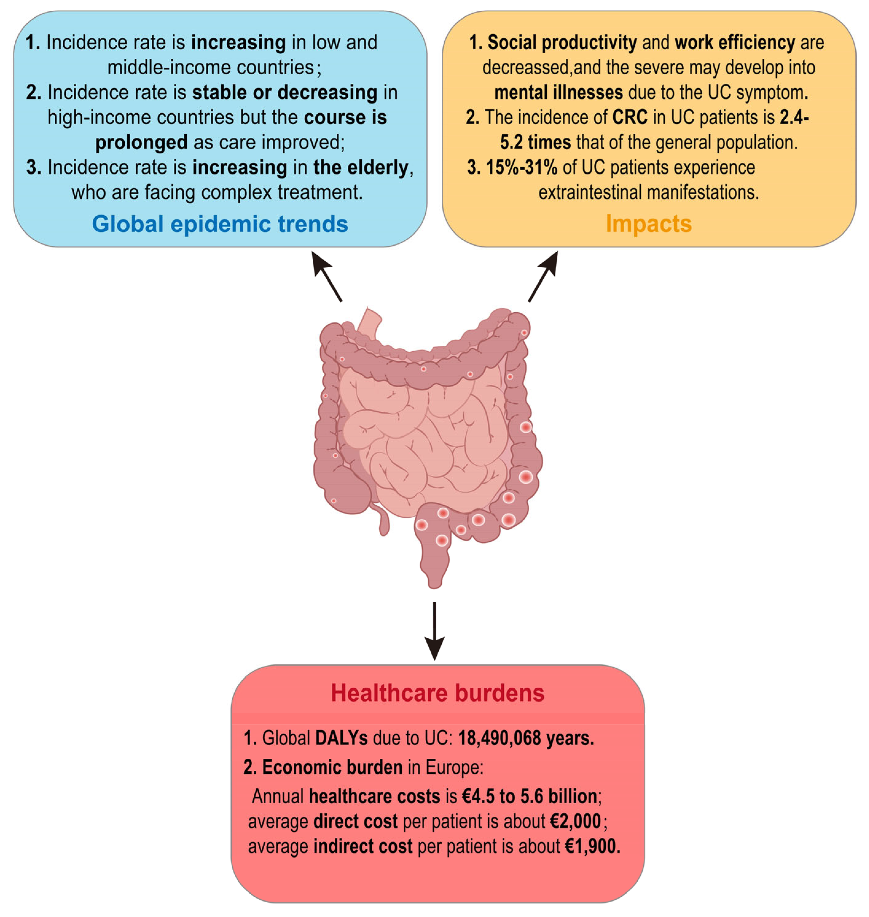
Ulcerative colitis (UC) is a chronic inflammatory disease affecting the colorectum, posing a significant global health burden. Recent studies highlight the critical role of gut microbiota and its metabolites, particularly bile acids (BAs), in UC’s pathogenesis. The relationship between BAs and gut microbiota is bidirectional: microbiota influence BA composition, while BAs regulate microbiota diversity and activity through receptors like Farnesoid X receptor (FXR) and Takeda G protein-coupled receptor 5 (TGR5). Targeting bile acid metabolism to reshape gut microbiota presents a promising therapeutic strategy for UC. This review examines the classification and synthesis of BAs, their interactions with gut microbiota, and the potential of nutritional and microbial interventions. By focusing on these therapies, we aim to offer innovative approaches for effective UC management.

Keywords: 
;  ;  ;  

1. Introduction

Ulcerative colitis (UC), a major type of inflammatory bowel disease (IBD), is a chronic lifelong inflammatory disease of the colon. The hallmark clinical symptom of UC is bloody diarrhea which characterized by an inflammatory reaction occurring in the colorectal mucosa with alternating periods of remission and active phases[1,2]. The Montreal classification divides UC into three subgroups: E1, in which inflammation is confined to the rectum and does not reach the sigmoid colon; E2, in which inflammation has involved the left half of the colon (far from the splenic flexure); and E3, in which extensive colonic inflammation involves the proximity of the splenic flexure and even the entire colon. E3 is the most predominant form of the phenotype observed in Asia[3]. The pathogenesis of UC is still unclear. Multiple factors, such as environmental factors genetic background and mucosal dysfunction, have been suggested to contribute to UC pathogenesis. Individuals with genetic predisposition after environmental exposures can triggers an inappropriate immune response by the interaction of intestinal microorganisms and their metabolites, which is currently considered to be the main cause of UC[1,4].

2. The Current Status of UC

Until 2000, UC was generally considered a disease predominantly affected populations in North America, Europe, and Oceania. Nowadays, with the globalization of the economy, changes in environmental risk factors and shifts in dietary patterns, the incidence of UC has been rising dramatically in both low and middle-income newly industrialized countries. Additionally, the prevalence of UC is higher in urban areas compared to rural regions[1,3,5]. The incidence rates in high-income countries have generally stabilized or even shown a downward trend. However, the highest UC incidence rates are still occurring in North America and Northern Europe, ranging from 8.8-23.14 per 100,000 person-years and 1.7-57.9 per 100,000 person-years, respectively[5]. The overall incidences of UC in both Europe and Asia show a north-south gradient, which may be related to the population density[6,7,8].
UC is a lifelong disease. The recurrent abdominal pain and diarrhea are prone to cause persistent weakness and psychological negativity in UC patients’ body. These symptoms affect the social productivity and interactions, reduce work efficiency, restrict social and recreational activities[6,7], and even develop into mental disorders such as depression and anxiety[8,9]. In addition, with the increased global life expectancy and aging population, the elderly have become a fast-growing group of UC patients. And the elderly face more severe immune system alterations, sarcopenia, and syndromes of underlying diseases, which increase the complexity of UC treatment to some extent. Although the treatment options for UC have expanded over the past decade, the five-year survival rate for patients without colectomy is no significant difference from that of the general population[3]. 5%-10% of patients with UC may still need surgery to treat their disease within five years of diagnosis[10]. The incidence of colorectal cancer (CRC) in patients with UC is approximately 1.21/1000[11], which is 2.4-5.2 times higher than the general population[12], with a risk ratio of 1.66 (95% CI, 1.57-1.76)[13]. CRC ranks as the second highest mortality rate globally, and the five-year survival rate of CRC in patients with UC is lower than that of the general patients[12]. Furthermore, about 15%-31% of UC patients are accompanied by one or several extraintestinal manifestations associated with metabolic derangements due to immune-related or intestinal malabsorption[10]. Mesoaxial and peripheral arthritis in the joints, scleral epiphora and uveitis in the eyes, low bone mass and osteoporosis in the bones, primary sclerosing cholangitis in the gallbladder and fatty liver in the liver are most common extraintestinal manifestations in UC patients[14]. All of the above undoubtedly decrease the quality of life of patients with UC, increase the burden of disease, and pose greater challenges to healthcare systems around the world.
Besides, according to the Global Burden of Disease Study, the annual healthcare burden due to UC in Europe is about 4.5 to 5.6 billion euros, and the global disability-adjusted life years (DALYs) of UC is 18,490,068 years[15,16], with an average of about €2,000 of direct healthcare costs per UC patient (including hospitalization and surgical expenditures and drug-related expenditures), and about €1,900 of the indirect healthcare costs (including reduced income due to, e.g., reduced work efficiency due to illness)[10]. In summary, the global epidemic trend of UC is generally rising, with far-reaching impacts and significant healthcare burdens (Figure 1).
Figure 1. Summary of current status of ulcerative colitis.
Preprints 150510 g001

3. Bile Acid Metabolism

In recent years, with the development of macrogenomics, the link between gut microbiota and UC has drawn increasing attention. Microbial metabolites, such as bile acids (BAs), short chain fatty acids (SCFAs) and tryptophan metabolites[17], bridge the microbiota and the host and also play essential roles in the gut barrier and intestinal inflammatory homeostasis. BAs, important components of bile, are synthesized from cholesterol in hepatocytes of the liver through a series of enzymatic reactions. This process is the primary pathway of cholesterol metabolism in mammals. With the clearer insight of BAs in recent years, studies have unanimously shown that BAs disorders are correlated with intestinal barrier dysfunction and mucosal inflammation, as well as that BAs can act as a signaling molecule to regulate inflammation and immune response of the body[18,19,20].

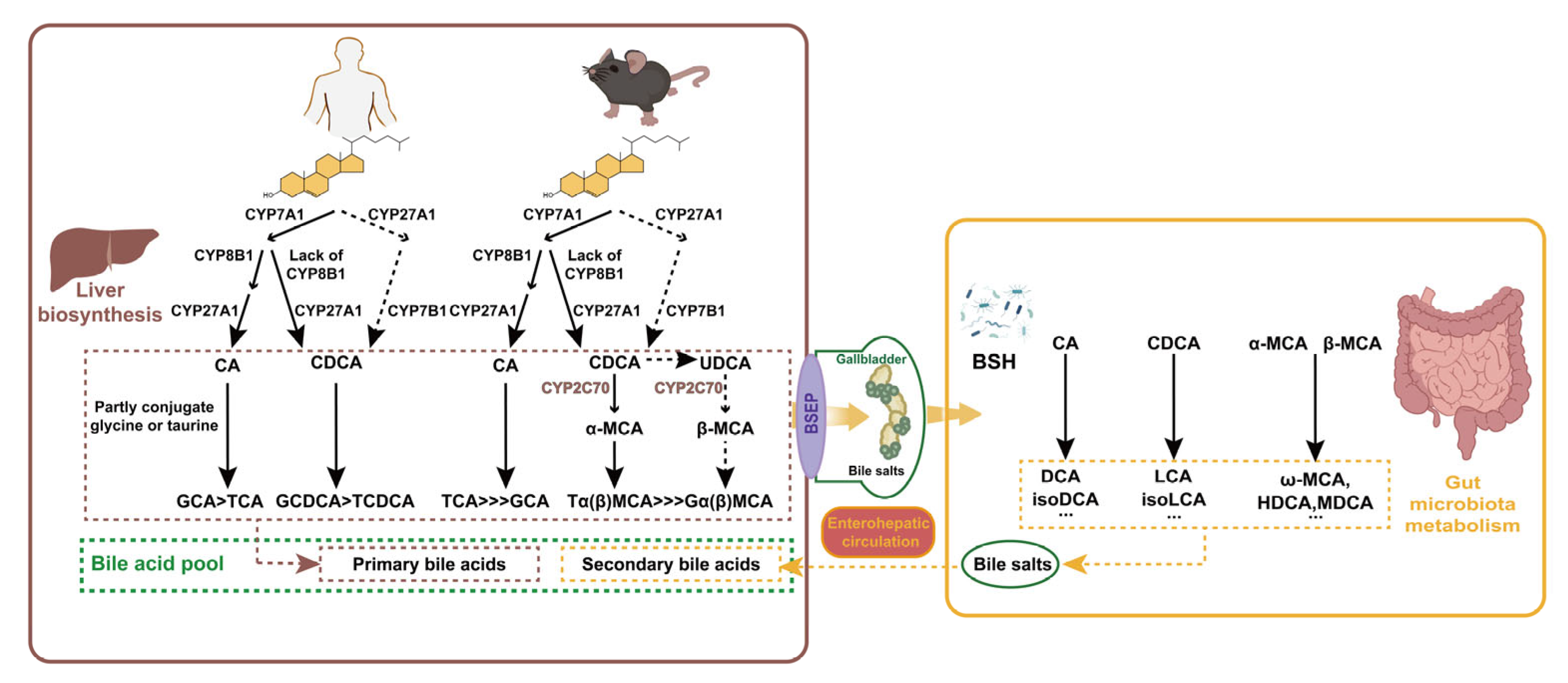
3.1. Classification and Synthesis of BAs

Bile acids can be classified as free and conjugated bile acids in terms of their structure. Free BAs include cholic acid (CA), chenodeoxycholic acid (CDCA), deoxycholic acid (DCA) and lithocholic acid (LCA). The above free BAs respectively combine with glycine (G) or taurine (T) to form a diversity of conjugated BAs (glycine is mainly in humans, taurine mainly in mice). The conjugated BAs include glycocholic acid (GCA), taurocholic acid (TCA), taurodeoxycholic acid (TDCA) and so forth, which usually exist as sodium salts within the body, making them more water-soluble and stable than free counterparts.
According to their origins, BAs can be categorized as primary BAs and secondary BAs. Primary BAs are synthesized directly in the liver from cholesterol, while secondary BAs are derived from primary bile acids in response to intestinal microorganisms. In human, primary BAs are CA, CDCA and their corresponding conjugated ones; secondary BAs include DCA, LCA, ursodeoxycholic acid (UDCA) and also their conjugated ones. The BA profiles vary between human and mice (Figure 2). In mice, β-muricholic acid (β-MCA) and CA are the predominant primary bile acids, while α-muricholic acid (α-MCA), CDCA and UDCA constitute a relatively small portion[21]. In addition to DCA and LCA, mice also possess ω-muricholic acid (ω-MCA), hyocholic acid (HCA), hyodeoxycholic acid (HDCA), and murine deoxycholic acid (MDCA) as secondary BAs[22].
Figure 2. Metabolism of bile acids in human and in mice. Bile acids are synthesized in the liver from cholesterol through enzymatic pathways involving CYP7A1, CYP27A1, and CYP8B1. These bile acids are conjugated with glycine or taurine to form conjugated bile acids. In the gut, bile salt hydrolase (BSH) enzymes produced by microbiota deconjugate bile acids. Primary bile acids are converted into secondary bile acids by gut bacteria. Secondary bile acids are reabsorbed into the bloodstream and transported back to the liver completing the enterohepatic circulation.
Preprints 150510 g002
Liver is the exclusive organ that synthesizes bile acids, and cytochrome P450 (CYP450) in its parenchymal cells mediates cholesterol oxidation to produce primary BAs. There are two main pathways for its synthesis: the cholesterol 7α-hydroxylase (CYP7A1)-mediated “classical pathway” (also known as the neutral pathway) and the sterol-27-hydroxylase (CYP27A1)-mediated “alternative pathway” (also known as the acidic pathway). In the classical pathway, CYP7A1 is the rate-limiting enzyme, determining the yield of BAs. Most cholesterol is initially hydroxylated by CYP7A1, and subsequently converted to CA through the action of microsomal sterol 12α-hydroxylase (CYP8B1) and mitochondrial CYP27A1. In the absence of CYP8B1, the intermediates are instead converted into CDCA. Alternatively, in the acidic pathway, cholesterol is first transformed into 2,7α-hydroxycholesterol by CYP27A1, and then further hydroxylated into CDCA by the rate-limiting enzyme sterol 7α-hydroxylase (CYP7B1). The ratio between the two primary bile acids, CA and CDCA, is determined by CYP8B1 which is not regulated by microbial influences[23]. In healthy individuals, at least 75% of primary bile acids are produced via the classical pathway; in rodents, particularly in mice, the alternative pathway may account for up to 50% of bile acids production[24]. In addition to CA and CDCA, certain lines of research propose that UDCA may also function as a primary bile acid in mice—as it can serve as a biosynthetic precursor to β-MCA—even though it makes up only about 1% of the total bile acid pool[21,23]. Most of the CDCA and UDCA are converted to α-MCA and β-MCA in mice, respectively, by sterol-6β-hydroxylase (CYP2C70)[22].
Conjugated bile acids can be actively transported into the bile by the bile salt export pump (BSEP) and stored in the gallbladder. Food intake or other external stimuli trigger gallbladder contraction, releasing bile into the duodenum This process forms mixed micelles of bile acids, cholesterol, and phospholipids, which aid in the emulsification and absorption of nutrients in the gut. Multiple species of anaerobic bacteria in the ileum and colon are able to generate bile salt hydrolase (BSH), which facilitates the deconjugation of conjugated BAs. Once deconjugated, these BAs can be converted to secondary BAs by intestinal microbes through a series of processes such as dehydroxylation, oxidation, or epimerization. For example, C7 dehydroxylation of CA and CDCA generate DCA and LCA, respectively; DCA and LCA can be transformed into iso-DCA and iso-LCA through epimerization. In humans, UDCA, a secondary bile acid, is derived from a small amount of CDCA through the catalytic action of 7α-hydroxysteroid dehydrogenase (7α-HSDH) and 7β-HSDH[25]. In mice, β-MCA can be oxidized and undergo epimerization by the gut microbiota to form 3α, 6α, 7β-trihydroxy bile acids (ω-MCA), which is the unique secondary bile acid in mice[21]. Besides, unconjugated α, β, and ω-MCA can undergo 7α or 7β-dehydroxylation by gut bacterial to yield HDCA or MDCA[26]. The generation of secondary bile acids increases the diversity and hydrophobicity of the bile acid pool. Compared to humans, mice also have a higher proportion of primary BAs, as they can express testosterone 7α-hydroxylase (CYP2A12), which can convert secondary bile acids back to CA and CDCA[27]. Over 90% of bile acids are reabsorbed from the distal small intestine into the blood circulation and eventually back to the liver via transporters such as the Organic Anion Transport Proteins (OATPs), the Na+-taurocholate cotransporting polypeptide (NTCP), BSEP and apical sodium-dependent bile acid transporter (ASBT)[21]. These BAs, along with newly synthesized ones, are then conveyed to the duodenum to complete the enterohepatic circulation[28].
Both primary and secondary bile acids can modulate host metabolism and immune response. Gut microorganisms can regulate secondary bile acid production, which in turn impacts bile acid-related signaling pathways, thereby modulating body metabolism and immune function.

3.2. Mutual Regulation of Microbiota and BAs

Uncoupling of gut microbiota, which involves removing the conjugation of BAs to glycine and taurine, prevents ASBT from recapturing BAs from the small intestine. BAs uncoupling is performed by bacteria with BSH activity. Metagenomics studies reveal that active BSH is found in nearly all major bacterial and archaeal groups in the human gut, including Firmicutes, Bacteroidetes, Bifidobacterium, Actinobacteria and archaeon Methanobrevibacter[29,30]. Uncoupled primary BAs enter the colon, where they are metabolized into secondary BAs by colonic symbiotic microorganisms. In this progress, the critical C7 dehydroxylation is mediated by bile acid-inducible (bai) manipulator proteins produced by bacteria with bai genes, such as Clostridium and Eubacterium[30]. Another important microbial transformation of bile acid is the oxidation of the hydroxyl on C3, C7, or C12. This transformation is catalyzed by hydroxysteroid dehydrogenases (HSDHs), which are found in Actinobacteria, Firmicutes, Proteobacteria, and Bacteroidetes. These oxidations are reversible and ultimately lead to epimerization, For instance, in humans, HSDHs produced by Eubacterium lentum and Clostridium perfringens can catalyze the 3α/β-hydroxy isomerization of LCA and DCA to form iso-LCA and iso-DCA, which are present in human serum and urine, with abundance only inferior to that of LCA and DCA[30].
Alterations in the gut microbiota can affect BAs metabolism. Factors such as antibiotics, exercise, diet, or other unfavorable states of intestinal microbial symbiosis will lead to alterations in the composition or activity of the intestinal microbiota[31], thereby disrupting BA metabolism. On the other hand, there is increasing evidence that BAs significantly shape the gut flora and are essential determinants of microbiota abundance, diversity, and metabolic activity. van Best N et al. found that during developmental period of the newborn, an increase in the concentration of primary BAs led to greater abundance of bacteria with genes for BA metabolism in the small intestine[32]. Studies from both human populations and mice have shown that cholestasis reduces the diversity of the host microbiota[33,34], although the exact mechanism remains unclear. Furthermore, BAs can modify the function of the microbiota; for instance, sublethal concentrations of DCA, TCA and TDCA can disrupt nucleotide and carbohydrate metabolism of mouse gut microbiome[35].

3.3. Receptors Involved in BA Metabolism

One of the mechanisms by which bile acid regulate intestinal physiology is controlling the metabolism and transport of BAs through several key host BA receptors. The nuclear receptors, including Farnesoid X receptor (FXR), vitamin D receptor (VDR), pregnane X receptor (PXR) and constitutive androstane receptor (CAR), serve as bridges between BAs and nuclear receptor target genes. These genes are involved in lipid and glucose homeostasis, xenobiotic metabolism and immunomodulatory pathways. Additionally, Several G protein-coupled receptors (GPCRs), such as Takeda G protein-coupled receptor 5 (TGR5, also known as G protein-coupled bile acid receptor 1, GPBAR1), muscarinic acetylcholine receptor M3 (M3R) and sphingosine-1-phosphate receptor 2 (S1PR2), also bind to BAs and primarily exerting immunomodulatory functions[36,37]. Among these receptors, FXR and TGR5 are the most extensively studied.

3.3.1. FXR

FXR is recognized as the first bile acid receptor to be discovered and its activation mediates the classical pathway of bile acid synthesis. Bile acids serve as natural ligands for FXR, and bile acid synthesis is tightly regulated by negative feedback inhibition of FXR[38]. The most potent ligand for FXR is CDCA, followed by CA, DCA and LCA[39]. In contrast, UDCA inhibits FXR activation[40]. Also, murine taurine-conjugated primary bile acids, TαMCA and TβMCA, have been identified as natural antagonists of FXR[23].
FXR is expressed in a variety of tissues, with the highest expression in the liver and ileum[41], followed by kidney, heart, ovary, thymus, eye, spleen and testis[42]. In the liver, BAs activate FXR and induce the expression of small heterodimer partner (SHP), which binds to liver receptor homolog-1 (LRH-1), thereby inhibiting Cyp7a1 gene expression[38,43,44]. In addition, hepatic FXR regulates the Cyp8b1 gene thus regulating CA formation[45,46,47]. Besides its local effects in the liver, FXR can also be activated by bile acids in the distal ileum, where it induces the secretion of fibroblast growth factor 15 (FGF15)/FGF19. FGF15/19 reaches the liver through the portal veins, binds to FGFR4/b-klotho heterodimeric complexes and triggers the c-Jun N-terminal kinase (JNK) 1/2 and extracellular signal-regulated kinase (ERK) 1/2 signaling cascades to inhibit Cyp7a1 expression[48,49]. Moreover, excess uncoupled BAs can be toxic, and in that situation, FXR activation plays a key role in hepatic detoxification. FXR can reduce BAs biosynthesis and promote the export from the liver; FXR induces the expression of bile acid coenzyme A: amino acid N-acyltransferase (BAAT) in the liver, thereby preventing the toxic accumulation of uncoupled BAs and enhances hepato-intestinal circulation[50]; FXR also helps eliminate hepatic BAs by increasing BSEP expression and decreasing the expression of hepatic transport proteins that uptake BAs, such as organic anion-transporting polypeptide (OATP) 1[38].

3.3.2. VDR, PXR and CAR

VDR, PXR and CAR are three closely related receptors that play similar roles in BA detoxification and elimination and they are regulated by FXR. However, unlike FXR, bile acids are not the primary ligands for these receptors. Nevertheless, all three nuclear receptors facilitate the removal of hepatotoxic LCA[51,52].
VDR is activated only by LCA and 1,25 dihydroxy-vitamin D3[53,54]. When activated, VDR induces the activation of cytochrome P450 3A4 (CYP3A4) activation to detoxify LCA, inhibits CYP7A1 expression in the liver, reduces hepatic bile acid synthesis, and increases ASBT expression[55,56].
PXR is mainly activated by exogenous drugs such as rifampicin, though its bile acid ligand is also LCA. PXR is responsible for inducing phase I and phase II metabolism of a variety of compounds including bile acids. Activation of PXR can induce the expression of detoxification enzymes, including the CYP3A family, as well as enzymes involved in BAs sulfation and conjugation[57,58]. CAR can cooperate with PXR in the pathway of LCA detoxification in the liver and promote OATP2 and multidrug resistance protein 3 (MRP3) expression. Additionally, PXR activation induces FGF19 expression in colonocytes, suggesting that PXR also contributes to inhibiting hepatic bile acid synthesis and promoting BA homeostasis[59].

3.3.3. TGR5

TGR5 is generally highly expressed in gallbladder, placenta, lung, spleen, intestine, liver, brown and white adipose tissue, skeletal muscle and bone marrow[30]. Bile acids are natural endogenous ligands for TGR5, with the potency in the order of LCA>DCA>CDCA>CA[57]. TGR5 identifies BAs regardless of their substitution and conjugation status[60], whereas S1PR2 only recognizes conjugated BAs[61]. As a membrane receptor, TGR5 can be internalized into the cytoplasm upon binding to its ligands and plays an essential role in cellular signaling pathways such as nuclear factor-κB (NF-κB), protein kinase B (AKT) and ERK.
Unlike FXR, hepatocytes do not express TGR5[62], and the function of TGR5 is tissue-specific. In gastric neurons, DCA, LCA or the selective TGR5 agonist oleanolic acid stimulates colonic motility in mice[63]. In monocytes and macrophages, TGR5 activation inhibits NF-κB-mediated inflammatory responses[64]. In splenic B cells, TGR5 promotes energy consumption and insulin sensitivity, and is closely associated with glucose homeostasis: the activation of TGR5 has been demonstrated to control hyperglycemia and hyperinsulinemia in both a diet-induced obesity mouse model and human studies[63,65,66].

3.3.4. M3R and S1PR2

M3R can be activated by DCA and LCA along with their glycine and taurine conjugates, in addition to acetylcholine[67]. M3R is involved in triggering the proliferation of intestinal epithelial cells and promoting tumor progression[68]. In human colon cancer cells, BAs activate M3R, which stimulates interaction with the epidermal growth factor receptor through matrix metalloproteinase 7 (MMP-7) activation. In ASBT-deficient mice, significantly elevated colonic bile acid level due to bile acid malabsorption induced overexpression of the Mmp7 gene. Consequently, the number and size of colonic tumors increased significantly while ASBT-deficient mice treated with carcinogen azoxymethane (AOM), indicating that M3R was involved in inducing proliferation of intestinal epithelial cells and promoting tumor progression[68].
S1PR2 can be activated by conjugated bile acids. Several studies have shown that bound bile acids rely on the Gαi protein subunit to activate ERK1/2 and AKT-dependent pathways in hepatocytes, inducing the activity of sphingosine kinase 2 (Sphk2) and resulting in an increase in sphingosine-1-phosphate (S1P)[61]. The increase in S1P activates S1PR2 and inhibits specific histone deacetylases (HDACs), which may lead to altered gene expression. S1PR2-deficient mice are more susceptible to fatty liver when fed a high-fat diet, indicating that the bile acid-S1PR2-Sphk2 axis is also involved in lipid metabolism processes[69]. Also, activation of S1PR2 by bile acids appears to be associated with the development of cholangiocarcinoma[70]. Most studies on bile acids and S1PR2 have focused on the liver, but a recent study has shown that S1PR2 is also expressed in intestinal epithelial cells, where its activation by bound bile acids promotes cell proliferation[71]. However, the role of S1PR2 in intestinal physiology requires further investigation.

4. UC and BA Metabolism

4.1. Disrupted BA Metabolism and Intestinal Flora Drive UC

During IBD episodes, the colonic epithelium generates the inflammatory response, compromising barrier integrity lost. The colonic epithelium serves as a physical boundary between the complex luminal environment and the host tissue. In addition, the colon epithelium includes components such as the outer mucus layer, the intestinal microbiota that colonizes the surface of the colonic epithelium, and the subepithelial components such as T cells, B cells, eosinophils, mast cells, dendritic cells, and macrophages. Together, these form the chemical, biological, and immune barriers in the intestinal mucosal barrier[72]. In UC patients, there are typical disruptions and changes in tight junction structure and protein composition and function of the colonic epithelium. The disruption of the intestinal barrier function in UC leads to permeability defects, allowing entry of microorganisms, increasing the influx of toxins and allergens, and further exacerbating immune cell infiltration into intestinal tissues and the inflammatory response[73,74]. BAs not only play a role in colonic epithelial permeability, but also participate in maintaining the intestinal microbial ecological balance, suggesting a close relationship between BAs and IBD development.
Studies have shown significant alterations in the gut microbiota of UC patients, including reduced biodiversity, instability in microbial composition, and a decreased abundance of Firmicutes involved in BA metabolism. Among these, Faecalibacterium prausnitzii is thought to have a protective effect against intestinal inflammation in UC. Dysbiosis of the gut microbiota leads to disrupted BA metabolism, resulting in elevated levels of conjugated and primary BAs, along with reduced levels of secondary BAs because of impaired deconjugation and conversion[75]. Furthermore, studies have found that increased levels of conjugated TCA in the feces of UC patients are positively correlated with elevated levels of tumor necrosis factor-α (TNF-α), a key pro-inflammatory cytokine[76].

4.2. Regulation of UC by BA Receptors

Bile acids, through FXR and TGR5 signaling, help maintain epithelial barrier integrity, modulate cytokine production, and orchestrate immune cell activity, thereby shaping the overall inflammatory and immune responses (Figure 3).
Figure 3. FXR and TGR5 regulate inflammation and immune responses. BAs activate FXR leading to the regulation of tight junction proteins(TJs) such as occludin and claudin. This strengthens the intestinal barrier and reduces bacterial translocation (BT); FXR activation also inhibits inflammatory signaling pathways, such as AP-1 and NF-κB, thereby reducing the production of pro-inflammatory cytokines like TNF-α. TGR5 activation modulates macrophage polarization, promoting a shift from the pro-inflammatory M1 phenotype (producing IFN-γ and TNF-α) to the anti-inflammatory M2 phenotype (producing IL-10).
Preprints 150510 g003

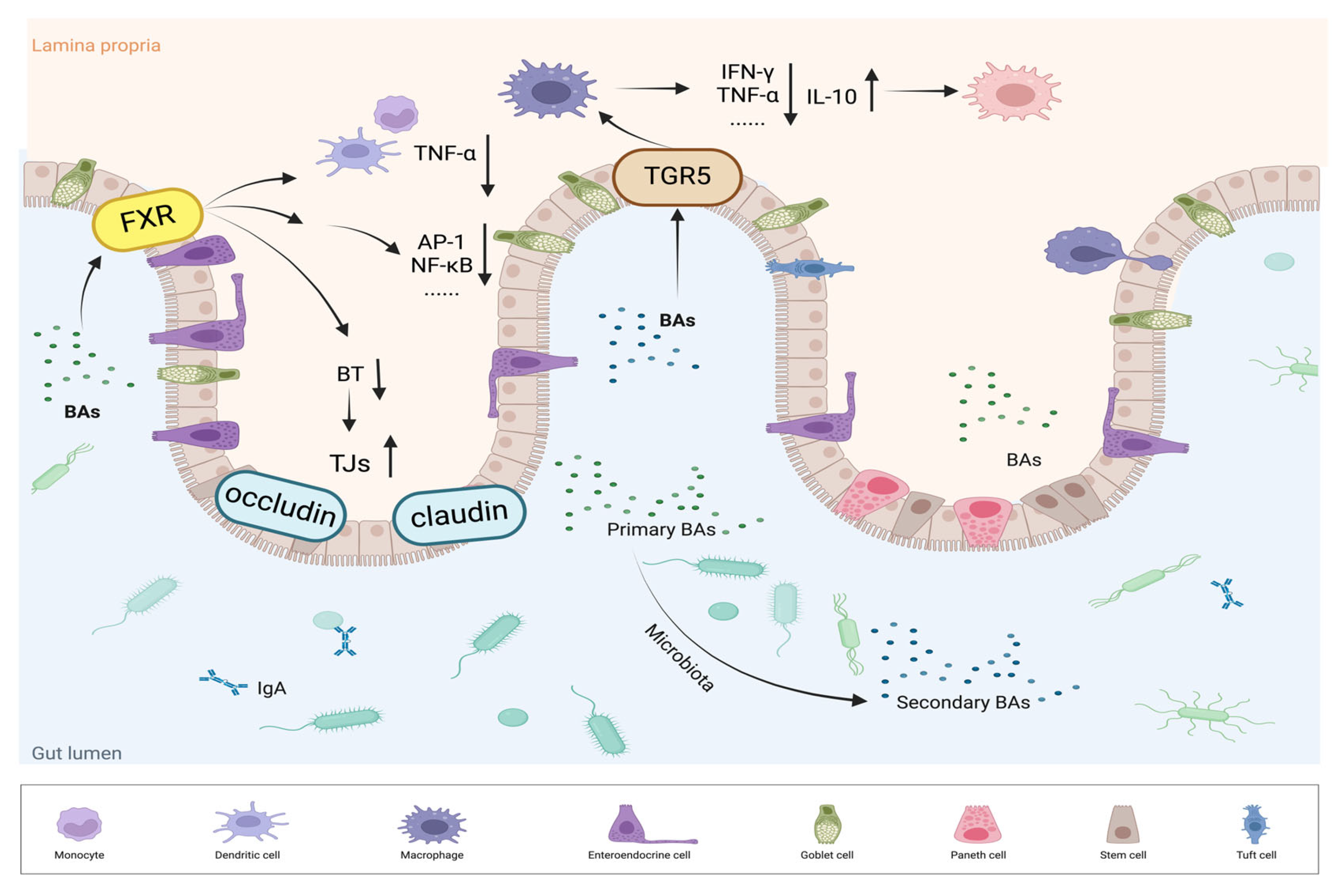
4.2.1. FXR Regulates the Immune Response and Intestinal Health

Apart from its important role in controlling hepatic bile acid synthesis, FXR also regulates the immune response to control inflammation, reduces bacterial overgrowth and translocation to prevent intestinal mucosal from injury, and regulates the secretion of FGF19 and chloride channels to alleviate diarrhea. Additionally, FXR is closely linked to the development of colorectal cancer, making it an important target for treatment of UC.
Studies by Calmus et al. demonstrated the role of bile acids in immune cell regulation: incubation of monocytes with CDCA inhibited lipopolysaccharide(LPS)-stimulated cytokine secretion[77]. FXR along with other nuclear receptors was found to be expressed in human peripheral blood mononuclear cells (PBMCs) and monocyte subpopulations such as CD4+ T cells, CD8+ T cells, and CD14+ monocyte subpopulations, suggesting the regulatory role of FXR in the immune response. FXR agonists such as INT-747 have been shown to reduce TNF-α secretion from activated human PBMCs and CD14+ monocytes, and from colon lamina propria mononuclear cells in IBD patients. This indicates that FXR agonists not only inhibit active mononuclear immune cells secreting cytokine, but also reduce inflammatory cytokine expression in differentiated intestinal epithelial cells[78]. Moreover, FXR activation suppresses inflammation through various signaling pathways, particularly through interactions with proteins such as activator protein 1 (AP-1) and signal transducers and activators of transcription 3 (STAT3), and by inhibiting transcription factors like NF-κB and AP-1, which are critical regulators of immune response genes[79,80,81]. Collectively, these findings provide strong evidence for FXR’s role in both innate and adaptive immunity, as well as its contribution to the pathophysiology of chronic intestinal inflammatory diseases.
In addition to its immune regulatory functions, FXR protects the intestinal mucosa by attenuating bacterial overgrowth and translocation while maintaining epithelial integrity. Blocking the secretion of bile acids and decreasing the concentration in the colonic lumen can promote bacterial proliferation and impair epithelial barrier function, leading to increased bacterial translocation across the colonic epithelium. For instance, oral administration of BAs (the endogenous ligand of FXR) to rats with obstructive jaundice resulted in decreased bacterial overgrowth and translocation, suggesting that FXR may act as a regulatory factor in bacterial dynamics[82]. In mouse models of bile duct ligation, treatment with the FXR agonist GW4064 led to diminished bacterial overgrowth and translocation, while FXR-deficient mice exhibited increased bacterial proliferation and translocation to mesenteric lymph nodes[83]. Furthermore, FXR activation elevated the expression of antimicrobial genes, including angiopoietin, carbonic anhydrase 12, inducible nitric oxide, and interleukin 18 (IL-18) in epithelial cells[80]. The selective agonist INT-747 enhanced the expression of tight junction proteins, such as claudin-1 and occludin, reduced intestinal inflammation, and normalized intestinal permeability, highlighting FXR’s protective role in the gut-liver axis[84].
FXR plays a crucial role in maintaining intestinal homeostasis and regulating various gastrointestinal disorders. Its involvement in bile acid metabolism and signaling pathways highlights its significance in both bile acid-induced diarrhea (BAD) and colorectal cancer progression. FXR significantly alleviates BAD by regulating the secretion of FGF19 and chloride channels. A reduction in ileocolonic FGF19/15 level disrupts feedback inhibition, leading to an overproduction of hepatic bile acids, elevated bile acid concentrations in the colon, and subsequent chloride secretion that impairs fluid absorption, resulting in diarrhea. The activation of intestinal FXR stimulates FGF19/15 secretion, suggesting the feasibility of using FXR agonists in the treatment of BAD[85]. Recent studies supporting this hypothesis have shown that INT-747 is well tolerated by patients with BAD, reducing hepatic bile acid synthesis and improving diarrhea symptoms[86]. Additionally, FXR has been indicated to reduce the expression of the chloride channel cystic fibrosis transmembrane conductance regulator (CFTR) and inhibit Na+/K+-ATPase activity in epithelial cells[87]. Decreased FXR expression is also closely associated with colorectal cancer progression. Research by Bailey et al. revealed diminished FXR expression in samples from patients with precancerous lesions and nearly absent expression in advanced colon adenocarcinoma samples. This FXR silencing may result from FXR promoter hypermethylation and V-Ki-ras2 Kirsten rat sarcoma viral oncogene homolog (KRAS) signaling, indicating that FXR inhibits epithelial-mesenchymal transition and other oncogenic signaling pathways. In chronic colitis models, the absence of FXR may activate Wnt signaling via neutrophil infiltration and increased TNF-α secretion from macrophages, which correlated to enhanced tumor progression and early mortality[88]. Treatment with cholestyramine, a bile acid-conjugated resin, in FXR knockout mice did not promote tumorigenicity, clearly demonstrating that FXR deficiency, rather than increased bile acids, is responsible for the high susceptibility to tumorigenicity[89]. Moreover, excessive dietary fat intake has been shown to stimulate hepatic bile acid synthesis and alter bile acid metabolism by gut microbes, ultimately modifying the overall bile acid pool. Notably, elevated levels of DCA are closely associated with both high-fat diets and an increased risk of CRC. DCA displays antagonist-like activity against FXR, whereas FXR agonists have demonstrated the potential to slow tumor progression, suggesting that FXR is likely involved in preserving colon health under high-fat dietary conditions[90]. These findings (Table 1) provide new insights into the potential role of FXR as a therapeutic target in UC, underscoring its multifaceted role in gut health and disease management.
Table 1. Overview of current research on FXR, emphasizing its immunological effects and mechanisms in gut diseases.
Study design Reference Main findings
Mouse colon and enterocyte-like cells treated with medium or INT-747. Gadaleta et al.,2011[78] FXR activation inhibits the secretion of TNF-α in immune cells (such as PBMCs and CD14+ monocytes).
Fxr-/- and WT mice treated with GW4064 or vehicle for 2 days and then subjected to BDL or sham operation. Inagaki et al.,2006[83] FXR activation increases genes involved in enterοprotection, decreases bacterial overgrowth and mucosal injury in ileum.
Rats subjected to BDL or not, and treated with vehicle 5 mg/kg GW4064 or UDCA. Verbeke et al.,2015[84] FXR activation normalizes ileal permeability and reduces bacterial translocation, resulting in a significant decrease in natural killer cells and INF-γ expression.
Patients with BAD (n=28) received oral INT-747 acid 25 mg daily for 2 weeks. Walters et al.,2015[86] FXR activation induced by INT-747 reduces hepatic bile acid synthesis and improving diarrhea symptoms.
IHC was used to detect FXR in tissues from human normal samples (n = 238), polyps (n = 32), and adenocarcinomas staged I-IV (n = 43, 39, 68, and 9, respectively). Bailey et al.,2014[88] FXR expression decreases in samples from patients with precancerous lesions and is nearly absent in advanced colon adenocarcinoma samples.
Fxr-/- and WT mice intraperitoneally injected with sterile saline with or without AOM once a week for 6 weeks. Maran et al.,2009[89] FXR deficiency promotes cell proliferation, inflammation, and tumorigenesis in the intestine, suggesting that FXR may protect against intestinal carcinogenesis.
PBMCs: peripheral blood mononuclear cells. WT: wild-type. BDL: bile duct ligation. BAD: bile acid-induced diarrhea. IHC: immunohistochemistry. AOM: azoxymethane.

4.2.2. TGR5 Bridges Inflammation and Immune Responses

Improvement of UC symptoms by TGR5 agonists primarily involves the modulation of inflammatory responses through two key mechanisms: (1) TGR5 inhibits NOD-like receptor protein 3 (NLRP3) inflammasome activation. Secondary BAs, such as DCA and LCA, activate TGR5, triggering the TGR5-cAMP-PKA signaling cascade, which promotes NLRP3 ubiquitination and reduces its activity[91]. (2) In monocytes and macrophages, TGR5 suppresses LPS-induced inflammatory responses. TGR5 agonists decrease TNF-α production via the mammalian target of rapamycin (mTOR) pathway. Tgr5-/- macrophages exhibited heightened cytokine secretion and increased migration, highlighting the role of TGR5 in chronic inflammation[92]. The latest research shows that UDCA reduced pro-inflammatory cytokine levels in macrophages by inducing suppressor of cytokine signaling 1 (SOCS1) expression in a TGR5-dependent manner. In the AOM-DSS mouse model, UDCA also significantly lowers the expression of cancer-related genes and inflammation in the colon[93]. Biagioli et al. also demonstrated that the small molecule TGR5 activator, BAR501, significantly alleviated the level of intestinal inflammation, downregulated the expression of pro-inflammatory cytokines and chemokines (IL-1β, IL-6, interferon-γ, and TNF-α) in UC mouse model. BAR501 enhanced the expression of the anti-inflammatory factors IL-10 and TGF-β, facilitating the shift of macrophages from the M1 pro-inflammatory to the M2 anti-inflammatory phenotype. The anti-inflammatory effects of BAR501 were absent in IL-10-/- mice, indicating that the M1/M2 macrophage phenotype transition is IL-10-dependent[94].

5. BA-Based Therapies

5.1. Dietary and Phytochemical Strategies

The goals of UC treatment are primarily to induce clinical response and biomarker normalization rapidly, and secondarily to maintain clinical remission and achieve endoscopic normalization to prevent long-term disability[1]. UC is usually managed with treatments based on the severity and location of the disease. 5-aminosalicylic acid (5-ASA) is indicated for mild to moderate UC and can serve as maintenance therapy. Corticosteroids (e.g., prednisone) are used for moderate to severe active UC, but are not commonly used as long-term maintenance therapy owing to the serious side effects. Immunomodulators, such as azathioprine and 6-mercaptopurine, are suitable for patients who have poor response to 5-ASA or corticosteroids to maintain remission. Biological agents, including anti-TNF antibodies (e.g., infliximab), integrin receptor antagonists (e.g., vedolizumab), and IL-12/23 inhibitors (e.g., ustekinumab), are applicable to patients with moderate to severe UC. Small molecule drugs, such as JAK inhibitors (e.g., tofacitinib) and S1P receptor (e.g., etrasimod) modulators, are used in patients who have failed to respond to other treatments[1].
Although these medicines can effectively manage UC symptoms, they are associated with high failure rates, serious side effects, and substantial costs. Statistics show that more than 10% of UC patients eventually require surgical intervention[1]. Therefore, the development of new strategies for preventing and treating UC remains a critical area of research. Phytochemicals, naturally occurring compounds found in plants and easily accessible, have demonstrated health benefits and are considered a promising source of cost-effective, high-efficiency therapies.
Dietary intake is essential for sustaining human life, yet patients with ulcerative colitis often experience nutrient deficiencies due to recurrent diarrhea and chronic inflammation. Proper dietary interventions can ensure sufficient intake of essential nutrients, thereby promoting tissue repair and supporting immune function. Furthermore, dietary composition can modulate and sustain the structure and relative abundance of the gut microbiota. Adopting a balanced diet promotes the proliferation of beneficial microbial populations, enhances the production of key metabolites (e.g., SCFAs and BAs), and helps maintain intestinal stability. Researches indicate that excessive dietary fat intake can alter the bile acid pool and is associated with colorectal cancer development[90], whereas dietary fiber consumption exerts anti-inflammatory and anticancer effects, potentially via SCFAs. The interaction between bile acids and SCFAs is likely mediated by changes in the gut microbiota[95]. Additionally, certain dietary polyphenols can regulate BA metabolism and improve intestinal barrier function, thereby alleviating symptoms of UC and offering potential value as preventive or adjunctive treatments for the condition. For instance, apple polyphenol extracts can modulate circadian rhythms, influencing bile acid metabolism and gut microbiota in high-fat diet mice, which leads to anti-inflammatory effects and improved intestinal homeostasis. Polyphenols found in oats, such as avenanthramides and flavonoids, can alleviate metabolic syndrome and inflammatory responses induced by high-fat diets through the regulation of bile acid metabolism. Furthermore, compounds like anthocyanins, proanthocyanidins, and caffeic acid have been shown to inhibit pro-inflammatory signaling pathways (e.g., NF-κB and NLRP3) and enhance the activity or expression of FXR and TGR5[96]. In summary, a high-fiber, low-fat diet with more beneficial polyphenols may be particularly suitable for patients with UC.
Recent studies have also increasingly highlighted the role of other specific phytochemicals in alleviating symptoms of ulcerative colitis (UC) through the regulation of bile acid metabolism. These findings offer promising new insights into potential therapeutic strategies for managing UC. For instance, berberine has been shown to alleviate UC symptoms by inhibiting the colonization of harmful bacteria, promoting primary bile acid metabolism, and restoring intestinal barrier function via the bile acid/S1PR2/RhoA/ROCK signaling pathway[97]. Pulsatilla decoction improves bile acid homeostasis and mitigates UC symptoms by activating the FXR-ASBT pathway and upregulating the expression of FXR, TGR5, CYP7A1, and FGF15 proteins[98]. Lycium barbarum polysaccharides may increase the abundance of Dubosiella in the gut microbiota, which enhances the production of secondary bile acids (such as LCA and DCA) and upregulates TGR5 expression, ultimately strengthening the intestinal barrier[99]. Similarly, carvacrol and thymol promote the production of secondary bile acids, such as HDCA and 12-ketodeoxycholic acid (12-KCAC), by increasing the abundance of Parabifidobacterium in the colon, thereby exerting anti-inflammatory effects[100]. In conclusion, the ability of these phytochemicals to modulate bile acid metabolism and alleviate UC symptoms, provides novel research directions and potential therapeutic strategies for the treatment of UC.

5.2. Fecal Microbial Transplantation (FMT) and Probiotic Measurements

FMT involves transferring fecal microorganisms from a healthy donor to a patient, which was initially intended for the treatment of recurrent Clostridium difficile infections, but is now also used for other intestinal diseases, such as IBD[101]. The success of FMT in treatment of IBD may partly be attributed to the restoration of BSH-dependent secondary BA production, which reduces C. difficile colonization[102,103]. However, FMT has shown with mixed results in the treatment of UC[104]. The lack of specificity in modulating the microbiota through FMT may lead to unexpected adverse effects, especially since the full impact of the intact bacterial community on human health is not yet fully understood, making it challenging to design successful FMT treatments.
Probiotics offer an advantage over FMT by more specifically improving the bile acid (BA) pool in conditions of dysbiosis and disease. They influence the BA pool by modulating the activation of BA receptors such as FXR, PXR, and VDR. The probiotic mixture VSL#3, containing BSH-expressing bacteria, enhances BA deconjugation and excretion while upregulating hepatic BA biosynthesis through inhibition of the FXR-FGF15 pathway, thereby helping treat UC[105]. Similarly, the probiotic strain GR-4, isolated from the traditional Chinese fermented food “jiangshui”, selectively enriches bacterial taxa associated with BA metabolism and enhances the gut microbiota’s ability to modify BAs, leading to reduced conjugated BAs and increased secondary BAs, which helps alleviate UC symptoms[106]. Using biologically active secondary BAs, such as UDCA, which may have similar beneficial effects[107], however, the positive effects of direct BAs supplementation may only last during treatment, whereas altering the microbiota to enhance bile metabolism may provide longer-lasting benefits[31].

6. Summary and Perspective

Ulcerative colitis is a chronic disease that remains difficult to cure. Ongoing research continues to elucidate the complex interplay between UC, gut microbiota, and bile acid metabolism, offering new avenues for therapeutic intervention. However, identifying highly effective treatments with minimal side effects remains a priority to ensure both cost-effectiveness and favorable long-term outcomes. Building on advances in dietary and phytochemical strategies, and FMT and probiotic measurements detailed in this review, modulating bile acid pathways presents a promising tactic to alleviate inflammation and restore intestinal homeostasis. Moving forward, further well-designed studies are required to deepen our mechanistic understanding of UC and BA and to guide the development of safe and effective therapies for UC management.

Author Contributions

Conceptualization, writing—original draft preparation, X. J.; writing−review and editing, J.R; formal analysis and validation, G. Y. and W.W; visualization, M. C.; investigation, writing-review and editing, Y. Z.; writing-review and editing, supervision, financial support C. H. All authors have read and agreed to the published version of the manuscript.

Funding

This review was funded by the National Natural Science Foundation of China, grant number No. 82103819.

Data Availability Statement

Data availability statement is not applicable to this article as no datasets were generated or analyzed during the current study.

Conflicts of Interest

The authors declare no conflicts of interest.

Abbreviations

The following abbreviations are used in this manuscript:
UC Ulcerative colitis
IBD Inflammatory bowel disease
BAs Bile acids
FXR Farnesoid X receptor
TGR5 Takeda G protein-coupled receptor 5
DALYs Disability-adjusted life years
SCFAs Short chain fatty acids
CA Cholic acid
CDCA Chenodeoxycholic acid
DCA Deoxycholic acid
LCA Lithocholic acid
UDCA Ursodeoxycholic acid
α/β-MCA α/β-muricholic acid
HCA Hyocholic acid
HDCA Hyodeoxycholic acid
MDCA Murine deoxycholic acid
BSEP Bile salt export pump
BSH Bile salt hydrolase
7α-HSDH 7α-hydroxysteroid dehydrogenase
OATPs Organic Anion Transport Proteins
ASBT Apical sodium-dependent bile acid transporter
HSDHs Hydroxysteroid dehydrogenases
VDR Vitamin D receptor
PXR Pregnane X receptor
CAR Constitutive androstane receptor
GPCRs G protein-coupled receptors
M3R Muscarinic acetylcholine receptor M3
S1PR2 Sphingosine-1-phosphate receptor 2
SHP Small heterodimer partner
LRH-1 Liver receptor homolog-1
FGF15/19 Fibroblast growth factor 15/19
JNK c-Jun N-terminal kinase
ERK Extracellular signal-regulated kinase
BAAT Bile acid coenzyme A: amino acid N-acyltransferase
MRP3 Multidrug resistance protein 3
AKT Protein kinase B
AOM Azoxymethane
LPS Lipopolysaccharide
PBMCs Peripheral blood mononuclear cells
AP-1 Activator protein 1
STAT3 Signal transducers and activators of transcription 3
BAD Bile acid-induced diarrhea.
mTOR Mammalian target of rapamycin
NLRP3 NOD-like receptor protein 3

References

  1. Le Berre, C.; Honap, S.; Peyrin-Biroulet, L. Ulcerative colitis. Lancet 2023, 402, 571–584. [Google Scholar] [CrossRef] [PubMed]
  2. Wei, S.C.; Sollano, J.; Hui, Y.T.; Yu, W.; Santos Estrella, P.V.; Llamado, L.J.Q.; Koram, N. Epidemiology, burden of disease, and unmet needs in the treatment of ulcerative colitis in Asia. Expert Rev. Gastroenterol. Hepatol. 2021, 15, 275–289. [Google Scholar] [CrossRef] [PubMed]
  3. Du, L.; Ha, C. Epidemiology and Pathogenesis of Ulcerative Colitis. Gastroenterol. Clin. North Am. 2020, 49, 643–654. [Google Scholar] [CrossRef]
  4. Molodecky, N.A.; Soon, I.S.; Rabi, D.M.; Ghali, W.A.; Ferris, M.; Chernoff, G.; Benchimol, E.I.; Panaccione, R.; Ghosh, S.; Barkema, H.W. , et al. Increasing incidence and prevalence of the inflammatory bowel diseases with time, based on systematic review. Gastroenterology 2012, 142, 46–54. [Google Scholar] [CrossRef]
  5. Jeuring, S.F.; van den Heuvel, T.R.; Zeegers, M.P.; Hameeteman, W.H.; Romberg-Camps, M.J.; Oostenbrug, L.E.; Masclee, A.A.; Jonkers, D.M.; Pierik, M.J. Epidemiology and Long-term Outcome of Inflammatory Bowel Disease Diagnosed at Elderly Age-An Increasing Distinct Entity? Inflamm. Bowel Dis. 2016, 22, 1425–1434. [Google Scholar] [CrossRef] [PubMed]
  6. Benchimol, E.I.; Bernstein, C.N.; Bitton, A.; Murthy, S.K.; Nguyen, G.C.; Lee, K.; Cooke-Lauder, J.; Siddiq, S.; Windsor, J.W.; Carroll, M.W. , et al. The Impact of Inflammatory Bowel Disease in Canada 2018: A Scientific Report from the Canadian Gastro-Intestinal Epidemiology Consortium to Crohn’s and Colitis Canada. J. Can. Assoc. Gastroenterol. 2019, 2, S1–S5. [Google Scholar] [CrossRef]
  7. Ghosh, S.; Sensky, T.; Casellas, F.; Rioux, L.C.; Ahmad, T.; Marquez, J.R.; Vanasek, T.; Gubonina, I.; Sezgin, O.; Ardizzone, S. , et al. A Global, Prospective, Observational Study Measuring Disease Burden and Suffering in Patients with Ulcerative Colitis Using the Pictorial Representation of Illness and Self-Measure Tool. J. Crohns Colitis 2020, 15, 228–237. [Google Scholar] [CrossRef]
  8. Moulton, C.D.; Pavlidis, P.; Norton, C.; Norton, S.; Pariante, C.; Hayee, B.; Powell, N. Depressive symptoms in inflammatory bowel disease: an extraintestinal manifestation of inflammation? Clin Exp Immunol 2019, 197, 308–318. [Google Scholar] [CrossRef]
  9. Mikocka-Walus, A.; Andrews, J.M. It is high time to examine the psyche while treating IBD. Nat. Rev. Gastroenterol. Hepatol. 2018, 15, 329–330. [Google Scholar] [CrossRef]
  10. Zhao, M.; Gonczi, L.; Lakatos, P.L.; Burisch, J. The Burden of Inflammatory Bowel Disease in Europe in 2020. J. Crohns Colitis 2021, 15, 1573–1587. [Google Scholar] [CrossRef]
  11. Mabila, S.L.; McQuistan, A.A.; Murray, J.H. Incidence of Colorectal Cancer Among Active Component Service Members, 2010-2022. MSMR 2023, 30, 2–6. [Google Scholar]
  12. Marabotto, E.; Kayali, S.; Buccilli, S.; Levo, F.; Bodini, G.; Giannini, E.G.; Savarino, V.; Savarino, E.V. Colorectal Cancer in Inflammatory Bowel Diseases: Epidemiology and Prevention: A Review. Cancers (Basel) 2022, 14. [Google Scholar] [CrossRef] [PubMed]
  13. Olen, O.; Erichsen, R.; Sachs, M.C.; Pedersen, L.; Halfvarson, J.; Askling, J.; Ekbom, A.; Sorensen, H.T.; Ludvigsson, J.F. Colorectal cancer in ulcerative colitis: a Scandinavian population-based cohort study. Lancet 2020, 395, 123–131. [Google Scholar] [CrossRef] [PubMed]
  14. Harbord, M.; Annese, V.; Vavricka, S.R.; Allez, M.; Barreiro-de Acosta, M.; Boberg, K.M.; Burisch, J.; De Vos, M.; De Vries, A.M.; Dick, A.D. , et al. The First European Evidence-based Consensus on Extra-intestinal Manifestations in Inflammatory Bowel Disease. J. Crohns Colitis 2016, 10, 239–254. [Google Scholar] [CrossRef]
  15. Disease, G.B.D.; Injury, I.; Prevalence, C. Global, regional, and national incidence, prevalence, and years lived with disability for 354 diseases and injuries for 195 countries and territories, 1990-2017: a systematic analysis for the Global Burden of Disease Study 2017. Lancet 2018, 392, 1789–1858. [Google Scholar] [CrossRef]
  16. Burisch, J.; Jess, T.; Martinato, M.; Lakatos, P.L.; EpiCom, E. The burden of inflammatory bowel disease in Europe. J. Crohns Colitis 2013, 7, 322–337. [Google Scholar] [CrossRef]
  17. Lavelle, A.; Sokol, H. Gut microbiota-derived metabolites as key actors in inflammatory bowel disease. Nat. Rev. Gastroenterol. Hepatol. 2020, 17, 223–237. [Google Scholar] [CrossRef] [PubMed]
  18. Kuipers, F.; de Boer, J.F.; Staels, B. Microbiome Modulation of the Host Adaptive Immunity through Bile Acid Modification. Cell Metab. 2020, 31, 445–447. [Google Scholar] [CrossRef]
  19. Sinha, S.R.; Haileselassie, Y.; Nguyen, L.P.; Tropini, C.; Wang, M.; Becker, L.S.; Sim, D.; Jarr, K.; Spear, E.T.; Singh, G. , et al. Dysbiosis-Induced Secondary Bile Acid Deficiency Promotes Intestinal Inflammation. Cell Host Microbe 2020, 27, 659–670. [Google Scholar] [CrossRef]
  20. Song, X.; Sun, X.; Oh, S.F.; Wu, M.; Zhang, Y.; Zheng, W.; Geva-Zatorsky, N.; Jupp, R.; Mathis, D.; Benoist, C. , et al. Microbial bile acid metabolites modulate gut RORgamma(+) regulatory T cell homeostasis. Nature 2020, 577, 410–415. [Google Scholar] [CrossRef]
  21. Li, J.; Dawson, P.A. Animal models to study bile acid metabolism. Biochim Biophys Acta Mol Basis Dis 2019, 1865, 895–911. [Google Scholar] [CrossRef] [PubMed]
  22. Takahashi, S.; Fukami, T.; Masuo, Y.; Brocker, C.N.; Xie, C.; Krausz, K.W.; Wolf, C.R.; Henderson, C.J.; Gonzalez, F.J. Cyp2c70 is responsible for the species difference in bile acid metabolism between mice and humans. J. Lipid Res. 2016, 57, 2130–2137. [Google Scholar] [CrossRef]
  23. Sayin, S.I.; Wahlstrom, A.; Felin, J.; Jantti, S.; Marschall, H.U.; Bamberg, K.; Angelin, B.; Hyotylainen, T.; Oresic, M.; Backhed, F. Gut microbiota regulates bile acid metabolism by reducing the levels of tauro-beta-muricholic acid, a naturally occurring FXR antagonist. Cell Metab. 2013, 17, 225–235. [Google Scholar] [CrossRef] [PubMed]
  24. Yang, Y.; Zhang, J. Bile acid metabolism and circadian rhythms. Am. J. Physiol. Gastrointest. Liver Physiol. 2020, 319, G549–G563. [Google Scholar] [CrossRef]
  25. Ridlon, J.M.; Kang, D.J.; Hylemon, P.B. Bile salt biotransformations by human intestinal bacteria. J. Lipid Res. 2006, 47, 241–259. [Google Scholar] [CrossRef]
  26. Eyssen, H.J.; De Pauw, G.; Van Eldere, J. Formation of hyodeoxycholic acid from muricholic acid and hyocholic acid by an unidentified gram-positive rod termed HDCA-1 isolated from rat intestinal microflora. Appl Environ Microbiol 1999, 65, 3158–3163. [Google Scholar] [CrossRef]
  27. Honda, A.; Miyazaki, T.; Iwamoto, J.; Hirayama, T.; Morishita, Y.; Monma, T.; Ueda, H.; Mizuno, S.; Sugiyama, F.; Takahashi, S. , et al. Regulation of bile acid metabolism in mouse models with hydrophobic bile acid composition. J. Lipid Res. 2020, 61, 54–69. [Google Scholar] [CrossRef]
  28. Xiang, T.; Deng, Z.; Yang, C.; Tan, J.; Dou, C.; Luo, F.; Chen, Y. Bile acid metabolism regulatory network orchestrates bone homeostasis. Pharmacol. Res. 2023, 196, 106943. [Google Scholar] [CrossRef] [PubMed]
  29. Jones, B.V.; Begley, M.; Hill, C.; Gahan, C.G.; Marchesi, J.R. Functional and comparative metagenomic analysis of bile salt hydrolase activity in the human gut microbiome. Proc. Natl. Acad. Sci. U. S. A. 2008, 105, 13580–13585. [Google Scholar] [CrossRef]
  30. Wahlstrom, A.; Sayin, S.I.; Marschall, H.U.; Backhed, F. Intestinal Crosstalk between Bile Acids and Microbiota and Its Impact on Host Metabolism. Cell Metab. 2016, 24, 41–50. [Google Scholar] [CrossRef]
  31. Collins, S.L.; Stine, J.G.; Bisanz, J.E.; Okafor, C.D.; Patterson, A.D. Bile acids and the gut microbiota: metabolic interactions and impacts on disease. Nat. Rev. Microbiol. 2023, 21, 236–247. [Google Scholar] [CrossRef] [PubMed]
  32. van Best, N.; Rolle-Kampczyk, U.; Schaap, F.G.; Basic, M.; Olde Damink, S.W.M.; Bleich, A.; Savelkoul, P.H.M.; von Bergen, M.; Penders, J.; Hornef, M.W. Bile acids drive the newborn’s gut microbiota maturation. Nat. Commun. 2020, 11, 3692. [Google Scholar] [CrossRef] [PubMed]
  33. Li, Y.; Tang, R.; Leung, P.S.C.; Gershwin, M.E.; Ma, X. Bile acids and intestinal microbiota in autoimmune cholestatic liver diseases. Autoimmun. Rev. 2017, 16, 885–896. [Google Scholar] [CrossRef]
  34. Cabrera-Rubio, R.; Patterson, A.M.; Cotter, P.D.; Beraza, N. Cholestasis induced by bile duct ligation promotes changes in the intestinal microbiome in mice. Sci. Rep. 2019, 9, 12324. [Google Scholar] [CrossRef]
  35. Tian, Y.; Gui, W.; Koo, I.; Smith, P.B.; Allman, E.L.; Nichols, R.G.; Rimal, B.; Cai, J.; Liu, Q.; Patterson, A.D. The microbiome modulating activity of bile acids. Gut Microbes 2020, 11, 979–996. [Google Scholar] [CrossRef] [PubMed]
  36. Thibaut, M.M.; Bindels, L.B. Crosstalk between bile acid-activated receptors and microbiome in entero-hepatic inflammation. Trends Mol. Med. 2022, 28, 223–236. [Google Scholar] [CrossRef] [PubMed]
  37. Ticho, A.L.; Malhotra, P.; Dudeja, P.K.; Gill, R.K.; Alrefai, W.A. Bile Acid Receptors and Gastrointestinal Functions. Liver Res 2019, 3, 31–39. [Google Scholar] [CrossRef]
  38. Sinal, C.J.; Tohkin, M.; Miyata, M.; Ward, J.M.; Lambert, G.; Gonzalez, F.J. Targeted disruption of the nuclear receptor FXR/BAR impairs bile acid and lipid homeostasis. Cell 2000, 102, 731–744. [Google Scholar] [CrossRef]
  39. Wang, H.; Chen, J.; Hollister, K.; Sowers, L.C.; Forman, B.M. Endogenous bile acids are ligands for the nuclear receptor FXR/BAR. Mol. Cell 1999, 3, 543–553. [Google Scholar] [CrossRef]
  40. Mueller, M.; Thorell, A.; Claudel, T.; Jha, P.; Koefeler, H.; Lackner, C.; Hoesel, B.; Fauler, G.; Stojakovic, T.; Einarsson, C. , et al. Ursodeoxycholic acid exerts farnesoid X receptor-antagonistic effects on bile acid and lipid metabolism in morbid obesity. J. Hepatol. 2015, 62, 1398–1404. [Google Scholar] [CrossRef]
  41. Lefebvre, P.; Cariou, B.; Lien, F.; Kuipers, F.; Staels, B. Role of bile acids and bile acid receptors in metabolic regulation. Physiol. Rev. 2009, 89, 147–191. [Google Scholar] [CrossRef]
  42. Teodoro, J.S.; Rolo, A.P.; Palmeira, C.M. Hepatic FXR: key regulator of whole-body energy metabolism. Trends Endocrinol Metab 2011, 22, 458–466. [Google Scholar] [CrossRef] [PubMed]
  43. Goodwin, B.; Jones, S.A.; Price, R.R.; Watson, M.A.; McKee, D.D.; Moore, L.B.; Galardi, C.; Wilson, J.G.; Lewis, M.C.; Roth, M.E. , et al. A regulatory cascade of the nuclear receptors FXR, SHP-1, and LRH-1 represses bile acid biosynthesis. Mol. Cell 2000, 6, 517–526. [Google Scholar] [CrossRef] [PubMed]
  44. Lu, T.T.; Makishima, M.; Repa, J.J.; Schoonjans, K.; Kerr, T.A.; Auwerx, J.; Mangelsdorf, D.J. Molecular basis for feedback regulation of bile acid synthesis by nuclear receptors. Mol. Cell 2000, 6, 507–515. [Google Scholar] [CrossRef]
  45. Kim, I.; Ahn, S.H.; Inagaki, T.; Choi, M.; Ito, S.; Guo, G.L.; Kliewer, S.A.; Gonzalez, F.J. Differential regulation of bile acid homeostasis by the farnesoid X receptor in liver and intestine. J. Lipid Res. 2007, 48, 2664–2672. [Google Scholar] [CrossRef] [PubMed]
  46. Kong, B.; Wang, L.; Chiang, J.Y.; Zhang, Y.; Klaassen, C.D.; Guo, G.L. Mechanism of tissue-specific farnesoid X receptor in suppressing the expression of genes in bile-acid synthesis in mice. Hepatology 2012, 56, 1034–1043. [Google Scholar] [CrossRef]
  47. Zhu, Y.; Li, F.; Guo, G.L. Tissue-specific function of farnesoid X receptor in liver and intestine. Pharmacol. Res. 2011, 63, 259–265. [Google Scholar] [CrossRef]
  48. Inagaki, T.; Choi, M.; Moschetta, A.; Peng, L.; Cummins, C.L.; McDonald, J.G.; Luo, G.; Jones, S.A.; Goodwin, B.; Richardson, J.A. , et al. Fibroblast growth factor 15 functions as an enterohepatic signal to regulate bile acid homeostasis. Cell Metab. 2005, 2, 217–225. [Google Scholar] [CrossRef]
  49. Potthoff, M.J.; Kliewer, S.A.; Mangelsdorf, D.J. Endocrine fibroblast growth factors 15/19 and 21: from feast to famine. Genes Dev 2012, 26, 312–324. [Google Scholar] [CrossRef]
  50. Pircher, P.C.; Kitto, J.L.; Petrowski, M.L.; Tangirala, R.K.; Bischoff, E.D.; Schulman, I.G.; Westin, S.K. Farnesoid X receptor regulates bile acid-amino acid conjugation. J. Biol. Chem. 2003, 278, 27703–27711. [Google Scholar] [CrossRef]
  51. Stedman, C.A.; Liddle, C.; Coulter, S.A.; Sonoda, J.; Alvarez, J.G.; Moore, D.D.; Evans, R.M.; Downes, M. Nuclear receptors constitutive androstane receptor and pregnane X receptor ameliorate cholestatic liver injury. Proc. Natl. Acad. Sci. U. S. A. 2005, 102, 2063–2068. [Google Scholar] [CrossRef] [PubMed]
  52. Saini, S.P.; Sonoda, J.; Xu, L.; Toma, D.; Uppal, H.; Mu, Y.; Ren, S.; Moore, D.D.; Evans, R.M.; Xie, W. A novel constitutive androstane receptor-mediated and CYP3A-independent pathway of bile acid detoxification. Mol. Pharmacol. 2004, 65, 292–300. [Google Scholar] [CrossRef]
  53. Makishima, M.; Lu, T.T.; Xie, W.; Whitfield, G.K.; Domoto, H.; Evans, R.M.; Haussler, M.R.; Mangelsdorf, D.J. Vitamin D receptor as an intestinal bile acid sensor. Science 2002, 296, 1313–1316. [Google Scholar] [CrossRef]
  54. Adachi, R.; Shulman, A.I.; Yamamoto, K.; Shimomura, I.; Yamada, S.; Mangelsdorf, D.J.; Makishima, M. Structural determinants for vitamin D receptor response to endocrine and xenobiotic signals. Mol. Endocrinol. 2004, 18, 43–52. [Google Scholar] [CrossRef] [PubMed]
  55. Han, S.; Li, T.; Ellis, E.; Strom, S.; Chiang, J.Y. A novel bile acid-activated vitamin D receptor signaling in human hepatocytes. Mol. Endocrinol. 2010, 24, 1151–1164. [Google Scholar] [CrossRef]
  56. Chen, X.; Chen, F.; Liu, S.; Glaeser, H.; Dawson, P.A.; Hofmann, A.F.; Kim, R.B.; Shneider, B.L.; Pang, K.S. Transactivation of rat apical sodium-dependent bile acid transporter and increased bile acid transport by 1alpha,25-dihydroxyvitamin D3 via the vitamin D receptor. Mol. Pharmacol. 2006, 69, 1913–1923. [Google Scholar] [CrossRef]
  57. Schaap, F.G.; Trauner, M.; Jansen, P.L. Bile acid receptors as targets for drug development. Nat. Rev. Gastroenterol. Hepatol. 2014, 11, 55–67. [Google Scholar] [CrossRef] [PubMed]
  58. Li, T.; Chiang, J.Y. Nuclear receptors in bile acid metabolism. Drug Metab. Rev. 2013, 45, 145–155. [Google Scholar] [CrossRef]
  59. Wistuba, W.; Gnewuch, C.; Liebisch, G.; Schmitz, G.; Langmann, T. Lithocholic acid induction of the FGF19 promoter in intestinal cells is mediated by PXR. World J. Gastroenterol. 2007, 13, 4230–4235. [Google Scholar] [CrossRef]
  60. Sato, H.; Macchiarulo, A.; Thomas, C.; Gioiello, A.; Une, M.; Hofmann, A.F.; Saladin, R.; Schoonjans, K.; Pellicciari, R.; Auwerx, J. Novel potent and selective bile acid derivatives as TGR5 agonists: biological screening, structure-activity relationships, and molecular modeling studies. J. Med. Chem. 2008, 51, 1831–1841. [Google Scholar] [CrossRef]
  61. Nagahashi, M.; Takabe, K.; Liu, R.; Peng, K.; Wang, X.; Wang, Y.; Hait, N.C.; Wang, X.; Allegood, J.C.; Yamada, A. , et al. Conjugated bile acid-activated S1P receptor 2 is a key regulator of sphingosine kinase 2 and hepatic gene expression. Hepatology 2015, 61, 1216–1226. [Google Scholar] [CrossRef]
  62. Fiorucci, S.; Carino, A.; Baldoni, M.; Santucci, L.; Costanzi, E.; Graziosi, L.; Distrutti, E.; Biagioli, M. Bile Acid Signaling in Inflammatory Bowel Diseases. Dig Dis Sci 2021, 66, 674–693. [Google Scholar] [CrossRef] [PubMed]
  63. Alemi, F.; Poole, D.P.; Chiu, J.; Schoonjans, K.; Cattaruzza, F.; Grider, J.R.; Bunnett, N.W.; Corvera, C.U. The receptor TGR5 mediates the prokinetic actions of intestinal bile acids and is required for normal defecation in mice. Gastroenterology 2013, 144, 145–154. [Google Scholar] [CrossRef]
  64. Wang, Y.D.; Chen, W.D.; Yu, D.; Forman, B.M.; Huang, W. The G-protein-coupled bile acid receptor, Gpbar1 (TGR5), negatively regulates hepatic inflammatory response through antagonizing nuclear factor kappa light-chain enhancer of activated B cells (NF-kappaB) in mice. Hepatology 2011, 54, 1421–1432. [Google Scholar] [CrossRef] [PubMed]
  65. Watanabe, M.; Houten, S.M.; Mataki, C.; Christoffolete, M.A.; Kim, B.W.; Sato, H.; Messaddeq, N.; Harney, J.W.; Ezaki, O.; Kodama, T. , et al. Bile acids induce energy expenditure by promoting intracellular thyroid hormone activation. Nature 2006, 439, 484–489. [Google Scholar] [CrossRef]
  66. Potthoff, M.J.; Potts, A.; He, T.; Duarte, J.A.; Taussig, R.; Mangelsdorf, D.J.; Kliewer, S.A.; Burgess, S.C. Colesevelam suppresses hepatic glycogenolysis by TGR5-mediated induction of GLP-1 action in DIO mice. Am. J. Physiol. Gastrointest. Liver Physiol. 2013, 304, G371–380. [Google Scholar] [CrossRef] [PubMed]
  67. Schledwitz, A.; Sundel, M.H.; Alizadeh, M.; Hu, S.; Xie, G.; Raufman, J.P. Differential Actions of Muscarinic Receptor Subtypes in Gastric, Pancreatic, and Colon Cancer. Int. J. Mol. Sci. 2021, 22. [Google Scholar] [CrossRef]
  68. Raufman, J.P.; Dawson, P.A.; Rao, A.; Drachenberg, C.B.; Heath, J.; Shang, A.C.; Hu, S.; Zhan, M.; Polli, J.E.; Cheng, K. Slc10a2-null mice uncover colon cancer-promoting actions of endogenous fecal bile acids. Carcinogenesis 2015, 36, 1193–1200. [Google Scholar] [CrossRef]
  69. Kwong, E.; Li, Y.; Hylemon, P.B.; Zhou, H. Bile acids and sphingosine-1-phosphate receptor 2 in hepatic lipid metabolism. Acta. Pharm. Sin. B. 2015, 5, 151–157. [Google Scholar] [CrossRef]
  70. Wang, Y.; Aoki, H.; Yang, J.; Peng, K.; Liu, R.; Li, X.; Qiang, X.; Sun, L.; Gurley, E.C.; Lai, G. , et al. The role of sphingosine 1-phosphate receptor 2 in bile-acid-induced cholangiocyte proliferation and cholestasis-induced liver injury in mice. Hepatology 2017, 65, 2005–2018. [Google Scholar] [CrossRef]
  71. Chen, T.; Huang, Z.; Liu, R.; Yang, J.; Hylemon, P.B.; Zhou, H. Sphingosine-1 phosphate promotes intestinal epithelial cell proliferation via S1PR2. Front Biosci (Landmark Ed) 2017, 22, 596–608. [Google Scholar] [CrossRef] [PubMed]
  72. Salim, S.Y.; Soderholm, J.D. Importance of disrupted intestinal barrier in inflammatory bowel diseases. Inflamm. Bowel Dis. 2011, 17, 362–381. [Google Scholar] [CrossRef] [PubMed]
  73. Schmitz, H.; Barmeyer, C.; Fromm, M.; Runkel, N.; Foss, H.D.; Bentzel, C.J.; Riecken, E.O.; Schulzke, J.D. Altered tight junction structure contributes to the impaired epithelial barrier function in ulcerative colitis. Gastroenterology 1999, 116, 301–309. [Google Scholar] [CrossRef] [PubMed]
  74. D’Inca, R.; Di Leo, V.; Corrao, G.; Martines, D.; D’Odorico, A.; Mestriner, C.; Venturi, C.; Longo, G.; Sturniolo, G.C. Intestinal permeability test as a predictor of clinical course in Crohn’s disease. Am. J. Gastroenterol. 1999, 94, 2956–2960. [Google Scholar] [CrossRef]
  75. Duboc, H.; Rajca, S.; Rainteau, D.; Benarous, D.; Maubert, M.A.; Quervain, E.; Thomas, G.; Barbu, V.; Humbert, L.; Despras, G. , et al. Connecting dysbiosis, bile-acid dysmetabolism and gut inflammation in inflammatory bowel diseases. Gut 2013, 62, 531–539. [Google Scholar] [CrossRef]
  76. Yang, Z.H.; Liu, F.; Zhu, X.R.; Suo, F.Y.; Jia, Z.J.; Yao, S.K. Altered profiles of fecal bile acids correlate with gut microbiota and inflammatory responses in patients with ulcerative colitis. World J. Gastroenterol. 2021, 27, 3609–3629. [Google Scholar] [CrossRef]
  77. Calmus, Y.; Guechot, J.; Podevin, P.; Bonnefis, M.T.; Giboudeau, J.; Poupon, R. Differential effects of chenodeoxycholic and ursodeoxycholic acids on interleukin 1, interleukin 6 and tumor necrosis factor-alpha production by monocytes. Hepatology 1992, 16, 719–723. [Google Scholar] [CrossRef]
  78. Gadaleta, R.M.; van Erpecum, K.J.; Oldenburg, B.; Willemsen, E.C.; Renooij, W.; Murzilli, S.; Klomp, L.W.; Siersema, P.D.; Schipper, M.E.; Danese, S. , et al. Farnesoid X receptor activation inhibits inflammation and preserves the intestinal barrier in inflammatory bowel disease. Gut 2011, 60, 463–472. [Google Scholar] [CrossRef]
  79. Wagner, E.F.; Eferl, R. Fos/AP-1 proteins in bone and the immune system. Immunol. Rev. 2005, 208, 126–140. [Google Scholar] [CrossRef]
  80. Ding, L.; Yang, L.; Wang, Z.; Huang, W. Bile acid nuclear receptor FXR and digestive system diseases. Acta. Pharm. Sin. B. 2015, 5, 135–144. [Google Scholar] [CrossRef]
  81. Fiorucci, S.; Cipriani, S.; Mencarelli, A.; Renga, B.; Distrutti, E.; Baldelli, F. Counter-regulatory role of bile acid activated receptors in immunity and inflammation. Curr. Mol. Med. 2010, 10, 579–595. [Google Scholar] [CrossRef]
  82. Ding, J.W.; Andersson, R.; Soltesz, V.; Willen, R.; Bengmark, S. The role of bile and bile acids in bacterial translocation in obstructive jaundice in rats. Eur. Surg. Res. 1993, 25, 11–19. [Google Scholar] [CrossRef]
  83. Inagaki, T.; Moschetta, A.; Lee, Y.K.; Peng, L.; Zhao, G.; Downes, M.; Yu, R.T.; Shelton, J.M.; Richardson, J.A.; Repa, J.J. , et al. Regulation of antibacterial defense in the small intestine by the nuclear bile acid receptor. Proc. Natl. Acad. Sci. U. S. A. 2006, 103, 3920–3925. [Google Scholar] [CrossRef] [PubMed]
  84. Verbeke, L.; Farre, R.; Verbinnen, B.; Covens, K.; Vanuytsel, T.; Verhaegen, J.; Komuta, M.; Roskams, T.; Chatterjee, S.; Annaert, P. , et al. The FXR agonist obeticholic acid prevents gut barrier dysfunction and bacterial translocation in cholestatic rats. Am. J. Pathol. 2015, 185, 409–419. [Google Scholar] [CrossRef]
  85. Keely, S.J.; Walters, J.R. The Farnesoid X Receptor: Good for BAD. Cell. Mol. Gastroenterol. Hepatol. 2016, 2, 725–732. [Google Scholar] [CrossRef] [PubMed]
  86. Walters, J.R.; Johnston, I.M.; Nolan, J.D.; Vassie, C.; Pruzanski, M.E.; Shapiro, D.A. The response of patients with bile acid diarrhoea to the farnesoid X receptor agonist obeticholic acid. Aliment. Pharmacol. Ther. 2015, 41, 54–64. [Google Scholar] [CrossRef] [PubMed]
  87. Mroz, M.S.; Keating, N.; Ward, J.B.; Sarker, R.; Amu, S.; Aviello, G.; Donowitz, M.; Fallon, P.G.; Keely, S.J. Farnesoid X receptor agonists attenuate colonic epithelial secretory function and prevent experimental diarrhoea in vivo. Gut 2014, 63, 808–817. [Google Scholar] [CrossRef]
  88. Bailey, A.M.; Zhan, L.; Maru, D.; Shureiqi, I.; Pickering, C.R.; Kiriakova, G.; Izzo, J.; He, N.; Wei, C.; Baladandayuthapani, V. , et al. FXR silencing in human colon cancer by DNA methylation and KRAS signaling. Am. J. Physiol. Gastrointest. Liver Physiol. 2014, 306, G48–58. [Google Scholar] [CrossRef]
  89. Maran, R.R.; Thomas, A.; Roth, M.; Sheng, Z.; Esterly, N.; Pinson, D.; Gao, X.; Zhang, Y.; Ganapathy, V.; Gonzalez, F.J. , et al. Farnesoid X receptor deficiency in mice leads to increased intestinal epithelial cell proliferation and tumor development. J Pharmacol Exp Ther 2009, 328, 469–477. [Google Scholar] [CrossRef]
  90. Ocvirk, S.; O’Keefe, S.J.D. Dietary fat, bile acid metabolism and colorectal cancer. Semin. Cancer Biol. 2021, 73, 347–355. [Google Scholar] [CrossRef]
  91. Guo, C.; Xie, S.; Chi, Z.; Zhang, J.; Liu, Y.; Zhang, L.; Zheng, M.; Zhang, X.; Xia, D.; Ke, Y. , et al. Bile Acids Control Inflammation and Metabolic Disorder through Inhibition of NLRP3 Inflammasome. Immunity 2016, 45, 802–816. [Google Scholar] [CrossRef] [PubMed]
  92. Perino, A.; Pols, T.W.; Nomura, M.; Stein, S.; Pellicciari, R.; Schoonjans, K. TGR5 reduces macrophage migration through mTOR-induced C/EBPbeta differential translation. J. Clin. Invest. 2014, 124, 5424–5436. [Google Scholar] [CrossRef]
  93. Fathima, A.; Jamma, T. UDCA ameliorates inflammation driven EMT by inducing TGR5 dependent SOCS1 expression in mouse macrophages. Sci. Rep. 2024, 14, 24285. [Google Scholar] [CrossRef]
  94. Biagioli, M.; Carino, A.; Cipriani, S.; Francisci, D.; Marchiano, S.; Scarpelli, P.; Sorcini, D.; Zampella, A.; Fiorucci, S. The Bile Acid Receptor GPBAR1 Regulates the M1/M2 Phenotype of Intestinal Macrophages and Activation of GPBAR1 Rescues Mice from Murine Colitis. J. Immunol. 2017, 199, 718–733. [Google Scholar] [CrossRef]
  95. Makki, K.; Deehan, E.C.; Walter, J.; Backhed, F. The Impact of Dietary Fiber on Gut Microbiota in Host Health and Disease. Cell Host Microbe 2018, 23, 705–715. [Google Scholar] [CrossRef] [PubMed]
  96. Jiang, K.; Bai, Y.; Hou, R.; Chen, G.; Liu, L.; Ciftci, O.N.; Farag, M.A.; Liu, L. Advances in dietary polyphenols: Regulation of inflammatory bowel disease (IBD) via bile acid metabolism and the gut-brain axis. Food Chem. 2025, 472, 142932. [Google Scholar] [CrossRef]
  97. Yu, J.; Zheng, Y.; Liu, C.; Xie, Z.; Liu, Q.; Yang, S.; Tian, Q.; Song, C.; Chen, S. Multi-omics reveals the alleviating effect of berberine on ulcerative colitis through modulating the gut microbiome and bile acid metabolism in the gut-liver axis. Front. Pharmacol. 2024, 15, 1494210. [Google Scholar] [CrossRef]
  98. Xiao, Y.; Jia, Y.Q.; Liu, W.J.; Niu, C.; Mai, Z.H.; Dong, J.Q.; Zhang, X.S.; Yuan, Z.W.; Ji, P.; Wei, Y.M. , et al. Pulsatilla decoction alleviates DSS-induced UC by activating FXR-ASBT pathways to ameliorate disordered bile acids homeostasis. Front. Pharmacol. 2024, 15, 1399829. [Google Scholar] [CrossRef]
  99. Li, R.; Yang, P.; Liu, B.; Ye, Z.; Zhang, P.; Li, M.; Gong, Y.; Huang, Y.; Yang, L.; Li, M. Lycium barbarum polysaccharide remodels colon inflammatory microenvironment and improves gut health. Heliyon 2024, 10, e30594. [Google Scholar] [CrossRef]
  100. Zhang, K.; Xu, Y.; Zheng, Y.; Zhang, T.; Wu, Y.; Yan, Y.; Lei, Y.; Cao, X.; Wang, X.; Yan, F. , et al. Bifidobacterium pseudolongum-Derived Bile Acid from Dietary Carvacrol and Thymol Supplementation Attenuates Colitis via cGMP-PKG-mTORC1 Pathway. Adv Sci (Weinh) 2024, 11, e2406917. [Google Scholar] [CrossRef]
  101. van Nood, E.; Vrieze, A.; Nieuwdorp, M.; Fuentes, S.; Zoetendal, E.G.; de Vos, W.M.; Visser, C.E.; Kuijper, E.J.; Bartelsman, J.F.; Tijssen, J.G. , et al. Duodenal infusion of donor feces for recurrent Clostridium difficile. N. Engl. J. Med. 2013, 368, 407–415. [Google Scholar] [CrossRef] [PubMed]
  102. Mullish, B.H.; McDonald, J.A.K.; Pechlivanis, A.; Allegretti, J.R.; Kao, D.; Barker, G.F.; Kapila, D.; Petrof, E.O.; Joyce, S.A.; Gahan, C.G.M. , et al. Microbial bile salt hydrolases mediate the efficacy of faecal microbiota transplant in the treatment of recurrent Clostridioides difficile infection. Gut 2019, 68, 1791–1800. [Google Scholar] [CrossRef]
  103. Seekatz, A.M.; Theriot, C.M.; Rao, K.; Chang, Y.M.; Freeman, A.E.; Kao, J.Y.; Young, V.B. Restoration of short chain fatty acid and bile acid metabolism following fecal microbiota transplantation in patients with recurrent Clostridium difficile infection. Anaerobe 2018, 53, 64–73. [Google Scholar] [CrossRef]
  104. Paramsothy, S.; Nielsen, S.; Kamm, M.A.; Deshpande, N.P.; Faith, J.J.; Clemente, J.C.; Paramsothy, R.; Walsh, A.J.; van den Bogaerde, J.; Samuel, D. , et al. Specific Bacteria and Metabolites Associated With Response to Fecal Microbiota Transplantation in Patients With Ulcerative Colitis. Gastroenterology 2019, 156, 1440–1454. [Google Scholar] [CrossRef] [PubMed]
  105. Bibiloni, R.; Fedorak, R.N.; Tannock, G.W.; Madsen, K.L.; Gionchetti, P.; Campieri, M.; De Simone, C.; Sartor, R.B. VSL#3 probiotic-mixture induces remission in patients with active ulcerative colitis. Am. J. Gastroenterol. 2005, 100, 1539–1546. [Google Scholar] [CrossRef] [PubMed]
  106. Jingjing, F.; Weilin, J.; Shaochen, S.; Aman, K.; Ying, W.; Yanyi, C.; Pengya, F.; Byong-Hun, J.; El-Sayed, S.; Zhenmin, L. , et al. A Probiotic Targets Bile Acids Metabolism to Alleviate Ulcerative Colitis by Reducing Conjugated Bile Acids. Mol. Nutr. Food Res. 2024, 68, e2300731. [Google Scholar] [CrossRef]
  107. Luo, L.; Zhao, Y.; Zhang, G.; Dong, S.; Xu, Y.; Shi, H.; Zhang, M.; Liu, X.; Wang, S.; Luo, H. , et al. Tauroursodeoxycholic Acid Reverses Dextran Sulfate Sodium-Induced Colitis in Mice via Modulation of Intestinal Barrier Dysfunction and Microbiome Dysregulation. J Pharmacol Exp Ther 2024, 390, 116–124. [Google Scholar] [CrossRef]
Disclaimer/Publisher’s Note: The statements, opinions and data contained in all publications are solely those of the individual author(s) and contributor(s) and not of MDPI and/or the editor(s). MDPI and/or the editor(s) disclaim responsibility for any injury to people or property resulting from any ideas, methods, instructions or products referred to in the content.
Copyright: This open access article is published under a Creative Commons CC BY 4.0 license, which permit the free download, distribution, and reuse, provided that the author and preprint are cited in any reuse.
Prerpints.org logo

Preprints.org is a free preprint server supported by MDPI in Basel, Switzerland.

Subscribe

Disclaimer

Terms of Use

Privacy Policy

Privacy Settings

© 2025 MDPI (Basel, Switzerland) unless otherwise stated