Preprint
Article

This version is not peer-reviewed.

SARS-CoV-2 mRNA Vaccines Induce Cross-Reactive Antibodies to NL63 Coronavirus but do not Boost Pre-Existing Immunity Anti-NL63 Antibody Responses

A peer-reviewed article of this preprint also exists.

Submitted:

20 February 2025

Posted:

21 February 2025

You are already at the latest version

Abstract

MRNA vaccines have been shown to provide strong immune responses against SARS-CoV-2, but the extent of their antibody cross-reactivity against human seasonal coronaviruses, such as NL63, remains unclear. Furthermore, it is unknown whether pre-existing antibody responses against NL63 might affect the outcome of SARS-CoV-2 mRNA vaccination. This study used a flow cytometry-based serological assay and an in vitro neutralization assay to analyze NL63 antibody responses in sera from SARS-CoV-2 mRNA-vaccinated mice and plasma samples from a vaccinated human cohort. We found that the Moderna mRNA-1273 vaccine can generate cross-reactive antibodies against NL63. Importantly, SARS-CoV-2 mRNA vaccination did not boost pre-existing anti-NL63 responses in humans, and pre-existing NL63 antibody levels did not affect the antibody response induced by SARS-CoV-2 mRNA vaccination. These findings suggest that while SARS-CoV-2 mRNA vaccination can induce cross-reactive antibodies against NL63, pre-existing immunity to this seasonal coronavirus does not appear to significantly impact the effectiveness of the vaccine. This contributes to our understanding of the complex interplay between pre-existing immunity to seasonal coronaviruses and the immune response generated by SARS-CoV-2 mRNA vaccines.

Keywords: 
;  ;  ;  ;  ;  ;  

1. Introduction

The COVID-19 pandemic has highlighted the urgent need for effective vaccines to fight coronavirus infections. Messenger RNA (mRNA) vaccines have demonstrated strong immunogenicity and high efficacy against COVID-19 severe diseases. However, the effect of SARS-CoV-2 mRNA vaccines on other coronaviruses, including common cold-causing human seasonal coronaviruses like NL63, remains largely unknown [1,2]. NL63 (HCoV-NL63) causes respiratory infections in humans, which can be severe in vulnerable populations such as children or immunocompromised individuals [1]. Although NL63 belongs to a different coronavirus genus (alphacoronavirus) than SARS-CoV-2 (betacoronavirus), both viruses use the ACE2 receptor for host cell entry, raising the question of whether SARS-CoV-2 vaccination might induce cross-reactive immunity against NL63 [3]. Understanding this potential cross-reactivity is important for a comprehensive evaluation of the immunological impact of SARS-CoV-2 mRNA vaccines. Although initial data regarding the impact of SARS-CoV-2 mRNA vaccines on other coronaviruses, such as NL63, were limited, recent studies have begun to examine their effects, particularly in relation to antibody and T cell cross-reactivity [4,5,6,7].
Here, we have investigated whether SARS-CoV-2 mRNA vaccination generates cross-reactive antibodies against NL63 and how this may affect pre-existing anti-NL63 antibody responses. We have also examined whether pre-existing NL63 antibody levels influence the antibody response induced by SARS-CoV-2 mRNA vaccination. Our findings provide valuable insights into the complex interplay between SARS-CoV-2 vaccination and immunity to seasonal coronaviruses, with implications for future vaccine strategies and public health policies.

2. Materials and Methods

2.1. Mice

Six- to eight-week-old female C57BL/6 mice, purchased from Invivos (Singapore), were used in this study. The mice were housed in a specific pathogen-free environment at the A*STAR Biological Resource Centre (BRC), Singapore. All experiments and procedures were performed with approval from the Institutional Animal Care and Use Committee (IACUC), IACUC # 211673, in accordance with the Animal & Veterinary Service (AVS) and the National Advisory Committee for Laboratory Animal Research (NACLAR) of Singapore. BNT162b2 (Pfizer) and mRNA-1273 (Moderna) vaccines (approved for use by the Ministry of Health, Singapore), were obtained from the Ministry of Health for this study. Mice were immunized intramuscularly with 1 µg of either BNT162b2 or mRNA-1273 diluted in phosphate-buffered saline (PBS) to a final volume of 50 μl. This two-dose immunization regimen (day 0 and day 21) followed established protocols from previous studies [8,9]. Control mice received PBS only. Blood samples were collected via retro-orbital sampling on day 28 to assess humoral responses. For NL63 virus immunization, 7 x 10⁴ TCID50/50μl of purified NL63 in PBS was mixed with 50 μl of complete Freund’s Adjuvant (first dose) or incomplete Freund’s Adjuvant (subsequent doses) and injected subcutaneously into each mouse on days 0, 21, 35, and 49. Blood samples were collected via retro-orbital sampling, on anesthetized mice, on day 56 after the first dose to assess humoral responses.

2.2. Ethics Statement and Study Population

Written and signed informed consent was obtained from participants in accordance with the tenets of the Declaration of Helsinki. Serum and plasma samples were collected in two different studies previously reported. Fifteen samples came from the PROTECT study where individuals received 3 doses of BNT162b2 vaccines. The second dose was injected 3 weeks after the first dose, and the third dose was injected one year after. Samples were collected on day 0 (before vaccination), 3 months, 6 months after second dose and 1 to 3 months after 3rd dose (Figure 2a) [10]. The PROTECT study design and protocol were previously reviewed and approved by the National Healthcare Group Institutional Review Board, Domain-specific review board, Singapore (DSRB #2012/00917, approval granted the 01/09/2012 and renewed the 28/12/2023). Fifteen samples came from the PRIBIVAC study where participants were previously recruited and enrolled for a clinical trial at the National Centre for Infectious Diseases in Singapore for a clinical trial studying prime-boost vaccination strategies (ClinicalTrials.gov identifier: NCT05142319). The trial design and first results of this trial have been described previously [11,12]. This study was reviewed and approved by the National Healthcare Group Institutional Review Board, Domain-specific review board, Singapore, (DSRB #2021/00821, approval granted the 21/10/2021). In the PRIBIVAC study, Individuals were selected if they had received two doses of the BNT162b2 vaccine between 174 and 327 days (median 228 days) prior to enrolment. Participants were excluded if they had: previous SARS-CoV-1 or SARS-CoV-2 infection (confirmed by the absence of antibodies to the SARS-CoV-2 nucleocapsid protein), a history of being immunocompromised (e.g., requiring immunosuppressant medication, undergoing chemotherapy, or diagnosed with leukaemia). Each participant was given one dose of mRNA-1273 booster after enrollment. Samples were collected before mRNA-1273 booster, and 7 days and 1 month after booster (Figure 2a).

2.3. Virus

The NL63 virus was obtained from BEI Resources (#NR-470)(USA). NL63 was cultured in the LLC-MK2 cell line under 33 °C in DMEM/F-12 medium (Thermo Scientific). For virus purification, virus supernatant was collected when ~50% CPE was observed in the culture. Thirty ml of supernatant was added to each Ultra-Clear Centrifuge Tube (Beckman Coulter). Four ml of 20% sucrose in 1x TNE was slowly added to the bottom of each tube. The tubes were then placed in ultracentrifuge cases and loaded to a Model L-100k Series Ultracentrifuge (Beckman Coulter). The virus was ultracentrifuged at 28,000 rpm for 4 hours at 4°C, with maximum acceleration and no-break deceleration. After ultracentrifugation, the bulk supernatant in each tube was removed by decanting, and tubes were kept inverted on a paper towel for drip drying. One hundred μl of PBS was added to resuspend and wash the virus, and the tube was kept vertically on ice for 30 minutes. The viral preparations in all tubes were mixed and kept under -80 °C before usage.

2.4. Spike Protein Flow Cytometry-Based (SFB) Assay

The SFB assay was performed as previously described [13]. HEK293T cells, engineered to stably express the NL63 spike protein on their surface, were seeded at a density of 2.5 x 10⁵ cells per well in 96-well V-bottom plates (Thermo Scientific). Cells were incubated with human plasma or mouse serum samples (diluted 1:500 in PBS containing 10% fetal bovine serum) for 30 minutes at 4°C. This was followed by secondary staining with Alexa Fluor 647-conjugated anti-human IgG (for human samples) or Alexa Fluor 647-conjugated anti-mouse IgG (for mouse samples) at a 1:700 dilution (Thermo Scientific), and propidium iodide (1 μg/ml; Sigma Aldrich) for 30 minutes at 4°C. Cells were washed with PBS containing 10% fetal bovine serum after each staining step. Flow cytometry data were acquired using a CytoFLEX Flow Cytometer (Beckman Coulter) and analyzed with FlowJo software. The cell population of interest was gated based on forward scatter area (FSC-A) versus side scatter area (SSC-A). The percentage of gated cells positive for Alexa Fluor 647 and proprium iodide negative (excluding dead cells) was used to quantify the level of antibodies binding to the NL63 or the SARS-CoV-2 spike proteins [14].

2.5. Neutralization Assay

Eighteen thousand LLC-MK2 cells were seeded into each well of a 96-well, black, clear-bottom plate (Corning) one day prior to the assay. NL63 virus (2000 TCID50/ml) was serially diluted in Dulbecco’s Modified Eagle Medium/Nutrient Mixture F-12 (DMEM/F12) containing 2% fetal bovine serum. Equal volumes of the diluted virus and serially diluted reagents were mixed and incubated at 33°C for 1 hour with three replicates per dilution (n=3). The cell culture media was removed, and 100 μl of the virus-reagent mixture was added to each well. After a 7-day incubation, cell viability was measured using the CellTiter-Glo Luminescent Cell Viability Assay (Promega) and a GloMax Explorer plate reader (Promega), according to the manufacturer’s instructions. NL63 typically requires at least 6 days to cause observable cytopathic effects (CPE) in cell culture, and similar conditions have been described previously [3]. In our experiments, 6 days of incubation resulted in minimal CPE, with 7 days proving optimal. A “no virus” control was included in each experiment to assess cell viability using the CellTiter-Glo kit. The EC50, corresponding to the antibody dilution that inhibits 50% of viral infection, was then calculated.

2.5. Statistical Analysis
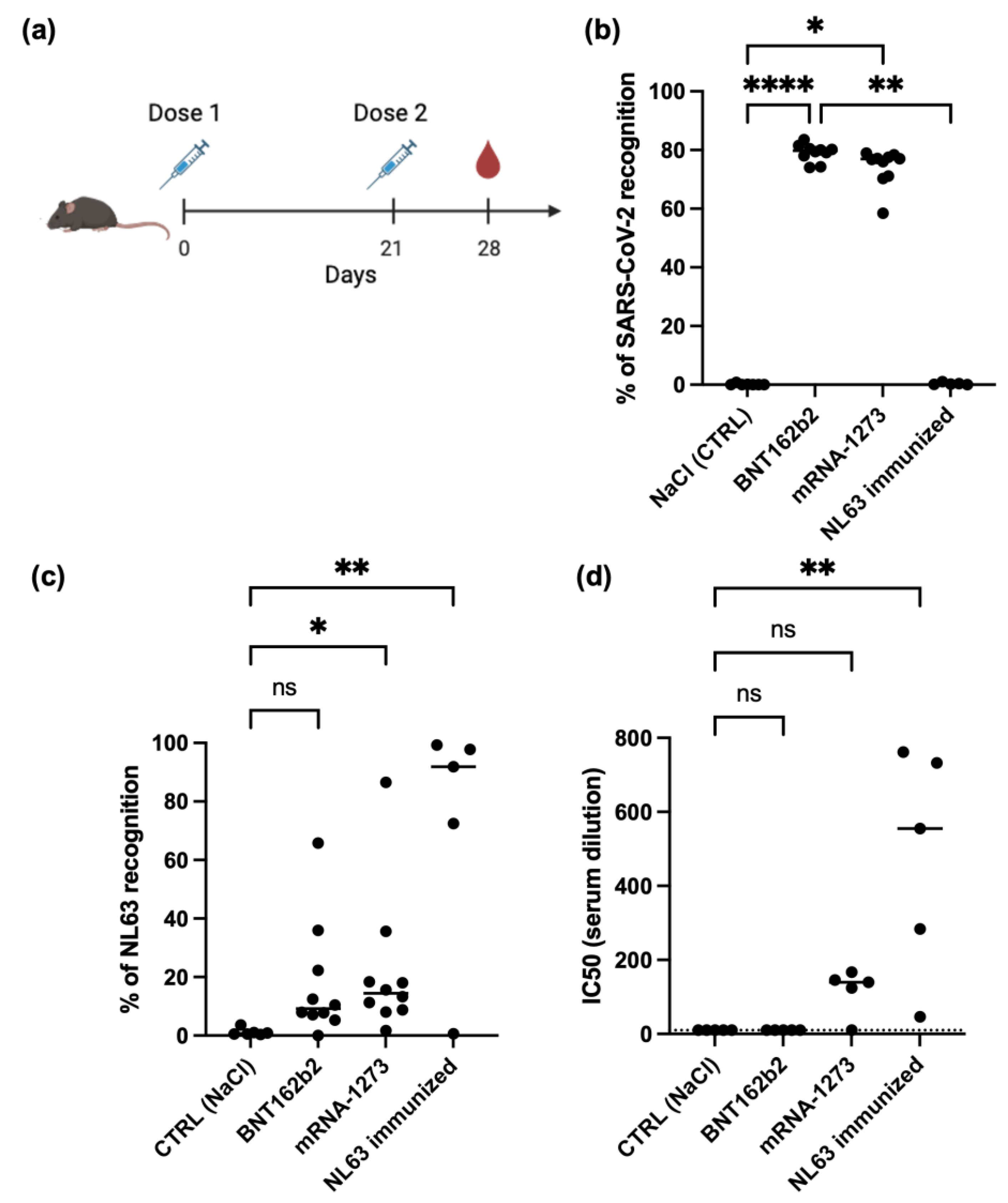
Statistical analyses were performed using GraphPad Prism 7 software. Kruskal-Wallis tests, followed by Dunn’s multiple comparison post hoc tests, were used for comparisons between multiple groups. Friedman tests, with Dunn’s multiple comparison post hoc tests, were used for comparisons between multiple time points. A p-value of less than 0.05 was considered statistically significant. Spearman’s correlation analysis was used to assess the correlation between antibody responses against the wild-type SARS-CoV-2 spike protein and the NL63 spike protein. Least squares regression was used to analyze the neutralization assay results.

3. Results

3.1. SARS-CoV-2 mRNA Vaccine Can Generate Cross-Reactive Antibody Against Human Seasonal Coronavirus NL63

We first conducted experiments in mice, as it is difficult to confirm the absence of prior exposure to seasonal coronaviruses like NL63 in adult humans. Mice were immunized with two doses of either the BNT162b2 or mRNA-1273 vaccine, with a 3-week interval between doses. Serum samples were collected 7 days after the second dose (Figure 1a). IgG antibody levels against the spike proteins of SARS-CoV-2 and NL63 were then measured using the SFB assay (Figure 1b) [13,14]. mRNA-1273 vaccination induced a significantly higher level of anti-NL63 spike antibodies (Figure 1c). This suggested that these vaccines might be capable of inducing cross-reactive and neutralizing antibodies against coronaviruses. While an increase in anti-NL63 spike antibodies was also observed after BNT162b2 vaccination, this increase was not statistically significant (p = 0.17). A live virus NL63 neutralization assay showed that the cross-reactive antibodies generated by mRNA-1273 vaccination had detectable albeit weak neutralizing activity against NL63 (Figure 1d).
Taken together, these results demonstrate that SARS-CoV-2 mRNA vaccination, particularly with mRNA-1273, can elicit a cross-reactive antibody response against NL63.

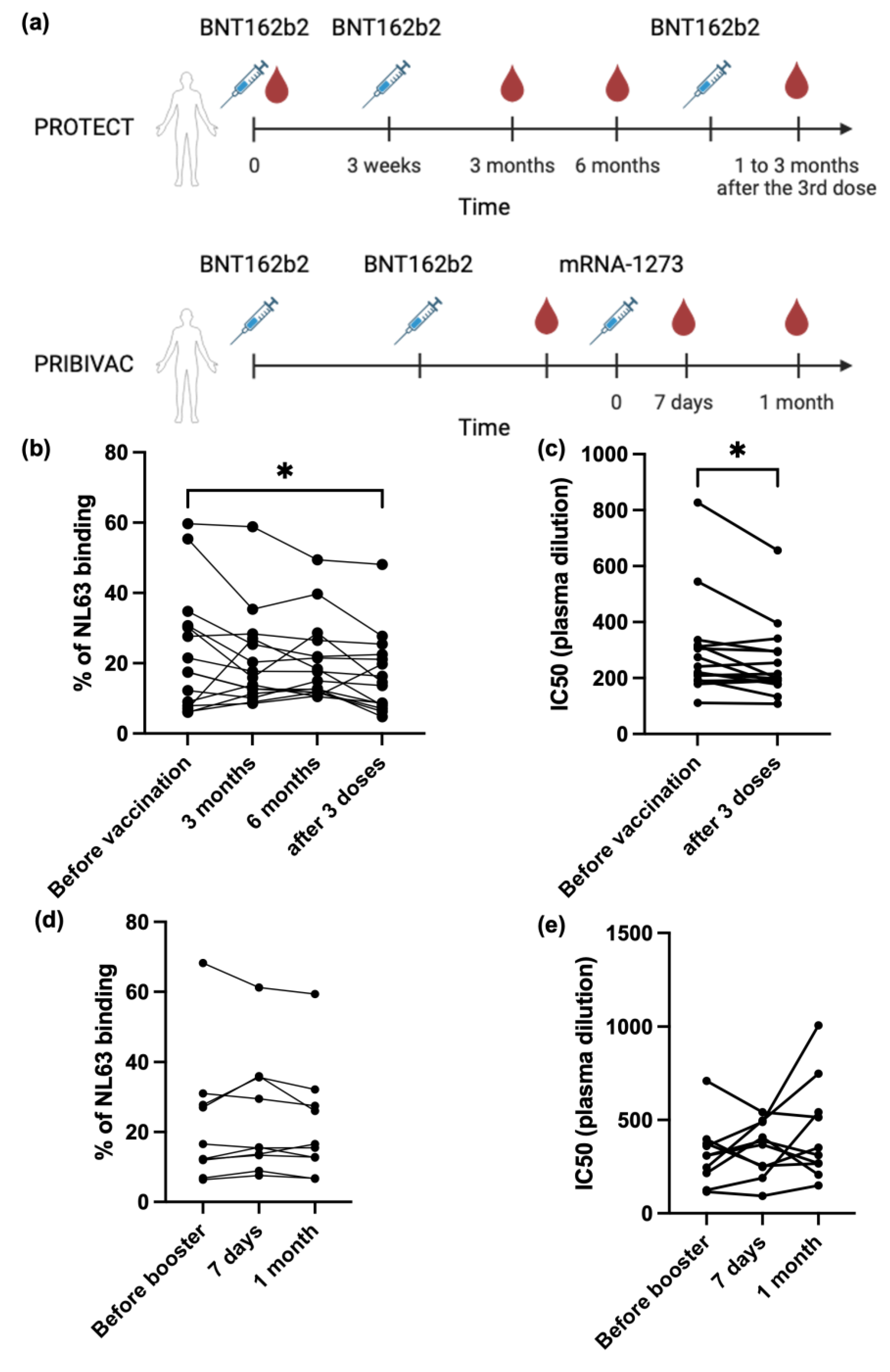
3.2. SARS-CoV-2 mRNA Vaccine Cannot Boost Pre-Existing NL63 Antibody Responses

Samples from 15 individuals who received three doses of the BNT162b2 mRNA vaccine were assessed for IgG antibody responses against NL63 using the SFB assay (Figure 2a). For the PROTECT group, the median age was 33 years (95% CI of median 29-39; range 23-44), and volunteers were predominately female (93.3%) and Filipino (73.3%). For the PRIBIVAC group, the median age is 36 years (95% CI of median 31-59; range 23-76), and volunteers were predominantly Chinese (86.7%). Pre-existing NL63 antibodies were prevalent in human plasma samples before vaccination, with a median spike recognition of 17.5% (Figure 2b). However, after two doses of BNT162b2, there was no significant change in anti-NL63 antibody levels. Furthermore, anti-NL63 levels decreased after the third BNT162b2 dose. This observation is consistent with the decline in neutralizing antibody titers against NL63 after three doses of BNT162b2 (Figure 2c). This suggests a waning of the anti-NL63 antibody response in absence of boosting of NL-63 exposure and further confirms that the BNT162b2 vaccine has no significant influence on anti-NL63 antibody responses.
In the next experiment, samples from 10 more individuals who had previously received two doses of the BNT162b2 vaccine and had received a booster dose of the mRNA-1273 vaccine (Figure 2a) were tested. Neither the antibody response against NL63 nor the NL63 neutralizing antibody titer changed after the mRNA-1273 booster dose (Figure 2d,e).

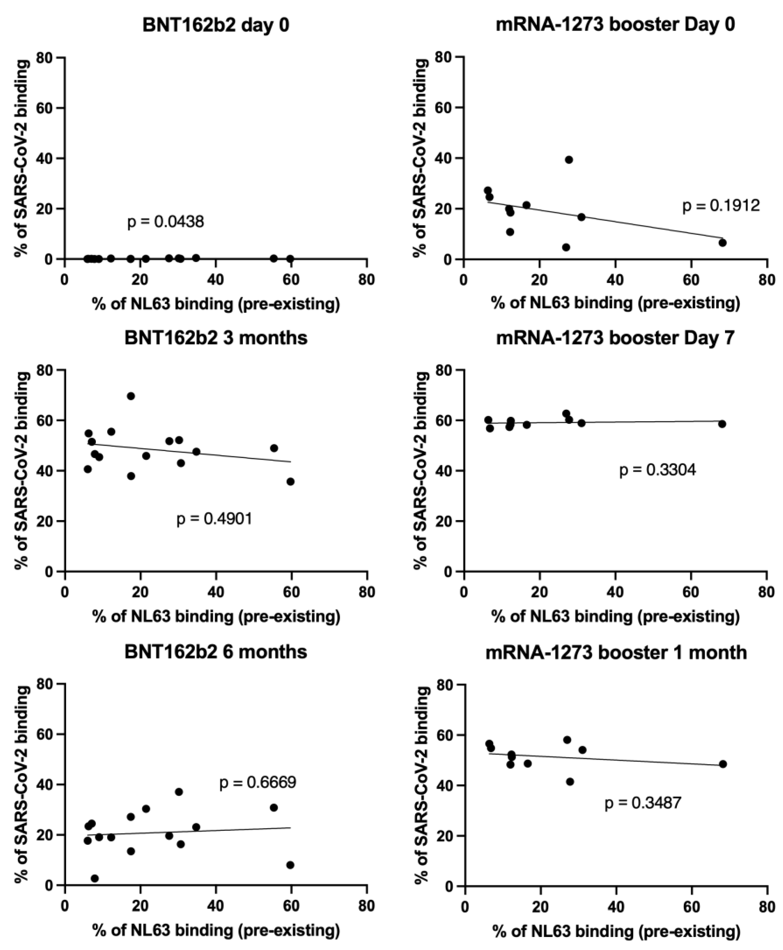
3.3. NL63 Pre-Existing Immune Response Did Not Affect Anti SARS-CoV-2 Antibody Response Imduced by Vaccination

To determine whether pre-existing antibody responses against NL63 affected SARS-CoV-2 vaccine immunogenicity, we analyzed the correlation between pre-existing anti-NL63 IgG levels and SARS-CoV-2 IgG antibody levels at different time points after vaccination with mRNA BNT162b2 vaccine alone or the prime-boost combination of the BNT162b2 vaccine followed by mRNA-1273. We found no significant correlation between NL63 and SARS-CoV-2 IgG levels (Figure 3), indicating that pre-existing NL63 antibody levels did not influence the priming, boosting, longevity, or waning of the antibody response in individuals receiving the BNT162b2 vaccine alone or the prime-boost combination of the BNT162b2 vaccine followed by mRNA-1273.

4. Discussion

Previous studies have shown that infections with the seasonal coronavirus NL63 are common in the human population [1]. These infections have been hypothesized to influence immune responses to subsequent SARS-CoV-2 infection or vaccination due to pre-existing antibodies binding to vaccine antigen and influencing processes such as antigen clearance and antigen presentation. Reciprocally, SARS-CoV-2 vaccination may also influence immune responses against seasonal coronaviruses (Table 1). Most current research has focused on cross-reactivity among individuals recovering from COVID-19 rather than on vaccination outcomes. According to published literature, SARS-CoV-2 infection generally does not generate cross-reactive antibodies against NL63 (Table 1). While some studies have reported higher anti-NL63 antibody levels in COVID-19 patients compared to healthy individuals, this effect is likely due to the boosting of pre-existing anti-NL63 memory responses rather than the generation of cross-reactive anti-SARS-CoV-2 antibodies. This is supported by the observation that there is no correlation between anti-SARS-CoV-2 and anti-NL63 antibody levels after infection [15].
To address these questions, we used two experimental approaches. First, we immunized mice with SARS-CoV-2 mRNA vaccines (both Pfizer and Moderna). This approach was chosen because most humans have been exposed to seasonal coronaviruses and may possess residual immune memory, even if they are seronegative. A recent study has shown that an adenovirus vaccine (FAdV-9-S19) generated low-level protection against NL63 and OC43 in K18-hACE2 mice [31]. Here, we found that immunization with both mRNA vaccines generated cross-reactive antibodies against the human seasonal coronavirus NL63. However, only the mRNA-1273 (Moderna) vaccine induced neutralizing antibodies. This suggests that the two mRNA vaccines elicit different antibody repertoires, with the mRNA-1273 vaccine inducing more cross-neutralizing antibodies against NL63.
Although these two vaccines share the same amino acid sequence for the spike protein [32], they differ in their lipid nanoparticle formulations and mRNA modifications. These differences may affect immunogenicity and the breadth of the antibody response, as has been seen previously when assessing breadth against variants of SARS-CoV-2 [33].
Our data also implies that these vaccines may also induce antibody responses against other human coronaviruses. Cross-reactive antibodies against various seasonal coronaviruses induced by SARS-CoV-2 viral infection have been demonstrated previously [32]. Conserved sequences possibly containing shared epitopes have been reported but their importance in anti-coronavirus immunity remains to be validated [32].
In the second set of experiments, we investigated the effect of SARS-CoV-2 mRNA vaccination on pre-existing anti-NL63 responses in humans. We observed that vaccination did not boost NL63 antibody responses, and that these anti-NL63 responses did not affect SARS-CoV-2 antibody level induced by vaccination. Previous studies have also reported that the immune imprinting of other coronaviruses was not boosted after vaccination of the inactivated COVID-19 vaccine [32], and that pre-existing immunity against seasonal coronaviruses had no negative effect for SARS-CoV-2 infection in mice [4]. One limitation of this study is that we did not have access to a cohort of mRNA-1273-only vaccinated humans. In our cohort, individuals received two BNT162b2 doses and one mRNA-1273 booster dose. However, it was reported elsewhere that two doses of mRNA-1273 vaccination can boost NL63 antibody response compared to one dose of vaccination [31]. Therefore, we postulate that one dose of mRNA-1273 booster vaccination dose is not sufficient to boost cross-reactive anti-NL63 response.
An additional limitation of this study is that only cross-reactive antibody response was evaluated. It was reported elsewhere that when T cell responses were assessed, SARS-CoV-2 vaccination or exposure boosted cross-reactive T cell responses against other coronaviruses (Table 1). One study showed that SARS-CoV-2 mRNA vaccination increased the T cell response to NL63 by 3 folds, while no responses to other seasonal coronaviruses were boosted [5]. Another study found that cross-reactive T cell responses were higher in seronegative health-care workers than healthy individuals, but the level decreased after SARS-CoV-2 infection, suggesting that exposure to SARS-CoV-2 might interfere with the cross-reactive T cell responses [35]. On the other hand, pre-existing T cell responses to SARS-CoV-2 have also been studied. These cross-reactive T cells widely existed before SARS-CoV-2 infection, and their epitopes can target various SARS-CoV-2 proteins, such as the spike, nucleocapsid and RTC [36,37,38,39]. Interestingly, one study identified the highly conserved Spike fusion peptide sequence as a T cell epitope, suggesting fusion peptide as a potential vaccine candidate to generate broad immune responses among coronaviruses for both B cell and T cell activation [38]. Another study identified S1200-1226 as the epitope for pre-existing cross-reactive T cells from healthy donors, which spanned across the heptad repeat 2 (HR2) and the transmembrane (TM) segment [41]. Additional studies in a small number of individuals suggested that pre-existing CD4+ and CD8+ T cells against alpha common cold coronavirus (229E) but no other seasonal coronaviruses were associated with protection from symptomatic and fatal SARS-CoV-2 infections in unvaccinated COVID-19 patients [40].

5. Conclusions

mRNA vaccines are effective tools against SARS-CoV-2 infections, but their ability for a broad protection have not been fully studied. The effect of BNT162b2 and mRNA-1273 vaccinations on the cross-reactive anti-NL63 were evaluated. In addition, the pre-existing anti-NL63 responses did not affect SARS-CoV-2 antibody level induced by SARS-CoV-2 vaccination.

Author Contributions

Conceptualization, W.T., L.R., L.N. and M.T.; methodology, W.T., Y.S.G., Z.W.C, C.W.L., P.X.H. and Y.J.T.; investigation, W.T., Y.S.G., Z.W.T, P.X.H., C.W.L., D.C.L., B.E.Y. and Y.T.; validation, W.T., Y.S.G., Z.W.C., C.W.L., P.X.H. and Y.T.; writing—original draft preparation, W.T.; writing—review and editing, W.T., L.R., Z.W.C, Y.S.G., C.W.L., P.X.H., D.C.L., B.E.Y., and M.T.; supervision, L.R., L.N. and M.T.; funding acquisition, B.E.Y., L.N. and L.R. NCID study group: Jocelyn Jin Yu, Zheng Kuang Soh, Yi Qing Chin, Jonathan Jordon Lim, Juwinda Ongko, Eshele Anak Libau, Celine Theo, Mohammed Ridzwan Bin Abdullah, Shiau Hui Diong, He Ping Yeo. COVID-19 cohort study group: Angeline Rouers, Matthew Zirui Tay, Anthony Torres-Ruesta, Nathan Wong, Yuling Huang, Alice Soh Meoy Ong, Adeline Chiew Yen Chua, Samantha Nguee, Yong Jie Tan, Vanessa Neo, Isaac Kai Jie Kam, Ajayanandan Yadunandan, Sooriya Kannan Selvam, Jarvis Goh, Ng Kah Ying, Sim Xin Yi, Wong Wei Lun, Anna Xinyi Loo, Liang Hui Loo. COVID clinicians’ team: Surinder Pada (Ng Teng Fong General Hospital, Singapore), Louisa Jin Sun (Infectious Diseases, Alexandra Hospital, Singapore), Desmond Luan Seng Ong (National University Polyclinic, Singapore), Jyoti Somani (Division of Infectious Diseases, Department of Medicine, National University Hospital, National University Health System, Singapore), Eng Sing Lee (National Healthcare Group Polyclinics, Singapore). All authors have read and agreed to the published version of the manuscript.

Funding

This study was funded by the Biomedical Research Council (BMRC), A*CRUSE (Vaccine monitoring project), A*ccelerate GAP-funded project (ACCL/19-GAP064-R20H-H) from Agency of Science, Technology and Research (A*STAR), Singapore National Medical Research Council (COVID19RF-001; COVID19RF-0011; COVID19RF-0018; COVID19RF-060; OFLCG19May-0034), US Food and Drug Administration (#75F40120C00085), and A*STAR COVID-19 Research funding (H/20/04/g1/006). YSG was supported by a Career Development Fund award by A*STAR (SC35/22-805100; #222D800035) and TF IPC Ltd under the grant titled “Temasek Foundation Infectious Diseases Programme for Surveillance and Diseases X Resilience”. LFPN was also supported by A*STAR Industry alignment Fund – Pre-positioning Programme (H22J1a0050). LR was also supported by a Start-up University Grant from Ministry of Health (#022388-00001). The funders did not have any role in the writing of the manuscript or the decision to submit it for publication.

Institutional Review Board Statement

Written informed consent was obtained from participants in accordance with the tenets of the Declaration of Helsinki. For COVID-19 serum/plasma collection, the study was proved by a domain specific review board (DSRB) under study number 2012/00917. The other cohort was recruited and enrolled at the National Centre for Infectious Diseases in Singapore (ClinicalTrials.gov identifier: NCT05142319). The animal study protocol conducted in accordance with the corresponding guidelines of National Advisory Committee for Laboratory Animal Research (NACLAR) and Animal & Veterinary Service (AVS), Singapore and approved by Biomedical Resource Centre (BRC), Singapore (IACUC #211623).

Informed Consent Statement

Informed consent was obtained from all subjects involved in the study. Written informed consent has been obtained from the patient(s) to publish this paper.

Data Availability Statement

All the data and materials can be found in the manuscript. Other requirements can be obtained by contacting the author.

Acknowledgments

We acknowledge all participants in this study.

Conflicts of Interest

A patent application for the SFB assay has been filed (Singapore patent #10202009679P: A Method of Detecting Antibodies and Related Products) by YSG, LR, and LFPN. All other authors declare no competing interests.

References

  1. Dijkman, R.; Jebbink, M.F.; El Idrissi, N.B.; Pyrc, K.; Muller, M.A.; Kuijpers, T.W.; Zaaijer, H.L.; van der Hoek, L. Human coronavirus NL63 and 229E seroconversion in children. J Clin Microbiol 2008, 46, 2368–2373. [CrossRef]
  2. Pyrc, K.; Berkhout, B.; Van Der Hoek, L. Antiviral strategies against human coronaviruses. IDDT 2007, 7, 59–66. [CrossRef]
  3. Milewska, A.; Nowak, P.; Owczarek, K.; Szczepanski, A.; Zarebski, M.; Hoang, A.; Berniak, K.; Wojarski, J.; Zeglen, S.; Baster, Z.; et al. Entry of human coronavirus NL63 into the cell. J Virol 2018, 92, e01933-17. [CrossRef]
  4. Amanat, F.; Clark, J.; Carreño, J.M.; Strohmeier, S.; Yellin, T.; Meade, P.S.; Bhavsar, D.; Muramatsu, H.; Sun, W.; Coughlan, L.; et al. Immunity to seasonal coronavirus spike proteins does not protect from SARS-CoV-2 challenge in a mouse model but has no detrimental effect on protection mediated by COVID-19 mRNA Vaccination. J Virol 2023, 97, e0166422. [CrossRef]
  5. Woldemeskel, B.A.; Garliss, C.C.; Blankson, J.N. SARS-CoV-2 mRNA vaccines induce broad CD4+ T Cell Responses that recognize SARS-CoV-2 variants and HCoV-NL63. J Clin Invest 2021, 131, e149335. [CrossRef]
  6. Hu, C.; Wang, Z.; Ren, L.; Hao, Y.; Zhu, M.; Jiang, H.; Wang, S.; Li, D.; Shao, Y. Pre-Existing Anti-HCoV-OC43 immunity influences the durability and cross-reactivity of humoral response to SARS-CoV-2 vaccination. Front Cell Infect Microbiol 2022, 12, 978440. [CrossRef]
  7. Murray, S.M.; Ansari, A.M.; Frater, J.; Klenerman, P.; Dunachie, S.; Barnes, E.; Ogbe, A. The Impact of pre-Existing cross-reactive immunity on SARS-CoV-2 infection and vaccine responses. Nat Rev Immunol 2023, 23, 304–316. [CrossRef]
  8. Adami, F.L.; de Castro, M.V.; Almeida, B. da S.; Daher, I.P.; Yamamoto, M.M.; Souza Santos, K.; Zatz, M.; Naslavsky, M.S.; Rosa, D.S.; Cunha-Neto, E.; et al. Anti-RBD IgG antibodies from endemic coronaviruses do not protect against the acquisition of SARS-CoV-2 infection among exposed uninfected individuals. Front Immunol 2024, 15. [CrossRef]
  9. Shrwani, K.; Sharma, R.; Krishnan, M.; Jones, T.; Mayora-Neto, M.; Cantoni, D.; Temperton, N.J.; Dobson, S.L.; Subramaniam, K.; McNamara, P.S.; et al. Detection of serum cross-reactive antibodies and memory response to SARS-CoV-2 in prepandemic and post–COVID-19 convalescent samples. J Infect Dis 2021, 224, 1305–1315. [CrossRef]
  10. Renia, L.; Goh, Y.S.; Rouers, A.; Le Bert, N.; Chia, W.N.; Chavatte, J.-M.; Fong, S.-W.; Chang, Z.W.; Zhuo, N.Z.; Tay, M.Z.; et al. Lower vaccine-acquired immunity in the elderly population following two-dose BNT162b2 vaccination is alleviated by a third vaccine dose. Nat Commun 2022, 13, 4615. [CrossRef]
  11. Poh, X.Y.; Tan, C.W.; Lee, I.R.; Chavatte, J.-M.; Fong, S.-W.; Prince, T.; Hartley, C.; Yeoh, A.Y.Y.; Rao, S.; Chia, P.Y.; et al. Antibody response of heterologous vs homologous messenger RNA vaccine boosters against the severe acute respiratory syndrome coronavirus 2 omicron variant: interim results from the PRIBIVAC study, a randomized clinical trial. Clin Inf Dis 2022, 75, 2088–2096. [CrossRef]
  12. Poh, X.Y.; Lee, I.R.; Lim, C.; Teo, J., Rao, S.; Chia, P.Y.; Ong, S.W.X.; Lee, T.H.; Lin, R.J.H.; Ng, L.F.P.; et al. Evaluation of the safety and immunogenicity of different COVID-19 vaccine combinations in healthy individuals: study protocol for a randomized, subject-blinded, controlled phase 3 trial [PRIBIVAC]. Trials 2022, 23, 498. [CrossRef]
  13. Goh, Y.S.; Ng, L.F.P.; Renia, L. A Flow cytometry-based assay for serological detection of anti-spike antibodies in COVID-19 patients. STAR Protoc 2021, 2, 100671. [CrossRef]
  14. Goh, Y.S.; Chavatte, J.M.; Lim, J.A.; Lee, B.; Hor, P.X.; Amrun, S.N.; Lee, C.Y., Chee, R.S.; Wang, B., Lee, C.Y.; et al. Sensitive detection of total anti-spike antibodies and isotype switching in asymptomatic and symptomatic individuals with COVID-19. Cell Rep Med 2021, 16, 100193. [CrossRef]
  15. Grobben, M.; van der Straten, K.; Brouwer, P.J.; Brinkkemper, M.; Maisonnasse, P.; Dereuddre-Bosquet, N.; Appelman, B.; Lavell, A.A.; van Vught, L.A.; Burger, J.A.; et al. Cross-reactive antibodies after SARS-CoV-2 infection and vaccination. eLife 2021, 10, e70330. [CrossRef]
  16. Imai, K.; Matsuoka, M.; Tabata, S.; Kitagawa, Y.; Nagura-Ikeda, M.; Kubota, K.; Fukada, A.; Takada, T.; Sato, M.; Noguchi, S.; et al. Cross-reactive humoral immune responses against seasonal human coronaviruses in COVID-19 patients with different disease severities. Int J Infect Dis 2021, 111, 68–75. [CrossRef]
  17. Denninger, V.; Xu, C.K.; Meisl, G.; Morgunov, A.S.; Fiedler, S.; Ilsley, A.; Emmenegger, M.; Malik, A.Y.; Piziorska, M.A.; Schneider, M.M.; et al. Microfluidic antibody affinity profiling reveals the role of memory reactivation and cross-reactivity in the defense against SARS-CoV-2. ACS Infect Dis 2022, 8, 790–799. [CrossRef]
  18. Pattinson, D.; Jester, P.; Guan, L.; Yamayoshi, S.; Chiba, S.; Presler, R.; Rao, H.; Iwatsuki-Horimoto, K.; Ikeda, N.; Hagihara, M.; et al. A novel method to reduce ELISA serial dilution assay workload applied to SARS-CoV-2 and seasonal HCoVs. Viruses 2022, 14, 562. [CrossRef]
  19. Adams, O.; Andrée, M.; Rabl, D.; Ostermann, P.N.; Schaal, H.; Lehnert, E.; Ackerstaff, S.; Müller, L.; Fischer, J.C. Humoral response to SARS-CoV-2 and seasonal coronaviruses in COVID-19 patients. J Med Virol 2022, 94, 1096–1103. [CrossRef]
  20. Guo, L.; Wang, Y.; Kang, L.; Hu, Y.; Wang, L.; Zhong, J.; Chen, H.; Ren, L.; Gu, X.; Wang, G.; et al. Cross-reactive antibody against human coronavirus OC43 spike protein correlates with disease severity in COVID-19 patients: a retrospective study. Emerg Microb Infect 2021, 10, 664–676. [CrossRef]
  21. Iyer, A.S.; Jones, F.K.; Nodoushani, A.; Kelly, M.; Becker, M.; Slater, D.; Mills, R.; Teng, E.; Kamruzzaman, M.; Garcia-Beltran, W.F.; et al. Persistence and decay of human antibody responses to the receptor binding domain of SARS-CoV-2 spike protein in COVID-19 patients. Sci Immunol 2020, 5, eabe0367. [CrossRef]
  22. Wells, D.A.; Cantoni, D.; Mayora-Neto, M.; Genova, C.D.; Sampson, A.; Ferrari, M.; Carnell, G.; Nadesalingam, A.; Smith, P.; Chan, A.; et al. Human seasonal coronavirus neutralization and COVID-19 severity. J Med Virol 2022, 94, 4820–4829. [CrossRef]
  23. Camerini, D.; Randall, A.Z.; Trappl-Kimmons, K.; Oberai, A.; Hung, C.; Edgar, J.; Shandling, A.; Huynh, V.; Teng, A.A.; Hermanson, G.; et al. Mapping SARS-CoV-2 antibody epitopes in COVID-19 patients with a multi-coronavirus protein microarray. Microbiol Spectr 2021, 9, e01416-21. [CrossRef]
  24. Struck, F.; Schreiner, P.; Staschik, E.; Wochinz-Richter, K.; Schulz, S.; Soutschek, E.; Motz, M.; Bauer, G. Incomplete IgG avidity maturation after seasonal coronavirus infections. J Med Virol 2022, 94, 186–196. [CrossRef]
  25. Peng, Y.; Liu, Y.; Hu, Y.; Chang, F.; Wu, Q.; Yang, J.; Chen, J.; Teng, S.; Zhang, J.; He, R.; et al. Monoclonal Antibodies constructed from COVID-19 convalescent memory B cells exhibit potent binding activity to MERS-CoV spike S2 subunit and other human coronaviruses. Front Immunol 2022, 13. [CrossRef]
  26. Lin, C.-Y.; Wolf, J.; Brice, D.C.; Sun, Y.; Locke, M.; Cherry, S.; Castellaw, A.H.; Wehenkel, M.; Crawford, J.C.; Zarnitsyna, V.I.; et al. Pre-Existing Humoral Immunity to human common cold coronaviruses negatively impacts the protective SARS-CoV-2 antibody response. Cell Host Microbe 2022, 30, 83-96.e4. [CrossRef]
  27. Wang, J.; Li, D.; Zhou, Q.; Wiltse, A.; Zand, M.S. Antibody mediated immunity to SARS-CoV-2 and human coronaviruses: multiplex beads assay and volumetric absorptive microsampling to generate immune repertoire cartography. Front Immunol 2021, 12. [CrossRef]
  28. Stanley, A.M.; Aksyuk, A.A.; Wilkins, D.; Green, J.A.; Lan, D.; Shoemaker, K.; Tieu, H.-V.; Sobieszczyk, M.E.; Falsey, A.R.; Kelly, E.J. Seasonal human coronavirus humoral responses in AZD1222 (ChaAdOx1 nCoV-19) COVID-19 vaccinated adults reveal limited cross-immunity. Front Immunol 2024, 15. [CrossRef]
  29. Kolehmainen, P.; Huttunen, M.; Iakubovskaia, A.; Maljanen, S.; Tauriainen, S.; Yatkin, E.; Pasternack, A.; Naves, R.; Toivonen, L.; Tähtinen, P.A.; et al. Coronavirus spike protein-specific antibodies indicate frequent infections and reinfections in infancy and among BNT162b2-vaccinated healthcare workers. Sci Rep 2023, 13, 8416. [CrossRef]
  30. Amanat, F.; Thapa, M.; Lei, T.; Ahmed, S.M.S.; Adelsberg, D.C.; Carreño, J.M.; Strohmeier, S.; Schmitz, A.J.; Zafar, S.; Zhou, J.Q.; et al. SARS-CoV-2 mRNA vaccination induces functionally diverse antibodies to NTD, RBD, and S2. Cell 2021, 184, 3936-3948.e10. [CrossRef]
  31. Cantoni, D.; Siracusano, G.; Mayora-Neto, M.; Pastori, C.; Fantoni, T.; Lytras, S.; Di Genova, C.; Hughes, J.; Lopalco, L.; Temperton, N. Analysis of antibody neutralisation activity against SARS-CoV-2 variants and seasonal human coronaviruses NL63, HKU1, and 229E induced by three different COVID-19 vaccine platforms. Vaccines 2022, 11, 58. [CrossRef]
  32. Asamoah-Boaheng, M.; Grunau, B.; Karim, M.E.; Jassem, A.N.; Bolster, J.; Marquez, A.C.; Scheuermeyer, F.X.; Goldfarb, D.M. Are higher antibody levels against seasonal human coronaviruses associated with a more robust humoral immune response after SARS-CoV-2 vaccination? Front Immunol 2022, 13. [CrossRef]
  33. Singh, G.; Abbad, A.; Kleiner, G.; Srivastava, K.; Gleason, C.; Group, P.S.; Carreño, J.M.; Simon, V.; Krammer, F. The Post-COVID-19 population has a high prevalence of cross-reactive antibodies to spikes from all orthocoronavirinae genera. mBio 2024. [CrossRef]
  34. Jacob-Dolan, C.; Feldman, J.; McMahan, K.; Yu, J.; Zahn, R.; Wegmann, F.; Schuitemaker, H.; Schmidt, A.G.; Barouch, D.H. coronavirus-specific antibody cross reactivity in rhesus macaques following SARS-CoV-2 vaccination and infection. J Virol 2021, 95, 10.1128/jvi.00117-21. [CrossRef]
  35. Wirsching, S.; Harder, L.; Heymanns, M.; Gröndahl, B.; Hilbert, K.; Kowalzik, F.; Meyer, C.; Gehring, S. Long-Term, CD4+ memory t cell response to SARS-CoV-2. Front Immunol 2022, 13. [CrossRef]
  36. Soni, M.K.; Migliori, E.; Fu, J.; Assal, A.; Chan, H.T.; Pan, J.; Khatiwada, P.; Ciubotariu, R.; May, M.S.; Pereira, M.R.; et al. The prospect of universal coronavirus immunity: characterization of reciprocal and non-reciprocal T cell responses against SARS-CoV2 and common human coronaviruses. Front Immunol 2023, 14. [CrossRef]
  37. Lineburg, K.E.; Grant, E.J.; Swaminathan, S.; Chatzileontiadou, D.S.M.; Szeto, C.; Sloane, H.; Panikkar, A.; Raju, J.; Crooks, P.; Rehan, S.; et al. CD8+ T cells specific for an immunodominant SARS-CoV-2 nucleocapsid epitope cross-react with selective seasonal coronaviruses. Immunity 2021, 54, 1055-1065.e5. [CrossRef]
  38. Naghibosadat, M.; Babuadze, G.G.; Pei, Y.; Hurst, J.; Salvant, E.; Gaete, K.; Biondi, M.; Moloo, B.; Goldstein, A.; Avery, S.; et al. Vaccination against SARS-CoV-2 provides low-level cross-protection against common cold coronaviruses in mouse and non-human primate animal models. J Virol 2025, 0, e01390-24. [CrossRef]
  39. Klompus, S.; Leviatan, S.; Vogl, T.; Mazor, R.D.; Kalka, I.N.; Stoler-Barak, L.; Nathan, N.; Peres, A.; Moss, L.; Godneva, A.; et al. Cross-reactive antibodies against human coronaviruses and the animal coronavirome suggest diagnostics for future zoonotic spillovers. Sci Immunol 2021, 6, eabe9950. [CrossRef]
  40. Devaux, C.A.; Fantini, J. Unravelling antigenic cross-reactions toward the world of coronaviruses: extent of the stability of shared epitopes and SARS-CoV-2 anti-spike cross-neutralizing antibodies. Pathogens 2023, 12, 713. [CrossRef]
  41. Yin, D.; Han, Z.; Lang, B.; Li, Y.; Mai, G.; Chen, H.; Feng, L.; Chen, Y.; Luo, H.; Xiong, Y.; et al. Effect of seasonal coronavirus immune imprinting on the immunogenicity of inactivated COVID-19 vaccination. Front Immunol 2023, 14. [CrossRef]
  42. da Silva Antunes, R.; Pallikkuth, S.; Williams, E.; Dawen Yu, E.; Mateus, J.; Quiambao, L.; Wang, E.; Rawlings, S.A.; Stadlbauer, D.; Jiang, K.; et al. Differential T-cell reactivity to endemic coronaviruses and SARS-CoV-2 in community and health care workers. J Infect Dis 2021, 224, 70–80. [CrossRef]
  43. Loyal, L.; Braun, J.; Henze, L.; Kruse, B.; Dingeldey, M.; Reimer, U.; Kern, F.; Schwarz, T.; Mangold, M.; Unger, C.; et al. Cross-Reactive CD4+ T Cells enhance SARS-CoV-2 immune responses upon infection and vaccination. Science 2021, 374, eabh1823. [CrossRef]
  44. Le Bert, N.; Tan, A.T.; Kunasegaran, K.; Tham, C.Y.L.; Hafezi, M.; Chia, A.; Chng, M.H.Y.; Lin, M.; Tan, N.; Linster, M.; et al. SARS-CoV-2-Specific T cell immunity in cases of COVID-19 and SARS, and uninfected controls. Nature 2020, 584, 457–462. [CrossRef]
  45. Swadling, L.; Diniz, M.O.; Schmidt, N.M.; Amin, O.E.; Chandran, A.; Shaw, E.; Pade, C.; Gibbons, J.M.; Le Bert, N.; Tan, A.T.; et al. Pre-existing polymerase-specific t cells expand in abortive seronegative SARS-CoV-2. Nature 2022, 601, 110–117. [CrossRef]
  46. Shimizu, K.; Iyoda, T.; Sanpei, A.; Nakazato, H.; Okada, M.; Ueda, S.; Kato-Murayama, M.; Murayama, K.; Shirouzu, M.; Harada, N.; et al. Identification of TCR repertoires in functionally competent cytotoxic t cells cross-reactive to SARS-CoV-2. Commun Biol 2021, 4, 1365. [CrossRef]
  47. Coulon, P.-G.; Prakash, S.; Dhanushkodi, N.R.; Srivastava, R.; Zayou, L.; Tifrea, D.F.; Edwards, R.A.; Figueroa, C.J.; Schubl, S.D.; Hsieh, L.; et al. High frequencies of alpha common cold coronavirus/SARS-CoV-2 cross-reactive functional CD4+ and CD8+ memory T cells are associated with protection from symptomatic and fatal SARS-CoV-2 infections in unvaccinated COVID-19 patients. Front Immunol 2024, 15, 1343716. [CrossRef]
Figure 1. Cross-reactive anti-SARS-CoV-2 antibodies against NL063 induced by mRNA vaccines. (a) Mouse vaccination regime. (b and c) Antibody responses against SARS-CoV-2 WT (b) and NL63 (c) measured by the SFB assay. Serum samples were collected after immunization with BNT162b2 (n = 10), mRNA-1273 (n = 10), heat-inactivated NL063 virus (n = 5) or NaCl as control (n = 5). Data are expressed as the percentage of SARS-CoV-2 or NL63 spike protein recognition, which corresponds to the percentage of cells expressing the SARS-CoV-2 or NL63 spike proteins that are recognized by antibodies specific for these proteins. (d) Neutralization antibody titers against NL063 in immunized mouse sera (n = 5). Median values are indicated by the line. ns: not significant, *: p < 0.05, **: p<0.01, ****: p<0.0001; by Kruskal-Wallis test.
Preprints 150077 g001
Figure 2. No boosting effect of NL63 antibody after SARS-CoV-2 mRNA vaccination. (a) Vaccination regime for individuals recruited in this study. (b and d) Antibody response against NL63 for the PROTECT (b) (n = 15) and PRIBIVAC (d) (n = 10) group measured by the SFB assay. Data are expressed as the percentage of NL63 spike protein recognition, which corresponds to the percentage of cells expressing the NL63 spike proteins that are recognized by antibodies specific for these proteins. (c and e) Neutralization antibody titer against NL63 in the PROTECT (c) (n = 15) and PRIBIVAC (e) (n = 10) group. *: p < 0.05; by Kruskal-Wallis test (b) or Wilcoxon test (c).
Preprints 150077 g002
Figure 3. Correlation between pre-existing NL63 antibody levels and SARS-CoV-2 antibody levels after BNT162b2 vaccination (n = 15) or mRNA-1273 booster (n = 10). No response to SARS-CoV-2 was observed on day 0 for BNT162b2, and hence it is not included in the analysis. The p value was calculated by Spearman correlation.
Preprints 150077 g003
Table 1. Previous studies on whether SARS-CoV-2 infection or vaccination affects NL63 antibody response.
Experiment group Control group Antibody or T cells? Tested antigen on NL63 Result Ref
PCR-positive PCR-negative close contacts IgG antibody RBD No difference [8]
PCR-positive Pre-COVID samples IgG antibody RBD No difference [9]
COVID-19 severe COVID-19 mild IgG antibody S and N No difference [16]
COVID-19 convalescents Healthy individuals IgG antibody S1 No difference [17]
COVID-19 convalescents Healthy individuals IgG antibody S and N No difference [18]
COVID-19 convalescents Healthy individuals IgG antibody N No difference [19]
COVID-19 convalescents Healthy individuals IgG antibody S No difference [20]
COVID-19 convalescents Healthy individuals IgG antibody RBD No difference [21]
COVID-19 patients Healthy individuals IgG antibody Pseudovirus Higher neutralization [22]
COVID-19 convalescents Pre-COVID samples IgG antibody S2 and N Higher response [23]
COVID-19 convalescents Healthy individuals IgG antibody N Higher response [24]
COVID-19 convalescents COVID-19 admission IgG antibody S and N Higher against N in severe patients [25]
COVID-19 convalescents Healthy individuals IgG antibody S Higher response [15]
COVID-19 convalescents Healthy individuals IgG mAbs S 3 strongly cross-reactive mAbs [25]
Health care workers with direct/indirect contact to COVID-19 patients Health care workers with no contact to COVID-19 patients IgG, IgM and IgA antibody S Higher IgM between direct and no contact group [26]
COVID-19 convalescents or BNT162b2 vaccination (2 doses) Pre-COVID samples IgG antibody S and N No difference [27]
AZD1222 primary vaccination and/or booster Placebo IgG antibody S No difference [28]
BNT162b2 vaccination (2 and 3 doses) Before vaccination (same individuals) IgG antibody S1 No difference [29]
BNT162b2 vaccination (2 doses) Before vaccination (same individuals) IgG antibody S No difference [30]
AZD1222, BNT162b2 or mRNA-1273 vaccinated (2 doses) AZD1222, BNT162b2 or mRNA-1273 vaccinated (1 dose) IgG antibody Pseudovirus Higher neutralization for mRNA-1273 [31]
BNT162b2 or mRNA-1273 Healthy individuals IgG antibody S No difference [32]
BNT162b2 or mRNA-1273 (2 doses) Before vaccination (same individuals) IgG antibody S No difference [33]
After 1-dose vaccination (did not mention what vaccine) Before vaccination (same individuals) IgG antibody Pseudovirus Higher neutralization [22]
BBIBP-CorV vaccinated (2 doses) Before vaccination (same individuals) IgG antibody S Higher response [6]
Infected, Ad26-vaccinated or DNA-vaccinated + re-infected macaques Before treatment (same animals) IgG antibody S Higher response [33]
COVID-19 convalescents Healthy individuals CD4+ T cells Pool peptide library S1 and S2 No difference [34]
COVID exposed individuals Healthy individuals CD4+ T cells Pool peptide library S1, S2, M and NP Higher against S1, M and NP [35]
BNT162b2 or mRNA-1273 vaccinated Healthy individuals CD4+ T cells Pool peptide library (whole virus) Higher response [36]
COVID-19 convalescents Healthy individuals CD8+ T cells Homologous N105–113 (PPKVHFYYL) No difference [37]
Disclaimer/Publisher’s Note: The statements, opinions and data contained in all publications are solely those of the individual author(s) and contributor(s) and not of MDPI and/or the editor(s). MDPI and/or the editor(s) disclaim responsibility for any injury to people or property resulting from any ideas, methods, instructions or products referred to in the content.
Copyright: This open access article is published under a Creative Commons CC BY 4.0 license, which permit the free download, distribution, and reuse, provided that the author and preprint are cited in any reuse.
Prerpints.org logo

Preprints.org is a free preprint server supported by MDPI in Basel, Switzerland.

Subscribe

Disclaimer

Terms of Use

Privacy Policy

Privacy Settings

© 2025 MDPI (Basel, Switzerland) unless otherwise stated