Submitted:
18 February 2025
Posted:
18 February 2025
You are already at the latest version
Abstract
Targeted nanomaterials are at the forefront of advancements in nanomedicine due to their unique and versatile properties. These include nanoscale size, shape, surface chemistry, mechanical flexibility, fluorescence, optical behaviour, magnetic, and electronic characteristics as well as biocompatibility and biodegradability. These attributes enable their application across diverse fields such as drug delivery, bioimaging, sensing, disease diagnostics, tissue engineering, cosmetics, and electronics. This review explores the fundamental characteristics of nanomaterials and emphasize their importance into clinical applications. It further delves into methodologies for nanoparticles programming alongside discussions on clinical trials and case studies. We discussed some of promising nanomaterials such as polymeric nanoparticles, carbon-based nanoparticles and metallic nanoparticles with their role in biomedical applications. The review underscores significant advancements in translating nanomaterials into clinical applications and highlight the potential of these innovative approaches in revolutionizing the medical field.
Keywords:
Nanomaterials
; Clinical Applications
; Drug Delivery
1. Introduction
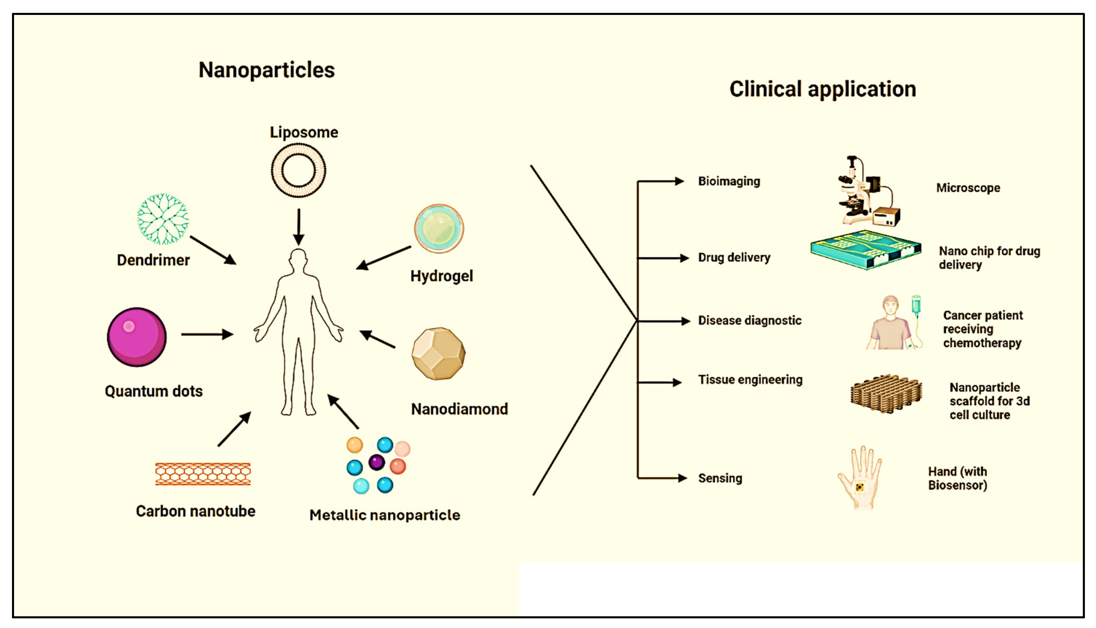
In recent years, nanotechnology has become popular and positively influenced preclinical medical development in medicine and shipping in the emerging field of nanomedicine. Today, nanomaterials formulate Nano-systems and nanocarriers that help drugs deliver to the target site and cure diseases. Based on the use and applications of nanoparticles (NPs) on a nanoscale, there are serval benefits and applications, including bioimaging, drug delivery [1], regenerative medicine [2], cosmetics [3], electrochemical DNA detection [4], polishing [5], energy storage [6], sunscreen protrction [7], and sensing [8] (Figure 1).
Figure 1.
Different types of nanomaterials and their clinical applications.
The nanoscale dimensions of these particles have unique and advantageous properties, chemical, physical, and biological domains, which can surpass those of their larger equivalent [9]. Nanoscale medicines are especially beneficial since various significant biological molecules, including water, enzymes, haemoglobin, antibodies, proteins, glucose, and receptors, fall within this size spectrum [10]. Various essential nanomaterial platforms for biological uses, such as drug delivery and cancer treatment, have been created and examined [11]. Nanoparticles, standards under the nanoscale, can transport various payloads, including small molecular drugs, nucleic acids, proteins, imaging agents, and other substances [12]. Nanoparticles are specifically programmed to enhance the safety and efficacy of drug delivery [13,14]. Engineered nanoparticles offer potential in disease diagnosis and treatment but face biological barriers. Advances in nanoparticle engineering and understanding their properties create new therapeutic opportunities [15]. Pharmaceutical nanocarriers like liposomes and micelles exhibit longevity, targeting, penetration, and contrast properties. Multifunctional nanocarriers could significantly enhance therapeutic and diagnostic efficacy [16]. Potential nanotechnological carrier platforms include quantum dots, dendrimers, polymers, carbon nanotubes, organic NPs, metallic NPs, liposomes, nanogels, and peptide NPs.
NPs smaller than 5 nm are easily eliminated from the systemic flow, while those bigger than 10 nm tend to be added to the bloodstream. Expanding nanomedicine from small-scale to large-scale production while maintaining stability presents notable challenges [17]. PEG is a polymer with chains that resist protein absorption, but subsequent opsonization and macrophage clearance still occur. Liposomes with albumin attached to their surface demonstrate longer circulation times than unmodified or PEGylated versions [18]. Cationic polymers encapsulate siRNA, increasing cellular uptake despite their toxicity, thus overcoming barriers to targeted drug delivery [19]. Liposomes with stimuli-responsive characteristics like light, pH, temperature, enzymatic reaction, ultrasound, or radiation are effective for multimodal nanoscale functions [20]. Cationic PAMAM dendrimers interact with negatively charged molecules, showing toxicity, but zwitterionic dendrimers with phosphorylcholine surfaces reduce cytotoxicity [21]. Hydrogels’ ability to stretch and swell makes them highly biocompatible, playing a crucial role in medical applications [22]. Gene delivery transplants genetic material to regulate and function by removing unwanted genes and introducing the desired ones [23]. QDs conjugated with proteins and biotin functionalization enable specific binding, cellular uptake, and intracellular imaging [24,25]. Metallic nanoparticles (MNPs) used in cancer detection can conjugate with recognition molecules like antibodies, providing outstanding sensitivity [26]. Tian et al. synthesized biocompatible gold nanoparticles with red fluorescence using egg whites and microwaves, which exhibited fluorescence in the presence of viable cancer cells and showed potential for cancer cell detection [27].
Clinical trials are research studies that test new treatments and their effects on human health. Volunteers participate in these trials, including testing drugs, devices, and procedures. There are four phases of clinical trials. Phase I tests safety and dosage on a small group. Phase II expands the group to monitor side effects. Phase III involves large populations across regions to confirm effectiveness. Phase IV occurs post-approval to gather long-term safety data [28]. Examples include the phase III trials of albumin-bound paclitaxel for metastatic breast cancer, pegylated liposomal doxorubicin for various cancers, patisiran for hereditary transthyretin amyloidosis, and nab-paclitaxel plus gemcitabine for pancreatic cancer.
CALAA-01, the first targeted nanoparticle (NP) for siRNA delivery, completed preclinical trials and entered human clinical trials. Designed for systemic nucleic acid administration in cancer therapy, it uses cyclodextrin-containing polymer (CDP) to form nanoparticles (~70 nm) [29]. These NPs are functionalized for stability, targeting, and endosomal escape. The two-vial formulation allows rapid self-assembly at the point of care, protecting siRNA from degradation. In murine models, CALAA-01 demonstrated effective gene targeting and tumour inhibition without eliciting immune responses.
This review covers the historical background and characteristics of nanoparticles (NPs), requirements for programming NPs, various types of nanoparticles, clinical trials and case studies, their clinical applications, and challenges in NPs programming. We examine the evolution of nanoparticle technology, the diverse types and their uses in medicine, and the technical hurdles faced in optimizing NPs functionality.
2. Historical Background of Programmable Nanomaterials
In 1950, The first synthesis of the molecule that is considered a nanoparticle and conjugates with a polymer-drug, for example- formulated a polyvinylpyrrolidone mescaline conjugate compound that consists of a small peptide spacer between polymer and drug [30]. In 1970s, Ringsdorf gave a theory about the conjugation of target drugs, describing the key principles of conjugating the drug molecule (Figure 2). In 1972, albumin-based nanoparticles were reported [31,32]. This is the first protein-built regulatory approved albumin-bound paclitaxel that is accepted by the US Food and Drug Administration (FDA), used for medication for breast cancer [32]. The second-gen polymer-based NPs were invented in 1776 [33]. In 1980, the polymer-drug poly (styrene-co-maleic acid) was conjugated with the cytotoxic drug neocarzinostain. Compared to free neocarzinostatin, the SMANCS conjugate demonstrated greater effectiveness at the tumor site due to the enhanced permeability and retention (EPR) effect [34]. The first NP-based drug was permitted by the FDA in 1980. Cytotoxic anticancer medication paclitaxel (Taxol; Bristol-Myers Squibb) is also made with cremophor [35].In 1989, goserelin acetate, in an implantable form, became the first controlled-release polymer component to receive FDA approval. Marketed by AstraZeneca under the brand name Zoladex, this synthetic luteinizing hormone-releasing hormone analogue is used to treat certain forms of breast and prostate cancer [35]. These controlled-release polymer-drug components, called “drug depots”, permit a medication or other molecule to be encapsulated inside a polymer matrix and gradually released as it diffuses out over an extended period. This innovative approach was pioneered in Langer’s laboratory [36]. Beginning with non-degradable polymer matrices in initial research, Langer’s laboratory advanced to develop a drug, including a biodegradable depot form of the cytotoxic drug. In 1996, the FDA approved this breakthrough for the treatment of brain cancer [37].
Figure 2.
Historical progress in nanoparticles based therapies.
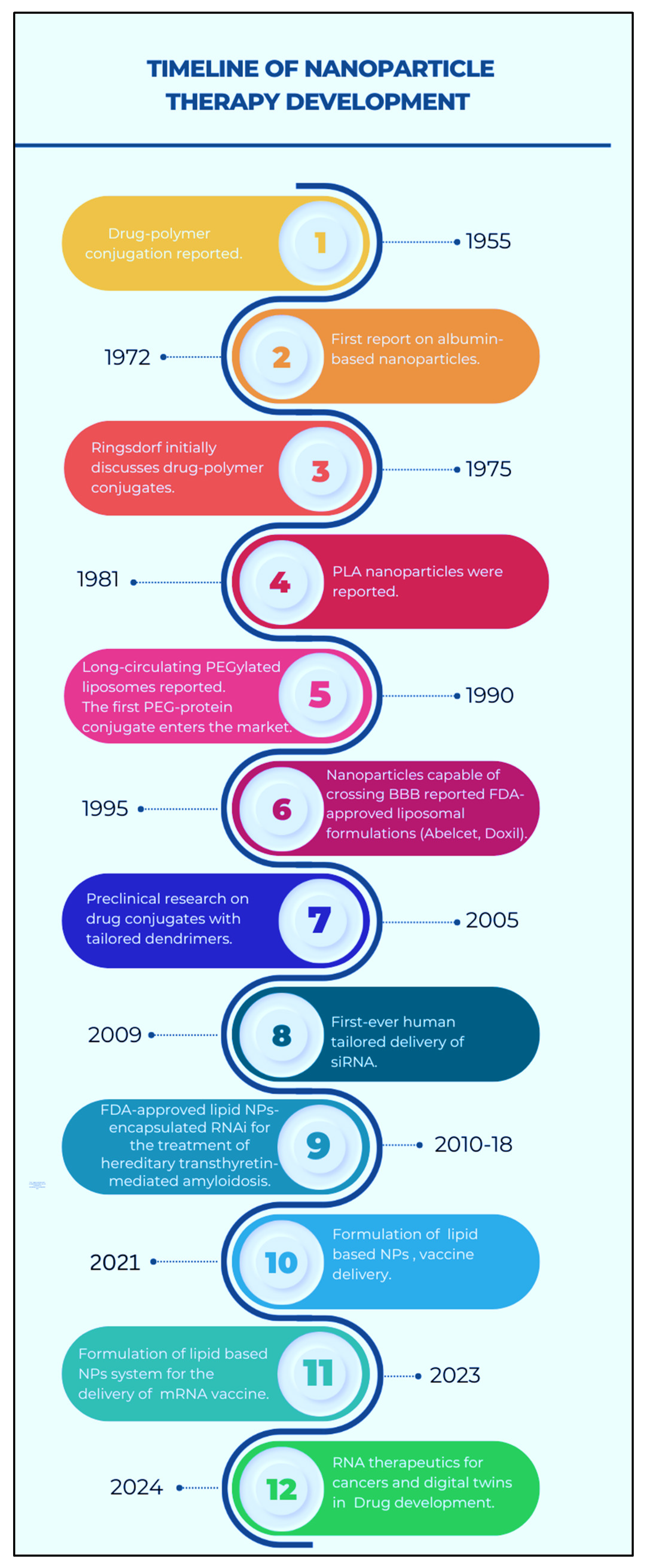
The evolution of NPs-based drug delivery systems has seen significant milestones since 1999 [38]. In that year, the first anti-tumour dendrimer–drug conjugate was reported [35], followed by the entry of synthetic polymer anticancer drug conjugates into clinical trials. By 2002, targeted polymer-drug conjugates had also begun clinical trials. In 2004 [39], The initial creation of polymer-based micelles containing doxorubicin was successfully brought into clinical use, and a polymeric micelle formulation of paclitaxel, free from Cremophor, successfully completed Phase I clinical trials.
The year 2005 witnessed a preclinical trial of targeted dendrimer conjugated with drug [40], the introduction of shape-specific NPs for drug delivery, and the FDA approval of the first protein-based nanoparticle, Abraxane. In 2006, preclinical studies on bow-tie doxorubicin-conjugated dendrimers and initial in vivo studies using drug-loaded polymersomes were carried out. In 2009 [41], In humans, the first targeted delivery of siRNA was accomplished. Between 2010 and 2018 [42], the FDA approved lipid nanoparticles (NPs) encapsulated RNAi for treating hereditary transthyretin-mediated amyloidosis. In 2021, lipid-based NP formulations were utilized for vaccine delivery, and by 2023 [43], these systems were employed for mRNA vaccine delivery. In 2024 [44], advancements include RNA therapeutics for cancers and the use of digital twins in drug development.
3. Characteristics of Nanomaterials
Nanoparticles have unique properties that make them applicable to various clinical applications. The most essential properties of NPs are their small nanoscale size (1-100nm), which permits them to circulate throughout the body without affecting blood circulation (Figure 3). Cellular uptake is directly influenced by the size and shape of nanomaterials [45]. Particle sizes less than 5 nm are easily eluted out from the systemic circulation by renal clearance [46] 10nm to 100nm, accumulating mainly in the spleen, liver, and bone marrow [47,48,49]. The nonpartial nature in size around 10nm -100nm differs from the biodistribution, with cellular uptake within this range primarily dependent on the type of cell [50,51]. The propensity of nanomaterial accumulation in the cell is detected by the specific type of protein that absorbs in vivo on the surface of the nanomaterial [52,53,54,55]. This propensity happened due to the surface modification of nanomaterials [56,57]. The absorption of protein by the nanomaterial, known as opsonisation, starts when the nanomaterial and plasma come into contact.
Figure 3.
Characteristics of nanomaterials.
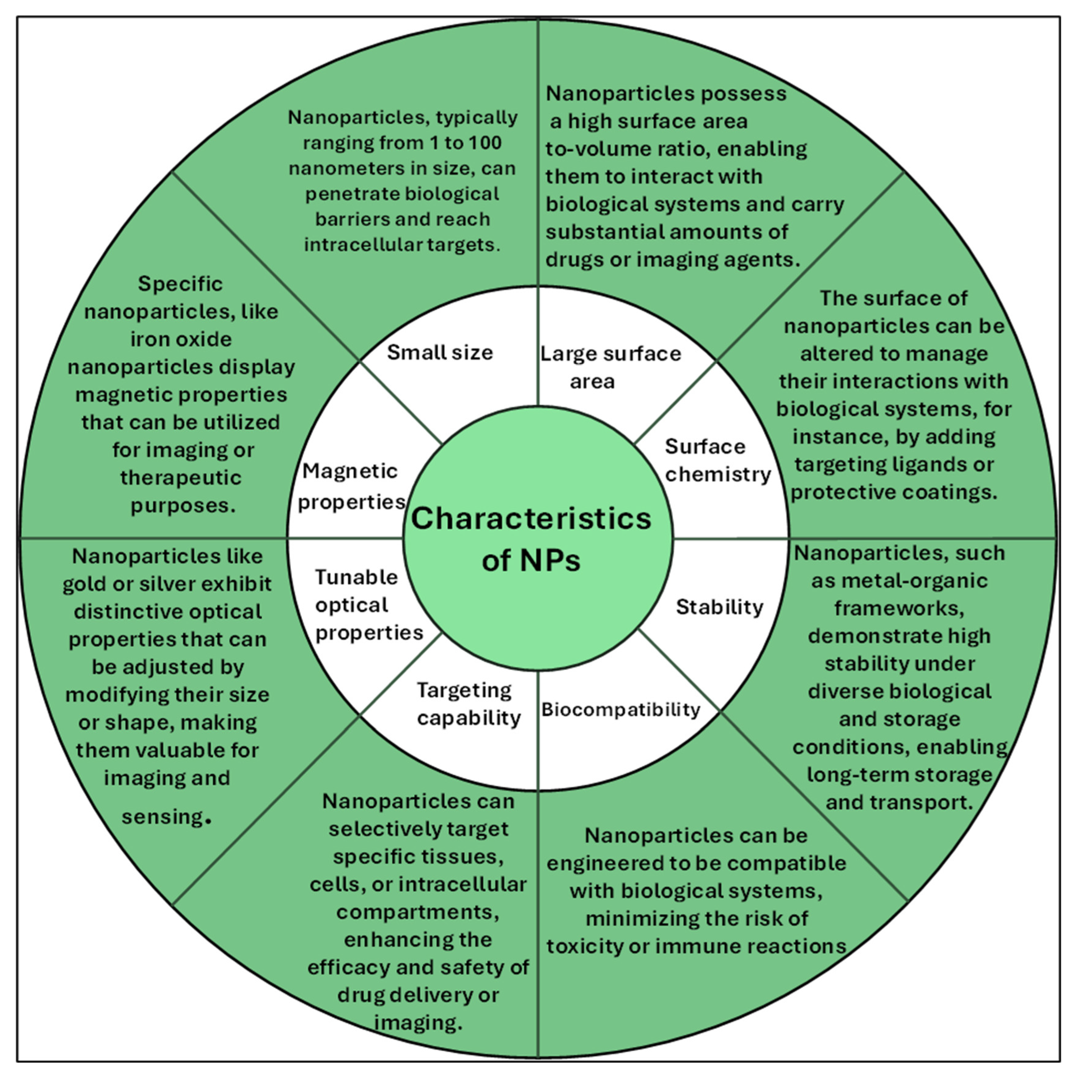
Metallic NPs have fascinated researchers for decades and are now broadly utilised in biomedical engineering. These NPs possess unique properties, such as distinctive optical and electronic characteristics. This is due to the free electrons in the metal NPs, which are collectively excited in response to incident light [58]. Specifically, metallic and metal oxide nanoparticles possess magnetic properties, making them suitable for various applications, including imaging, cell separation, targeting, and drug delivery. Nanomedicine formation is easy on a small scale, but Expanding nanomedicine from small-scale to large-scale production while maintaining stability presents notable challenges [17].
Recent approaches to mitigating the negative effects of opsonisation have focused on slowing the process by making the particle surface more hydrophilic or by balancing the surface charge of the NPs. The most common process is Adsorbing or grafting a hydrophilic polymeric coat, for example- PEG, on the nanoparticle surface [59,60,61]. PEG is a polymer that has polymer chains, depending on their density, and these chains work as steric bras that provide resistance to protein absorption. Still, while the PEG impact is fleeting, subsequent opsonisation and macrophage clearance nevertheless occur. In a particular study, liposomes with albumin covalently attached to their surface ex vivo demonstrated longer flow times than their unmodified or PEGylated versions [18]. When exposed to plasma, the researchers attributed this extended flow to decreased opsonin binding.
Cell culture and body fluids are mainly made from salts, mineral ions and biomolecules. In a study, Allouni et al. studied that the stability of nanomaterials is determined by many factors in culture media, for example, the concentration of nanoparticles, protein abundance and ionic strength; these salt and mineral ions are intermingled with a nanomaterial for their charge compatibility, frequently leading to precipitation and agglomeration of nanomaterial [62].
4. What is Needed to Program Nanoparticles
Nanomedicine involves the manipulation of human biological systems at the molecular level using nanoscale or nanostructured materials. These materials interact effectively with biological systems, providing promising solutions to various challenging health problems through nano-diagnostics and nanotherapeutics. Various nanoparticles have been created with modified functional surfaces and bioactive cores. This design provides several beneficial therapeutic and diagnostic properties, including improved permeation and retention in the circulatory system, targeted drug delivery, highly efficient gene transfection, and advanced bioimaging capabilities [63].
Many programable nanoparticles (nanomedicine) directly interact with genetic material, and these NPs’ interaction with biomolecules helps in normal cell division and gene function [64]; these may cause mutagenicity and toxicity [65]. Toxicity leads to inflammatory responses of macrophages and neutrophils by forming reactive nitrogen and oxygen, which causes nitrosative and oxidative stress [66]. The adhesion of such types of free radicals causes massive damage to the body system [67]. Several types of damage occur; for example, Cancer is caused by lipid peroxidation and protein denaturation, strand breaking due to oxidative DNA damage, transcription of the fibrosis and cancer-causing genes, and transcription of the genes that cause cancer and fibrosis [68]. When the nanomedicine is administered intravenously, wealth data shows an accumulation of these NPs in the liver and migrations through cardiovascular, central nervous, and renal systems [69]. These nanoparticles could be traced after administration, and many unknown potentials of NPs may help in the threats against safety. Currently, many interactions between nanoparticles and biological systems remain unclear. As a result, deciphering, characterizing, or making inferences about the physicochemical and toxicological properties of nanomedicines remains challenging. This is an area where progress is likely to be gradual in the absence of standard regulatory guidelines. It is crucial to understand that there is no “one size fits all” solution because the distinct characteristics seen at the nanoscale rely heavily on the kind of nanoparticle, its surface characteristics, how it is administered, and most importantly, its variety of morphologies. Because of this variability, the regulatory procedure is very intricate.
5. Different Types of Programmable Nanomaterials
Programmable nanomaterials are engineered for functions and applications. This review explores various types of nanoparticles (NPs) and their biological applications, including polymeric NPs, carbon-based NPs, and metallic NPs.
5.1. Polymeric Nanomaterials
Polymeric nanomaterials are evenly distributed and derived from organic matter. Biodegradable, nontoxic and biocompatible polymeric nanoparticles (For example- human serum albumin, bovine serum albumin, and chitosan) are widely used in receptor-mediated drug delivery and therapeutic [70]. Polymeric hybrid nanomaterials have achieved significant interest because of their versatile applications in the biomedical area. They are instrumental in modifying biological entities, serving as transporters for hydrophobic drugs, and acting as non-viral vectors for nucleic acid delivery. A simple process involves the polymer (polyethene glycol PEG) binding with protein, and this approach maintains the pharmacokinetic without changing the biological function of proteins. The potential of self-assembly of nanoparticles is not utilised. There are several types of polymers, and they may be degradable and non-degradable. Natural and synthetic polymers are utilised to make polymeric vesicles and polymeric micelles as drug delivery vehicles [71,72,73,74]. Compared to natural polymers, some specific synthetic polymers have long lifetimes, so they help distinguish the disease location. Therefore, Synthetic polymers are used in long-term drug-release (over several days or weeks) systems [75]. Amphiphilic polymers or graft copolymers are constructed in a liquid medium and form NPs. Polymers, for example, PGA (ploy -glycolic acid), PLA (poly-lactic acid), and PLA (poly-lactic acid), and their copolymer are the most considerably studied.
For the past 30 years, these nanoparticles have been utilized in formulation, biocompatibility studies, and surgical applications, demonstrating their biocompatibility [76]. The rate at which the drug is released is regulated by the degradation of the polymer, which can be controlled by altering the mass of the polymer nanoparticles. For copolymers, their nanostructure and composition are used to control drug release [77]. There are several anticancer drugs, such as tamoxifen and paclitaxel. These small polymeric micelles (10–100 nm) are highly water-insoluble compared to polymeric vesicles, which enhances their accumulation in tumour tissue and improves permeability and retention time within the tumour [78]. Specific Cationic polymers can bind with nucleic acid, and the main target is to achieve siRNA transport and cell transfection. This work holds promise for developing novel therapeutic strategies for treating severe illnesses, including cancer and inherited metabolic diseases. For example, Mao et al. demonstrated the use of a cationic triblock copolymer in cancer therapy to deliver siRNA targeting the acid ceramidase gene. They illustrated how to create siRNA/biodegradable micellar triblock copolymer complexes that effectively penetrate cancer cells and silence genes [79]. Cationic polymers encapsulate siRNA, which has been shown to have toxicity, making their clinical use difficult [19]. However, the positively charged catatonic polymer increases the cellular uptake; therefore, this approach overcomes the variers that target specific drug delivery [80].
Clinical Examples:
Genexol-PM & NK105
Genexol-PM, developed by Samyang Biopharm in South Korea, is a lyophilized polymeric micellar nanocarrier containing paclitaxel (PTX) drug, that is approved for the treatment of MBC, ovarian cancer, pancreatic cancer, and advanced NSCLC. This nanomedicine allows the administration of the higher maximum tolerable dose and reduces the risk of allergic or adverse reactions to paclitaxel [81,82,83,84,85]. NK105, another nanomedicine developed by Nippon Kayaku Co., Japan. Particle size is an 85 nm nanoparticle formulation of paclitaxel (PTX) with a hydrophilic PEG outer shell and a hydrophobic core for efficient drug encapsulation [85,86,87].
Abraxane (Nab-paclitaxel)
Albumin-bound nanoparticle formulation of paclitaxel (nab-paclitaxel), takes advantage of enhanced albumin delivery to tumors via receptor-mediated transport or transcytosis. Nab-paclitaxel promotes caveolin-1 expression and the development of caveolae by binding to the endothelial cells’ gp60 albumin receptor [88]. Studies suggest that increased survival in pancreatic cancer with nab-Paclitaxel and Gemcitabine. In a phase 3 study, patients with metastatic pancreatic cancer were randomly assigned to receive nab-paclitaxel (125 mg/m²) plus gemcitabine (1000 mg/m²) or gemcitabine alone. The primary endpoint was overall survival. Among 861 patients, the nab-paclitaxel–gemcitabine group had a median overall survival of 8.5 months compared to 6.7 months in the gemcitabine group (hazard ratio, 0.72; P<0.001). The one-year survival rate was 35% versus 22%, respectively. Progression-free survival was 5.5 months versus 3.7 months (hazard ratio, 0.69; P<0.001). The response rate was 23% in the combination group versus 7% in the monotherapy group (P<0.001). Higher rates of neutropenia, fatigue, and neuropathy were observed with the combination treatment [89].
5.1.1. Liposomes
Liposomes are a vesicular carrier system based on phospholipids that can be used as a possible drug delivery vehicle. Liposomes have several benefits over traditional delivery systems, including the capacity to target specific sites, manage release patterns, boost stability, and have lower associated toxicity. Many researchers have worked to create primary or modified liposomes in the last ten years to deliver a range of therapeutic substances efficiently [90]. Liposomes’ main structural elements include a phospholipid bilayer, cholesterol, lipoproteins, hydrophilic core, and targeting ligands like amino acid fragments, antibodies, or proteins for specific cell targeting [91].
The new approach, for example, can alter the surface characteristics that liposomes can be used for target-specific cancer and other unhealthy cells without harming the immune system and blood circulation. Due to their inability to spread, specific liposomes function as site-specific drug accumulators. In certain conditions, they can also be used for pulmonary or topical delivery of liposome aerosols. However, most other applications face a significant challenge due to this quick clearance [92]. Liposomes are employed in gene therapy and transfection, immune response enhancement, medication delivery, and treating various diseases [93].In sophisticated drug delivery systems, liposomes that are modified to exhibit stimuli-responsive characteristics such as light, pH, temperature, enzymatic reaction, ultrasound, or radiation-sensitive nanoparticles work well as carriers for multimodal nanoscale trigger and effector functions [20,94,95,96,97].
Clinical Examples:
Doxil®
[98,99]Pegylated liposomal doxorubicin (Doxil® or Caelyx®) uses polyethene glycol polymers to limit reticuloendothelial uptake, resulting in increased circulation, lower distribution volume, and better tumour uptake. Preclinical studies revealed one- or two-phase plasma concentration-time profiles for pegylated liposomal doxorubicin, with an elimination half-life of 20-30 hours and a volume of distribution comparable to blood volume. Due to its aqueous nature, doxorubicin is located in the inner core of the nanoparticle. The surrounding lipid bilayer acts as a protective barrier, preventing early degradation of the medication, while the PEG coating effectively camouflages the particle from the immune system. This nanoparticle property not only prolongs the drug’s circulation in the bloodstream but also minimizes its uptake by the mononuclear phagocyte system [98,99]. The AUC is enhanced 60-fold, with preferential accumulation in implanted tumours and xenografts, resulting in higher tumour drug concentrations than free doxorubicin. Clinical trials with pegylated liposomal doxorubicin in humans, including ARKS and different carcinomas, have revealed a pharmacokinetic profile with greatly longer circulation, reduced clearance, and volume of distribution, which improves dosage scheduling potential [100].
Vyxeos™ and Onivyde™
Vyxeos (CPX-351), developed by Jazz Pharmaceuticals. It is the first FDA-approved dual-drug containing liposomal nanomedicine that delivers Cytarabine and daunorubicin 5:1 molar ratio. In phase III clinical trial in AML patients, this drug showed better potential and a higher survival rate than the conventional treatment with free cytarabine and daunorubicin [101].
Onivyde® (MM-398 or PEP02) FDA approved drug, developed by Merrimack Pharmaceuticals, is irinotecan containing a liposomal drug used in solid tumors and also metastatic pancreatic cancer [102].
Marqibo®
Vincristine sulfate liposome injection (VSLI; Marqibo®), developed by Talon Therapeutics in the USA, is a vincristine sulfate containing sphingomyelin and cholesterol-based nanomedicine. It is majorly used in the treatment of hematologic malignancies and solid tumors such as leukemia, Hodgkin’s disease, and non-Hodgkin lymphoma (NHL).
This liposomal drug was specially engineered to address the dosing restrictions and pharmacokinetic challenges associated with traditional nonliposomal VCR. This innovative delivery method permits the administration of the higher maximum tolerable dose with accurate delivery in targeted tissues, prolongs the drug circulation time, and slows the release of the drug in the tumor interstitium [103].
5.1.2. Dendrimers
Dendrimers are unique nanostructures or nanoparticles made up of branching layers that resemble the skin of an onion. These nanostructures grow in concentric layers outward from the core, producing a gradual increase in size comparable to the size of many globular proteins found in vivo. “Generations” refers to these concentric layers that resemble branched trees. Every dendrimer has an exact number of functional groups on its outer generation that can serve as a monodispersed platform to design advantageous interactions between nanoparticles and drugs and between nanoparticles and tissues. These properties have garnered substantial attention in the medical field as nanocarriers for conventional tiny medicines, DNA/RNA, proteins, and, in certain circumstances, naturally active nano-size pharmaceuticals. Dendrimer-based drugs and imaging and diagnostic agents present promising options for various nanomedicine applications [104]. The PAMAM (poly amidoamine) [21,105,106] is the most renowned dendrimer for clinical applications. Cationic polyamidoamine dendrimer interacts with a negatively charged molecule, showing a toxic effect. To reduce this effect, Jia et al. studied the work of phosphocholine and zwitterion present in a cell member’s outer surface and found some lipid ends [107]. Compared to native PAMAM dendrimers, these zwitterionic dendrimers with a phosphorylcholine surface effectively reduce cytotoxicity [108]. Clinical investigations on dendrimers have been conducted. Their size makes them highly distinct from polymeric systems, micelles, and liposomes, which causes them to behave differently after injection [109]. In terms of mechanical stability, dendrimers that form a single covalent bond structure surpass liposomes. They also provide an internal hydrophobic cavity for payload encapsulation and functional switching. However, compared to micelles and lipid/polymer vesicles, the payload-to-carrier weight ratio is smaller, limiting the integration of space-consuming smart sensors or nano-size switches [110]. The lack of clinical expertise poses a hurdle to developing commercial dendrimer-based nanoscale systems since it casts doubt on drug development, regulatory approval, and clinical success, all of which influence industrial decision-making [109].
5.1.3. Nano Hydrogel
Hydrogels are polymeric materials composed of interlinked polymeric chains, forming a hydrophilic structure that can retain and absorb large quantities of water. Hydrogels closely mimic living tissue due to their highwater content, soft consistency, and porosity. These versatile materials can be fabricated into various forms, for example, microparticles (MPs), slabs, coatings, NPs, and films, to suit different applications. In medicine and clinical practice, cosmetic technology, hydrogels have a broad range of uses, involving tissue engineering, drug delivery, cell and biomolecule separation, diagnostics, and cell immobilization. They can also serve as barriers to drug accumulation, holding substantial amounts of biological fluids and swelling in the process. The ability of hydrogels to stretch and swell gives them a squishy texture like real tissue, making them highly biocompatible. This unique combination of properties-water retention, flexibility, and biocompatibility enables hydrogels to play an important role in modern medical and clinical applications [22].
One of the primary uses of hydrogels is in the production of soft contact lenses. Unlike glass lenses, hydrogel lenses enable gas diffusion and maintain moisture on the surface of the retina [111]. Clinical trials are ongoing with new hydrogel lenses for different results to improve the wear time, add pigment, and optimize the geometry of the lenses.
Moreover, the hyaluronic acid-containing hydrogel is used in facial correction. Juvéderm® is the leading product in the market; it uses the correction of age-related volume loss and lip augmentation, and it is sensible enough to serve facial wrinkles [112].
Nano hydrogels are also utilized in sensing applications. For instance, Ionic poly (N-isopropyl acrylamide-co-methacrylic acid) (PNM) hydrogels serve as protein receptors due to their significant refractive index change upon protein binding. These hydrogels are synthesized on the surface of silica gold nanoshells (AuNSs) to create a composite material (AuNS@PNM). This combined material is employed to detect levels of two proteins, lysozyme and lactoferrin, which serve as markers for chronic dry eye. Given that lactoferrin and lysozyme have high isoelectric points, indicating their positive charge, they are attracted to the negatively charged PNM hydrogels. When these proteins attach to the hydrogels, AuNS@PNM exhibits a noticeable, concentration-dependent red shift in the Localized Surface Plasmon Resonance (LSPR) wavelength. This shift enables the identification of clinically significant changes in protein levels in human tears [113].
The physicochemical properties of nanoscale hydrogel networks, such as membrane disruption, cytocompatibility, and critical phase transition pH, can be finely tuned by adjusting the polymer composition. These tailored properties are crucial for designing intracellular drug delivery vehicles. By understanding how these vehicles internalize and function within cells, we can optimize their physicochemical characteristics to achieve more effective treatments [114].
Moreover, the breakdown of pH-responsive Nanogel in reductive conditions formed a cross-linker. With minimal impact on the pH-responsive nano gel physiochemical characteristics, this crosslinker was added under the trade name PDESSB30 when these nanogels meet normal quantities of glutathione Based on the light scattering measurement. It was shown that functional siRNA could be delivered to Caco-2 cells using both degradable (PDESSB30) and non-degradable (PDETB30) nanogels, achieving gene silencing of 47% and 83%, respectively. PDETB30 and PDESB30 are appealing candidates for additional improvement as therapeutic siRNA delivery systems due to their favourable physicochemical characteristics and siRNA delivery efficiency [115].
5.2. Carbon-Based Nanomaterials
Carbon NPs, including nanodiamonds, carbon nanofibers, graphene, carbon quantum dots, and carbon nanotubes, have unique qualities that make them promise for utilize in clinical applications. These applications include the treatment of cancers that are resistant to chemotherapy, improving magnetic resonance imaging, tissue regeneration, stem cell banking, and more. Furthermore, methods for enhancing the administration of drugs and imaging through carbon nanomaterials have been examined. These methods include creating endothelial leakiness and using artificial intelligence to create the best nanoparticle-based drug combination delivery system [116].
5.2.1. Nanodiamonds (NDs)
Nanodiamonds are a class of carbon-based NPs. They have unique properties that make them applicable for clinical applications. Nanodiamonds (NDs) can be synthesised using various methods. For example, the most common methods of NDs synthesis are detonation, laser ablation, Chemical vapour deposition (CVD), high pressure and high temperature (HPHT). NDs are commonly used in clinical applications due to their detonation nanosized and fluorescence [117]. The fluorescence of NDs makes them applicable to several clinical applications, for example, bioimaging, diagnostic applications, and drug delivery [118,119,120].
NDs most prominent application is to improve the delivery of chemotherapeutics medication, mainly chemotherapeutics cancer. The most common process of chemoresistance is the active release of chemotherapy drugs from cancerous cells via ATP-binding cassette transporters (5′-triphosphate), decreasing the number of chemotherapeutic medications that are effective in cancer cells, mainly cancer (stem cells) [121].
NDs have a large surface area due to NDs’ use in absorption and targeted release of several anticancer drugs, for example, tetracyclines [122,123], 4-hydroxytamoxifen [124], and paclitaxel [125]. When the drug is encapsulated on the surface of the NDs and enters the cell absorbed from NDs inside the cell, it maintains the concentration of the drug, a result that was utilized to restore the cancer cells’ medication resistance to conventional chemotherapy [126]. Combination chemotherapy employing a mixture of drugs (cocktail) is the most effective treatment for tumors that are mutated and resistant to multiple drugs [127]. Recently, feedback system control (FSC) technology has been used to determine the best drug combination for ND-bleomycin, ND-DOX, unmodified paclitaxel, and ND-mitoxantrone [128]. FSC tasted millions of formulations and 57 combinations. These are tested on the different types of breast cancer cell lines and consistently outperformed single drugs in every case.
Gene delivery involves transferring genetic material or gene therapy into cells to replace unwanted genes with desired ones to regulate and enhance cellular functions [23].
Nanodiamonds (NDs) are widely investigated for their applications in gene delivery, leveraging interactions between polymeric agents on their surface and negatively charged nucleic acids [129,130,131]. For example, the ND-PEI vector efficiently delivers pEGFPLuc plasmids encoding green fluorescent protein (GFP) and luciferase into the cytoplasm [132].
5.2.2. Quantum Dots (QDs)
Quantum dots are semiconductor NPs with unique shapes, sizes, and tuneable optoelectronic and electric properties. Because of this property, in recent years, quantum dots have become attractive in biomedical engineering, where they can be used for real-time bioimaging, single-molecule probes, drug delivery, intracellular tracking, vivo imaging, and diagnostics [133]. The optical properties of quantum dots QDs depend on the composition and size, quantum yield, multiplexing capacity, surface area-to-volume ratio, and resistance to photobleaching. Traditional organic dyes are highly susceptible to photobleaching, and quantum yield can regularly be less than 15% in biological environments [134]. Moreover, mainly conventional organic label dyes are opposed; QDs probably have a wide range of fluorescence, from near ultraviolet (UV) to infrared. Quantum dots (QDs) emitting in the near-infrared range have significantly enhanced the potential of fluorescence in biomedicine due to their reduced tissue absorption and comparatively low autofluorescence [134,135]. The primary challenge with using quantum dots (QDs) in biomedical and theragnostic applications is their insolubility in water, which is crucial for bioimaging. To address this, many researchers have the main aim of encapsulating QDs with hydrophilic, soluble materials. For example, QDs can be encapsulated in hydrogels or polymeric matrices. Additionally, surface modification techniques, such as adding ligands or functional groups, can be applied to hydrophobic QDs to improve their solubility in water [24]. Finally, the coating material is selected for the desired applications and environments.
The QDs are conjugated proteins, and biotin functionalisation could make specific binding, cellular uptake, and intracellular imaging possible. QDs can be conjugated with many biomaterials, for example, oligonucleotide, antibodies, small molecule ligands, and proteins, to directly import and export the therapeutics in their in vivo or in vitro pathway to specific targets [24,136]. The conjugation on the surface of quantum dots (QDs) depends on the surface chemistry of the conjugate biomaterial and the functional group used for attachment, which can involve either covalent or non-covalent bonding [137].Moreover, significant work has been done to improve QDs further to enable more exact site-specific bioconjugation. Additional concerns remaining referred include improving the efficacy of bioconjugation and continuing the high quantum yield.
QDs are increasingly being used in biological applications, particularly for in vivo studies. When administered through intravenous injection, these nano-sized colloidal particles face a more complex set of challenges compared to conventional organic dyes. Upon entering the circulatory system, QDs encounter various biological barriers at both the organ and cellular levels. These barriers can hinder the accumulation of QDs at the desired target sites and reduce the efficiency of their interactions. Consequently [138,139], overcoming these biological barriers is crucial for enhancing the efficacy of QDs in targeted applications.
5.2.3. Carbon Nanotube (CNTs)
Carbon nanotubes (CNTs) have garnered significant attention as a promising nanomedicine drug candidate due to their distinctive and exceptional mechanical, electrical, and physicochemical properties. Over the past decade, this emerging nanomaterial has piqued the interest of many scientific disciplines. CNTs offer numerous potential applications in cancer treatment, including drug delivery, imaging, and combination therapy [140].
In 1991, Iijima Sumio envented carbon nanotubes. The arc discharge is one of the primary methods utilised to create carbon nanotubes [141]. In addition, the surface modification and functionalisation of CNTs make them low toxic, reduce immunogenicity, and increase the drug loading capacity [142].
There are several techniques for applying CNTs: use as a template, mount the sensing agent such as an antibody, and then modify the optical or electronic properties of a particular stimulus (for example, a cancer protein). Liu et al. Demonstrate the application of the CNTs (use as a nanocarrier) for treating pancreatic and liver cancer [143]. Commonly used markers for liver cancer medication include α-fetoprotein variant (AFP-l3), α-fetoprotein (AFP), and aberrant plasminogen (APT). Li and other researchers developed gold-coated CNTs that bind with antibodies labelled with a redox probe to serve as markers. The principle involves the biomarker binding to the antibody-immobilized CNTs to generate a signal. DOGU et al. reported that this occurs due to the unique properties of hepatocellular carcinoma cells [144]. Extremely differentiated cells (HUH7) have an excellent chance to bind compared to weakly differentiated cells (SNU182). Using these characteristics, I developed an imageable carbon nanotube surface to diagnose liver cancer cell invasion levels.
With almost 184,000 fatalities yearly, ovarian cancer is the second most frequent gynaecological disease worldwide. Mijin Kim et al. make Nano sensor arrays to detect critical diseases. It is an innovative device that encapsulates a single-walled carbon nanotube (SWVNT) to use Functionalized ssDNA; with the potential to completely change the course of therapy and prognosis for this terrible illness, this design holds great promise for prompt and precise ovarian cancer identification [145]. First, they selected spectral variables of nanotube arrays as features, computing the data based on the response of the nanotube arrays to the serum sample.
The use of five mainly used machine learning algorithms, for example, artificial neural networks [146], stochastic sen [147], logistic regression [148], support vector machines [149], and decision trees [150] for binary classification, where the goal is to separate patients from either healthy individual with ovarian cancer or patients from patients with other disorders [151].
During the cancer progression, heterogeneity rises, driven by cellular and non-cellular changes in the tumour microenvironment (TME), leading to proliferation, growth, and cell death resistance. Carbon-based nanotube (CNT) nanoplatforms hold significant potential for cancer diagnosis. Yang et al. developed oxidized multi-walled CNTs with a large diameter to encapsulate the anticancer drug cisplatin on the inner surface. The outer surface was coated with Doxorubicin (DOX), folic acid, and polyethylene glycol (PEG) to prevent the early release of cisplatin [152]. Wang et al. improved MRI-guided, TME-responsive phototherapy by modifying multi-walled nanotube (MWNTs) with manganese dioxide and Ce6, increasing cytotoxicity in acidic TME through catalysis and photothermal effects to destroy tumour cells [153].
5.3. Metallic Nanoparticles
The creation of tailored nanoparticles has been a major factor in advancing nanotechnology. Because of their substantial inertness and nanoscale shapes, which are comparable in size to many biological molecules, different metallic NPs have been thoroughly investigated for biomedical applications. By changing specific particle parameters, including size, environment, shape, aspect ratio, functionalization qualities, and synthesis techniques. one can modify their intrinsic features, which include optical, electrical, surface plasmon resonance, and physicochemical. various applications, such as sensing, drug administration, photodynamic therapy, imaging, and the fusion of several applications, have been made possible by these tunable properties. The different characteristics of metallic NPs and their application in cancer treatment [26].
Metallic nanoparticles are used in sensing to detect cancer; these can easily conjugate with a recognition molecule; for example, antibodies bind on the surface of biomolecules for their recognition. The metallic NPs give excellent sensitivity for the recognition of tumour cells [27]. Tian et al. synthesized gold (Au) nanoparticles with red fluorescence using egg white and microwave irradiation. These nanoparticles demonstrated fluorescence signals in the presence of viable cancer cells (HepG2), while normal cells did not exhibit fluorescence. The fluorescence intensity increased over time. Biocompatibility was confirmed by the MTT assay, showing that more than 80% of cells remained viable after 24 hours at concentrations ranging from 0 to 1.2 mg/ml. These findings indicate that the synthesized Au nanoparticles hold promise for detecting cancer cells [154]. Saeed et al. developed DNA-modified gold nanoparticles (AuNPs) and graphene oxide-based electrodes for the early detection of breast cancer markers. They achieved high sensitivity for CD24 and ERBB2 using amperometric detection methods [155]. In another study, an aptamer-nanoparticle strip biosensor (ANSB) was created to quickly, accurately, sensitively, and affordably detect circulating tumour cells. Aptamers were chosen and coupled with AuNPs using the cell-SELEX process, resulting in ANSBs on a lateral flow device. In less than 15 minutes, they could visually identify at least 4000 Ramos cells and around 800 cells using a portable strip reader [156].In another study, authors developed a three-dimensional electrochemical DNA biosensor using 3D graphene-functionalized AgNPs, achieving high sensitivity (1.0 × 10⁻¹⁴ M) and effectively detecting CYFRA21-1 DNA in lung cancer clinical samples [157]. Additionally, a biosensor was developed using a magnetic bar carbon paste electrode modified with iron oxide and silver nanoparticles (MBCPE/Fe3O4@Ag/ssDNA). This biosensor showed a wide linear range and an extremely low detection limit (0.1 fM) for the BRCA1 5382 mutation [158]. Small changes in size can significantly impact the optical properties of magnetic nanoparticles (MNPs) due to the surface plasmon absorption phenomenon. Alterations in size and shape also influence the energy difference between the conductive and valence bands of the material. These variations result in distinct tunable properties beneficial for bioimaging applications [159].
A new Janus nano platform combining AuNPs, and Fe3O4 NPs/mesoporous silica core@shell, modified with a targeting peptide and fluorescent dye, enabled tumour-targeted multimodal imaging (CT, MRI, and fluorescent tracking) in a fibrosarcoma-bearing mouse model [160].Iron alloy cores, unlike the toxic manganese, have shown promising results in clinical trials, with research focusing on cytotoxicity assessment and direct imaging of FePt nanocrystals using quantum interference to monitor their stability and metal interactions [161]. Nanoparticles loaded with curcumin and conjugated with folic acid, synthesized using the Turkevitch method, exhibited 80% drug release under acidic pH conditions. These nanoparticles showed no toxicity in human breast epithelial and mouse fibroblast cell lines. Moreover, they demonstrated enhanced anticancer effects in vivo against MCF-7 breast cancer cells [161,162]. Using stirring synthesis, paclitaxel siRNA-loaded polyethyleneimine and PEGylated anisamide-capped AuNPs increased siRNA exposure and synergistically suppressed prostate cancer in a PC-3 xenograft mouse model [163].
Clinical Examples:
NanoTherm
Nanotherm is a magnetic nanoparticle, developed by MagForce Nanotechnologies AG in Germany. This drug is made of superparamagnetic iron oxide nanoparticles (SPIONs) coated with amino silane; this drug is used for cancer thermal therapy [164,165]. The treatment uses localized heating (41–46 °C) to make cancer cells more sensitive to therapy or applies higher heat (>46 °C) to directly destroy them and surrounding tissues [166].

Table 1.
Some examples of clinically approved nanomedicine.
| Name | Drug | Carrier property | Indications | Manufacture Company |
| Myocet® | Doxorubicin | Liposome | Breast Cancer | Teva |
| Marqibo® | Vincristine sulphate | Liposome | Acute lymphoblastic leukemia (ALL) | Talon Therapeutics |
| Ambisome | Amphotericin B | Liposome | Fungal infection | Gilead Sciences |
| Onivyde® | Irinotecan | Liposome | Pancreatic cancer | Merrimack Pharmaceuticals |
| Doxil® | Doxorubicin | Liposome | Various cancers, ALL, AML, Breast cancer, Ovarian cancer | Johnson &Johnson |
| Vyxeos® | Cytarabine and daunorubicin |
Liposome | AML | Jazz Pharmaceuticals |
| Abraxane® | Paclitaxel | Albumin | Various cancers, Breast cancer | Abraxis BioScience |
Table 2.
Nanomedicines in clinical trials in Phases II and III.
| Name | Drug | Carrier property | Indications | Manufacture Company |
| Genexol-PM® | Paclitaxel | Polymeric micelles | Breast,lung, ovarian cancer | Samyang |
| NK-105® | Paclitaxel | Micelle: PEG- poly aspartate |
Metastatic Breast Cancer | Nippon Kayaku Co. |
| NanoTherm | Aminosilane- coated SPIONs |
Metallic nanoparticle | GBM & prostate cancer | MagForce Nanotechnologies |
| ThermoDox | Doxorubicin | Thermosensitive liposome | Hepatocellular carcinoma | Celsion |
| Lipoplatin® | Cisplatin | Liposome | Breast, pancreatic, urinary bladder, and gastrointestinal cancer |
Regulon Inc. |
6. The Challenge in Programming the Nanoparticles
The primary challenge for the nanomaterial program is that governing bodies like the FDA possess data on bulk materials, which do not exhibit the same pharmacokinetics and pharmacodynamics as programmable nanoparticles used in nanomedicine [167]. This data is stored based on the efficacy and safety of bulk materials, which does not accurately represent the behaviour of nanoparticles used in clinical conditions after-market authorization. Consequently, this creates difficulties in setting efficacy and safety regulations for nanomedicines. While a non-nano version might meet regulatory guidelines, its nanomedicine counterpart might not [168].
In 2009, Owen and Raynard highlighted that a single-size standard is not suitable for all types of nanoparticles due to differences in clinical requirements, physiology [169], and application routes. However, this warning has been largely ignored by current regulatory authorities. The complexity in the structure, size, form, and clinical application of programmable nanoparticles poses significant challenges to the regulatory system in classifying and characterizing nanoparticles. For example, dynamic light scattering (DLS) is used to determine hydrodynamic size, but this technique assumes that the particles scattering light are spherical, making it inaccurate for rod-shaped materials. Additionally, other techniques used for size measurement may not accurately reflect the form of nanomaterials as experienced in the human body. For instance, using a transmission electron microscope involves drying samples, which may alter their shape or size compared to their form in solution. Protein coronas commonly form when nanomaterials are injected into the bloodstream, leading to significant underestimation of their true size in a physiological context. There is no consensus in the literature on the best standards for nanometrology or characterization [170]. As a result, preclinical nanomedicine development will likely continue without stringent clinical regulatory guidance or intervention.
Cellular and nanotoxicity response is another challenge faced [171] (Figure 4). There are several ideas for overcoming cytotoxicity. Traditional toxicity testing on large animals, which was previously used for small drug molecules, has been deemed unethical, excessively expensive, and impractical for evaluating nanotoxicity [172]. In vitro toxicity techniques are employed to assess nanoparticles. They offer cost-effective and time-efficient managed experimental conditions compared to animal testing. However, various assays are utilized to bypass the complexities of the human body [173]. It employs compensatory mechanisms and pathological responses to handle toxins, along with intricate metabolic processes. Additionally, growing evidence indicates that traditional in vitro tests for small compounds are unsuitable for nanomaterials [174]. Nanomaterials interact with the reagents used in in vitro assays. Their characteristics—such as optical properties, high absorption, acidity or alkalinity, catalytic activity, dissolution, and magnetic properties—result in interactions with the reagents in these tests [175]. Consequently, new tests are necessary to evaluate the toxicity of nanomaterials and nanomedicines before appropriate regulatory guidelines can be established. This need is significantly hindering progress in the field [176]. When designing nanoparticles for clinical applications, the drug delivery process and mechanism require preclinical trial data for approval, including information on advanced effects [177].
Figure 4.
Challenges in programable nanoparticle.
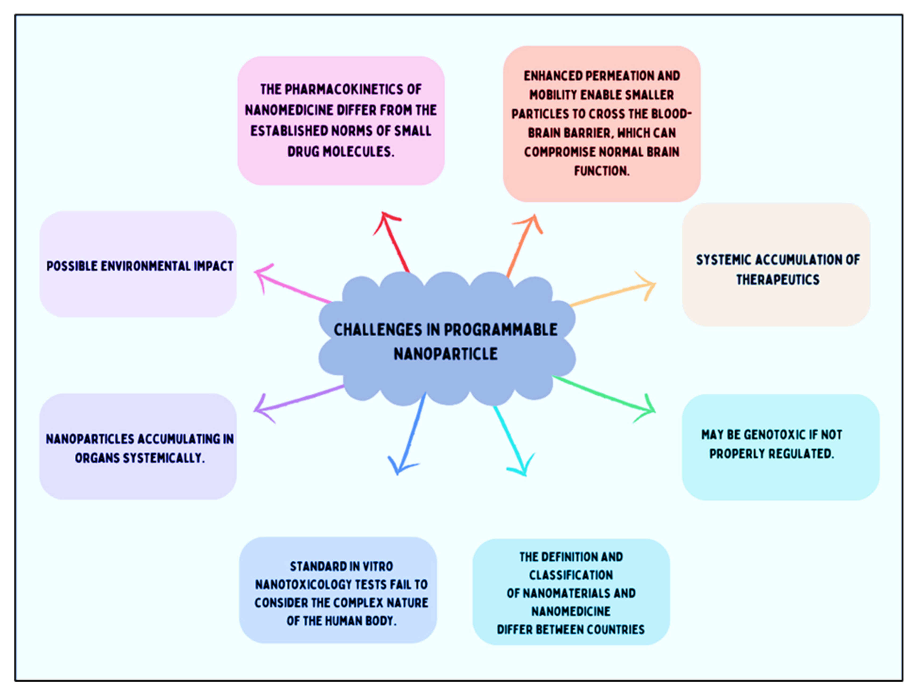
The toxicity effect may be caused by specific Organs or cells and the occurrence of antibiotics due to high drug dosing. Moreover, the size of the NPs is more mobile, where they are larger targeted sites. The mobility of NPs allows them to pass the blood-brain barrier and promote the function of the brain for a long duration [178]. This factor is crucial. Without adequate information, we cannot assert that programmable nanomedicine is safe, though it may overcome these challenges in the future. Describing the pharmacokinetics of nano-drugs is causing the main challenge in the regulations [179,180]. Nanomedicines exhibit unconventional behavior compared to small drug molecules, leading to prolonged bioavailability. This extended presence in the body presents substantial health risks if these products were to be sold over the counter. Consequently, regulatory authorities must meticulously assess whether nanomedicines warrant strict monitoring or can be available as over-the-counter products. However, arriving at a definitive decision is difficult due to the current absence of toxicity data and information.
7. Conclusions and Outlook
The rapid progression of nanotechnology has significantly bolstered preclinical development, especially in the area of nanomedicine. Programmable nanomaterials have important characteristics for example, shape, fluorescence, size, mechanical strength, surface chemistry, and surface area. These characteristics help to improve drug delivery, tissue engineering, bioimaging, nanomedicine, therapeutics, and numerous other biological applications. Nanoparticles should possess characteristics such as biocompatibility, bioavailability, biodegradability, targeted and controlled drug release that exceed those of their larger counterparts. These properties are provided to address the difficult challenges and improve therapeutics. Polymeric nanomaterial, liposomes, dendrimer and nano hydrogel are for the front of the revolution. Carbon-based NPs, for example, nanodiamonds, carbon quantum dots, and carbon nanotubes offer unique, electronic, magnetic and optical properties that are highly advantageous for drug delivery, bioimaging and cancer treatment. Nanodiamonds have shown promise in chemotherapy resistance and fascinating gene delivery, underscoring their potential in treating resistant and genetic diseases. Metallic nanoparticles have potential biological applications.
Despite these advancements, translating nanomedicine from research to clinical practice faces significant challenges. One major hurdle is the biological barriers that nanoparticles encounter, affecting their distribution, cellular uptake, and overall efficacy. The shape, size, and surface chemistry of nanoparticles must be carefully engineered to maximize their interactions with biological systems. Looking ahead, the future of programmable nanomaterials in clinical applications is promising yet contingent on overcoming these challenges. Continued research and development are essential to optimize nanoparticle design, enhance targeting and delivery mechanisms, and mitigate potential toxicity. Collaboration between researchers, clinicians, and regulatory authorities will be crucial in establishing comprehensive guidelines and protocols for the safe and effective use of nanomedicines. In conclusion, targeted nanomaterials has promising clinical applications in the biomedical sectors.
Acknowledgments
We extend our sincere gratitude to all members of the DB lab for their feedback on this manuscript. AK and PY thank IITGN-MoES Gol for MTech and PhD fellowships, respectively. R.S. acknowledges the Science and Engineering Research Board (SERB), Government of India, for financial support through the National Post-Doctoral Fellowship (NPDF). DB thanks SERB-CRG, Gujcost GSBTM and STARS-MoES for research grants.
Conflicts of Interest
The authors do not declare any conflict of interest.
References
- Wilczewska, A.Z.; Niemirowicz, K.; Markiewicz, K.H.; Car, H. Nanoparticles as drug delivery systems. Pharmacological Reports 2012, 64, 1020–1037. [Google Scholar] [CrossRef] [PubMed]
- Hofmann-Amtenbrink, M.; Grainger, D.W.; Hofmann, H. Nanoparticles in medicine: Current challenges facing inorganic nanoparticle toxicity assessments and standardizations. Nanotechnology, Biology, and Medicine 2015, 11, 1689–1694. [Google Scholar] [CrossRef] [PubMed]
- Chiari-Andréo, B.G.; et al. Nanoparticles for cosmetic use and its application. In Nanoparticles in Pharmacotherapy; Elsevier, 2019; pp. 113–146. [Google Scholar] [CrossRef]
- Wang, J. Nanoparticle-based electrochemical DNA detection. Anal Chim Acta 2003, 500, 247–257. [Google Scholar] [CrossRef]
- Peng, W.; et al. Improvement of magnetorheological finishing surface quality by nanoparticle jet polishing. 2013, 52, 043401. [Google Scholar] [CrossRef]
- Ghosh, S.; et al. Nanoparticle-enhanced multifunctional nanocarbons—recent advances on electrochemical energy storage applications. J Phys D Appl Phys 2022, 55, 413001. [Google Scholar] [CrossRef]
- Hanigan, D.; et al. Trade-offs in ecosystem impacts from nanomaterial versus organic chemical ultraviolet filters in sunscreens. Water Res 2018, 139, 281–290. [Google Scholar] [CrossRef] [PubMed]
- Willner, I.; Baron, R.; Willner, B. Integrated nanoparticle–biomolecule systems for biosensing and bioelectronics. Biosens Bioelectron 2007, 22, 1841–1852. [Google Scholar] [CrossRef]
- Szelenyi, I. Nanomedicine: evolutionary and revolutionary developments in the treatment of certain inflammatory diseases. [CrossRef]
- Seigneuric, R.; et al. From Nanotechnology to Nanomedicine: Applications to Cancer Research. Curr Mol Med 2010, 10, 640–652. [Google Scholar] [CrossRef]
- Peer, D.; et al. Nanocarriers as an emerging platform for cancer therapy. Nat Nanotechnol 2007, 2, 751–760. [Google Scholar] [CrossRef]
- Park, T.; Jeong, J.; reviews, S.K.-A. drug delivery; 2006, undefined. Current status of polymeric gene delivery systems. ElsevierTG Park, JH Jeong, SW KimAdvanced drug delivery reviews, 2006•Elsevier.
- Park, T.; Jeong, J.; reviews, S.K.-A. drug delivery & 2006, undefined. Current status of polymeric gene delivery systems. ElsevierTG Park, JH Jeong, SW KimAdvanced drug delivery reviews, 2006•Elsevier.
- Parveen, S.; Sahoo, S.K. Polymeric nanoparticles for cancer therapy. J Drug Target 2008, 16, 108–123. [Google Scholar] [CrossRef]
- Petros, R.A.; DeSimone, J.M. Strategies in the design of nanoparticles for therapeutic applications. Nat Rev Drug Discov 2010, 9, 615–627. [Google Scholar] [CrossRef] [PubMed]
- TORCHILIN, V. Multifunctional nanocarriers☆. Adv Drug Deliv Rev 2006, 58, 1532–1555. [Google Scholar] [CrossRef] [PubMed]
- Paliwal, R.; Babu, R.J.; Palakurthi, S. Nanomedicine Scale-up Technologies: Feasibilities and Challenges. Ageing Int 2014, 15, 1527–1534. [Google Scholar] [CrossRef]
- FURUMOTO, K.; et al. Effect of coupling of albumin onto surface of PEG liposome on its in vivo disposition. Int J Pharm 2007, 329, 110–116. [Google Scholar] [CrossRef] [PubMed]
- Jeong, J.; Mok, H.; Oh, Y.; chemistry, T.P.-B. & 2009, undefined. siRNA conjugate delivery systems. ACS PublicationsJH Jeong, H Mok, YK Oh, TG ParkBioconjugate chemistry, 2009•ACS Publications 2009, 20, 5–14. [Google Scholar]
- Elegbede, A.I.; et al. Mechanistic studies of the triggered release of liposomal contents by matrix metalloproteinase-9. ACS PublicationsAI Elegbede, J Banerjee, AJ Hanson, S Tobwala, B Ganguli, R Wang, X Lu, DK SrivastavaJournal of the American Chemical Society, 2008•ACS Publications 2008, 130, 10633–10642. [Google Scholar] [CrossRef]
- Patil, M.; Zhang, M.; nano, T.M.-A. & 2011, undefined. Multifunctional triblock nanocarrier (PAMAM-PEG-PLL) for the efficient intracellular siRNA delivery and gene silencing. ACS PublicationsML Patil, M Zhang, T MinkoACS nano, 2011•ACS Publications 2011, 5, 1877–1887. [Google Scholar]
- Malpure, P.S.; Patil, S.S.; More, Y.M.; Nikam, P.P. A Review On-Hydrogel. A Review On-Hydrogel. American Journal of PharmTech Research 2018, 8. [Google Scholar] [CrossRef]
- Coonrod, A.; Li, F.Q.; Horwitz, M. On the mechanism of DNA transfection: efficient gene transfer without viruses. Gene Ther 1997, 4, 1313–1321. [Google Scholar] [CrossRef]
- Mattoussi, H.; Palui, G.; reviews, H.N.-A. drug delivery & 2012, undefined. Luminescent quantum dots as platforms for probing in vitro and in vivo biological processes. ElsevierH Mattoussi, G Palui, HB NaAdvanced drug delivery reviews, 2012•Elsevier.
- Smith, A.; Duan, H.; Mohs, A.; reviews, S.N.-A. drug delivery & 2008, undefined. Bioconjugated quantum dots for in vivo molecular and cellular imaging. ElsevierAM Smith, H Duan, AM Mohs, S NieAdvanced drug delivery reviews, 2008•Elsevier.
- Khursheed, R.; et al. Biomedical applications of metallic nanoparticles in cancer: Current status and future perspectives. Biomedicine & Pharmacotherapy 2022, 150, 112951. [Google Scholar]
- Elahi, N.; Kamali, M.; Talanta, M.B.-. & 2018, undefined. Recent biomedical applications of gold nanoparticles: A review. ElsevierN Elahi, M Kamali, MH BaghersadTalanta, 2018•Elsevier 2018, 184, 537–556. [Google Scholar]
- Clinical trials. Available online: https://www.who.int/health-topics/clinical-trials#tab=tab_1.
- Davis, M.E. The first targeted delivery of siRNA in humans via a self-assembling, cyclodextrin polymer-based nanoparticle: from concept to clinic. Mol Pharm 2009, 6, 659–668. [Google Scholar] [CrossRef] [PubMed]
- JATZKEWITZ, H. [Incorporation of physiologically-active substances into a colloidal blood plasma substitute. I. Incorporation of mescaline peptide into polyvinylpyrrolidone]. Hoppe Seylers Z Physiol Chem 1954, 297, 149–56. [Google Scholar] [CrossRef]
- Scheffel, U.; Rhodes, B.A.; Natarajan, T.K.; Wagner, H.N. Albumin microspheres for study of the reticuloendothelial system. J Nucl Med 1972, 13, 498–503. [Google Scholar]
- Gradishar, W.J.; et al. Phase III Trial of Nanoparticle Albumin-Bound Paclitaxel Compared With Polyethylated Castor Oil–Based Paclitaxel in Women With Breast Cancer. Journal of Clinical Oncology 2005, 23, 7794–7803. [Google Scholar] [CrossRef] [PubMed]
- KREUTER, J. Nanoparticles—a historical perspective. Int J Pharm 2007, 331, 1–10. [Google Scholar] [CrossRef]
- Maeda, H.; Greish, K.; Fang, J. The EPR effect and polymeric drugs: A paradigm shift for cancer chemotherapy in the 21st century. Advances in Polymer Science 2006, 193, 103–121. [Google Scholar]
- Kim, T.-Y.; et al. Phase I and pharmacokinetic study of Genexol-PM, a cremophor-free, polymeric micelle-formulated paclitaxel, in patients with advanced malignancies. Clin Cancer Res 2004, 10, 3708–16. [Google Scholar] [CrossRef]
- Brem, H.; et al. Biocompatibility of a biodegradable, controlled-release polymer in the rabbit brain. Sel Cancer Ther 1989, 5, 55–65. [Google Scholar] [CrossRef]
- Brem, H.; et al. Placebo-controlled trial of safety and efficacy of intraoperative controlled delivery by biodegradable polymers of chemotherapy for recurrent gliomas. The Polymer-brain Tumor Treatment Group. Lancet 1995, 345, 1008–12. [Google Scholar] [CrossRef]
- Malik, N.; Evagorou, E.G.; Duncan, R. Dendrimer-platinate: a novel approach to cancer chemotherapy. Anticancer Drugs 1999, 10, 767–76. [Google Scholar] [CrossRef] [PubMed]
- Danson, S.; et al. Phase I dose escalation and pharmacokinetic study of pluronic polymer-bound doxorubicin (SP1049C) in patients with advanced cancer. Br J Cancer 2004, 90, 2085–2091. [Google Scholar] [CrossRef] [PubMed]
- Kukowska-Latallo, J.F.; et al. Nanoparticle targeting of anticancer drug improves therapeutic response in animal model of human epithelial cancer. Cancer Res 2005, 65, 5317–5324. [Google Scholar] [CrossRef]
- Davis, M.E. The First Targeted Delivery of siRNA in Humans via a Self-Assembling, Cyclodextrin Polymer-Based Nanoparticle: From Concept to Clinic. [CrossRef]
- Laina, A.; Vlachogiannis, N.; Stamatelopoulos, K.; Stellos, K. RNA therapies for cardiovascular disease. The Vasculome: From Many, One 2022, 413–425. [Google Scholar] [CrossRef]
- Tenchov, R.; Bird, R.; Curtze, A.E.; Zhou, Q. Lipid Nanoparticles from Liposomes to mRNA Vaccine Delivery, a Landscape of Research Diversity and Advancement. ACS Nano 2021, 15, 16982–17015. [Google Scholar] [CrossRef]
- Expediting Drug Development of Novel Therapeutics: Regulatory and Commercialization Implications of Digital Twin Technology in Clinical Trials | Vanderbilt JETLaw | Vanderbilt University. Available online: https://www.vanderbilt.edu/jetlaw/2024/01/24/expediting-drug-development-of-novel-therapeutics-regulatory-and-commercialization-implications-of-digital-twin-technology-in-clinical-trials/.
- Hoshyar, N.; Gray, S.; Han, H.; Bao, G. The Effect of Nanoparticle Size on In Vivo Pharmacokinetics and Cellular Interaction. Nanomedicine 2016, 11, 673–692. [Google Scholar] [CrossRef]
- Vinogradov, S.V.; Bronich, T.K.; Kabanov, A.V. Nanosized cationic hydrogels for drug delivery: Preparation, properties and interactions with cells. Adv Drug Deliv Rev 2002, 54, 135–147. [Google Scholar] [CrossRef]
- Ilium, L.; et al. Blood clearance and organ deposition of intravenously administered colloidal particles. The effects of particle size, nature and shape. Int J Pharm 1982, 12, 135–146. [Google Scholar] [CrossRef]
- Moghimi, S.M.; Hedeman, H.; Muir, I.S.; Illum, L.; Davis, S.S. An investigation of the filtration capacity and the fate of large filtered sterically-stabilized microspheres in rat spleen. Biochimica et Biophysica Acta (BBA) - General Subjects 1993, 1157, 233–240. [Google Scholar] [CrossRef]
- Porter, C.J.H.; Moghimi, S.M.; Illum, L.; Davis, S.S. The polyoxyethylene/polyoxypropylene block co-polymer Poloxamer-407 selectively redirects intravenously injected microspheres to sinusoidal endothelial cells of rabbit bone marrow. FEBS Lett 1992, 305, 62–66. [Google Scholar] [CrossRef]
- Mitragotri, S.; Lahann, J. Physical approaches to biomaterial design. Nat Mater 2009, 8, 15–23. [Google Scholar] [CrossRef] [PubMed]
- Barua, S.; Rege, K. Cancer Cell Phenotype Dependent Differential Intracellular Trafficking of Unconjugated Quantum Dots. [CrossRef]
- Frank, M.M.; Fries, L.F. The role of complement in inflammation and phagocytosis. Immunol Today 1991, 12, 322–326. [Google Scholar] [CrossRef]
- Leu, D.; Manthey, B.; Kreuter, J.; Speiser, P.; Delucax, P.P. Distribution and Elimination of Coated Polymethyl [2-14C]Methacrylate Nanoparticles After Intravenous Injection in Rats. J Pharm Sci 1984, 73, 1433–1437. [Google Scholar] [CrossRef] [PubMed]
- Göppert, T.M.; Müller, R.H. Polysorbate-stabilized solid lipid nanoparticles as colloidal carriers for intravenous targeting of drugs to the brain: Comparison of plasma protein adsorption patterns. J Drug Target 2005, 13, 179–187. [Google Scholar] [CrossRef]
- Moghimi, S.M.; Patel, H.M. Serum-mediated recognition of liposomes by phagocytic cells of the reticuloendothelial system-The concept of tissue specificity. Adv Drug Deliv Rev 1998, 32, 45–60. [Google Scholar] [CrossRef] [PubMed]
- DOBROVOLSKAIA, M.A.; McNEIL, S.E. Immunological properties of engineered nanomaterials. Nanoscience and Technology 2009, 278–287. [Google Scholar] [CrossRef]
- Serda, R.E. 1:2;1-J Volume 1 | Number 2 | 2009 Nanoscale Pages 173-288 www. rsc. 2009, 1, 173–288. [Google Scholar]
- Xia, Y.; Halas, N.J. Shape-Controlled Synthesis and Surface Plasmonic Properties of Metallic Nanostructures. MRS Bull 2005, 30, 338–348. [Google Scholar] [CrossRef]
- Torchilin, V.P.; Trubetskoy, V.S. Which polymers can make nanoparticulate drug carriers long-circulating? Adv Drug Deliv Rev 1995, 16, 141–155. [Google Scholar] [CrossRef]
- Adams, M.L.; Lavasanifar, A.; Kwon, G.S. Amphiphilic block copolymers for drug delivery. J Pharm Sci 2003, 92, 1343–1355. [Google Scholar] [CrossRef]
- Otsuka, H.; Nagasaki, Y.; Kataoka, K. PEGylated nanoparticles for biological and pharmaceutical applications. Adv Drug Deliv Rev 2003, 55, 403–419. [Google Scholar] [CrossRef] [PubMed]
- Allouni, Z.E.; Cimpan, M.R.; Høl, P.J.; Skodvin, T.; Gjerdet, N.R. Agglomeration and sedimentation of TiO2 nanoparticles in cell culture medium. Colloids Surf B Biointerfaces 2009, 68, 83–87. [Google Scholar] [CrossRef] [PubMed]
- Ma, X.; Zhao, Y.; Liang, X.-J. Theranostic Nanoparticles Engineered for Clinic and Pharmaceutics. Acc Chem Res 44, 1114. [CrossRef] [PubMed]
- Zhang, X.Q.; et al. Interactions of nanomaterials and biological systems: Implications to personalized nanomedicine. Adv Drug Deliv Rev 2012, 64, 1363–1384. [Google Scholar] [CrossRef]
- Singh, N.; et al. NanoGenotoxicology: the DNA damaging potential of engineered nanomaterials. Biomaterials 2009, 30, 3891–3914. [Google Scholar] [CrossRef]
- Smolkova, B.; Dusinska, M.; Gabelova, A. Nanomedicine and epigenome. Possible health risks. Food Chem Toxicol 2017, 109, 780–796. [Google Scholar] [CrossRef]
- Lobo, V.; Patil, A.; Phatak, A.; Chandra, N. Free radicals, antioxidants and functional foods: Impact on human health. Pharmacogn Rev 2010, 4, 118–126. [Google Scholar] [CrossRef]
- Manke, A.; Wang, L.; Rojanasakul, Y. Mechanisms of nanoparticle-induced oxidative stress and toxicity. Biomed Res Int 2013, 2013. [Google Scholar] [CrossRef]
- Kermanizadeh, A.; Balharry, D.; Wallin, H.; Loft, S.; Møller, P. Nanomaterial translocation--the biokinetics, tissue accumulation, toxicity and fate of materials in secondary organs--a review. Crit Rev Toxicol 2015, 45, 837–872. [Google Scholar] [CrossRef]
- Nagati, V.; Tenugu, S.; Pasupulati, A.K. Stability of therapeutic nano-drugs during storage and transportation as well as after ingestion in the human body. In Advances in Nanotechnology-Based Drug Delivery Systems; Elsevier, 2022; pp. 83–102. [Google Scholar] [CrossRef]
- Tang, M.; Lei, L.; Guo, S.; Cancer, W.H.-C. J. & 2010, undefined. Recent progress in nanotechnology for cancer therapy. scholar.archive.orgMF Tang, L Lei, SR Guo, WL HuangChin J Cancer, 2010•scholar.archive.org.
- Hu, X.; et al. Biodegradable block copolymer-doxorubicin conjugates via different linkages: preparation, characterization, and in vitro evaluation. ACS PublicationsX Hu, S Liu, Y Huang, X Chen, X JingBiomacromolecules, 2010•ACS Publications 2010, 11, 2094–2102. [Google Scholar] [CrossRef]
- Hans, M.; Materials, A.L.-C. O. in S. S. and & 2002, undefined. Biodegradable nanoparticles for drug delivery and targeting. Elsevier.
- Wang, L.; Zeng, R.; Li, C.; biointerfaces, R.Q.-C. and S. B. & 2009, undefined. Self-assembled polypeptide-block-poly (vinylpyrrolidone) as prospective drug-delivery systems. ElsevierL Wang, R Zeng, C Li, R QiaoColloids and Surfaces B: biointerfaces, 2009•Elsevier.
- Parveen, S.; Sahoo, S.K. Polymeric nanoparticles for cancer therapy. J Drug Target 2008, 16, 108–123. [Google Scholar] [CrossRef]
- Gilding, D.K.; Reed, A.M. Biodegradable polymers for use in surgery—polyglycolic/poly(actic acid) homo- and copolymers: 1. Polymer (Guildf) 1979, 20, 1459–1464. [Google Scholar] [CrossRef]
- Duncan, R. Polymer conjugates as anticancer nanomedicines. Nat Rev Cancer 2006, 6, 688–701. [Google Scholar] [CrossRef]
- Blanco, E.; Kessinger, C.W.; Sumer, B.D.; Gao, J. Multifunctional micellar nanomedicine for cancer therapy. Exp Biol Med 2009, 234, 123–131. [Google Scholar] [CrossRef]
- Mao, C.; et al. A biodegradable amphiphilic and cationic triblock copolymer for the delivery of siRNA targeting the acid ceramidase gene for cancer therapy. ElsevierCQ Mao, JZ Du, TM Sun, YD Yao, PZ Zhang, EW Song, J WangBiomaterials, 2011•Elsevier.
- Park, M.R.; et al. Degradable polyethylenimine-alt-poly(ethylene glycol) copolymers as novel gene carriers. Journal of Controlled Release 2005, 105, 367–380. [Google Scholar] [CrossRef]
- Tam, Y.T.; Gao, J.; Kwon, G.S. Oligo(lactic acid) n -Paclitaxel Prodrugs for Poly(ethylene glycol)- block -poly(lactic acid) Micelles: Loading, Release, and Backbiting Conversion for Anticancer Activity. J Am Chem Soc 2016, 138, 8674–8677. [Google Scholar] [CrossRef]
- Cho, H.; Gao, J.; Kwon, G.S. PEG- b -PLA micelles and PLGA- b -PEG- b -PLGA sol–gels for drug delivery. Journal of Controlled Release 2016, 240, 191–201. [Google Scholar] [CrossRef]
- Werner, M.E.; et al. Preclinical Evaluation of Genexol-PM, a Nanoparticle Formulation of Paclitaxel, as a Novel Radiosensitizer for the Treatment of Non-Small Cell Lung Cancer. International Journal of Radiation Oncology*Biology*Physics 2013, 86, 463–468. [Google Scholar] [CrossRef]
- Bernabeu, E.; Cagel, M.; Lagomarsino, E.; Moretton, M.; Chiappetta, D.A. Paclitaxel: What has been done and the challenges remain ahead. Int J Pharm 2017, 526, 474–495. [Google Scholar] [CrossRef]
- Kim, T.-Y.; et al. Phase I and Pharmacokinetic Study of Genexol-PM, a Cremophor-Free, Polymeric Micelle-Formulated Paclitaxel, in Patients with Advanced Malignancies. Clinical Cancer Research 2004, 10, 3708–3716. [Google Scholar] [CrossRef]
- Negishi, T.; et al. NK105, a paclitaxel-incorporating micellar nanoparticle, is a more potent radiosensitising agent compared to free paclitaxel. Br J Cancer 2006, 95, 601–606. [Google Scholar] [CrossRef]
- Hamaguchi, T.; et al. NK105, a paclitaxel-incorporating micellar nanoparticle formulation, can extend in vivo antitumour activity and reduce the neurotoxicity of paclitaxel. Br J Cancer 2005, 92, 1240–1246. [Google Scholar] [CrossRef]
- Yardley, D.A. nab-Paclitaxel mechanisms of action and delivery. Journal of Controlled Release 2013, 170, 365–372. [Google Scholar] [CrossRef]
- Von Hoff, D.D.; et al. Increased Survival in Pancreatic Cancer with nab-Paclitaxel plus Gemcitabine. NEJM.org. N Engl J Med 2013, 18, 1691–703. [Google Scholar] [CrossRef]
- Basu, B.; Prajapati, B.; Dutta, A.; Paliwal, H. Medical Application of Liposomes. J Explor Res Pharmacol 2024, 9, 30–39. [Google Scholar] [CrossRef]
- Wagner, A.; et al. GMP production of liposomes--a new industrial approach. J Liposome Res 2006, 16, 311–319. [Google Scholar] [CrossRef]
- Weiner, N.; Martin, F.; Riaz, M. Liposomes as a drug delivery system. Drug Dev Ind Pharm 1989, 15, 1523–1554. [Google Scholar] [CrossRef]
- Anderson, M.; Omri, A. The effect of different lipid components on the in vitro stability and release kinetics of liposome formulations. Drug Deliv 2004, 11, 33–39. [Google Scholar] [CrossRef]
- Jr, E.G.; Mar, N. Del, Owens, J.; letters, E.H.-N. & 1995, undefined. Transfecting neurons and glia in the rat using pH-sensitive immunoliposomes. ElsevierEE Geisert Jr, NA Del Mar, JL Owens, EG HolmbergNeuroscience letters, 1995•Elsevier.
- Christie, J.; Today, U.K.-D. D. & 2008, undefined. Ophthalmic light sensitive nanocarrier systems. ElsevierJG Christie, UB KompellaDrug Discovery Today, 2008•Elsevier.
- Suzuki, R.; Oda, Y.; … N. U.-Y. Z., J. & 2010, undefined. Development of ultrasonic cancer therapy using ultrasound sensitive liposome. europepmc.orgR Suzuki, Y Oda, N Utoguchi, K MaruyamaYakugaku Zasshi: Journal Of The Pharmaceutical Society Of Japan, 2010•europepmc.org.
- Lehner, R.; Wang, X.; Wolf, M.; release, P.H.-J. of controlled & 2012, undefined. Designing switchable nanosystems for medical application. ElsevierR Lehner, X Wang, M Wolf, P HunzikerJournal of controlled release, 2012•Elsevier.
- Goins, B.; Bao, A.; Phillips, W.T. Techniques for Loading Technetium-99m and Rhenium-186/188 Radionuclides into Preformed Liposomes for Diagnostic Imaging and Radionuclide Therapy. in 155–178 (2017). [CrossRef]
- Barenholz, Y. (Chezy). Doxil® — The first FDA-approved nano-drug: Lessons learned. Journal of Controlled Release 2012, 160, 117–134. [Google Scholar] [CrossRef]
- Gabizon, A.; Shmeeda, H.; Barenholz, Y. Pharmacokinetics of Pegylated Liposomal Doxorubicin. Clin Pharmacokinet 2003, 42, 419–436. [Google Scholar] [CrossRef]
- Lancet, J.E.; et al. CPX-351 (cytarabine and daunorubicin) Liposome for Injection Versus Conventional Cytarabine Plus Daunorubicin in Older Patients With Newly Diagnosed Secondary Acute Myeloid Leukemia. Journal of Clinical Oncology 2018, 36, 2684–2692. [Google Scholar] [CrossRef]
- Passero, F.C.; Grapsa, D.; Syrigos, K.N.; Saif, M.W. The safety and efficacy of Onivyde (irinotecan liposome injection) for the treatment of metastatic pancreatic cancer following gemcitabine-based therapy. Expert Rev Anticancer Ther 2016, 16, 697–703. [Google Scholar] [CrossRef]
- Silverman, J.A.; Deitcher, S.R. Marqibo® (vincristine sulfate liposome injection) improves the pharmacokinetics and pharmacodynamics of vincristine. Cancer Chemother Pharmacol 2013, 71, 555–564. [Google Scholar] [CrossRef]
- Kannan, R.M.; Nance, E.; Kannan, S.; Tomalia, D.A. Emerging concepts in dendrimer-based nanomedicine: From design principles to clinical applications. J Intern Med 2014, 276, 579–617. [Google Scholar] [CrossRef]
- Pavan, G.M.; et al. PAMAM dendrimers for siRNA delivery: Computational and experimental Insights. Chemistry - A European Journal 2010, 16, 7781–7795. [Google Scholar] [CrossRef]
- Navarro, G.; Nanotechnology, C. de Il.-N.; and, B. & 2009, undefined. Activated and non-activated PAMAM dendrimers for gene delivery in vitro and in vivo. ElsevierG Navarro, CT de ILarduyaNanomedicine: Nanotechnology, Biology and Medicine, 2009•Elsevier.
- Jia, L.; Xu, J.; Wang, H.; Biointerfaces, J.J.-C. and S. B. & 2011, undefined. Polyamidoamine dendrimers surface-engineered with biomimetic phosphorylcholine as potential drug delivery carriers. ElsevierL Jia, JP Xu, H Wang, J JiColloids and Surfaces B: Biointerfaces, 2011•Elsevier.
- Zhou, W.; et al. Zwitterionic phosphorylcholine as a better ligand for gold nanorods cell uptake and selective photothermal ablation of cancer cells. pubs.rsc.orgW Zhou, J Shao, Q Jin, Q Wei, J Tang, J JiChemical communications, 2010•pubs.rsc.org 2010, 46, 1479–1481. [Google Scholar] [CrossRef]
- Mccarthy, T.D.; et al. Dendrimers as drugs: discovery and preclinical and clinical development of dendrimer-based microbicides for HIV and STI prevention. ACS PublicationsTD McCarthy, P Karellas, SA Henderson, M Giannis, DF O’Keefe, G Heery, JRA PaullMolecular pharmaceutics, 2005•ACS Publications 2005, 2, 312–318. [Google Scholar] [CrossRef]
- O’Loughlin, J.; Millwood, I.; … H., M.-S. transmitted & 2010, undefined. Safety, tolerability, and pharmacokinetics of SPL7013 gel (VivaGel®): a dose ranging, phase I study. journals.lww.comJ O’Loughlin, IY Millwood, HM McDonald, CF Price, JM Kaldor, JRA PaullSexually transmitted diseases, 2010•journals.lww.com.
- Maulvi, F.; Soni, T.; delivery, D.S.-D. & 2016, undefined. A review on therapeutic contact lenses for ocular drug delivery. Taylor & FrancisFA Maulvi, TG Soni, DO ShahDrug delivery, 2016•Taylor & Francis 2016, 23, 3017–3026. [Google Scholar]
- Tezel, A.; Fredrickson, G.H. The science of hyaluronic acid dermal fillers. Journal of Cosmetic and Laser Therapy 2008, 10, 35–42. [Google Scholar] [CrossRef]
- Culver, H.R.; Wechsler, M.E.; Peppas, N.A. Label-Free Detection of Tear Biomarkers Using Hydrogel Coated Gold Nanoshells in a Localized Surface Plasmon Resonance-Based Biosensor HHS Public Access. ACS Nano 2018, 12, 9342–9354. [Google Scholar] [CrossRef]
- Liechty, W.B.; Scheuerle, R.L.; Vela Ramirez, J.E.; Peppas, N.A. Uptake and function of membrane-destabilizing cationic nanogels for intracellular drug delivery. Bioeng Transl Med 2019, 4, 17–29. [Google Scholar] [CrossRef]
- Liechty, W.B.; Scheuerle, R.L.; Vela Ramirez, J.E.; Peppas, N.A. Cytoplasmic delivery of functional siRNA using pH-Responsive nanoscale hydrogels. Int J Pharm 2019, 562, 249–257. [Google Scholar] [CrossRef] [PubMed]
- Loh, K.P.; et al. Clinical Applications of Carbon Nanomaterials in Diagnostics and Therapy. Advanced Materials 2018, 30. [Google Scholar] [CrossRef] [PubMed]
- Mochalin, V.N.; Shenderova, O.; Ho, D.; Gogotsi, Y. The properties and applications of nanodiamonds. Nature Nanotechnology 2011 7:1 2011, 7, 11–23. [Google Scholar]
- Hsiao, W.W.-W.; Lin, H.-H.; Chang, H.-C. Diamond Nanoparticles for Drug Delivery and Monitoring. 2017; 119–140. [Google Scholar] [CrossRef]
- Rosenholm, J.M.; Vlasov, I.I.; Burikov, S.A.; Dolenko, T.A.; Shenderova, O.A. Nanodiamond-Based Composite Structures for Biomedical Imaging and Drug Delivery. J Nanosci Nanotechnol 2014, 15, 959–971. [Google Scholar] [CrossRef] [PubMed]
- Su, L.-J.; et al. Fluorescent nanodiamonds enable quantitative tracking of human mesenchymal stem cells in miniature pigs. ( 2017. [CrossRef]
- Boon Toh, T.; Jieh Lim, J.; Kai-Hua Chow, E. Epigenetics in cancer stem cells. [CrossRef]
- Giammarco, J.; Mochalin, V.N.; Haeckel, J.; Gogotsi, Y. The adsorption of tetracycline and vancomycin onto nanodiamond with controlled release. J Colloid Interface Sci 2016, 468, 253–261. [Google Scholar] [CrossRef]
- Giammarco, J.; Mochalin, V.N.; Haeckel, J.; Gogotsi, Y. The adsorption of tetracycline and vancomycin onto nanodiamond with controlled release. J Colloid Interface Sci 2016, 468, 253–261. [Google Scholar] [CrossRef]
- Chen, M.; et al. Nanodiamond-mediated delivery of water-insoluble therapeutics. ACS Nano 2009, 3, 2016–2022. [Google Scholar] [CrossRef]
- Lim, D.G.; Jung, J.H.; Ko, H.W.; Kang, E.; Jeong, S.H. Paclitaxel-Nanodiamond Nanocomplexes Enhance Aqueous Dispersibility and Drug Retention in Cells. ACS Appl Mater Interfaces 2016, 8, 23558–23567. [Google Scholar] [CrossRef]
- Chow, E.K.; et al. Nanodiamond therapeutic delivery agents mediate enhanced chemoresistant tumor treatment. Sci Transl Med 2011, 3. [Google Scholar] [CrossRef]
- Dong, X.; Mumper, R.J. Nanomedicinal strategies to treat multidrug-resistant tumors: current progress. Nanomedicine (Lond) 2010, 5, 597–615. [Google Scholar] [CrossRef] [PubMed]
- Wang, H.; et al. Mechanism-independent optimization of combinatorial nanodiamond and unmodified drug delivery using a phenotypically driven platform technology. ACS Nano 2015, 9, 3332–3344. [Google Scholar] [CrossRef]
- Chen, M.; et al. Nanodiamond vectors functionalized with polyethylenimine for siRNA delivery. Journal of Physical Chemistry Letters 2010, 1, 3167–3171. [Google Scholar] [CrossRef]
- Alhaddad, A.; et al. Nanodiamond as a vector for siRNA delivery to Ewing sarcoma cells. Small 2011, 7, 3087–3095. [Google Scholar] [CrossRef] [PubMed]
- Alwani, S.; et al. Lysine-functionalized nanodiamonds as gene carriers: development of stable colloidal dispersion for in vitro cellular uptake studies and siRNA delivery application. Int J Nanomedicine 2016, 11, 687. [Google Scholar] [PubMed]
- Zhang, X.Q.; et al. Polymer-functionalized nanodiamond platforms as vehicles for gene delivery. ACS Nano 2009, 3, 2609–2616. [Google Scholar] [CrossRef]
- Wagner, A.M.; Knipe, J.M.; Orive, G.; Peppas, N.A. Quantum dots in biomedical applications. Acta Biomater 2019, 94, 44–63. [Google Scholar] [CrossRef]
- Girija Aswathy, R.; Yoshida, Y.; Maekawa, T.; Sakthi Kumar, D. Near-infrared quantum dots for deep tissue imaging. SpringerRG Aswathy, Y Yoshida, T Maekawa, DS KumarAnalytical and bioanalytical chemistry, 2010•Springer 2003, 397, 1417–1435. [Google Scholar]
- Aswathy: Near-infrared quantum dots for deep tissue imaging - Google Scholar. Available online: https://scholar.google.com/scholar_lookup?journal=Anal.+Bioanal.+Chem&title=Near-infrared+quantum+dots+for+deep+tissue+imaging&author=RG+Aswathy&author=Y+Yoshida&author=T+Maekawa&author=DS+Kumar&volume=397&issue=4&publication_year=2010&pages=1417-1435&pmid=20349348&.
- Li, C.; et al. In vivo real-time visualization of tissue blood flow and angiogenesis using Ag2S quantum dots in the NIR-II window. ElsevierC Li, Y Zhang, M Wang, Y Zhang, G Chen, L Li, D Wu, Q WangBiomaterials, 2014•Elsevier 2014, 35, 393–400. [Google Scholar] [CrossRef]
- Wang, Q.; et al. Quantum dot bioconjugation during core-shell synthesis. ics.purdue.eduQ Wang, Y Liu, Y Ke, H YanANGEWANDTE CHEMIE-INTERNATIONAL EDITION IN ENGLISH-, 2008•ics.purdue.edu. [CrossRef]
- Medintz, I.L.; Mattoussi, H.; Clapp, A.R.; Clapp, A.; Medintz, I. Potential clinical applications of quantum dots. Taylor & FrancisIL Medintz, H Mattoussi, AR ClappInternational journal of nanomedicine, 2008•Taylor & Francis 2008, 3, 151–167. [Google Scholar]
- Derfus, A.; Chan, W.; letters, S.B.-N. & 2004, undefined. Probing the cytotoxicity of semiconductor quantum dots. ACS PublicationsAM Derfus, WCW Chan, SN BhatiaNano letters, 2004•ACS Publications 2004, 4, 11–18. [Google Scholar]
- Gao, S.; Xu, B.; Sun, J.; Zhang, Z. Nanotechnological advances in cancer: therapy a comprehensive review of carbon nanotube applications. Front Bioeng Biotechnol 2024, 12. [Google Scholar] [CrossRef]
- Naik, S.; Lee, S.; Theerthagiri, J.; … Y., Y.-J. of H. & 2021, undefined. Rapid and highly selective electrochemical sensor based on ZnS/Au-decorated f-multi-walled carbon nanotube nanocomposites produced via pulsed laser technique. ElsevierSS Naik, SJ Lee, J Theerthagiri, Y Yu, MY ChoiJournal of Hazardous Materials, 2021•Elsevier.
- Mohanta, D.; Patnaik, S.; Sood, S.; analysis, N.D.-J. of pharmaceutical & 2019, undefined. Carbon nanotubes: Evaluation of toxicity at biointerfaces. ElsevierD Mohanta, S Patnaik, S Sood, N DasJournal of pharmaceutical analysis, 2019•Elsevier.
- Liu, J.; Li, X.; psychiatry, X.L.-B. & 2021, undefined. Proteome-wide association study provides insights into the genetic component of protein abundance in psychiatric disorders. Elsevier.
- Kucukayan-Dogu, G.; Gozen, D.; Bitirim, V.; … K. A.-A., S. & 2015, undefined. A new tool for differentiating hepatocellular cancer cells: Patterned carbon nanotube arrays. ElsevierG Kucukayan-Dogu, D Gozen, V Bitirim, KC Akcali, E BenguApplied Surface Science, 2015•Elsevier 2015, 351, 27–32. [Google Scholar]
- Kim, M.; et al. Detection of ovarian cancer via the spectral fingerprinting of quantum-defect-modified carbon nanotubes in serum by machine learning. nature.comM Kim, C Chen, P Wang, JJ Mulvey, Y Yang, C Wun, M Antman-Passig, HB Luo, S ChoNature biomedical engineering, 2022•nature.com.
- Zhang, Q.; Yu, H.; Barbiero, M.; … B. W.-L., S. & & 2019, undefined. Artificial neural networks enabled by nanophotonics. nature.comQ Zhang, H Yu, M Barbiero, B Wang, M GuLight: Science & Applications, 2019•nature.com. [CrossRef]
- Kumar, R.; Kumar, A.; Structures, D.K.-C. & 2023, undefined. Buckling response of CNT based hybrid FG plates using finite element method and machine learning method. ElsevierR Kumar, A Kumar, DR KumarComposite Structures, 2023•Elsevier.
- science, J.H.-I. encyclopedia of statistical & 2011, undefined. Logistic regression. encyclopediaofmath.org 2008, 117, 2395–2399. [Google Scholar]
- Kumar, R.; Kumar, A.; Structures, D.K.-C. & 2023, undefined. Buckling response of CNT based hybrid FG plates using finite element method and machine learning method. ElsevierR Kumar, A Kumar, DR KumarComposite Structures, 2023•Elsevier.
- Quinlan, J.R. Learning Decision Tree Classifiers. ( 1996. [CrossRef]
- Greenhill, S.; Rana, S.; Gupta, S.; … P., V.-I. & 2020, undefined. Bayesian optimization for adaptive experimental design: A review. ieeexplore.ieee.orgS Greenhill, S Rana, S Gupta, P Vellanki, S VenkateshIEEE access, 2020•ieeexplore.ieee.org.
- Yan, H.; et al. Toxicity of Carbon Nanotubes as Anti-Tumor Drug Carriers. Int J Nanomedicine 2019, 14, 10179–10194. [Google Scholar] [CrossRef]
- Wang, J.; Sun, P.; Bao, Y.; Liu, J.; An, L. Cytotoxicity of single-walled carbon nanotubes on PC12 cells. Toxicol In Vitro 2011, 25, 242–250. [Google Scholar] [CrossRef]
- Tian, L.; et al. Multi-talented applications for cell imaging, tumor cells recognition, patterning, staining and temperature sensing by using egg white-encapsulated gold nanoclusters. ElsevierL Tian, W Zhao, L Li, Y Tong, G Peng, Y LiSensors and Actuators B: Chemical, 2017•Elsevier.
- Saeed, A.; Sánchez, J.; O’Sullivan, C.; Bioelectrochemistry, M.A.-. & 2017, undefined. DNA biosensors based on gold nanoparticles-modified graphene oxide for the detection of breast cancer biomarkers for early diagnosis. Elsevier.
- Liu, G.; et al. Aptamer-nanoparticle strip biosensor for sensitive detection of cancer cells. Anal Chem 2009, 81, 10013–10018. [Google Scholar] [CrossRef]
- Chen, M.; et al. Three-dimensional electrochemical DNA biosensor based on 3D graphene-Ag nanoparticles for sensitive detection of CYFRA21-1 in non-small cell lung cancer. ElsevierM Chen, Y Wang, H Su, L Mao, X Jiang, T Zhang, X DaiSensors and Actuators B: Chemical, 2018•Elsevier.
- Benvidi, A.; Chemistry, S.J.-J. of E. & 2016, undefined. Self-assembled monolayer of SH-DNA strand on a magnetic bar carbon paste electrode modified with Fe3O4@ Ag nanoparticles for detection of breast cancer. Elsevier.
- Mirabello, V.; Calatayud, D.G.; Arrowsmith, R.L.; Ge, H.; Pascu, S.I. Metallic nanoparticles as synthetic building blocks for cancer diagnostics: from materials design to molecular imaging applications. pubs.rsc.orgV Mirabello, DG Calatayud, RL Arrowsmith, H Ge, SI PascuJournal of Materials Chemistry B, 2015•pubs.rsc.org. [CrossRef]
- Sánchezsánchez, A.; et al. Hybrid decorated core@ shell janus nanoparticles as a flexible platform for targeted multimodal molecular bioimaging of cancer. ACS PublicationsA Sánchez, K Ovejero Paredes, J Ruiz-Cabello, P Martínez-Ruíz, JM Pingarrón, R VillalongaACS applied materials & interfaces, 2018•ACS Publications 2018, 10, 31032–31043. [Google Scholar]
- Materials, S.S.-A. & 2006, undefined. Recent advances in chemical synthesis, self-assembly, and applications of FePt nanoparticles. Wiley Online LibraryS SunAdvanced Materials, 2006•Wiley Online Library 2006, 18, 393–403. [Google Scholar]
- Mahalunkar, S.; et al. Functional design of pH-responsive folate-targeted polymer-coated gold nanoparticles for drug delivery and in vivo therapy in breast cancer. Taylor & FrancisS Mahalunkar, AS Yadav, M Gorain, V Pawar, R Braathen, S Weiss, B Bogen, SW GosaviInternational journal of nanomedicine, 2019•Taylor & Francis 2019, 14, 8285–8302. [Google Scholar]
- Luan, X.; et al. Anisamide-targeted PEGylated gold nanoparticles designed to target prostate cancer mediate: Enhanced systemic exposure of siRNA, tumour growth suppression and a synergistic therapeutic response in combination with paclitaxel in mice. European Journal of Pharmaceutics and Biopharmaceutics 2019, 137, 56–67. [Google Scholar] [CrossRef] [PubMed]
- El-Boubbou, K. Magnetic Iron Oxide Nanoparticles As Drug Carriers: Clinical Relevance. Nanomedicine 2018, 13, 953–971. [Google Scholar] [CrossRef]
- Maier-Hauff, K.; et al. Intracranial Thermotherapy using Magnetic Nanoparticles Combined with External Beam Radiotherapy: Results of a Feasibility Study on Patients with Glioblastoma Multiforme. J Neurooncol 2007, 81, 53–60. [Google Scholar] [CrossRef]
- Cardoso, V.F.; et al. Advances in Magnetic Nanoparticles for Biomedical Applications. Adv Healthc Mater 2018, 7. [Google Scholar] [CrossRef]
- Vol. Handbook of Clinical Nanomedicine : Law, Business, Regulation, Safety, and Risk. In Handbook of Clinical Nanomedicine; 2017. [CrossRef]
- Bawa, R. Regulating nanomedicine - can the FDA handle it? Curr Drug Deliv 2011, 8, 227–234. [Google Scholar] [CrossRef]
- Rannard, S.; Owen, A. Nanomedicine: Not a case of “One size fits all”. Nano Today 2009, 4, 382–384. [Google Scholar] [CrossRef]
- Ali, F. Regulatory perspectives of nanomaterials for theranostic application. Nanotheranostics for Treatment and Diagnosis of Infectious Diseases 2022, 373–384. [Google Scholar] [CrossRef]
- Paradise, J. Regulating Nanomedicine at the Food and Drug Administration. AMA J Ethics 2019, 21, 347–355. [Google Scholar]
- Kroll, A.; et al. Cytotoxicity screening of 23 engineered nanomaterials using a test matrix of ten cell lines and three different assays. Part Fibre Toxicol 2011, 8, 9. [Google Scholar] [CrossRef]
- Edmondson, R.; Broglie, J.J.; Adcock, A.F.; Yang, L. Three-dimensional cell culture systems and their applications in drug discovery and cell-based biosensors. Assay Drug Dev Technol 2014, 12, 207–218. [Google Scholar] [CrossRef]
- Dickinson, A.M.; Godden, J.M.; Lanovyk, K.; Ahmed, S.S. Assessing the safety of nanomedicines: A mini review. https://eprints.ncl.ac.uk 2019, 5, 114–122. [Google Scholar] [CrossRef]
- Flühmann, B.; Ntai, I.; Borchard, G.; Simoens, S.; Mühlebach, S. Nanomedicines: The magic bullets reaching their target? Eur J Pharm Sci 2019, 128, 73–80. [Google Scholar] [CrossRef] [PubMed]
- Siegrist, S.; et al. Preclinical hazard evaluation strategy for nanomedicines. Nanotoxicology 2019, 13, 73–99. [Google Scholar] [CrossRef] [PubMed]
- Siegrist, S.; et al. Preclinical hazard evaluation strategy for nanomedicines. Nanotoxicology 2019, 13, 73–99. [Google Scholar] [CrossRef] [PubMed]
- Sharma, H.S.; Hussain, S.; Schlager, J.; Ali, S.F.; Sharma, A. Influence of nanoparticles on blood-brain barrier permeability and brain edema formation in rats. Acta Neurochir Suppl 2010, 106, 359–364. [Google Scholar]
- Agrahari, V.; Hiremath, P. Challenges associated and approaches for successful translation of nanomedicines into commercial products. Nanomedicine (Lond) 2017, 12, 819–823. [Google Scholar] [CrossRef]
- Limaye, V.; Fortwengel, G.; Limaye, D. REGULATORY ROADMAP FOR NANOTECHNOLOGY BASED MEDICINES. International Journal of Drug Regulatory Affairs 2014, 2, 33–41. [Google Scholar] [CrossRef]
Disclaimer/Publisher’s Note: The statements, opinions and data contained in all publications are solely those of the individual author(s) and contributor(s) and not of MDPI and/or the editor(s). MDPI and/or the editor(s) disclaim responsibility for any injury to people or property resulting from any ideas, methods, instructions or products referred to in the content. |
© 2025 by the authors. Licensee MDPI, Basel, Switzerland. This article is an open access article distributed under the terms and conditions of the Creative Commons Attribution (CC BY) license (http://creativecommons.org/licenses/by/4.0/).
Copyright: This open access article is published under a Creative Commons CC BY 4.0 license, which permit the free download, distribution, and reuse, provided that the author and preprint are cited in any reuse.


