Preprint
Article

This version is not peer-reviewed.

Cytokine Profiling in Vaginal Specimens from Asymptomatic Women with Human Papillomavirus and/or Sexually Transmitted Infections

Submitted:

18 February 2025

Posted:

18 February 2025

You are already at the latest version

Abstract

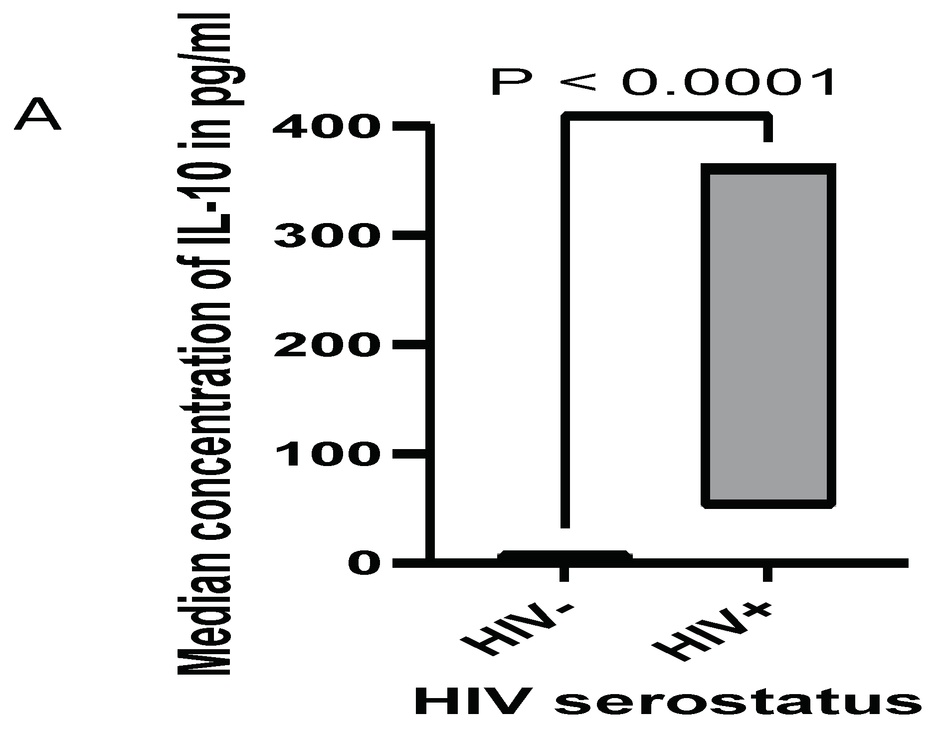
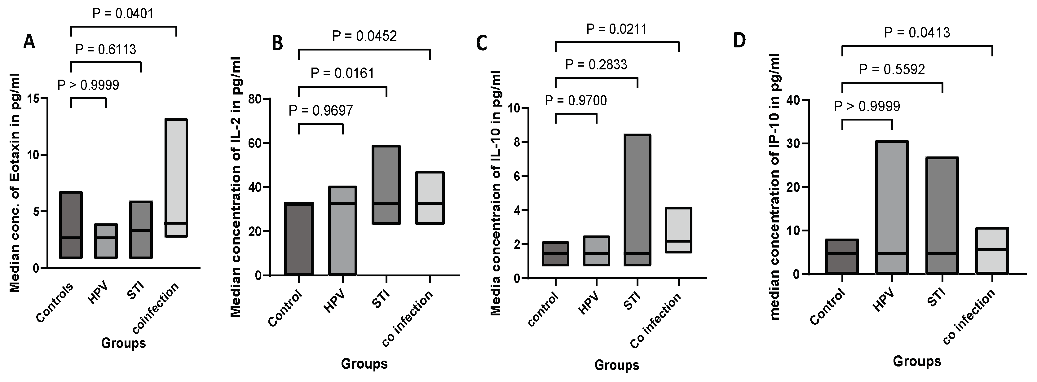
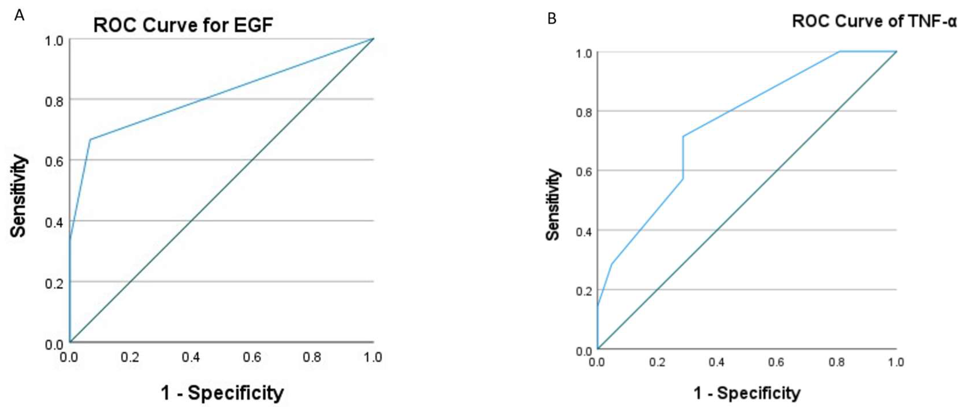
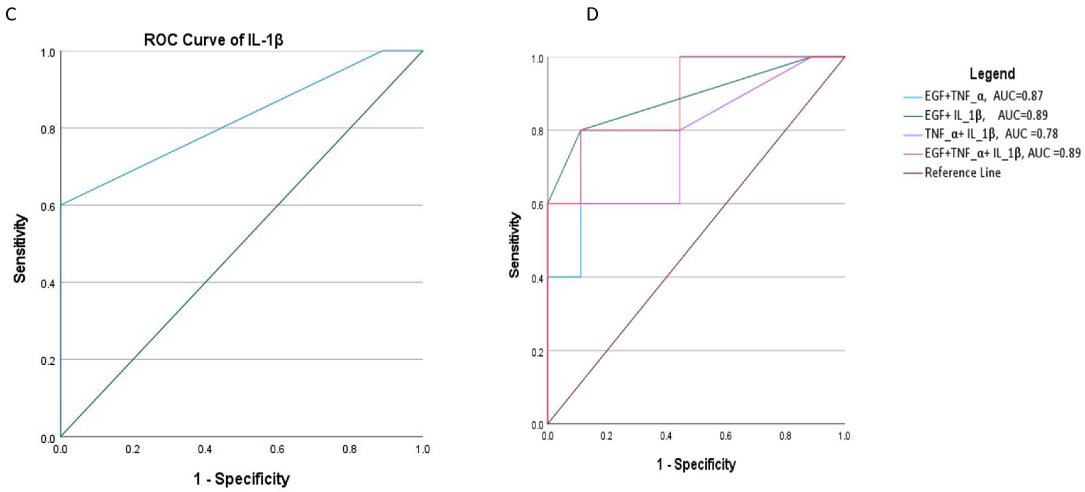
Background: The primary cause of cervical cancer is the high-risk human papillomavirus (hrHPV); the infection patterns may vary depending on the state of the vaginal immune system. Sexually transmitted infections (STIs) such as gonorrhea, trichomoniasis, and chlamydia can alter the local immune response, which in turn may affect the persistence and progression rate of hrHPV infections. The objective of this study was to compare cytokine levels from vaginal specimens collected from hrHPV positive, STI positive, and healthy controls, and to explore correlations with clinical parameters. Methods: Vaginal samples were collected from women diagnosed with hrHPV, asymptomatic STIs, and healthy controls. HrHPV and STIs status were confirmed through multiplex real time PCR. Vaginal cytokines and various growth factors were analyzed by Invitrogen ProcartaPlex™ Multiplex Immunoassay. Statistical analyses were performed to compare cytokine levels across different groups and to explore correlations with clinical parameters. Results: Women having STIs and hrHPV coinfection(n=26) had increased median concentration levels of Eotaxin (P=0.04), IL-2 (P=0.01), IL-10 (P=0.02), and IP-10 (P=0.04) compared to the control groups(n=22). Asymptomatic women with STIs (n=16) exhibited a distinct cytokine profile characterized by elevated levels of IL-2 (P=0.02) compared with control groups. Women with hrHPV(n=16) had lower levels of IL-15 (P=0.02) compared to controls. The median concentration levels of TNF-α (P=0.04), IL-1β (P=0.02), and EGF (P=0.01) were significantly greater in ≥CIN2+(n=8) than ≤CIN1(n=25). The area under the curve (AUC) of EGF, IL-1β, and the combination of EGF+IL-1β +TNF-α was good predictor for high-grade cervical lesions. Conclusion: The cytokine profiling revealed significant alterations in the vaginal immune environment in women with asymptomatic STIs and co-infection with hrHPV. The findings suggest that an inflammatory response in hrHPV infection may be modulated by co-existing STIs, potentially influencing disease progression. Elevated levels of EGF, IL-1β, and TNF-α in vaginal swabs suggest an inflammatory and proliferative environment that can contribute to the development and progression of cervical dysplasia.

Keywords: 
;  ;  ;  ;  

1. Introduction

Human papillomavirus (HPV) and sexually transmitted diseases (STIs), including gonorrhea, chlamydia, trichomoniasis, syphilis, and herpes, are linked to a host of adverse health outcomes, including cancer, infertility, chronic infection, and an increased risk of acquiring other infections(1,2). The host immune system and these infections have a complex and heterogeneous interaction, and understanding the mechanisms that underline them is essential to develop targeted prevention and treatment strategies (3).
An effective response to infections depends on the balance and regulation of cytokines. Cytokine profiling has enabled the understanding of the immunological components of a variety of incurable and infectious diseases and is becoming increasingly relevant because of its central role in influencing the immune response (4). Thus, cytokine profiling in a biological specimens provides insights into the host’s inflammatory milieu and immune status (5). This process is especially important for STIs and hrHPV infections because both local and systemic immune responses may have a significant impact on the development, dissemination, and clinical outcomes of these infectious diseases(6). Individual differences in the cytokine milieu can be attributed to a variety of factors, including the type and the location of the disease, co-infections, and the host’s immune status(7).
A high-risk HPV infection exhibits a specific cytokine profile(8). Inflammation is not a strong enough immune response to hrHPV, which could let the virus spread and avoid immune detections(9). Research has demonstrated a correlation between hrHPV infection and altered cytokine profiles, including reduced interferon-gamma (IFN-γ), increased interleukin-10 (IL-10), and other immunosuppressive cytokines. This immunosuppressive environment may help to progress hrHPV infection, cervical dysplasia, and malignancy(8,9).
The appropriate response to sexually transmitted infections is typically demonstrated by a careful balancing act between pro- and anti-inflammatory cytokines. Increased levels of cytokines like TNF-α, IL-1, and IL-6 indicate bacterial pathogen infections(10), such as those brought on by Neisseria gonorrhoeae and Chlamydia trachomatis, almost always result in a strong pro-inflammatory response(6). These cytokines are fundamental in attracting and energizing immune cells to the site of the infections, aiding in the pathogen’s clearance. Conversely, excessive inflammation can lead to tissue damage and chronic problems like as infertility and pelvic inflammatory diseases (11,12).
Cytokine profiling provides a view into the intricate relationships that hrHPV and STIs have with host immune system. The immunological mechanisms that characterize human interactions with STI and hrHPV appear to be an exciting new research area that is revealing new cytokine profiles (13). Through evidence of distinct cytokine signatures associated with hrHPV and STIs infections, and clinical outcomes, we can get a significant understanding of the development of these diseases. Moreover, this data may help design antibodies and targeted treatment strategies that improve clinical outcomes, by modifying the immune response(6,13). With meticulous cytokine profiling, we might be possible to identify underutilized biomarkers for disease diagnosis, prognosis, and treatment. This kind of research is scarce in Ethiopia. This study aimed to determine and compare the vaginal profile of cytokines in asymptomatic women, with either hrHPV, STIs, hrHPV/STIs co-infections, and healthy controls among sexually active asymptomatic women in Ethiopia. We also explored changes in vaginal cytokine levels of women with cervical intraepithelial lesions and determined the value of cytokine detection in assessing cervical lesions.

2. Methods and Materials

2.1. Study Setting and Study Population

The samples used in the study were collected from vagina in women of reproductive age who were screened for high-risk human papillomavirus (hrHPV) and sexually transmitted infections (STIs) such as Trichomonas vaginalis, Neisseria gonorrhea, Mycoplasma genitalium, Mycoplasma hominis, Ureaplasma urealyticum, Ureaplasma parvum, and Chlamydia trichomatis. Eighty women form attending cervical cancer screening in Adama cohort were included in this study. It comprised 16 women who tested positive for hrHPV, 16 women who tested positive for other STIs, 26 women who were co-infected, and 22 women who were negative controls. Among 42 hrHPV positive women, 33 had histology biopsy results, with 25 were negative for intraepithelial lesions or malignancy (NILM) or CIN1, and 8 had CIN2+ or invasive cervical cancer.

2.2. Data Collection and Laboratory Procedures

Sample collection: Copan eNat® swab collection and universal transport medium (Copan Italia SpA, Brescia, Italy) was used to collect and transport the vaginal samples for molecular investigations and cytokine profiling. The samples were collected from November 2021 to June 2022.
HPV detection and typing: HPV detection and typing was performed using a commercial kit, Anyplex II HPV HR (Seegene, Korea), which can detect 14 hr HPV types (i.e., 16, 18, 31, 33, 35, 39, 45, 51,52, 56, 58, 59, 66, and 68) in a single reaction tube by means of real-time PCR assays on the CFX96 real-time PCR instrument (Bio-Rad, Hercules, CA, USA).
STI detection: The STI detection was performed by using the Allplex™ STI-EA real-time multiplex kit with the CFX96™ thermal cycler (Bio-Rad, CA, USA).
Histology: Cervical biopsies were collected and handled routinely. The microscopic slides were examined and revealed the presence of benign alterations, low-grade squamous intraepithelial lesion (CIN1), high-grade squamous intraepithelial lesion (CIN2+), and invasive cervical cancer, as well as normal histology or no dysplasia (NILM).
Determination cytokine concentration: Invitrogen ProcartaPlex™ Multiplex Immunoassay (Lot number: 256805-001) was used for the determination of 45 cytokines, chemokines and growth factors including BDNF, EGF, Eotaxin, FGF-2, GM-CSF, GRO alpha, HGF, IFN alpha, IFN gamma, IL-1RA IL-1 alpha, IL-1 beta IL-2, IL-4, IL-5, IL-6, IL-7, IL-8, IL-9, IL-10, IL-12p70, IL-13, IL-15, IL-17A, IL-18, IL-21, IL-22, IL-23, IL-27, IL-31, IP-10, LIF, MCP-1, MIP-1 alpha, MIP-1 beta, NGF beta, RANTES, PDGF-BB, PIGF-1, SCF, SDF-1 alpha, TNF alpha, TNF beta, VEGF-A and VEGF-D.
ProcartaPlex™ Multiplex Immunoassay, a multiplex quantitative cytokine measurement technique, utilizes magnetic bead-based technology that allows for the simultaneous detection and quantitation of multiple cytokines from a single sample. Vaginal swab sample was used to measure cytokine, chemokine and growth factors using the manufacturer protocol. Luminex™ MAGPIX detection system from Invitrogen was used to assess the quantities cytokines. In a 96-well plate, all 45 analytes were detected at the same time following the manufacturers instruction (14).
Three distinct groups of beads were mixed and 50 µL of the mix were added per well, following the manufactories instructions. To keep the beads in place, a magnetic plate at the bottom of the plate was used for one wash. Addition of samples was done at 50 µL/well, and was kept at 4°C overnight.
In the next day, the plate was incubated for 30 minutes at room temperature with 500 rpm shaking. Following three washings, 25 µL of detection buffer per well was added, and the mixture was shaken at 500 rpm for 30 minutes at room temperature. Following three washings, 50 µL/well of the streptavidin–phycoerythrin (SAPE) solution was added, and the mixture was shaken at 500 rpm for 30 minutes at room temperature. Reading buffer (120 µL/well) was added after three washings. A MAGPIX Luminex equipment was used to read the plate following five minutes of 500 rpm shaking. The xPONENT® software Version 4.3 was used to obtain and visualized the MAGPIX Luminex result(14).
Normalization and standard curve: The concentration of the samples was calculated by plotting the expected concentration of the standards against the median florescent intensity (MFI) generated by each standard. A 5PL algorithm is recommended for the best curve fit. The result was analyzed according to the operation manual for the Luminex™ instrument. Normalization and standard curve were determined after export the run data in.csv format and navigate to the ProcartaPlex™ Analysis App on ThermoFisher Connect website: https://apps.thermofisher.com/apps/procartaplex.
Measurements below detection limit were set to the lowest standard value, values above detection limit were set to the highest standard concentration(14).

2.3. Statistical Analysis

All cytokine levels underwent log-transformation to ensure raw values were homogeneous and normal. The different cytokine levels were compared with the Kruskal–Wallis’s test. Kruskal–Wallis’s test was also used to compare cytokine levels of HIV negative, HIV positive, and HIV/HPV co-infected groups. Dunn’s test used to correct for multiple comparison groups of Kruskal–Wallis’s test results. Mann–Whitney U statistical tests used to compare differences between the two histological grades (≤CIN1 and ≥CIN2), and HIV positive and negative groups.

3. Result

Receiver operating characteristic (ROC) and area under the curve (AUC) analysis was used to identify optimal the minimum threshold values of cytokines levels to identify with maximum sensitivity and specificity for detection of ≥CIN2+ histological grades. All comparisons were two-sided, and a 5% level of significance was used. Statistical analysis (median, mean, standard error (SE), confidence interval (CI), AUC, ROC) was carried out with GraphPad Prism v10.3 was used (GraphPad Software Inc. USA).

3.1. Characteristics of the Study Population

The study included 80 women diagnosed with hrHPV, other sexually transmitted infections, co-infections, and healthy controls (Table 1). Of the participants, 10(13%) women lived with HIV. Thirty-three hrHPV positive women had biopsy results. The median age of the participants was with 32, with a range of 24-44 years. All participants were married.

3.2. Expression Levels of Cytokines

Out of the 45 cytokines analyzed, 42 (93%) were expressed, with at least one of the swab samples with cytokine showing detectable levels. Eighteen cytokines (40%), were expressed in more than half of the vaginal swabs. Eight (10%) cytokines, including Eotaxin, hepatocyte growth factor (HGF), Tumor Necrosis Factor-alpha (TNF-α), Leukemia Inhibitory Factor (LIF), Interleukin-12p70 (IL-12p70), IL-17 receptor A (IL-17RA), IL-4, and IL-10 were expressed in all samples (100%). Placenta Growth Factor 1 (PIGF-1), TNF-beta, and VEGF-D expression were not detected at any level in either the patients or control groups (Table 2).
Eighteen cytokines, including epidermal growth factor (EGF), Eotaxin, HGF, Interleukin-17 Receptor A (IL-1RA), Interleukin 2(IL-2), Interleukin-4 (IL-4), Interleukin-10 (IL-10), Interleukin-12 p70(IL-12p70), IL-13, IL-15, IL-17A (CTLA-8), IL-27, Interferon Gamma-Induced Protein 10 (IP-10 (CXCL10)), LIF, Monocyte Chemoattractant Protein 1 (MCP-1 (CCL2)), Stem Cell Factor (SCF), TNF alpha, and Vascular Endothelial Growth Factor A (VEGF-A) expression were higher in the swabs tested(Table 2).

3.3. Cytokine Expression in Women Live with HIV, and HIV+HPV

Women live with HIV (WLHIV) present a distinctive pattern of cytokines compared to HIV-negative women. WLHIV had higher levels of IL-10 than HIV-negative women (p<0.0001) (Figure 1). There has been no observed difference in cervical cytokine expression between HIV positive and HIV negative women for cytokines like EGF, Eotaxin, IL-2, IL-4, TNF-α, IP-10, INF-gamma, TGF-beta and so on.
Vaginal swab samples from women with HIV/hrHPV co-infection showed a positive correlation with IL-10 levels.

3.4. Cytokine Concentration in hrHPV, STI, hrHPV and STI Coinfection, and Controls

Women having STIs and coinfection with hrHPV had increased concentration of EGF, Eotaxin (CCL11), GM-CSF, HGF, IL-2, IL-4, IL-10, IP-10, and VEGF-A compared with controls. All of the chemokines tested, Eotaxin, HGF, TNF alpha, LIF, IL-12p70, IL-17RA, IL-4, and IL-10 were detected in all of the groups analyzed. HrHPV and STI co-infected participants showed significantly higher levels of IP-10, IL-10, IL-2, and Eotaxin (Table 3).
In this study, we analysis cytokines expression levels hrHPV + STI against controls (without hrHPV and STI), The median expression levels of the targeted cytokines were not statically different from controls.
In STI positive women, we found significantly higher levels of Eotaxin median concentration compared to controls (p=0.025) (Figure 2A). The median concentration level of IL-2 was significantly differed in STI (p=0.016) and coinfected women (p=0.045) compared to controls (Figure 2B). The median concentration levels of IP-10 (p=0.041) and IL-10 (p=0.021) differed significantly in coinfected women compared to controls, (Figure 2C,D).

3.5. Cytokine Concentration in NILM/CIN1 and CIN2+ and Cervical Cancer

We analyzed the relationships between cytokine levels (pg/ml) and the degree of cervical lesion (Table 4). The median vaginal swab concentration levels of Eotaxin, IL-1RA, IL-2, IL-4, IL-γ, IP-10, LIF, MCP-1, and VEGF were higher in the ≥CIN2+ and invasive cervical cancer groups than in the ≤CIN1 group. However, compared to the ≤CIN1 group, the median concentration levels of SCF and IL-27 was lower than in CIN2+ and cervical cancer groups. The median concentration levels of EGF, IL-1β, and TNF-α differed significantly among the two histological groups (≤CIN1 vs. ≥CIN2; P < 0.05). According to these findings, women with ≥CIN2 (CIN2+ and cervical cancer) had increased median concentrations of EGF (P = 0.01), IL-1β (P = 0.02), and TNF-α (p = 0.04) than those with ≤CIN1. No statistically significant differences were found in the median concentration levels of other cytokines with histological grades (P > 0.05).

3.6. Cytokine Score Effectiveness in Diagnosing Advanced Cervical Lesions

Table 5 displays the cytokine receiver operator characteristic (ROC) curve analysis used to distinguish between invasive cervical cancer and ≥CIN2+ from ≤CIN1. In the diagnosis of CIN2+ and invasive cervical cancer, the AUCs of EGF, TNF-α, and IL-1β, were 0.81 (95% CI: 0.57–1.0), 0.75(95% CI: 0.56–0.96), and 0.82 (95% CI: 0.56–1.0), respectively. When the three cytokines were pooled, the AUC was 0.89 (95% CI: 0.70-1.0) for differentiated CIN2+ and cervical cancer.
According to the ROC curve of the EGF, TNF-α, and IL-1β, sensitivities to detect CIN2+ and/or invasive cervical cancer at thresholds of >35.7, >16.8, and >8.0 were 0.67(95% CI:0.30-0.94), 1.0(95% CI:0.68-1.0), and 1.0 (95% CI:0.57-1.0), respectively, and specificities to detect CIN2+ and/or invasive cervical cancer at thresholds of >35.7, >16.8, and >8.0 were 0.93(95% CI:0.70-0.99), 0.14(95% CI:0.05-0.33), and 0.11(95% CI:0.01-0.44),respectively. The minimum threshold criterion was 8.0 pg/ml set to screen out CIN2+ and/or cervical cancer groups (Figure 3A–D).

Discussion

In our study, high-risk human papillomavirus (HrHPV) was associated with lover levels of IL-15 in the vagina, indicating a possible immunosuppressive environment. This finding is supported by reports which emphases on the immune evasion mechanisms of hrHPV infection(15). However, this is in contrast with a study conducted in Tanzanian women infected with HPV16/18 which reported elevated levels IL-15 in cervical tissues(8). IL-15 is a key cytokine involved in the activation and maintenance of natural killer (NK) cells and CD8+ T cells, both critical for controlling viral infections(16). In hrHPV infections, the observed decrease in IL-15 may reflect the virus’s ability to evade immune surveillance, fostering an immunosuppressive microenvironment that facilitates persistent infection and the progression of histological abnormalities(8). Studies suggest that reduced IL-15 levels are associated with higher rates of HPV persistence and progression to cervical intraepithelial neoplasia (CIN) or cervical cancer. However, IL-15 levels are generally higher in cervical cancer tissues(8). Immunological tolerance is necessary to ensure the persistence of HPV infection. Several studies have been conducted to test vaginal cytokine and chemokine levels in the past, HPV infection had the modest systemic impact (17,18).
In our study, co-infected individuals demonstrated a complex cytokine milieu with elevated levels of IL-2, IL-10, and IP-10. Studies have found that women with hrHPV coinfections have significantly higher levels of IL-10 and IL-2 in cervical samples, reflecting an active altered immune response aimed at viral clearance or lesion containment(19). Elevated Eotaxin levels in STI-positive women suggest increased eosinophilic activity or inflammation in the presence of STIs(20).
In the present study, we demonstrated that IL-2 was significantly elevated in co-infected individuals compared with controls, suggesting an ongoing inflammatory response. These findings are consistent with prior studies that have reported altered cytokine profiles, including elevated IL-2, in individuals with multiple infections. Studies have shown similar immune dysregulation in co-infected patients with HPV and Chlamydia trachomatis(21,22), as well as in those with HIV and HPV(23), where IL-2 are often elevated, indicating a complex and potentially exaggerated immune response. This finding suggests that the immune response in co-infection may be more robust or dysregulated, potentially contributing to the pathophysiology of co-infection. In comparison, the expression of IL-2 is typically lower in cervical cancer patients(24). Elevated IL-2 levels, known for their role in T cell activation and proliferation, might indicate an enhanced immune response or an attempt to combat the simultaneous infections(25).
Our finding revealed that anti-inflammatory IL-10 levels in co-infected individuals were significantly higher than in controls groups which is in agreement with prior studies. According to Syrjänen et al. (2009), high-grade CIN was most frequently linked to IL-10 up-regulation in tissue samples (26). This was further supported by a study conducted on Tanzanian women infected with HPV16/18 where elevated level of IL-10 was observed in the cervical tissue (8). Previous research has demonstrated that interleukin-10 (IL-10) is frequently overexpressed in cervical cancer tissues and in the blood of patients with this disease. IL-10 suppresses the immune response, potentially enabling cancer cells to evade immune detection and facilitate tumor growth. Elevated IL-10 expression is often linked to a shift towards a Th2-type immune response, which is typically associated with a non-inflammatory immune profile, as opposed to a Th1-type response that is more supportive of antitumor immunity(27,28).
Interferon induced protein-10 (IP-10, also known as CXCL10) was elevated in hrHPV/STI co-infected individuals compared with healthy controls. This elevation is significant because IP-10 is a chemokine that plays a crucial role in immune responses, particularly in attracting immune cells to sites of infection or inflammation. Elevated levels of IP-10 in co-infected individuals suggest a heightened immune response, which could be due to the combined effects of hrHPV and STIs on the immune system. Evidences showed that overexpression of IP-10 in infectious diseases and tumor development(29,30). However, in advanced cervical cancer, IP-10 levels have shown to be decreased(31). IP-10 has a strong thymus-dependent antitumor effect and primarily regulates the migration of T-cells, natural killer (NK) cells, and monocytes. IP-10, on the other hand, is crucial for controlling angiogenesis (30).
The significant elevation of EGF, IL-1β, and TNF-α in women with ≥CIN2 compared to ≤CIN1 highlights their potential role in lesion progression and cervical carcinogenesis. Elevated EGF may drive cellular proliferation in advanced lesions, while IL-1β and TNF-α likely contribute to inflammatory and tissue remodeling processes that exacerbate disease severity(32). Studies have shown that women with high-grade cervical intraepithelial neoplasia (CIN) or cervical cancer often exhibit altered EGF, IL-1β, and TNF-α expression in the cervix(33). Other research has indicated that cytokines such as TNF-α and IP-10 are significantly associated with hrHPV infections(34), further emphasizing the role of immune dysregulation in coinfections(35,36). This evidence collectively supports the assertion of a distinct cytokine profile in women with these infections, contributing to the pathogenesis and potential progression of cervical neoplasia(36). Elevated EGF levels may promote chronic inflammation and the creation of an immune-suppressive tumor microenvironment. Recent research highlights the significant role of inflammatory cytokines and growth factors, such as EGF, IL-1β, and TNF-α, in the progression from normal or lower-grade cervical intraepithelial neoplasia (≤CIN1) to higher-grade lesions (≥CIN2+)(32,37). Elevated EGF contributes to enhanced cell proliferation, while IL-1β and TNF-α promote chronic inflammation and tissue remodeling, both of which are critical in advancing cervical dysplasia. Persistent HPV infection, combined with heightened inflammatory signaling, appears to accelerate the progression to CIN2+ by creating a pro-tumorigenic microenvironment(38).
Studies indicated that WLHIV show increased levels of IL-10(39). Women with acute HIV infection have been found to possess significantly elevated levels of IL-10 in genital tract specimens(40). These findings highlight the heightened inflammatory response during the early stages of HIV infection. However, a study comparing cervical cytokine expression between HIV-positive and HIV-negative women have found no significant differences in the expression of levels INF-γ, TGF-β, IL-4, and IL-12(41). This result is consistent with our findings. The expression levels of the vast majority of the targeted cytokines did not differ significantly in our study. This suggested that HIV infection may not uniformly alter cytokine expressions in the cervical swabs.
In our study, a positive association between IL-10 concentration levels and HPV/HIV co-infection has been observed in vaginal swab samples. Previous studies have showed increased levels of IL-10 concentration in HPV/HIV co-infection compared to HIV negative ones (42). This suggests a predominance of Th2-type immune responses in HIV/HPV co-infected patients, potentially influencing disease outcomes.
The AUC of IL-1β and EGF were above 0.75, thus leading to a very good predictive value for CIN2+ and invasive cervical cancers. However, the AUC of TNF-α was below 0.75, thus leading to poor predictive value for CIN2+ and invasive cervical cancers. When the three cytokines were combined, AUC was slightly improved to detect CIN2+ and invasive cervical cancer. Further investigation is required to explore these links because statistical results may be impacted by small sample sizes.
One of the study’s limitations is the small number of CIN2+ and ICC cases we examined, which hinders us from drawing firm conclusions. We cannot rule out all other diseases such as bacterial vaginosis and vulvovaginitis, contributing factors to the immune response. Several cytokines were found to be below the baseline in more than half of the samples studied, which may have influenced the reliability and precision of the findings. Furthermore, the analysis lacks any follow-up studies, which would be useful in analyzing the rates of change of the cytokines over time and the development of associated diseases.

4. Conclusion

The findings reveal distinct cytokine profiles associated with sexually transmitted infections (STIs), co-infections, and the progression of cervical lesions, underscoring the role of immune modulation in these conditions. Differences in Eotaxin, IL-2, IP-10, and IL-10 between controls and coinfected individuals indicate shifts in immune signaling pathways, with IL-10 suggesting an anti-inflammatory response and IP-10 reflecting enhanced immune cell recruitment during coinfections. The significant elevation of EGF, IL-1β, and TNF-α in women with ≥CIN2+ compared to ≤CIN1 highlights their potential role in lesion progression and cervical carcinogenesis. These findings suggest a targeted immunological response in STI-positive individuals and those with advanced cervical lesions, offering insights into potential biomarkers for disease monitoring and progression. According to our findings, IL-1β and EGF may be utilized as predictors for severe cervical lesions. As a result, cytokine testing may serve as indicators of the progression of lesions. Further research is needed to explore these cytokines’ mechanistic roles and their potential as therapeutic targets in STI-related cervical disease.

Author Contributions

HB: and AM: research concept and design; HB, NA, and SM: collection and/or assembly of data; HB and DT: laboratory work and result interpretation; HB-data analysis and interpretation; drafting and writing of the manuscript; OF, CB, TA, AD, and MJ: critical revision of the manuscript and final approval of the article.

Ethics approval and consent to participate

This study was approved by ethical review committees at Addis Ababa University, the Department of Microbiology, Immunology, and Parasitology (DERC/04/2021), the College Ethical Review Board (IRB) (054/21/DMIP), the AHRI/ALERT Ethics Review Committee (AAERC) (PO/15/20), and the National Ethical Research Review Committee (NERC) (Ref. No. RAS/14.2/8382/21). The study was carried out, in accordance with the Declaration of Helsinki. Before recruitment, each subject provided written informed consent. The HPV and STI results were communicated with healthcare providers at Adama Referral Hospital Medical College for further evaluation and treatment. ClinicalTrials.gov ID: NCT05125380.

Availability of data and material

The data that support the findings of this study are available from the corresponding author upon reasonable request.

Acknowledgments

The authors would like to acknowledge all the collaborators of this work, Lund University, Addis Ababa University, and Armauer Hansen Research Institute.

Abbreviations

5PL Five parameter logistic algorism
AUC Area under curve
CIN1 Cervical Intraepithelial Neoplasia 1
CIN2+ Cervical Intraepithelial Neoplasia 2 or 3
HrHPV High-risk human Papillomavirus
ICC Invasive cervical cancer
NILM Negative for Intraepithelial Lesion or Malignancy
PCR Polymerase chain rection
PID Pelvic inflammatory disease
ROC Receiver operator characteristic
STIs Sexually transmitted infections

References

  1. Martinelli M, Musumeci R, Sechi I, Sotgiu G, Piana A, Perdoni F, et al. Prevalence of human papillomavirus (HPV) and other sexually transmitted infections (STIS) among italian women referred for a colposcopy. Int J Environ Res Public Health. 2019;16(24).
  2. WHO. Sexually transmitted infections (STIs);Key facts. 2024; Available from: https://www.who.int/news-room/fact-sheets/detail/sexually-transmitted-infections-(stis).
  3. Castle PE, Giuliano AR. Chapter 4: Genital tract infections, cervical inflammation, and antioxidant nutrients--assessing their roles as human papillomavirus cofactors. J Natl Cancer Inst Monogr [Internet]. 2003 Jun 1;2003(31):29–34. Available from: https://academic.oup.com/jncimono/article-lookup/doi/10.1093/oxfordjournals.jncimonographs.a003478.
  4. Duque GA, Descoteaux A. Macrophage cytokines: Involvement in immunity and infectious diseases. Front Immunol. 2014;5(OCT):1–12.
  5. Liu C, Chu D, Kalantar-Zadeh K, George J, Young HA, Liu G. Cytokines: From Clinical Significance to Quantification. Adv Sci. 2021;8(15).
  6. Masson L, Barnabas S, Deese J, Lennard K, Dabee S, Gamieldien H, et al. Inflammatory cytokine biomarkers of asymptomatic sexually transmitted infections and vaginal dysbiosis: A multicentre validation study. Sex Transm Infect. 2019;95(1):5–12.
  7. ter Horst R, Jaeger M, Smeekens SP, Oosting M, Swertz MA, Li Y, et al. Host and Environmental Factors Influencing Individual Human Cytokine Responses. Cell. 2016;167(4):1111-1124.e13.
  8. Vidal AC, Skaar D, Maguire R, Dodor S, Musselwhite LW, Bartlett JA, et al. IL-10, IL-15, IL-17, and GMCSF levels in cervical cancer tissue of Tanzanian women infected with HPV16/18 vs. non-HPV16/18 genotypes Clinical oncology. Infect Agent Cancer. 2015;10(1):1–7.
  9. Hibma, MH. The Immune Response to Papillomavirus During Infection Persistence and Regression. Open Virol J. 2013;6(1):241–8.
  10. Arnold KB, Burgener A, Birse K, Romas L, Dunphy LJ, Shahabi K, et al. Increased levels of inflammatory cytokines in the female reproductive tract are associated with altered expression of proteases, mucosal barrier proteins, and an influx of HIV-susceptible target cells. Mucosal Immunol [Internet]. 2016;9(1):194–205. [CrossRef]
  11. Mwatelah R, McKinnon LR, Baxter C, Abdool Karim Q, Abdool Karim SS. Mechanisms of sexually transmitted infection-induced inflammation in women: implications for HIV risk. J Int AIDS Soc. 2019;22(S6):32–9.
  12. Buder S, Schöfer H, Meyer T, Bremer V, Kohl PK, Skaletz-Rorowski A, et al. Bacterial sexually transmitted infections. JDDG - J Ger Soc Dermatology. 2019;17(3):287–315.
  13. Arnold KB, Burgener A, Birse K, Romas L, Dunphy LJ, Shahabi K, et al. Increased levels of inflammatory cytokines in the female reproductive tract are associated with altered expression of proteases, mucosal barrier proteins, and an influx of HIV-susceptible target cells. Mucosal Immunol [Internet]. 2016;9(1):194–205. [CrossRef]
  14. Thermo Fisher Scientific. ProcartaPlex TM Multiplex Immunoassay Multiplex immunoassays using magnetic bead technology for. 2022;0(41).
  15. Alcaide ML, Rodriguez VJ, Brown MR, Pallikkuth S, Arheart K, Martinez O, et al. High Levels of Inflammatory Cytokines in the Reproductive Tract of Women with BV and Engaging in Intravaginal Douching: A Cross-Sectional Study of Participants in the Women Interagency HIV Study. AIDS Res Hum Retroviruses. 2017;33(4):309–17.
  16. Perera PY, Lichy JH, Waldmann TA, Perera LP. The role of interleukin-15 in inflammation and immune responses to infection: Implications for its therapeutic use. Microbes Infect [Internet]. 2012;14(3):247–61. [CrossRef]
  17. Torres-Poveda K, Bahena-Román M, Delgado-Romero K, Madrid-Marina V. A prospective cohort study to evaluate immunosuppressive cytokines as predictors of viral persistence and progression to pre-malignant lesion in the cervix in women infected with HR-HPV: Study protocol. BMC Infect Dis. 2018;18(1):1–9.
  18. Sasagawa T, Takagi H, Makinoda S. Immune responses against human papillomavirus (HPV) infection and evasion of host defense in cervical cancer. J Infect Chemother. 2012;18(6):807–15.
  19. Adapen C, Réot L, Menu E. Role of the human vaginal microbiota in the regulation of inflammation and sexually transmitted infection acquisition: Contribution of the non-human primate model to a better understanding? Front Reprod Heal. 2022;4(December):1–19.
  20. Folci M, Ramponi G, Arcari I, Zumbo A, Brunetta E. Eosinophils as Major Player in Type 2 Inflammation: Autoimmunity and Beyond. In 2021. p. 197–219. Available from: https://link.springer.com/10.1007/5584_2021_640.
  21. Lugo LZA, Puga MAM, Jacob CMB, Padovani CTJ, Nocetti MC, Tupiná MS, et al. Cytokine profiling of samples positive for Chlamydia trachomatis and Human papillomavirus. PLoS One. 2023;18(3 March):1–15.
  22. Buckner LR, Lewis ME, Greene SJ, Foster TP, Quayle AJ. Chlamydia trachomatis infection results in a modest pro-inflammatory cytokine response and a decrease in T cell chemokine secretion in human polarized endocervical epithelial cells. Cytokine. 2013;63(2):151–65.
  23. Schindler S, Magalhães RA, Costa ELF, Brites C. Genital Cytokines in HIV/Human Papillomavirus Co-Infection: A Systematic Review. AIDS Res Hum Retroviruses [Internet]. 2022 Sep 1;38(9):683–91. Available from: https://www.liebertpub.com/doi/10.1089/aid.2021.0139.
  24. Cai Y, Zhai J, Wu Y, Chen R, Tian X. The Role of IL-2, IL-4, IL-10 and IFN-γ Cytokines Expression in the Microenvironment of Cervical Intraepithelial Neoplasia. Adv Reprod Sci. 2022;10(04):106–14.
  25. Spolski R, Li P, Leonard WJ. Biology and regulation of IL-2: from molecular mechanisms to human therapy. Nat Rev Immunol [Internet]. 2018;18(10):648–59. [CrossRef]
  26. Syrjänen S, Naud P, Sarian L, Derchain S, Roteli-Martins C, Longatto-Filho A, et al. Immunosuppressive cytokine Interleukin-10 (IL-10) is up-regulated in high-grade CIN but not associated with high-risk human papillomavirus (HPV) at baseline, outcomes of HR-HPV infections or incident CIN in the LAMS cohort. Virchows Arch. 2009;455(6):505–15.
  27. Scott ME, Ma Y, Kuzmich L, Mescicki AB. Diminished IFN-γ and IL-10 and elevated Foxp3 mRNA expression in the cervix are associated with CIN 2 or 3. Int J Cancer. 2009;124(6):1379–83.
  28. WANG Y, LIU X-H, LI Y-H, LI O. The paradox of IL-10-mediated modulation in cervical cancer. Biomed Reports. 2013;1(3):347–51.
  29. Liu M, Guo S, Hibbert JM, Jain V, Singh N, Wilson NO, et al. CXCL10/IP-10 in infectious diseases pathogenesis and potential therapeutic implications. Cytokine Growth Factor Rev [Internet]. 2011;22(3):121–30. [CrossRef]
  30. Yang L, Chen P, Luo S, Li J, Liu K, Hu H, et al. CXC-chemokine-ligand-10 gene therapy efficiently inhibits the growth of cervical carcinoma on the basis of its anti-angiogenic and antiviral activity. Biotechnol Appl Biochem [Internet]. 2009 Jul 23;53(3):209–16. Available from: https://iubmb.onlinelibrary.wiley.com/doi/10.1042/BA20090012.
  31. Sato E, Fujimoto J, Toyoki H, Sakaguchi H, Alam SM, Jahan I, et al. Expression of IP-10 related to angiogenesis in uterine cervical cancers. Br J Cancer. 2007;96(11):1735–9.
  32. Lin W, Niu Z, Zhang H, Kong Y, Wang Z, Yang X, et al. Imbalance of Th1/Th2 and Th17/Treg during the development of uterine cervical cancer. Int J Clin Exp Pathol [Internet]. 2019;12(9):3604–12. Available from: http://www.ncbi.nlm.nih.gov/pubmed/31934210%0Ahttp://www.pubmedcentral.nih.gov/articlerender.fcgi?artid=PMC6949808.
  33. Vitkauskaite A, Urboniene D, Celiesiute J, Jariene K, Skrodeniene E, Nadisauskiene RJ, et al. Circulating inflammatory markers in cervical cancer patients and healthy controls. J Immunotoxicol [Internet]. 2020;17(1):105–9. [CrossRef]
  34. Ali KS, Ali HYM, Jubrael JMS. Concentration levels of IL-10 and TNFα cytokines in patients with human papilloma virus (HPV) DNA+ and DNA- cervical lesions. J Immunotoxicol. 2012;9(2):168–72.
  35. Kobayashi A, Weinberg V, Darragh T, Smith-McCune K. Evolving immunosuppressive microenvironment during human cervical carcinogenesis. Mucosal Immunol [Internet]. 2008;1(5):412–20. [CrossRef]
  36. Song D, Li H, Li H, Dai J. Effect of human papillomavirus infection on the immune system and its role in the course of cervical cancer (Review). Oncol Lett. 2015;10(2):600–6.
  37. Fernandes APM, Gonçalves MAG, Duarte G, Cunha FQ, Simões RT, Donadi EA. HPV16, HPV18, and HIV infection may influence cervical cytokine intralesional levels. Virology. 2005;334(2):294–8.
  38. Bukowski A, Hoyo C, Vielot NA, Graff M, Kosorok MR, Brewster WR, et al. Epigenome-wide methylation and progression to high-grade cervical intraepithelial neoplasia (CIN2+): a prospective cohort study in the United States. BMC Cancer [Internet]. 2023;23(1):1–14. [CrossRef]
  39. Akoto C, Norris SA, Hemelaar J. Maternal HIV infection is associated with distinct systemic cytokine profiles throughout pregnancy in South African women. Sci Rep [Internet]. 2021;1–15. [CrossRef]
  40. Bebell LM, Passmore JA, Williamson C, Mlisana K, Iriogbe I, Van Loggerenberg F, et al. Relationship between levels of inflammatory cytokines in the genital tract and CD4+ cell counts in women with acute HIV-1 infection. J Infect Dis. 2008;198(5):710–4.
  41. Guimarães MVMIB, Michelin MA, Lucena AADS, Lodi CTDC, Lima MIDM, Murta EFC, et al. Cytokine expression in the cervical stroma of HIV-positive and HIV-negative women with cervical intraepithelial neoplasia. Viral Immunol. 2014;27(7):350–5.
  42. Schindler S, Netto E, Deminco F, Figueiredo CA, de Andrade CM, Alves AR, et al. Detection of cytokines in cervicovaginal lavage in HIV-infected women and its association with high-risk human papillomavirus. Front Immunol. 2024;15(June):1–7.
Figure 1. The median cytokine levels in women with HIV, and HIV +HPV (A, B). P value was calculated by using Mann-Whitney U test.
Preprints 149733 g001
Figure 2. The median concentration levels of cytokines that are significantly linked infections status (A, B, C, D). P values < 0.05, were calculated using Karskulis-Wallis’s test and correct for multiple comparison using Dunn’s test.
Preprints 149733 g002
Figure 3. ROC curves of EGF, TNF-α, IL-1β, and pooling of the three cytokines for the prediction of ≥CIN2+ and invasive cervical cancer.
Preprints 149733 g003aPreprints 149733 g003b
Table 1. Characteristics of the study participants (n=80).
Variable Responses Count Percent
Median age in years 32
Age group in years < 30 31 38.8
≥30 49 61.2
High-risk Human papillomavirus (hrHPV)§ Positive 42 52.5
Negative 38 47.5
Histology result (n=33) NILM or CIN1 25 75.8
CIN2+ and cervical cancer 8 24.2
Sexually transmitted infections (STI)|* Positive 42 52.5
Negative 38 47.5
HIV serostatus Positive 10 12.5
Negative 70 87.5
Infection Status of Participants HPV only 16 20
STIs only 16 20
Co-infection 26 32.5
Health controls 22 27.5
Note: *NG(n=1), CT(n=2), TV(n=5), MG(n=2), MH(n=25), UP(n=24), UU(n=20). §HrHPV16(n=5),18(n=1), 31(n=7), 33(n=2), 35(n=5), 39(n=2), 45(n=0), 51(n=6), 52 (n=5), 56(n=6), 58(n=2), 59(n=1), 66 (n=4), 68(n=4).
Table 2. The descriptive statics of targeted 45 cytokines (n=80).
Cytokines Number of samples expressed n (%) IQR(Q3-Q1) Median Mean ±SE
BDNF 2(3) 0.72-4.72 2.72 2.7±2.0
EGF 52(65) 24.25-24.25 24.25 35.7±4.78
Eotaxin (CCL11) 80(100) 2.68-3.96 2.68 3.4±0.18
FGF-2 2(3) 13.59-50.04 31.82 31.8±18.23
GM-CSF 20(25) 42.95-42.95 42.95 49.8±4.60
GRO alpha (CXCL1) 2(3) 8.17-31.12 19.65 19.7±11.48
HGF 80(100) 19.21-31.32 19.21 26.2±1.76
IFN alpha 11(14) 0.61-0.97 0.61 1.03±0.25
IFN gamma 25(31) 3.78-3.78 3.78 4.69±0.61
IL-1 alpha 2(3) 0.23-0.62 0.425 0.43±0.20
IL-1 beta 31(39) 10.3-18.4 10.3 14.2±1.67
IL-1RA 68(85) 144.2-362.1 144.2 259±38.98
IL-2 78(98) 23.0-33.69 32.66 32.2±0.93
IL-4 80(100) 8.36-10.73 8.36 8.83±0.31
IL-5 1(1) 15.35-15.35 15.35 15.4±0
IL-6 1(1) 26.3-26.3 26.3 26.3±0
IL-7 11(14) 1.07-2.18 1.64 1.62±0.20
IL-8 (CXCL8) 1(1) 79.97-79.97 79.97 80±0
IL-9 1(1) 36.78-36.78 36.78 36.8±0
IL-10 79(99) 1.47-2.18 1.47 1.89±0.12
IL-12p70 80(100) 2.45-3.283 2.45 2.70±0.13
IL-13 69(86) 8.62-15.21 8.62 11.6±0.92
IL-15 70(88) 3.39-6.19 3.39 5.69±0.61
IL-17A (CTLA-8) 80(100) 4.94-6.96 4.94 6.14±0.34
IL-18 6(8) 14.37-33.73 20.22 25.3±6.70
IL-21 1(1) 9.48-9.48 9.48 9.48±0
IL-22 6(8) 5.21-14.63 5.21 11.5±6.28
IL-23 1(1) 138.4-138.4 138.4 138±0
IL-27 76(95) 23.69-49.88 49.88 43.6±3.19
IL-31 1(1) 117.3-117.3 117.3 117±0
IP-10 (CXCL10) 67(84) 4.75-6.61 4.75 6.29±0.52
LIF 80(100) 6.76-8.47 6.76 8.11±0.42
MCP-1 (CCL2) 44(55) 6.4-7.585 6.4 8.65±1.26
MIP-1 alpha (CCL3) 3(4) 1.01-3.96 2.12 2.36±0.86
MIP-1 beta (CCL4) 2(3) 10.77-56.66 33.72 33.7±22.95
NGF beta 3(4) 1.51-3.97 2.78 2.75±0.71
PDGF-BB 2(3) 8.08-15.02 11.55 11.6±3.47
PlGF-1 0(0)
RANTES (CCL5) 1(1) 1.59-1.59 1.59 1.59±0
SCF 73(91) 0.66-1.26 0.66 1.07±0.10
SDF-1 alpha 1(1) 113.9-113.9 113.9 114±0
TNF alpha 79(99) 20.12-26.19 20.12 23.2±1.0
TNF beta 0(0)
VEGF-A 66(83) 4.52-7.53 6.06 77.4±67.99
VEGF-D 0(0)
Note: Interquartile Range (IQR), Q1 (First Quartile), Q3 (Third Quartile), SE, Standard error, unit of cytokine concentration pg/ml.
Table 3. Cytokine levels in the vaginal swabs of women with hr-HPV, sexually transmitted infections, hrHPV/STI co-infection, and controls.
Cytokine classification Cytokines Median (IQR) (pg/ml)
Groups
Controls HrHPV STI Co-infection
Chemokines MCP-1 6.4(6.4-6.4) 6.4(4.4-33.4) 6.4(6.4-8.6) 6.4(6.4-9.0)
P value - >0.99 >0.99 >0.99
IP-10 4.8 (4.8-5.5) 4.8(2.4-4.8) 4.8(4.8-6.6) 5.7(4.8-7.8)
P value - >0.99 >0.99 0.04*
Eotaxin 2.7(2.7-3) 2.7(2.7-3) 3.9 (2.7-5.0) 4.0 (4.0-5.0)
P value - >0.99 0.61 0.04*
Growth factors GM-CSF 43(43-43) 43(43-43) 53.1 (43-63.3) 43(43-63.3)
P value - >0.99 0.35 0.20
HGF 19.2(19.2-31.3) 19.2(19.2-31.3) 19.2(19.2-42.9) 19.2(31.3-31.3)
P value - >0.99 >0.99 >0.99
EGF 24.3(24.3-39.1) 24.3(24.3-24.3) 24.3(24.3-41.4) 24.3(24.3-47.2)
P value - 0.79 >0.99 >0.99
SCF 0.7(0.7-1.26 0.7(0.7-1.1) 1.3(0.7-1.3) 1.3(0.7-1.5)
P value - >0.99 0.33 0.056
VEGF-A 6.1(4.1-7.5) 6.1(0.0-6.1) 4.5(2.9-6.8) 6.1(2.9-9.3)
P value - >0.99 >0.99 0.50
Interferons INF-γ 3.8(3.8-8.1) 3.8(3.8-3.8) 3.8(3.8-3.8) 3.8(2.9-5.3)
P value - >0.99 >0.99 >0.99
Anti-Inflammatory or Immunoregulatory Cytokines IL-10 1.5(1.5-2.2) 1.5(1.5-1.8) 1.5(1.5-2.0) 2.2(1.5-2.9)
P value - 0.93 >0.99 0.02*
IL-1RA 144(144-144) 144(144.2-199) 144(144-144.2) 144(306-362.1)
P value - >0.99 >0.99 0.25
T-Cell Differentiation Cytokines IL-2 33(23-33) 33(23-33) 33(23-41) 33(33-41)
P value - >0.99 0.02* 0.01*
IL-4 8.4(8.4-9) 8.4(6-11) 8.4(7.4-10.7) 8.4(8.4-13)
P value - 0.94 0.99 0.56
IL-13 8.6(8.6-14) 8.6(8.6-10.3) 8.6(8.6-15.2) 8.6(8.6-15.2)
P value - >0.99 >0.99 >0.99
IL-17A 4.9(4.9-5.5) 4.9(3.3-4.9) 6.0(4.9-8.4) 5.5(4.9-7.4)
P value - 0.73 0.19 0.11
Pro-Inflammatory Cytokines IL-1β 10.3(10.3-10.3) 8.0(5.7-10.3) 14.5(10.3-25.9) 10.3(10.3-18.4)
P value - 0.95 0.22 0.61
TNF-α 20.1(18.6-26.2) 20.1(13.5-25.5) 20.1(20.1-26.2) 26.2(20.1-27.6)
P value - 0.99 0.99 0.08
Other
Cytokines
IL-12P70 2.5(2.5-2.5) 2.5(1.5-2.5) 2.5(2.5-3.4) 2.5(2.5-3.6)
P value - 0.83 >0.99 0.24
IL-15 6.2 (3.4-6.2) 3.4(3.4-3.4) 3.4(3.4-8.7) 6.2(3.4-6.2)
P value - 0.02* >0.99 >0.99
IL-27 23.7(23.7-49.9) 23.7(23.7-43.3) 23.7(49.9-49.9) 23.7(49.9-55.6)
P value - 0.83 >0.99 0.22
LIF 6.8(6.8-8.5) 6.8 (5.1-8.5) 6.8(5.4-8.5) 8.5(6.8-10)
- >0.99 >0.99 0.46
*P value < 0.05, p values were calculated using Kruskal-Wallis’s test and correct for multiple comparison using Dunn’s test.
Table 4. Cytokine median concentration levels of histological grades (≤CIN1 and ≥CIN2+).
S. No Cytokines Median (IQR) in pg/ml P value
≤CIN1 ≥CIN2+
1 EGF 24(24-24) 47(24-125) 0.01*
2 Eotaxin 2.7(2.7-4.0) 3.3(2.7-5.0) 0.14
3 GM-CSF 43(43-43) 43(43-130) 0.33
4 HGF 19(19-31) 19(19-40) 0.90
5 INF-γ 3.8(2.1-4.5) 3.8(3.8-14) 0.27
6 IL-1β 10(10-10) 14(10-36) 0.02*
7 IL-1RA 144(144-362) 362(144-2037) 0.99
8 IL-2 33(23-31) 37(33-41) 0.06
9 IL-4 8.4(5.8-11) 9.0(6.4-12) 0.47
10 IL-10 1.7(1.5-2.2) 2.2(1.6-2.4) 0.28
11 IL-12p70 2.5(1.9-3.4) 2.5(2.5-4.3) 0.27
12 IL-13 8.6(8.6-10) 8.6(8.6-14) 0.56
13 IL-15 3.4(3.4-6.2) 3.4(3.4-35) 0.56
14 IL-17A 4.9(4.9-7) 4.9(4.9-8.4) 0.55
15 IL-27 50(24-50) 37(24-73) 0.61
16 IP-10 4.8(4.8-6.6) 5.7(4.8-7.8) 0.47
17 LIF 6.8(6.8-8.5) 8.5(6.8-9.7) 0.46
18 MCP-1 6.4(6.4-6.4) 9.3(6.4-13) 0.10
19 SCF 0.97(0.66-1.3) 0.66(0.66-2.2) 0.97
20 TNF-α 20(20-26) 26(21-30) 0.04*
21 VEGF-A 6.1(4.5-7.5) 7.5(6.1-36) 0.12
Note: ≤CIN1 = Negative for Intraepithelial Lesion or Malignancy and Cervical Intraepithelial Neoplasia 1, ≥CIN2+=Cervical Intraepithelial Neoplasia 2/3 and invasive cervical cancer, * p value <0.05, which was determined using Mann-Whitney U test.
Table 5. ROC curve analysis of cytokines to differentiate CIN2+ and cervical cancer from ≤ CIN1.
Cytokines AUC(95%CI) P value
EGF 0.81(0.57-1.0) 0.029
TNF-α 0.75(0.56-0.96) 0.05
IL-1β 0.82(0.56-1.0) 0.053
EGF +TNF-α 0.87(0.67-1.0) 0.028
EGF +IL-1β 0.89(0.68-1.0) 0.02
TNF-α + IL-1β 0.78(0.49-1.0) 0.096
EGF +TNF-α + IL-1β 0.89(0.70-1.0) 0.02
AUC, Area under the curve, CI, Confidence interval.
Disclaimer/Publisher’s Note: The statements, opinions and data contained in all publications are solely those of the individual author(s) and contributor(s) and not of MDPI and/or the editor(s). MDPI and/or the editor(s) disclaim responsibility for any injury to people or property resulting from any ideas, methods, instructions or products referred to in the content.
Copyright: This open access article is published under a Creative Commons CC BY 4.0 license, which permit the free download, distribution, and reuse, provided that the author and preprint are cited in any reuse.
Prerpints.org logo

Preprints.org is a free preprint server supported by MDPI in Basel, Switzerland.

Subscribe

Disclaimer

Terms of Use

Privacy Policy

Privacy Settings

© 2025 MDPI (Basel, Switzerland) unless otherwise stated