
Submitted:
04 February 2025
Posted:
05 February 2025
You are already at the latest version
Abstract
Ulcerative colitis (UC) is the most prevalent inflammatory bowel disease (IBD) in the USA. Current treatments present clinical limitations, underscoring the need for innovative therapeutics that promote an anti-inflammatory immune response. This study evaluates the anti-inflammatory potential of Fh15, a recombinant Fasciola hepatica fatty acid binding protein in a DSS-induced UC mouse model. Our results demonstrated that Fh15 treatment significantly ameliorated the severity of colitis by reducing the disease activity index (DAI) and histopathological scores. Moreover, Fh15 also decreased serum levels of myeloperoxidase (MPO), and chitinase-3-like protein 1 (CHI3L1), and the expression of S100A9, a calcium-and zinc binding protein, which is an important marker for the pathogenesis of UC. Furthermore, Fh15 downregulated pro-inflammatory cytokines TNFα and IL-1β in the distal colon, suggesting modulation of macrophage activity. Immunohistochemistry analysis revealed significantly reduced neutrophil and macrophage infiltration in UC Fh15-treated mice. These findings highlight the therapeutic potential of Fh15 for UC, as it modulates inflammatory responses, reduces leukocyte infiltration, and preserves colon integrity.
Keywords:
Ulcerative colitis
; Fasciola hepatica
; fatty acid binding protein
; myeloperoxidase
; chitinase-3 like-protein-1
; S100A9
; TNF-α
; IL-1β
; neutrophils
; macrophages
1. Introduction
Ulcerative colitis (UC) is the most common inflammatory bowel disease (IBD) affecting approximately 286 individuals for every 100,000 persons per year in USA [1]. Its high prevalence also extents to Canada and many countries in Western Europe. Although the underlying etiology of UC remains unclear, it is thought to result from a combination of factors, including genetic predisposition, immunoregulatory issues, environmental influences, and gut microbiota [2]. These factors contribute to a chronic inflammatory state driven by colitogenic, pro-inflammatory T helper type-1 (Th1) cells, characterized by the production of large amounts of interferon-gamma (IFN-γ) and tumor necrosis factor (TNF) [3]. There is no cure for UC, and existing treatments have significant limitations that prevent many patients from achieving remission [4,5]. Therefore, there is a need to incorporate new treatments for UC.
Previous epidemiological studies and clinical trials have suggested that helminth infections might protect people from IBDs [6]. Helminths have co-evolved with the humans for millions of years and exert a strong immunomodulatory effect on their host. They polarize the immune response towards an anti-inflammatory Th2/T-regulatory response, which can only be achieved by suppressing the Th1-inflammatory response [7]. This strategy led helminths to establish a prolonged chronic infection in their mammalian host [8]. The prevalence of helminth infections contrasts with the patterns observed for IBDs and other autoimmune diseases. Helminth infections are endemic in underdeveloped countries but are rarely reported in Western countries. Interestingly, the noticeable rise in IBD cases in developing countries has been linked, among other factors, to the widespread application of intensive anti-parasitic treatments aimed at deworming the population. It is believed that such deworming campaign might have disrupted the long-term regulatory network established by helminths and may be in part responsible for the emergence of immunological disorders [9].
Considering that T-cells are critical for IBD and helminth infections, the reciprocal cross-regulation between Th1 and Th2 cells suggests that the polarization of a Th2 response by helminths could prevent or minimize the effects of Th1-mediated diseases. It has been demonstrated that the infection of mice with the nematode Trichinella spiralis ameliorates subsequent induced colitis, which was associated with a downregulation of the Th1 response [10]. Similarly, an infection with Fasciola hepatica, one of the most widely distributed trematodes worldwide, has been shown to attenuate the clinical symptoms of murine autoimmune encephalomyelitis [11] and prevent type-1 diabetes development in the non-obese diabetic mouse model [12]. Experimental infections with schistosome cercariae have been shown to confer protection against type-1 diabetes [13], encephalomyelitis [14] and colitis [15]. Moreover, clinical trials have provided evidence that the therapy with ova from the nematode Trichuris suis is effective for treating Crohn’s disease and ulcerative colitis without any adverse effects [16]. However, the use of ova, cysts, or deliberate exposure to helminth larvae as a therapeutic approach may be unappealing to patients and ethically unacceptable to many. The inability to control the progression of such helminthic infections remains a major concern. Additionally, the strong polarizing immune response toward Th2 induced by these parasites is highly nonspecific, potentially rendering the host incapable of responding effectively to concurrent infections that require a Th1-mediated immune response for protection [12,17]. In this sense, the identification of helminth-derived molecules that ultimately mediate host immune modulation could be a more attractive and feasible therapeutic option [18,19].
Our research group has identified Fh15, the recombinant form of a member of the F. hepatica fatty acid binding protein family and demonstrated that it exhibits powerful anti-inflammatory properties. A single dose of Fh15 administered intraperitoneally to mice exposed to lethal doses of lipopolysaccharide (LPS)- E. coli can significantly suppress the cytokine storm [20] and increase the survival rate [21]. When Fh15 was administered prophylactically via intravenous infusion to non-human primates challenged with live E. coli, it was shown to suppress the cytokine storm, bacteremia, endotoxemia, and other inflammatory markers associated with sepsis [22]. Although sepsis and ulcerative colitis are distinct diseases, they share common characteristics: both can be caused by bacterial infections, trigger excessive inflammatory responses, and result in severe health complications. In this study, we investigated the potential of Fh15 as a therapeutic agent to suppress intestinal inflammation caused by ulcerative colitis using a DSS-induced colitis model in C57BL/6 mice. Our results demonstrate that Fh15 can mitigate intestinal inflammation by modulating the colonic immune cell infiltration and suppressing pro-inflammatory markers associated with the severity of UC pathology. Thus, Fh15 holds promise for further research aimed at developing a new helminth-derived treatment against ulcerative colitis.
2. Materials and Methods
2.1. Animals and Ethics Statement
A total of 50 C57BL/6 male mice (6–8-week-old) were obtained from Charles River Laboratories and maintained under standard conditions at 21°C and 12-hour light-dark cycle, with access to food and water ad libitum. All procedures were conducted according to the regulations of the Ethics Institutional Animal Care and Use Committee of the University of Puerto Rico-Medical Sciences Campus (Protocol No. 7870123). Euthanasia was performed under deep anesthesia.
2.2. Recombinant F. hepatica FABP (Fh15)
The recombinant Fh15 used in the present study was obtained as previously described [22] and consisted of 10 ml of purified Fh15 (2.29mg/ml, <0.4 EU/mg) with >90% purity confirmed by LC-MS. Fh15 was expressed in Bacillus subtilis as a fusion protein with a 6XHis at the amino terminus and purified by a Ni+-agarose column. The aliquots of purified Fh15 used in the present study remained stored at -80oC until use.
2.3. Dextran Sulfate Sodium (DSS) Colitis Induction and Fh15 Treatment
First, 5 animals were randomly assigned into 1 of 6 groups (PBS, DSS, Fh15, DSS-Fh15 50µg, DSS-Fh15 100 µg, and DSS-Fh15 150 µg) to perform a dose-response experiment. The healthy control group, named PBS, drank normal water, and received three intraperitoneal (i.p.) injections on days 1, 3, and 5 of 50 µL 0.1 M phosphate-buffered saline (PBS) pH 7.2 endotoxin free. The group designated as Fh15 drank normal water and received three i.p. injections of 50 µg Fh15 diluted in 50 µL PBS on days 1, 3 and 5. Acute colitis was induced in groups labeled as DSS and DSS-Fh15 by providing ad libitum access to autoclaved drinking water with 4% (w/v) dextran sulfate sodium (DSS; 40kDa, Sigma-Aldrich) for 7 days as described by Chaissang et al. (2014) [23]. The DSS-Fh15 groups received three i.p. doses of 50 µg (2.0mg/kg), 100 µg (4.0 mg/kg), or 150 µg (5.0 mg/kg) of Fh15 according to their respective group. These doses were administered on days 1, 3, and 5 of DSS-water consumption (Figure 1). The number of Fh15 injections was determined based on studies reported in the literature using other helminth antigens [19]. At the end of the experimental period (Day 7), animals were anesthetized and bled via the orbital vein using capillary tubes. The serum was obtained by blood centrifugation (10,000 x g, 4oC, 10 minutes). After bleeding, animals were euthanized and necropsied to collect their colons.
2.4. Disease Activity Index (DAI)
Disease activity index (DAI) was used as primary criterion to assess colitis severity, as described by Wang et al. (2017) [18]. Mice were monitored daily for changes in body weight, stool consistency, and the presence of blood in stool. Disease severity was assessed by a clinical scoring system as follows: weight loss, 0 (< 2%), 1 ( ≥ 2% - < 5%), 2 (≥ 5% - <10%), 3 (≥10% - < 15%) or 4 (>15%); stool consistency, 0 (normal), 1 (softer stool), 2 (moderate diarrhea), or 3 (diarrhea); and the presence of blood in stool, 0 (no rectal bleeding), 1 (positive Hemoccult/no visible blood), 2 (visible blood in stool), or 3 (fresh rectal bleeding) (Table 1). The summed scores for weight loss, stool consistency, and the presence of blood in stool were used as the DAI.
2.5. Macroscopic Score and Histopathological Scoring
Colons removed from each animal on euthanasia day were macroscopically examined by using a modified established scoring system [24] to indirectly measure inflammation. We measured colon length in centimeters (cm) from the cecum to the rectum, adhesion, inflamed length (cm), bowel thickness (mm) and observed the presence of hemorrhage, fecal blood, and diarrhea on euthanasia day (Table 2). All scores per mouse were summed to determine the final macroscopic score. Using the Swiss-rolling technique, mice colons were fixed 48 hours in 10% formalin. Tissues were embedded in paraffin and cut into 4-µm-thick sections on positively charged slides. Dewaxed sections were routinely stained with hematoxylin-eosin (H&E) dyes for histological evaluation [25]. The histopathological score was calculated according to well-accepted parameters described elsewhere [26] in a blinded fashion by an expert pathologist (J.T.A.). The parameters used for evaluation are described in Table 3. All scores per parameter and per mouse were summed to determine the final histopathological score.
2.6. Serum Myeloperoxidase and Chitinase-3 Like-Protein-1 Concentration
To measure levels of myeloperoxidase (MPO) in serum, an enzyme released primarily by neutrophils [27], we utilized a Mouse Myeloperoxidase SimpleStep ELISA® Kit (Abcam; ab275109). Chitinase-3-like protein 1 (CHI3L1) levels in serum were measured using a Mouse YKL-40/CHI3L1 ELISA® kit (Abcam; ab238262). CHI3L1 is secreted by different cell types and is considered an important marker of inflammation [28]. Absorbance for both MPO and CHI3L1 was measured spectrophotometrically at 450 nm, with concentrations reported in pg/mL.
2.7. Colonic Cytokines Gene Expression
TNF-α and IL-1β expression were measured in distal colon sections of each mouse. Tissue was stored in TRIzol™ reagent and kept at -80oC until used. Total RNA was extracted using the AllPrep® DNA/RNA/Protein kit (Qiagen, Germany). Because DSS can inhibit the qRT-PCR by altering the binding between the reverse transcriptase and primed RNA [29], we removed DSS by purifying the mRNA from total RNA using the RNeasy® Pure mRNA Bead kit (Qiagen, Germany). Purified mRNA (10ng) was used to generate cDNA using the Applied Biosystems (Thermo Fisher Scientific, USA) Power SYBR™ Green RNA-to Ct™ 1 Step kit. The cDNA was subjected to real-time quantitative PCR using the QuantStudio™ 3 system with the QuantStudio™ Design and Analysis software (v1.5.2). Cytokines primers sequences by Sigma Aldrich are shown in Table 4. The relative expression of each gene was calculated by 2-ΔΔCt (RQ) using GAPDH as an internal reference.
2.8. Colon Immune Cells Infiltration and Calcium Binding Protein Marker
Dewaxed colon sections were subjected to standard immunohistochemistry (IHC) procedures to determine immune cell infiltration and measure levels of S100 calcium binding A9 protein (S100A9), which is a protein secreted by granulocytes and is highly elevated in patients with UC [30]. The primary antibodies used were rabbit anti-mouse F4/80 (macrophage), anti-Ly6G (neutrophil), and anti-S100A9 (Table 5). As the secondary antibody, we used an anti-rabbit antibody-horseradish peroxidase (HRP) polymer conjugate (MACH2, Cat# RHRP520MM, Biocare) and as the chromogenic substrate, we used 3,3’-diaminobenzidine (DAB) (Cat# SK-4103-400, Vector Labs). IHC slides were counterstained with hematoxylin. Brightfield images were captured with an inverted confocal microscope equipped with a 40X objective. The percentage of antibody positive tissue areas were evaluated using Nikon NIS Elements AR Software v.5.20 [31]. First, a binary layer threshold was created to exclude unstained colonic tissue from antibody-stained tissue area. Then, three regions of interest (ROIs) were created, each with a total area of 5057.61 µm² and randomly distributed across the tissue area. This process allows to quantify ROI’s area fraction occupied by antibody staining. A total of three images from different colon regions, per animal, were analyzed for each antibody of interest. The percentage area of the thresholder signal was exported and normalized to the average value of non-treated tissues.
2.9. Statistical Analysis
Results are expressed as mean ± SE values. Statistical significance was determined by one-way or two-way ANOVA with Dunnett's multiple comparisons using GraphPad Prism software v.8 (GraphPad Software Inc., La Jolla, CA, USA). Data with a p < 0.05 was considered statistically significant.
3. Results
3.1. Fh15 Treatment Reduces Disease Activity Index in DSS-Induced UC Mice
We used C57BL/6 mice, the prototypical Th1-type mouse strain [32,33], to develop the DSS-induced UC mouse model. To optimize Fh15 treatment dose we first accomplished a dose-response experiment in which three different DSS-groups received i.p. injections with 50μg, 100μg or 150μg of Fh15 on days 1, 3 and 5. Each Fh15 administration corresponded to doses of 2.0, 4.0, or 5.0 mg/kg body weight, respectively. Animals were monitored daily, and the disease activity index (DAI) was calculated based on weight loss, stool consistency, and presence of blood in stool (Table 1). Compared to the baseline, mice in the DSS group exhibited a significant weight loss, with mice showing a mean loss of 19.39% by day 7 (Figure 2A, Table S1). The DSS group also displayed the highest scores for rectal bleeding and diarrhea, achieving a maximum Disease Activity Index of 10 (Figure 2B, Figure S1), indicating a clear and progressive intestinal damage. In contrast, animals that received the treatment with Fh15, irrespectively of the dose assayed, showed significant reduction in DAI (p < 0.0001) from day 2 to 7 (Figure S2A). Since no differences in DAI reductions were observed among the Fh15 doses, it is not possible to determine the optimal dose at this time. However, it is important to mention that DSS mice treated with 50μg of Fh15 experienced a significant reduction in weight loss, averaging 10.62% compared to untreated colitis mice on day 7 (Figure 2A, Table S1). These findings indicate that Fh15 effectively alleviates clinical scores associated with DSS-induced ulcerative colitis.
3.2. Fh15 Significantly Prevents Colon Shortening and Decreases Macroscopic Score in DSS-Induced UC Mice
Since mice with acute colitis tend to exhibit a colon shortening, we measured the colon length in centimeters as an additional criterion to assess the therapeutic effect of Fh15. Consistent with the DAI results, a significant colonic shortening was observed for the DSS group (Figure 3A). For the DSS groups treated with Fh15 at different concentrations, the results indicated that a dose of 2.0 mg/kg (DSS-Fh15 50μg group) of Fh15 was the only one to show a significant effect compared to the DSS group (p < 0.001). In contrast, groups of 100 μg and 150 μg demonstrated little to no impact on colon shortening (Figure S2B). Although all Fh15 doses significantly reduced DAI and the macroscopic score (Figure S2C), the dose of 2.0 mg/kg body weight was selected for subsequent experiments based on its effect on colon shortening. We also measured adhesion, inflamed length (cm), bowel thickness (mm) and observed the presence of hemorrhage, fecal blood, and diarrhea on day 7 to calculate the macroscopic damage with a modified established scoring system (Table 2) [24]. The higher score for the DSS group was 11.10 (Table S2). In comparison, the mean colon macroscopic scores for DSS-Fh15-treated mice were significantly lower, with a minimum score of 5.90, indicating substantial suppression (Figure 3B, Table S2). This data indicate that Fh15 reduces colon shortening and lowers macroscopic score, decreasing UC inflammation.
3.3. Fh15 Ameliorates Histological Alterations in DSS-Induced UC Mice
To evaluate if Fh15 was attenuating histological alterations induced by DSS administration, colonic hematoxylin and eosin staining was used and double-blinded scored by a pathologist (Table 3). Consistent with the results described above, colonic histological alterations were observed in the DSS group, which had the highest scores for each assessment criteria (Table S3). DSS-Fh15 mice exhibited a lower histopathological score compared to DSS group, suggesting a potential advantageous effect of the treatment. As shown, DSS-Fh15 displayed less extent of inflammation, mucosa with a better goblet cell architecture, and less crypt distortion (Figure 3C, Table S3). Therefore, Fh15 treatment helps to reduce structural changes associated with ulcerative colitis in the colons of mice, helping to preserve intestinal crypts and epithelial cells.
3.4. Fh15 Decreases Serum Levels of Myeloperoxidase and CHI3L1, While Suppressing S100A9 and Pro-Inflammatory Cytokines in Colonic Tissues of DSS-Induced UC Mice
Consistent with the anti-inflammatory effects showed by Fh15, the levels of MPO (p<0.05) and CHI3L1 (p<0.01) were significantly reduced in colitis mice treated with Fh15 (Figure 4A, 4B). Moreover, IHC analysis revealed a significant increase in S100A9 levels in the colon of untreated DSS mice, which were found significantly reduced (p<0.01) in DSS-Fh15 mice (Figure 4C). We next determined the distal colon expression of two key pro-inflammatory cytokines, TNFα and IL-1β, which were found overexpressed in untreated DSS mice (Figure 4D, 4E). Importantly, the expression of TNFα and IL-1β were found significantly reduced in the DSS-Fh15 group (p<0.0001 and p<0.01, respectively). Healthy animals that were treated with Fh15 showed background levels (similar to PBS) of MPO, CHI3L1, S100A9, TNFα, and IL-1β, which indicate that Fh15 alone does not contribute to the inflammation or overexpression of any inflammatory marker (Figure 4). These results indicate that Fh15 reduces important pro-inflammatory markers in DSS mice, indicating its potential to mitigate intestinal inflammation associated with ulcerative colitis.
3.6. Fh15 Modulates Colonic Tissue Immune Cell Infiltration of DSS-Induced UC Mice
To evaluate the effect of Fh15 on the intestinal immune cells’ infiltration we performed IHC analysis using primary antibodies specific for macrophages (F4/80+), and neutrophils (Ly6G+). As our results showed (Figure 5), there was a large macrophage and neutrophil infiltration in the colonic tissue of DSS mice, which were found significantly reduced (p<0.05 and p<0.01, respectively) in those animals that received Fh15 treatment. Thus, Fh15 decreases macrophages and neutrophils infiltration in colonic tissue of DSS-induced UC mice.
4. Discussion
Fasciola hepatica is recognized as “master of immune regulation” [34]. From very early stages of infection, this parasite secretes myriads of molecules termed excretory secretory products (ESPs) that are responsible for inducing major Th2 responses with concurrent suppression of Th1 responses [17,35]. It is therefore unsurprising that F. hepatica infection [11], its excretory-secretory products [12], or extracellular vesicles [19], have been utilized to prevent or relieve autoimmune diseases, including ulcerative colitis. Our research group has characterized members of the F. hepatica fatty acid-binding proteins (FABPs), particularly Fh15, as some of the most immunomodulatory molecules with potential biotherapeutic applications. In various experimental models of sepsis, including non-human primates, the treatment with Fh15 was able to suppress inflammatory markers associated to Th1-responses without apparent side effects and excellent tolerability [22]. Therefore, Fh15 represents one of the best characterized helminth-derived molecules, ready for use in clinical trials, owning to its immune-modulating properties. These properties led us to investigate the effects of Fh15 treatment on experimentally induced ulcerative colitis.
In this study, colitis was induced by providing animals with ad libitum access to drinking water containing 4% DSS, a colitogenic chemical with anticoagulant properties. This method of DSS-induced colitis in mice closely resembles human UC [36]. The mechanism by which DSS induces intestinal inflammation is unclear, but it is likely to result in colonic epithelial monolayer lining damage allowing the dissemination of proinflammatory intestinal contents (e.g. bacteria and their products) into underlying tissue. Our data show that the administration of 2.0 mg/kg Fh15 to colitis-mice three time in a week significantly attenuated the disease activity index (DAI), macroscopical and histological features of acute colonic inflammation. Our results are consistent with those reported by Roig et al. 2014 [19] using extracellular vesicles from F. hepatica (FhEV) and were also like those obtained by others that used defined recombinant antigens from Schistosoma mansoni (GST28) [37], Trichinella spiralis (TsSp) [38], and Ascaris lumbricoides (Al-CPI) [39] in mouse models of colitis. However, in our study, Fh15 was administered intraperitoneally (i.p.) concurrently with colitis induction, whereas in other studies, treatments with GST28, TsSp, or Al-CPI were administered prophylactically via subcutaneous or intraperitoneal routes several days or weeks before colitis induction. These observations suggest that, regardless of the type of antigen or the route of administration, helminth-antigen therapy can be effective in both prophylactic and therapeutic applications, alleviating the pathological effects of colitis.
Because macrophages and neutrophils play essential but different roles in ulcerative colitis, we proceed to examine the effect of Fh15 on these cells. Macrophages are particularly abundant in the gastrointestinal mucosa, especially in the lamina propria near the epithelium [40] and can promote inflammation depending on their phenotype. Macrophages can be classically activated (M1 macrophages) or alternatively activated (M2 macrophages) being M1 the pro-inflammatory phenotype and M2 the anti-inflammatory phenotype [41]. In the inflammatory stage of UC, excessive macrophage infiltration can aggravate inflammation. Although characterizing the macrophage phenotype was beyond the scope of our study, it can be assumed that intestinal macrophages in the DSS-colitis group are polarized toward the M1 phenotype, like the predominance of M1-type macrophages observed in the colons of patients with UC. [42]. M1-type macrophages contribute to inflammation by secreting pro-inflammatory cytokines such as TNFα and IL-1β. TNFα plays a crucial role in immune cells expansion by enhancing NF-κB activation [43]. Additionally, TNFα increases intestinal permeability, allowing the passage of antigens and toxic substances, which leads to intestinal inflammation [44]. IL-1β contributes to intestinal epithelial barrier disruption [45] and promotes Th17 cell differentiation [46], a key factor in IBD inflammation. Therefore, the observation that the treatment with Fh15 significantly reduced the expression of TNFα and IL-1β in colonic tissue suggest that Fh15 might have a role modulating the number of tissue macrophages, particularly the predominance of M1-type macrophages. This assumption is consistent with our previous studies in the sepsis model, which demonstrated that Fh15 increase the population of large peritoneal macrophages (LPMs), which are essentially anti-inflammatory, to perpetuate the steady state or homeostasis in the setting of an inflammatory stimuli [20].
Neutrophils are the largest population of myeloid leukocytes and the first in immediately to the infection site providing a “first line” of defense against pathogens [47]. They contain a huge number of antimicrobial granules that allows them to destroy pathogens during phagocytosis or outside the cells [47]. Under physiological conditions, neutrophils are absent from healthy intestinal mucosa. However, at the onset of the intestinal inflammation neutrophils are rapidly recruited from circulation. Macrophages play a critical role in the recruitment of neutrophils during UC by secreting IL-1β, and TNF-α among other chemotactic signals [48]. Neutrophil can destroy pathogens outside cells by releasing neutrophil extracellular traps (NETs) through a process termed NETosis [49]. NETs consist of modified chromatin “decorated” with bactericidal proteins such as myeloperoxidase, elastase and histones [27]. NETs components are indiscriminately cytotoxic and pro-inflammatory [50] and when are released in excess they activate and exacerbate a wide range of pathologies including ulcerative colitis [51]. Activated neutrophils can also expressed S100A9 [52], a protein that makes up almost half of the intracellular protein content. S100A9 is a calcium-binding protein is constitutively expressed by neutrophils, dendritic cells, and monocytes. S100A9 is released extracellularly during inflammatory conditions and is thought to act as a damage-associated molecular pattern (DAMP) [53]. The observation that Fh15 treatment significantly reduced the levels of CHI3L1 and S100A9 is therefore consistent with the substantial reduction in the number of neutrophils and macrophages in colonic tissue of colitis mice. On the other hand, the observation that Fh15 also suppress the levels of MPO in serum could also suggest that Fh15 could has an impact in the NETs production, an effect that currently is being investigated.
5. Conclusion
In conclusion, this study demonstrates that Fh15, a recombinant Fasciola hepatica fatty acid-binding protein, significantly reduces inflammation in a DSS-induced ulcerative colitis mouse model. Fh15 administration mitigated clinical symptoms, decreased pro-inflammatory markers, and reduced macrophage and neutrophil infiltration in the colon, contributing to the restoration of intestinal mucosal integrity and reduction of inflammatory damage. These findings highlight the therapeutic potential of Fh15 for modulating the immune response and treating UC, making it a promising candidate for drug development targeting inflammatory bowel diseases and related conditions.
Supplementary Materials
The following supporting information can be downloaded at the website of this paper posted on Preprints.org. Figure S1: Stool consistency and bleeding is significantly reduced in DSS-induced UC mice that received Fh15 treatment; Figure S2: Fh15 dose-response disease activity index, colon length, and macroscopic score index; Table S1:Therapeutic effects of Fh15 in a DSS-induced ulcerative colitis mouse model, using a clinical scoring system to assess the disease activity index (DAI) on day 7; Table S2: Evaluation of Fh15 therapeutic efficacy in decreasing macroscopic colon damage and inflammation on day 7 after euthanasia; Table S3: Average histopathological score with standard deviation per evaluated parameter by experimental group.
Author Contributions
Conceptualization, A.M.E.; data curation, M.F.G. and A.E.M.; methodology, A.M.E.; M.F.G.; and A.O.; validation, M.F.G; formal analysis, M.F.G.; investigation, M.F.G; C.M.R.L.; R.P.S.; resources, A.M.E.; writing—original draft preparation, M.F.G. and A.M.E..; writing—review and editing M.F.G., A.M.E., C.M.R.L., C.O.M., A.O., M.D.S., J.T.A., and R.P.S.; visualization; M.F.G.; supervision, A.E.M.; project administration A.E.M. All authors have read and agreed to the published version of the manuscript.
Funding
This research was supported by NIAID grant SC1 AI155439-01.
Institutional Review Board Statement
The study was conducted in accordance with the Declaration of Helsinki and approved by the Ethics Institutional Animal Care and Use Committee of the University of Puerto Rico-Medical Sciences Campus (Protocol No. 7870123).
Data Availability Statement
All data is presented in this manuscript and provided as supplementary information
Acknowledgments
We want to give special thanks to Albersy Armina and Riseilly Ramos for the constant support. Thanks to Dr. Brian Gulbransen for his invaluable insights into chemically induced UC mouse models. We also thank Lcd. Bismark Madera from the Neuroimaging and Electrophysiology Facility at UPR-Molecular Sciences Research Center for his guidance and support with microscopy techniques and facilities. Additional support for MFG’s training was provided by the NIGMS-RISE Research Initiative for Scientific Enhancement (R25GM061838). In addition, histology services were facilitated by the Mouse Histology and Phenotyping Laboratory (MHPL) at Northwestern University, supported by the National Cancer Institute (NCI) grant P30-CA060553 awarded to the Robert H. Lurie Comprehensive Cancer Center.
Conflicts of Interest
The authors declare no conflict of interest.
References
- Ng, S.C.; Shi, H.Y.; Hamidi, N.; Underwood, F.E.; Tang, W.; Benchimol, E.I.; et al. Worldwide incidence and prevalence of inflammatory bowel disease in the 21st century: A systematic review of population-based studies. Lancet 2017, 390, 2769–2778. [Google Scholar] [CrossRef]
- Basso, P.J.; Fonseca, M.T.C.; Bonfá, G.; Alves, V.B.F.; Sales-Campos, H.; Nardini, V.; et al. Association among genetic predisposition, gut microbiota, and host immune response in the etiopathogenesis of inflammatory bowel disease. Brazilian J Med Biol Res 2014, 47, 727–737. [Google Scholar] [CrossRef]
- Tatiya-aphiradee, N.; Chatuphonprasert, W.; Jarukamjorn, K. Immune response and inflammatory pathway of ulcerative colitis. J Basic Clin Physiol Pharmacol 2018, 30, 1–10. [Google Scholar] [CrossRef]
- Reinink, A.R.; Lee, T.C.; Higgins, P.D.R. Endoscopic mucosal healing predicts favorable clinical outcomes in inflammatory bowel disease. Inflamm Bowel Dis 2016, 22, 1859–1869. [Google Scholar] [CrossRef]
- Pugliese, N.; Roda, G.; Peyrin-Biroulet, L.; Danese, S. Emerging therapies for the treatment of ulcerative colitis. Expert Opin Emerg Drugs 2020, 25, 71–79. [Google Scholar] [CrossRef]
- Capron, M.; Béghin, L.; Leclercq, C.; Labreuche, J.; Dendooven, A.; Standaert, A.; et al. Safety of P28GST, a protein derived from a schistosome helminth parasite, in patients with Crohn’s disease: A pilot study (ACROHNEM). J Clin Med 2019, 9, 41. [Google Scholar] [CrossRef]
- Radtke, D.; Thuma, N.; Schülein, C.; Kirchner, P.; Ekici, A.B.; Schober, K.; et al. Th2 single-cell heterogeneity and clonal distribution at distant sites in helminth-infected mice. Elife 2022, 11. [Google Scholar] [CrossRef]
- Maizels, R.M.; McSorley, H.J. Regulation of the host immune system by helminth parasites. J Allergy Clin Immunol 2016, 138, 666–675. [Google Scholar] [CrossRef]
- Weinstock, J.V.; Summers, R.W.; Elliott, D.E.; Qadir, K.; Urban, J.F.; Thompson, R. The possible link between de-worming and the emergence of immunological disease. J Lab Clin Med 2002, 139, 334–338. [Google Scholar] [CrossRef]
- Khan, W.I.; Blennerhasset, P.A.; Varghese, A.K.; Chowdhury, S.K.; Omsted, P.; Deng, Y.; et al. Intestinal nematode infection ameliorates experimental colitis in mice. Infect Immun 2002, 70, 5931–5937. [Google Scholar] [CrossRef]
- Walsh, K.P.; Brady, M.T.; Finlay, C.M.; Boon, L.; Mills, K.H.G. Infection with a helminth parasite attenuates autoimmunity through TGF-β-mediated suppression of Th17 and Th1 responses. J Immunol 2009, 183, 1577–1586. [Google Scholar] [CrossRef]
- Lund, M.E.; O’Brien, B.A.; Hutchinson, A.T.; Robinson, M.W.; Simpson, A.M.; Dalton, J.P.; et al. Secreted proteins from the helminth Fasciola hepatica inhibit the initiation of autoreactive T cell responses and prevent diabetes in the NOD mouse. PLoS ONE 2014, 9, e86289. [Google Scholar] [CrossRef] [PubMed]
- Cooke, A.; Tonks, P.; Jones, F.M.; O'Shea, H.; Hutchings, P.; Fulford, A.J.C.; et al. Infection with Schistosoma mansoni prevents insulin dependent diabetes mellitus in non-obese diabetic mice. Parasite Immunol 1999, 21, 169–176. [Google Scholar] [CrossRef]
- Kuijk, L.M.; Klaver, E.J.; Kooij, G.; van der Pol, S.M.A.; Heijnen, P.; Bruijns, S.C.M.; et al. Soluble helminth products suppress clinical signs in murine experimental autoimmune encephalomyelitis and differentially modulate human dendritic cell activation. Mol Immunol 2012, 51, 210–218. [Google Scholar] [CrossRef]
- Moreels, T.G. Concurrent infection with Schistosoma mansoni attenuates inflammation induced changes in colonic morphology, cytokine levels, and smooth muscle contractility of trinitrobenzene sulphonic acid induced colitis in rats. Gut 2004, 53, 99–107. [Google Scholar] [CrossRef]
- Summers, R.W.; Elliott, D.E.; Urban, J.F.; Thompson, R.A.; Weinstock, J.V. Trichuris suis therapy for active ulcerative colitis: A randomized controlled trial. Gastroenterology 2005, 128, 825–832. [Google Scholar] [CrossRef]
- Flynn, R.J.; Mulcahy, G.; Welsh, M.; Cassidy, J.P.; Corbett, D.; Milligan, C.; et al. Co-infection of cattle with Fasciola hepatica and Mycobacterium bovis—Immunological consequences. Transbound Emerg Dis 2009, 56, 269–274. [Google Scholar] [CrossRef]
- Wang, L.; Yu, Z.; Wan, S.; Wu, F.; Chen, W.; Zhang, B.; et al. Exosomes derived from dendritic cells treated with Schistosoma japonicum soluble egg antigen attenuate DSS-induced colitis. Front Pharmacol 2017, 8. [Google Scholar] [CrossRef]
- Roig, J.; Saiz, M.L.; Galiano, A.; Trelis, M.; Cantalapiedra, F.; Monteagudo, C.; et al. Extracellular vesicles from the helminth Fasciola hepatica prevent DSS-induced acute ulcerative colitis in a T-lymphocyte independent mode. Front Microbiol 2018, 9. [Google Scholar] [CrossRef]
- Ramos-Benitez, M.J.; Ruiz-Jimenez, C.; Rosado-Franco, J.J.; Ramos-Pérez, W.D.; Mendez, L.B.; Osuna, A.; et al. Fh15 blocks the lipopolysaccharide-induced cytokine storm while modulating peritoneal macrophage migration and CD38 expression within spleen macrophages in a mouse model of septic shock. mSphere 2018, 3. [Google Scholar] [CrossRef]
- Armina-Rodriguez, A.; Ocasio-Malavé, C.; Méndez-Torres, L.B.; Valdés-Fernández, B.; Espino, A.M. Fasciola hepatica Fh15 promotes survival in a mouse septic shock model and downregulates inflammatory cytokines. J Immunol 2023, 210, 82.02–82.02. [Google Scholar] [CrossRef]
- Rosado-Franco, J.J.; Armina-Rodriguez, A.; Marzan-Rivera, N.; Burgos, A.G.; Spiliopoulos, N.; Dorta-Estremera, S.M.; et al. Recombinant Fasciola hepatica fatty acid binding protein as a novel anti-inflammatory biotherapeutic drug in an acute gram-negative nonhuman primate sepsis model. Microbiol Spectr 2021, 9, e0191021. [Google Scholar] [CrossRef] [PubMed]
- Chassaing, B.; Aitken, J.D.; Malleshappa, M.; Vijay-Kumar, M. Dextran sulfate sodium (DSS)-induced colitis in mice. Curr Protoc Immunol 2014, 104. [Google Scholar] [CrossRef]
- Storr, M.A.; Keenan, C.M.; Zhang, H.; Patel, K.D.; Makriyannis, A.; Sharkey, K.A. Activation of the cannabinoid 2 receptor (CB2) protects against experimental colitis. Inflamm Bowel Dis 2009, 15, 1678–1685. [Google Scholar] [CrossRef] [PubMed]
- Carson, F.L.; Hladik Cappellano, C. Histotechnology: A Self-Instructional Text, 3rd ed.; American Society for Clinical Pathology Press: Hong Kong, 2009. [Google Scholar]
- Sann, H.; von Erichsen, J.; Hessmann, M.; Pahl, A.; Hoffmeyer, A. Efficacy of drugs used in the treatment of IBD and combinations thereof in acute DSS-induced colitis in mice. Life Sci 2013, 92, 708–718. [Google Scholar] [CrossRef]
- Schaid, T.R.; LaCroix, I.; Hansen, K.C.; D’Alessandro, A.; Moore, E.E.; Sauaia, A.; et al. A proteomic analysis of NETosis in trauma: Emergence of SerpinB1 as a key player. J Trauma Acute Care Surg 2022. [Google Scholar] [CrossRef]
- Mizoguchi, E. Chitinase 3–like-1 exacerbates intestinal inflammation by enhancing bacterial adhesion and invasion in colonic epithelial cells. Gastroenterology 2006, 130, 398–411. [Google Scholar] [CrossRef]
- Viennois, E.; Chen, F.; Laroui, H.; Baker, M.T.; Merlin, D. Dextran sodium sulfate inhibits the activities of both polymerase and reverse transcriptase: Lithium chloride purification, a rapid and efficient technique to purify RNA. BMC Res Notes 2013, 6, 360. [Google Scholar] [CrossRef]
- Zhang, X.; Wei, L.; Wang, J.; Qin, Z.; Wang, J.; Lu, Y.; et al. Suppression colitis and colitis-associated colon cancer by anti-S100A9 antibody in mice. Front Immunol 2017, 8. [Google Scholar] [CrossRef]
- Choi, S.G.; Tittle, T.; Garcia-Prada, D.; Kordower, J.H.; Melki, R.; Killinger, B.A. Alpha-synuclein aggregates are phosphatase resistant. bioRxiv Prepr Serv Biol 2024. [CrossRef]
- Heinzel, F.P.; Sadick, M.D.; Holaday, B.J.; Coffman, R.L.; Locksley, R.M. Reciprocal expression of interferon gamma or interleukin 4 during the resolution or progression of murine leishmaniasis. Evidence for expansion of distinct helper T cell subsets. J Exp Med 1989, 169, 59–72. [Google Scholar] [CrossRef] [PubMed]
- Jovicic, N.; Jeftic, I.; Jovanovic, I.; Radosavljevic, G.; Arsenijevic, N.; Lukic, M.L.; et al. Differential immunometabolic phenotype in Th1 and Th2 dominant mouse strains in response to high-fat feeding. PLoS ONE 2015, 10, e0134089. [Google Scholar] [CrossRef] [PubMed]
- Corral-Ruiz, G.M.; Sánchez-Torres, L.E. Fasciola hepatica-derived molecules as potential immunomodulators. Acta Trop 2020, 210, 105548. [Google Scholar] [CrossRef]
- Donnelly, S.; Stack, C.M.; O’Neill, S.M.; Sayed, A.A.; Williams, D.L.; Dalton, J.P. Helminth 2-Cys peroxiredoxin drives Th2 responses through a mechanism involving alternatively activated macrophages. FASEB J 2008, 22, 4022–4032. [Google Scholar] [CrossRef] [PubMed]
- Okayasu, I.; Hatakeyama, S.; Yamada, M.; Ohkusa, T.; Inagaki, Y.; Nakaya, R. A novel method in the induction of reliable experimental acute and chronic ulcerative colitis in mice. Gastroenterology 1990, 98, 694–702. [Google Scholar] [CrossRef]
- Driss, V.; El Nady, M.; Delbeke, M.; Rousseaux, C.; Dubuquoy, C.; Sarazin, A.; et al. The schistosome glutathione S-transferase P28GST, a unique helminth protein, prevents intestinal inflammation in experimental colitis through a Th2-type response with mucosal eosinophils. Mucosal Immunol 2016, 9, 322–335. [Google Scholar] [CrossRef]
- Long, S.R.; Liu, R.D.; Kumar, D.V.; Wang, Z.Q.; Su, C.-W. Immune protection of a helminth protein in the DSS-induced colitis model in mice. Front Immunol 2021, 12. [Google Scholar] [CrossRef]
- Coronado, S.; Barrios, L.; Zakzuk, J.; Regino, R.; Ahumada, V.; Franco, L.; et al. A recombinant cystatin from Ascaris lumbricoides attenuates inflammation of DSS-induced colitis. Parasite Immunol 2017, 39. [Google Scholar] [CrossRef]
- De Schepper, S.; Verheijden, S.; Aguilera-Lizarraga, J.; Viola, M.F.; Boesmans, W.; Stakenborg, N.; et al. Self-maintaining gut macrophages are essential for intestinal homeostasis. Cell 2018, 175, 400–415.e13. [Google Scholar] [CrossRef]
- Yunna, C.; Mengru, H.; Lei, W.; Weidong, C. Macrophage M1/M2 polarization. Eur J Pharmacol 2020, 877, 173090. [Google Scholar] [CrossRef]
- Lissner, D.; Schumann, M.; Batra, A.; Kredel, L.-I.; Kühl, A.A.; Erben, U.; et al. Monocyte and M1 macrophage-induced barrier defect contributes to chronic intestinal inflammation in IBD. Inflamm Bowel Dis 2015, 21, 1297–1305. [Google Scholar] [CrossRef]
- Webb, L.V.; Ley, S.C.; Seddon, B. TNF activation of NF-κB is essential for development of single-positive thymocytes. J Exp Med 2016, 213, 1399–1407. [Google Scholar] [CrossRef] [PubMed]
- Al-Sadi, R.; Guo, S.; Ye, D.; Ma, T.Y. TNF-α modulation of intestinal epithelial tight junction barrier is regulated by ERK1/2 activation of Elk-1. Am J Pathol 2013, 183, 1871–1884. [Google Scholar] [CrossRef] [PubMed]
- Al-Sadi, R.; Ye, D.; Dokladny, K.; Ma, T.Y. Mechanism of IL-1β-induced increase in intestinal epithelial tight junction permeability. J Immunol 2008, 180, 5653–5661. [Google Scholar] [CrossRef] [PubMed]
- Zhou, L.; Chu, C.; Teng, F.; Bessman, N.J.; Goc, J.; Santosa, E.K.; et al. Innate lymphoid cells support regulatory T cells in the intestine through interleukin-2. Nature 2019, 568, 405–409. [Google Scholar] [CrossRef]
- Rosales, C.; Demaurex, N.; Lowell, C.A.; Uribe-Querol, E. Neutrophils: Their role in innate and adaptive immunity. J Immunol Res 2016, 2016, 1469780. [Google Scholar] [CrossRef]
- Ren, X.; Manzanares, L.D.; Piccolo, E.B.; Urbanczyk, J.M.; Sullivan, D.P.; Yalom, L.K.; et al. Macrophage–endothelial cell crosstalk orchestrates neutrophil recruitment in inflamed mucosa. J Clin Invest 2023, 133. [Google Scholar] [CrossRef]
- Vorobjeva, N.V.; Chernyak, B.V. NETosis: Molecular mechanisms, role in physiology and pathology. Biochem 2020, 85, 1178–1190. [Google Scholar] [CrossRef]
- Aratani, Y. Myeloperoxidase: Its role for host defense, inflammation, and neutrophil function. Arch Biochem Biophys 2018, 640, 47–52. [Google Scholar] [CrossRef]
- Dinallo, V.; Marafini, I.; Di Fusco, D.; Laudisi, F.; Franzè, E.; Di Grazia, A.; et al. Neutrophil extracellular traps sustain inflammatory signals in ulcerative colitis. J Crohn’s Colitis 2019, 13, 772–784. [Google Scholar] [CrossRef]
- Yoshioka, Y.; Mizutani, T.; Mizuta, S.; Miyamoto, A.; Murata, S.; Ano, T.; et al. Neutrophils and the S100A9 protein critically regulate granuloma formation. Blood Adv 2016, 1, 184–192. [Google Scholar] [CrossRef] [PubMed]
- Simard, J.-C.; Simon, M.-M.; Tessier, P.A.; Girard, D. Damage-associated molecular pattern S100A9 increases bactericidal activity of human neutrophils by enhancing phagocytosis. J Immunol 2011, 186, 3622–3631. [Google Scholar] [CrossRef] [PubMed]
Figure 1.
DSS-induced ulcerative colitis and Fh15 treatment administration in C57BL/6 mice. Colitis was induced in C57BL/6 male mice by providing 4% (w/v) dextran sulfate sodium in autoclaved drinking water for 7 days to the DSS and DSS-Fh15 groups. PBS and Fh15 groups received normal drinking water. The Fh15 group received three intraperitoneal (i.p.) injections of Fh15 [2 mg/kg body weight]. DSS-Fh15 group was divided into three subgroups that received different i.p. doses of Fh15: 50 µg, 100 µg, and 150 µg, corresponding to 2.0 mg/kg, 4.0 mg/kg, and 5.0 mg/kg body weight, respectively. Control groups (PBS and DSS) received an equivalent volume of PBS endotoxin free. Fh15 or PBS was administered on 1, 3, and 5. Red cross: euthanasia day. Blue syringe: PBS administration. Orange syringe: Fh15 administration. Gray syringe: Day of administration. Created in https://BioRender.com.
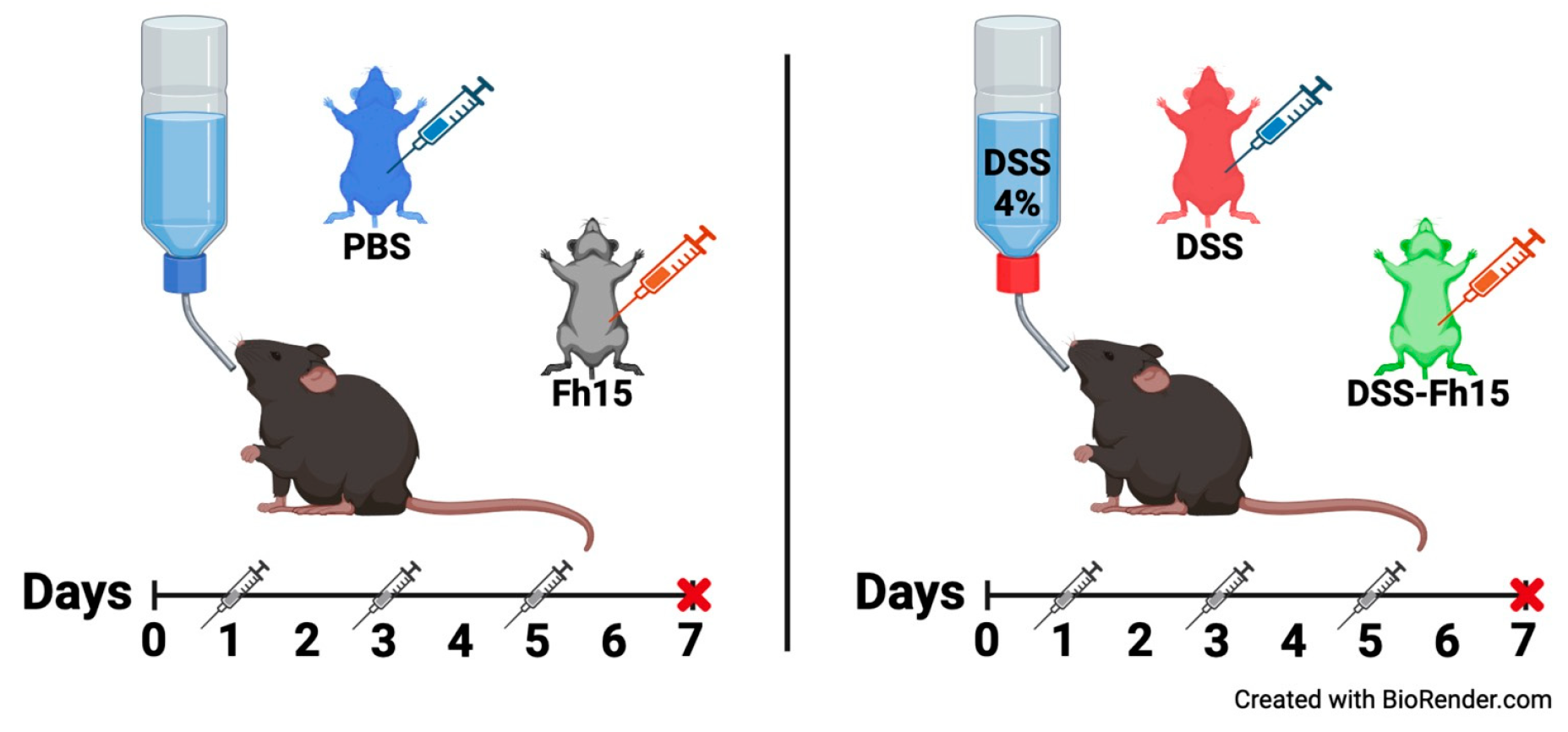
Figure 2.
Fh15 treatment significantly reduces disease activity index and body weight loss in ulcerative colitis mice. (A) Daily body weight percentage of C57BL/6 male mice with ulcerative colitis treated with Fh15 (2 mg/kg body weight) on days 1, 3, and 5. (B) Daily disease activity index. Statistical significance between groups was determined using two-way ANOVA with Dunnett's multiple comparisons test, using the DSS group as the reference. ****p < 0.0001, ***p < 0.001, **p < 0.01, *p < 0.05.
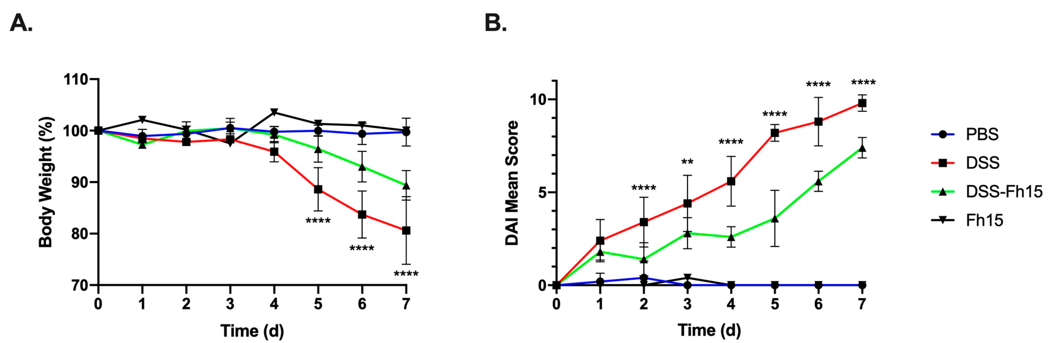
Figure 3.
Fh15 significantly prevents colon shortening and reduces macroscopic score with notable decrease in histological alterations in male mice with DSS-induced ulcerative colitis. (A) Males colon length after three doses of Fh15 (2.0mg/kg body weight). Colon measurement in centimeters was used as an indirect marker of inflammation. (B) Mean macroscopic score. (C) H&E staining of colon samples representing histological assessment observed with a 40X objective. Statistical significance between groups was assessed using one-way ANOVA with Dunnett's multiple comparisons using DSS as reference group. ***p<0.001, *p<0.05. Bar represents 20μm.
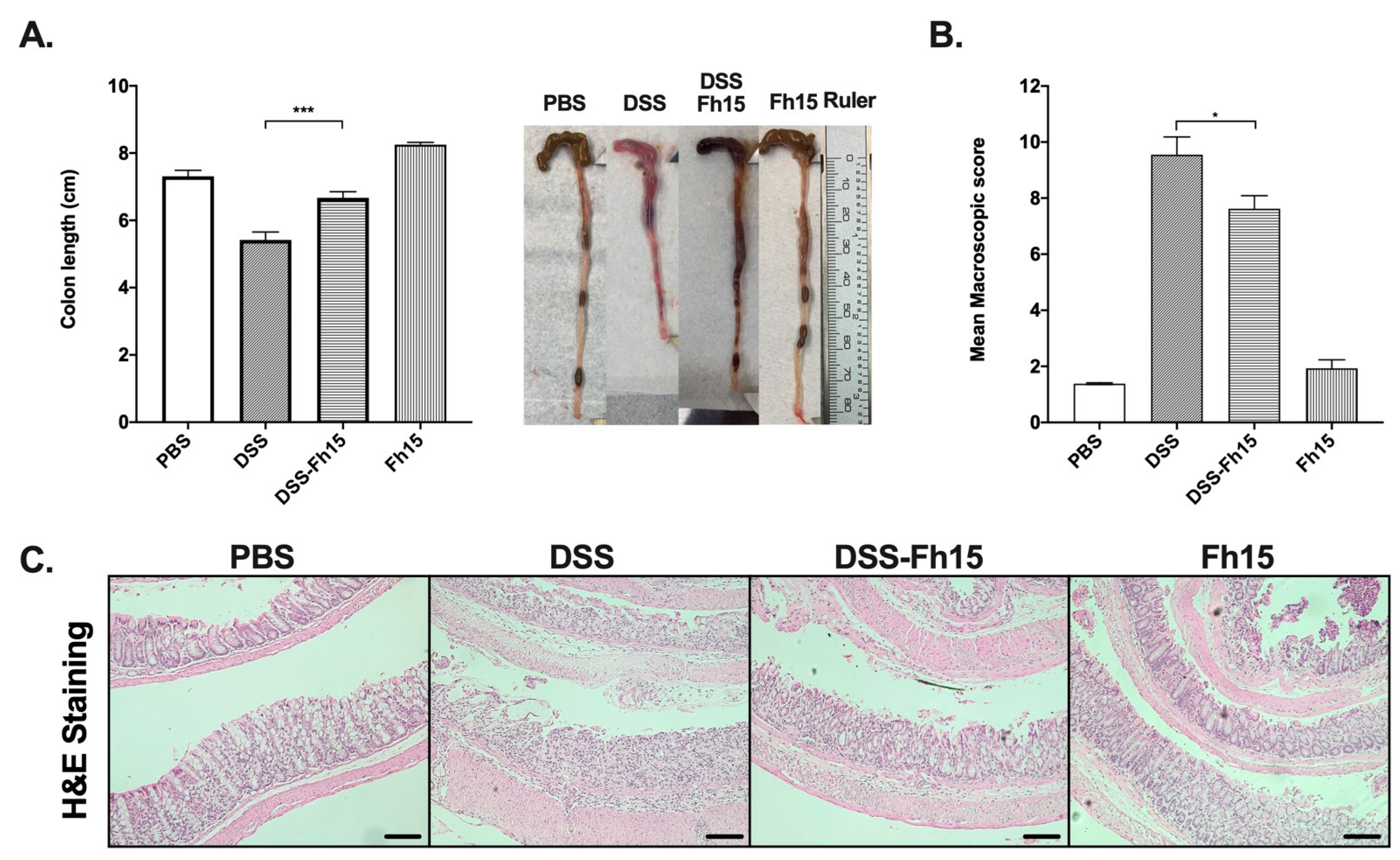
Figure 4.
Fh15 reduces pro-inflammatory markers in male mice with DSS-Induced ulcerative colitis. (A) Serum concentrations of myeloperoxidase and (B) chitinase-3-like protein were measured in picograms per milliliter (pg/mL) using ELISA. (C) Representative immunostaining images of S100A9 and the mean percentage of S100A9+ area in colonic samples are shown. mRNA expression levels of (D) TNFα and (E) IL-1β in the distal colon were assessed via RT-PCR, with GAPDH used as housekeeping gene for internal reference. Statistical significant differences between groups were determined by using one-way ANOVA with Dunnett's multiple comparisons, using DSS as reference group. ****p<0.0001, **p<0.01, *p<0.05. Bar represents 100μm.
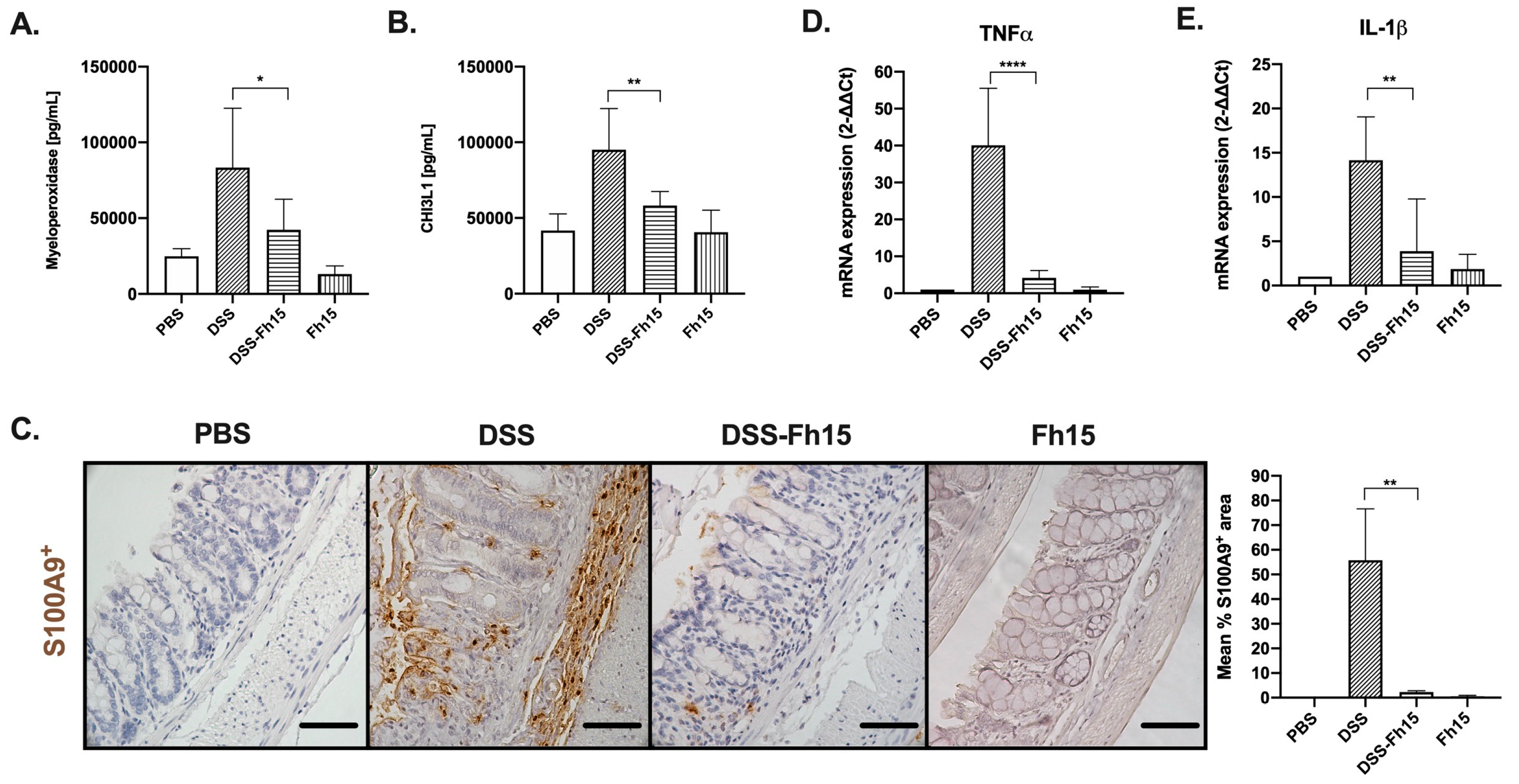
Figure 5.
Fh15 effectively decreases macrophages and neutrophils populations in male mice with DSS-induced ulcerative colitis. Immunostaining images representating macrophages (F4/80+), and neutrophils (Ly6G+), including the mean percentage of marker+ areas per group. Statistical significant differences between groups were determined by using one-way ANOVA with Dunnett's multiple comparisons, using DSS as reference group. **p<0.01, *p<0.05. Bar represents 100μm.
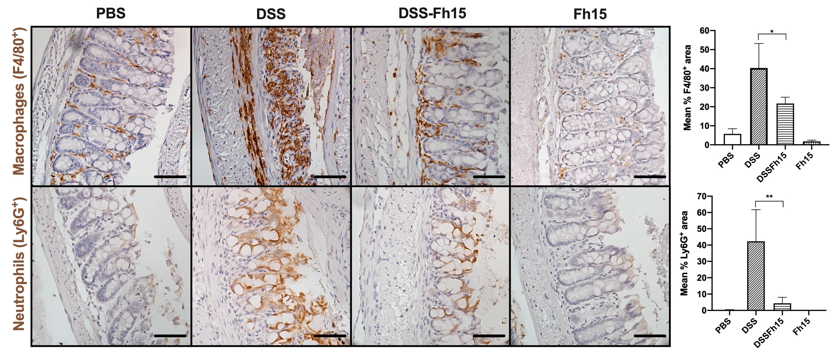

Table 1.
Assessment of Disease Activity Index.
| Weight loss | Stool Consistency | Bleeding | Score |
|---|---|---|---|
| < 2% | Normal | Negative hemoccult | 0 |
| > 2% - <5% | Softer stool | Positive hemoccult/ no visible blood | 1 |
| > 5% - <10% | Moderate Diarrhea | Visual blood in stool | 2 |
| > 10% - <15% | Diarrhea | Fresh rectal bleeding | 3 |
| > 15% | - | - | 4 |
Table 2.
Assessment of colon macroscocpic score on day 7 after euthanasia, modified from Storr, M. et al 2009 [1].
Table 2.
Assessment of colon macroscocpic score on day 7 after euthanasia, modified from Storr, M. et al 2009 [1].
| Macroscopic damage | Score |
|---|---|
| Colon length | >=6cm = 0pt, <6cm = 1pt, or <5cm = 2pt |
| Inflamed length | Centimeters measurement |
| Bowel thickness | Millimeters measurement |
| Adhesion | 0= No Adhesion, 1= Mild, 2= Moderate, or 3= Severe |
| Hemorrhage | Present= 1 or Absent = 0 |
| Fecal blood | Present= 1 or Absent = 0 |
| Diarrhea | Present= 1 or Absent = 0 |
Table 3.
Assessment of histopathological scoring, retrieved from Sann, H. et al 2013 [2].
Table 3.
Assessment of histopathological scoring, retrieved from Sann, H. et al 2013 [2].
| Score | Extent of inflammation |
Infiltration neutrophils + lympho-histiocytes | Extent of crypt damage | Crypt abscesses | Sub-mucosal edema | Loss of goblet cells |
Reactive epithelial hyperplasia |
|---|---|---|---|---|---|---|---|
| 0 | None | None | None | None | None | None | None |
| 1 | Mucosa | Focal | Basal one third | Focal | Focal | Focal | Focal |
| 2 | Mucosa+submucosa | Multifocal | Basal two thirds | Multifocal | Multifocal | Multifocal | Multifocal |
| 3 | Mucosa+submucosa+ muscle layer |
Diffuse | Entire crypt damage | - | Diffuse | Diffuse | Diffuse |
| 4 | Transmura | - | Crypt damage +ulceration |
- | - | - | - |
Table 4.
RT-qPCR primers.
| Gene | Primers | Sequence 5’- 3’ |
|---|---|---|
| GAPDH | Forward | CATGGCCTTCCGTGTTCCTA |
| Reverse | CCTGCTTCACCACCTTCTTGAT | |
| TNF-α | Forward | AAGCCTGTAGCCCACGTCGTA |
| Reverse | AGGTACAACCCATCGGCTGG | |
| IL-1β | Forward | GAAATGCCACCTTTTGACAGTG |
| Reverse | TGGATGCTCTCATCAGGACAG |
Table 5.
Primary antibodies (IgGs) used for chromogenic immunohistochemistry (IHC).
| Marker | Target | Host species | IHC Dilution | Cat # | Vendor |
|---|---|---|---|---|---|
| F4/80 | Macrophages | Rabbit | 1:100 | 70076S | CST |
| LY6G | Neutrophils | Rabbit | 1:75 | 87048S | CST |
| S100A9 | Calcium binding protein A9 | Rabbit | 1:800 | 73425S | CST |
Disclaimer/Publisher’s Note: The statements, opinions and data contained in all publications are solely those of the individual author(s) and contributor(s) and not of MDPI and/or the editor(s). MDPI and/or the editor(s) disclaim responsibility for any injury to people or property resulting from any ideas, methods, instructions or products referred to in the content. |
© 2025 by the authors. Licensee MDPI, Basel, Switzerland. This article is an open access article distributed under the terms and conditions of the Creative Commons Attribution (CC BY) license (http://creativecommons.org/licenses/by/4.0/).
Copyright: This open access article is published under a Creative Commons CC BY 4.0 license, which permit the free download, distribution, and reuse, provided that the author and preprint are cited in any reuse.


