
Submitted:
30 January 2025
Posted:
31 January 2025
You are already at the latest version
Abstract
Pediatric stroke, a significant cause of long-term neurological deficits in children, often arises from disruptions within neurovascular unit (NVU) components. The NVU, a dynamic ensemble of astrocytes, endothelial cells, pericytes, and microglia, is vital for maintaining cerebral homeostasis and regulating vascular brain development. Its structural integrity, particularly at the blood-brain barrier (BBB), depends on intercellular junctions and the basement membrane, which together restrict paracellular transport and shield the brain from systemic insults. Dysfunction in this intricate system is increasingly linked to pediatric stroke and related cerebrovascular conditions.
Mutations disrupting endothelial cell adhesion or pericyte-endothelial interactions can compromise BBB stability, leading to pathological outcomes such as intraventricular hemorrhage in the germinal matrix, a hallmark of vascular brain immaturity. Additionally, inflammation, ferroptosis, necroptosis, and autophagy are key cellular processes influencing brain damage and repair. Excessive activation of these mechanisms can exacerbate NVU injury, whereas targeted therapeutic modulation offers potential pathways to mitigate damage and support recovery.
This review explores the cellular and molecular mechanisms underlying NVU dysfunction, BBB disruption, and subsequent brain injury in pediatric stroke. Understanding the interplay between genetic mutations, environmental stressors, and NVU dynamics provides new insights into stroke pathogenesis. The susceptibility of the germinal matrix to vascular rupture further emphasizes the critical role of NVU integrity in early brain development. Targeting inflammatory pathways and cell death mechanisms presents promising strategies to preserve NVU function and improve outcomes for affected neonates.
Keywords:
fetal
; pediatric
; perinatal stroke
; neurovascular unit
; blood-brain barrier
; vascular brain development
INTRODUCTION
Brain development involves complex processes such as neuronal and glial growth, myelination, and the formation of cortical structures, which are guided by conserved genetic mechanisms across mammals. While animal studies have advanced understanding of these processes, humans exhibit unique, species-specific brain features that support higher-order functions [1]. Disruptions in these genetic programs, especially in preterm infants, can lead to neurodevelopmental and psychiatric disorders, with the brain's maturity at birth being a critical factor [2].
Preterm birth, defined as occurring before 37 weeks of gestation, varies by gestational age: moderate or late preterm (32–37 weeks), very preterm (28–32 weeks), and extremely preterm (less than 28 weeks) [3]. Globally, about 15 million preterm births occur annually, representing 10% of live births. The incidence and causes differ significantly across racial, ethnic, and geographic groups [4].
The NVU and germinal matrix (GM) are essential for neurogenesis, vascular development, and brain maturation during the fetal and neonatal periods, making them highly susceptible to injury during perinatal stroke. The dynamic nature of brain development influences the severity and progression of damage after an ischemic or hemorrhagic event. The stage of postnatal brain maturation and the development pace of central nervous system (CNS) cell types at the time of stroke result in distinct excitotoxic and inflammatory responses. Therefore, it is crucial to target pathophysiological pathways in a maturation-specific manner to protect brain function and support repair in the developing brain after stroke [5].
Pediatric Stroke
Pediatric stroke is a serious neurological condition that can result in long-term morbidity, including sensorimotor deficits, intellectual disability, and epilepsy [6]. The risk of pediatric stroke is highest during the fetal or neonatal period and is influenced by neuroplasticity, with recovery patterns shaped by the timing, location, and associated comorbidities of the stroke [7].
Pediatric strokes are categorized by age into perinatal strokes (20 weeks’ gestation to 28 days after birth) and childhood strokes (28 days to 18 years) [8-10]. Perinatal strokes include acute perinatal stroke (APS), evident shortly after birth, and presumed perinatal stroke (PPS), diagnosed later due to symptoms like seizures or early handedness [8]. Unlike older patients, perinatal strokes lack clear clinical signs and require neuroimaging for diagnosis.
As in adults, pediatric strokes are also classified by cause as ischemic or hemorrhagic [11]. Ischemic strokes include arterial ischemic stroke (AIS) and venous infarction, typically caused by cerebral or cortical vein thrombosis (CSVT) [8,9]. They are further categorized by timing: fetal ischemic stroke (diagnosed before birth), neonatal ischemic stroke (within 28 days of birth), and presumed perinatal ischemic stroke (PIS, diagnosed later but presumed to have occurred between the 20th week of gestation and 28 days postnatal) [12,13]. Hemorrhagic strokes can manifest as intracerebral (ICH), intraventricular (IVH), or subarachnoid (SAH) hemorrhage, and are classified as primary (due to vascular anomalies or bleeding disorders) or secondary (arising from ischemic infarction) [8,14]. Distinguishing these forms may be challenging without prompt imaging [12,15,16]. The impact of any stroke is influenced by the neurodevelopmental stage at the time of the event [7].
The annual incidence of pediatric stroke ranges from 3 to 25 per 100,000 children in developed countries [8], with a higher rate in neonates (25-40 per 100,000) and even greater in premature neonates (approximately 100 per 100,000) [9]. Perinatal stroke is a leading cause of hemiparetic cerebral palsy and has lifelong impacts, including motor, cognitive, and epileptic issues [10]. Stroke is more common in boys than girls and in Black, Asian, and Hispanic children compared to white children [17,18].
Risk factors for perinatal stroke include maternal conditions (e.g., preeclampsia, infections, diabetes) and neonatal factors (e.g., cardiac disease, prematurity, clotting disorders), though a direct cause is rarely identified [19]. For childhood stroke, common causes are arteriopathy, congenital heart disease, and prothrombotic disorders, with additional risks like infection, trauma, and sickle cell disease. Pediatric stroke differs from adult stroke not only in demographics and risk factors but also in the unique vulnerability and recovery patterns of the developing brain [19,20].
Human Brain Development
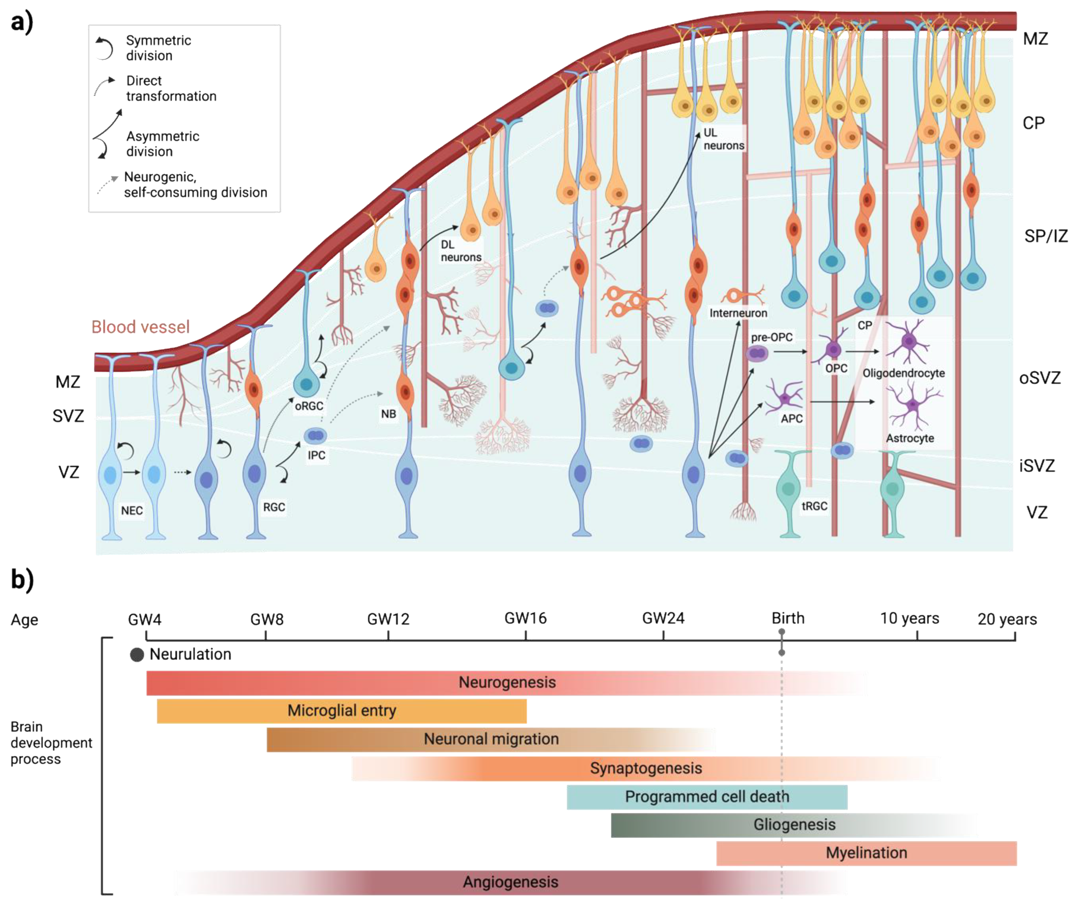
Comprehensive reviews detailing early neurogenesis in the perinatal brain have been recently published [1,21,22]. Human brain development begins around the second to third gestational week (GW) and continues through childhood, orchestrated by a complex interplay of genetic, epigenetic, and environmental factors. Early in the first trimester, the process of neurulation sets the stage for CNS formation as neuroepithelial cells in the ectoderm form the neural plate. By the third GW, the plate folds and fuses into a closed neural tube, marking the foundation of the CNS [23,24]. Shortly after, the brain’s vasculature develops, separating the CNS from the periphery around the fifth GW [21] (Figure 1).
As the neocortex begins to form between the eighth and eighteenth GW, neurogenesis intensifies. Neural epithelial cells (NECs) initially divide symmetrically to expand their population, then switch to asymmetric divisions to produce the first neurons. These early neurons migrate radially into a structure called the preplate [25,26] (Figure 1a). Over time, NECs evolve into radial glia cells, which act as scaffolds for the migration of excitatory neurons to the developing cortical layers, while inhibitory neurons migrate tangentially into the cortex. By the ninth to thirteenth GWs, neurogenesis peaks, supported by intermediate progenitor cells in the subventricular zone [27] (Figure 1a). In the third trimester, neuronal axons and glial cells, such as astrocytes and oligodendrocytes, are produced and integrated into neural circuits [28]. Synaptogenesis, the formation of synaptic connections, begins as early as the eighth GW [29], with microglia and astrocytes shaping neural networks through synaptic pruning [30,31]. Microglia, derived from the hematopoietic lineage, appear by the fifth GW (Figure 1b) and contribute to early brain development even before vascular sprouting [32]. Astrocytes and oligodendrocyte progenitors emerge later, between the fifteenth-seventeenth GW [33,34]. Myelination, the process of insulating nerve fibers, starts mid-gestation and continues into late adolescence, supporting the brain’s growing complexity and efficiency (Fig. 1b). Postnatally, the brain’s structure and plasticity continue to evolve, enabling lifelong adaptation and learning [2,5].
Human Vascular Development
The development and expansion of vascular beds are essential for embryonic growth, as vascular defects during this stage can result in severe brain malformations [35]. In humans, brain vascularization progresses in parallel with brain development, following a precise sequence of angiogenic processes. By GW eight, the primitive capillary network in the perineural vascular plexus begins to expand into the CNS parenchyma [36] (Figure 1).
Figure 1.
Key stages of human brain development: from conception to adulthood. a) Neurovascular communication during CNS development. Neocortical development begins with neurogenesis, where neural progenitor cells differentiate into neuroblasts, and eventually to neurons, oligodendrocytes, and astrocytes. Simultaneously, the formation of the cerebral vasculature is initiated as sprouting blood vessels from the perineural vascular plexus extend radial branches toward the ventricular zone. These initial branches further subdivide (anastomosis) and interconnect, forming a complex and intricately organized vascular network essential for supporting the metabolic demands of the developing neocortex. APC, astrocyte precursor cell; CP, cortical plate; DL, deep layer; IPC, intermediate progenitor cell; iSVZ, inner SVZ; IZ, intermediate zone; MZ, marginal zone; NB, neuroblast; NEC, neuroepithelial cell; OPC, oligodendrocyte precursor cell; oRGC, outer radial glial cell; oSVZ, outer SVZ; RGC, radial glial cell; SP, sublplate; SVZ, subventricular zone; tRGC, truncated radial glial cell; UL, upper layer; VZ, ventricular zone. b) This diagram highlights the major stages and timeline of human brain development, starting with neurulation, followed by neurogenesis, microglial entry, neuronal migration, and synaptogenesis. By 18 GW, apoptosis eliminates excess cells to refine populations and establish proper synaptic connectivity. During the third trimester, glial cells support the myelination of neurons, a process that continues postnatally into adulthood. Angiogenesis, essential for brain development, begins in the early embryonic stages, peaks during rapid growth, stabilizes by birth, and continues postnatally with limited adaptations, supporting brain function throughout life.
During early neuroepithelial development, endothelial cells (ECs) originate from mesodermal angioblasts and migrate into the neuroepithelium via the perineural vascular plexus, guided by Vascular Endothelial Growth Factor (VEGF) [37]. Alongside VEGF, several key signaling pathways contribute to angiogenic sprouting and vessel remodeling, including the Angiopoietin-Tie system, the Hedgehog pathway, Ephrin receptor tyrosine kinases, Dll4/Notch1 signaling, and the Wnt signaling pathway [38-43]. At this stage, the developing blood vessels exhibit short, primitive tight junctions (TJs), lack fenestrations, and limit the movement of blood-derived proteins into the CNS environment [37]. TJs proteins, such as occludin and claudin-5, critical for CNS barriers, begin to emerge by the sixteenth GW [5,36]. As development progresses, ECs aggregate into primitive structures that mature into functional blood vessels [44].
The formation of vascular networks occurs through sprouting angiogenesis, where new blood vessels grow outward and connect to other vessels or sprouts via anastomosis. At the cellular level, this process is characterized by dynamic and highly regulated behaviors (reviewed by [45,46]). In response to pro-angiogenic signals, ECs are activated, remodeling their actin cytoskeletons and downregulating junctional adhesion molecules to enable cell motility. Throughout this process, ECs maintain contact with neighboring cells and the parent vessel, ensuring vascular integrity and proper apical-basal cell polarization [47]. Fine-tuning these cellular responses to angiogenic signals is crucial for the successful formation and maturation of vascular networks.
NEUROVASCULAR UNIT CELL-CELL INTERACTION
Two comprehensive reviews detailing the cellular components and function of the NVU in the perinatal brain were recently published [48,49]. The NVU is formed by interactions between blood vessels and neural and glial cells in the CNS, including microvascular ECs, neurons, microglia, astrocytes, pericytes, and matrisomes [50,51] (Figure 2). These components engage in close and intricate interactions, allowing the NVU to function as a cohesive unit. The NVU maintains a highly selective BBB, ensuring cerebral homeostasis, regulates local blood flow through neurovascular coupling, provides trophic support, produces growth factors and paracrine signals, guides neuronal differentiation, and recruits oligodendrocytes [48,52-54].
The NVU plays a crucial role in regulating the local blood supply to meet neuronal metabolic needs by adjusting the diameter of blood vessels [48,55]. In this process, astrocytes and pericytes are activated by glutamate released from neurons. Glutamate, in turn, triggers the release of vasoactive mediators that modulate vascular tone (Figure 2b). The balance between vasoconstrictors and vasodilators controls the tone of the surrounding vasculature, thereby regulating local blood flow [48].
The second major function of the NVU is maintaining the BBB [48]. The BBB serves as both a physical and metabolic barrier between the bloodstream and neuroglia [55]. It acts as a physical barrier due to the presence of complex TJs [49,51] and adherents junctions (AJs) between ECs [55]. This forces most molecular traffic to traverse the BBB via a transcellular route, rather than passing between cells via the junctions typically found in other endothelial tissues [51]. Consequently, paracellular transport of water-soluble or polar compounds is limited, small ion transport is restricted, and EC polarity is preserved [55]. The role of the BBB depends on the interactions and coordination among various cellular components and the extracellular matrix (ECM) which together create a functional and anatomical barrier system [56].
Astrocyte, pericytes and ECs interactions
The BBB is established and maintained through intricate interactions between ECs, pericytes, and astrocytes. These cellular components of the NVU work together to regulate angiogenesis and ensure the barrier’s protective and selective permeability, critical for brain function [57] (Figure 2).
During late embryogenesis and postnatal development, the BBB undergoes progressive maturation. TJs formed by ECs become increasingly complex, creating impermeable barriers essential for maintaining brain homeostasis. By GW12 in humans, proteins such as occludin, claudins, and junctional adhesion molecules (JAMs) organize into functional TJs, linking to the cytoskeleton via zonula occludens (ZO)-1, ZO-2, and ZO-3 [58]. AJs, composed of VE-cadherin, PECAM-1, and nectin, provide structural stability, while gap junctions (GJs), formed by connexin hemichannels, facilitate direct intercellular communication [59] (Figure 2b). These features mature by embryonic day 15 (E15) in rodents, and by E19, TJs are fully differentiated [60], setting the stage for robust BBB functionality [61,62]. Astrocytic contributions to TJ tightness start closer to birth and become more prominent during postnatal development [63].
Both ECs and pericytes are situated in the basement membrane (BM) and anchored to it via integrins (Figure 2b). Pericytes play essential roles in BBB development and maintenance. They promote TJ formation [61], regulate vascular development [64], and contribute to inflammatory processes [65]. Signaling between ECs and pericytes involves platelet-derived growth factor BB (PDGF-BB) and its receptor (PDGFR-β), adhesion via N-cadherin, and GJs that facilitate the exchange of ions and small molecules [66,67] (Figure 2b). These interactions ensure BBB integrity and permeability control during embryogenesis and adulthood. While pericytes are indispensable, studies suggest that CNS-derived signals can independently induce BBB-specific molecule expression in ECs, highlighting additional regulatory mechanisms [43,61].
Astrocytes, located furthest from the vascular lumen within the NVU, interface with the BM and ECM through their endfeet, contributing ECM proteins and regulating vascular tone via calcium signaling [57] (Figure 2). They influence TJ protein expression by secreting factors like Src-suppressed C-kinase substrate (SSeCKS) and maintain the integrity of the BBB by modulating matrix metalloproteinase-9 (MMP-9) activity in pericytes [68,69]. Apolipoprotein E (ApoE3), secreted by astrocytes, binds to low-density lipoprotein receptor-related protein 1 (LRP1) in pericytes, suppressing the pro-inflammatory CypA-NF-κB-MMP-9 pathway [69]. This prevents degradation of TJ and BM proteins, protecting the BBB, whereas ApoE4 lacks this protective effect [69]. Astrocytes also metabolize glucose taken up by ECs via Glut1, converting it to lactate, which is then supplied to neurons through monocarboxylate transporters MCT1 and MCT4 [59] (Figure 2b).
ECs, integral to the BBB, form the physical barrier and facilitate selective nutrient exchange. Their connections through TJs, AJs, and GJs are critical for maintaining the barrier’s integrity. Knockout studies reveal that astrocyte-derived laminin is essential for TJ protein expression and overall BBB functionality [70]; its absence leads to barrier leakage [71], particularly under pathological conditions such as perinatal stroke.
MECHANISMS OF BRAIN DAMAGE AFTER A PERINATAL STROKE
Pathophysiological Mechanisms of a Hypoxic-Ischemic Injury
Hypoxic-ischemic (HI) injury during the perinatal period triggers a cascade of cellular and molecular processes, leading to necrotic and apoptotic cell death. Unlike in adults, the BBB in neonates is not fully developed, contributing to the unique pathophysiology of neonatal strokes. Despite its early developmental stage, studies show that the BBB remains largely impermeable to very small molecules, maintaining a degree of selective permeability [72,73].
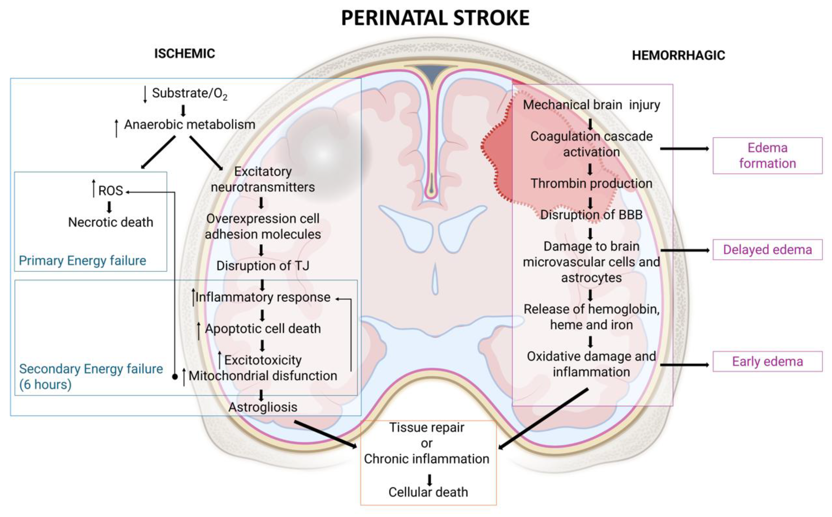
HI injury results in neuronal death through two primary phases [74] (Figure 3): First, a primary energy failure occurs due to the deprivation of oxygen and glucose, which disrupts normal metabolic processes. This disruption leads to anaerobic metabolism, intracellular lactate accumulation, and the production of reactive oxygen species (ROS), ultimately resulting in necrotic cell death. For several cells essential for maintaining CNS homeostasis—such as pericytes, astrocytes, microglia, and ECs—damage to the BBB plays a critical role in the pathogenesis of NVU dysfunction, subsequently causing downstream perivascular damage [70,75]. Furthermore, the uncontrolled release of excitatory neurotransmitters exacerbates neuronal damage [73,75]. Second, if the insult persists, it leads to secondary energy failure, which occurs at least 6 hours post-injury and may continue for days after the initial event. During this phase, most cell deaths result from apoptosis, driven by excitotoxicity, oxidative stress, mitochondrial dysfunction, and inflammation [73,74,76].
After the acute injury phase, the chronic phase is characterized by astrogliosis and chronic inflammation, with processes aimed at restoring CNS integrity, although often incomplete or maladaptive [9,70,74] (Figure 3).
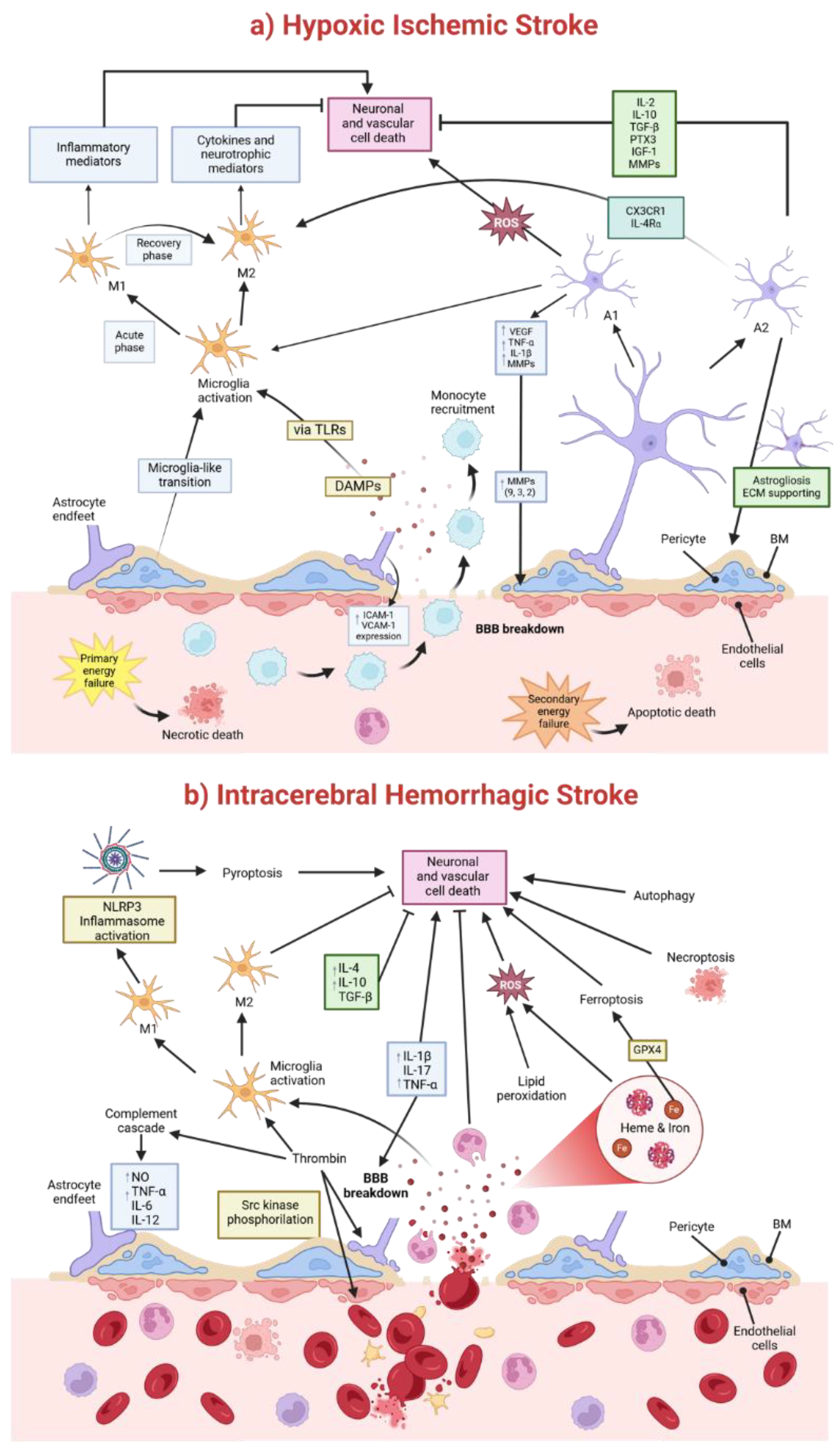
Neonatal stroke triggers distinct immune responses compared to adults due to the immaturity of immune pathways and age-dependent mediator effects. Microglia play a dual role in neonatal HI injury. Initially, they exacerbate damage, but later, they contribute to recovery through processes such as debris clearance and extracellular matrix remodeling. Following injury, microglia are activated via toll-like receptors (TLRs) in response to damage-associated molecular patterns (DAMPs), transitioning from a resting to an active state [77] (Figure 4a). These cells polarize into either pro-inflammatory (M1) or anti-inflammatory (M2) phenotypes. M1 microglia release damaging inflammatory mediators, while M2 microglia promote repair through cytokines and neurotrophic factors [77]. This dynamic polarization shifts from M1 dominance during acute phase of injury to M2 activity during recovery. Imbalances in this transition impact neonatal HI injury outcomes (Figure 4a).
Like microglia, astrocytes also exhibit two polarization states in response to HI injury: neurotoxic A1 (pro-inflammatory) and neuroprotective A2 (anti-inflammatory). Pro-inflammatory astrocytes contribute to BBB disruption by increasing the expression of factors like VEGF, cytokines (e.g., TNF-α, IL-1β), ROS, and MMPs. Additionally, astrocytes recruit immune cells by up-regulating ICAM-1 and VCAM-1 expression and activating microglia, thereby indirectly intensifying inflammation-induced BBB damage (reviewed in [78]). Following stroke, pro-inflammatory astrocytes, together with infiltrating leukocytes and activated microglia, secrete matrix metalloproteinases (MMPs) [79,80]. Particularly MMP-9, MMP-3, and MMP-2, mediate ECM breakdown, degrading TJs, laminin, collagen, and fibronectin. This degradation results in vasogenic edema, BBB leakage, and immune cell infiltration [81-85] (Figure 4). Upregulation of MMP-9 has been observed in neonatal rodent brains 24 hours after ischemic injury [86]. Moreover, MMPs facilitate adhesion molecule signaling and ECs activation, processes critical for monocyte recruitment and the inflammatory cascades seen in neonatal brain injuries [5,87,88]. Anti-inflammatory astrocytes, in contrast, promote BBB repair by resolving inflammation. They secrete protective cytokines like IL-2, IL-10, and TGF-β, and release PTX3 and IGF-1, which protect the BBB and support immune cell polarization toward anti-inflammatory states (reviewed in [78]) (Figure 4a). Anti-inflammatory astrocytes also release MMPs, but those instead promote tissue repair [89,90]. Moreover, anti-inflammatory astrocytes also regulate microglial activation via CX3CR1 and IL-4Rα signaling [91] (Figure 4a). Notably, neuroprotective astrocytes, which dominate three days post-stroke, support ECM integrity and scar formation through astrogliosis. This process limits immune cell migration and confines inflammation within the infarct region [92].
Pericytes, another key cellular responder to ischemic stroke damage, also exhibit remarkable plasticity under ischemic conditions. They can transition into microglia-like or other cell types through a multipotent stem cell intermediate state, meaning their loss worsens leukocyte infiltration [93-95].
Strategies aimed at promoting anti-inflammatory polarization of both microglia and astrocytes, or inhibit harmful pro-inflammatory responses, hold promise for the management of neonatal HI injury.
Pathophysiological Mechanisms of Brain Damage after Intracerebral Hemorrhage (ICH)
ICH triggers complex pathological processes, resulting in acute and delayed brain damage. These processes include mechanical injury, inflammatory and immune responses, BBB disruption, oxidative stress, and cell death, with significant implications for perihematomal edema and long-term functional outcomes [96,97] (Figure 3). ICH initially causes mechanical injury through increased intracranial pressure and reduced local cerebral blood flow, leading to ischemia and brain tissue compression. This mechanical stress exacerbates secondary damage by promoting edema formation and cytotoxicity [96]. The activation of the coagulation cascade is a major contributor to early edema formation after ICH. Thrombin, generated by the breakdown of prothrombin due to BBB disruption, induces BBB dysfunction by activating Src kinase phosphorylation, damaging brain microvascular ECs and astrocytes [97]. While Src kinase activity initially exacerbates injury, it later contributes to BBB restoration and edema resolution after 2–6 days [98]. Thrombin also activates the complement cascade, leading to the release of nitric oxide (NO), tumor necrosis factor-alpha (TNF-α), and interleukins (e.g., IL-6, IL-12), which amplify inflammatory responses and promote secondary injury [98] (Figure 4b).
Around three days post-ICH, delayed edema arises, primarily mediated by red blood cell breakdown and hemoglobin degradation products, such as heme and iron. These compounds, along with enzymes like carbonic anhydrase, contribute to oxidative stress and further edema development [97,99] (Figure 3, Figure 4b).
The inflammatory response is central to ICH pathophysiology, involving both cellular (microglia, astrocytes, macrophages, leukocytes) and molecular (cytokines, chemokines, reactive oxygen species, extracellular proteases) mediators [99,100]. Proinflammatory cytokines, such as IL-1β, IL-17, and TNF-α, exacerbate injury by amplifying inflammation and increasing BBB permeability (Figure 4b). Conversely, anti-inflammatory cytokines (e.g., IL-4, IL-10, TGF-β) facilitate tissue repair [100]. IL-6, a dual-function cytokine, participates in both pro- and anti-inflammatory pathways depending on the stage of injury [100] (Figure 4b). Neuroinflammation after IHC also involves a complex interplay of M1 and M2 macrophages/microglia. While M1 cells drive detrimental neuroinflammation, M2 cells promote hematoma resolution, edema resorption, and white matter repair [101]. The NLRP3 inflammasome, a key protein complex in inflammation following ICH, is activated in M1 microglia through mechanisms such as the complement system. Once activated, it triggers caspase-1, which converts pro-IL-1β and pro-IL-18 into their active forms, IL-1β and IL-18, leading to pyroptosis and elevated pro-inflammatory cytokines [100]. Neutrophils also exert dual roles, contributing to ICH growth and injury but aiding recovery through lactoferrin-mediated iron scavenging and edema reduction [100] (Figure 4b).
As mentioned, the BBB undergoes significant dysfunction in ICH, exacerbating edema formation and allowing infiltration of immune cells and neurotoxic molecules. Treatments like hyperosmotic solutions (e.g., mannitol) and glibenclamide, a sulfonylurea receptor inhibitor, show promise in mitigating perihematomal edema [102].
Cell death pathways associated with ICH include autophagy, ferroptosis and necroptosis. Oxidative stress, driven by hemoglobin-derived iron and lipid peroxidation, plays a central role in neuronal and vascular injury. Ferroptosis, a non-apoptotic, iron-dependent form of cell death mediated by glutathione peroxidase 4 (GPX4), is a key contributor [103]. Experimental studies demonstrate that lipid peroxidation inhibitors, such as ferrostatin-1 and liproxstatin-1, reduce ICH-induced brain atrophy, neuronal death, and functional deficits [103].
Figure 4.
Key mechanisms underlying hypoxic-ischemic (HI) stroke and neonatal intracerebral haemorrhagic stroke (ICH). a) HI stroke is characterized by energy failure, leading to both necrotic and apoptotic cell death. Pro-inflammatory astrocytes (A1) and microglia (M1) exacerbate damage through cytokine release, while neuroprotective astrocytes (A2) and microglia (M2) facilitate recovery by providing extracellular matrix (ECM) support and promoting anti-inflammatory signaling. b) ICH is defined by thrombin activation, oxidative stress caused by heme and iron deposition, and activation of cell death pathways such as pyroptosis, necroptosis, and ferroptosis, which collectively contribute to neuronal and vascular damage. BM, basement membrane; DAMPs, Damage-Associated Molecular Patterns; ECM, extracellular matrix; Glutathione Peroxidase 4; ICAM-1, Intercellular Adhesion Molecule 1; M1, pro-inflammatory microglia; M2, anti-inflammatory microglia; NLRP3, Pyrin Domain-Containing Protein 3; ROS, Reactive Oxygen Species, GPX4, TLRs, Toll-Like Receptors; VCAM-1, Vascular Cell Adhesion Molecule 1; VEGF, Vascular Endothelial Growth Factor.
NVU AND MONOGENIC NEUROLOGICAL DISORDERS
Several genetic diseases originate within specific cell types of the NVU and are closely linked to critical roles in BBB development, function, and regulation. Although these diseases are rare, they provide valuable insights into pathogenic mechanisms and casual relationships. Below, we outline some of these disorders, the associated cell types, and the potential pathogenic role of BBB dysfunction.
Pediatric stroke caused by mutations in genes expressed in ECs
Mutations in genes critical for ECs and the endothelium-derived ECM play a significant role in brain hemorrhage. These genes encode proteins vital for the structure and regulation of EC-cell junctions, vascular basement membranes, and transporters essential for BBB integrity. Biallelic variants in TJ-associated genes, such as JAM2 [104], JAM3 [72], and OCLN (occludin) [105-108], have been linked to increased BBB permeability, resulting in uncontrolled leakage of solutes and plasma proteins, along with excessive trans-endothelial leukocyte migration. This disruption leads to neuroinflammation. JAM-C normally inhibits this process [109]. Patients harboring such mutations often present with brain hemorrhage, calcification, movement disorders, and cognitive or neurobehavioral abnormalities [110,111].
The ECM and basement membranes not only provide structural support but also act as reservoirs for growth factors that regulate cellular functions. Type IV collagens, a major component of basement membranes, are vital for preserving BBB structure and function. For instance, mutations in the α1(IV) chain (COL4A1) cause perinatal cerebral hemorrhage [112].
Mutations in COL4A1/A2 manifest as lacunar ischemic strokes, deep ICH and white matter hyperintensities [59]. A murine model demonstrated that Col4A1 mutations predisposed both neonatal and adult mice to ICH. Durrani et al. (2017) reported a term infant with extensive intrauterine stroke, encephalomalacia, and anterior segment dysgenesis caused by a de novo mutation in COL4A1 [113].
Intracranial or intraventricular hemorrhage (ICH/IVH) is a significant type of perinatal stroke, particularly in preterm infants. These hemorrhages originate in the GM, a highly vascularized region located between the caudate nucleus and the thalamus near the foramen of Monro. The GM microvasculature is especially fragile due to immature basal lamina, sparse pericyte coverage, deficient TJs, and limited astrocytic support [114] (Figure 2a). Lecca et al. identified bi-allelic loss-of-function mutations in the ESAM gene in a cohort of thirteen perinatal stroke cases, including four fetuses, from eight unrelated families [111]. Furthermore, four additional perinatal stroke patients from two unrelated families have recently been reported to harbor homozygous ESAM variants, including the first described missense mutation [115]. ESAM (endothelial cell adhesion molecule), a TJ protein critical for BBB integrity, was not previously linked to rare human disease traits. Patients harboring ESAM mutations exhibit severe global developmental delay, epilepsy, spasticity, ventriculomegaly, ICH, and cerebral calcifications. This phenotype closely resembles conditions associated with other TJ-related gene mutations, such as JAM2, JAM3, and OCLN [106-108,116]. ESAM, a member of the immunoglobulin receptor family, facilitates endothelial cell adhesion through homophilic interactions. Its expression is predominantly restricted to embryonic and adult vasculature, where it regulates endothelial permeability and neutrophil extravasation. Research by Sauteur et al. (2017) highlighted the overlapping but distinct roles of ESAM and VE-cadherin in blood vessel formation and endothelial cell recognition during vascular anastomosis [47]. In a murine model, Duong et al. (2020) demonstrated that ESAM is crucial for maintaining endothelial junction integrity, particularly in the lungs. Gene inactivation of Esam increased vascular permeability in the lung, though it did not affect the heart, skin, or brain [117].
Additionally, vascular malformations, including cerebral cavernous malformations (CCMs) and arteriovenous malformations (AVMs), significantly increase ICH risk. CCMs are characterized by fragile venous capillary clusters lacking smooth muscle support, rendering them prone to rupture. Genetic mutations in KRIT1/CCM1, CCM2, and CCM3 underlie most CCMs [118]. AVMs, congenital anomalies of direct arterial-to-venous connections, are driven by excessive VEGF signaling and mutations in RAS/MAPK pathway genes like KRAS. Hereditary hemorrhagic telangiectasia (HHT) is a classic example of a syndrome associated with AVMs. Most individuals meeting the clinical diagnostic criteria for HHT harbor pathogenic variants in four TGFb pathway genes: Endoglin (ENG), Activin Receptor Like 1 (ACVRL1 or ALK1), SMAD family member 4 (SMAD4) and Growth Differentiation Factor 2 (GDF2 or BMP9)] [119]. Another genetic disorder involving AVMs is capillary malformation-arteriovenous malformation (CM-AVM) syndrome, which follows an autosomal dominant inheritance pattern due to loss of function variants in RAS P21 Protein Activator 1 (RASA1) and Ephrin Receptor B4 (EPHB4)] [120,121].
Sturge Weber Syndrome (SWS), a rare neurocutaneous disorder, also contributes to ICH risk through vascular malformations. Unlike inherited disorders such as HHT and CM-AVM, SWS is caused by a somatic activating mutation in GNAQ, which encodes the G protein subunit alpha-q (Gαq). This mutation, GNAQ p.R183Q, located in the switch 1 domain of Gαq, leads to constitutive activation of the protein and is present in approximately 88% of brain lesions in SWS patients [122,123]. SWS is characterized by abnormal capillary and venous blood vessels, with associated leptomeningeal vascular malformations.
This array of vascular malformations highlights the diverse genetic underpinnings contributing to ICH risk, emphasizing the importance of mutations in genes regulating endothelial cell function, vascular development, and inflammatory processes.
Pediatric Stroke caused by mutations in genes expressed in Vascular Mural Cells and Pericytes
Cerebral autosomal dominant arteriopathy with subcortical infarcts and leukoencephalopathy (CADASIL) is a relatively common autosomal-dominant stroke syndrome, affecting approximately 2–4 per 100,000 individuals. It is caused by mutations in the NOTCH3 gene, which is specifically expressed in vascular mural cells. These cells include vascular smooth muscle cells (VSMCs) and pericytes, playing a vital role in maintaining the integrity of blood vessels. Over 200 Notch3 mutations have been associated with CADASIL, though the causative role of loss-of-function mutations remains uncertain [124]. The CADASIL phenotype is predominantly associated with the expression of Notch3 in VSMCs, suggesting a key role in vascular health. Studies in Notch3-/- mice have demonstrated a progressive decline in VSMCs, mirroring the vascular pathology observed in CADASIL [125]. This regression of VSMCs is associated with decreased vessel wall thickness, loss of extracellular matrix, and weakening of the vessel wall. A key hypothesis posits that Notch3 facilitates communication and cell-cell interactions between VSMCs and arterial ECs, crucial for vascular stability. Experimental evidence supports this hypothesis, with transgenic mice expressing the CADASIL R90C mutation specifically in VSMCs recreating the classic CADASIL pathology. These findings underline the essential role of Notch3 in the function and integrity of VSMCs [125]. Notch3 is also expressed in pericytes. However, its role in pericyte function remains controversial. Henshall et al. reported that Notch3-/- knockout mice showed BBB disruption and leakage but found no impact on pericytes [125]. In contrast, Wang et al. demonstrated that Notch3 is necessary for pericyte proliferation in the zebrafish brain, indicating a potential role in pericyte dynamics [126]. These conflicting findings highlight the need for further research to clarify the role of Notch3 in pericyte biology. A significant connection between Notch3 and PDGFR-β has been established. Notch3 regulates PDGFR-β expression, and Notch3-/- mice exhibit reduced levels of this receptor and AVMs [127,128]. Similarly, PDGFR-β levels are decreased in individuals with CADASIL [127]. Since PDGFR-β is expressed by both ECs and mural cells, including pericytes and VSMCs, its role in vascular health is critical. Studies have shown that Pdgf-b-/- mice lack pericytes in their vasculature, leading to microaneurysm formation and rupture during gestation [129]. Heterozygous Pdgfr-β mice exhibit reduced pericyte numbers, suggesting a direct correlation between PDGFR-β expression and pericyte density [130]. This evidence further supports the intricate relationship between Notch3 signaling, PDGFR-β levels, and mural cell function. While the role of Notch3 in VSMC maintenance and vascular integrity is well established, its effect on pericytes remains less clear, with contradictory findings warranting further investigation.
Moyamoya disease (MMD), a progressive vasculopathy characterized by the narrowing and eventual occlusion of the intracranial internal carotid arteries, is a significant cause of childhood stroke. Rare, damaging variants in two VSMCs-specific genes have been found in non-East Asian patients with MMD: DIAPH1 (diaphanous-1), a critical effector of actin remodeling in vascular cells [131] and ACTA2, which encodes the smooth muscle-specific isoform of a-actin in VSMCs [132]. Both genes converge on pathways regulating the cytoskeleton, highlighting their essential role in maintaining vascular integrity and function.
FUTURE PERSPECTIVES
The intricate interplay between components of the NVU and GM underscores their central role in neurogenesis, vascular development, and brain maturation during critical periods of fetal and neonatal growth. This complexity also highlights their vulnerability to perinatal ischemic injuries. Understanding the dynamic and maturation-dependent pathophysiological responses within the NVU is essential to developing targeted therapeutic strategies. By focusing on age-specific mechanisms—such as the modulation of the BBB, neurovascular coupling, and astrocyte-pericyte-endothelial interactions—future research and clinical interventions can more effectively preserve neural integrity and promote repair in the developing brain. This approach holds promise for mitigating the long-term impact of perinatal stroke and fostering resilience in the CNS.
Genetic insights into NVU-associated disorders offer valuable opportunities to explore novel therapeutic strategies. For instance, rare damaging variants in DIAPH1 and ACTA2 in non-East Asian MMD patients emphasize the critical role of actin cytoskeleton regulation in vascular integrity and function. Understanding these pathways could guide the development of targeted interventions to prevent or manage cerebrovascular diseases. Similarly, targeting specific junction proteins (e.g., ESAM, JAMs, occludin) or ECM degradation pathways mediated by MMPs may mitigate BBB disruption in ischemic and hemorrhagic injuries. Moreover, therapies focusing on Notch3 signaling, PDGFR-β, and the modulation of mural cell maintenance have potential applications for conditions like CADASIL and MMD.
Emerging therapeutic modalities, including lipid peroxidation inhibitors (e.g., ferrostatin-1), immunomodulators, and gene-editing technologies, hold promise for addressing both acquired and genetic NVU dysfunctions. Preclinical models remain critical to evaluating these interventions, but translating these findings into clinical practice will require robust multicenter trials focused on safety, efficacy, and long-term outcomes.
Acknowledgements
This work was supported by grants from the Instituto de Salud Carlos III, PRiDe Project (PI23/000890) and PReDICT Project (PMPER24/00021), RICORS-ICTUS (RD21/0006/0004 and RD24/0009/0022) together with Next-Generation EU funds that finance the actions of the Recovery and Resilience Mechanism. TGM is supported by a Follium fellowship, DGG by a Junior fellowship and MOF by a JuniorTech fellowship, all from IdISBa/Impost Turisme Sostenible/Agència d’Estratègia Turística de les Illes Balears/Govern de les Illes Balears.
References
- Zhou, Y.; Song, H.; Ming, G.L. Genetics of human brain development. Nat Rev Genet 2024, 25, 26–45. [Google Scholar] [CrossRef]
- Doi, M.; Usui, N.; Shimada, S. Prenatal Environment and Neurodevelopmental Disorders. Front Endocrinol (Lausanne) 2022, 13, 860110. [Google Scholar] [CrossRef]
- Quinn, J.A.; Munoz, F.M.; Gonik, B.; Frau, L.; Cutland, C.; Mallett-Moore, T.; Kissou, A.; Wittke, F.; Das, M.; Nunes, T.; et al. Preterm birth: Case definition & guidelines for data collection, analysis, and presentation of immunisation safety data. Vaccine 2016, 34, 6047–6056. [Google Scholar] [CrossRef]
- Purisch, S.E.; Gyamfi-Bannerman, C. Epidemiology of preterm birth. Semin Perinatol 2017, 41, 387–391. [Google Scholar] [CrossRef]
- Di Martino, E.; Rayasam, A.; Vexler, Z.S. Brain Maturation as a Fundamental Factor in Immune-Neurovascular Interactions in Stroke. Transl Stroke Res 2024, 15, 69–86. [Google Scholar] [CrossRef] [PubMed]
- Nelson, K.B. Perinatal ischemic stroke. Stroke 2007, 38, 742–745. [Google Scholar] [CrossRef] [PubMed]
- Hill, N.M.; Malone, L.A.; Sun, L.R. Stroke in the Developing Brain: Neurophysiologic Implications of Stroke Timing, Location, and Comorbid Factors. Pediatr Neurol 2023, 148, 37–43. [Google Scholar] [CrossRef]
- Ferriero, D.M.; Fullerton, H.J.; Bernard, T.J.; Billinghurst, L.; Daniels, S.R.; DeBaun, M.R.; deVeber, G.; Ichord, R.N.; Jordan, L.C.; Massicotte, P.; et al. Management of Stroke in Neonates and Children: A Scientific Statement From the American Heart Association/American Stroke Association. Stroke 2019, 50, e51–e96. [Google Scholar] [CrossRef]
- Jankovic, M.; Petrovic, B.; Novakovic, I.; Brankovic, S.; Radosavljevic, N.; Nikolic, D. The Genetic Basis of Strokes in Pediatric Populations and Insight into New Therapeutic Options. Int J Mol Sci 2022, 23. [Google Scholar] [CrossRef] [PubMed]
- Srivastava, R.; Mailo, J.; Dunbar, M. Perinatal Stroke in Fetuses, Preterm and Term Infants. Semin Pediatr Neurol 2022, 43, 100988. [Google Scholar] [CrossRef] [PubMed]
- Dunbar, M.; Kirton, A. Perinatal stroke: mechanisms, management, and outcomes of early cerebrovascular brain injury. Lancet Child Adolesc Health 2018, 2, 666–676. [Google Scholar] [CrossRef]
- Lee, C.C.; Lin, J.J.; Lin, K.L.; Lim, W.H.; Hsu, K.H.; Hsu, J.F.; Fu, R.H.; Chiang, M.C.; Chu, S.M.; Lien, R. Clinical Manifestations, Outcomes, and Etiologies of Perinatal Stroke in Taiwan: Comparisons between Ischemic, and Hemorrhagic Stroke Based on 10-year Experience in A Single Institute. Pediatr Neonatol 2017, 58, 270–277. [Google Scholar] [CrossRef]
- Raju, T.N.; Nelson, K.B.; Ferriero, D.; Lynch, J.K. Ischemic perinatal stroke: summary of a workshop sponsored by the National Institute of Child Health and Human Development and the National Institute of Neurological Disorders and Stroke. Pediatrics 2007, 120, 609–616. [Google Scholar] [CrossRef]
- Govaert, P.; Ramenghi, L.; Taal, R.; de Vries, L.; Deveber, G. Diagnosis of perinatal stroke I: definitions, differential diagnosis and registration. Acta Paediatr 2009, 98, 1556–1567. [Google Scholar] [CrossRef] [PubMed]
- Bogousslavsky, J.; Regli, F.; Uské, A.; Maeder, P. Early spontaneous hematoma in cerebral infarct: is primary cerebral hemorrhage overdiagnosed? Neurology 1991, 41, 837–840. [Google Scholar] [CrossRef] [PubMed]
- Choi, P.M.; Ly, J.V.; Srikanth, V.; Ma, H.; Chong, W.; Holt, M.; Phan, T.G. Differentiating between Hemorrhagic Infarct and Parenchymal Intracerebral Hemorrhage. Radiol Res Pract 2012, 2012, 475497. [Google Scholar] [CrossRef] [PubMed]
- Fullerton, H.J.; Wu, Y.W.; Zhao, S.; Johnston, S.C. Risk of stroke in children: ethnic and gender disparities. Neurology 2003, 61, 189–194. [Google Scholar] [CrossRef]
- Mallick, A.A.; Ganesan, V.; Kirkham, F.J.; Fallon, P.; Hedderly, T.; McShane, T.; Parker, A.P.; Wassmer, E.; Wraige, E.; Amin, S.; et al. Childhood arterial ischaemic stroke incidence, presenting features, and risk factors: a prospective population-based study. Lancet Neurol 2014, 13, 35–43. [Google Scholar] [CrossRef] [PubMed]
- Malone, L.A.; Felling, R.J. Pediatric Stroke: Unique Implications of the Immature Brain on Injury and Recovery. Pediatr Neurol 2020, 102, 3–9. [Google Scholar] [CrossRef]
- Lynch, J.K.; Hirtz, D.G.; DeVeber, G.; Nelson, K.B. Report of the National Institute of Neurological Disorders and Stroke workshop on perinatal and childhood stroke. Pediatrics 2002, 109, 116–123. [Google Scholar] [CrossRef]
- Budday, S.; Steinmann, P.; Kuhl, E. Physical biology of human brain development. Front Cell Neurosci 2015, 9, 257. [Google Scholar] [CrossRef] [PubMed]
- Degl'Innocenti, E.; Dell'Anno, M.T. Human and mouse cortical astrocytes: a comparative view from development to morphological and functional characterization. Front Neuroanat 2023, 17, 1130729. [Google Scholar] [CrossRef]
- O'Rahilly, R.; Müller, F. The Embryonic Human Brain: An Atlas of Developmental Stages, Third Edition. The Embryonic Human Brain: An Atlas of Developmental Stages, Third Edition 2005, 1–358. [Google Scholar] [CrossRef]
- Stiles, J.; Jernigan, T.L. The basics of brain development. Neuropsychol Rev 2010, 20, 327–348. [Google Scholar] [CrossRef] [PubMed]
- Baggiani, M.; Dell'Anno, M.T.; Pistello, M.; Conti, L.; Onorati, M. Human Neural Stem Cell Systems to Explore Pathogen-Related Neurodevelopmental and Neurodegenerative Disorders. Cells 2020, 9. [Google Scholar] [CrossRef]
- Gao, P.; Sultan, K.T.; Zhang, X.J.; Shi, S.H. Lineage-dependent circuit assembly in the neocortex. Development 2013, 140, 2645–2655. [Google Scholar] [CrossRef]
- Tau, G.Z.; Peterson, B.S. Normal development of brain circuits. Neuropsychopharmacology 2010, 35, 147–168. [Google Scholar] [CrossRef] [PubMed]
- Lanzone, A.; Ferrazzani, S.; Botta, A. Delivery and late preterm birth. Italian Journal of Pediatrics 2014, 40, A1. [Google Scholar] [CrossRef]
- Molliver, M.E.; Kostović, I.; van der Loos, H. The development of synapses in cerebral cortex of the human fetus. Brain Res 1973, 50, 403–407. [Google Scholar] [CrossRef] [PubMed]
- Riccomagno, M.M.; Kolodkin, A.L. Sculpting neural circuits by axon and dendrite pruning. Annu Rev Cell Dev Biol 2015, 31, 779–805. [Google Scholar] [CrossRef]
- Jithoo, A.; Penny, T.R.; Pham, Y.; Sutherland, A.E.; Smith, M.J.; Petraki, M.; Fahey, M.C.; Jenkin, G.; Malhotra, A.; Miller, S.L.; et al. The Temporal Relationship between Blood-Brain Barrier Integrity and Microglial Response following Neonatal Hypoxia Ischemia. Cells 2024, 13. [Google Scholar] [CrossRef] [PubMed]
- Kierdorf, K.; Erny, D.; Goldmann, T.; Sander, V.; Schulz, C.; Perdiguero, E.G.; Wieghofer, P.; Heinrich, A.; Riemke, P.; Hölscher, C.; et al. Microglia emerge from erythromyeloid precursors via Pu.1- and Irf8-dependent pathways. Nat Neurosci 2013, 16, 273–280. [Google Scholar] [CrossRef] [PubMed]
- Roessmann, U.; Gambetti, P. Astrocytes in the developing human brain. An immunohistochemical study. Acta Neuropathol 1986, 70, 308–313. [Google Scholar] [CrossRef]
- Yeung, M.S.; Zdunek, S.; Bergmann, O.; Bernard, S.; Salehpour, M.; Alkass, K.; Perl, S.; Tisdale, J.; Possnert, G.; Brundin, L.; et al. Dynamics of oligodendrocyte generation and myelination in the human brain. Cell 2014, 159, 766–774. [Google Scholar] [CrossRef] [PubMed]
- Raybaud, C. Normal and abnormal embryology and development of the intracranial vascular system. Neurosurg Clin N Am 2010, 21, 399–426. [Google Scholar] [CrossRef] [PubMed]
- Ballabh, P.; Hu, F.; Kumarasiri, M.; Braun, A.; Nedergaard, M. Development of tight junction molecules in blood vessels of germinal matrix, cerebral cortex, and white matter. Pediatr Res 2005, 58, 791–798. [Google Scholar] [CrossRef]
- Liebner, S.; Czupalla, C.J.; Wolburg, H. Current concepts of blood-brain barrier development. Int J Dev Biol 2011, 55, 467–476. [Google Scholar] [CrossRef] [PubMed]
- Adams, R.H. Vascular patterning by Eph receptor tyrosine kinases and ephrins. Semin Cell Dev Biol 2002, 13, 55–60. [Google Scholar] [CrossRef] [PubMed]
- Alvarez, J.I.; Dodelet-Devillers, A.; Kebir, H.; Ifergan, I.; Fabre, P.J.; Terouz, S.; Sabbagh, M.; Wosik, K.; Bourbonnière, L.; Bernard, M.; et al. The Hedgehog pathway promotes blood-brain barrier integrity and CNS immune quiescence. Science 2011, 334, 1727–1731. [Google Scholar] [CrossRef]
- Daneman, R.; Agalliu, D.; Zhou, L.; Kuhnert, F.; Kuo, C.J.; Barres, B.A. Wnt/beta-catenin signaling is required for CNS, but not non-CNS, angiogenesis. Proc Natl Acad Sci U S A 2009, 106, 641–646. [Google Scholar] [CrossRef]
- Liebner, S.; Corada, M.; Bangsow, T.; Babbage, J.; Taddei, A.; Czupalla, C.J.; Reis, M.; Felici, A.; Wolburg, H.; Fruttiger, M.; et al. Wnt/beta-catenin signaling controls development of the blood-brain barrier. J Cell Biol 2008, 183, 409–417. [Google Scholar] [CrossRef]
- Shawber, C.J.; Kitajewski, J. Notch function in the vasculature: insights from zebrafish, mouse and man. Bioessays 2004, 26, 225–234. [Google Scholar] [CrossRef] [PubMed]
- Alvarez, J.I.; Katayama, T.; Prat, A. Glial influence on the blood brain barrier. Glia 2013, 61, 1939–1958. [Google Scholar] [CrossRef] [PubMed]
- Lee, H.S.; Han, J.; Bai, H.J.; Kim, K.W. Brain angiogenesis in developmental and pathological processes: regulation, molecular and cellular communication at the neurovascular interface. Febs j 2009, 276, 4622–4635. [Google Scholar] [CrossRef] [PubMed]
- Betz, C.; Lenard, A.; Belting, H.G.; Affolter, M. Cell behaviors and dynamics during angiogenesis. Development 2016, 143, 2249–2260. [Google Scholar] [CrossRef]
- Wacker, A.; Gerhardt, H. Endothelial development taking shape. Curr Opin Cell Biol 2011, 23, 676–685. [Google Scholar] [CrossRef] [PubMed]
- Sauteur, L.; Affolter, M.; Belting, H.G. Distinct and redundant functions of Esama and VE-cadherin during vascular morphogenesis. Development 2017, 144, 1554–1565. [Google Scholar] [CrossRef]
- Bell, A.H.; Miller, S.L.; Castillo-Melendez, M.; Malhotra, A. The Neurovascular Unit: Effects of Brain Insults During the Perinatal Period. Front Neurosci 2019, 13, 1452. [Google Scholar] [CrossRef] [PubMed]
- Wu, B.A.; Chand, K.K.; Bell, A.; Miller, S.L.; Colditz, P.B.; Malhotra, A.; Wixey, J.A. Effects of fetal growth restriction on the perinatal neurovascular unit and possible treatment targets. Pediatr Res 2024, 95, 59–69. [Google Scholar] [CrossRef] [PubMed]
- Iadecola, C. Neurovascular regulation in the normal brain and in Alzheimer's disease. Nat Rev Neurosci 2004, 5, 347–360. [Google Scholar] [CrossRef] [PubMed]
- Abbott, N.J.; Rönnbäck, L.; Hansson, E. Astrocyte-endothelial interactions at the blood-brain barrier. Nat Rev Neurosci 2006, 7, 41–53. [Google Scholar] [CrossRef] [PubMed]
- Schaeffer, S.; Iadecola, C. Revisiting the neurovascular unit. Nat Neurosci 2021, 24, 1198–1209. [Google Scholar] [CrossRef]
- Iadecola, C. The Neurovascular Unit Coming of Age: A Journey through Neurovascular Coupling in Health and Disease. Neuron 2017, 96, 17–42. [Google Scholar] [CrossRef]
- Rizzuti, M.; Melzi, V.; Brambilla, L.; Quetti, L.; Sali, L.; Ottoboni, L.; Meneri, M.; Ratti, A.; Verde, F.; Ticozzi, N.; et al. Shaping the Neurovascular Unit Exploiting Human Brain Organoids. Mol Neurobiol 2024, 61, 6642–6657. [Google Scholar] [CrossRef] [PubMed]
- Manu, D.R.; Slevin, M.; Barcutean, L.; Forro, T.; Boghitoiu, T.; Balasa, R. Astrocyte Involvement in Blood-Brain Barrier Function: A Critical Update Highlighting Novel, Complex, Neurovascular Interactions. Int J Mol Sci 2023, 24. [Google Scholar] [CrossRef] [PubMed]
- Disdier, C.; Stonestreet, B.S. Hypoxic-ischemic-related cerebrovascular changes and potential therapeutic strategies in the neonatal brain. J Neurosci Res 2020, 98, 1468–1484. [Google Scholar] [CrossRef] [PubMed]
- Langen, U.H.; Ayloo, S.; Gu, C. Development and Cell Biology of the Blood-Brain Barrier. Annu Rev Cell Dev Biol 2019, 35, 591–613. [Google Scholar] [CrossRef]
- Wolburg, H.; Lippoldt, A. Tight junctions of the blood–brain barrier: development, composition and regulation. Vascular Pharmacology 2002, 38, 323–337. [Google Scholar] [CrossRef]
- Zhao, Z.; Nelson, A.R.; Betsholtz, C.; Zlokovic, B.V. Establishment and Dysfunction of the Blood-Brain Barrier. Cell 2015, 163, 1064–1078. [Google Scholar] [CrossRef]
- Nico, B.; Quondamatteo, F.; Herken, R.; Marzullo, A.; Corsi, P.; Bertossi, M.; Russo, G.; Ribatti, D.; Roncali, L. Developmental expression of ZO-1 antigen in the mouse blood-brain barrier. Brain Res Dev Brain Res 1999, 114, 161–169. [Google Scholar] [CrossRef]
- Daneman, R.; Zhou, L.; Kebede, A.A.; Barres, B.A. Pericytes are required for blood-brain barrier integrity during embryogenesis. Nature 2010, 468, 562–566. [Google Scholar] [CrossRef] [PubMed]
- Armulik, A.; Genové, G.; Betsholtz, C. Pericytes: developmental, physiological, and pathological perspectives, problems, and promises. Dev Cell 2011, 21, 193–215. [Google Scholar] [CrossRef] [PubMed]
- Qian, X.; Shen, Q.; Goderie, S.K.; He, W.; Capela, A.; Davis, A.A.; Temple, S. Timing of CNS cell generation: a programmed sequence of neuron and glial cell production from isolated murine cortical stem cells. Neuron 2000, 28, 69–80. [Google Scholar] [CrossRef]
- Dore-Duffy, P.; Cleary, K. Morphology and properties of pericytes. Methods Mol Biol 2011, 686, 49–68. [Google Scholar] [CrossRef] [PubMed]
- Dalkara, T.; Gursoy-Ozdemir, Y.; Yemisci, M. Brain microvascular pericytes in health and disease. Acta Neuropathol 2011, 122, 1–9. [Google Scholar] [CrossRef] [PubMed]
- Armulik, A.; Abramsson, A.; Betsholtz, C. Endothelial/pericyte interactions. Circ Res 2005, 97, 512–523. [Google Scholar] [CrossRef]
- Kemp, S.S.; Aguera, K.N.; Cha, B.; Davis, G.E. Defining Endothelial Cell-Derived Factors That Promote Pericyte Recruitment and Capillary Network Assembly. Arterioscler Thromb Vasc Biol 2020, 40, 2632–2648. [Google Scholar] [CrossRef]
- Lee, S.W.; Kim, W.J.; Choi, Y.K.; Song, H.S.; Son, M.J.; Gelman, I.H.; Kim, Y.J.; Kim, K.W. SSeCKS regulates angiogenesis and tight junction formation in blood-brain barrier. Nat Med 2003, 9, 900–906. [Google Scholar] [CrossRef]
- Bell, R.D.; Winkler, E.A.; Singh, I.; Sagare, A.P.; Deane, R.; Wu, Z.; Holtzman, D.M.; Betsholtz, C.; Armulik, A.; Sallstrom, J.; et al. Apolipoprotein E controls cerebrovascular integrity via cyclophilin A. Nature 2012, 485, 512–516. [Google Scholar] [CrossRef] [PubMed]
- Paz, A.A.; González-Candia, A. Potential pharmacological target of tight junctions to improve the BBB permeability in neonatal Hypoxic-Ischemic encephalopathy Diseases. Biochem Pharmacol 2023, 207, 115356. [Google Scholar] [CrossRef] [PubMed]
- Yao, Y.; Chen, Z.L.; Norris, E.H.; Strickland, S. Astrocytic laminin regulates pericyte differentiation and maintains blood brain barrier integrity. Nat Commun 2014, 5, 3413. [Google Scholar] [CrossRef] [PubMed]
- Saunders, N.R.; Dziegielewska, K.M.; Mollgard, K.; Habgood, M.D. Physiology and molecular biology of barrier mechanisms in the fetal and neonatal brain. J Physiol 2018, 596, 5723–5756. [Google Scholar] [CrossRef] [PubMed]
- Martinez-Biarge, M.; Ferriero, D.M.; Cowan, F.M. Perinatal arterial ischemic stroke. Handb Clin Neurol 2019, 162, 239–266. [Google Scholar] [CrossRef] [PubMed]
- Li, B.; Concepcion, K.; Meng, X.; Zhang, L. Brain-immune interactions in perinatal hypoxic-ischemic brain injury. Prog Neurobiol 2017, 159, 50–68. [Google Scholar] [CrossRef]
- Greco, P.; Nencini, G.; Piva, I.; Scioscia, M.; Volta, C.A.; Spadaro, S.; Neri, M.; Bonaccorsi, G.; Greco, F.; Cocco, I.; et al. Pathophysiology of hypoxic-ischemic encephalopathy: a review of the past and a view on the future. Acta Neurol Belg 2020, 120, 277–288. [Google Scholar] [CrossRef] [PubMed]
- Fernández-López, D.; Natarajan, N.; Ashwal, S.; Vexler, Z.S. Mechanisms of perinatal arterial ischemic stroke. J Cereb Blood Flow Metab 2014, 34, 921–932. [Google Scholar] [CrossRef]
- Zhang, G.; Zhao, A.; Zhang, X.; Zeng, M.; Wei, H.; Yan, X.; Wang, J.; Jiang, X.; Dai, Y. Glycolytic reprogramming in microglia: A potential therapeutic target for ischemic stroke. Cell Signal 2024, 124, 111466. [Google Scholar] [CrossRef] [PubMed]
- Lu, W.; Wen, J. Crosstalk Among Glial Cells in the Blood-Brain Barrier Injury After Ischemic Stroke. Mol Neurobiol 2024, 61, 6161–6174. [Google Scholar] [CrossRef] [PubMed]
- Gidday, J.M.; Gasche, Y.G.; Copin, J.C.; Shah, A.R.; Perez, R.S.; Shapiro, S.D.; Chan, P.H.; Park, T.S. Leukocyte-derived matrix metalloproteinase-9 mediates blood-brain barrier breakdown and is proinflammatory after transient focal cerebral ischemia. Am J Physiol Heart Circ Physiol 2005, 289, H558–568. [Google Scholar] [CrossRef]
- McColl, B.W.; Rothwell, N.J.; Allan, S.M. Systemic inflammation alters the kinetics of cerebrovascular tight junction disruption after experimental stroke in mice. J Neurosci 2008, 28, 9451–9462. [Google Scholar] [CrossRef]
- Asahi, M.; Asahi, K.; Jung, J.C.; del Zoppo, G.J.; Fini, M.E.; Lo, E.H. Role for matrix metalloproteinase 9 after focal cerebral ischemia: effects of gene knockout and enzyme inhibition with BB-94. J Cereb Blood Flow Metab 2000, 20, 1681–1689. [Google Scholar] [CrossRef] [PubMed]
- Rosenberg, G.A.; Estrada, E.Y.; Dencoff, J.E. Matrix metalloproteinases and TIMPs are associated with blood-brain barrier opening after reperfusion in rat brain. Stroke 1998, 29, 2189–2195. [Google Scholar] [CrossRef] [PubMed]
- Asahi, M.; Sumii, T.; Fini, M.E.; Itohara, S.; Lo, E.H. Matrix metalloproteinase 2 gene knockout has no effect on acute brain injury after focal ischemia. Neuroreport 2001, 12, 3003–3007. [Google Scholar] [CrossRef] [PubMed]
- Lo, E.H.; Dalkara, T.; Moskowitz, M.A. Mechanisms, challenges and opportunities in stroke. Nat Rev Neurosci 2003, 4, 399–415. [Google Scholar] [CrossRef]
- Tong, W.; Zhang, L. Fetal hypoxia and programming of matrix metalloproteinases. Drug Discov Today 2012, 17, 124–134. [Google Scholar] [CrossRef] [PubMed]
- Borjini, N.; Sivilia, S.; Giuliani, A.; Fernandez, M.; Giardino, L.; Facchinetti, F.; Calzà, L. Potential biomarkers for neuroinflammation and neurodegeneration at short and long term after neonatal hypoxic-ischemic insult in rat. J Neuroinflammation 2019, 16, 194. [Google Scholar] [CrossRef]
- Lai, J.C.Y.; Rocha-Ferreira, E.; Ek, C.J.; Wang, X.; Hagberg, H.; Mallard, C. Immune responses in perinatal brain injury. Brain Behav Immun 2017, 63, 210–223. [Google Scholar] [CrossRef] [PubMed]
- Zonneveld, R.; Martinelli, R.; Shapiro, N.I.; Kuijpers, T.W.; Plötz, F.B.; Carman, C.V. Soluble adhesion molecules as markers for sepsis and the potential pathophysiological discrepancy in neonates, children and adults. Crit Care 2014, 18, 204. [Google Scholar] [CrossRef] [PubMed]
- Zhao, B.Q.; Wang, S.; Kim, H.Y.; Storrie, H.; Rosen, B.R.; Mooney, D.J.; Wang, X.; Lo, E.H. Role of matrix metalloproteinases in delayed cortical responses after stroke. Nat Med 2006, 12, 441–445. [Google Scholar] [CrossRef] [PubMed]
- Vaillant, C.; Didier-Bazès, M.; Hutter, A.; Belin, M.F.; Thomasset, N. Spatiotemporal expression patterns of metalloproteinases and their inhibitors in the postnatal developing rat cerebellum. J Neurosci 1999, 19, 4994–5004. [Google Scholar] [CrossRef]
- Norden, D.M.; Fenn, A.M.; Dugan, A.; Godbout, J.P. TGFbeta produced by IL-10 redirected astrocytes attenuates microglial activation. Glia 2014, 62, 881–895. [Google Scholar] [CrossRef] [PubMed]
- Rakers, C.; Schleif, M.; Blank, N.; Matuskova, H.; Ulas, T.; Handler, K.; Torres, S.V.; Schumacher, T.; Tai, K.; Schultze, J.L.; et al. Stroke target identification guided by astrocyte transcriptome analysis. Glia 2019, 67, 619–633. [Google Scholar] [CrossRef] [PubMed]
- Nakagomi, T.; Kubo, S.; Nakano-Doi, A.; Sakuma, R.; Lu, S.; Narita, A.; Kawahara, M.; Taguchi, A.; Matsuyama, T. Brain vascular pericytes following ischemia have multipotential stem cell activity to differentiate into neural and vascular lineage cells. Stem Cells 2015, 33, 1962–1974. [Google Scholar] [CrossRef] [PubMed]
- Sakuma, R.; Kawahara, M.; Nakano-Doi, A.; Takahashi, A.; Tanaka, Y.; Narita, A.; Kuwahara-Otani, S.; Hayakawa, T.; Yagi, H.; Matsuyama, T.; et al. Brain pericytes serve as microglia-generating multipotent vascular stem cells following ischemic stroke. J Neuroinflammation 2016, 13, 57. [Google Scholar] [CrossRef]
- Nirwane, A.; Yao, Y. SMA(low/undetectable) pericytes differentiate into microglia- and macrophage-like cells in ischemic brain. Cell Mol Life Sci 2022, 79, 264. [Google Scholar] [CrossRef] [PubMed]
- Bai, Q.; Sheng, Z.; Liu, Y.; Zhang, R.; Yong, V.W.; Xue, M. Intracerebral haemorrhage: from clinical settings to animal models. Stroke Vasc Neurol 2020, 5, 388–395. [Google Scholar] [CrossRef]
- Xi, G.; Keep, R.F.; Hoff, J.T. Pathophysiology of brain edema formation. Neurosurg Clin N Am 2002, 13, 371–383. [Google Scholar] [CrossRef]
- Zheng, H.; Chen, C.; Zhang, J.; Hu, Z. Mechanism and Therapy of Brain Edema after Intracerebral Hemorrhage. Cerebrovasc Dis 2016, 42, 155–169. [Google Scholar] [CrossRef]
- Wilkinson, D.A.; Pandey, A.S.; Thompson, B.G.; Keep, R.F.; Hua, Y.; Xi, G. Injury mechanisms in acute intracerebral hemorrhage. Neuropharmacology 2018, 134, 240–248. [Google Scholar] [CrossRef] [PubMed]
- Zhu, H.; Wang, Z.; Yu, J.; Yang, X.; He, F.; Liu, Z.; Che, F.; Chen, X.; Ren, H.; Hong, M.; et al. Role and mechanisms of cytokines in the secondary brain injury after intracerebral hemorrhage. Prog Neurobiol 2019, 178, 101610. [Google Scholar] [CrossRef] [PubMed]
- Guo, Y.; Dai, W.; Zheng, Y.; Qiao, W.; Chen, W.; Peng, L.; Zhou, H.; Zhao, T.; Liu, H.; Zheng, F.; et al. Mechanism and Regulation of Microglia Polarization in Intracerebral Hemorrhage. Molecules 2022, 27. [Google Scholar] [CrossRef]
- Simard, J.M.; Geng, Z.; Woo, S.K.; Ivanova, S.; Tosun, C.; Melnichenko, L.; Gerzanich, V. Glibenclamide reduces inflammation, vasogenic edema, and caspase-3 activation after subarachnoid hemorrhage. J Cereb Blood Flow Metab 2009, 29, 317–330. [Google Scholar] [CrossRef] [PubMed]
- Zhou, S.Y.; Cui, G.Z.; Yan, X.L.; Wang, X.; Qu, Y.; Guo, Z.N.; Jin, H. Mechanism of Ferroptosis and Its Relationships With Other Types of Programmed Cell Death: Insights for Potential Interventions After Intracerebral Hemorrhage. Front Neurosci 2020, 14, 589042. [Google Scholar] [CrossRef] [PubMed]
- Fernández-López, D.; Faustino, J.; Daneman, R.; Zhou, L.; Lee, S.Y.; Derugin, N.; Wendland, M.F.; Vexler, Z.S. Blood-brain barrier permeability is increased after acute adult stroke but not neonatal stroke in the rat. J Neurosci 2012, 32, 9588–9600. [Google Scholar] [CrossRef]
- Abdel-Hamid, M.S.; Abdel-Salam, G.M.H.; Issa, M.Y.; Emam, B.A.; Zaki, M.S. Band-like calcification with simplified gyration and polymicrogyria: report of 10 new families and identification of five novel OCLN mutations. J Hum Genet 2017, 62, 553–559. [Google Scholar] [CrossRef] [PubMed]
- O'Driscoll, M.C.; Daly, S.B.; Urquhart, J.E.; Black, G.C.; Pilz, D.T.; Brockmann, K.; McEntagart, M.; Abdel-Salam, G.; Zaki, M.; Wolf, N.I.; et al. Recessive mutations in the gene encoding the tight junction protein occludin cause band-like calcification with simplified gyration and polymicrogyria. Am J Hum Genet 2010, 87, 354–364. [Google Scholar] [CrossRef] [PubMed]
- Cen, Z.; Chen, Y.; Chen, S.; Wang, H.; Yang, D.; Zhang, H.; Wu, H.; Wang, L.; Tang, S.; Ye, J.; et al. Biallelic loss-of-function mutations in JAM2 cause primary familial brain calcification. Brain 2020, 143, 491–502. [Google Scholar] [CrossRef]
- Schottlaender, L.V.; Abeti, R.; Jaunmuktane, Z.; Macmillan, C.; Chelban, V.; O'Callaghan, B.; McKinley, J.; Maroofian, R.; Efthymiou, S.; Athanasiou-Fragkouli, A.; et al. Bi-allelic JAM2 Variants Lead to Early-Onset Recessive Primary Familial Brain Calcification. Am J Hum Genet 2020, 106, 412–421. [Google Scholar] [CrossRef]
- Lamagna, C.; Meda, P.; Mandicourt, G.; Brown, J.; Gilbert, R.J.; Jones, E.Y.; Kiefer, F.; Ruga, P.; Imhof, B.A.; Aurrand-Lions, M. Dual interaction of JAM-C with JAM-B and alpha(M)beta2 integrin: function in junctional complexes and leukocyte adhesion. Mol Biol Cell 2005, 16, 4992–5003. [Google Scholar] [CrossRef] [PubMed]
- Stamatovic, S.M.; Johnson, A.M.; Keep, R.F.; Andjelkovic, A.V. Junctional proteins of the blood-brain barrier: New insights into function and dysfunction. Tissue Barriers 2016, 4, e1154641. [Google Scholar] [CrossRef]
- Lecca, M.; Pehlivan, D.; Suner, D.H.; Weiss, K.; Coste, T.; Zweier, M.; Oktay, Y.; Danial-Farran, N.; Rosti, V.; Bonasoni, M.P.; et al. Bi-allelic variants in the ESAM tight-junction gene cause a neurodevelopmental disorder associated with fetal intracranial hemorrhage. Am J Hum Genet 2023, 110, 681–690. [Google Scholar] [CrossRef]
- Gould, D.B.; Phalan, F.C.; Breedveld, G.J.; van Mil, S.E.; Smith, R.S.; Schimenti, J.C.; Aguglia, U.; van der Knaap, M.S.; Heutink, P.; John, S.W. Mutations in Col4a1 cause perinatal cerebral hemorrhage and porencephaly. Science 2005, 308, 1167–1171. [Google Scholar] [CrossRef] [PubMed]
- Durrani-Kolarik, S.; Manickam, K.; Chen, B. COL4A1 Mutation in a Neonate With Intrauterine Stroke and Anterior Segment Dysgenesis. Pediatr Neurol 2017, 66, 100–103. [Google Scholar] [CrossRef]
- Garvey, A.A.; Walsh, B.H.; Inder, T.E. Pathogenesis and prevention of intraventricular hemorrhage. Semin Perinatol 2022, 46, 151592. [Google Scholar] [CrossRef]
- Abdel-Salam, G.M.H.; Esmail, A.; Nagy, D.; Abdel-Ghafar, S.F.; Abdel-Hamid, M.S. Novel homozygous ESAM variants in two families with perinatal strokes showing variable neuroradiologic and clinical findings. J Hum Genet 2024. [Google Scholar] [CrossRef] [PubMed]
- Mochida, G.H.; Ganesh, V.S.; Felie, J.M.; Gleason, D.; Hill, R.S.; Clapham, K.R.; Rakiec, D.; Tan, W.H.; Akawi, N.; Al-Saffar, M.; et al. A homozygous mutation in the tight-junction protein JAM3 causes hemorrhagic destruction of the brain, subependymal calcification, and congenital cataracts. Am J Hum Genet 2010, 87, 882–889. [Google Scholar] [CrossRef] [PubMed]
- Duong, C.N.; Nottebaum, A.F.; Butz, S.; Volkery, S.; Zeuschner, D.; Stehling, M.; Vestweber, D. Interference With ESAM (Endothelial Cell-Selective Adhesion Molecule) Plus Vascular Endothelial-Cadherin Causes Immediate Lethality and Lung-Specific Blood Coagulation. Arterioscler Thromb Vasc Biol 2020, 40, 378–393. [Google Scholar] [CrossRef] [PubMed]
- Stahl, S.; Gaetzner, S.; Voss, K.; Brackertz, B.; Schleider, E.; Surucu, O.; Kunze, E.; Netzer, C.; Korenke, C.; Finckh, U.; et al. Novel CCM1, CCM2, and CCM3 mutations in patients with cerebral cavernous malformations: in-frame deletion in CCM2 prevents formation of a CCM1/CCM2/CCM3 protein complex. Hum Mutat 2008, 29, 709–717. [Google Scholar] [CrossRef]
- McDonald, J.; Wooderchak-Donahue, W.; VanSant Webb, C.; Whitehead, K.; Stevenson, D.A.; Bayrak-Toydemir, P. Hereditary hemorrhagic telangiectasia: genetics and molecular diagnostics in a new era. Front Genet 2015, 6, 1. [Google Scholar] [CrossRef] [PubMed]
- Wooderchak-Donahue, W.L.; Johnson, P.; McDonald, J.; Blei, F.; Berenstein, A.; Sorscher, M.; Mayer, J.; Scheuerle, A.E.; Lewis, T.; Grimmer, J.F.; et al. Expanding the clinical and molecular findings in RASA1 capillary malformation-arteriovenous malformation. Eur J Hum Genet 2018, 26, 1521–1536. [Google Scholar] [CrossRef] [PubMed]
- Amyere, M.; Revencu, N.; Helaers, R.; Pairet, E.; Baselga, E.; Cordisco, M.; Chung, W.; Dubois, J.; Lacour, J.P.; Martorell, L.; et al. Germline Loss-of-Function Mutations in EPHB4 Cause a Second Form of Capillary Malformation-Arteriovenous Malformation (CM-AVM2) Deregulating RAS-MAPK Signaling. Circulation 2017, 136, 1037–1048. [Google Scholar] [CrossRef]
- Shirley, M.D.; Tang, H.; Gallione, C.J.; Baugher, J.D.; Frelin, L.P.; Cohen, B.; North, P.E.; Marchuk, D.A.; Comi, A.M.; Pevsner, J. Sturge-Weber syndrome and port-wine stains caused by somatic mutation in GNAQ. N Engl J Med 2013, 368, 1971–1979. [Google Scholar] [CrossRef]
- Nasim, S.; Bichsel, C.; Pinto, A.; Alexandrescu, S.; Kozakewich, H.; Bischoff, J. Similarities and differences between brain and skin GNAQ p.R183Q driven capillary malformations. Angiogenesis 2024, 27, 931–941. [Google Scholar] [CrossRef] [PubMed]
- Di Donato, I.; Bianchi, S.; De Stefano, N.; Dichgans, M.; Dotti, M.T.; Duering, M.; Jouvent, E.; Korczyn, A.D.; Lesnik-Oberstein, S.A.; Malandrini, A.; et al. Cerebral Autosomal Dominant Arteriopathy with Subcortical Infarcts and Leukoencephalopathy (CADASIL) as a model of small vessel disease: update on clinical, diagnostic, and management aspects. BMC Med 2017, 15, 41. [Google Scholar] [CrossRef] [PubMed]
- Henshall, T.L.; Keller, A.; He, L.; Johansson, B.R.; Wallgard, E.; Raschperger, E.; Mae, M.A.; Jin, S.; Betsholtz, C.; Lendahl, U. Notch3 is necessary for blood vessel integrity in the central nervous system. Arterioscler Thromb Vasc Biol 2015, 35, 409–420. [Google Scholar] [CrossRef]
- Wang, Y.; Pan, L.; Moens, C.B.; Appel, B. Notch3 establishes brain vascular integrity by regulating pericyte number. Development 2014, 141, 307–317. [Google Scholar] [CrossRef]
- Jin, S.; Hansson, E.M.; Tikka, S.; Lanner, F.; Sahlgren, C.; Farnebo, F.; Baumann, M.; Kalimo, H.; Lendahl, U. Notch signaling regulates platelet-derived growth factor receptor-beta expression in vascular smooth muscle cells. Circ Res 2008, 102, 1483–1491. [Google Scholar] [CrossRef] [PubMed]
- Nadeem, T.; Bogue, W.; Bigit, B.; Cuervo, H. Deficiency of Notch signaling in pericytes results in arteriovenous malformations. JCI Insight 2020, 5. [Google Scholar] [CrossRef]
- Lindahl, P.; Johansson, B.R.; Leveen, P.; Betsholtz, C. Pericyte loss and microaneurysm formation in PDGF-B-deficient mice. Science 1997, 277, 242–245. [Google Scholar] [CrossRef] [PubMed]
- Tallquist, M.D.; French, W.J.; Soriano, P. Additive effects of PDGF receptor beta signaling pathways in vascular smooth muscle cell development. PLoS Biol 2003, 1, E52. [Google Scholar] [CrossRef] [PubMed]
- Kundishora, A.J.; Peters, S.T.; Pinard, A.; Duran, D.; Panchagnula, S.; Barak, T.; Miyagishima, D.F.; Dong, W.; Smith, H.; Ocken, J.; et al. DIAPH1 Variants in Non-East Asian Patients With Sporadic Moyamoya Disease. JAMA Neurol 2021, 78, 993–1003. [Google Scholar] [CrossRef] [PubMed]
- Guo, D.C.; Papke, C.L.; Tran-Fadulu, V.; Regalado, E.S.; Avidan, N.; Johnson, R.J.; Kim, D.H.; Pannu, H.; Willing, M.C.; Sparks, E.; et al. Mutations in smooth muscle alpha-actin (ACTA2) cause coronary artery disease, stroke, and Moyamoya disease, along with thoracic aortic disease. Am J Hum Genet 2009, 84, 617–627. [Google Scholar] [CrossRef]
Figure 2.
Graphical representation of the BBB structure. a) Schematic representations of a cross-section of a CNS capillary, with key characteristics observed during the perinatal period highlighted in red. b) NVU unit cell-cell interaction. ECs are connected with each other through the TJ, AJ and GJ proteins, together with cytoplasmatic proteins, ZO maintaining BBB function. Pericytes partially cover the ECs and are embedded in the basement membrane. The pericytes act as contractile components in the brain's microvascular BBB, sensible to astrocyte inputs. AJ, adherent junctions; BM, basement membrane; ECM, extracellular matrix; GJ, gap junctions; TJ, tight junctions; ZO, zonula occludens.
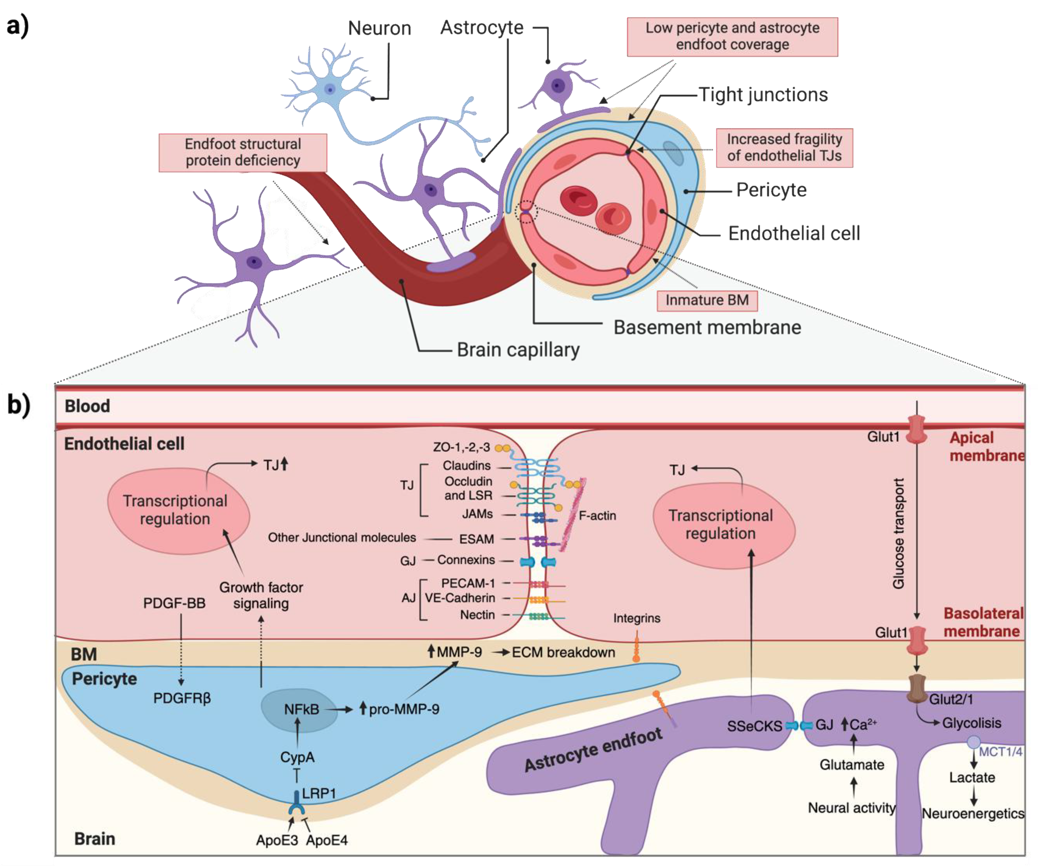
Figure 3.
Schematic overview of the mechanisms underlying brain damage after a perinatal stroke. Hypoxic-ischemic injury (left) involves energy failure, ROS production, inflammation, and cell death. Intracerebral hemorrhagic injury (right) includes BBB disruption, thrombin activation, oxidative stress, and edema formation. Both lead to tissue repair or chronic inflammation and cell death. ROS, reactive oxygen species; TJ, tight junctions; BBB, blood–brain barrier.
Disclaimer/Publisher’s Note: The statements, opinions and data contained in all publications are solely those of the individual author(s) and contributor(s) and not of MDPI and/or the editor(s). MDPI and/or the editor(s) disclaim responsibility for any injury to people or property resulting from any ideas, methods, instructions or products referred to in the content. |
© 2025 by the authors. Licensee MDPI, Basel, Switzerland. This article is an open access article distributed under the terms and conditions of the Creative Commons Attribution (CC BY) license (http://creativecommons.org/licenses/by/4.0/).
Copyright: This open access article is published under a Creative Commons CC BY 4.0 license, which permit the free download, distribution, and reuse, provided that the author and preprint are cited in any reuse.


