Preprint
Article

This version is not peer-reviewed.

Immune Responses Against West Nile Virus and Mosquito Salivary Proteins in Wild Birds from St. Tammany Parish, Louisiana

A peer-reviewed article of this preprint also exists.

Submitted:

22 January 2025

Posted:

24 January 2025

You are already at the latest version

Abstract

Though a variety of methods are used to conduct West Nile virus (WNV) surveillance, accurate prediction and prevention of outbreaks remains a global challenge. Previous studies have established that the concentration of antibodies to mosquito saliva is directly related to the intensity of exposure to mosquito bites and can be a good proxy to determine risk of infection in human populations. To assess exposure characteristics and transmission dynamics among avian communities, we tested the levels of IgY antibodies against whole salivary glands of Aedes albopictus and Culex quinquefasciatus, as well as WNV antigen, in 300 Northern cardinals sampled from April 2019 to October 2019 in St. Tammany Parish, Louisiana. Though there were no significant differences in antibody responses among sex or age groups, exposure to Ae. albopictus bites was more positively associated with exposure to WNV compared with Cx. quinquefasciatus exposure (ρ = 0.2525, p <0.001; ρ = 0.1752, p = 0.02437). This association was more pronounced among female birds (ρ = 0.3004, p = 0.0075), while no significant relationship existed between exposure to either mosquito vector and WNV among male birds in the study. In general, two seasonal trends in exposure were found, noting that exposure to Ae. albopictus becomes less intense throughout the season (ρ = -0.1529, p = 0.04984), while recaptured birds in the study were found to have increased exposure to Cx. quinquefasciatus by the end of the season (ρ = 0.277, p = 0.0468). Additionally, we report the identification of several immunogenic salivary proteins, including D7 family proteins, from both mosquito vectors among the birds. Our results suggest the role of Ae. albopictus as an early season enzootic vector of WNV, facilitated by Northern cardinal breeding behaviors, enhancing the potential to increase infections among Cx. quinquefasciatus mosquitoes in the late season contributing to human disease incidence and epizootic spillover in the environment.

Keywords: 
;  ;  ;  ;  ;  ;  

1. Introduction

In the 25 years since its emergence in the United States, West Nile virus (WNV) has become the most frequent mosquito-transmitted pathogen in the country and the most widely distributed arboviral disease in the world [1]. Transmission of WNV occurs primarily through the bite of infected Culex female mosquitoes, however the predominant vector species varies regionally across the continental United States [2]. Birds, particularly Passeriformes (songbirds) of the Corvidae (crows, jays, magpies, etc.) and Cardinalidae (cardinals) families are the main vertebrate reservoir hosts [2,3]. A previous study where North American birds were experimentally infected with WNV suggests that passerine bird species, particularly those in the Corvidae family, develop and sustain the highest viremia levels throughout the course of infection in comparison to bird species from nine other orders [3]. They were also more likely to develop clinical signs of infection and were more susceptible to mortality than any other bird species included in the study [3]. Previous studies have demonstrated host competency as an important factor contributing to an individual species role as amplifying hosts in enzootic and epizootic WNV transmission cycles, as is the case with several passerine species [4]. Conversely, studies suggest that Northern cardinals display low to moderate host competence in comparison to other passerine species due to the development of mild viremia [4]. However, these levels are strong enough to infect mosquito vectors, such that this species is important in sustaining enzootic WNV cycles but likely have limited influence in epizootic transmission of the virus [4].
As a result of their ornithophagic preferences and competency to transmit the virus, Culex mosquitoes are the most common vectors of WNV [5]. However, Aedes albopictus demonstrates less rigid preferences in its feeding behavior as these mosquitoes will readily feed from both mammalian and avian hosts, making it a potential bridge vector to consider in epizootic WNV transmission [5,6,7]. As a result, previous serological testing suggests WNV infection in a wide variety of mammals including deer, dogs, rodents, as well as horses and humans which are considered incidental hosts [2]. Though humans do develop clinical signs and symptoms, most cases are subclinical and remain undetected because of the asymptomology [2]. Approximately 25% of cases develop a mild self-limiting febrile illness, West Nile fever, characterized by a papular rash [8,9]. Unfortunately, approximately 1% of symptomatic cases progress to the more severe form of neurologic disease, commonly referred to as West Nile neuroinvasive disease [9].
In Louisiana, Culex quinquefasciatus is the primary mosquito vector implicated in WNV transmission, although mosquitoes from other genera also demonstrate vector competence, including several species of Anopheles, Culiseta, and Psorophora [2,10,11]. West Nile virus is endemic to all parishes within Louisiana but historically, Caddo, East Baton Rouge, and St. Tammany parishes have reported the highest number of human cases [10,12]. Previously, surveillance of sentinel chicken flocks and testing of dead birds was performed to predict and monitor WNV outbreaks as a measure of risk of transmission to humans in the state [13]. Due to the endemicity of the virus, testing dead birds for the presence of the virus is no longer conducted as a surveillance method for WNV unless requested by local mosquito abatement districts as part of a more comprehensive mosquito control and surveillance program [12]. Thus, current WNV surveillance relies primarily on molecular testing of pooled mosquito surveillance samples and the passive health seeking behaviors of patients with symptomatic infections [12]. Although, veterinary detection of equine infections and screening of blood center donations supplement these measures [12]. Though some human cases of WNV may be identified and allow for the estimation of local WNV transmission, the majority of asymptomatic infections remain undetected leading to the gross underestimations of WNV disease incidence in Louisiana [14].
Since WNV is mainly transmitted to humans through mosquito bite, current research is also focused on understanding the role of mosquito factors in the establishment of infection [15]. In this regard, several studies point to a major role of mosquito saliva in enhancement of not only viral replication but also enhancement of clinical characteristics of disease [15]. During the blood feeding process, mosquitoes introduce saliva into the vertebrate host eliciting an antibody response that can be measured through immunologic assays, including enzyme linked immunosorbent assays (ELISA) [16,17]. Previous studies have established that these antibodies can effectively measure intensity of vector-host interaction and can be a suitable proxy in determining risk of disease transmission in the human population [16,17,18]. Similar to the mammalian immune system, birds produce antibodies in response to these salivary antigens [19]. Immunoglobulin (Ig)Y is the primary antibody isotype found in amphibian, reptile, and avian species [19,20,21]. These antibodies have shared structural and functional homology with mammalian IgG and IgE and are primarily responsible for neutralization of antigens though they also participate in anaphylactic host responses [19,20,21].
In this study, we evaluated the avian IgY antibody responses of wild Northern cardinals (Cardinalis cardinalis) from St. Tammany Parish to Cx. quinquefasciatus and Ae. albopictus whole salivary gland extract (SGE) to measure the intensity of biting exposure and to identify the most immunogenic proteins in the saliva of these mosquito vectors. We also tested the IgY antibodies against WNV whole cell lysate antigen among the sampled birds. Our aim was to determine the intensity of mosquito and virus exposure among wild-caught cardinals in St. Tammany Parish to inform and improve understanding of regional WNV transmission dynamics, serving to enhance and support local mosquito surveillance and control measures pertaining to WNV.

2. Materials and Methods

2.1. Sample Collection

200μL of whole blood was collected by jugular venipuncture from 300 Northern cardinals captured via mist nets at 3 sites in St. Tammany Parish, Louisiana during a six-month period from April 2019 to October 2019 as part of local WNV surveillance efforts. Samples were collected on a biweekly basis in the initial four months of sample collection (April 2019 – August 2019) after which they were collected weekly for the remainder of the sample collection period (August 2019 – October 2019). All birds were banded for identification and released back to the area where they were trapped after sample collection (USFWS Permit Number MB679047-0; USGS Permit Number 24238). Complete epidemiological information was only available for 165 birds and 26 birds included in this study were recaptured during the sampling period. Baseline and recapture serum samples for each bird were tested and included in both aggregate and separate analyses. Available characteristics of the sampled birds are presented in Table 1. Data for PCR detection of current WNV infection were not available for these samples.

2.2. Aedes albopictus and Culex quinquefasciatus Mosquito Rearing and Salivary Gland Extract (SGE) Preparation

Aedes albopictus (Gainesville strain) and Cx. quinquefasciatus (Sebring strain) mosquitoes were reared at Tulane University’s Health Science campus in New Orleans, Louisiana. In this process, Ae. albopictus and Cx. quinquefasciatus eggs were hatched and emerging larvae were kept at insectary conditions of room temperature at 25 - 27°C, relative humidity 75-80%, and a 16:8 L:D cycle. Mosquitoes were allowed unrestricted access to a 10% sucrose solution throughout adult stages. Female mosquitoes were cold anesthetized, washed in 70% ethanol, and placed in 1x phosphate buffered saline (PBS) for salivary gland dissection. Salivary glands were placed and kept in 1x PBS, frozen at -80°C and thawed at 4°C for three cycles to promote cell rupture and release of proteins. The resulting SGE was kept at -80°C until use. Protein concentration was quantified using an Implen N50 Nanophotometer (Implen, Westlake Village, CA).

2.3. ELISA Testing Against Culex quinquefasciatus and Aedes albopictus Salivary Gland Extract

ELISA conditions were standardized as published elsewhere [22]. The ELISA tests were used to measure total IgY antibody titers against whole SGE from Cx. quinquefasciatus and Ae. albopictus mosquitoes. 96-well ELISA plates (High Binding Multiwell ELISA Microplates – UltraCruz®) (Santa Cruz-Biotechnology) were coated with 50μL/well of 1μg/mL of either Cx. quinquefasciatus or Ae. albopictus SGE in 1x PBS and incubated overnight at 4°C. Following overnight incubation, plates were washed three times with wash buffer (1x PBS and 0.1% Tween) and blocked using a 2% milk solution for 30 minutes at room temperature. Blocked plates were incubated with 50μL/well of 1:100 serum dilution in blocking buffer overnight at 4°C. After overnight incubation, plates were washed three times with wash buffer and incubated with 50μL/well of horseradish peroxidase (HRP)-conjugated goat anti-bird IgY antibody (Abcam, Cambridge, UK) diluted at 1:1000 in blocking buffer and incubated for 2 hours at 37°C on a plate shaker. Colorimetric development was achieved by using tetra-methyl-benzidine (TMB) (Abcam, Cambridge, UK) as substrate and was stopped with 1M H2SO4. Plates were measured at 450nm absorbance. Each sample was tested in duplicate. Additionally, three controls were included on each plate: 1) a control blank consisting of two wells without antigen or sample as a control for nonspecific color development for any of the reagents used in the assays; 2) a negative control consisting of two wells with antigen and without sample as a control for nonspecific binding of the detection antibody used in the assays; and 3) a positive control on each plate to evaluate plate to plate variation and normalize OD (optical density) values. The positive control used throughout all assays was a serum sample from rooster blood in ethylenediaminetetraacetic acid (EDTA) (Lampire Biologicals).

2.4. ELISA Testing Against West Nile Virus Whole Cell Lysate Antigen

96-well ELISA plates (High Binding Multiwell ELISA Microplates – UltraCruz®) were coated with 50μL/well of Concanavalin A (MP Biomedicals) prepared at 25μg/mL in 10mM Hepes (Thermofisher Scientific) in deionized water, adapted from previous methods described by Robinson et al [23]. Plates were then washed three times with wash buffer and coated with 50μL/well of WNV cell lysate (ZeptoMatrix, LLC) at 0.5μg/mL in 1x PBS and incubated overnight at 4°C. Plates were washed three times with wash buffer and blocked using a 2% milk solution for 30 minutes at room temperature. Blocked plates were incubated with 50μL/well of 1:100 serum dilution in blocking buffer overnight at 4°C. After overnight incubation, plates were washed three times with wash buffer and incubated with 50μL/well of HRP-conjugated goat anti-bird IgY antibody (Abcam, Cambridge, UK) diluted at 1:1000 in blocking buffer and incubated for 2 hours at 37°C on a plate shaker. Colorimetric development was achieved by using TMB (Abcam, Cambridge, UK) as substrate and was stopped with 1M H2SO4. Plates were measured at 450nm absorbance. Each sample was tested in duplicate. Controls were included as described above.

2.5. Mosquito SGE Protein Electrophoresis and Immunoblotting

Equal amounts (10μg) of Ae. albopictus and Cx. quinquefasciatus SGE were loaded into miniprotean TGX 4-15% polyacrylamide gels (BioRad). A pre-stained protein marker (Precision Plus Protein™ (10 – 250 kDa) Dual Color) was used as a molecular weight marker and the gel was run at 120V for 75 minutes. The gel was then fixed in an acetic acid solution and silver stained using the Silver Stain Plus™ kit (BioRad) according to the manufacturer’s instructions for visualization of separated proteins. An additional gel was used to transfer salivary proteins to a PVDF membrane. The PVDF membrane containing mosquito salivary proteins was blocked for one hour in a 2% milk solution and incubated overnight on a plate shaker at 4°C in a 1:1000 serum dilution made using serum from the positive control in ELISA blocking buffer. Following overnight incubation, the membrane was washed three times using the ELISA wash buffer and incubated in a 1:1000 HRP-conjugated goat anti-bird IgY antibody (Abcam, Cambridge, UK) dilution in blocking buffer for 2h at room temperature. Then the membrane was washed three times in 1x PBS and incubated in 1-step™ Ultra TMB-Blotting Solution (Thermofisher Scientific) until desired development was achieved. The reaction was stopped using deionized water. Reactive proteins were visualized on a GelDocXR+ (BioRad).

2.6. In-Gel Digestion and LCMS Preparation

Immunogenic bands in the Western blot from Ae. albopictus and Cx. quinquefasciatus SGE were sent for sequencing at the Tulane School of Medicine Proteomics Core Facility. Two independent bands were sent for analysis from each mosquito vector. Individual protein bands were excised from the polyacrylamide gel in a fume hood using a surgical knife prior to destaining. Excised gel samples were destained by washing twice in deionized water followed by three washes with 25 mM ammonium bicarbonate (ABC) and 50% acetonitrile (ACN). Destained gel samples were digested by adding 100% ACN, vortexing for five minutes, and drying the gel samples at 60°C for two minutes. 25 mM dithiothreitol in 25 mM ABC was added to the dried gels and incubated for one hour at room temperature. After incubation, 55 mM indole-3-acetic acid (IAA) in 25 mM ABC was added to gel samples and incubated in a dark room for one hour, then the gel samples were washed using deionized water for five minutes to remove any remaining IAA. Samples were dehydrated by adding 25 mM ABC in 50% ACN and vortexing for five minutes. This was repeated using 100% ACN and samples were allowed to dry afterward. 200μL 25 mM ABC and 1.5μL trypsin were added to the dried samples and incubated at 37°C overnight. The following day, digested peptides were extracted, and digestion was stopped using 20% ferulic acid (FA). The peptide solution was dried completely at 60°C for 1-2 hours. The dried peptide solution was reconstituted in LCMS buffer (2% ACN/0.1% FA) and centrifuged for 20 minutes at room temperature prior to performing LCMS analysis.

2.7. Protein Identification

To increase the number of identified proteins, the results of the LCMS analysis were referenced against all reviewed mosquito databases in UniProt for identification. Proteins identified in these analyses were filtered based on expected molecular weight and percent coverage. Filtered results were searched by ID code in the UniProt database for protein and molecular weight confirmation and amino acid sequence accession. A protein BLAST (pBLAST) analysis was performed on the sequences of uncharacterized proteins to determine proteins with similar sequence identities and to infer possible identity and function of the immunogenic proteins among our samples.

2.8. Data Analysis

Antibody concentrations used in our analyses are expressed as the calculated adjusted optical density (OD) which was determined by subtracting the average optical density of the negative control and blank wells from the average OD value of the duplicate values for each sample [17]. Shapiro-Wilkes tests were performed on all variables to determine normality at an α level of 0.05. All variables were found to be significant (p < 0.05) by the Shapiro-Wilkes test, indicating non-parametric data. As such, Mann-Whitney tests were used to test for differences among two groups, including analyses by sex and age group. Matched pairs analyses were completed using the Wilcoxon matched pairs test to compare antibody responses among individual recaptured birds. Spearman correlation coefficients were calculated to determine the strength of association between two variables. All statistical testing was performed using R statistical software version 4.3.3 at an α level of 0.05 [24].

3. Results

3.1. Description of the Study Population

The main characteristics of the study population are provided in Table 1. Throughout the 28-week sampling period, serum samples and epidemiologic data were collected from 165 birds. These birds were nearly equal by sex with 78 (47.3%) female birds and 80 (48.5%) males (information was not available for 7 (4.2%) birds). By age, birds were less evenly split with 64 (38.8%) hatch year (juvenile) birds compared with 101 (61.2%) after hatch year (adult) birds collected. Sample collection by site was strongly biased, 116 (70.3%) samples were collected from Site 1, 38 (23%) from Site 3, and the remaining 11 (6.7%) samples from Site 2. Due to the overrepresentation of one site, statistical testing was not conducted by site of sample collection. However, even with the unequal sample size by site, the average IgY response to each antigen was consistent among all three locations, as well as every other variable included in this study (Supplementary Table S1). Of the 165 observations used in the analyses, 30 samples were taken from 26 recaptured birds throughout the sampling period, with only two birds having been recaptured more than once, the highest number of recaptures for a single bird at three. Samples were collected at initial capture and every subsequent capture throughout the study. Using the IgY response of the initial and final captures for the recaptured birds, Wilcoxon signed rank tests were performed to test for the presence of any influential differences among the matched pairs in their response to Cx. quinquefasciatus, Ae. albopictus, and WNV before including these results in further analyses (Figure 1). No significant differences were found among the matched pair samples to any of the assayed antigens (p = 0.80, 0.94, and 0.72, respectively) (Figure 1). Wilcoxon signed rank matched pairs tests were also performed for the recaptured female, male, hatch year, and after hatch year birds against Cx. quinquefasciatus, Ae. albopictus, and WNV. No significant results were found in any comparison among any group to any antigen (Supplementary Figures S1 and S2). It is important to note that elapsed time between initial and final captures vary among individual birds.

3.2. WNV-Infected Mosquito Pools

In 2020, St. Tammany submitted 4,729 mosquito pools for testing and 7 pools tested positive for WNV representing 5 different mosquito species (2 Cx. nigripalpus, 2 Cx. quinquefasciatus, 1 Cx. salinarius, 1 Cx. erraticus, and 1 Ae. vexans). The first positive pool was collected in early August (Cx. nigripalpus), while the first WNV-positive Cx. quinquefasciatus were not detected until the third week in September. Since sampling was conducted as part of routine arboviral surveillance for WNV and was not completed as part of this study, sampling methods are biased towards Culex spp. and therefore other mosquito species are not frequently sampled in numbers high enough to determine an accurate estimate of WNV positivity among other mosquito species present in the area. Even though these data were collected throughout the height of the COVID-19 pandemic, it does not appear that any interruptions occurred in sampling as St. Tammany Parish still reports submitting mosquito pools for testing throughout the earliest months of the COVID-19 pandemic in the U.S.

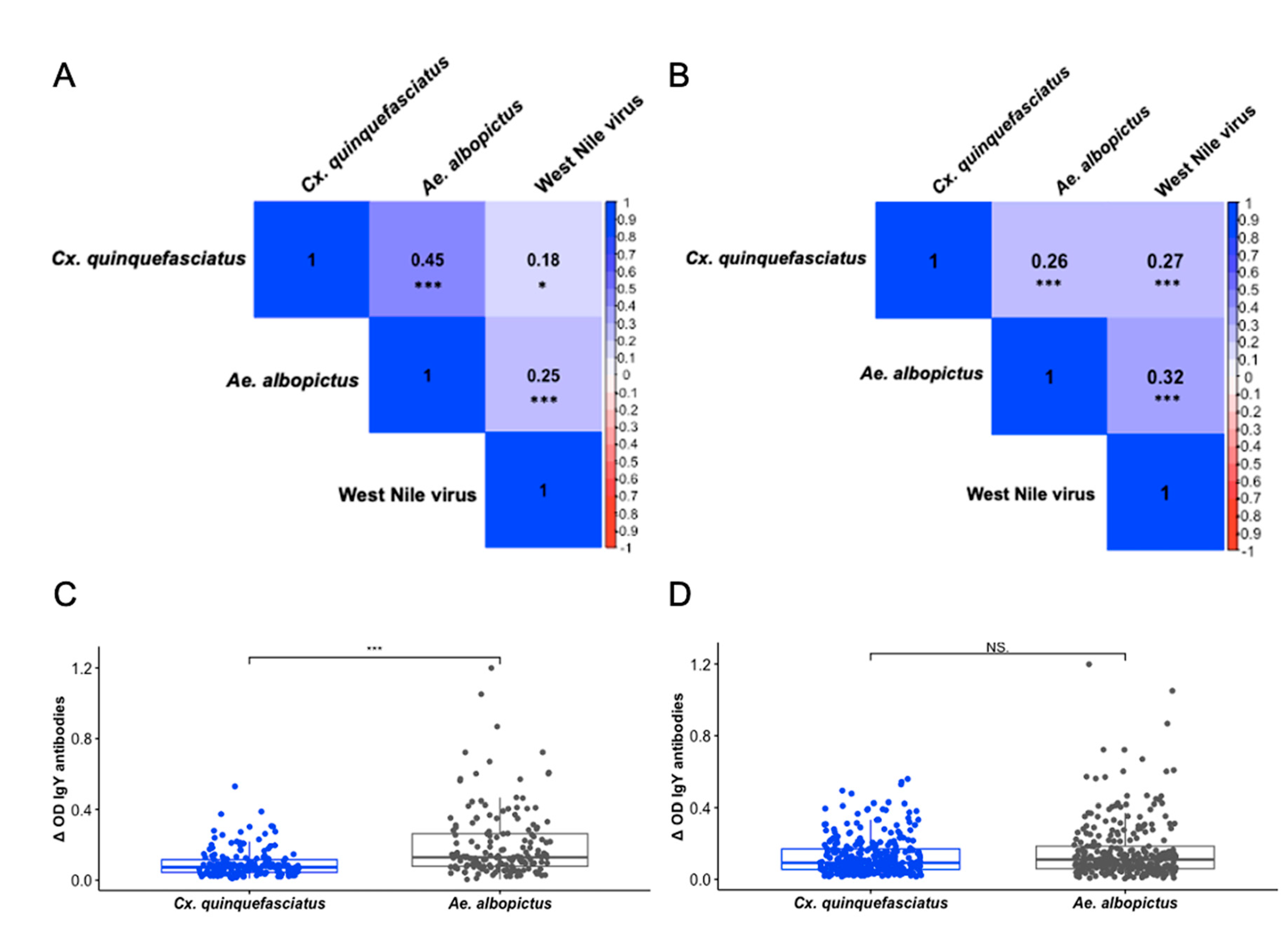
3.3. IgY Responses Are Associated Among Culex quinquefasciatus, Aedes albopictus, and West Nile Virus

Individual IgY responses were tested to determine if associations between exposure to WNV and the mosquito vectors were present in our study population. Initially, we analyzed results on 165 birds for which corresponding epidemiologic variables were available (Figure 2A). As expected, there were positive associations between exposure to Cx. quinquefasciatus and Ae. albopictus (ρ = 0.4471, p < 0.001), as well as exposure to Cx. quinquefasciatus and WNV (ρ = 0.1752, p = 0.02437). However, exposure to Ae. albopictus and WNV among these birds yields a stronger positive association than was demonstrated between Cx. quinquefasciatus and WNV (ρ = 0.2525, p <0.001) (Figure 2A). Subsequently, we compared the IgY response to Cx. quinquefasciatus against the IgY response to Ae. albopictus among the same 165 birds and found a significantly higher IgY response to Ae. albopictus (p <0.001) (Figure 2C). Considering that the significant difference in IgY response between the mosquito vectors may have influenced the results of our correlations, we performed the same analyses on all 300 samples we assayed (Figure 2B and 2D). The results of these correlations reveal similar relationships among Cx. quinquefasciatus, Ae. albopictus, and WNV while no significant difference was present in the IgY response to Cx. quinquefasciatus and Ae. albopictus (p > 0.05). Though the relationship between Cx. quinquefasciatus and WNV was much stronger among all 300 samples (ρ = 0.2714, p <0.001), a noticeably stronger association remained between Ae. albopictus and WNV (ρ = 0.3196, p <0.001). Additionally, the association of exposure between the mosquito vectors was substantially diminished (ρ = 0.2562, p <0.001) compared to the relationship found among the initial test using only 165 birds.

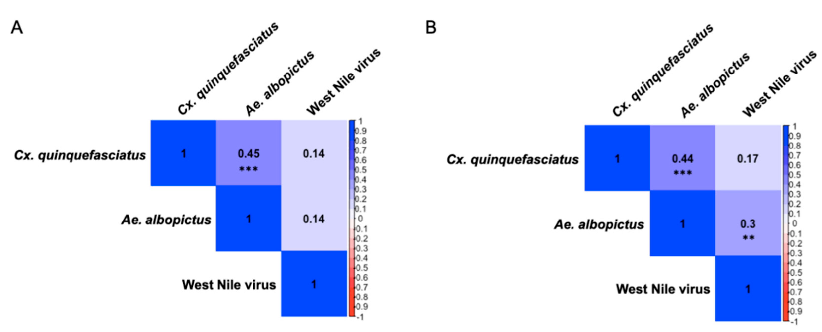
3.4. Presence of Sex-Specific Associations of IgY Response to Aedes albopictus and West Nile virus

When testing the same associations between antigens among male and female bird groups, sex-specific differences emerged (Figure 3). Parallel to our analyses in the entire study population, strong positive associations were present between exposure to the mosquito vectors in both male and female birds (ρ = 0.4534, p < 0.001; ρ = 0.4433, p < 0.001, respectively). Spearman correlations of IgY response between either Cx. quinquefasciatus or Ae. albopictus and WNV among male birds were weak and not statistically significant, though positively associated (ρ = 0.1427, p = 0.2065; ρ = 0.1358, p = 0.2297, respectively). In female birds, the positive association between Cx. quinquefasciatus and WNV was comparable to the equivalent association in their male counterparts (ρ = 0.1661, p = 0.146), while the positive association between Ae. albopictus and WNV in this group was the only significant association among either mosquito vector with WNV in either sex category (ρ = 0.3004, p = 0.0075).

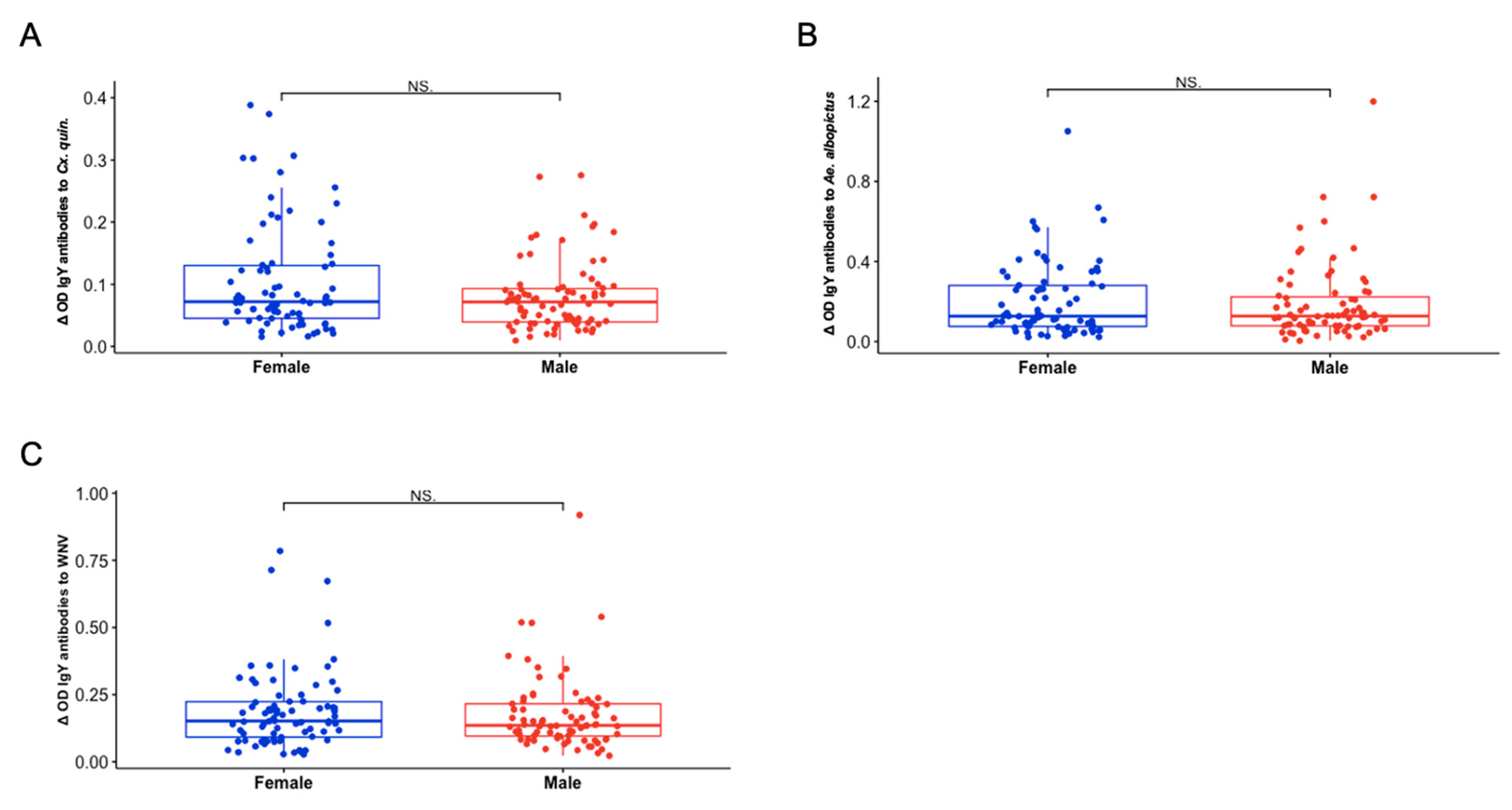
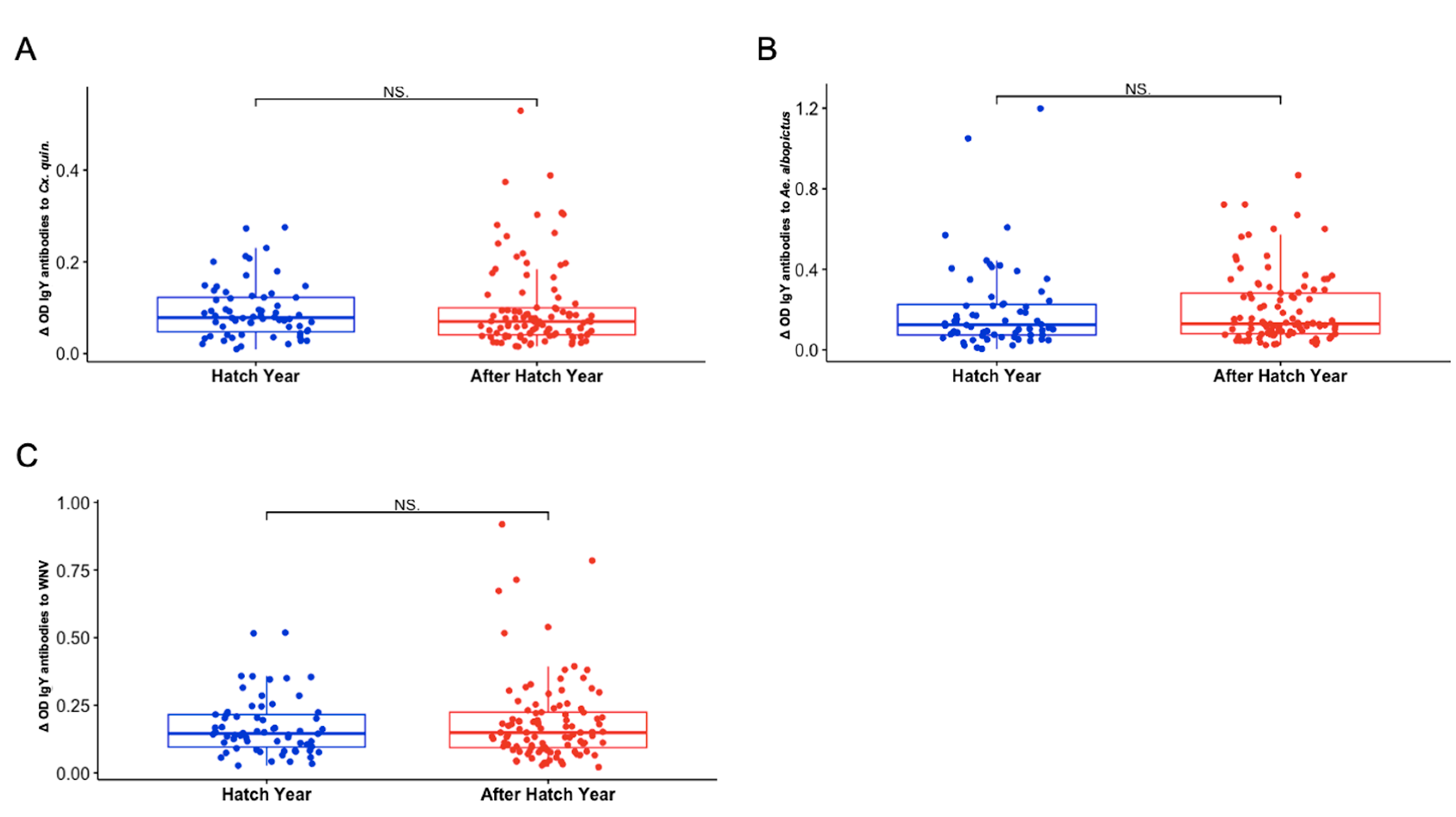
3.5. Sex and Hatch Year Are Not Important Variables Defining Exposure to Aedes albopictus or Culex quinquefasciatus Mosquito Bites Among Northern Cardinals

We observed no significant differences in IgY response by sex to Cx. quinquefasciatus SGE, Ae. albopictus SGE, and WNV antigen (p = 0.2904, 0.7543, and 0.3884, respectively) (Figure 4). Similarly, there were no significant differences in IgY response by age group to Cx. quinquefasciatus SGE, Ae. albopictus SGE, and WNV antigen (p = 0.3846, 0.4773, and 0.8976, respectively) (Figure 5). Consistent with our other findings, the highest average IgY response across both sex and age group categories was to Ae. albopictus SGE, while IgY response to Cx. quinquefasciatus SGE were the weakest among all groups.

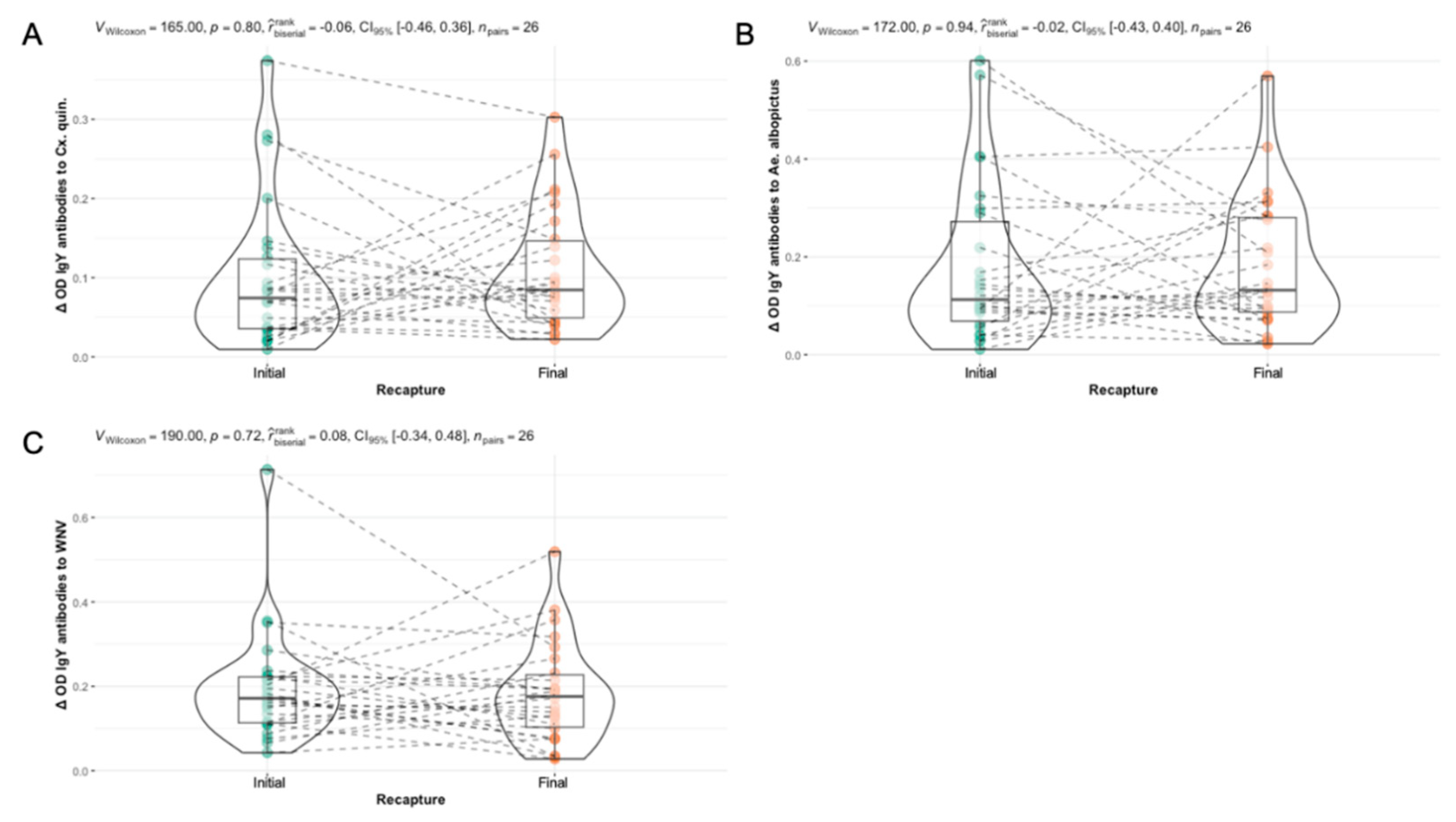
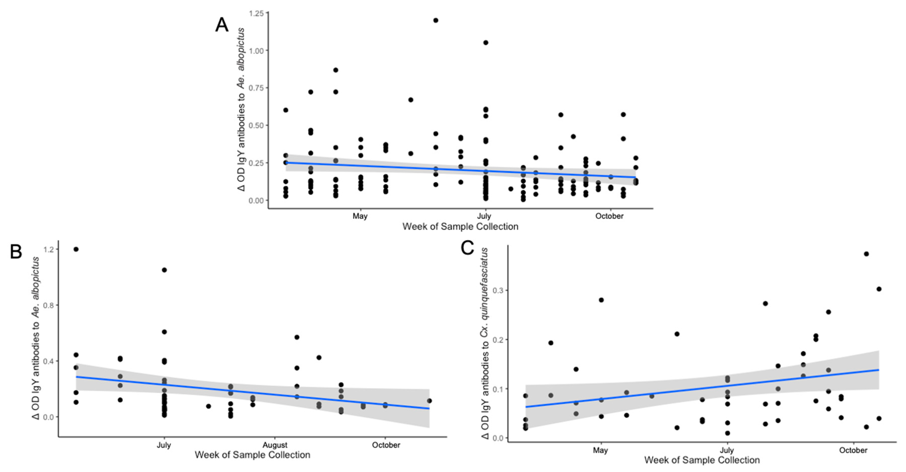
3.6. Differences in Seasonal Exposure to Aedes albopictus and Culex quinquefasciatus Among Northern Cardinals

This study yielded three significant temporal findings among IgY responses throughout the sampling period. The results demonstrate an overall negative association in IgY response to Ae. albopictus by week of sample collection (ρ = -0.1529, p = 0.04984) (Figure 6A). When examining the same association by age group, the negative temporality of IgY response to Ae. albopictus is only present in hatch year birds (ρ = -0.2482, p = 0.04799) and was not observed in the after hatch year birds. (Figure 6B). Samples from juvenile birds collected earlier in the calendar year generally displayed stronger IgY responses to Ae. albopictus than samples collected later in the calendar year. The same relationship was not present among any other antigen in the hatch year birds or with any antigen in the samples collected from after hatch year birds (Table 4). However, when determining exposure temporality by using the initial and final serum samples from the 26 recaptured birds in our study (n = 52 samples) only a positive association of IgY response to Cx. quinquefasciatus by week of sample collection exists (ρ = 0.277, p = 0.0468) (Figure 6). No other significant temporal associations were found among these birds.
Table 2. Results of all Spearman correlations of IgY antibody titers in the total study population, hatch year, after hatch year, female, male, and recaptured birds against Cx. quinquefasciatus and Ae. albopictus SGE, as well as WNV by week of sample collection. P-values are denoted as *(p<0.05), **(p<0.01), ***(p<0.001).
Cx. quinquefasciatus Ae. albopictus West Nile virus
All Birds (n = 165) 0.0797
p = 0.3087
-0.1529
p = 0.04984*
0.0606
p = 0.4394
After Hatch Year (n = 101) 0.0599
p = 0.5513
-0.0826
p = 0.4117
0.1215
p = 0.2262
Hatch Year (n = 64) 0.1546
p = 0.2226
-0.2482
p = 0.04799*
-0.0277
p = 0.828
Male (n = 80) -0.0277
p = 0.828
-0.1709
p = 0.1295
0.0093
p = 0.9349
Female (n = 78) 0.1592
p = 0.1639
-0.1185
p = 0.3015
0.1099
p = 0.3377
Recaptured Birds (n = 52)1 0.2770
p = 0.0468*
-0.0132
p = 0.9259
0.1126
p = 0.4267
1 Comparisons among 26 recaptured birds include baseline and final serum samples of each recaptured bird during the study period.

3.7. Identification of Several Pharmacologically Active Immunogenic Proteins

The immunoblot results testing the reactivity of bird serum against whole salivary gland extract from Ae. albopictus and Cx. quinquefasciatus reveals stronger reactivity to a greater number of salivary proteins from Ae. albopictus SGE compared to those revealed among Cx. quinquefasciatus SGE (Figure 7). Mass spectrometry identified 2,251 proteins among all four bands. Both structural and secreted proteins were found among the analyzed samples since our study used whole salivary gland extract rather than saliva alone. After filtering the results by corresponding molecular weight and those with percent coverage greater than 25%, 60 total proteins were identified. The list provided in Table 3 has been filtered by genus to remove any redundant proteins from other mosquito genera. Any duplicate proteins within a genus were removed manually to include proteins identified to the species of interest only.
Mass spectrometry and sequencing of the proteins in the band at ~38-44 kDa from the Ae. albopictus sample identified six proteins corresponding to the Ae. albopictus proteome. Of the identified proteins, three displayed enzymatic function, two were uncharacterized, and one was found to have binding activity. A pBLAST analysis on both uncharacterized proteins (A0AAB0A6X6 and A0A182G0N4) showed strong identities (≥95%) with the D7L1 salivary protein of Ae. albopictus and Aed a 2-like 37 kDa salivary gland allergen. The Ae. albopictus band identified at ~26-34 kDa revealed 13 proteins with identities to the Ae. albopictus proteome. Five of these were related to enzymatic functions, while another five were structural proteins, and three of the proteins were uncharacterized. A pBLAST analysis on the uncharacterized protein, A0A182GDW0, confirms a 100% identity to tropomyosin-1 in the Ae. albopictus proteome. The other uncharacterized proteins from this band all had strong identities (>97%) to enzymatic proteins including a dehydrogenase (A0A182H2F8) and a reductase (A0A182G1S0) from the Ae. albopcitus proteome.
Analysis of the protein band at ~68-70 kDa in the Cx. quinquefasciatus sample resulted in the identification of three proteins including one secreted protein (A0A8D8JS99), an enzyme (A0A8D8DSN3), and a molecular chaperone (B0X501). Of these three proteins, one was identified from the Cx. quinquefasciatus (B0X501) proteome and two from the Cx. pipiens proteome. The ~30-36 kDa band from the Cx. quinquefasciatus SGE yields seven proteins including three proteins with enzymatic activity, three structural proteins, and one secreted protein. None of the final proteins identified in the Cx. quinquefasciatus SGE were uncharacterized. A full list of the identified secreted proteins before coverage and genus filtering is available in Supplementary Table S2.

4. Discussion

The use of avian serology to monitor West Nile virus in the environment is a well established method of WNV surveillance, having been performed since its emergence in the United States, though no studies have explored IgY responses to arthropod saliva in wild bird populations. In our study, we describe the dynamics of IgY responses in Northern cardinals in St. Tammany Parish, Louisiana against salivary proteins of two local WNV vectors, Ae. albopictus and Cx. quinquefasciatus, as well as WNV whole cell lysate antigen during a single transmission season. Our findings reveal that Northern cardinals generally had higher IgY titers to salivary proteins from Ae. albopictus rather than Cx. quinquefasciatus. The high immunogenicity of Ae. albopictus SGE, in addition to the identification of a greater number of immunogenic salivary proteins compared to Cx. quinquefasciatus in our immunoblot suggests a more novel exposure to Ae. albopictus in the tested birds. We suspect that several mechanisms are simultaneously contributing to these outcomes. The first of these being that consistent exposure to avian blood meals has influenced the adaptation of less immunogenic saliva in Cx. quinquefasciatus as vertebrate blood has been shown to effect immunologic selection pressure on arthropod saliva, noting that vectors more frequently exposed to a host develop less inflammatory salivary profiles over time to facilitate successful acquisition of a bloodmeal [26]. Secondly, that the seasonal reemergence of Ae. albopictus in the spring after overwintering in egg diapause is contributing to the difference in IgY responses among the mosquito vectors, as it generates a prolonged period of nonexposure between Ae. albopictus and local bird species, preventing both the mosquito and the host from becoming more immunologically adapted to one another [27,28,29,30]. This hypothesis is further evidenced by the negative temporal associations reported among the cardinals in this study between IgY response to Ae. albopictus SGE and date of sample collection, suggesting that local bird populations become less immunologically adapted to tolerate Ae. albopictus saliva throughout winter months when vector-host contact is minimal but become less antigenically stimulated by it throughout the summer months when exposure is more consistent. Though, Cx. quinquefascitus exhibits quiescence behavior in the winter, they are not fully dormant throughout part of the year and are much more abundant in the local environment than Ae. albopictus, such that birds are more consistently exposed to their bites year-round providing a possible explanation for the absence of this association and the reduced IgY response among the birds included in this study to their saliva [27].
Further examination of this temporal trend by age group demonstrates a more pronounced negative association between IgY response to Ae. albopictus SGE and date of sample collection in hatch year birds. This finding elucidates an additional component to consider in the early season involvement of Ae. albopictus in which hatch year birds may have a disproportionate role as amplifying hosts compared to their adult counterparts as has been reported previously [31]. However, if this were true, we expected to find a corresponding association in hatch year birds between IgY response to WNV and date of sample collection, as well as differences in IgY response between adult and hatch year birds. The absence of these findings leads us to doubt a role favoring hatch year birds as early season amplifying WNV hosts in our study area, though it is important to highlight that this finding may fluctuate annually as environmental conditions for transmission vary. The absence of IgY response differences among sex groups is not surprising given that previous work has noted no observable differences in avian immunity by sex, however avian immune profiles have been reported to vary seasonally by breeding status in both male and female birds [32,33]. Northern cardinals typically raise 1-4 broods annually, occurring in spring (March-July), such that the breeding season falls in the six-month period between April and October when most of the sample collection for our study took place [34]. Similar to our analyses by sex, the absence of IgY response differences among age groups are supported by a previous study published in 2013 which reported no statistically significant differences in WNV seropositivity among 1st year hatchling and adult Northern cardinals in east Illinois, though adults of other epidemiologically relevant bird species were found to have higher antibody responses than juvenile birds [35]. Although, several earlier studies have reported higher antibody titers among adult birds of other species in comparison to first year hatchlings of the same species [35,36]. Beyond this, other important considerations for the decreasing IgY titers to Ae. albopictus SGE as hatch year birds became older throughout the sampling period are neutralizing antibody decay and maternal antibody transfer. A study published in 2015 noted a faster rate of neutralizing WNV antibody decay in hatch year birds in comparison to adult birds, but this alone still does not hold as an explanation for the absence of any association to WNV exposure in this group. In addition, the exact age of the birds in this study was not available, so we must also consider that maternal antibody acquisition may be influencing the results from hatch year birds, especially those captured within 1 month of hatching, and may not be representative of true exposure [37,38].
Our study found a stronger relationship between IgY response to WNV and Ae. albopictus SGE rather than the established local WNV vector, Cx. quinquefasciatus, among Northern cardinals in the area. It is possible that the identification of two immunogenic D7-like secreted proteins in the Ae. albopictus sample compared to a single D7 protein from the Cx. quinquefasciatus sample is contributing to these observations. A previous study utilizing a recombinant D7 protein vaccine noted enhanced WNV pathogenesis in mice vaccinated with this protein [15]. The presence of more of these immunogenic proteins from Ae. albopictus could promote more intense WNV viremia and immunologic response among birds who were infected by Ae. albopictus bite. Considering that the feeding behaviors of Ae. albopictus are increasingly flexible, it is possible that these mosquitoes are infected with WNV more often due to their diverse host range and viral susceptibility. However, a previous study investigating vector competence of three WNV vectors, including Cx. pipiens and Ae. albopictus but excluding Cx. quinquefasciatus, found that Ae. albopictus required higher virus titers to become infected than was necessary for Cx. pipiens [39]. Though mammalian species have been documented with viremic titers higher than expected, no mammal has demonstrated viremia as strong as those observed in avian hosts, such that it is more probable that Ae. albopictus in the area are becoming infected with WNV predominantly through avian bloodmeals rather than a variety of mammalian hosts [40]. A study published in 2014, hypothesized the early season (April – June) involvement of Cx. restuans in restarting seasonal WNV transmission in the Northeast, prior to the late season (June – October) involvement and viral amplification commonly demonstrated by Cx. pipiens in the region [41]. Though this hypothesis was unsupported by the fact that Cx. restuans was found to frequently feed on avian hosts of poor WNV competency rather than known amplifying hosts, we believe it is an important consideration for the role of Ae. albopictus in the WNV transmission cycle of our study area, as a previous study in St. Tammany Parish reported Northern cardinals as the most abundant resident bird species in neighborhoods of previous focal WNV transmission and found high prevalence of WNV seropositivity and neutralizing antibodies in this species [42]. It remains unclear whether mammals are capable of developing viremia high enough to infect mosquito vectors, especially Ae. albopictus, though we believe it is reasonable to conclude these results indicate Ae. albopictus vectors WNV among avian communities more often than has been previously understood in southeast Louisiana and their role as early season enzootic vectors of WNV should be investigated further.
Given that maternal antibody acquisition cannot be completely separated from the results reported in the hatch year birds, we examined IgY response associations between antigens by sex category, though we found no temporal associations of note among either group. However, when we observed associations between the IgY responses to each antigen among the two sex groups, the relationship between IgY response to WNV and Ae. albopictus SGE only existed among female birds. The same study in which the early season involvement of Cx. restuans was hypothesized also reported an increase in bloodmeals on female birds while brooding their young [41]. We believe these exposure characteristics strongly reflect Northern cardinal brooding behaviors, which favor exposure to Ae. albopictus bites rather than Cx. quinquefasciatus, as Northern cardinals build outdoor nests at low altitudes, where Ae. albopictus is more likely to feed [34,43]. These findings, in conjunction with our own regarding the temporality of Ae. albopictus exposure, further support the thought that Ae. albopictus has a crucial role in early season transmission and circulation of WNV among avian hosts, particularly vectoring the pathogen among brooding females and their offspring, before bloodmeals by Cx. quinquefasciatus become more intense in the late season.
The positive association to Cx. quinquefascitus saliva in the recaptured birds demonstrates the intensification of Culex feeding behaviors in the later summer months (August – October). Though, this finding has been reported among other studies, it is important to confirm its occurrence in our study area. In a study examining the temporal effects of Culex feeding behaviors on WNV transmissibility, researchers found that Culex spp. mosquitoes in Georgia exhibited a pronounced shift in host preference late in the summer season from feeding on American Robins (Turdus migratorius) to Northern cardinals which made up the majority of Culex bloodmeals in the latter half of the season (August – October) [44]. We believe the increased frequency of exposure to Cx. quinquefasciatus later in the summer is also a reflection of the breeding behaviors of Northern cardinals, which end their breeding season in July, when they become more mobile and return to higher altitudes after brooding their young [34]. These behaviors make them less accessible to Ae. albopictus in the area and more likely to be fed on by Cx. quinquefasciatus which typically feed at higher altitudes [6,43,45]. Unsurprisingly, the increase in Culex bloodmeals on Northern cardinals corresponds to the seasonality of human cases in southeast Louisiana, likely due to their increased competency to amplify WNV compared to Ae. albopictus.
Taken together, our results indicate the possible role of Ae. albopictus as an early season enzootic vector of WNV in southeast Louisiana, particularly vectoring WNV among brooding female Northern cardinals in the environment as they are relatively immobile and residing at a low altitude favorable for Ae. albopictus feeding. The broad circulation of WNV among these birds favors transmission into the Cx. quinquefasciatus population as birds become more mobile and return to higher altitudes in the late summer after the end of their breeding season. Transmission to Cx. quinquefasciatus allows for rapid amplification in the environment and increases the likelihood of human infections, particularly symptomatic infection, corresponding to previous seasonal trends of human WNV case detection established in the area [46].
The primary strength of our study is the selective use of Northern cardinals, rather than other bird species in the area, to survey vector and pathogen exposure. Northern cardinals are a non-migratory species corroborating the notion that exposure to both mosquito vectors and WNV occurred within the region from which the birds were sampled and limits the concern that the results of this study are due to exposure elsewhere. Use of immunologic techniques to evaluate mosquito and pathogen exposure among wild-caught birds yields a more representative depiction of the transmission dynamics and mosquito-pathogen interface in southeast Louisiana than has been previously allowed by surveillance of sentinel bird flocks, equine testing, and monitoring of symptomatic human WNV cases.
However, the study is limited in two ways which are important to consider when interpreting the results presented in this work. The first limitation is that the information presented in the study only represents live birds. No dead birds were sampled throughout the study period such that the results of the study only represent immunologic characteristics of birds that had limited exposure to the vectors of interest in the study, were unexposed to WNV, or survived WNV infection. Additionally, the use of mist nets in sample collection procedures also bias our sampling as it excludes infected passerines which are likely to have impaired flight and activity patterns due to WNV infection.

5. Conclusions

West Nile virus poses a significant public health threat, not only to human health but also veterinary health in the U.S. and abroad. However, implementation of robust surveillance methods for the prediction and prevention of WNV outbreaks is challenging due to the complex relationships of vectors, amplifying reservoir hosts, and the environmental conditions influencing virus transmission. Routine surveillance of WNV is presently conducted primarily by mosquito pool testing and passive reporting of symptomatic equine and human infections, however both methods are strongly biased and incomplete, which has major consequences on conclusions and practices determined from their interpretation. While four vaccines have been approved and licensed for equine prevention of WNV, notwithstanding their own limitations, there is no approved medical countermeasure to prevent or treat WNV infection in humans [47]. Additionally, standard clinical management strategies are palliative, further highlighting the value of reliable and holistic surveillance techniques to inform accurate predictions of epizootic WNV outbreaks. To do this, several studies have previously surveyed wild birds for the presence of IgY antibodies to WNV [48]. However, this study is the first to assess vector exposure by measuring IgY antibody titers against mosquito SGE in wild birds, in addition to pathogen exposure.

Supplementary Materials

The following supporting information can be downloaded at the website of this paper posted on Preprints.org. Figure S1: Wilcoxon signed ranks matched pairs tests among recaptured female (L; n = 12) and male (R; n=14) birds using initial and final IgY antibody titers against Cx. quinquefasciatus SGE (A and D), Ae. albopictus SGE (B and E), and WNV antigen (C and F) [25]; Figure S2: Wilcoxon signed ranks matched pairs tests among recaptured hatch year (L; n = 8) and after hatch year (R; n=12) birds using initial and final IgY antibody titers against Cx. quinquefasciatus SGE (A and D), Ae. albopictus SGE (B and E), and WNV antigen (C and F) [25]; Table S1: Average IgY antibody titers measured in Δ in optical density (OD) values across all variables among 165 birds; Table S2: Complete list of secreted proteins identified by mass spectrometry in immunogenic bands from Cx. quinquefasciatus and Ae. albopictus whole salivary gland extract.

Author Contributions

Conceptualization, B.L.-R. and K.A.C.; Methodology, A.R.S. and B.L.-R.; Formal analysis, A.R.S. and B.L.-R.; Investigation, A.R.S., S.H., Z.J., and B.L.-R.; Resources, D.M.W, J.D.V., K.A.C., S.B.J., and B.L.-R.; Writing—original draft preparation, A.R.S., K.A.C., S.R.M., and B.L.-R.; Writing—review and editing, A.R.S., S.H., Z.J., J.D.V, S.B.J., D.M.W., K.A.C., S.R.M., and B.L.-R.; Visualization, A.R.S.; Supervision, B.L.-R.; All authors have read and agreed to the published version of the manuscript.

Funding

This research received no external funding.

Institutional Review Board Statement

Not applicable.

Informed Consent Statement

Not applicable.

Data Availability Statement

The raw data supporting the conclusions of this article will be made available by the authors on request.

Acknowledgments

We would like to thank the members of the Fan laboratory for their expertise, efforts, and dedication throughout the protein identification portion of this project, in addition to the members of the Robinson laboratory for providing valuable insights and guidance related to the ELISA methodologies used in this work. We also acknowledge and appreciate the St. Tammany Parish Mosquito Abatement team, particularly Benjamin Duckworth, for the collection of the avian blood samples. The following reagent was obtained through BEI Resources, NIAID, NIH: Aedes albopictus, Strain Gainesville, MRA-804, contributed by Sandra A. Allan. Culex quinquefasciatus (Sebring) colony material was kindly provided by Nicole Foley and Sean Masters from the Division of Vector-Borne Diseases at the Centers for Disease Control and Prevention in Fort Collins, CO.

Conflicts of Interest

The authors declare no conflicts of interest.

Abbreviations

The following abbreviations are used in this manuscript:
ABC Ammonium bicarbonate
ACN Acetonitrile
Ae. Aedes
Cx. Culex
ELISA Enzyme linked immunosorbent assay
FA Ferulic acid
IAA Indole-3-acetic acid
IgY Immunoglobulin Y
LCMS Liquid chromatography-mass spectrometry
OD Optical density
PBS Phosphate buffered saline
PVDF Polyvinylidene fluoride
SGE Salivary gland extract
WNV West Nile virus

References

  1. CDC Epidemiology and Ecology | Mosquitoes | . (2022, June 29). https://www.cdc.gov/mosquitoes/guidelines/west-nile/epidemiology-ecology.html.
  2. Hayes, E.B.; Komar, N.; Nasci, R.S.; Montgomery, S.P.; O’Leary, D.R.; Campbell, G.L. Epidemiology and Transmission Dynamics of West Nile Virus Disease. Emerging Infectious Diseases. 2005, 11, 1167–1173. [Google Scholar] [CrossRef]
  3. Komar, N.; Langevin, S.; Hinten, S.; Nemeth, N.; Edwards, E.; Hettler, D.; Davis, B.; Bowen, R.; Bunning, M. Experimental Infection of North American Birds with the New York 1999 Strain of West Nile Virus. Emerging Infectious Diseases 2003, 9, 311–322. [Google Scholar] [CrossRef] [PubMed]
  4. Levine, R.S.; Mead, D.G.; Kitron, U.D. Limited Spillover to Humans from West Nile Virus Viremic Birds in Atlanta, Georgia. Vector Borne and Zoonotic Diseases. 2013, 13, 812–817. [Google Scholar] [CrossRef] [PubMed]
  5. Faraji, A.; Egizi, A.; Fonseca, D.M.; Unlu, I.; Crepeau, T.; Healy, S.P.; Gaugler, R. Comparative Host Feeding Patterns of the Asian Tiger Mosquito, Aedes albopictus, in Urban and Suburban Northeastern USA and Implications for Disease Transmission. PLOS Neglected Tropical Diseases. 2014, 8, e3037. [Google Scholar] [CrossRef]
  6. Hutchinson, R.A.; West, P.A.; Lindsay, S.W. Suitability of two carbon dioxide-baited traps for mosquito surveillance in the United Kingdom. Bulletin of Entomological Research. 2007, 97, 591–597. [Google Scholar] [CrossRef] [PubMed]
  7. Savage, H.M.; Niebylski, M.L.; Smith, G.C.; Mitchell, C.J.; Craig, G.B., Jr. Host-Feeding Patterns of Aedes albopictus (Diptera: Culicidae) at a Temperate North American Site. Journal of Medical Entomology. 1993, 30, 27–34. [Google Scholar] [CrossRef]
  8. Vouillon, A.; Barthelemy, J.; Lebeau, L.; Nisole, S.; Savini, G.; Lévêque, N.; Simonin, Y.; Garcia, M.; Bodet, C. Skin tropism during Usutu virus and West Nile virus infection: An amplifying and immunological role. Journal of Virology. 2023, 98, e01830-23. [Google Scholar] [CrossRef]
  9. Rossi, S.L.; Ross, T.M.; Evans, J.D. West Nile Virus. Clinics in Laboratory Medicine. 2010, 30, 47–65. [Google Scholar] [CrossRef] [PubMed]
  10. Ratard, R. (2021). West Nile Virus Annual Report.
  11. Rochlin, I.; Faraji, A.; Healy, K.; Andreadis, T.G. West Nile Virus Mosquito Vectors in North America. Journal of Medical Entomology. 2019, 56, 1475–1490. [Google Scholar] [CrossRef]
  12. West Nile Virus | La Dept. Of Health. (n.d.). Retrieved May 16. 2024, from https://ldh.la.gov/page/west-nile-virus.
  13. Louisiana Arbovirus Surveillance Summary 2020. (n.d.). Retrieved from https://ldh.la.gov/assets/oph/Center-PHCH/Center-CH/infectious-epi/Arboviral/arboweekly/2020_wnv_reports/ARBO_2049.pdf.
  14. West Nile Virus | La Dept. Of Health. (n.d.). Retrieved May 16. 2024, from https://ldh.la.gov/assets/oph/Center-PHCH/Center-CH/infectious-epi/Arboviral/arboweekly/2023_WNV_Reports/ARBO_2352.pdf.
  15. Reagan, K.L.; Machain-Williams, C.; Wang, T.; Blair, C.D. Immunization of Mice with Recombinant Mosquito Salivary Protein D7 Enhances Mortality from Subsequent West Nile Virus Infection via Mosquito Bite. PLOS Neglected Tropical Diseases 2012, 6, e1935. [Google Scholar] [CrossRef]
  16. Londono-Renteria, B.L.; Eisele, T.P.; Keating, J.; James, M.A.; Wesson, D.M. Antibody Response Against Anopheles albimanus (Diptera: Culicidae) Salivary Protein as a Measure of Mosquito Bite Exposure in Haiti. Journal of Medical Entomology. 2010, 47, 1156–1163. [Google Scholar] [CrossRef] [PubMed]
  17. Londono-Renteria, B.; Cardenas, J.C.; Cardenas, L.D.; Christofferson, R.C.; Chisenhall, D.M.; Wesson, D.M.; McCracken, M.K.; Carvajal, D.; Mores, C.N. Use of Anti-Aedes aegypti Salivary Extract Antibody Concentration to Correlate Risk of Vector Exposure and Dengue Transmission Risk in Colombia. PLOS ONE. 2013, 8, e81211. [Google Scholar] [CrossRef] [PubMed]
  18. Maldonado-Ruiz, L.P.; Montenegro-Cadena, L.; Blattner, B.; Menghwar, S.; Zurek, L.; Londono-Renteria, B. Differential Tick Salivary Protein Profiles and Human Immune Responses to Lone Star Ticks (Amblyomma americanum) From the Wild vs. A Laboratory Colony. Frontiers in Immunology. 2019, 10. [Google Scholar] [CrossRef] [PubMed]
  19. Taylor, A.I.; Gould, H.J.; Sutton, B.J.; Calvert, R.A. Avian IgY Binds to a Monocyte Receptor with IgG-like Kinetics Despite an IgE-like Structure *. Journal of Biological Chemistry. 2008, 283, 16384–16390. [Google Scholar] [CrossRef]
  20. Lee, W.; Syed Atif, A.; Tan, S.C.; Leow, C.H. Insights into the chicken IgY with emphasis on the generation and applications of chicken recombinant monoclonal antibodies. Journal of Immunological Methods. 2017, 447, 71–85. [Google Scholar] [CrossRef] [PubMed]
  21. Pacheco, B.L.B.; Nogueira, C.P.; Venancio, E.J. IgY Antibodies from Birds: A Review on Affinity and Avidity. Animals. 2023, 13, 19. [Google Scholar] [CrossRef]
  22. Londono-Renteria, B.; Patel, J.C.; Vaughn, M.; Funkhauser, S.; Ponnusamy, L.; Grippin, C.; Jameson, S.B.; Apperson, C.; Mores, C.N.; Wesson, D.M.; Colpitts, T.M.; Meshnick, S.R. Long-Lasting Permethrin-Impregnated Clothing Protects Against Mosquito Bites in Outdoor Workers. The American journal of tropical medicine and hygiene 2015, 93, 869–874. [Google Scholar] [CrossRef] [PubMed]
  23. Robinson, J.E.; Holton, D.; Liu, J.; McMurdo, H.; Murciano, A.; Gohd, R. A novel enzyme-linked immunosorbent assay (ELISA) for the detection of antibodies to HIV-1 envelope glycoproteins based on immobilization of viral glycoproteins in microtiter wells coated with concanavalin A. Journal of immunological methods 1990, 132, 63–71. [Google Scholar] [CrossRef] [PubMed]
  24. R Core Team (2024). R: A Language and Environment for Statistical Computing. R Foundation for Statistical Computing, Vienna, Austria.
  25. Patil, I. Visualizations with statistical details: The 'ggstatsplot' approach. Journal of Open Source Software. 2021, 6, 3167. [Google Scholar] [CrossRef]
  26. Arcà, B.; Struchiner, C.J.; Pham, V.M.; Sferra, G.; Lombardo, F.; Pombi, M.; Ribeiro, J.M.C. Positive selection drives accelerated evolution of mosquito salivary genes associated with blood-feeding. Insect Molecular Biology. 2014, 23, 122–131. [Google Scholar] [CrossRef] [PubMed]
  27. Diniz, D.F.A.; de Albuquerque, C.M.R.; Oliva, L.O.; de Melo-Santos, M.A.V.; Ayres, C.F.J. Diapause and quiescence: Dormancy mechanisms that contribute to the geographical expansion of mosquitoes and their evolutionary success. Parasites & Vectors. 2017, 10, 310. [Google Scholar] [CrossRef]
  28. Doucoure, S.; Mouchet, F.; Cournil, A.; Goff, G.L.; Cornelie, S.; Roca, Y.; Giraldez, M.G.; Simon, Z.B.; Loayza, R.; Misse, D.; Flores, J.V.; Walter, A.; Rogier, C.; Herve, J.P.; Remoue, F. Human Antibody Response to Aedes aegypti Saliva in an Urban Population in Bolivia: A New Biomarker of Exposure to Dengue Vector Bites. The American Journal of Tropical Medicine and Hygiene. 2012, 87, 504–510. [Google Scholar] [CrossRef] [PubMed]
  29. Montiel, J.; Carbal, L.F.; Tobón-Castaño, A.; Vásquez, G.M.; Fisher, M.L.; Londono-Rentería, B. IgG antibody response against Anopheles salivary gland proteins in asymptomatic Plasmodium infections in Narino, Colombia. Malaria Journal. 2020, 19, 42. [Google Scholar] [CrossRef]
  30. Buezo Montero, S.; Gabrieli, P.; Montarsi, F.; Borean, A.; Capelli, S.; De Silvestro, G.; Forneris, F.; Pombi, M.; Breda, A.; Capelli, G.; Arcà, B. IgG Antibody Responses to the Aedes albopictus 34k2 Salivary Protein as Novel Candidate Marker of Human Exposure to the Tiger Mosquito. Frontiers in Cellular and Infection Microbiology 2020, 10. [Google Scholar] [CrossRef]
  31. Hamer, G.L.; Walker, E.D.; Brawn, J.D.; Loss, S.R.; Ruiz, M.O.; Goldberg, T.L.; Schotthoefer, A.M.; Brown, W.M.; Wheeler, E.; Kitron, U.D. Rapid amplification of West Nile virus: the role of hatch-year birds. Vector Borne Zoonotic Dis. 2008, 8, 57–67. [Google Scholar] [CrossRef] [PubMed]
  32. Valdebenito, J.O.; Halimubieke, N.; Lendvai, Á.Z.; Figuerola, J.; Eichhorn, G.; Székely, T. Seasonal variation in sex-specific immunity in wild birds. Scientific Reports. 2021, 11, 1349. [Google Scholar] [CrossRef]
  33. Owen, J.C.; Nakamura, A.; Coon, C.A.; Martin, L.B. The effect of exogenous corticosterone on West Nile virus infection in Northern Cardinals (Cardinalis cardinalis). Veterinary Research. 2012, 43, 34. [Google Scholar] [CrossRef] [PubMed]
  34. Breeding—Northern Cardinal—Cardinalis cardinalis—Birds of the World. (n.d.). Retrieved December 3, 2024, from https://birdsoftheworld.org/bow/species/norcar/cur/breeding.
  35. Lampman, R.L.; Krasavin, N.M.; Ward, M.P.; Beveroth, T.A.; Lankau, E.W.; Alto, B.W.; Muturi, E.; Novak, R.J. West Nile Virus Infection Rates and Avian Serology in East-Central Illinois. Journal of the American Mosquito Control Association. 2013, 29, 108–122. [Google Scholar] [CrossRef]
  36. West Nile Virus Antibodies in Avian Species of Georgia, USA: 2000–2004. (n.d.). [CrossRef]
  37. Kaspers, B.; Göbel, T.W.; Schat, K.A.; Vervelde, L. Avian Immunology (3rd ed.) (Academic Press, 2022).
  38. Lamb, J.S.; Tornos, J.; Lejeune, M.; Boulinier, T. Rapid loss of maternal immunity and increase in environmentally mediated antibody generation in urban gulls. Scientific Reports. 2024, 14, 4357. [Google Scholar] [CrossRef] [PubMed]
  39. Tiawsirisup, S.; Platt, K.B.; Evans, R.B.; Rowley, W.A. A Comparision of West Nile Virus Transmission by Ochlerotatus trivittatus (COQ.), Culex pipiens (L.), and Aedes albopictus (Skuse). Vector-Borne and Zoonotic Diseases. 2005, 5, 40–47. [Google Scholar] [CrossRef] [PubMed]
  40. Root, J.J.; Bosco-Lauth, A.M. West Nile Virus Associations in Wild Mammals: An Update. Viruses. 2019, 11, Article 5. [Google Scholar] [CrossRef] [PubMed]
  41. Egizi, A.M.; Farajollahi, A.; Fonseca, D.M. Diverse Host Feeding on Nesting Birds May Limit Early-Season West Nile Virus Amplification. Vector-Borne and Zoonotic Diseases. 2014, 14, 447–453. [Google Scholar] [CrossRef] [PubMed]
  42. Komar, N.; Panella, N.A.; Langevin, S.A.; Brault, A.C.; Amador, M.; Edwards, E.; Owen, J.C. AVIAN HOSTS FOR WEST NILE VIRUS IN ST. TAMMANY PARISH, LOUISIANA, 2002. The American Journal of Tropical Medicine and Hygiene 2005, 73, 1031–1037. [Google Scholar] [CrossRef] [PubMed]
  43. Obenauer, P.J.; Kaufman, P.E.; Allan, S.A.; Kline, D.L. Host-Seeking Height Preferences of Aedes albopictus (Diptera: Culicidae) in North Central Florida Suburban and Sylvatic Locales. Journal of Medical Entomology. 2009, 46, 900–908. [Google Scholar] [CrossRef] [PubMed]
  44. Levine, R.S.; Mead, D.G.; Hamer, G.L.; Brosi, B.J.; Hedeen, D.L.; Hedeen, M.W.; McMillan, J.R.; Bisanzio, D.; Kitron, U.D. Supersuppression: Reservoir Competency and Timing of Mosquito Host Shifts Combine to Reduce Spillover of West Nile Virus. The American Journal of Tropical Medicine and Hygiene. 2016, 95, 1174–1184. [Google Scholar] [CrossRef] [PubMed]
  45. Savage, H.M.; Anderson, M.; Gordon, E.; Mcmillen, L.; Colton, L.; Delorey, M.; Sutherland, G.; Aspen, S.; Charnetzky, D.; Burkhalter, K.; Godsey, M. Host-Seeking Heights, Host-Seeking Activity Patterns, and West Nile Virus Infection Rates for Members of the Culex pipiens Complex at Different Habitat Types Within the Hybrid Zone, Shelby County, TN, 2002 (Diptera: Culicidae). Journal of Medical Entomology 2008, 45, 276–288. [Google Scholar] [CrossRef] [PubMed]
  46. Louisiana Arbovirus Surveillance Summary 2021. (2022, January 1). Retrieved from https://ldh.la.gov/assets/oph/Center-PHCH/Center-CH/infectious-epi/Arboviral/arboweekly/2021_WNV_Reports/ARBO_2152.pdf.
  47. Cendejas, P.M.; Goodman, A.G. Vaccination and Control Methods of West Nile Virus Infection in Equids and Humans. Vaccines. 2024, 12, 485. [Google Scholar] [CrossRef] [PubMed]
  48. McMillan, J.R.; Hamer, G.L.; Levine, R.S.; Mead, D.G.; Waller, L.A.; Goldberg, T.L.; Walker, E.D.; Brawn, J.D.; Ruiz, M.O.; Kitron, U.; Vazquez-Prokopec, G. Multi-Year Comparison of Community- and Species-Level West Nile Virus Antibody Prevalence in Birds from Atlanta, Georgia and Chicago, Illinois, 2005–2016. The American Journal of Tropical Medicine and Hygiene 2023, 108, 366–376. [Google Scholar] [CrossRef]
Figure 1. Wilcoxon signed ranks matched pairs tests among all recaptured birds using initial and final IgY antibody titers against (A) Cx. quinquefasciatus SGE, (B) Ae. albopictus SGE, and (C) WNV antigen [25].
Preprints 146987 g001
Figure 2. (A) Correlogram of Spearman correlation of IgY antibody titers in subset of samples for which epidemiological variables were available (n=165) against Cx. quinquefasciatus and Ae. albopictus mosquito SGE and WNV antigen. (B) Correlogram of Spearman correlation of IgY antibody titers among all samples that were tested (n=300) against Cx. quinquefasciatus and Ae. albopictus mosquito SGE and WNV antigen. Positive correlations are indicated in gradient blue according to strength of association (darker blue represents a stronger positive correlation) while negative correlations are indicated in gradient red according to strength of association (darker red represents a stronger negative correlation). (C) Mann-Whitney U test comparing IgY antibody titers in subset of samples for which epidemiological variables were available (n=165) between Cx. quinquefasciatus and Ae. albopictus. (D) Mann-Whitney U test comparing IgY antibody titers among all samples that were tested (n=300) between Cx. quinquefasciatus and Ae. albopictus. IgY response measured in Δ in optical density (OD) values. P-values are denoted as *(p<0.05), **(p<0.01), ***(p<0.001).
Preprints 146987 g002
Figure 3. (A) Correlogram of Spearman correlation of IgY antibody titers in samples from male birds (n=80) against Cx. quinquefasciatus and Ae. albopictus mosquito SGE and WNV antigen. (B) Correlogram of Spearman correlation of IgY antibody titers in samples from female birds (n=78) against Cx. quinquefasciatus and Ae. albopictus mosquito SGE and WNV antigen. Positive correlations are indicated in gradient blue according to strength of association (darker blue represents a stronger positive correlation) while negative correlations are indicated in gradient red according to strength of association (darker red represents a stronger negative correlation). P-values are denoted as *(p<0.05), **(p<0.01), ***(p<0.001).
Preprints 146987 g003
Figure 4. Mann-Whitney tests by sex. (A) IgY responses to Cx. quinquefasciatus SGE, (B) IgY responses to Ae. albopictus SGE, (C) IgY responses to WNV. Results from female (n=78) and male (n=80) birds. Data for sex was not available for 7 samples, which were not included in these analyses. IgY responses measured in Δ in optical density (OD) values.
Preprints 146987 g004
Figure 5. Mann-Whitney tests by age. (A) IgY responses to Cx. quinquefasciatus SGE, (B) IgY responses to Ae. albopictus SGE, (C) IgY responses to WNV. Results from hatch year (n=64) and after hatch year (n=101) birds. IgY responses measured in Δ in OD values.
Preprints 146987 g005
Figure 6. Results of Spearman correlations by week of sample collection. (A) IgY responses in total study population (n=165) against Ae. albopictus SGE. (B) IgY responses in hatch year birds (n=64) against Ae. albopictus SGE. (C) IgY responses in recaptured birds (n=52) against Cx. quinquefasciatus SGE.
Preprints 146987 g006
Figure 7. Aedes albopictus and Culex quinquefasciatus immunoblot results.
Preprints 146987 g007
Table 1. Characteristics of the study population.
Site
n (%)
1 2 3
116 (70.3) 11 (6.7) 38 (23)
Sex
n (%)
Male Female Not available
80 (48.5) 78 (47.3) 7 (4.2)
Age
n (%)
Hatch Year After Hatch Year Not available
64 (38.8) 101 (61.2) 0 (0)
Mass
(g)
Average Minimum Maximum
39.022 3 46
Table 3. List of proteins with greater than 25% peptide coverage identified by mass spectrometry in immunogenic bands from Culex quinquefasciatus and Aedes albopictus whole salivary gland extract.
Protein Name ID MW (Da)
Aedes albopictus
~38-44 kDa band
Uncharacterized protein (Aedes albopictus) A0AAB0A6X6 38,495
Uncharacterized protein (Aedes albopictus) A0A182G0N4 41,526
Pyruvate dehydrogenase E1 component subunit beta A0A023ERL5 38,453
Fructose-bisphosphate aldolase A0A023EQM6 39,152
Glycerol-3-phosphate dehydrogenase [NAD(+)] A0A182GGV8 38,604
Putative actin filament-coating protein tropomyosin A0A023ETF0 43,859
Aedes albopictus
~26-34 kDa band
14-3-3 protein epsilon A0A023ENU3 29,418
Enoyl-CoA hydratase, mitochondrial A0A023EPZ2 31,511
ATP synthase subunit gamma A0A023ENY5 32,711
Uncharacterized protein (Aedes albopictus) A0A182GDW0 29,466
ADP/ATP translocase A0A023EP24 32,904
Proteasome subunit alpha type A0A023ENX8 28,843
Regulator of microtubule dynamics protein 1 A0A182GHN2 26,069
Electron transfer flavoprotein subunit beta A0A023ENM9 27,466
N-acetyltransferase domain-containing protein A0A023EKP0 27,121
Proteasome subunit alpha type A0A023EL07 27,671
Putative 11beta-hydroxysteroid dehydrogenase type 1 A0A023ENR7 27,211
Uncharacterized protein (Aedes albopictus) A0A182H2F8 26,847
Uncharacterized protein (Aedes albopictus) A0A182G1S0 27,190
Culex quinquefasciatus
~68-70 kDa band
H(+)-transporting two-sector ATPase A0A8D8DSN3 68,188
Transmembrane protease serine 9 A0A8D8JS99 70,089
Heat shock protein 70 B2 B0X501 69,919
Culex quinquefasciatus
~30-36 kDa band
Regucalcin A0A8D8BZB5 33,670
Long form salivary protein D7L2 B0X6Z1 36,051
Tropomyosin-2 A0A8D8A960 35,025
Probable elongation factor 1-beta A0A8D8KQV6 31,741
Enoyl-CoA hydratase ECHA12 B0W6D4 33,912
Malate dehydrogenase A0A1Q3FI32 35,186
ADP/ATP translocase B0WFA5 32,972
Disclaimer/Publisher’s Note: The statements, opinions and data contained in all publications are solely those of the individual author(s) and contributor(s) and not of MDPI and/or the editor(s). MDPI and/or the editor(s) disclaim responsibility for any injury to people or property resulting from any ideas, methods, instructions or products referred to in the content.
Copyright: This open access article is published under a Creative Commons CC BY 4.0 license, which permit the free download, distribution, and reuse, provided that the author and preprint are cited in any reuse.
Prerpints.org logo

Preprints.org is a free preprint server supported by MDPI in Basel, Switzerland.

Subscribe

Disclaimer

Terms of Use

Privacy Policy

Privacy Settings

© 2025 MDPI (Basel, Switzerland) unless otherwise stated