Preprint
Review

This version is not peer-reviewed.

Exploring the Therapeutic Potential of BRCA1 and BRCA2 as Targets in Canine Oncology: A Comprehensive Review of Their Role in Cancer Development and Treatment

Submitted:

19 January 2025

Posted:

20 January 2025

You are already at the latest version

Abstract

Tumor diseases represent a significant global health challenge, impacting both humans and companion animals, notably dogs. The parallels observed in the pathophysiology of cancer between humans and dogs underscore the importance of advancing comparative oncology and translational research methodologies. Moreover, dogs serve as valuable models for human cancer research due to shared environments, genetics, and treatment responses. Notably, breast cancer gene 1 (BRCA1) and breast cancer gene 2 (BRCA2), which are pivotal in human oncology, also influence the development and progression of canine tumors. The role of BRCA1 and BRCA2 in canine cancers remains underexplored, but their potential significance as therapeutic targets is strongly considered. This systematic review aims to broaden the discussion of BRCA1 and BRCA2 beyond mammary tumors, exploring their implications across various canine cancers. By emphasizing shared genetic underpinnings between species and advocating for a comparative approach, the review indicates the potential of BRCA genes as targets for innovative cancer therapies in dogs, contributing to advancements in both human and veterinary oncology.

Keywords: 
;  ;  ;  ;  

1. Introduction

Cancer, characterized by the uncontrolled growth and proliferation of cells, significantly impacts health systems worldwide (Brown et al. 2023). Despite the transient shift in mortality patterns due to COVID-19 in 2020, cancer consistently ranks as a leading cause of death, second only to cardiovascular diseases in the United States and Europe (Heron and Anderson 2016; Siegel et al. 2021). This global health issue extends beyond humans (Townsend et al. 2016), with cancer also recognized as the leading cause of disease-related mortality in dogs across developed nations (Moore et al. 2001; Proschowsky et al. 2003; Bonnett et al. 2005; Egenvall et al. 2005; Fleming et al. 2011; Adams et al. 2010; Dobson 2013) . As of 2023, the global dog population is estimated at 900 million (Gompper 2023), with approximately 470 million dogs living as pets (Harvey et al. 2019). Retrospective studies have shown that cancer accounts for about 30% of all canine deaths (Melissa et al. 2008; Fleming et al. 2011; Alvarez 2014), underscoring its prevalence and impact in the canine population. The shared environments and lifestyles between dogs and their human companions have facilitated the emergence of dogs as valuable models for understanding human cancers. The spontaneous development of cancers in dogs, mirroring those in humans, along with similar responses to treatment, positions canine cancer research as a crucial parallel to human oncological studies (Kol et al. 2015; Paynter et al. 2021). Hence, the comparative oncology field has recognized that the naturally occurring cancers in dogs offer invaluable insights into human oncology, especially given the common pathophysiology of these diseases and their similarity in terms of risk factors, tumor biology, and response to treatment. Recent genomic studies have highlighted significant similarities between canine and human cancers, revealing common genetic underpinnings and offering insights into tumor development across species. Notably, cancer types, such as osteosarcoma, melanoma, non-Hodgkin lymphoma, and bladder cancer, share histological and molecular characteristics between dogs and humans, suggesting a shared oncogenic pathway and potential for comparative oncology research (Cadieu and Ostrander 2007; Paoloni et al. 2009; Thomas et al. 2009).
Among the various cancers affecting dogs in Europe, mammary tumors are particularly prevalent, especially in uncastrated female dogs (Arnesen 2001; Egenvall 2005). Research has identified several candidate genes associated with canine mammary tumor development, including breast cancer gene 1 (BRCA1) and breast cancer gene 2 (BRCA2) (Rivera 2009; Borge 2011). These genes, known for their role in human breast and ovarian cancers, have also been implicated in canine mammary tumors, highlighting the genetic commonalities between canine and human cancers (Rivera et al. 2009; Enginler et al. 2014; Yoshikawa et al. 2015). However, despite the established importance of BRCA1 and BRCA2 as biomarkers and therapeutic targets in human oncology, their role in canine cancers remains underexplored.
This review aims to expand the discussion of BRCA1 and BRCA2 beyond their association with CMT, exploring their broader implications in the oncogenesis of various cancers in dogs. By highlighting the frequency and impact of cancer in dogs, drawing parallels with human oncology, and delving into the genetic underpinnings shared between species, this review seeks to emphasize the potential of BRCA genes as targets for innovative cancer therapies. Furthermore, it emphasizes the importance of a comparative approach, recognizing the value of canine models in elucidating the molecular mechanisms of cancer and in the development of targeted treatments.

2. Methods

To ensure a comprehensive and current overview, a systematic literature search spanning from 1990 to 2023 was conducted across PubMed and the Cochrane Library, the search were “canine mammary tumors”, “BRCA1 and BRCA2”, “PARPi”, and “DNA repair mechanisms”. This methodological approach facilitated the inclusion of relevant clinical trials, reviews, meta-analyses, and other pivotal studies, that supported review’s investigation in reference to therapeutic meaning of BRCA1 and BRCA2 as a potential target in canine oncology.

3. Main Text

3.1. BRCA1 and BRCA2: Structural, Biological and Molecular Functions

The Role of BRCA1 and BRCA2
BRCA1 (Figure 1a) and BRCA2 (Figure 1b) are pivotal tumor suppressor genes, each playing a significant role in cellular processes crucial for maintaining genomic stability. BRCA1 encodes proteins that are involved in various cellular processes, including DNA repair mechanisms, particularly through homologous recombination (HR), which is essential for ensuring genomic stability (Jhanwar-Uniyal 2003). Beyond its role in DNA repair, BRCA1 also regulates cell cycle progression and transcriptional control, influencing cell growth and division, thereby preventing abnormal cell proliferation (Bochar et al. 2000). Similarly, BRCA2 is involved in DNA repair mechanisms, with its encoded protein facilitating mainly HR. It interacts with other proteins to repair DNA double-strand breaks (DSB), a critical function for maintaining genomic integrity (Jensen et al. 2010). Together, BRCA1 and BRCA2 cooperate in a concerted effort to repair damaged DNA, regulate cell growth, and prevent the development of cancer by ensuring the cell’s genetic material remains stable and intact.

3.2. Molecular and Biological Functions of BRCA1 and BRCA2 in Cancer: A Comparative View Between Humans and Canines

In humans, mutations in BRCA1 and BRCA2 genes are strongly associated with an increased risk of breast, ovarian, prostate, pancreatic, melanoma, and peritoneal cancers, highlighting their essential role in DNA repair mechanisms, cell cycle control, and tumor suppression (Hall et al. 1990; Futreal et al. 1994; Miki 1994; Bhattacharyya et al. 2000; Deng and Brodie 2000; Deng and Scott 2000). Despite being located on different chromosomes, both genes share similar functions in DNA damage repair, transcriptional regulation, and maintenance of genomic integrity (Bhattacharyya et al. 2000; Deng and Scott 2000), thus preventing cancer onset.
In canines, the BRCA1 and BRCA2 genes share structural and functional homology with their human counterparts (Figure 2), indicating their potential significance in the field of comparative oncology. Recent studies have begun to uncover the significance of these genes in dogs, particularly their association with mammary tumors (Egenvall et al. 2005; Rivera et al. 2009). Germline mutations in canine BRCA1 and BRCA2 have been linked to a heightened risk of mammary tumors (Ochiai et al. 2001), analogous to their role in human breast and ovarian cancers. However, research into these mutations and their functional implications in dogs is still evolving, with findings signifying both similarities and unique aspects of canine cancer pathogenesis. Expression analyses of BRCA1 and BRCA2 in CMT reveal complex interactions with DNA repair proteins such as RAD51 Recombinase (RAD51) (Kato et al. 2000; Ochiai et al. 2001), mirroring their human functions but also indicating species-specific regulatory mechanisms. For instance, RAD51 overexpression in canine tumors suggests a distinct regulatory loop that may differ from human cancer pathways. Contrary to the human context, where BRCA1 expression is a strong indicator of malignancy, canine oncology presents a more complex landscape. Recent advancements have shown a distinctive expression pattern involving BMP2, LTBP4, and DERL1 genes in canine mammary tumors. This intricate mRNA expression signature offers a promising malignancy pattern in dogs, highlighting the diversity of cancer genetics across species (Perou et al. 2000; Klopfleischet al. 2010).
Such insights are pivotal as they expand our understanding of tumor biology in canines, potentially guiding the application of targeted therapies like PARP inhibitors. The detailed comparative analysis of the BRCA1 and BRCA2 genes in humans and canines indicates the critical roles these genes play in maintaining genomic integrity and preventing cancer across species. Table 1 illustrates not only the genetic locations of these genes BRCA1 on human chromosome 17q21 and canine chromosome 9, and BRCA2 on human chromosome 13q13.2 and canine chromosome 25, but also their substantial genetic lengths, which in humans translates to a protein comprising 1,863 amino acids for BRCA1 and 3,418 for BRCA2.
The table further reveals the high degree of evolutionary conservation of these genes, highlighting their fundamental role in cellular defense mechanisms. For instance, BRCA1 is highly conserved between humans and canines, suggesting a shared mechanism of tumor suppression and DNA repair, as evidenced by the presence of RING and BRCT domains which are crucial for these processes (Deng and Brodie 2000; Nieto et al. 2003; Rebbeck et al. 2011; Rivera and Euler 2011; Caestecker et al. 2013; Qiu et al. 2015). Similarly, BRCA2’s conservation is reflected in its BRC repeats and helicase domains, indicating a conserved function in DNA repair (Thorlacius et al. 1996; Bertwistle et al.1997; Easton et al. 1997; Yoshikawa et al. 2012; Yasunaga Yoshikawa et al. 2015; Thirthagiri et al. 2016; Thumser-Henner et al. 2020).

3.3. Targeting BRCA1 and BRCA2 Using PARP Inhibitors (PARPi) for Personalized Therapies

The therapeutic landscape of cancer treatment has been profoundly influenced by recognizing the BRCA1 and BRCA2 mutations and targeting them, leading to innovative approaches and advancements in cancer therapy. These genetic markers have paved the way for personalized medicine in oncology, notably through the application of PARPi. Specifically, the mechanism of action of PARPi targets the enzyme PARP (Poly [ADP-ribose] polymerase), crucial for repairing single-strand DNA breaks. By inhibiting PARP, these drugs prevent the repair of DNA damage, leading to the accumulation of DNA breaks. In cells with BRCA1 or BRCA2 mutations, which already lack the ability to repair double-strand breaks via homologous recombination, this accumulation of DNA damage becomes lethal, causing cancer cell death (Wang 1995; Bryant 2005; Farmer 2005; De Bono 2017; Evans et al. 2017). The groundbreaking aspect of using Olaparib in human medicine for treating cancers with BRCA1 and BRCA2 mutations was demonstrated in patients with metastatic breast tumor harboring BRCA1/2 mutations, achieving an overall response rate (OR) of 50%. This approach exploited the absence of homologous recombination DNA repair in cancer cells (Tung et al. 2020).
In veterinary field, companies like Vidium Animal Health, a subsidiary of the Translational Genomics Research Institute utilized a comprehensive next-generation sequencing (NGS) panel to identify genomic mutations in dogs diagnosed with various cancers. This panel facilitates the identification of genetic aberrations such as single nucleotide variants, insertions, or deletions, copy number variants, and internal tandem duplications across a comprehensive spectrum of cancer-related genes. For example, it can detect mutations in BRCA1/BRCA2, associated with breast and ovarian cancer risk, followed by mutations in genes, including TP53, known for its role in cell cycle regulation and apoptosis, and EGFR which mutations are relevant in non-small cell lung cancer development. This comprehensive and specific strategy showcases its capability to cover a broad range of genetic variations implicated in oncogenesis (Dong and Wang 2012). The recent study collected clinical data and analyzed 336 unique mutations across 89 genes in 26 different cancer types. The study found that mutations in six specific genes (CCND1, CCND3, SMARCB1, FANCG, CDKN2A/B, and MSH6) were significantly associated with shorter progression-free survival (PFS) in canine patients (Chon et al. 2023).
Furthermore, the research highlighted the potential benefits of using olaparib as targeted treatment. Dogs that received targeted therapy before their first cancer progression (n=45) experienced significantly longer PFS compared to those that did not receive such treatments (n=82, P=.01). This benefit was also observed in a subset of 29 dogs that received genomically informed targeted treatment, showing a statistically significant improvement in PFS compared to those who did not receive such personalized treatments (P=.05) (Chon et al. 2023). The findings show the importance of genomic analysis and precision medicine in veterinary oncology, demonstrating that dogs with cancer can benefit from targeted treatments based on specific genomic mutations.
The reported clinical trials involving PARPi and their observed efficacy in various cancer types in humans (Table 2), provide a reference for the potential translational impact these findings may have on the treatment of canine cancers. The experiences from human oncology, including the development of resistance to PARPi and strategies to overcome it (Yalon et al. 2016; Kim et al. 2017; Miller et al. 2022), provide valuable insights for designing effective treatments in veterinary oncology. Such trials will not only validate the use of these drugs in both humans and dogs, but also enhance our understanding of the molecular underpinnings of different types of canine cancers. They may also reveal novel therapeutic targets and lead to the development of new treatment regimens, which could include combination therapies involving PARPi (Pulliam et al. 2018) and immune checkpoint inhibitors or other DNA damage response targeting agents (Shen et al. 2018; Peyraud and Italiano 2020).
Interestingly, BRCA mutations are not the only indication for the use of PARPi. The possibility of effective use of PARPi in the treatment of cancer without BRCA mutations is related to the occurrence of a phenomenon known as “BRCAness”. The “BRCAness” concept pertains to the phenotype observed in certain tumors. These tumors, despite lacking mutations in the BRCA1 or BRCA2 genes, exhibit similar functional deficiencies in DNA repair mechanisms, particularly in the HR pathway (Deng et al. 2000; Shen et al. 2018). In both humans and canines, this phenomenon is crucial as it influences the response to certain therapies, notably PARPi. The deficiencies typically involve genes other than BRCA1/2 but are essential for effective HR, such as ATM, ATR, PALB2, and RAD51 (Chalasani and Livingston 2013; Misenko et al. 2018; Byrum et al. 2019). These genes play pivotal roles in the detection, signaling, and repair of DNA double-strand breaks, and their dysfunction leads to a characteristic inability to properly repair DNA, similar to that seen with BRCA mutations.
To screen for ’BRCAness’ in canine cancers which is a multi-faceted approach is often employed, combining genetic, genomic, and functional assays. Genetic screening can be done through next-generation sequencing (NGS) to identify mutations in genes associated with HR, beyond BRCA1/2 (Trujillano et al. 2015). Genomic approaches, such as comparative genomic hybridization (CGH), can detect genomic scars—patterns of deletions, duplications, and rearrangements indicative of a history of defective DNA repair (Inazawa et al. 2004). Additionally, functional assays, such as the RAD51/ℽH2Ax foci formation assay, directly evaluate the competency of tumor cells to mount an effective DNA repair response after induced DNA damage (Shah et al. 2014). Through these methods, veterinary oncologists can pinpoint canine tumors that, although BRCA mutation-negative, exhibit a ’BRCAness’ profile, thereby extending the potential application of PARPi therapy.
The cross-application of therapeutic strategies between human and veterinary oncology, particularly for dogs with cancer associated with BRCA mutations but also other, broader cancer types, offers a promising path to improve treatment outcomes.

3.4. Advancing BRCA-Targeted Therapies in Veterinary Oncology

There is not much information in the literature about the mechanism of action and possible use of PARPi in dogs. Existing in-vitro experiments have laid the groundwork, demonstrating the susceptibility of canine cancer cells with deficient DNA repair mechanisms to PARPi, mirroring the success observed in human cancer cells. For instance, our recent study explored the effects of olaparib, on specific canine lymphoma and leukemia cell lines, namely CLBL-1 and GL-1. The aim of this study was to evaluate whether olaparib could exploit vulnerabilities within the DNA repair mechanisms of these cancer cells, similar to its proven success in human oncology. Our team employed a series of in-vitro assays to assess the impact of olaparib: the MTT assay for cell proliferation, flow cytometry for analyzing cell cycle progression and apoptosis, and detection of phosphorylated histone H2AX to indicate DNA damage. The results were significant, showing that olaparib markedly inhibited the proliferation of the tested canine cancer cell lines in both concentration- and time-dependent manners. This inhibition was characterized by a substantial arrest of cells in the G2/M phase of the cell cycle, alongside with an increase in apoptosis. Such outcomes suggest that the efficacy of olaparib in inducing synthetic lethality likely arises from its interference with the DNA repair pathways, exploiting the intrinsic deficiencies within the cells’ repair mechanisms (Pawlak et al. 2023).
Despite the small number of studies on the possibility of using PARPi in dogs, olaprib is increasingly used in veterinary oncology, based on the results of genetic analyses. For instance, a one case study highlighted the effectiveness of olaparib, administered at a dosage of 3 milligrams per kilogram of the dog’s body weight daily, in treating a spayed female pug diagnosed with malignant oral melanoma (Hull 2022a). A further method was implemented, such as FidoCure’s DNA sequencing, which identified ATM and BRCA1 mutations, leading to the use of olaparib as a potential treatment. This treatment resulted in significant positive changes, including the reduction in the size of affected lymph nodes and a noticeable shrinkage of the tumor mass (Hull 2022a). Similarly, another clinical study, facilitated by FidoCure®, involved a 13-year-old Shih Tzu diagnosed with adrenocortical carcinoma. The patient’s medical journey began in June 2020 with symptoms leading to the discovery of an adrenal mass, subsequently diagnosed after surgical removal and chemotherapy treatments. Despite initial efforts, patients experienced local recurrence and metastasis, including a significant liver mass identified as metastatic adrenocortical carcinoma. In a shift toward precision medicine, FidoCure®’s DNA sequencing revealed mutations in BRCA1 and PTEN in within the tumor, indicating that targeted therapies with PARPi olaparib, and mTOR inhibitor rapamycin, can be beneficial and effective (Hull 2023b).
Starting in May 2023, olaparib was administered, showing no adverse effects and demonstrating significant clinical improvement, including a reduction in the size of the liver metastasis and stabilization of the adrenal gland mass (Hull 2023b). These results emphasize the potential that precision medicine in veterinary oncology, particularly for managing complex and advanced-stage cancers. These studies underscore the importance of genetic testing in identifying targeted therapies that can lead to better outcomes for pets with cancer.
Still, determining the optimal dosage of PARPi (Mateo et al. 2016) particularly in canines, where significant genetic diversity exists among breeds, is crucial for maximizing therapeutic efficacy while minimizing potential side effects. However, long-term studies are also essential to evaluate the safety and overall benefit of these treatments in the veterinary setting.
The potential of PARPi and combination therapies (Dréan et al. 2016) specifically in canine cancer treatment highlights a significant advance in veterinary oncology, drawing parallels to their successful application in human cancer treatment. The move towards more personalized medicine in dogs is not only expected to enhance treatment outcomes but also to provide a deeper understanding of cancer biology that transcends species boundaries. Establishing reliable diagnostics, understanding breed-specific genetic predispositions, and fostering interdisciplinary collaborations are essential steps in integrating these advanced therapies into everyday veterinary care. Ultimately, these efforts aim to enhance the quality of life for dogs diagnosed with cancer and contribute to the broader fight against this complex disease.

4. Conclusion and Future Directions

Evidence suggests that BRCA1 and BRCA2 are crucial targets for innovative canine cancer therapies, offering promising parallels to human medicine and underlining the importance of a comparative approach. Despite the findings discussed on the evolutionary conservation and functional similarities between species, distinct aspects of canine cancer biology and genetics necessitate further investigation. Future research should focus on expanding our understanding of BRCA mutations in diverse canine cancers, improving diagnostic techniques, investigating ’BRCAness’ across canine cancers, and developing targeted therapies, including PARPi. Such an approach may enhance better diagnosis, treatment outcomes and cancer prognosis. The significance of this research will enrich the understanding of comparative oncology, offering novel insights into cancer biology and fostering advancements in both veterinary and human medicine.

Author Contributions

J.P. conceived the article, performed a literature search, and drafted the manuscript. A.P. and A.S. critically reviewed and supervised the work. All authors modified and approved the final manuscript.

Funding

The authors declare that no funds, grants, or other support were received for the preparation of this manuscript.

Institutional Review Board Statement

Not applicable.

Informed Consent Statement

Not applicable.

Data Availability Statement

According to the review nature of the manuscript all sources are open and are available in the references.

Acknowledgments

The authors acknowledge the support extended from the Department of Pharmacology and Toxicology of the Faculty of Veterinary Medicine and the Doctoral School of Wrocław University of Environmental and Life Sciences, Poland.

Conflicts of Interest

The authors have no relevant financial or non-financial interests to disclose.

References

  1. Adams, V.; Evans, K.M.; Sampson, J.; et al. Methods and mortality results of a health survey of purebred dogs in the UK. J Small Anim Pract 2010, 51, 512–524. [Google Scholar] [CrossRef]
  2. Alvarez, C.E. Naturally Occurring Cancers in Dogs: Insights for Translational Genetics and Medicine. ILAR Journal 2014, 55, 16–45. [Google Scholar] [CrossRef] [PubMed]
  3. Arnesen, K.; Glattre, E.; Grøndalem, J.; et al. The Norwegian canine cancer register 1990–1998. Report from the Project “Cancer in dog”. EJCAP 2001, 11, 159–169. [Google Scholar]
  4. Audeh, M.W.; Carmichael, J.; et al. Oral poly(ADP-ribose) polymerase inhibitor olaparib in patients with BRCA1 or BRCA2 mutations and recurrent ovarian cancer: a proof-of-concept trial. Lancet 2010, 376, 245–251. [Google Scholar] [CrossRef] [PubMed]
  5. Bertwistle, D.; Swift, S.; Marston, N.J.; et al. Nuclear location and cell cycle regulation of the BRCA2 protein. Cancer Research 1997, 57, 5485–5488. [Google Scholar] [PubMed]
  6. Bhattacharyya, A.; Ear, U.S.; Koller, B.H.; et al. The breast cancer susceptibility gene BRCA1 is required for subnuclear assembly of Rad51 and survival following treatment with the DNA cross-linking agent cisplatin. J Biol Chem 2000, 275, 23899–23903. [Google Scholar] [CrossRef] [PubMed]
  7. Bochar, D.; Wang, L.; Beniya, H.; et al. BRCA1 Is Associated with a Human SWI/SNF-Related Complex Linking Chromatin Remodeling to Breast Cancer. Cell 2000, 102, 257–265. [Google Scholar] [CrossRef]
  8. Bonnett, B.N.; Egenvall, A.; Hedhammar, A.; Olson, P. Mortality in over 350,000 insured Swedish dogs from 1995-2000: I. Breed-, gender-, age- and cause-specific rates. Acta Vet Scand 2005, 46, 105–120. [Google Scholar] [CrossRef] [PubMed]
  9. Borge, K.S.; Lingaas, F.; et al. Identification of genetic variation in 11 candidate genes of canine mammary tumour. Vet Comp Oncol 2011, 9, 241–250. [Google Scholar] [CrossRef] [PubMed]
  10. Brown, J.S.; Amend, S.R.; Austin, R.H.; et al. Updating the Definition of Cancer. Mol Cancer Res 2023, 21, 1142–1147. [Google Scholar] [CrossRef]
  11. Bryant, H.E.; Thomas, H.D.; Parker, K.M.; et al. Specific killing of BRCA2-deficient tumours with inhibitors of poly(ADP-ribose) polymerase. Nature 2005, 434, 913–917. [Google Scholar] [CrossRef] [PubMed]
  12. Byrum, A.K.; Vindigni, A.; Mosammaparast, N. Defining and Modulating ’BRCAness’. Trends in Cell Biology 2019. [Google Scholar] [CrossRef] [PubMed]
  13. Cadieu, E.; Ostrander, E.A. Canine genetics offers new mechanisms for the study of human cancer. Cancer Epidemiol Biomarkers Prev 2007, 16, 2181–2183. [Google Scholar] [CrossRef]
  14. Caestecker, K.W.; Van de Walle, G.R. The role of BRCA1 in DNA double-strand repair: past and present. Experimental Cell Research 2013, 319, 575–587. [Google Scholar] [CrossRef] [PubMed]
  15. Chalasani, P.; Livingston, R.B. Differential chemotherapeutic sensitivity for breast tumors with “BRCAness”: A review. The Oncologist 2013, 18, 909–916. [Google Scholar] [CrossRef]
  16. Chon, E.; Sakthikumar, S.; Tang, M.; et al. Novel genomic prognostic biomarkers for dogs with cancer. J Vet Intern Med 2023, 37, 2410–2421. [Google Scholar] [CrossRef]
  17. Chon, E.; Wang, G.; Whitley, D.; et al. Genomic tumor analysis provides clinical guidance for the management of diagnostically challenging cancers in dogs. Journal of the American Veterinary Medical Association 2023, 8, 1–10. [Google Scholar] [CrossRef] [PubMed]
  18. De Bono, J.R.; Mina, L.; Chugh, R.; et al. Phase I, dose-escalation, two-part trial of the PARP inhibitor talazoparib in patients with advanced germline BRCA1/2 mutations and selected sporadic cancers. Cancer Discov 2017, 7, 620–629. [Google Scholar] [CrossRef] [PubMed]
  19. Deng, C.; Brodie, S.G. Roles of BRCA1 and its interacting proteins. BioEssays 2000, 22, 728–737. [Google Scholar] [CrossRef] [PubMed]
  20. Deng, C.; Scott, F. Role of the tumor suppressor gene Brca1 in genetic stability and mammary gland tumor formation. Oncogene 2000, 19, 1059–1064. [Google Scholar] [CrossRef]
  21. Dobson, J.M. Breed-predispositions to cancer in pedigree dogs. ISRN 2013, 41275. [Google Scholar] [CrossRef]
  22. Dong, H.; Wang, S. Exploring the cancer genome in the era of next-generation sequencing. Frontiers of Medicine 2012, 6, 48–55. [Google Scholar] [CrossRef]
  23. Dréan, A.; Lord, C.J.; Ashworth, A. PARP inhibitor combination therapy. Critical Reviews in Oncology/Hematology 2016, 108, 73–85. [Google Scholar] [CrossRef]
  24. Easton, D.F.; Steele, L.; Fields, P.; et al. Cancer risks in two large breast cancer families linked to BRCA2 on chromosome 13q12-13. American Journal of Human Genetics 1997, 61, 120–128. [Google Scholar] [CrossRef] [PubMed]
  25. Egenvall, A.; Bonnett, B.N.; Hedhammar, A.; Olson, P. Mortality in over 350,000 insured Swedish dogs from 1995-2000: II. Breed-specific age and survival patterns and relative risk for causes of death. Acta Vet Scand 2005, 46, 121–136. [Google Scholar] [CrossRef] [PubMed]
  26. Egenvall, A.; Bonnett, B.N.; Ohagen, P.; et al. Incidence of and survival after mammary tumors in a population of over 80,000 insured female dogs in Sweden from 1995 to 2002. Prev Vet Med 2005, 69, 109–127. [Google Scholar] [CrossRef] [PubMed]
  27. Enginler, S.O.; Akış, I.; Toydemir, T.S.; et al. Genetic variations of BRCA1 and BRCA2 genes in dogs with mammary tumours. Vet Res Commun 2014, 38, 21–27. [Google Scholar] [CrossRef]
  28. Evans, T.; Matulonis, U. PARP inhibitors in ovarian cancer: evidence, experience and clinical potential. Ther Adv Med Oncol 2017, 9, 253–267. [Google Scholar] [CrossRef] [PubMed]
  29. Farmer, H.; Lord, C.J.; Tutt, A.; et al. Targeting the DNA repair defect in BRCA mutant cells as a therapeutic strategy. Nature 2005, 434, 917–921. [Google Scholar] [CrossRef]
  30. Fleming, J.; Creevy, K.E.; Promislow, D.E. Mortality in north american dogs from 1984 to 2004: an investigation into age-, size-, and breed-related causes of death. J Vet Intern Med 2011, 25, 187–198. [Google Scholar] [CrossRef]
  31. Fong, P.C.; Boss, D.S.; Yap, T.A.; et al. Inhibition of poly(ADP-ribose) polymerase in tumors from BRCA mutation carriers. N Engl J Med 2009, 361, 123–134. [Google Scholar] [CrossRef]
  32. Futreal, P.A.; Liu, Q.; Shattuck-Eidens, D.; Cochran, C.; et al. BRCA1 mutations in primary breast and ovarian carcinomas. Science 1994, 266, 120–122. [Google Scholar] [CrossRef] [PubMed]
  33. Garbe, P.L. The companion animal as a sentinel for environmentally related human diseases. Acta Vet Scand Suppl 1988, 84, 290–292. [Google Scholar] [PubMed]
  34. Gompper, M.E. The dog–human–wildlife interface: assessing the scope of the problem; Oxford University Press, 2023; pp. 9–54. [Google Scholar]
  35. Hall, J.M.; Lee, M.K.; Newman, B.; Morrow, J.E.; et al. Linkage of early-onset familial breast cancer to chromosome 17q21. Science 1990, 250, 1684–1689. [Google Scholar] [CrossRef]
  36. Harvey, N.D.; Shaw, S.C.; Craigon, P.J.; et al. Environmental risk factors for canine atopic dermatitis: a retrospective large-scale study in Labrador and golden retrievers. Veterinary Dermatology 2019. [Google Scholar] [CrossRef] [PubMed]
  37. Heron, M.; Anderson, R.N. Changes in the Leading Cause of Death: Recent Patterns in Heart Disease and Cancer Mortality. NCHS Data Brief 2016, 254, 1–8. [Google Scholar]
  38. Hull, A. Case Study: Malignant Oral Melanoma. Fido Cure Publishing Web, VTS ECC. 2022. Available online: https://blog.fidocure.com/fidocure-blog/malignant-oral-melanoma (accessed on 3 April 2022).
  39. Hull, A. Adrenocortical Carcinoma Case Study: Pixelle. Fido Cure Publishing Web, VTS ECC. 2023. Available online: https://blog.fidocure.com/fidocure-blog/adrenocortical-carcinoma-case-study-pixelle (accessed on 8 December 2023).
  40. Inazawa, J.; Inoue, J.; Imoto, I. Comparative genomic hybridization (CGH)-arrays pave the way for identification of novel cancer-related genes. Cancer Science 2004, 95, 1. [Google Scholar] [CrossRef] [PubMed]
  41. Isakoff, S.; Overmoyer, B.; Tung, N.; et al. A phase II trial of the PARP inhibitor veliparib (ABT888) and temozolomide for metastatic breast cancer. Journal of Clinical Oncology 2010, 28, 1019–1019. [Google Scholar] [CrossRef]
  42. Jensen, R.B.; Carreira, A.; Kowalczykowski, S.C. Purified human BRCA2 stimulates RAD51-mediated recombination. Nature 2010, 467, 678–683. [Google Scholar] [CrossRef]
  43. Jhanwar-Uniyal, M. BRCA1 in cancer, cell cycle and genomic stability. Frontiers in Bioscience : a Journal and Virtual Library 2003, 8, 1107–1117. [Google Scholar] [CrossRef]
  44. Kato, M.; Yano, K.; Matsuo, F.; et al. Identification of Rad51 alteration in patients with bilateral breast cancer. J Hum Genet 2000, 45, 133–137. [Google Scholar] [CrossRef] [PubMed]
  45. Kaufman, B.; Shapira-Frommer, R.; Schmutzler, R.K.; et al. Olaparib monotherapy in patients with advanced cancer and a germline BRCA1/2 mutation. J Clin Oncol 2015, 33, 244–250. [Google Scholar] [CrossRef]
  46. Kim, Y.; Sharip, A.; Jiang, J.; et al. Reverse the Resistance to PARP Inhibitors. International Journal of Biological Sciences 2017, 13, 198–208. [Google Scholar] [CrossRef] [PubMed]
  47. Klopfleisch, R.; Klose, P.; Gruber, A.D. The combined expression pattern of BMP2, LTBP4, and DERL1 discriminates malignant from benign canine mammary tumors. Vet Pathol 2010, 47, 446–454. [Google Scholar] [CrossRef] [PubMed]
  48. Kol, A.; Arzi, B.; Athanasiou, K.A.; et al. Companion animals: Translational scientist’s new best friends. Science Translational Medicine 2015, 7, 321–322. [Google Scholar] [CrossRef]
  49. Moe, L. Population-based incidence of mammary tumours in some dog breeds. J Reprod Fertil Suppl 2001, 439–443. [Google Scholar]
  50. Mateo, J.; Moreno, V.; Gupta, A.; et al. An Adaptive Study to Determine the Optimal Dose of the Tablet Formulation of the PARP Inhibitor Olaparib. Target Oncol 2016, 11, 401–415. [Google Scholar] [CrossRef]
  51. Miki, Y.J.; Shattuck-Eidens, D.; Futreal, P.A.; et al. A strong candidate for the breast and ovarian cancer susceptibility gene BRCA1. Science 1994, 266, 66–71. [Google Scholar] [CrossRef] [PubMed]
  52. Miller, R.E.; El-Shakankery, K.H.; Lee, J.Y. PARP inhibitors in ovarian cancer: overcoming resistance with combination strategies. Journal of Gynecologic Oncology 2022, 33, 12. [Google Scholar] [CrossRef] [PubMed]
  53. Misenko, S.M.; Patel, D.S.; Her, J.; Bunting, S.F. DNA repair and cell cycle checkpoint defects in a mouse model of ‘BRCAness’ are partially rescued by 53BP1 deletion. Cell Cycle 2018, 17, 881–891. [Google Scholar] [CrossRef]
  54. Moore, G.E.; Burkman, K.D.; Carter, M.N.; Peterson, M.R. Causes of death or reasons for euthanasia in military working dogs: 927 cases (1993-1996). J Am Vet Med Assoc 2001, 219, 209–214. [Google Scholar] [CrossRef] [PubMed]
  55. Nieto, A.; Perez-Alenza, M.D.; Del Castillo, N.; et al. BRCA1 expression in canine mammary dysplasias and tumours: relationship with prognostic variables. Journal of Comparative Pathology 2003, 128, 260–268. [Google Scholar] [CrossRef]
  56. Ochiai, K.; Morimatsu, M.; Tomizawa, N.; Syuto BCloning sequencing full length of canine, B.r.c.a. 2.; Rad51, c.D.N.A. J Vet Med Sci 2001, 63, 1103–1108. [Google Scholar] [CrossRef] [PubMed]
  57. Paoloni, M.; Davis, S.; Lana, S.; et al. Canine tumor cross-species genomics uncovers targets linked to osteosarcoma progression. BMC Genomics 2009, 10, 625. [Google Scholar] [CrossRef]
  58. Paoloni, M.; Khanna, C. Translation of new cancer treatments from pet dogs to humans. Nature Reviews Cancer 2008, 8, 147–156. [Google Scholar] [CrossRef] [PubMed]
  59. Paynter, A.N.; Dunbar, M.D.; Creevy, K.E.; Ruple, A. Veterinary Big Data: When Data Goes to the Dogs. Animals 2021, 1, 7. [Google Scholar] [CrossRef] [PubMed]
  60. Pawlak, A.; Pasaol, J.; Bajzert, A.; et al. PARP inhibition as a potential target for canine lymphoma and leukemia. Journal of Veterinary Pharmacology and Therapeutics 2023, 46, 38–39. [Google Scholar] [CrossRef]
  61. Penson, R.; Whalen, C.; Lasonde, B.; et al. A phase II trial of iniparib (BSI-201) in combination with gemcitabine/carboplatin (GC) in patients with platinum-sensitive recurrent ovarian cancer. Journal of Clinical Oncology 2011, 29, 5004–5004. [Google Scholar] [CrossRef]
  62. Perou, C.M.; Sørlie, T.; Eisen, M.; et al. Molecular portraits of human breast tumours. Nature 2000, 406, 747–752. [Google Scholar] [CrossRef]
  63. Peyraud, F.; Italiano, A. Combined PARP Inhibition and Immune Checkpoint Therapy in Solid Tumors. Cancers 2020, 12, 1. [Google Scholar] [CrossRef] [PubMed]
  64. Plummer, R.; Jones, C.; Middleton, M.; et al. Phase I study of the poly(ADP-ribose) polymerase inhibitor, AG014699, in combination with temozolomide in patients with advanced solid tumors. Clin Cancer Res 2008, 14, 7917–7923. [Google Scholar] [CrossRef]
  65. Plummer, R.; Lorigan, P.; Steven, N.; et al. A phase II study of the potent PARP inhibitor, Rucaparib (PF-01367338, AG014699), with temozolomide in patients with metastatic melanoma demonstrating evidence of chemopotentiation. Cancer Chemother Pharmacol 2013, 71, 1191–1199. [Google Scholar] [CrossRef]
  66. Proschowsky, H.F.; Rugbjerg, H.; Ersbøll, A.K. Mortality of purebred and mixed-breed dogs in Denmark. Prev Vet Med 2003, 58, 63–74. [Google Scholar] [CrossRef]
  67. Pulliam, N.; Fang, F.; Ozes, A.; et al. An Effective Epigenetic-PARP Inhibitor Combination Therapy for Breast and Ovarian Cancers Independent of BRCA Mutations. Clinical Cancer Research 2018, 24, 3163–3175. [Google Scholar] [CrossRef]
  68. Qiu, H.; Sun, W.; Yang, X.; et al. Promoter mutation and reduced expression of BRCA1 in canine mammary tumors. Research in Veterinary Science 2015, 103, 143–148. [Google Scholar] [CrossRef] [PubMed]
  69. Rebbeck, T.R.; Mitra, N.; Domchek, S.M.; et al. Modification of BRCA1-Associated Breast and Ovarian Cancer Risk by BRCA1-Interacting Genes. Cancer Research 2011, 71, 5792–5805. [Google Scholar] [CrossRef]
  70. Rivera, P.; Euler, H.P. Molecular Biological Aspects on Canine and Human Mammary Tumors. Veterinary Pathology 2011, 48, 132–146. [Google Scholar] [CrossRef] [PubMed]
  71. Rivera, P.; Melin, M.; Biagi, T.; et al. Mammary tumor development in dogs is associated with BRCA1 and BRCA2. Cancer Res 2009, 69, 8770–8774. [Google Scholar] [CrossRef]
  72. Rivera, P.M.; Biagi, T.; Fall, T.; et al. Mammary tumor development in dogs is associated with BRCA1 and BRCA2. Cancer Res 2009, 69, 8770–8774. [Google Scholar] [CrossRef]
  73. Shah, M.; Dobbin, Z.C.; Nowsheen, S. An ex vivo assay of XRT-induced Rad51 foci formation predicts response to PARP-inhibition in ovarian cancer. Gynecologic Oncology 2014, 134, 331–337. [Google Scholar] [CrossRef] [PubMed]
  74. Shen, J.; Zhao, W.; Ju, Z.; et al. PARPi Triggers the STING-Dependent Immune Response and Enhances the Therapeutic Efficacy of Immune Checkpoint Blockade Independent of BRCAness. Cancer Research 2018, 79, 311–319. [Google Scholar] [CrossRef] [PubMed]
  75. Siegel, R.L.; Miller, K.D.; Fuchs, H.E.; et al. Cancer Statistics 2021. CA Cancer J Clin 2021, 71, 7–33. [Google Scholar] [CrossRef] [PubMed]
  76. Thirthagiri, E.; Klarmann, K.D.; Shukla, A.; et al. BRCA2 minor transcript lacking exons 4-7 supports viability in mice and may account for survival of humans with a pathogenic biallelic mutation. Human Molecular Genetics 2016, 25, 1934–1945. [Google Scholar] [CrossRef]
  77. Thomas, R.; Duke, S.E.; Wang, H.J.; et al. ’Putting our heads together’: insights into genomic conservation between human and canine intracranial tumors. J Neurooncol 2009, 94, 333–349. [Google Scholar] [CrossRef] [PubMed]
  78. Thorlacius, S.; Olafsdóttir, G.H.; Tryggvadottir, L.; et al. A single BRCA2 mutation in male and female breast cancer families from Iceland with varied cancer phenotypes. Nature Genetics 1996, 13, 117–119. [Google Scholar] [CrossRef] [PubMed]
  79. Thumser-Henner, P.; Nytko, K.; Rohrer, C. Mutations of BRCA2 in canine mammary tumors and their targeting potential in clinical therapy. BMC Veterinary Research 2020, 16. [Google Scholar] [CrossRef]
  80. Townsend, N.; Wilson, L.; Bhatnagar, P.; et al. Cardiovascular disease in Europe: epidemiological update 2016. Eur Heart J 2016, 37, 3232–3245. [Google Scholar] [CrossRef] [PubMed]
  81. Trujillano, D.; Weiss, M.E.; Schneider, J.; et al. Next-generation sequencing of the BRCA1 and BRCA2 genes for the genetic diagnostics of hereditary breast and/or ovarian cancer. The Journal of Molecular Diagnostics 2015, 17, 162–170. [Google Scholar] [CrossRef]
  82. Tung, N.M.; Boughey, J.C.; Pierce, L.J.; et al. Management of Hereditary Breast Cancer: American Society of Clinical Oncology, American Society for Radiation Oncology, and Society of Surgical Oncology Guideline. J Clin Oncol 2020, 38, 2080–2106. [Google Scholar] [CrossRef]
  83. Tutt, A.; Robson, M.; Garber, J.E.; et al. Oral poly(ADP-ribose) polymerase inhibitor olaparib in patients with BRCA1 or BRCA2 mutations and advanced breast cancer: a proof-of-concept trial. Lancet 2010, 376, 235–244. [Google Scholar] [CrossRef]
  84. Wang, Z.Q.; Stingl, L.; Berghammer, H. Mice lacking ADPRT and poly(ADP-ribosyl)ation develop normally but are susceptible to skin disease. Genes Dev 1995, 9, 509–520. [Google Scholar] [CrossRef] [PubMed]
  85. Yalon, M.; Tuval-Kochen, L.; Castel, D.; et al. Overcoming Resistance of Cancer Cells to PARP-1 Inhibitors with Three Different Drug Combinations. PLoS ONE 2016, 11. [Google Scholar] [CrossRef] [PubMed]
  86. Yoshikawa, Y.; Morimatsu, M.; Ochiai, K.; et al. Reduced canine BRCA2 expression levels in mammary gland tumors. BMC Vet Res 2015, 11, 159. [Google Scholar] [CrossRef]
  87. Yoshikawa, Y.; Ochiai, K.; Morimatsu, M.; et al. Effects of the Missense Mutations in Canine BRCA2 on BRC Repeat 3 Functions and Comparative Analyses between Canine and Human BRC Repeat 3. PLoS ONE 2012, 7. [Google Scholar] [CrossRef] [PubMed]
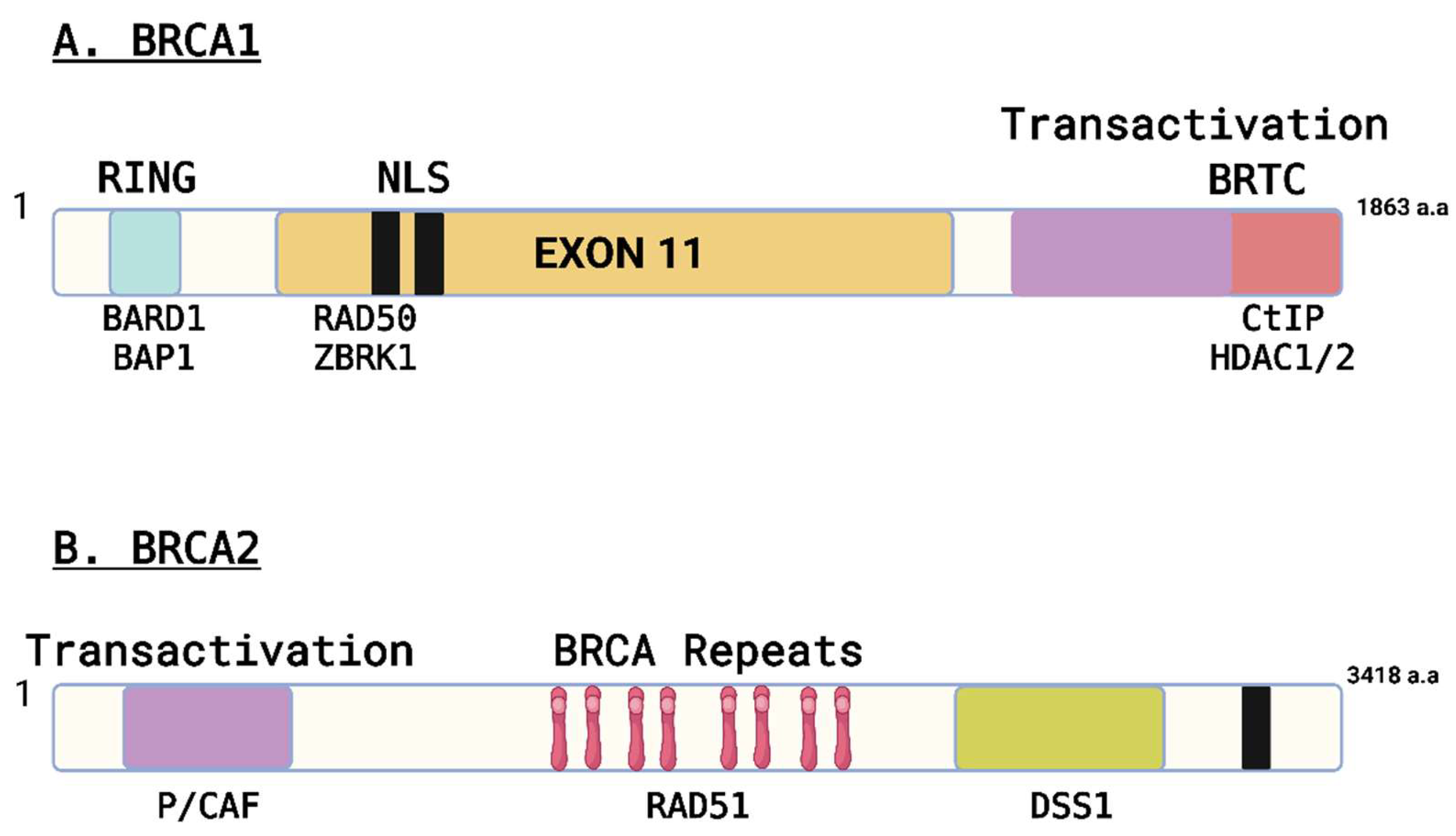
Figure 1. Structural and functional motifs recognized in BRCA1 and BRCA2. (A) Schematic structure of BRCA1. The RING domain, the transactivation domain, the BRCT domain, nuclear localization signals (NLS), and exon 11 coding regions are indicated. Representative proteins that interact with BRCA1 in three different regions are shown. (B) Schematic structure of BRCA2. The transactivation domain, BRC repeats, and NLS are indicated. Representative BRCA2-interacting proteins are indicated.
Preprints 146602 g001
Figure 2. Summary of BRCA1 and BRCA2 essential information: 3D protein structure, chromosomal location, and primary function.
Preprints 146602 g002
Table 1. Comparative Analysis of BRCA1 and BRCA2 Genes in Humans and Canines.
BRCA1 (Human) BRCA1 (Canine) BRCA2 (Human) BRCA2 (Canine)
Chromosome Location 17q21 Chr.9 13q12.3 Chr.25
Gene Length (base pairs) ~100,000 ~85,000 ~84,000 ~81,000
Protein Length (amino acids) 1863 1832 3418 3414
Conservation across species Highly conserved Conserved Highly conserved Conserved
Biological Function Tumor suppressor Tumor suppressor DNA repair DNA repair
Protein Domains RING, BRCT, SQ/TQ cluster RING, BRCT, SQ/TQ cluster BRC repeats, DNA-binding, helicase BRC repeats, DNA-binding, helicase
Association with Cancer Breast, ovarian, other cancers Mammary gland, ovarian, other cancers Breast, ovarian, other cancers Mammary gland, ovarian, other cancers
Genetic Variants Numerous pathogenic variants identified Limited studies on genetic variants Numerous pathogenic variants identified Limited studies on genetic variants
Mutational Spectrum Point mutations, insertions, deletions Point mutations, insertions, deletions Point mutations, insertions, deletions Point mutations, insertions, deletions
Disease Risk Increased risk of breast, ovarian, and other cancers Increased risk of mammary gland, ovarian, and other cancers Increased risk of breast, ovarian, and other cancers Increased risk of mammary gland, ovarian, and other cancers
References (Deng and Brodie 2000; Rebbeck et al.2011; Caestecker et al. 2013) (Nieto et al. 2003; Rivera and Euler 2011; Qiu et al. 2015) (Thorlacius et al.1996; Bertwistle et al. 1997; Thirthagiri et al. 2016) (Yoshikawa et al. 2012; Yoshikawa et al. 2015; Thumser-Henner et al. 2020)
Implications of BRCA1 and BRCA2 in Cancer Therapy.
Table 2. Synopsis of reported clinical trials involving PARPi.
PARP inhibitor tested Cancer type Efficacy Refs
Olaparib Solid tumors (ovarian: 35%) Clinical benefit for 63% (in BRCA mutations carriers’ patients) (Fong et al. 2009)
Breast ORR*: 41% (Tutt et al. 2010)
Ovarian ORR*: 33% (Audeh et al. 2010)
Ovarian, breast, pancreatic and prostate Tumor response rate*: 26.2% (Kaufman et al. 2015)
Rucaparib (Temozolomide) Metastatic melanoma Clinical benefit for 34.8% of the patients (Plummer et al. 2013)
Advanced solid
malignancies
CR: 1/32 (melanoma)
PR: 2/32 (melanoma, desmoid tumor)
(Plummer et al. 2008)
Veliparib Metastatic breast cancer ORR (CR+PR) 12.5% (3/24) (Isakoff et al. 2010)
Iniparib Metastatic TNBC ORR (CR+PR) 32% (Penson et al. 2011)
ORR* (Objective Response Rate): according to RECIST, with confirmation of response at least 28 days apart by CT scan and RECIST. Tumor response rate*: according to RECIST, with confirmation of response at least 28 days apart; CR – complete response; PR – partial response.
Disclaimer/Publisher’s Note: The statements, opinions and data contained in all publications are solely those of the individual author(s) and contributor(s) and not of MDPI and/or the editor(s). MDPI and/or the editor(s) disclaim responsibility for any injury to people or property resulting from any ideas, methods, instructions or products referred to in the content.
Copyright: This open access article is published under a Creative Commons CC BY 4.0 license, which permit the free download, distribution, and reuse, provided that the author and preprint are cited in any reuse.
Prerpints.org logo

Preprints.org is a free preprint server supported by MDPI in Basel, Switzerland.

Subscribe

Disclaimer

Terms of Use

Privacy Policy

Privacy Settings

© 2025 MDPI (Basel, Switzerland) unless otherwise stated