Preprint
Review

This version is not peer-reviewed.

Neuroimmune Interactions in Pancreatic Cancer

A peer-reviewed article of this preprint also exists.

Submitted:

16 January 2025

Posted:

18 January 2025

You are already at the latest version

Abstract

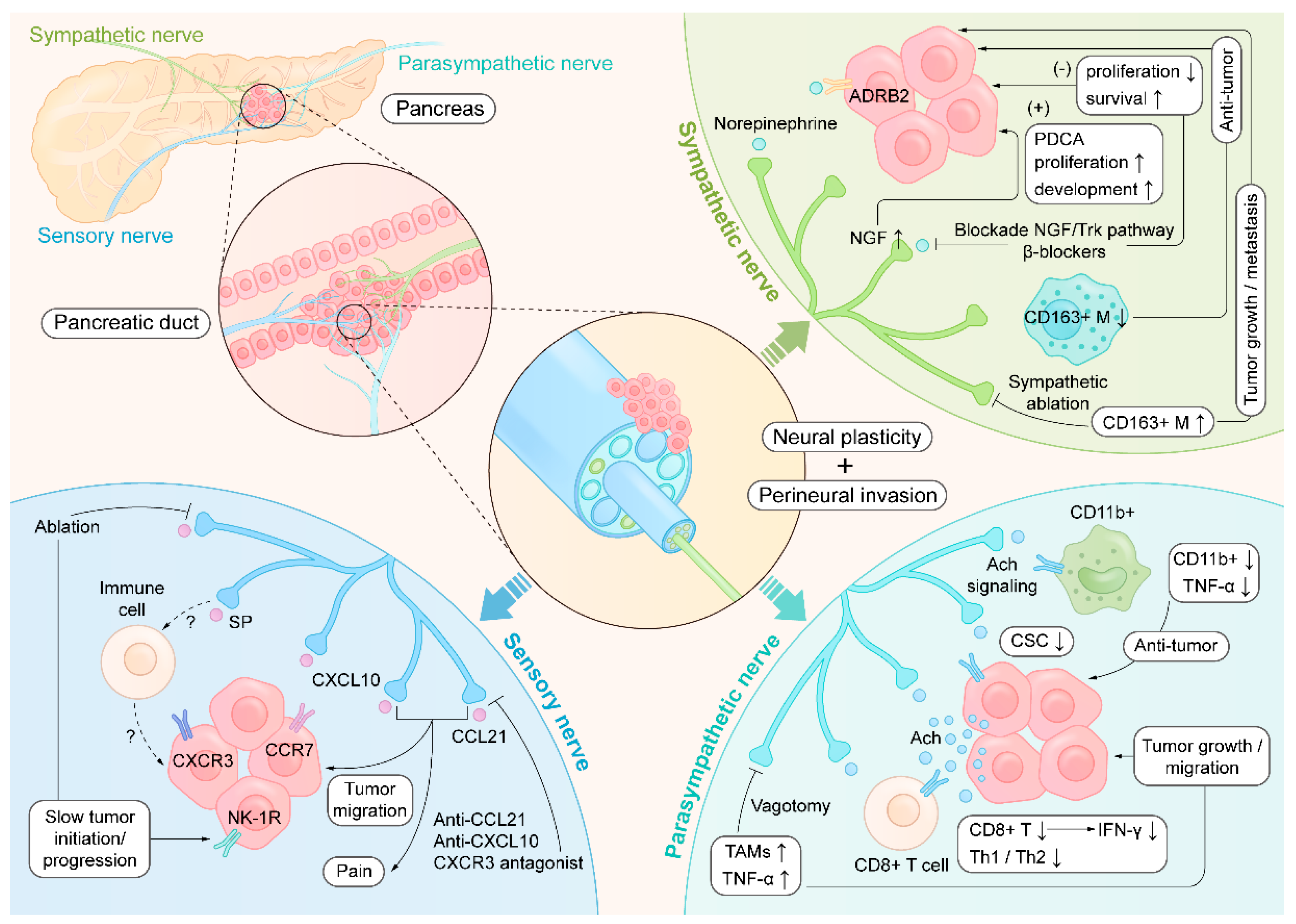
Pancreatic ductal adenocarcinoma (PDAC) is a highly aggressive primary malignancy, and recent technological advances in surgery have opened up more possibilities for surgical treatment. Novel data show that diverse immune and neural components play essential roles in the aggressive behavior of PDAC. Recent studies have shown that neural invasion, neural plasticity, and altered innervation of autonomic nerve fibers are involved in pancreatic neuropathy in PDAC patients and have clarified the functional structure of the nerves innervating pancreatic draining lymph nodes. Notably, research on the pathogenesis and therapeutic options for treating PDAC from the viewpoint of interactions of the neuroimmune network is at the cutting edge. In this review, we present a special focus on neuroimmune interactions that highlight the current state of knowledge and future challenges concerning the reciprocal relationship between the immune system and nervous system in PDAC. Understanding the molecular events governing the pancreatic neuroimmune signaling axes will enhance our knowledge of physiology and may provide novel therapeutic targets for treating PDAC.

Keywords: 
;  ;  ;  ;  

1. Introduction

The nervous system and immune system communicate throughout the body and in the central nervous system, which can recognize danger simultaneously and respond in both a simultaneous and coordinated fashion to regulate tissue and organ function in homeostasis and diseases [1,2,3,4,5]. However, the field of neuroimmunology has undergone a recent renaissance, and evidence suggests that nervous system and immune system communication is far more complex than previously realized.
Neuroimmunology studies to date have tended to focus on interactions between nerves and immune systems at the local level. Peripheral neuroimmune interactions critically regulate systemic and local tissue immunity. Novel data show that the immune system and nervous system play mutual roles in the body and are essential in immune, metabolism-related, neoplastic, and other diseases [6,7,8].
As an organ associated with neuroimmunity, the pancreas is densely innervated by a complex network of neurons that coordinate critical physiological functions. In addition, pancreatic draining lymph nodes have pancreatic innervation and exert some immunomodulatory effects [9]. Notably, research on the pathogenesis and therapeutic options for treating pancreatic ductal adenocarcinoma (PDAC), including recently identified local forms, from the viewpoint of crosstalk of the neuroimmune network and intervention techniques is at the cutting edge. Current evidence reveals the neuroimmune crosstalk occurring in PDAC, explaining the role of interactions between different types of nerve and immune cells in PDAC in terms of tumor formation, development, and metastasis [10,11,12].
In this review, we focus specifically on neuroimmune interactions associated with PDAC, aiming to elucidate the value of neuroimmune crosstalk in PDAC pathogenesis and intervention through the effects of different types of neural and immune cells on PDAC.

2. Neuroanatomy in the Pancreas and Draining Lymph Nodes

The anatomy of pancreatic innervation includes sympathetic efferent, parasympathetic efferent, vagal afferent, spinal afferent, and enteropancreatic innervation [13]. In the human pancreas, these different types of nerves have distinct innervation characteristics. Using three-dimensional imaging, Chien and colleagues revealed that nerve fibers positive for substance P as the afferent nerve are located at the base of the interlobular duct and that nerve fibers positive for vesicular acetylcholine transporter and tyrosine hydroxylase as the efferent nerve exist in the periacinar and perivascular spaces, which reach the islet along a blood vessel [14]. Neurons and glial networks enter the islet core, and sympathetic and parasympathetic nerves reside in the immediate microenvironment [15]. However, few parasympathetic cholinergic axons innervate the islets in humans, and the sympathetic nerves entering the islets preferentially innervate the vascular smooth muscle cells in islets [16]. The sympathetic nervous system constricts blood vessels in many parts of the body to ensure the stability of the circulatory system under special circumstances [16]. Nerves not only extend to pancreatic parenchyma cells but also some mesenchymal cells. However, further studies are needed to confirm the existence and function of unique connections between nerve cells and various mesenchymal cells.
More recently, novel findings on the functional structure of nerves innervating pancreatic draining lymph nodes have been reported in terms of neuroimmune regulation [9]. Lymph nodes serve as immune organs to filter lymphatic fluid and initiate local adaptive immune responses. The peripheral nervous system communicates specifically with the immune system through local interactions that provide the structural basis for complex immune and neural response networks [17]. By identifying a molecularly distinct and heterogeneous population of sensory neurons with the capacity to affect lymph node function and homeostasis, Huang et al. recently established lymph nodes as a point of convergence between the sensory nervous system and the immune system [18].
Regarding experimental animals, the presence of norepinephrine innervation in the popliteal and mesenteric lymph nodes of mice has long been detected by fluorescein histochemistry [19]. Sympathetic nerves, including perivascular and discrete structures, are observed in human inguinal lymph nodes, and the number of sympathetic nerves varies between compartments and between and within individuals [20]. According to a study of human pancreas specimens, sympathetic innervation of secondary lymphoid organs, such as lymph nodes, varies by species and is influenced by certain physiological or pathological conditions [21]. By placing a suction electrode and recording a field action potential with microelectrodes, projection of pancreatic nerves to mouse lymph nodes was demonstrated by stimulation of pancreatic nerves [9].

3. Neural Plasticity in PDAC

Neural plasticity is an inherent feature of PDAC and involves neuronal activation at the peripheral, spinal and supraspinal levels [22]. Sympathetic nerve innervation of the pancreas is significantly lower in patients with PDAC than in patients with a healthy pancreas, and the number of sympathetic fibers in PDAC patients without nerve invasion is markedly higher than in those with nerve invasion [23]. In contrast, there is no significant change in parasympathetic or cholinergic nerve distribution [23].
In terms of spatial variation in the pancreatic nerve distribution, increased nerve density and hypertrophy compared with those in the normal pancreas are typical features of PDAC [24]. The density of intrapancreatic nerves in patients with PDAC tends to decrease toward the center of the tumor [2]. In mice, nerve hypertrophy and infiltration gradually follow the progression of PDAC, and hypertrophy of the nerve is associated with fibrosis of the corresponding area and atrophy of pancreatic acinar cells [25]. The mean percentage of sympathetic nerve fibers per nerve in the pancreas of PDAC patients is significantly lower than that in the pancreas of healthy patients [23], which may be partly explained by a decrease in the number or volume of sympathetic nerves or both. In patients with PDAC, increased nerve size is associated with decreased numbers of cholinergic and adrenergic nerve fibers and enhanced neural nesting immunoreactivity [23]. Indeed, there is a correlation between the volume and number of pancreatic nerves. In addition to the neurological changes described above, changes in neurotrophic factor expression occur prior to tumor formation, and the nervous system is involved in all PDAC processes, including the period before cancer development [25].

4. Neuroimmune Crosstalk in PDAC

Neuroimmune crosstalk, which can serve as a crucial immunomodulatory hub, has been reported in both healthy and disease states [8,26,27,28]. Emerging evidence suggests that the somatosensory and autonomic nervous systems play an active role in host defense via direct regulation of immunological and inflammatory responses in the periphery [29,30]. For instance, enteric neurons can reduce surgery-induced intestinal inflammation and prevent postoperative ileus through regulation of mucosal macrophages [31]. Nerve fibers of mouse lymph nodes have close contacts or associations with immune cells, including dendritic cells, macrophages, and T or B lymphocytes [17]. Moreover, the impact of the neural system on immune function and immune cells via cytokine and trophic factor secretion can influence endogenous neural stem cell proliferation, differentiation and migration [29,30,32]. For example, enteric neurons maintain the development of muscularis macrophages in a steady state by secreting colony stimulatory factor 1; reciprocally, muscularis macrophages can activate intestinal neurons expressing bone morphogenetic protein receptors by secreting bone morphogenetic protein 2[33].
Neuroimmune regulation is related to neoplastic diseases and plays a specific role in the occurrence and development of tumors. In breast cancer, tumor-specific sympathetic denervation downregulates expression of immune checkpoint molecules, including programmed death-1 and programmed death ligand-1, on CD4+ or CD8+ T cells and upregulates that of interferon-γ on CD4+ or CD8+ cells, which have an active effector phenotype [34]. The sympathetic nervous system can maintain the inflammatory microenvironment and promote the occurrence of hepatocellular carcinoma by activating the a1-adrenergic receptors of Kupffer cells [35].
PDAC is a highly aggressive tumor, with a five-year survival rate of only approximately 10%[36]. Pathological and immunohistochemical examinations of PDAC tissue have revealed specific nervous and immune system features. The tumor microenvironment is closely related to neoplastic development [37,38,39,40,41], and neural signals play a role in the tumor microenvironment [42]. Neurotropism is an important histological feature of PDAC that is associated with frequent neurological invasion [43]. In addition, perineural invasion is a common feature of PDAC and is associated with impaired immune responses [11].
In pancreatic cancer, nerves are significantly infiltrated by cytotoxic T lymphocytes, macrophages, and mast cells, which account for 35%, 39%, and 21%, respectively, of all perineural inflammatory cells [5]. Perineural invasion can induce neuronal damage and inflammation [44]. Colocalization of nerve and immune cells may affect nerve and immune functions, and this neuroimmune regulation may lead to changes in disease. Nerves can regulate tumor growth in ways that affect tumor metabolism. For example, nerves promote PDAC growth in nutrient-deficient environments by releasing substances necessary for tumor growth, such as serine [45]. The neuroimmune interactions occurring in PDAC at the peripheral nerve level, including the regulation of immune cells and sympathetic, parasympathetic and sensory nerves, are summarized in Figure 1. Despite the promising achievements of tumor immunotherapy in many tumor types, it has not been very successfully applied in PDAC treatment [46]. Determining the mechanisms of neuroimmune regulation in pancreatic cancer may facilitate the progress of tumor immunotherapy for pancreatic cancer treatment.

4.1. Sympathetic Nerve

Different interactions between a variety of nerves and immune cells in the pancreas may produce different effects. Sympathetic nerves are known to promote prostate cancer, breast cancer and melanoma [1]. In the pancreas, sympathetic nervous system/β-adrenoceptor signaling promotes tumor growth [47], and neuromodulation contributes to PDAC at all stages, even before cancer appears [48]. Recent findings have provided insights into the mechanisms by which the sympathetic nervous system exerts its cancer-protective effects in a mouse model of PDAC, but these effects are not caused by nerves acting alone [10]. Indeed, research has demonstrated that sympathetic axons slow PDAC progression by locally inhibiting CD163+ macrophage subsets at the lesion site; ablation of innervated sympathetic nerves increases tumor growth and spread, which supports use of CD163+ macrophages as mediators of the protumoral effect of sympathetic resection in PDAC [10]. In melanoma-bearing mice, beta-adrenergic receptor blockers increase the frequency of effector CD8+ T cells in tumors and reduce expression of programmed death receptor-1[49].

4.2. Parasympathetic Nerves

The neural effects of sympathetic and parasympathetic nerves may be modulated by β-adrenergic and muscarinic receptors, respectively [1]. Recent research suggests that parasympathetic nervous system input inhibits primary and secondary tumorigenesis of PDAC through cholinergic signaling and that stimulation of muscarinic receptors may be helpful in treatment of PDAC [12]. In mice with established PDAC, stimulation of muscarinic type 1 receptors can suppress the cancer stem cell compartment, CD11b+ myeloid cells, tumor necrosis factor α (TNF-α) levels, and metastatic growth in the liver. Vagotomy significantly increases the number of tumor-associated macrophages and levels of TNF-α in tumors, promoting tumor growth and reducing survival in a murine pancreatic cancer model [50].

4.3. Sensory Nerves

In genetically engineered PDAC mouse models, sensory innervation increases during pancreatic intraepithelial neoplasia formation and increases further during cancer progression [25]. The chemokines CCL21 and CXCL10, which originate from sensory neurons, promote PDAC metastasis through their receptors CCR7 and CXCR3, respectively [51]. Ablation of sensory neurons in genetic models of PDAC delays formation of pancreatic intraepithelial neoplasia and ultimately prolongs survival [44]. In addition to nerve cells, Schwann cells of peripheral nerves are activated in the preneoplastic stage of cancer, possess strong affinity for cancer cells and initiate nerve cancer cell interactions [52,53]. TIMP1 from pancreatic cancer cells stimulates Schwann cells and promotes peripheral nerve invasion, and TIMP1 knockdown can suppress peripheral nerve invasion [54].
Major depressive disorder is four times more common in cancer patients than in the general population [55]. Psychosocial stress activates the sympathetic nervous system, hypothalamic–pituitary–adrenal axis, and neuroendocrine axis and subsequently regulates inflammatory responses through immune cells [56]. In cancer patients, psychosocial stress promotes inflammation and oxidative stress, decreased immunosurveillance, and dysfunctional activation of the autonomic nervous system and the hypothalamic‒pituitary‒adrenal axis [55]. Human and animal studies have shown that sympathetic and neuroendocrine responses to psychosocial stress significantly influence cancer by regulating inflammatory mediators [56]. For instance, psychological intervention in regional breast cancer patients enhance natural killer cell cytotoxicity and T-cell proliferation and reduce the risk of death after recurrence [57].

5. Neuroimmune Regulation Intervention

With advances in surgical techniques, surgical treatment can be used for an increasing number of patients with PDAC [58]. In addition, surgical denervation has long been reported for pain management in patients with PDAC who cannot undergo surgical tumor removal [59]. To date, many intervention studies related to neuroimmune interactions have been performed, and some of the intervention methods related to PDAC are summarized in Figure 1. In clinical practice, beta-adrenergic receptor blockers, such as propranolol, are the most commonly used drugs to attenuate sympathetic adrenergic function. Patients with PDAC administered beta-adrenergic receptor blockers had a lower cancer-specific mortality rate than did those not given these drugs [60]. Compared with conventional receptor blocker drugs, use of a liposome nanomedicine system to administer propranolol hydrochloride can avoid the adverse effects caused by inhibition of nontargeted adrenergic neural activity [61]. Furthermore, it has considerable therapeutic effects on PDAC, prostate cancer, and melanoma, with increased safety and efficiency [61].
There are very precise optogenetic and pharmacogenetic methods that allow neurons to be manipulated in very specific ways [62]. As a genetic local neuroengineering technology, adeno-associated virus vectors designed to carry transgenes downstream of neuron fiber type-specific promoters can stimulate tumor immunity, such as increased effector helper or cytotoxic T cells in primary tumor tissue, and achieve greater tumor suppression efficacy than administration of beta-adrenergic receptor blockers [34]. These methods may be developed for the pancreas in the future to achieve desired results. In general, pancreatic neuroimmune regulation will develop in the direction of less trauma, strong targeting and convenient use.
In addition to use of systemic drugs, there are many methods for direct neurological intervention, including neurotomy, nerve ablation, and nerve block. Perineural invasion is associated with an immunosuppressive microenvironment in PDAC, and intervention involving perineural invasion with bilateral subdiaphragmatic vagotomy is associated with increased CD8+ T cells, an increased Th1/Th2 ratio, and improved survival [11]. In contrast to surgery, ultrasound-guided celiac plexus block and neurolysis are used to relieve and treat pain in patients with PDAC [63].
In the past few decades, electrical stimulation therapy has attracted considerable attention as a new therapeutic method. The electric field enhances keratinocyte migration, strengthens immune defense, and improves mitochondrial function, and a weak electric field can be safe and has antibacterial effects on the human body [64]. Compared with traditional drugs, which require systematic administration and a certain amount of time for effect depending on the administration, electrical stimulation can act directly on nerve bundles to produce rapid effects [65]. As a bioelectric therapy, stimulation of the vagus nerve in epilepsy patients with an implanted device reduced levels of TNF, interleukin-1β, and interleukin-6 in peripheral blood [66]. More recently, electrical stimulation of the pancreatic nerve was shown to prevent autoimmune diabetes by inhibiting migration of diabetogenic T cells from pancreatic draining lymph nodes to islets, resulting in less severe insulitis [9].
In addition to confirming the research value, some experimental teams have obtained inconsistent results after repeating the above experiment [67]. Moreover, it should be kept in mind that stimulation of the vagus nerve involves afferent and efferent nerves and controls multiple tissues and organs; in fact, stimulation of the vagus nerve not only causes the desired therapeutic effects but also nerve stimulus-related side effects [9]. Therefore, enhancing targeting of electrical stimulation therapy to reduce side effects can improve the clinical application prospects of electrical stimulation. However, there are still many problems to be solved, such as the design of the electrode to stimulate the pancreatic nerve, the setting of electrical stimulation parameters, and the method of electrode implantation.

6. Concluding Remarks and Future Perspectives

The findings of neuroimmune network crosstalk and intervention techniques in PDAC open up a fertile area for detailed studies that have broad translational implications. Much of the information obtained to date involves the structural plasticity of pancreatic nerves, specifically under conditions of inflammation or cancer. Most previous studies have focused on peripheral neuroimmune crosstalk and aimed to understand the reciprocal relationship between the immune system and nervous system during pancreatic neoplasms. More recently, novel data on the functional structure of nerves innervating pancreatic draining lymph nodes have revealed neuronal communication with stromal and immune cells. Furthermore, recent studies have highlighted interventions targeting pancreatic neuroimmune signaling axes and presented an emerging avenue for treatment of pancreatic diseases. Neuroimmune interactions at the peripheral nerve level in PDAC patients, including interactions in the pancreas and draining lymph nodes, should be explored in depth to elucidate their implications, which will help scholars and clinicians to recognize and manage PDAC patients from a new perspective.
In general, the molecular mechanisms underlying the dynamic pathological conditions of neuroimmune crosstalk are poorly understood. Further research on the internal links and mechanisms of neuroimmune regulation and intervention in pancreatic physiology and pathophysiology will be helpful for understanding the pancreas and PDAC and provides a basis for diagnosis and treatment. Exciting areas for future research include gaining a more comprehensive understanding of the anatomical, molecular, and cellular basis of neuroimmune crosstalk in PDAC; revealing the innervation established during development and homeostasis maintenance of the pancreas; clarifying how neuronal communication to stromal and immune cells of the lymphatic vasculature is mediated [68]; identifying nerve-derived factors, in addition to neurotransmitters and immunomodulatory factors; and elucidating the cell types that are directly targeted by nerves that mediate effects on PDAC. Technological advances in chemical genetic tools, optogenetics and single-cell sequencing offer new opportunities to reveal the anatomical, molecular and cellular profiles of specific neuronal and immune subtypes projecting to the pancreas and associated circuits.

Author Contributions

Conceptualization, J.C. and Y.C.; writing—original draft preparation, R.W. and J.C.; writing—review and editing, R.W. and J.C.; supervision, Y.C.; funding acquisition, Y.C. All authors have read and agreed to the published version of the manuscript.

Funding

This work was supported in part by research grants from the National Natural Science Foundation of China (82071746).

Institutional Review Board Statement

Not applicable.

Informed Consent Statement

Not applicable.

Data Availability Statement

Not applicable.

Acknowledgments

We thank Hu Xinyue for optimizing the illustrations from August Fireflies Technology Co.Ltd, Chengdu.

Conflicts of Interest

The authors declare no conflicts of interest.

Abbreviations

The following abbreviations are used in this manuscript:
PDAC Pancreatic ductal adenocarcinoma
Ach Acetylcholine
ADRB2 Adrenoceptor beta 2
CSC Cancer stem cell
IFN-γ Interferon-γ
SP Substance P
TAMs Tumor-associated macrophages
TNF-α Tumor necrosis factor-α
NGF Nerve growth factor
NK-1R Neurokinin-1 receptor

References

  1. Kamiya, A.; Hiyama, T.; Fujimura, A.; Yoshikawa, S. Sympathetic and parasympathetic innervation in cancer: therapeutic implications. Clinical autonomic research : official journal of the Clinical Autonomic Research Society 2021, 31, 165–178. [Google Scholar] [CrossRef]
  2. Iwasaki, T.; Hiraoka, N.; Ino, Y.; Nakajima, K.; Kishi, Y.; Nara, S.; Esaki, M.; Shimada, K.; Katai, H. Reduction of intrapancreatic neural density in cancer tissue predicts poorer outcome in pancreatic ductal carcinoma. Cancer science 2019, 110, 1491–1502. [Google Scholar] [CrossRef] [PubMed]
  3. Kato, K.; Ikeura, T.; Yanagawa, M.; Tomiyama, T.; Fukui, T.; Uchida, K.; Takaoka, M.; Nishio, A.; Uemura, Y.; Satoi, S.; et al. Morphological and immunohistochemical comparison of intrapancreatic nerves between chronic pancreatitis and type 1 autoimmune pancreatitis. Pancreatology : official journal of the International Association of Pancreatology (IAP)... [et al.] 2017, 17, 403–410. [Google Scholar] [CrossRef]
  4. Mundinger, T.O.; Mei, Q.; Foulis, A.K.; Fligner, C.L.; Hull, R.L.; Taborsky, G.J., Jr. Human Type 1 Diabetes Is Characterized by an Early, Marked, Sustained, and Islet-Selective Loss of Sympathetic Nerves. Diabetes 2016, 65, 2322–2330. [Google Scholar] [CrossRef]
  5. Demir, I.E.; Schorn, S.; Schremmer-Danninger, E.; Wang, K.; Kehl, T.; Giese, N.A.; Algül, H.; Friess, H.; Ceyhan, G.O. Perineural mast cells are specifically enriched in pancreatic neuritis and neuropathic pain in pancreatic cancer and chronic pancreatitis. PloS one 2013, 8, e60529. [Google Scholar] [CrossRef] [PubMed]
  6. Veiga-Fernandes, H.; Pachnis, V. Neuroimmune regulation during intestinal development and homeostasis. Nature immunology 2017, 18, 116–122. [Google Scholar] [CrossRef] [PubMed]
  7. Pavlov, V.A.; Chavan, S.S.; Tracey, K.J. Molecular and Functional Neuroscience in Immunity. Annual review of immunology 2018, 36, 783–812. [Google Scholar] [CrossRef] [PubMed]
  8. Veiga-Fernandes, H.; Mucida, D. Neuro-Immune Interactions at Barrier Surfaces. Cell 2016, 165, 801–811. [Google Scholar] [CrossRef]
  9. Guyot, M.; Simon, T.; Ceppo, F.; Panzolini, C.; Guyon, A.; Lavergne, J.; Murris, E.; Daoudlarian, D.; Brusini, R.; Zarif, H.; et al. Pancreatic nerve electrostimulation inhibits recent-onset autoimmune diabetes. Nature biotechnology 2019, 37, 1446–1451. [Google Scholar] [CrossRef]
  10. Guillot, J.; Dominici, C.; Lucchesi, A.; Nguyen, H.T.T.; Puget, A.; Hocine, M.; Rangel-Sosa, M.M.; Simic, M.; Nigri, J.; Guillaumond, F.; et al. Sympathetic axonal sprouting induces changes in macrophage populations and protects against pancreatic cancer. Nature communications 2022, 13, 1985. [Google Scholar] [CrossRef] [PubMed]
  11. Yang, M.W.; Tao, L.Y.; Jiang, Y.S.; Yang, J.Y.; Huo, Y.M.; Liu, D.J.; Li, J.; Fu, X.L.; He, R.; Lin, C.; et al. Perineural Invasion Reprograms the Immune Microenvironment through Cholinergic Signaling in Pancreatic Ductal Adenocarcinoma. Cancer Res 2020, 80, 1991–2003. [Google Scholar] [CrossRef]
  12. Renz, B.W.; Tanaka, T.; Sunagawa, M.; Takahashi, R.; Jiang, Z.; Macchini, M.; Dantes, Z.; Valenti, G.; White, R.A.; Middelhoff, M.A.; et al. Cholinergic Signaling via Muscarinic Receptors Directly and Indirectly Suppresses Pancreatic Tumorigenesis and Cancer Stemness. Cancer Discov 2018, 8, 1458–1473. [Google Scholar] [CrossRef] [PubMed]
  13. Hampton, R.F.; Jimenez-Gonzalez, M.; Stanley, S.A. Unravelling innervation of pancreatic islets. Diabetologia 2022. [CrossRef] [PubMed]
  14. Chien, H.J.; Chiang, T.C.; Peng, S.J.; Chung, M.H.; Chou, Y.H.; Lee, C.Y.; Jeng, Y.M.; Tien, Y.W.; Tang, S.C. Human pancreatic afferent and efferent nerves: mapping and 3-D illustration of exocrine, endocrine, and adipose innervation. American journal of physiology. Gastrointestinal and liver physiology 2019, 317, G694–g706. [Google Scholar] [CrossRef]
  15. Tang, S.C.; Baeyens, L.; Shen, C.N.; Peng, S.J.; Chien, H.J.; Scheel, D.W.; Chamberlain, C.E.; German, M.S. Human pancreatic neuro-insular network in health and fatty infiltration. Diabetologia 2018, 61, 168–181. [Google Scholar] [CrossRef] [PubMed]
  16. Rodriguez-Diaz, R.; Abdulreda, M.H.; Formoso, A.L.; Gans, I.; Ricordi, C.; Berggren, P.O.; Caicedo, A. Innervation patterns of autonomic axons in the human endocrine pancreas. Cell metabolism 2011, 14, 45–54. [Google Scholar] [CrossRef]
  17. Hu, D.; Nicholls, P.K.; Claus, M.; Wu, Y.; Shi, Z.; Greene, W.K.; Ma, B. Immunofluorescence characterization of innervation and nerve-immune cell interactions in mouse lymph nodes. European journal of histochemistry : EJH 2019, 63. [CrossRef]
  18. Huang, S.; Ziegler, C.G.K.; Austin, J.; Mannoun, N.; Vukovic, M.; Ordovas-Montanes, J.; Shalek, A.K.; von Andrian, U.H. Lymph nodes are innervated by a unique population of sensory neurons with immunomodulatory potential. Cell 2021, 184, 441–459e425. [Google Scholar] [CrossRef] [PubMed]
  19. Felten, D.L.; Livnat, S.; Felten, S.Y.; Carlson, S.L.; Bellinger, D.L.; Yeh, P. Sympathetic innervation of lymph nodes in mice. Brain research bulletin 1984, 13, 693–699. [Google Scholar] [CrossRef] [PubMed]
  20. Cleypool, C.G.J.; Mackaaij, C.; Lotgerink Bruinenberg, D.; Schurink, B.; Bleys, R. Sympathetic nerve distribution in human lymph nodes. Journal of anatomy 2021. [CrossRef]
  21. Bellinger, D.L.; Lorton, D. Autonomic regulation of cellular immune function. Autonomic neuroscience : basic & clinical 2014, 182, 15–41. [Google Scholar] [CrossRef]
  22. Demir, I.E.; Friess, H.; Ceyhan, G.O. Neural plasticity in pancreatitis and pancreatic cancer. Nat Rev Gastroenterol Hepatol 2015, 12, 649–659. [Google Scholar] [CrossRef]
  23. Ceyhan, G.O.; Demir, I.E.; Rauch, U.; Bergmann, F.; Müller, M.W.; Büchler, M.W.; Friess, H.; Schäfer, K.H. Pancreatic neuropathy results in “neural remodeling” and altered pancreatic innervation in chronic pancreatitis and pancreatic cancer. Am J Gastroenterol 2009, 104, 2555–2565. [Google Scholar] [CrossRef] [PubMed]
  24. Ceyhan, G.O.; Bergmann, F.; Kadihasanoglu, M.; Altintas, B.; Demir, I.E.; Hinz, U.; Müller, M.W.; Giese, T.; Büchler, M.W.; Giese, N.A.; et al. Pancreatic neuropathy and neuropathic pain--a comprehensive pathomorphological study of 546 cases. Gastroenterology 2009, 136, 177–186e171. [Google Scholar] [CrossRef] [PubMed]
  25. Stopczynski, R.E.; Normolle, D.P.; Hartman, D.J.; Ying, H.; DeBerry, J.J.; Bielefeldt, K.; Rhim, A.D.; DePinho, R.A.; Albers, K.M.; Davis, B.M. Neuroplastic changes occur early in the development of pancreatic ductal adenocarcinoma. Cancer Res 2014, 74, 1718–1727. [Google Scholar] [CrossRef]
  26. Godinho-Silva, C.; Cardoso, F.; Veiga-Fernandes, H. Neuro-Immune Cell Units: A New Paradigm in Physiology. Annual review of immunology 2019, 37, 19–46. [Google Scholar] [CrossRef]
  27. Ordovas-Montanes, J.; Rakoff-Nahoum, S.; Huang, S.; Riol-Blanco, L.; Barreiro, O.; von Andrian, U.H. The Regulation of Immunological Processes by Peripheral Neurons in Homeostasis and Disease. Trends in immunology 2015, 36, 578–604. [Google Scholar] [CrossRef] [PubMed]
  28. Vasconcelos, D.P.; Jabangwe, C.; Lamghari, M.; Alves, C.J. The Neuroimmune Interplay in Joint Pain: The Role of Macrophages. Frontiers in immunology 2022, 13, 812962. [Google Scholar] [CrossRef]
  29. Kioussis, D.; Pachnis, V. Immune and nervous systems: more than just a superficial similarity? Immunity 2009, 31, 705–710. [Google Scholar] [CrossRef]
  30. Franco, R.; Pacheco, R.; Lluis, C.; Ahern, G.P.; O’Connell, P.J. The emergence of neurotransmitters as immune modulators. Trends in immunology 2007, 28, 400–407. [Google Scholar] [CrossRef] [PubMed]
  31. Stakenborg, N.; Labeeuw, E.; Gomez-Pinilla, P.J.; De Schepper, S.; Aerts, R.; Goverse, G.; Farro, G.; Appeltans, I.; Meroni, E.; Stakenborg, M.; et al. Preoperative administration of the 5-HT4 receptor agonist prucalopride reduces intestinal inflammation and shortens postoperative ileus via cholinergic enteric neurons. Gut 2019, 68, 1406–1416. [Google Scholar] [CrossRef]
  32. Dobrenis, K.; Gauthier, L.R.; Barroca, V.; Magnon, C. Granulocyte colony-stimulating factor off-target effect on nerve outgrowth promotes prostate cancer development. International journal of cancer 2015, 136, 982–988. [Google Scholar] [CrossRef]
  33. Muller, P.A.; Koscsó, B.; Rajani, G.M.; Stevanovic, K.; Berres, M.L.; Hashimoto, D.; Mortha, A.; Leboeuf, M.; Li, X.M.; Mucida, D.; et al. Crosstalk between muscularis macrophages and enteric neurons regulates gastrointestinal motility. Cell 2014, 158, 300–313. [Google Scholar] [CrossRef]
  34. Kamiya, A.; Hayama, Y.; Kato, S.; Shimomura, A.; Shimomura, T.; Irie, K.; Kaneko, R.; Yanagawa, Y.; Kobayashi, K.; Ochiya, T. Genetic manipulation of autonomic nerve fiber innervation and activity and its effect on breast cancer progression. Nature neuroscience 2019, 22, 1289–1305. [Google Scholar] [CrossRef] [PubMed]
  35. Huan, H.B.; Wen, X.D.; Chen, X.J.; Wu, L.; Wu, L.L.; Zhang, L.; Yang, D.P.; Zhang, X.; Bie, P.; Qian, C.; et al. Sympathetic nervous system promotes hepatocarcinogenesis by modulating inflammation through activation of alpha1-adrenergic receptors of Kupffer cells. Brain, behavior, and immunity 2017, 59, 118–134. [Google Scholar] [CrossRef]
  36. Mizrahi, J.D.; Surana, R.; Valle, J.W.; Shroff, R.T. Pancreatic cancer. Lancet (London, England) 2020, 395, 2008–2020. [Google Scholar] [CrossRef]
  37. Chen, K.; Wang, Q.; Li, M.; Guo, H.; Liu, W.; Wang, F.; Tian, X.; Yang, Y. Single-cell RNA-seq reveals dynamic change in tumor microenvironment during pancreatic ductal adenocarcinoma malignant progression. EBioMedicine 2021, 66, 103315. [Google Scholar] [CrossRef]
  38. Gola, M.; Sejda, A.; Godlewski, J.; Cieślak, M.; Starzyńska, A. Neural Component of the Tumor Microenvironment in Pancreatic Ductal Adenocarcinoma. Cancers (Basel) 2022, 14. [Google Scholar] [CrossRef]
  39. Aziz, H.M.; Saida, L.; de Koning, W.; Stubbs, A.P.; Li, Y.; Sideras, K.; Palacios, E.; Feliu, J.; Mendiola, M.; van Eijck, C.H.J.; et al. Spatial genomics reveals a high number and specific location of B cells in the pancreatic ductal adenocarcinoma microenvironment of long-term survivors. Frontiers in immunology 2022, 13, 995715. [Google Scholar] [CrossRef]
  40. Hosen, S.M.Z.; Uddin, M.N.; Xu, Z.; Buckley, B.J.; Perera, C.; Pang, T.C.Y.; Mekapogu, A.R.; Moni, M.A.; Notta, F.; Gallinger, S.; et al. Metastatic phenotype and immunosuppressive tumour microenvironment in pancreatic ductal adenocarcinoma: Key role of the urokinase plasminogen activator (PLAU). Frontiers in immunology 2022, 13, 1060957. [Google Scholar] [CrossRef] [PubMed]
  41. Gu, Z.; Du, Y.; Zhao, X.; Wang, C. Tumor microenvironment and metabolic remodeling in gemcitabine-based chemoresistance of pancreatic cancer. Cancer letters 2021, 521, 98–108. [Google Scholar] [CrossRef] [PubMed]
  42. Takahashi, R.; Ijichi, H.; Fujishiro, M. The Role of Neural Signaling in the Pancreatic Cancer Microenvironment. Cancers (Basel) 2022, 14. [Google Scholar] [CrossRef] [PubMed]
  43. Tan, X.; Sivakumar, S.; Bednarsch, J.; Wiltberger, G.; Kather, J.N.; Niehues, J.; de Vos-Geelen, J.; Valkenburg-van Iersel, L.; Kintsler, S.; Roeth, A.; et al. Nerve fibers in the tumor microenvironment in neurotropic cancer-pancreatic cancer and cholangiocarcinoma. Oncogene 2021, 40, 899–908. [Google Scholar] [CrossRef]
  44. Saloman, J.L.; Albers, K.M.; Li, D.; Hartman, D.J.; Crawford, H.C.; Muha, E.A.; Rhim, A.D.; Davis, B.M. Ablation of sensory neurons in a genetic model of pancreatic ductal adenocarcinoma slows initiation and progression of cancer. Proceedings of the National Academy of Sciences of the United States of America 2016, 113, 3078–3083. [Google Scholar] [CrossRef]
  45. Banh, R.S.; Biancur, D.E.; Yamamoto, K.; Sohn, A.S.W.; Walters, B.; Kuljanin, M.; Gikandi, A.; Wang, H.; Mancias, J.D.; Schneider, R.J.; et al. Neurons Release Serine to Support mRNA Translation in Pancreatic Cancer. Cell 2020, 183, 1202–1218e1225. [Google Scholar] [CrossRef] [PubMed]
  46. Principe, D.R.; Korc, M.; Kamath, S.D.; Munshi, H.G.; Rana, A. Trials and tribulations of pancreatic cancer immunotherapy. Cancer letters 2021, 504, 1–14. [Google Scholar] [CrossRef]
  47. Hutchings, C.; Phillips, J.A.; Djamgoz, M.B.A. Nerve input to tumours: Pathophysiological consequences of a dynamic relationship. Biochimica et biophysica acta. Reviews on cancer 2020, 1874, 188411. [Google Scholar] [CrossRef] [PubMed]
  48. Renz, B.W.; Takahashi, R.; Tanaka, T.; Macchini, M.; Hayakawa, Y.; Dantes, Z.; Maurer, H.C.; Chen, X.; Jiang, Z.; Westphalen, C.B.; et al. β2 Adrenergic-Neurotrophin Feedforward Loop Promotes Pancreatic Cancer. Cancer Cell 2018, 33, 75–90e77. [Google Scholar] [CrossRef] [PubMed]
  49. Bucsek, M.J.; Qiao, G.; MacDonald, C.R.; Giridharan, T.; Evans, L.; Niedzwecki, B.; Liu, H.; Kokolus, K.M.; Eng, J.W.; Messmer, M.N.; et al. β-Adrenergic Signaling in Mice Housed at Standard Temperatures Suppresses an Effector Phenotype in CD8(+) T Cells and Undermines Checkpoint Inhibitor Therapy. Cancer research 2017, 77, 5639–5651. [Google Scholar] [CrossRef] [PubMed]
  50. Partecke, L.I.; Käding, A.; Trung, D.N.; Diedrich, S.; Sendler, M.; Weiss, F.; Kühn, J.P.; Mayerle, J.; Beyer, K.; von Bernstorff, W.; et al. Subdiaphragmatic vagotomy promotes tumor growth and reduces survival via TNFα in a murine pancreatic cancer model. Oncotarget 2017, 8, 22501–22512. [Google Scholar] [CrossRef]
  51. Hirth, M.; Gandla, J.; Höper, C.; Gaida, M.M.; Agarwal, N.; Simonetti, M.; Demir, A.; Xie, Y.; Weiss, C.; Michalski, C.W.; et al. CXCL10 and CCL21 Promote Migration of Pancreatic Cancer Cells Toward Sensory Neurons and Neural Remodeling in Tumors in Mice, Associated With Pain in Patients. Gastroenterology 2020, 159, 665–681.e613. [Google Scholar] [CrossRef]
  52. Demir, I.E.; Boldis, A.; Pfitzinger, P.L.; Teller, S.; Brunner, E.; Klose, N.; Kehl, T.; Maak, M.; Lesina, M.; Laschinger, M.; et al. Investigation of Schwann cells at neoplastic cell sites before the onset of cancer invasion. Journal of the National Cancer Institute 2014, 106. [Google Scholar] [CrossRef]
  53. Deborde, S.; Gusain, L.; Powers, A.; Marcadis, A.; Yu, Y.; Chen, C.H.; Frants, A.; Kao, E.; Tang, L.H.; Vakiani, E.; et al. Reprogrammed Schwann cells organize into dynamic tracks that promote pancreatic cancer invasion. Cancer discovery 2022. [CrossRef] [PubMed]
  54. Tian, Z.; Ou, G.; Su, M.; Li, R.; Pan, L.; Lin, X.; Zou, J.; Chen, S.; Li, Y.; Huang, K.; et al. TIMP1 derived from pancreatic cancer cells stimulates Schwann cells and promotes the occurrence of perineural invasion. Cancer letters 2022, 546, 215863. [Google Scholar] [CrossRef] [PubMed]
  55. Bortolato, B.; Hyphantis, T.N.; Valpione, S.; Perini, G.; Maes, M.; Morris, G.; Kubera, M.; Köhler, C.A.; Fernandes, B.S.; Stubbs, B.; et al. Depression in cancer: The many biobehavioral pathways driving tumor progression. Cancer treatment reviews 2017, 52, 58–70. [Google Scholar] [CrossRef] [PubMed]
  56. Powell, N.D.; Tarr, A.J.; Sheridan, J.F. Psychosocial stress and inflammation in cancer. Brain, behavior, and immunity 2013, 30 Suppl, S41-47. [CrossRef]
  57. Andersen, B.L.; Thornton, L.M.; Shapiro, C.L.; Farrar, W.B.; Mundy, B.L.; Yang, H.C.; Carson, W.E., 3rd. Biobehavioral, immune, and health benefits following recurrence for psychological intervention participants. Clinical cancer research : an official journal of the American Association for Cancer Research 2010, 16, 3270–3278. [Google Scholar] [CrossRef] [PubMed]
  58. Schneider, M.; Hackert, T.; Strobel, O.; Büchler, M.W. Technical advances in surgery for pancreatic cancer. Br J Surg 2021, 108, 777–785. [Google Scholar] [CrossRef]
  59. Worsey, J.; Ferson, P.F.; Keenan, R.J.; Julian, T.B.; Landreneau, R.J. Thoracoscopic pancreatic denervation for pain control in irresectable pancreatic cancer. Br J Surg 1993, 80, 1051–1052. [Google Scholar] [CrossRef] [PubMed]
  60. Udumyan, R.; Montgomery, S.; Fang, F.; Almroth, H.; Valdimarsdottir, U.; Ekbom, A.; Smedby, K.E.; Fall, K. Beta-Blocker Drug Use and Survival among Patients with Pancreatic Adenocarcinoma. Cancer Res 2017, 77, 3700–3707. [Google Scholar] [CrossRef] [PubMed]
  61. Zhou, S.; Li, J.; Yu, J.; Wang, Y.; Wang, Z.; He, Z.; Ouyang, D.; Liu, H.; Wang, Y. Tumor microenvironment adrenergic nerves blockade liposomes for cancer therapy. Journal of controlled release : official journal of the Controlled Release Society 2022, 351, 656–666. [Google Scholar] [CrossRef] [PubMed]
  62. Makhmutova, M.; Caicedo, A. Optical Imaging of Pancreatic Innervation. Frontiers in endocrinology 2021, 12, 663022. [Google Scholar] [CrossRef]
  63. Yasuda, I.; Wang, H.P. Endoscopic ultrasound-guided celiac plexus block and neurolysis. Digestive endoscopy : official journal of the Japan Gastroenterological Endoscopy Society 2017, 29, 455–462. [Google Scholar] [CrossRef] [PubMed]
  64. Sen, C.K.; Mathew-Steiner, S.S.; Das, A.; Sundaresan, V.B.; Roy, S. Electroceutical Management of Bacterial Biofilms and Surgical Infection. Antioxidants & redox signaling 2020, 33, 713–724. [Google Scholar] [CrossRef]
  65. Ramadi, K.B.; Srinivasan, S.S.; Traverso, G. Electroceuticals in the Gastrointestinal Tract. Trends in pharmacological sciences 2020, 41, 960–976. [Google Scholar] [CrossRef]
  66. Koopman, F.A.; Chavan, S.S.; Miljko, S.; Grazio, S.; Sokolovic, S.; Schuurman, P.R.; Mehta, A.D.; Levine, Y.A.; Faltys, M.; Zitnik, R.; et al. Vagus nerve stimulation inhibits cytokine production and attenuates disease severity in rheumatoid arthritis. Proceedings of the National Academy of Sciences of the United States of America 2016, 113, 8284–8289. [Google Scholar] [CrossRef]
  67. Makhmutova, M.; Rodriguez-Diaz, R.; Caicedo, A. A Nervous Breakdown that May Stop Autoimmune Diabetes. Cell metabolism 2020, 31, 215–216. [Google Scholar] [CrossRef]
  68. Wang, P.L.; Czepielewski, R.S.; Randolph, G.J. Sensory Nerves Regulate Transcriptional Dynamics of Lymph Node Cells. Trends in immunology 2021, 42, 180–182. [Google Scholar] [CrossRef]
Figure 1. Neuroimmune interactions and interventions in pancreatic ductal adenocarcinoma at the peripheral nerve level. Ach, acetylcholine; ADRB2, adrenoceptor beta 2; CSC, cancer stem cell; IFN-γ, interferon-γ; PDAC, pancreatic ductal adenocarcinoma; SP, substance P; TAMs, tumor-associated macrophages; TNF-α, tumor necrosis factor-α; NGF, nerve growth factor; NK-1R, neurokinin-1 receptor.
Preprints 146348 g001
Disclaimer/Publisher’s Note: The statements, opinions and data contained in all publications are solely those of the individual author(s) and contributor(s) and not of MDPI and/or the editor(s). MDPI and/or the editor(s) disclaim responsibility for any injury to people or property resulting from any ideas, methods, instructions or products referred to in the content.
Copyright: This open access article is published under a Creative Commons CC BY 4.0 license, which permit the free download, distribution, and reuse, provided that the author and preprint are cited in any reuse.
Prerpints.org logo

Preprints.org is a free preprint server supported by MDPI in Basel, Switzerland.

Subscribe

Disclaimer

Terms of Use

Privacy Policy

Privacy Settings

© 2025 MDPI (Basel, Switzerland) unless otherwise stated