Preprint
Article

This version is not peer-reviewed.

Inflammation and Fibrosis at Pancreatic Resection Margin and Their Role In Post-Operative Pancreatic Fistula Development After Pancreaticoduodenectomy: A Pilot Study From A Single Institution

Submitted:

30 December 2024

Posted:

31 December 2024

You are already at the latest version
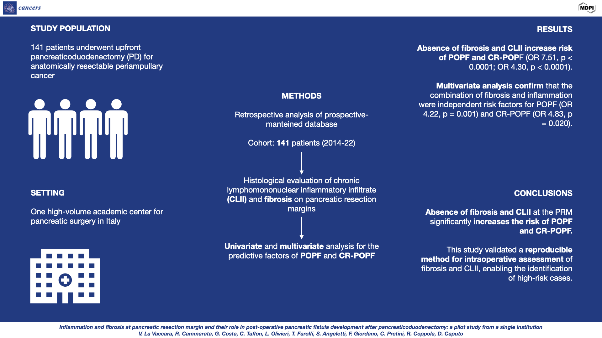
Abstract

Background/Objectives: The absence of pancreatic fibrosis is a known risk factor for post-operative pancreatic fistula (POPF) following pancreaticoduodenectomy (PD). Emerging evidence suggests inflammation plays a role in anastomotic healing. This study aimed to assess the role of chronic lymphomononuclear inflammatory infiltrate (CLII) and fibrosis at the pancreatic resection margin (PRM) on POPF development and explored a method for its intraoperative prediction. Materials and Methods: Data from 141 patients who underwent PD between 2014 and 2022 were retrospectively analyzed. PRM were histologically evaluated for fibrosis and CLII. Univariate and multivariate analyses were performed to identify potential predictors of postoperative pancreatic fistula (POPF) and clinically relevant postoperative pancreatic fistula (CR-POPF). Results: The histopathological analysis of intraoperative frozen sections of the PRM showed a strong association between the absence of fibrosis and CLII and increased risk of POPF and CR-POPF (OR 7.51, p < 0.0001; OR 4.30, p < 0.0001). Multivariate analysis further identified a main pancreatic duct diameter of less than 3 mm, as well as the combined presence of fibrosis and inflammation, as independent risk factors for developing POPF (OR 4.22, p = 0.001) and CR-POPF (OR 4.83, p = 0.020). Conclusion: The absence of fibrosis and CLII at the PRM significantly increases the risk of POPF and CR-POPF. This study validated a reproducible method for intraoperative assessment of fibrosis and CLII, enabling the identification of high-risk cases. Such a tool could guide surgical strategies to mitigate POPF-related complications, improving patient outcomes after PD.

Keywords: 
;  ;  ;  ;  ;  ;  ;  

1. Introduction

Pancreaticoduodenectomy (PD) is considered the gold standard treatment for resectable periampullary tumors, though it remains a complex surgical procedure associated with significant risk of complications (about 50%), even in high-volume centers [1].
Despite the high complication rate, post-operative mortality has decreased significantly to below 5% in specialized centers. Among the potential complications, post-operative pancreatic fistula (POPF) is one of the most frequent and feared, affecting approximately 30% of patients [2].
The development of POPF often leads to extended hospital stays and substantial increase in healthcare costs. It can also result in severe, life-threatening complications, such as bleeding and sepsis [3].
According to the International Study Group of Pancreatic Surgery (ISGPS), POPF is defined by the presence of pancreatic amylase in the drainage fluid at levels three times higher than the Upper Limit of Normal (ULN).
POPF is clinically classified into three grades (A, B, and C) based on its severity. Grade A, known as a Biochemical Leak (BL), does not affect the post-operative course, while grades B and C, referred to as Clinically Relevant Postoperative Pancreatic Fistula (CR-POPF), negatively impact post-operative course. The mortality rate for patients who develop CR-POPF ranges from 12% to 18.2% [4,5].
Given the challenge of predicting which patients will develop POPF at an early stage, there has been a recent shift toward a proactive approach centered on early diagnosis and complication prevention. This shift has highlighted the increasing need for tools that can assist in the early detection of CR-POPF [6].
Various risk factors for POPF have been identified, including elevated body mass index (BMI), poor preoperative nutrition, male gender, excessive intraoperative blood loss, tumor type, and limited surgical experience. Among these, the two most predictive factors - commonly used in risk score calculation - are the diameter of the main pancreatic duct (MPD) and the texture of the pancreas, as assessed by the surgeon. However, these evaluations are often subjective and lack standardized definitions, making it challenging to clearly differentiate between a "soft" and a "firm" pancreas. The increasing adoption of minimally invasive surgical approaches further complicates the assessment of pancreas texture due to limited tactile feedback [2,6,7].
The aim of this study is to establish objectively measurable predictive parameters for POPF development. Intraoperative frozen sections (IFS) of pancreatic resection margins (PRM), routinely analyzed to confirm a clear resection margin, were used. Histological findings, such as the degree of fibrosis of the pancreatic parenchyma and the extent of chronic lymphomononuclear inflammatory infiltrate (CLII), were correlated with POPF and CR-POPF in patients undergoing PD.

2. Materials and Methods

Data from patients who underwent PD at the General Surgery Unit of Fondazione Policlinico Universitario Campus Bio-Medico in Rome between December 2014 and November 2022 were retrospectively analyzed from a prospectively maintained database. The study received approval from the ethics committee of Università Campus Bio-Medico of Rome. The database included information such as gender, age, body mass index (BMI), alcohol consumption, smoking status, diabetes, POPF grade (A-B-C), pancreatic texture, MPD diameter, postoperative blood loss, type of neoplasm, and postoperative complications such as delayed gastric emptying (DGE) and post-pancreatectomy hemorrhage (PPH). Clinical data were obtained from the hospital's information system.
Inclusion criteria included patients over the age of 18 who underwent PD. Patients were excluded if pancreatic resection margin frozen sections were unavailable, or if they had clinical infections at the time of surgery, autoimmune diseases, or hematological disorders. Patients who received neoadjuvant treatments were also excluded. Patients were ultimately categorized into two groups: POPF-no vs. POPF-yes (Group 1) and CR-POPF-no vs. CR-POPF-yes (Group 2).
Prior to surgery, each patient underwent preoperative imaging for staging purposes, aimed at evaluating the extent of the disease and identifying any vascular anomalies [8]. If needed, multiorgan and/or vascular resections were performed concurrently. All patients underwent PD with pancreaticojejunal reconstruction using a duct-to-mucosa anastomosis technique as previously described [9].
All surgical procedures were concluded with the placement of two abdominal drains: one behind the hepaticojejunal anastomosis on the right side and the other behind the pancreaticojejunal anastomosis on the left side. The main pancreatic duct (MPD) diameter was measured intraoperatively by the surgical team, with a 3 mm cutoff used to distinguish high- or low-risk anastomoses for fistula formation, based on commonly cited literature. Pancreatic texture was also assessed intraoperatively and classified by the surgeon as either soft or firm [6].
All patients received antibiotic prophylaxis in line with clinical guidelines [11]. Pancreatic amylase levels in the drainage fluid were measured daily for the first five postoperative days, unless the drains were removed earlier [11].
Postoperative complications were assessed using the Clavien-Dindo classification and defined according to ISGPS criteria [5,12].
Hematoxylin and eosin (H&E) stained slides of pancreatic resection margins were reviewed by a pathology trainee under the supervision of a senior pathologist.
Based on the literature, a four-tier classification system for pancreatic fibrosis was used: Grade 0 = no fibrosis, with normal pancreatic parenchyma; Grade I = periductal fibrosis involving less than 10% of the pancreatic parenchyma, with preserved lobular architecture; Grade II = periductal and/or diffuse fibrosis affecting 10-90% of the parenchyma, with varying amounts of fibrotic septa surrounding groups of acini; and Grade III = diffuse fibrosis involving more than 90% of the parenchyma, with only scattered residual acini [13,14]. (Figure 1)
The inflammatory infiltrate was assessed based on the presence of chronic lymphomononuclear cells. A four-tier classification system was used: Grade 0 = no inflammatory infiltrate detected; Grade I = lymphomononuclear infiltrate affecting less than 10% of the pancreatic parenchyma; Grade II = infiltrate present in 10-70% of the parenchyma; and Grade III = infiltrate affecting more than 70% of the pancreatic parenchyma (Figure 2).
Statistical analysis was conducted using StataCorp 2019 STATA Statistical Software, release 16 (College Station, TX: StataCorp LLC). Categorical data were presented as frequencies, while continuous data were expressed as mean values ± standard deviation (SD) and/or medians with interquartile ranges (IQR, 25th–75th percentiles), along with minimum and maximum values. Differences in means were compared using the independent sample Student’s t-test or the Mann-Whitney U test, as appropriate. Fisher’s exact test or the Chi-square test (with or without Yates correction) was applied to assess differences in categorical data.
Receiver operating characteristic (ROC) curve analysis was used to determine the cut-off value for any relevant continuous variable by maximizing Youden’s index, converting it into a binary variable. Univariate and multivariate analyses were then performed to identify independent predictors of POPF and CR-POPF, with Odds Ratios (ORs) and 95% confidence intervals (CIs) calculated for significant variables.
Main pancreatic duct (MPD) diameter, absence of fibrosis, absence of CLII, and other known risk factors for POPF and CR-POPF were included in a backward and forward logistic regression analysis.
All tests were two-tailed, and a p-value ≤ 0.05 was considered statistically significant.

3. Results

3.1. Demographic Data

A total of 360 patients underwent PD during the study period. Of these, 141 met the inclusion criteria and were included in the analysis, as frozen sections of the pancreatic resection margin were unavailable for the remaining patients.
POPF occurred in 60 patients (42.5%), while CR-POPF was observed in 32 patients (22.7%).
Table 1 shows the main demographic and preoperative characteristics of the study groups. There were no significant differences between the groups in terms of age, gender, BMI, ASA score, alcohol consumption, smoking habits, or neoadjuvant therapy - factors that could be associated with higher risk of developing POPF or CR-POPF. Regarding operative details, no significant differences were found between the two groups in the type of procedure, operation time, or intraoperative blood loss. However, in the POPF vs. No POPF group, cholangiocarcinoma was significantly associated with an increased risk of POPF (p = 0.0350), while no significant differences were observed for other types of neoplasms (Table 1).

3.2. Correlation of Pancreatic Fibrosis, Lymphomononuclear Inflammatory Infiltrate, Main Pancreatic Duct Diameter and Pancreatic Texture with POPF

The main pancreatic duct (MPD) diameter, as estimated intraoperatively by the surgeon, being greater than 3 mm (p=0.0001 vs p=0.0031) and the presence of fibrosis in the histopathological analysis of the resection margin (p<0.0001 vs p<0.0001) were significantly associated with lower rates of POPF and CR-POPF. When these two parameters were combined, according to the updated ISGPS classification, a significant difference between the two groups was observed for both BL and CR-POPF (p<0.0001 vs p=0.0206).
Histopathological analysis of intraoperative frozen sections (IFS) from the pancreatic resection margin demonstrated a significant correlation between the degree of pancreatic parenchymal fibrosis (graded into four levels) and the presence of lymphomononuclear inflammatory infiltrate (also graded into four levels) with the risk of developing POPF and CR-POPF, both when evaluated individually and in combination (Table 2).
As shown in Table 3, the absence of pancreatic fibrosis was associated with a significant higher risk of POPF (OR 7.51; p<0.0001) and CR-POPF (OR 4.43; p<0.0001). Similarly, the absence of lymphomononuclear inflammatory infiltrate (LII) was linked to a higher risk of POPF (OR 4.30; p<0.0001) and CR-POPF (OR 3.40; p=0.0099). When neither inflammatory infiltrate nor pancreatic fibrosis were present, the risk of POPF and CR-POPF further increased [(OR 5.20; p<0.0001), (OR 4.83; p=0.020), respectively] (Table 3).
The degree of pancreatic fibrosis determined histopathologically has shown a significant correlation with the surgical assessment of pancreatic texture (p <0.0001), as depicted in Table 4 (Table 4).

3.3. Multivariate Analysis

The multivariate analysis identified a main pancreatic duct diameter of less than 3 mm and the combination of fibrosis and inflammation as independent risk factors for both POPF [OR 4.22 (1.80–9.88), p = 0.001] and CR-POPF [OR 3.1 (1.19–8.07), p = 0.020]. Additionally, the combination of fibrosis and inflammation was also a significant predictor for POPF [OR 5.44 (2.11–14.00)] and CR-POPF [OR 4.83 (1.27–18.3), p = 0.020]. The results of the multivariate analysis are detailed in Table 5 (Table 5).

4. Discussion

POPF remains a significant challenge in the surgical management of pancreatic cancer, being the leading cause of postoperative morbidity and mortality. Accurately predicting the risk of POPF, particularly CR-POPF, is essential for optimizing postoperative care strategies [2].
Historically, efforts to address this complication have focused on mitigating the potentially severe consequences of POPF through various management strategies [15].
In recent years, the emphasis has shifted towards identifying predictive factors for POPF risk, allowing for better preparation in managing patients at higher risk for serious complications. Examples of this proactive approach include establishing amylase cut-off levels in surgical drainage fluid and combining them with clinical and imaging parameters. This shift aims to improve outcomes by enhancing early detection and targeted intervention strategies for at-risk patients [9,10].
Research has increasingly focused on analyzing factors that precede the postoperative course. Numerous patient-related risk factors, both modifiable and non-modifiable, have been identified and, in several cases, incorporated into risk scores designed to predict the likelihood of POPF during the preoperative phase [16,17].
The effort to identify patients at high risk of POPF during the perioperative phase has led to the development of strategies aimed at reducing the likelihood of postoperative complications. As a result, some authors have proposed total pancreatectomy as a potential standard of care for patients with a high risk of POPF, particularly in cases involving pancreatojejunostomy [18].
The focus has shifted from preventing POPF to managing its risk, highlighting the importance of identifying patients with factors that make them susceptible to severe complications from POPF [19].
Among the numerous risk factors for POPF, the characteristics of the residual pancreatic parenchyma are the most strongly correlated. In particular, the presence of fibrosis, which increases tissue firmness, has been shown to enhance suture stability hence lowering the risk of pancreatojejunal anastomotic leakage [20].
However, the collection of this data is highly variable due to operator dependency, a challenge further compounded by the loss of tactile feedback in minimally invasive surgeries. Additionally, the absence of clear definitions makes this surgical assessment inherently subjective [21].
Our experience has shown that the pancreatic duct diameter and the extent of pancreatic parenchymal fibrosis can be accurately assessed through histological analysis of the intraoperative frozen section (IFS) of the pancreatic resection margin (PRM). In line with existing literature, our findings confirm that pancreatic fibrosis, followed by duct diameter, are independent predictors of both POPF and CR-POPF [13].
Simultaneously, the role of inflammation in the development of surgical complications has been widely studied. Numerous experiences in pancreatic surgery highlight the undeniable impact of systemic inflammation on surgical outcomes, especially in predicting the onset of CR-POPF [10].
Additionally, it has been suggested that acute inflammation of the residual pancreatic parenchyma after resection may contribute to the development of pancreatic fistula. In fact, the ISGPS has defined a new condition known as post-pancreatectomy acute pancreatitis (PPAP) [22].
While systemic and acute inflammation have been extensively studied, the role of chronic inflammation in this context remains underexplored. Nevertheless, it is well established that inflammatory infiltrates are linked to wound healing. Lymphocytes play a key role in this process by regulating the balance between inflammation and pathogen tolerance. This balance is critical to ensure that the necessary inflammatory response to fight infections and initiate tissue repair does not become excessive, causing damage to the tissues. Moreover, lymphocytes help modulate immune responses, influencing both cell proliferation and the formation of new tissue. These mechanisms are essential for achieving effective and balanced wound healing [23].
The presence of a robust chronic inflammatory component at the pancreatic transection margin could serve as an additional predictive marker for fibrotic pancreatic parenchyma, potentially offering protection against the risk of anastomotic leakage. A strong inflammatory infiltrate may contribute to creating a local environment favoring tissue healing, promoting quicker and more consistent tissue restoration [24].
To the best of our knowledge, this study is the first to present an effective and reproducible method for detecting chronic inflammatory infiltrates in the pancreatic parenchyma. Additionally, we have demonstrated that the absence of CLII in the residual pancreatic parenchyma is significantly associated with an increased risk of both POPF and CR-POPF. This association becomes even more pronounced when fibrosis is also absent. Therefore, we conclude that the lack of both CLII and fibrosis in the residual pancreatic parenchyma constitutes a significant risk factor for POPF, particularly CR-POPF.
These findings align with the well-established pathophysiological principle that a rich chronic inflammatory infiltrate is linked to enhanced tissue regeneration [25].

5. Conclusions

This study introduces several novel elements when considered in the context of current literature. While numerous studies have explored the role of systemic inflammation in predicting major surgical complications, few have specifically focused on the state of local inflammation, particularly in the tissues involved in the anastomosis [26].
The key innovation of this study is the development of an effective and reproducible intraoperative tool for predicting the risk of POPF by analyzing the presence of fibrosis and CLII. Its effectiveness, as demonstrated by the accuracy data in our study, lies in the immediate analysis of intraoperative frozen sections (IFS) of the pancreatic resection margin (PRM) by an experienced pathologist. This tool offers significant support to surgeons by influencing strategies aimed at mitigating the consequences of CR-POPF, which can be implemented during the procedure itself. In line with recent literature, identifying a high-risk pancreas - lacking fibrosis and CLII - could justify the placement of surgical drains or, in some cases, even a total pancreatectomy. On the other hand, a pancreas rich in fibrosis and inflammation might allow for a less aggressive approach, potentially avoiding the need for surgical drains altogether [27,28,29].
Finally, pancreatic surgery is increasingly adopting minimally invasive techniques. Much of the data on parenchymal texture currently relies on the surgeon's direct tactile assessment. However, with the advancements in laparoscopic and robotic surgery, we believe that developing intraoperative tools for objective POPF risk assessment - less dependent on the surgeon's tactile evaluation - is essential to preserve the benefits gained from these innovations [30].
We believe that introducing a tool that provides surgeons with objective data during pancreatic resection can significantly aid in determining the most appropriate strategy for each patient.
However, this study has some limitations. The small sample size has constrained the statistical power, and the retrospective, single-center design may have introduced potential biases. Nevertheless, patients who received neoadjuvant treatments have been excluded from the analysis. Further prospective studies, particularly on patients undergoing minimally invasive PD, are needed to validate these findings. Authors should discuss the results and how they can be interpreted from the perspective of previous studies and of the working hypotheses. The findings and their implications should be discussed in the broadest context possible. Future research directions may also be highlighted.

Author Contributions

Conceptualization: V.L.V., R.C. and D.C.; methodology: V.L.V., R.C., T.C., O.L. and D.C.; formal analysis: V.L.V., R.C., G.C. and D.C; data curation: V.L.V., R.C., G.F, P.C., F.T. and D.C; writing—original draft preparation V.L.V., R.C. and D.C; writing—review and editing, V.L.V., R.C. and D.C All authors have read and agreed to the published version of the manuscript.

Funding

This research received no external funding.

Institutional Review Board Statement

The study was conducted according to the guidelines of the Declaration of Helsinki and approved by the Ethics Committee of Università Campus Bio-Medico di Roma (protocol code 105.20 OSS.NOT; 2/12/2020).

Informed Consent Statement

Patient consent was waived due to the retrospective design of the study and considering that data are de-identified.

Data Availability Statement

The data presented in this study are available on request from the corresponding author.

Conflicts of Interest

The authors declare no conflicts of interest.

Abbreviations

The following abbreviations are used in this manuscript:
POPF Post-operative Pancreatic Fistula
CR-POPF Clinically Relevant Post-Operative Pancreatic Fistula
PD Pancreaticoduodenectomy
PRM Pancreatic Resection Margin
CLII Chronic Lymphomononuclear Inflammatory Infiltrate
IFS Intraoperative Frozen Sections
ASA American Society of Anesthesiologists classification
BMI Body Mass Index
DM Diabetes Mellitus
PDAC Pancreatic Ductal Adenocarcinoma
AC Ampullary Carcinoma
CCA Cholangiocarcinoma
NET Neuroendocrine Tumor
IPMN Intraductal Papillary Mucinous Neoplasm
MCN Mucinous Cystic Neoplasm
DGE Delayed Gastric Emptying
PPH Post-pancreatectomy Hemorrhage
PPAP Post-pancreatectomy Acute Pancreatitis
NAT Neoadjuvant Therapy
MPD Main Pancreatic Duct

References

  1. Smits F.J.; van Santvoort H.C.; Besselink M.G.; et al. Management of severe pancreatic fistula after pancreatoduodenectomy. JAMA Surg. 2017,152,540-548.
  2. McMillan M.T.; Soi S.; Asbun H.J. et al. Risk-adjusted outcomes of clinically relevant pancreatic fistula following pancreatoduodenectomy: a model for performance evaluation. Ann Surg. 2016, 264, 344-352.
  3. Harnoss J.C.; Ulrich A.B.; Harnoss J.M.; et al. Use and results of consensus definitions in pancreatic surgery: a systematic review. Surgery 2014, 155, 47–57.
  4. Bassi C.; Dervenis C.; Butturini G.; et al. Postoperative pancreatic fistula: an international study group (ISGPF) definition. Surgery 2005, 138(1), 8-13.
  5. Bassi C.; Marchegiani G; Dervenis C.; et al. The 2016 update of the International Study Group (ISGPS) definition and grading of postoperative pancreatic fistula: 11 Years After. Surgery 2017,161(3), 584-591.
  6. Chen J.Y.; Feng J.; Wang X.Q.; Cai S.W.; Dong J.H.; Chen Y.L.; Risk scoring system and predictor for clinically relevant pancreatic fistula after pancreaticoduodenectomy. World J Gastroenterol. 2015, 21(19), 5926-5933.
  7. Mungroop T.H.; van Rijssen L.B.; van Klaveren D.; et al. Alternative Fistula Risk Score for Pancreatoduodenectomy (a-FRS): Design and International External Validation. Ann Surg. 2019, 269(5), 937-943.
  8. La Vaccara V.; Coppola A.; Cammarata R.; Olivieri L.; Farolfi T.; Coppola R.; Caputo D.; Right hepatic artery anomalies in pancreatoduodenectomy - a risk for arterial resection but not for postoperative outcomes. J Gastrointest Oncol. 2023, 14(5), 2158-2166.
  9. Caputo D.; Coppola A.; La Vaccara V.; Passa R.; Carbone L.; Ciccozzi M.; Angeletti S.; Coppola R.; Validations of new cut-offs for surgical drains management and use of computerized tomography scan after pancreatoduodenectomy: The DALCUT trial. World J Clin Cases 2022, 10(15), 4836-4842.
  10. Coppola A.; La Vaccara V.; Angeletti S.; Spoto S.; Farolfi T.; Cammarata R.; Maltese G.; Coppola R.; Caputo D.; Postoperative procalcitonin is a biomarker for excluding the onset of clinically relevant pancreatic fistula after pancreaticoduodenectomy. J Gastrointest Oncol. 2023, 14(2), 1077-1086.
  11. Coppola A.; La Vaccara V.; Farolfi T.; Fiore M.; Cascone C.; Ramella S.; Spoto S.; Ciccozzi M.; Angeletti S.; Coppola R.; Caputo D.; Different Biliary Microbial Flora Influence Type of Complications after Pancreaticoduodenectomy: A Single Center Retrospective Analysis. J Clin Med. 2021, 10(10), 2180.
  12. Clavien P.A.; Barkun J.; de Oliveira M.L.; et al. The Clavien-Dindo classification of surgical complications: five-year experience. Ann Surg 2009, 250, 187-96.
  13. Timme S.; Kayser G.; Werner M.; et al. Surgeon vs Pathologist for Prediction of Pancreatic Fistula: Results from the Randomized Multicenter RECOPANC Study. J Am Coll Surg. 2021, 232(6), 935-945.
  14. Wellner U.F.; Kayser G.; Lapshyn H.; et al. A simple scoring system based on clinical factors related to pancreatic texture predicts postoperative pancreatic fistula preoperatively. HPB 2010, 12(10), 696-702.
  15. Malgras B.; Dokmak S.; Aussilhou B.; Pocard M.; Sauvanet A.; Management of postoperative pancreatic fistula after pancreaticoduodenectomy. J Visc Surg. 2023, 160(1), 39-51.
  16. McMillan M.T.; Allegrini V.; Asbun H.J.; et al. Incorporation of procedure-specific risk into the ACS-NSQIP surgical risk calculator improves the prediction of morbidity and mortality after pancreatoduodenectomy. Ann Surg. 2017, 265, 978e986.
  17. Pedrazzoli S.; Pancreatoduodenectomy (PD) and postoperative pancreatic fistula (POPF): a systematic review and analysis of the POPF-related mortality rate in 60.739 patients retrieved from the English literature published between 1990 and 2015. Medicine (Baltimore) 2017, 96e6858.
  18. Balzano G.; Zerbi A.; Aleotti F.; Capretti G.; Melzi R.; Pecorelli N.; Mercalli A.; Nano R.; Magistretti P.; Gavazzi F.; De Cobelli F.; Poretti D.; Scavini M.; Molinari C.; Partelli S.; Crippa S.; Maffi P.; Falconi M.; Piemonti L.; Total Pancreatectomy With Islet Autotransplantation as an Alternative to High-risk Pancreatojejunostomy After Pancreaticoduodenectomy: A Prospective Randomized Trial. Ann Surg. 2023, 277(6), 894-903.
  19. Parray A.M.; Chaudhari V.A.; Shrikhande S.V.; Bhandare M.S.; Mitigation strategies for post-operative pancreatic fistula after pancreaticoduodenectomy in high-risk pancreas: an evidence-based algorithmic approach"- a narrative review. Chin Clin Oncol. 2022, 11(1), 6.
  20. Tajima Y.; Kuroki T.; Tsuneoka N.; et al. Anatomy-specific pancreatic stump management to reduce the risk of pancreatic fistula after pancreatic head resection. World J Surg. 2009, 33(10), 2166-2176.
  21. Schuh F.; Mihaljevic A.L.; Probst P.; et al. A Simple Classification of Pancreatic Duct Size and Texture Predicts Postoperative Pancreatic Fistula: A classification of the International Study Group of Pancreatic Surgery. Ann Surg. 2023, 277(3), 597e608.
  22. Marchegiani G.; Barreto S.G.; Bannone E.; Sarr M.; Vollmer C.M.; Connor S.; Falconi M.; Besselink M.G.; Salvia R.; Wolfgang C.L.; Zyromski N.J.; Yeo C.J.; Adham M.; Siriwardena A.K.; Takaori K.; Hilal M.A.; Loos M.; Probst P.; Hackert T.; Strobel O.; Busch O.R.C.; Lillemoe K.D.; Miao Y.; Halloran C.M.; Werner J.; Friess H.; Izbicki J.R.; Bockhorn M.; Vashist Y.K.; Conlon K.; Passas I.; Gianotti L.; Del Chiaro M.; Schulick R.D.; Montorsi M.; Oláh A.; Fusai G.K.; Serrablo A.; Zerbi A.; Fingerhut A.; Andersson R.; Padbury R.; Dervenis C.; Neoptolemos J.P.; Bassi C.; Büchler M.W.; Shrikhande S.V.; International Study Group for Pancreatic Surgery. Postpancreatectomy Acute Pancreatitis (PPAP): Definition and Grading From the International Study Group for Pancreatic Surgery (ISGPS). Ann Surg. 2022, 275(4), 663-672.
  23. Seraphim P.M.; Leal E.C.; Moura J.; Gonçalves P.; Gonçalves J.P.; Carvalho E.; Lack of lymphocytes impairs macrophage polarization and angiogenesis in diabetic wound healing. Life Sci 2020, 254, 117813.
  24. Wynn T. Cellular and molecular mechanisms of fibrosis. J Pathol 2008, 214(2),199-210.
  25. Long H.; Lichtnekert J.; Andrassy J.; Schraml B.U.; Romagnani P.; Anders H.J.; Macrophages and fibrosis: how resident and infiltrating mononuclear phagocytes account for organ injury, regeneration or atrophy. Front Immunol 2023, 14, 1194988.
  26. Coppola A.; La Vaccara V.; Caggiati L.; et al. Utility of preoperative systemic inflammatory biomarkers in predicting postoperative complications after pancreaticoduodenectomy: Literature review and single center experience. World J Gastrointest Surg 2021,13(10), 1216-1225.
  27. McMillan M.T.; Malleo G.; Bassi C.; et al. Multicenter, Prospective Trial of Selective Drain Management for Pancreatoduodenec,tomy Using Risk Stratification. Ann Surg 2017, 265, 1209-18.
  28. Marchegiani G.; Perri G.; Burelli A.; Zoccatelli F.; Andrianello S.; Luchini C.; Donadello K.; Bassi C.; Salvia R.; High-risk Pancreatic Anastomosis Versus Total Pancreatectomy After Pancreatoduodenectomy: Postoperative Outcomes and Quality of Life Analysis. Ann Surg 2022, 276(6), e905-e913.
  29. Zaghal A.; Tamim H.; Habib S.; Jaafar R.; Mukherji D.; Khalife M.; Mailhac A.; Faraj W.; Drain or No Drain Following Pancreaticoduodenectomy: The Unsolved Dilemma. Scand J Surg 2020, 109(3), 228-237.
  30. Hilst J.; D'Hondt M.; Ielpo B.; Keck T.; Khatkov I.E.; Koerkamp B.G.; Lips D.J.; Luyer M.D.P.; Mieog J.S.D.; Morelli L.; Molenaar I.Q.; van Santvoort H.C.; Sprangers M.A.G.; Ferrari C.; Berkhof J.; Maisonneuve P.; Abu Hilal; Besselink M.G.; European Consortium on Minimally Invasive Pancreatic Surgery (E-MIPS). Minimally invasive versus open pancreatoduodenectomy for pancreatic and peri-ampullary neoplasm (DIPLOMA-2): study protocol for an international multicenter patient-blinded randomized controlled trial. Trials 2023, 24(1), 665.
Figure 1. Histopathological classification system of pancreatic fibrosis level (from right to left). (a) Grade 0 = no fibrosis, with normal pancreatic parenchyma; (b) Grade I = periductal fibrosis involving less than 10% of the pancreatic parenchyma, with preserved lobular architecture; (c) Grade II = periductal and/or diffuse fibrosis affecting 10-90% of the parenchyma, with varying amounts of fibrotic septa surrounding groups of acini; (d) Grade III = diffuse fibrosis involving more than 90% of the parenchyma, with only scattered residual acini.
Preprints 144540 g001
Figure 2. Histopathological classification system of CLII level in the pancreatic parenchyma (from right to left). (a) Grade 0 = no inflammatory infiltrate detected; (b) Grade I = lymphomononuclear infiltrate affecting less than 10% of the pancreatic parenchyma; (c) Grade II = infiltrate present in 10-70% of the parenchyma; and (d) Grade III = infiltrate affecting more than 70% of the pancreatic parenchyma.
Preprints 144540 g002
Table 1. Univariate analysis and relationship of demographics data with POPF and CR-POPF.
Variable Total POPF-no POPF-yes P-value CR-POPF-no CR-POPF-yes P-value
Total, n (%) 141 (100) 81 (57.4) 60 (42.5) 109 (77.3) 32 (22.7)
Age, y, (median) 72 72 71.5 0.6601 72 71 0.765
Sex, n (%)
• Female
• Male

63 (44.6)
78 (55.3)

36 (44.4)
45 (55.5)

27 (45)
33 (55)

0.9477

49 (44.9)
60 (55)

14 (437)
18 (56.2)

0.9041
ASA n (%)
• I
• II
• III
• IV

1 (1.23)
39 (48)
37 (45.6)
4 (4.9)

1 (1.66)
28 (46.6)
24 (40)
7 (11.6)


0.5090

1 (0.91)
53 (48.6)
50 (45.8)
5 (4.5)

1 (3.1)
14 (43.7)
11 (34.3)
6 (18.7)


0.0441*
BMI, kg/m2 (mediana) 28 28 28.5 0.6456 28 28.5 0.2353
Alcoholic Habit, n (%) 3 (2.1) 1 (1.2) 2 (3.3) 0.3932 1 (0.9) 2 (6.25) 0.6568
Smoking Habit, n(%) 43 (30.4) 23 (28.3) 20 (33.3) 0.5289 31 (28.4) 12 (37.5) 0.3277
DM (%) 31 (21.9) 20 (24.6) 11 (18.3) 0.3674 25 (22.9) 6 (18.7) 0.6152
Hypertension (%) 70 (49.6) 41 (50.6) 29 (48.3) 0.0766 54 (49.5) 16 (50) 0.9636
Neoadjuvant treatment (%) 10 (7.0) 7 (8.6) 3 (5) 0.4049 10 (9.1) 0 (0) 0.0755
Type of disease (%)
• PDAC
• AC
• CCA
• NET
• IPMN
• MCN
• Clear cells

110 (78.0)
3 (2.1)
11 (7.8)
7 (4.9)
6 (4.2)
3 (2.1)
1 (0.7)

68 (83.9)
1 (1.2)
3 (3.7)
2 (2.4)
4 (4.9)
3 (3.7)
0 (0)

42 (70)
2 (3.3)
8 (13.3)
5 (8.3)
2 (3.3)
0 (0)
1 (1.6)

0.0480*
0.3932
0.0350*
0.1130
0.6406
0.1319
0.2436

88 (80.7)
2 (1.83)
5 (4.5)
5 (4.5)
5 (4.5)
3 (2.7)
1 (0.9)

22 (68.7)
1 (3.1)
6 (18.7)
2 (6.2)
1 (3.1)
0 (0)
0 (0)

0.1501
0.6566
0.0086*
0.7034
0.7186
0.3428
0.5866
Intraoperative bleeding, mL, (median) 300 300 300 0.3233 300 300 0.4021
Operative time, median 375 371 376.5 0.6060 364 382.5 0.6501
DGE (%) 50 (35.4) 21 (25.9) 29 (48.3) 0.0060* 28 (25.6) 22 (68.7) <0.0001*
PPH (%) 24 (17.0) 4 (4.9) 20 (33.3) <0.0001* 8 (7.3) 15 (46.8) <0.0001*
90 days mortality (%) 17 (12.0) 6 (7.4) 11 (18.3) 0.0488* 11 (10) 6 (18.7) 0.1860
Tipe of surgery (%)
• Whipple
• Traverso

41 (29)
100 (71)

21 (25.9)
60 (74)

20 (33.3)
40 (66.6)

0.3383

31 (28.4)
78 (71.5)

10 (31.2)
22 (68.7)

0.7583
Reintervention (%) 19 (13.4) 6 (7.4) 13 (21.6) 0.5609 10 (9.1) 9 (28.1) 0.0058*
Length of Stay (%) 13 (9.2) 9 (11.1) 19.5 (32.5) 0.0001* 11 (10) 27 (84.3) 0.0001*
ASA, American Society of Anesthesiologists classification; BMI, body Max Index; DM, Diabetes Mellitus; PDAC, pancreatic ductal adenocarcinoma; AC, ampullary carcinoma; CCA, cholangiocarcinoma; NET, neuroendocrin tumor; IPMN, Intraductal Papillary Mucinous Neoplasm; MCN, mucinous cystic neoplasm; DGE, Delayed Gastric Empty; PPH, Post-Pancreatectomy Hemorrage.

Table 2. Univariate analysis and relationship of risk variables with POPF and CR-POPF.
Variable POPF-no
n (%)
POPF-yes
n (%)
P-value CR-POPF-no
n (%)
CR-POPF-yes
n (%)
P-value
MPD diameter
< 3 mm
> 3 mm

22 (27.1)
59 (72.8)

36 (60)
23 (38.3)

0.0001*

38 (34.8)
71 (65.1)

20 (62.5)
11 (34.3)

0.00031*
Pancreatic texture (evaluation by surgeon)
Soft
Hard


41 (50.6)
40 (49.3)


41 (68.3)
19 (31.6)


0.0350*


63 (57.7)
46 (42.2)


19 (59.3)
13 (40.6)


0.8737
New ISGPS classification
A (hard + ≥3 mm)
B (hard + <3 mm)
C (soft + ≥3 mm)
D (soft + <3 mm)

27 (33.3)
13 (16.0)
32 (39.5)
9 (11.1)

8 (13.3)
11 (18.3)
15 (25)
26 (43.3)
0.0005* 30 (27.5)
16 (14.6)
41 (37.6)
22 (20.1)

5 (15.6)
8 (25)
6 (18.7)
13 (40.6)
0.0206*
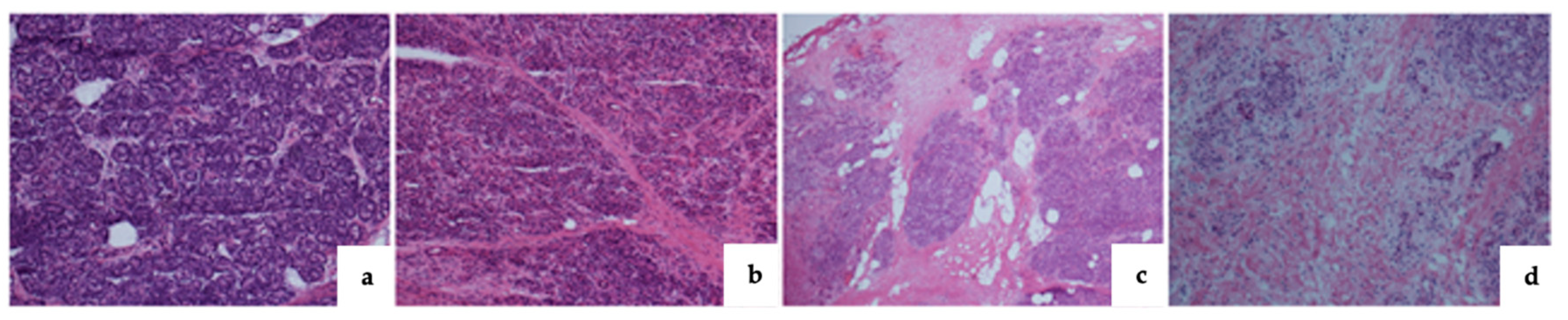
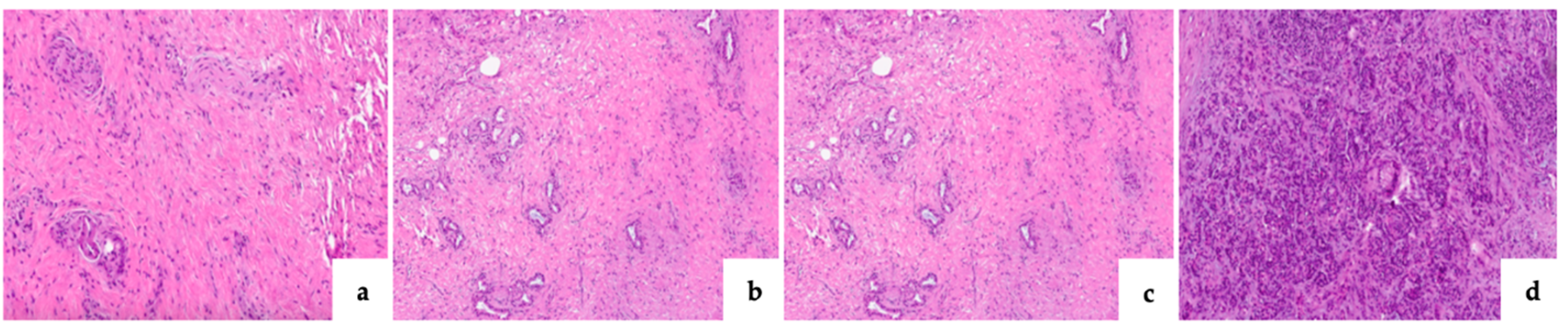
Pancreatic fibrosis
Grade 0
Grade I
Grade II
Grade III

12 (14.8)
17 (20.9)
16 (19.7)
34 (41.9)

34 (56.6)
17 (28.3)
8 (13.3)
1 (1.6)
<0.0001*
27 (24.7)
26 (23.8)
20 (18.3)
34 (31.1)

19 (59.3)
8 (25)
4 (12.5)
1 (3.1)
0.0062*
CLII
Grade 0
Grade I
Grade II
Grade III

39 (48.1)
32 (39.5)
7 (8.6)
1 (1.2)

48 (80)
11 (18.3)
1 (1.6)
0 (0)
0.0049*
61 (55.9)
38 (34.8)
7 (6.4)
1 (0.9)

26 (81.2)
5 (15.6)
1 (3.1)
0(0)
0.0934
*Statistically significant; MPD, main pancreatic duct
CLII, chronic lymphomononuclear inflammatory infiltrate; POPF, post-operative pancreatic fistuale; CR-POPF, clinically relevant POPF
Table 3. Correlation between fibrosis and CLII with POPF and CR-POPF.
Variable POPF-no Vs. POPF-yes CR-POPF-no Vs. CR-POPF-yes
OR 95% CI P-value OR 95% CI P-value
Grade 0 fibrosis 7.51 3.08 – 18.3 <0.0001* 4.43 1.85 – 10.6 0.0003*
Grade 0 CLII 4.30 1.90 – 9.73 0.0001* 3.40 1.26 – 9.19 0.0099*
Grade 0 fibrosis and CLII 5.20 2.23 – 12.1 <0.0001* 4.83 1.27 – 18.3 0.0200*
Table 4. Correlation of Surgical and Pathological Evaluation of Pancreatic Texture.
Histopathological evaluation Surgeon evaluation
Pancreatic fibrosis Pancreatic texture
Soft (82) Hard (59) P-value
<0.0001*
Grade 0 10 (12.1) 36 (61.0)
Grade I 9 (11.1) 25 (42.3)
Grade II 17 (20.7) 7 (11.8)
Grade III 23 (28.0) 12 (20.3)
*Statistically significant.
Table 5. Multivariate analysis.
Variable No POPF Vs. POPF No CR-POPF Vs. CR-POPF
OR 95% CI P-value OR 95% CI P-value
Age, y 1.02 0.97-1.07 0.329 0.99 0.94-1.04 0.754
Sex, male Vs. female 1.35 0.59-3.04 0.467 1.48 0.58-3.75 0.404
BMI > 30 kg/m2 2.61 0.71-9.59 0.148 3.12 0.59-16.4 0.178
NAT, yes Vs. not 1.35 0.52-3.50 0.536 1
Operative time 1.00 0.99-1.00 0.377 0.99 0.99-1.00 0.453
Blood loss, >300 mL 0.99 0.99-1.00 0.415 1.00 0.99-1.00 0.512
MPD < 3mm 4.22 1.80-9.88 0.0001* 3.1 1.19-8.07 0.020*
No fibrosis and CLII 5.44 2.11-14.00 0.0001* 4.83 1.27-18.3 0.020*
Disclaimer/Publisher’s Note: The statements, opinions and data contained in all publications are solely those of the individual author(s) and contributor(s) and not of MDPI and/or the editor(s). MDPI and/or the editor(s) disclaim responsibility for any injury to people or property resulting from any ideas, methods, instructions or products referred to in the content.
Copyright: This open access article is published under a Creative Commons CC BY 4.0 license, which permit the free download, distribution, and reuse, provided that the author and preprint are cited in any reuse.
Prerpints.org logo

Preprints.org is a free preprint server supported by MDPI in Basel, Switzerland.

Subscribe

Disclaimer

Terms of Use

Privacy Policy

Privacy Settings

© 2025 MDPI (Basel, Switzerland) unless otherwise stated