Preprint
Article

This version is not peer-reviewed.

Zephycandidine A and Synthetic Analogues – Synthesis and Evaluation of Biological Activity

A peer-reviewed article of this preprint also exists.

Submitted:

13 January 2025

Posted:

14 January 2025

You are already at the latest version

Abstract

A convenient total synthesis of the cytotoxic imidazo[1,2-f]phenanthridine-type Amaryllidaceae alkaloid zephycandidine A (3) was developed, which further allowed to perform modifications of substituents at the benzenoid ring A and the imidazole ring D. The biological activities of all synthesized compounds were evaluated, whereby reported activities of the parent alkaloid were poorly reproducible, while the closely related analogue THK-121 (11) showed a strong inhibitory effect on proliferation of leukemia cells. Additionally, our novel analogue significantly induced cell death via the intrinsic apoptosis pathway, evident by loss of mitochondrial membrane potential, increased mitochondrial oxidative stress and disrupted mitochondrial structure, in the same cells. At the same time, healthy cells were hardly affected by the treatment with THK-121 (11), indicating a potential therapeutic margin.

Keywords: 
;  ;  ;  ;  ;  ;  

1. Introduction

The Amaryllidaceae, a family of mainly perennial bulbous flowering plants with more than one thousand species found in tropical and subtropical regions of the world, are a rich source of bioactive alkaloids. Over decades, hundreds of alkaloids from diverse frameworks possessing acetylcholinesterase inhibitory, antimicrobial, antiviral, antitumor and other activities have been isolated [1]. Numerous types of Amaryllidaceae alkaloids contain modified phenanthrene partial structures, among them the lycorine (1) subtype with the pyrrolo[de]phenanthidine skeleton and the crinine subtype (e.g., haemanthamine (2)) incorporating the 5,10b-ethanophenanthridine skeleton (Figure 1).
In 2016, Zhan et al. [2] reported on the isolation of the first imidazo[1,2-f]phenanthridine-type alkaloid, zephycandidine A (3) from Zephyranthes candida (Lindl.) (Amaryllidaceae), a bulbous herb, which is cultured as an ornamental flower, and the whole plants are used as a folk medicine in China. Structurally distinct alkaloids from the same plant were unfortunately named zephycandidine I, II, III in 2017 [3].
This new minor alkaloid zephycandidine A (3) was described to exhibit significant cytotoxicity against five cancer cell lines with IC50 values ranging from 2 to 7 μM and to induce apoptosis in leukemia cells. Further, the authors claimed acetylcholinesterase inhibitory activity, but this activity could not be confirmed by the Banwell group in later investigations on synthetic zephycandidine A [4].
A first partial synthesis of zephycandidine A (3) was accomplished by Murphy et al. [5] in three steps from the alkaloid haemanthamine (2). Thermal degradation of 2 yielded phenanthrene 4 (Figure 1) in poor yield, subsequent treatment with 1,2-dibromoethane, liquid ammonia, and MnO2, following an annulation protocol of Cronin [6] gave the target compound. Recently, Banwell [4] reported on two distinct novel approaches to zephycandidine A (3). While the first method, including Pd-catalyzed cyclization of a 2-arylimidazole and 1,2-dihalobenzenes through a two-fold cross-coupling process afforded a poorly separable mixture of two isomeric products, a lower yielding (32% in the final step) second route, where regioselectivity was achieved by introduction of an additional bromo substituent and utilization of expensive 2-iodophenylboronic acid, gave zephycandidine A (3). Recently, another total synthesis going through a bromoaryl-imidazole and utilizing a non-aromatic ring C building block (1,3-cyclohexanedione) with limited variability and a complex catalyst system has been published by Lee et al. [7]. Two other general approaches to the imidazo[1,2-f]phenanthridine backbone (not yet applied to the total synthesis of zephycandidine A) utilize readily available 6-aminophenanthridine building blocks. Attachment of the 2,3-unsubstituted imidazole ring is conveniently achieved by treatment with chloroacetaldehyde [8,9], 3-substituted analogues are produced from 6-aminophenanthridines and aliphatic aldehydes/arylacetaldehydes through a novel, metal-free sulfur endorsed oxidative cyclization protocol developed by the Zhang group [10]. Further, 6-chlorophenanthridine can be condensed with aminoacetaldehyde dimethylacetal to give 2,3-unsubstituted imidazo[1,2-f]phenanthridines [11].
The reported antitumor activity of zephycandidine A (3) from the plant source prompted the Zhang group to perform studies on the activity of synthetic analogues they obtained in the course of the elucidation of the scope of their oxidative cyclization protocol [10]. The collection of test compounds was characterized by diverse substituents at C-3 (phenyl, benzyl, linear and branched alkyl residues), further, a few substituents were introduced at the benzenoid rings. Among these test substances, the 3-methyl- and the 3-tert-butyl imidazo[1,2-f]phenanthridines showed marked inhibitory activity against several tumor cell lines (top values about 2 µM). Unfortunately, zephycandidine A (3) was not included in this substance library, neither was the 9a,12a-methylenedioxy unit, a privileged structure motif in bioactive natural products, or similar oxygenation patterns (except one monomethoxy analogue) present among the investigated compounds. This prompted us to investigate the cytotoxic activities of a) methylenedioxy-decorated imidazo[1,2-f]phenanthridines bearing additional substituents at the imidazole ring (C-3), as well as b) analogues with modified oxygenation pattern at ring A and one congener with a pyridine as ring A in a panel of different cancer cell lines.

2. Results and Discussion

2.1. Chemistry

Zephycandidine A (3) and analogues with unsubstituted imidazole ring (ring D) and with modifications in ring A were synthesized by adapting the approach of Zhang et al. [8]. A new and effective total synthesis of zephycandidine A (3) was developed starting from commercially available building block 6-bromo-1,3-benzodioxole-5-carbonitrile (6a), which is alternatively prepared in one step from cheap 6-bromopiperonal (5a) by oximation/dehydration [12] in 96% yield. Suzuki-Miyaura cross-coupling of 6a and 2-(4,4,5,5-tetramethyl-1,3,2-dioxaborolan-2-yl)aniline (7) gave, after spontaneous intramolecular nucleophilic amine-to-nitrile addition, the according 6-aminophenanthridine 8a in 76% yield. Subsequent reaction with chloroacetaldehyde and Na2CO3 in isopropanol/water gave the alkaloid 3 in 89% yield (Scheme 1). In accordance with reports from other groups [4,5] the alkaloid was obtained as a high-melting solid, whereas Zhan et al. [2] surprisingly describe their compound isolated from the plant source as a colourless oil. Notably, Zhan’s published 1H and 13C NMR spectra are fragmentary (in the 1H NMR spectrum the part below 5.4 ppm, and in the 13C NMR spectrum the part below 54 ppm is missing), thus the presence of eventually bioactive – see discussion below – aliphatic impurities in the isolated natural product cannot be excluded. Our two-step procedure (68% overall yield) represents the most effective total synthesis of zephycandidine A at present.
This reaction sequence could easily be extended to the synthesis of zephycandidine A analogues with modified substitution patterns in ring A (methoxy, isopropoxy residues), since the required ortho-bromobenzonitriles 6b-d (for details see Experimental Section) were easily accessible from easy to prepare or optionally commercially available ortho-bromobenzaldehydes 5b-d via conversion to the oximes and concomitant dehydration in hot DMSO [12]. The benzodioxole ring system was in the same manner replaced by a pyridine ring (compound 12) by starting with 2-bromopyridine-3-carboxaldehyde (5e). The target compounds 912 obtained this way are shown in Scheme 1.
For the synthesis of analogues of zephycandidine A (3) bearing additional substituents at the imidazole ring D (C-3) (pertaining the methylenedioxy motif at ring A) we utilized intermediate 8a from the zephycandidine A synthesis (Scheme 1). Reaction with appropriate aldehydes (phenylacetaldehyde, propanal, 3-methylbutanal) in DMSO/cyclohexane with excess elemental sulfur at 120 °C [10] gave the desired products 13 - 15 in moderate yields (21 – 58%) (Scheme 2).
The so obtained library of zephycandidine A (3) and seven analogues was subjected to analysis of biological activity against several cancer cell lines.

2.2. Evaluation of Biological Activity

Since accumulating evidence demonstrated promising antitumoral effects of alkaloids derived from Amaryllidaceae plants [13,14,15,16], we elucidated the potential of zephycandidine A (3) and our novel analogues to inhibit the growth and survival of cancer cells. First, we tested their biological activity in a CellTiter®-Blue proliferation assay on various cancer cell lines. Initially, we tested 7 analogues on Jurkat (leukemia) (Figure 2A) and MCF-7 (breast cancer) (Figure 2B) cells, which resulted in IC50 values in the µM range (11.7 µM and 145 µM, respectively for zephycandidine A (3); 9.6 µM to 120 µM for the synthetic analogues) for inhibition of cancer cell proliferation (for details, see Supporting Information, Table S1). From these tested analogs, we chose three active compounds to characterize further: mixed diether 11 (internal code: THK-121), pyridine analogue 12, and methylimidazole 13. These synthetic compounds showed superior activity as compared to zephycandidine A (3) in decreasing cell proliferation in MCF-7 and were at least equipotent in inhibiting Jurkat cell proliferation. Analysis of their anti-proliferative capacity in CCRF-CEM (Figure 2C), HL-60 (Figure 2D) and non-cancerous HUVEC (Figure 2E) cells also led to IC50 values in the µM range (Supporting Information, Table S1). These results are in general in accordance with a previous report by Zhan et al., that shows an anti-proliferative potency of zephycandidine A in the low µM range against several cancer cell lines [2]. Like Zhan et al. reported selectivity of zephycandidine A for cancer cells over human bronchial epithelial Beas-2B cells, we found an increased anti-proliferative potential of the lead structure as well as analogues against cancer cell lines as compared to non-cancerous HUVEC cells. This indicates some degree of cancer selectivity (Figure 2E). Of note, IC50 values obtained for zephycandidine A in MCF7 and HL-60 cells were up to 100 fold higher in our experimental setup as compared to the values reported by Zhan et al. This discrepancy might be attributed to either a differences in the assays used or the origin of zephycandidine A. While Zhan et al. isolated zephycandidine A from Zephyranthes candida [2], we used our very pure synthesis product. As pointed out above, the published NMR data for the isolated alkaloid do not exclude aliphatic impurities, so we (in accordance with Banwell and co-workers [4], who could not reproduce the claimed acetylcholinesterase-inhibitory activity) suppose that reported biological activities of the alkaloid were falsified by undetected biologically active impurities.
Nevertheless, zephycandidine A (3) and, even to a higher extent, synthetic analogues exhibit the potential to inhibit cancer cell growth in the low µM range and exhibit a more pronounced effect on cancer proliferation as compared to healthy cells.
Next, we tested zephycandidine A and selected analogues on their potential to induce cell death in cancer cells. Based on the identified antiproliferative activity, we used concentrations at the respective IC50 values and analyzed cell death using flow cytometry. The lead structure induced moderate cell death in all tested lines (Figure 2 F-I), whereas the tested analogues showed an increased potential to induce cell death in leukemic cells (Figure 2 F,H,I).
To verify the cell death induction capacity of zephycandidine A and analogues, we analyzed established markers for apoptosis. Firstly, we analyzed the activation of effector caspase-3 [17] and its downstream substrate, DNA repair protein PARP (poly [ADP-ribose] polymerase) [18] by Western Blot analysis. While, the lead structure zephycandidine A exhibits neither detectable caspase-3 activation nor significant PARP cleavage, the closely related compound 11 displays activation of caspase-3, accompanied by PARP cleavage (Figure 3A), indicating significant apoptosis induction.
As our analogues induce apoptosis, we were interested in the mode of cell death induction. An early step in apoptosis is transcriptionally activating pro-apoptotic Bcl-2 family members (e.g. Bax, Bak, Bcl-xS, Mcl-1S) and repressing anti-apoptotic Bcl-2 proteins (e.g. Mcl-1L, Bcl-2, Bcl-xL). We checked for expression of Bax and Mcl-1L protein levels after treatment with zephycandidine A and selected analogues. Analysis of protein level in Jurkat cells (Figure 3B) revealed a downregulation of anti-apoptotic Mcl-1L and upregulation of pro-apoptotic Bax upon treatment with compound 11, whereas zephycandidine A did not display an effect. In breast cancer MCF7 cells, no effect of zephycandidine A or analogues on Bcl-2 family proteins was detected (Figure 3C).
Mechanistically, Mcl-1L and Bax interact with each other, as do other members of the Bcl-2 family to form a mitochondrial apoptosis-induced channel (MAC) in the mitochondrial outer membrane leading to release of cytochrome c and other pro-apoptotic factors from the mitochondria. Due to the subsequent apoptosome formation and activation of caspase 9, effector caspases 3 and 7 are activated thereby driving the apoptosis process [19] [20]. Along the line, Zhan et al. [2] described that zephycandidine A treatment of HL-60 cells led to an upregulation of Bax as well as a downregulation of anti-apoptotic Bcl-2, already implying involvement of mitochondria in the mediating of apoptosis. To assess, whether mitochondria are affected by our compounds, we analyzed mitochondrial integrity by flow cytometry. Therefore, we investigated whether zephycandidine A and its analogues facilitate loss of mitochondrial membrane potential (Δψm) and formation of mitochondrial superoxides, an indicator for mitochondrial oxidative stress. Δψm can be detected by JC-1 staining. If Δψm is intact, JC-1 is capable to accumulate in the healthy mitochondria forming aggregates that elicit a red fluorescence signal. In contrast, damaged mitochondria show mitochondrial depolarization indicated by a decreased red/green fluorescence intensity. We found a significant increase of dissipated mitochondria in both Jurkat and MCF-7 cells upon treatment with analogue 11, whereas Δψm was unaffected by zephycandidine A (Figure 3D-E). Using MitoSOXTM Red we revealed that concomitantly treatment with compound 11 results in an increased presence of mitochondrial superoxide in both cell lines (Figure 3F-G). These data indicate that the lead compound zephycandidine A does not significantly induce apoptosis in the tested cell lines, but the novel analogue 11 does cause apoptosis in a mitochondria-dependent manner.
To confirm the effect of compound 11 on mitochondrial function, we further analyzed mitochondrial morphology and biogenesis. Several studies report that mitochondrial morphology greatly affects mitochondrial function [21,22,23], hence we observed mitochondrial morphology by confocal imaging. Mitotracker staining revealed that the control cells display fiber-shaped, elongated mitochondria, which transform to round-shaped structures upon treatment with compound 11 (Figure 4A-B). This phenotype was also observed in the literature upon mitochondria-driven apoptosis, where it was reported to be induced by a reduction of cristae density, mtDNA depletion, and a disbalance of mitochondrial dynamics that finally aggravate mitochondrial function [23,24,25]. As a response to mitochondrial dysfunction cells frequently increase mitochondrial biogenesis [26]. Thus, we assessed the mitochondrial mass upon treatment, with no evident induction of mitochondrial biogenesis upon treatment with zephycandidine A or analogues (Figure 4C-D). In sum, compound 11 induces the intrinsic apoptosis pathway (Figure 3A-G, 4A-B) without inducing a regulatory upregulation in mitochondrial mass (Figure 4C-D), leading to cell death in leukemia cells (Figure 2F-I).

2.3. Discussion

Summarized, in this study, we have developed short approaches to zephycandidine A (3) and related imidazo[1,2-f]phenanthridines with modifications of substituents at the benzenoid ring A and the imidazole ring D. We show the antiproliferative properties of the parent alkaloid and novel synthetic analogues in four different cancer cell lines. We investigated the capacity of the parental molecule and its analogues to enable cell death and found that the novel analogue THK-121 (11) leads to induction of mitochondrial apoptosis, while the parental alkaloid does not. We hence describe novel zephycandidine A analogues with increased cytotoxic potential, preferentially targeting cancer cells. This will enable further analysis of zephycandidine A and a library of analogues on their biological activity for instance in cancer cell migration. Interestingly in that regard, Narva et. al have shown that synthetic analogues of zephycandidine A (3), mostly with additional substituents at ring D, not only facilitate antitumoral potential by inhibiting proliferation and inducing apoptosis but also by inhibiting cancer cell migration [10] Since dysfunctional mitochondria have been linked excessively to metastasis and migration [27], it would be interesting to check zephycandidine A analogues on their potential to interfere with migration.

3. Experimental Section

3.1. Chemistry

3.1.1. General Reagent and Analytical Information

All solvents used were purified according to standard procedures or of HPLC or p.a. grade purchased from commercial sources. Chemical reagents were purchased from Sigma Aldrich (Schnelldorf, Germany) TCI (Eschborn, Germany) and BLD Pharm (Kaiserslautern). IR spectra were recorded on a Perkin Elmer FTIR Paragon 1000 spectrometer. NMR spectra were recorded on Jeol JNMR-GX 400 (400 MHz), Jeol JNMR-GX 500 (500 MHz), Avance III HD Bruker BioSpin (400 MHz) and Avance III HD Bruker BioSpin (500 MHz) spectrometers. Spectra were recorded in deuterated solvents and signal assignments were carried out based on 1H, 13C, DEPT, HMQC, HMBC, and COSY spectra. Chemical shifts are reported in parts per million (ppm) and J values are reported in hertz. High- resolution mass spectra were performed by electrospray ionization (ESI) using a Thermo Finnigan LTQ FT Ultra spectrometer or electron impact (EI) at 70 eV on a Jeol GCmate II or on a Finnigan MAT 95 spectrometer. All reactions were monitored by GC/MS or thin-layer chromatography (TLC) using precoated plastic sheets POLYGRAM®SIL G/UV254 from Macherey-Nagel (Düren, Germany). Compounds on TLC plates were detected under UV light at 254 and 366 nm. Separations with flash column chromatography (FCC) were performed on Merck silica gel 60 as stationary phase. Melting points were determined by the open tube capillary method on a Büchi melting point B- 540 apparatus and are uncorrected. HPLC purities were determined using an HP Agilent 1100 HPLC with a diode array detector and an Agilent Zorbax Eclipse plus C18 column (150 × 4.6 mm; 5 μm) with acetonitrile/water in different proportions as mobile phase.
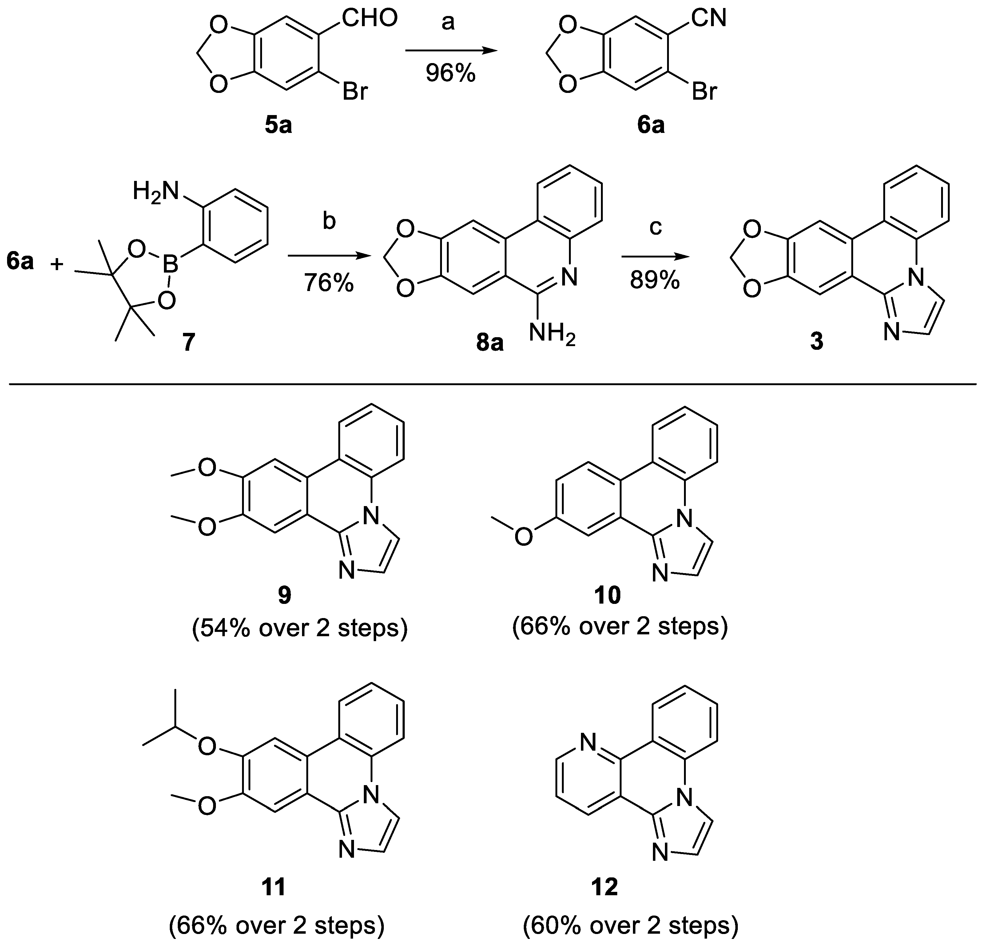
General procedure A for the conversion of aromatic aldehydes into nitriles. The aldehyde 5a-e (10 mmol, 1.0 eq) was dissolved in DMSO and hydroxylamine hydrochloride (15 mmol, 1.5 eq) was added. The mixture was heated to 90 °C and stirred for 1 h at this temperature. After cooling to room temperature water (20 mL) was added and the suspension was extracted with ethyl acetate (3 x 15 mL). The organic phases were pooled, washed with brine (3 x 15 mL), dried over Na2SO4 and evaporated. The crude product was purified by silica gel column chromatography with the declared eluent.
General procedure B for the synthesis of aminophenanthridines 7a-7e. Substituted 2-bromobenzonitrile 6a-e (2.5 mmol, 1.0 eq), 2-aminophenylboronic acid pinacol ester (7) (548 mg, 2.50 mmol, 1.00 eq) and bis(triphenylphosphine)palladium(II) chloride (88 mg, 0.013 mmol, 0.050 eq) were dissolved in 15 mL dry, degassed DMF in a Schlenk round-bottom flask under N2. Afterwards 4 mL of a shortly degassed aqueous 2 M Na2CO3 solution was added, and the yellow suspension was stirred at 80 °C for 16 h. After cooling to room temperature, the crude mixture was filtrated through a short plug of Celite, which was subsequently washed with 50 mL methylene chloride and 50 mL methylene chloride /methanol 9:1. Then the combined organic solution was mixed with 100 mL water and the organic phase was separated. The aqueous phase was extracted twice with 25 mL methylene chloride, the organic fractions were pooled, dried over anhydrous Na2SO4 and concentrated in vacuo. The crude product was purified by silica gel column chromatography with the declared eluent.
General procedure C for the cyclization to the imidazophenanthridine ring system. Substituted 6-aminophenanthridine 8a-e (0.42 mmol, 1.0 eq), chloroacetaldehyde (50% in water, 0.11 mL, 4.3 eq) and Na2CO3 (71 mg, 0.84 mmol, 2.0 eq) were mixed with 3 mL isopropanol and 3 mL water. The suspension was refluxed for 1 h. After cooling to room temperature 10 mL water were added and the suspension was extracted with ethyl acetate (3 x 10 mL). The organic phases were pooled, washed with brine (10 mL), dried over Na2SO4 and evaporated. The crude product was purified by silica column chromatography with the declared eluent.
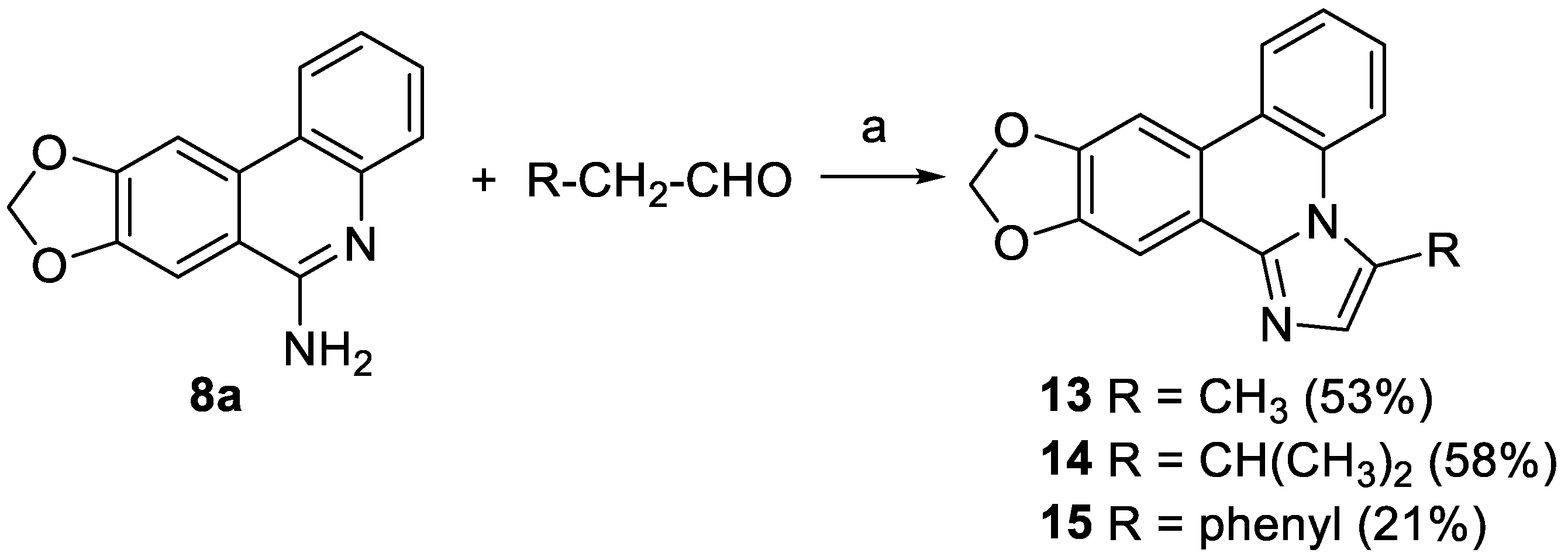
General procedure D for the cyclization to ring D-substituted imidazophenanthridines 13-15. Aminophenanthridine 8a (48 mg, 0.20 mmol, 1.0 eq) and sulfur (103 mg, 3.20 mmol, 16.0 eq) were suspended in 1 mL DMSO/cyclohexane (2:1). The appropriate aldehyde (0.30 mmol, 1.5 eq) was added and the mixture was heated at 120 °C overnight. After cooling to room temperature water (10 mL) was added and the black mixture was extracted with methylene chloride (3 x 10 mL). The combined organic phases were washed with brine (2 x 10 mL), dried over Na2SO4, and evaporated. The crude product was purified by silica column chromatography with the declared eluent.
6-Bromobenzo[d][1,3]dioxole-5-carbonitrile (6a). The synthesis was accomplished following General Procedure A starting from aldehyde 5a. Eluent for FSC isohexane/ethyl acetate 2:1 to afford 6a as a colourless solid (2.17 g, 9.05 mmol, 96%). m.p. 134-135 °C. 1H NMR (400 MHz, CDCl3) δ 7.09 (s, 1H, 6-H), 7.03 (s, 1H, 3-H), 6.10 (s, 2H, -OCH2O-). 13C NMR (101 MHz, CDCl3) δ 152.25 (C-2), 147.47 (C-1), 119.00 (C-4), 117.36 (-CN), 113.43 (C-6), 112.59 (C-3), 107.98 (C-5), 103.06 (-OCH2O-). IR (ATR): ṽmax/cm-1 = 2913, 2231, 1495, 1480, 1259, 1111, 1031, 922, 880, 838, 737. HRMS (EI): calcd. for C8H4BrNO2 (M)•+: 224.9425 found: 224.9423.
2-Bromo-4,5-dimethoxybenzonitrile (6b). The synthesis was accomplished following General Procedure A from 2-bromo-4,5-dimethoxybenzaldehyde (5b). Eluent for FSC isohexane/ethyl acetate 5:1 to afford 6b as a colourless solid (2.23 g, 9.22 mmol, 92%). m.p. 117-119 °C. 1H NMR (400 MHz, methylene chloride-d2) δ 7.10 (s, 1H, 3-H), 7.07 (s, 1H, 6-H), 3.89 (s, 3H, 1’-H), 3.84 (s, 3H, 2’H). 13C NMR (101 MHz, methylene chloride-d2) δ 153.46 (C-4), 148.74 (C-5), 117.62 (C-2), 117.21 (-CN), 115.53 (C-6), 115.44 (C-3), 106.63 (C-1), 56.41 (C-1’), 56.27 (C-2’).IR (ATR): ṽmax/cm-1 = 2942, 2229, 1592, 1505, 1377, 1261, 1219, 1168, 1035, 952, 874, 853, 794. HRMS (EI): calcd. for C9H8BrNO2 (M)•+: 240.9733 found: 240.9736.
2-Bromo-5-hydroxy-4-methoxybenzonitrile. The synthesis was accomplished following General Procedure A from commercially available 2-bromo-5-hydroxy-4-methoxybenzaldehyde. Eluent for FSC isohexane/ethyl acetate 1:1 to afford the target compound as a colourless solid (2.19 g, 9.62 mmol, 96%). m.p. 160-163 °C. 1H NMR (400 MHz, DMSO-d6) δ 10.07 (s, 1H, -OH), 7.35 (s, 1H, 6-H), 7.17 (s, 1H, 3-H), 3.87 (s, 3H, -OCH3). 13C NMR (101 MHz, DMSO-d6) δ 152.69 (C-4), 146.49 (C-5), 119.39 (C-3), 117.70 (C-2), 116.24 (C-6), 114.44 (-CN), 105.35 (C-1), 56.40 (-OCH3). IR (ATR): ṽmax/cm-1 = 3367, 2229, 1608, 1508, 1438, 1286, 1267, 1211, 1160, 1020, 867, 842, 804. HRMS (EI): calcd. for C8H6BrNO2 (M)•+: 226.9576 found: 226.9582.
2-Bromo-5-isopropoxy-4-methoxybenzonitrile (6d). 2-Bromo-5-hydroxy-4-methoxybenzonitrile (1.14 g, 5.00 mmol, 1.00 eq) was dissolved in dry acetone (50 mL), K2CO3 (1.24 g, 7.50 mmol, 1.50 eq) added, and the resulting suspension stirred for 15 minutes. 2-Iodopropane (1.5 mL, 15 mmol, 3.0 eq) was added and the mixture was stirred for 48 h at 50 °C. Brine (100 mL) was added and the suspension extracted with ethyl acetate (3x 50 mL). The organic phases were pooled, washed with brine (50 mL), dried over Na2SO4, and evaporated resulting in a yellow oil, which was purified by silica column chromatography (isohexane/ethyl acetate 2:1) to afford 6d as a colorless solid (1.28 g, 4.73 mmol, 95 %). m.p. 89-91 °C. 1H NMR (400 MHz, methylene chloride-d2) δ 7.10 (s, 1H, 3-H or 6-H), 7.09 (s, 1H, 3-H or 6-H), 4.49 (hept, J = 6.1 Hz, 1H, 1’-H), 3.87 (s, 3H, -OCH3), 1.33 (d, J = 6.1 Hz, 6H, 2’-H). 13C NMR (101 MHz, methylene chloride-d2) δ 154.68 (C-4), 146.83 (C-5), 119.18 (C-6), 117.65 (C-2), 117.10 (-CN), 116.01 (C-3), 106.62 (C-1), 72.16 (C-1’), 56.37 (-OCH3), 21.52 (C-2’). IR (ATR): ṽmax/cm-1 = 2922, 2223, 1588, 1504, 1437, 1377, 1268, 1259, 1217, 1166, 1138, 1027, 921, 860, 798. HRMS (EI): calcd. for C9H8BrNO2 (M)•+: 269.0046 found: 269.0049.
2-Bromonicotinonitrile (6e). The synthesis was accomplished following General Procedure A from commercially available 2-bromo-3-pyridinecarboxaldehyde. Eluent for FSC isohexane/ethyl acetate 5:1 to afford 6e as a colourless solid (1.72 g, 9.41 mmol, 94%). m.p. 107-109 °C. 1H NMR (400 MHz, methylene chloride-d2) δ 8.60 (dd, J = 4.9, 1.9 Hz, 1H, 6-H), 8.03 (dd, J = 7.7, 2.0 Hz, 1H, 4-H), 7.41 (dd, J = 7.7, 4.9 Hz, 1H, 5-H). 13C NMR (101 MHz, methylene chloride-d2) δ 152.92 (C-6), 152.52 (C-2), 142.72 (C-4), 122.38 (C-5), 114.67 (-CN), 110.82 (C-3).IR (ATR): ṽmax/cm-1 = 3081, 3065, 2236, 1577, 1398, 1145, 1131, 1079, 807, 7356, 672. HRMS (EI): calcd. for C6H3BrN2 (M)•+: 181.9474 found: 181.9478.
[1,3]Dioxolo[4,5-j]phenanthridin-6-amine (8a). The synthesis was accomplished following General Procedure B starting from nitrile 6a. Eluent for FSC isohexane/ethyl acetate 1:2 (containing 1% triethylamine) to afford 8a as a beige solid (455 mg, 1.91 mmol, 76%). m.p. 250-252 °C. 1H NMR (400 MHz, DMSO-d6) δ 8.35 (dd, J = 8.2, 1.4 Hz, 1H, 4-H), 8.13 (s, 1H, 11-H), 7.81 (s, 1H, 7-H), 7.52 – 7.39 (m, 2H, 1-H, 3-H), 7.21 (ddd, J = 8.3, 6.8, 1.5 Hz, 1H, 2-H), 6.78 (s, 2H, NH2), 6.22 (s, 2H, OCH2O). 13C NMR (101 MHz, DMSO-d6) 13C NMR (101 MHz, DMSO-d6) δ 155.06 (C-6), 150.41 (C-7a or C-10a), 147.63 (C-7a or C-10a), 144.56 (C-4a), 130.71 (C-11a), 127.81 (C-3), 125.50 (C-1), 122.37 (C-4), 121.44 (C-2), 120.58 (C-11b), 114.07 (C-6a), 102.25 (C-7), 101.91 (OCH2O), 100.81 (C-11). IR (ATR): ṽmax/cm-1 = 3484, 3059, 1660, 1454, 1233, 1035, 1029, 938, 753, 731. HRMS (EI): calcd. for C14H10N2O2 (M)•+: 238.0737 found: 238.0738.
8,9-Dimethoxyphenanthridin-6-amine (8b). The synthesis was accomplished following the General Procedure B starting from nitrile 6b. Eluent for FSC ethyl acetate/ triethylamine 99:1 to afford 8b as an off-white solid (403 mg, 1.58 mmol, 63%). m.p. 215-216 °C. 1H NMR (500 MHz, methylene chloride-d2) δ 8.29 (dd, J = 8.1, 1.4 Hz, 1H, 4-H), 7.86 (s, 1H, 7-H), 7.66 (dd, J = 8.2, 1.4 Hz, 1H, 1-H), 7.52 (ddd, J = 8.2, 7.0, 1.4 Hz, 1H, 2-H), 7.37 (ddd, J = 8.2, 7.0, 1.4 Hz, 1H, 3-H), 7.20 (s, 1H, 10-H), 5.20 – 5.15 (br s, 2H, -NH2), 4.08 (s, 3H, 2’-H), 4.00 (s, 3H, 1’-H). 13C NMR (126 MHz, methylene chloride-d2) δ 154.57 (C-6), 153.15 (C-9), 150.29 (C-8), 144.57 (C-4a), 130.00 (C-10a), 128.47 (C-3), 127.20 (C-2), 123.30 (C-1), 122.07 (C-4), 121.89 (C-10b), 113.56 (C-6a), 104.27 (C-10), 103.57 (C-7), 56.59 (C-2’), 56.51 (C-1’). IR (ATR): ṽmax/cm-1 = 3384, 3155, 1661, 1615, 1524, 1451, 1359, 1266, 1206, 1021, 803, 749, 732. HRMS (EI): calcd. for C15H14N2O2 (M)•+: 254.1050 found: 254.1051.
8-Methoxyphenanthridin-6-amine (8c). The synthesis was accomplished following the General Procedure B starting from commercially available 2-bromo-5-methoxybenzonitrile (6c). Eluent for FSC ethyl acetate/ triethylamine 99:1 to afford 8c as an off-white solid (450 mg, 2.01 mmol, 80%). m.p. 174-175 °C. 1H NMR (400 MHz, DMSO-d6) δ 8.56 (d, J = 9.0 Hz, 1H, 10-H), 8.37 (dd, J = 8.2, 1.4 Hz, 1H, 4-H), 7.77 (d, J = 2.6 Hz, 1H, 7-H), 7.51 (dd, J = 8.2, 1.4 Hz, 1H, 1-H), 7.43 (m, 2H, 9-H, 2-H), 7.24 (ddd, J = 8.2, 6.8, 1.4 Hz, 1H, 3-H), 6.99 (s, 2H, NH2), 3.93 (s, 3H, OCH3). 13C NMR (101 MHz, DMSO-d6) δ 158.45 (C-8), 155.11 (C-6), 143.97 (C-4a), 127.52 (C-10a), 127.31 (C-3), 125.46 (C-2), 124.28 (C-4), 121.73 (C-9, C-10), 120.37 (C-10b), 120.19 (C-1), 119.89 (C-6a), 105.82 (C-7), 55.70 (-OCH3). IR (ATR): ṽmax/cm-1 = 3392, 3099, 1616, 1532, 1407, 1345, 1254, 1221, 1035, 999, 821, 752, 745, 729. HRMS (EI): calcd. for C14H12N2O (M)•+: 224.0944 found: 224.0950.
8-Isopropoxy-9-methoxyphenanthridin-6-amine (8d). The synthesis was accomplished following the General Procedure B starting from nitrile 6d. Eluent for FSC ethyl acetate/ triethylamine 99:1 to afford 8d as an off-white solid (564 mg, 2.00 mmol, 80%). m.p. 208-210 °C. 1H NMR (400 MHz, DMSO-d6) δ 8.42 (dd, J = 8.2, 1.4 Hz, 1H, 4-H), 7.97 (s, 1H, 7-H), 7.74 (s, 1H, 10-H), 7.51 – 7.37 (m, 2H, 1-H, 3-H), 7.22 (ddd, J = 8.2, 6.8, 1.5 Hz, 1H, 2-H), 6.85 (s, 2H, -NH2), 4.86 (hept, J = 6.1 Hz, 1H, 1’-H), 4.01 (s, 3H, -OCH3), 1.34 (d, J = 6.0 Hz, 6H, 2’-H). 13C NMR (101 MHz, DMSO-d6) δ 155.42 (C-6), 153.19 (C-9), 147.54 (C-8), 144.97 (C-4a), 129.05 (C-10a), 127.96 (C-3), 125.91 (C-1), 122.69 (C-4), 121.65 (C-2), 120.79 (C-10b), 113.40 (C-6a), 108.52 (C-10), 104.04 (C-7), 70.66 (C-1’), 56.25 (-OCH3), 22.32 (C-2’). IR (ATR): ṽmax/cm-1 = 3460, 3133, 1646, 1506, 1450, 1263, 1203, 1106, 1022, 850, 761, 737 HRMS (EI): calcd. for C17H18N2O2 (M)•+: 282.1368 found: 282.1358.
Benzo[h][1,6]naphthyridin-5-amine (8e). The synthesis was accomplished following the General Procedure B starting from nitrile 6e. Eluent for FSC ethyl acetate/ triethylamine 99:1 to afford 8e as an off-white solid (475mg, 2.43 mmol, 97%). m.p. 128-130°C. 1H NMR (400 MHz, DMSO-d6) δ 9.09 (dd, J = 4.4, 1.6 Hz, 1H, 2-H), 8.75 (m, 2H, 4-H, 10-H), 7.71 (ddd, J = 8.3, 4.4, 0.7 Hz, 1H, 3-H), 7.64 – 7.53 (m, 2H, 7-H, 8-H), 7.32 (ddd, J = 8.2, 6.8, 1.4 Hz, 1H, 9-H), 7.23 (s, 2H, -NH2). 13C NMR (101 MHz, DMSO-d6) δ 156.05 (C-5), 153.16 (C-2), 149.71 (C-10b), 147.48 (C-6a), 133.25 (C-4), 130.72 (C-8), 125.72 (C-7), 123.71 (C-10), 122.85 (C-3), 122.40 (C-9), 121.77 (C-10a), 114.25 (C-4a). IR (ATR): ṽmax/cm-1 = 3102, 1652, 1470, 1104, 740. HRMS (EI): calcd. for C12H9N3 (M)•+: 195.0791 found: 195.0791.
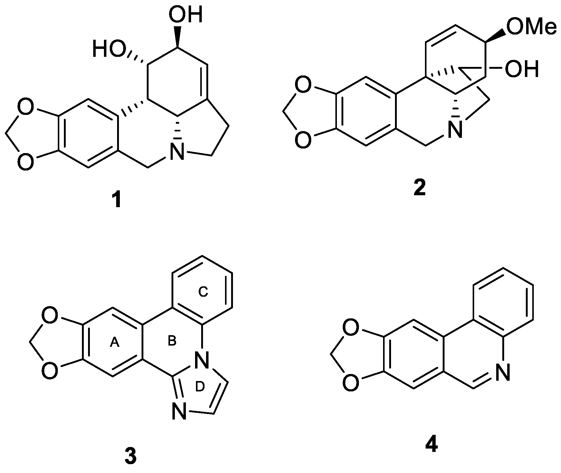
[1,3]Dioxolo[4,5-j]imidazo[1,2-f]phenanthridine (3, zephycandidine A). The synthesis was accomplished following General Procedure C using 8a as substituted phenanthridine. Eluent for FSC isohexane/ethyl acetate 1:1 to afford 3 as a colorless solid (98 mg, 0.37 mmol, 89%). m.p. 242-244 °C (lit. [4]: 242-243 °C, lit. [5]: 242-245 °C). 1H NMR (400 MHz, CDCl3) δ 8.24 (dd, J = 8.1, 1.4 Hz, 1H, 8-H), 8.02 (s, 1H, 9-H), 7.95 (d, J = 1.5 Hz, 1H, 2-H), 7.84 (dd, J = 8.2, 1.3 Hz, 1H, 5-H), 7.71 (s, 1H, 13-H), 7.60 – 7.54 (m, 2H, 3-H, 6-H), 7.48 (ddd, J = 8.4, 7.2, 1.3 Hz, 1H, 7-H), 6.13 (s, 2H, -OCH2O-). 13C NMR (101 MHz, CDCl3) δ 149.42 (C-9a), 148.76 (C-12), 142.63 (C-13b), 131.34 (C-2), 131.11 (C-4a), 128.01 (C-6), 124.96 (C-7), 123.83 (C-8), 123.31 (C-8b), 121.75 (C-13a), 119.50 (C-8a), 115.84 (C-5), 111.53 (C-3), 102.88 (C-9), 101.76 (-OCH2O-), 101.39 (C-13). IR (ATR): ṽmax/cm-1 = 2920, 1506, 1462, 1331, 1260, 1035, 848, 736 HRMS (EI): calcd. for C16H10N2O2 (M)•+: 262.0737 found: 262.0737. HPLC purity: >99%.
10,11-Dimethoxyimidazo[1,2-f]phenanthridine (9). The synthesis was accomplished following General Procedure C using 8b as substituted phenanthridine. Eluent for FSC ethyl acetate/triethylamine 99:1 to afford 9 as an off-white solid (99 mg, 0.36 mmol, 85%). m.p. 165-166 °C. 1H NMR (400 MHz, methylene chloride-d2) δ 8.34 (dd, J = 8.2, 1.4 Hz, 1H, 5-H), 8.01 – 7.98 (m, 2H, 3-H, 12-H), 7.88 (dd, J = 8.2, 1.3 Hz, 1H, 8-H), 7.71 (s, 1H, 2-H), 7.59 (ddd, J = 8.3, 7.2, 1.4 Hz, 1H, 6-H), 7.51 (s, 1H, 9-H), 7.51 (td, J = 7.0, 1.3 Hz, 1H, 7-H), 4.05 (s, 3H, OCH3), 4.04 (s, 3H, OCH3). 13C NMR (101 MHz, methylene chloride-d2) δ 151.22 (C-10 or C-11), 151.20 (C-10 or C-11), 142.94 (C-12b), 131.84 (C-4a), 131.59 (C-2), 128.36 (C-6), 125.36 (C-8), 124.15 (C-7), 122.12 (C-8b), 121.99 (C-8a), 118.61 (C-12a), 116.48 (C-5), 112.23 (C-3), 105.40 (C-12), 104.50 (C-9), 56.64 (-OCH3), 56.53 (-OCH3). IR (ATR): ṽmax/cm-1 = 2830, 1615, 1516, 1478, 1455, 1274, 1212, 1161, 1142, 1033, 1017, 854, 793, 753, 711. HRMS (EI): calcd. for C17H14N2O2 (M)•+: 278.1055 found: 278.1050. HPLC purity: >99%.
11-Methoxyimidazo[1,2-f]phenanthridine (10). The synthesis was accomplished following General Procedure C using 8c as substituted phenanthridine. Eluent for FSC ethyl acetate/triethylamine 99:1 to afford 10 as an off-white solid (85 mg, 0.34 mmol, 82%). m.p. 99°C. 1H NMR (400 MHz, methylene chloride-d2) δ 8.40 (dd, J = 8.1, 1.4 Hz, 1H, 9-H), 8.32 (d, J = 9.0 Hz, 1H, 5-H), 8.06 (d, J = 2.8 Hz, 1H, 2-H), 8.04 (d, J = 1.4 Hz, 1H, 3-H), 7.90 (dd, J = 8.2, 1.3 Hz, 1H, 8-H), 7.60 (ddd, J = 8.4, 7.2, 1.5 Hz, 1H, 6-H), 7.56 (d, J = 1.3 Hz, 1H, 12-H), 7.52 (ddd, J = 8.4, 7.2, 1.3 Hz, 1H, 7-H), 7.25 (dd, J = 9.0, 2.8 Hz, 1H, 10-H), 4.00 (s, 3H, -OCH3). 13C NMR (101 MHz, methylene chloride-d2) δ 160.02 (C-11), 142.03 (C-12b), 130.75 (C-4a or C-2), 130.70 (C-4a or C-2), 127.83 (C-7), 125.26 (C-6), 124.73 (C-12a), 124.26 (C-9), 123.62 (C-5), 121.86 (C-8a), 121.09 (C-8b), 118.38 (C-10), 115.83 (C-8), 112.35 (C-3), 105.19 (C-12), 55.76 (-OCH3).IR (ATR): ṽmax/cm-1 = 3434, 1617, 1472, 1325, 1291, 1036, 862, 750. HRMS (EI): calcd. for C16H12N2O (M)•+: 248.0944 found: 248.0943. HPLC purity: 99%.
11-Isopropoxy-10-methoxyimidazo[1,2-f]phenanthridine (11, THK-121). The synthesis was accomplished following General Procedure C using 8d as substituted phenanthridine. Eluent for FSC ethyl acetate/triethylamine 99:1 to afford 11 as an off-white solid (107 mg, 0.35 mmol, 83%). m.p. 73°C. 1H NMR (400 MHz, methylene chloride-d2) δ 8.36 (dd, J = 8.1, 1.5 Hz, 1H, 5-H), 8.03 (s, 1H, 12-H), 8.00 (d, J = 1.5 Hz, 1H, 3-H), 7.90 (dd, J = 8.2, 1.3 Hz, 1H, 8-H), 7.76 (s, 1H, 9-H), 7.60 (ddd, J = 8.3, 7.2, 1.4 Hz, 1H, 7-H), 7.54 – 7.50 (m, 2H, 2-H, 6-H), 4.87 (hept, 1H, 1’-H), 4.05 (s, 3H, -OCH3), 1.45 (d, J = 6.1 Hz, 6H, 2’-H). 13C NMR (101 MHz, methylene chloride-d2) δ 152.09 (C-10), 149.45 (C-11), 142.96 (C-12b), 131.83 (C-4a), 131.50 (C-2), 128.33 (C-6), 125.40 (C-8), 124.15 (C-7), 122.20 (C-8b), 121.87 (C-8a), 118.62 (C-12a), 116.50 (C-5), 112.23 (C-3), 108.01 (C-12), 105.03 (C-9), 71.56 (C-1’), 56.60 (-OCH3), 22.30 (C-2’). IR (ATR): ṽmax/cm-1 = 2976, 1614, 1514, 1453, 1384, 1263, 1209, 1107, 1022, 953, 924, 849, 798, 718. HRMS (EI): calcd. for C19H18N2O2 (M)•+: 306.1363 found: 306.1364. HPLC purity: >99%.
Benzo[h]imidazo[2,1-f][1,6]naphthyridine (12). The synthesis was accomplished following General Procedure C using 8e as substituted phenanthridine. Eluent for FSC ethyl acetate/ triethylamine 99:1 to afford 12 as an off-white solid (57 mg, 0.26 mmol, 62%). m.p. 253-255 °C. 1H NMR (400 MHz, methylene chloride-d2) δ 9.06 (dd, J = 8.1, 1.5 Hz, 1H, 5-H), 8.91 (dd, J = 4.5, 1.8 Hz, 1H, 10-H), 8.85 (dd, J = 8.0, 1.8 Hz, 1H, 12-H), 8.07 (d, J = 1.4 Hz, 1H, 3-H), 7.91 (dd, J = 8.3, 1.1 Hz, 1H, 8-H), 7.74 (ddd, J = 8.4, 7.1, 1.5 Hz, 1H, 6-H), 7.63 – 7.54 (m, 3H, 2-H, 7-H, 11-H). 13C NMR (101 MHz, methylene chloride-d2) δ 150.09 (C-10), 144.68 (C-8b), 141.38 (C-12b), 133.19 (C-4a), 131.93 (C-2), 131.37 (C-12), 130.48 (C-6), 126.07 (C-8), 125.32 (C-7), 123.35 (C-11), 122.73 (C-8a), 119.42 (C-12a), 115.30 (C-5), 112.61 (C-3). IR (ATR): ṽmax/cm-1 = 3319, 3148, 1653, 1581, 1479, 1472, 1395, 1295, 1080, 750, 726. HRMS (EI): calcd. for C14H9N3 (M)•+: 219.0791 found: 219.0790. HPLC purity: 98%.
3-Methyl-[1,3]dioxolo[4,5-j]imidazo[1,2-f]phenanthridine (13). The synthesis was accomplished following General Procedure D using propanal (23 µL, 0.30 mmol) as aldehyde. Eluent for FSC isohexane/ethyl acetate 1:1 to afford 13 as a colorless solid (29 mg, 0.11 mmol, 53%). m.p. 180-182 °C. 1H NMR (400 MHz, methylene chloride-d2) δ 8.34 (dd, J = 8.5, 1.3 Hz, 1H, 5-H), 8.30 (dd, J = 8.1, 1.6 Hz, 1H, 8-H), 7.97 (s, 1H, 13-H), 7.73 (s, 1H, 9-H), 7.56 (ddd, J = 8.5, 7.2, 1.6 Hz, 1H, 7-H or 6-H), 7.49 (ddd, J = 8.3, 7.2, 1.3 Hz, 1H, 7-H or 6-H), 7.21 (q, J = 1.1 Hz, 1H, 2-H), 6.12 (s, 2H, -OCH2O-), 2.91 (d, J = 1.1 Hz, 3H, -CH3). 13C NMR (101 MHz, methylene chloride-d2) δ 149.78 (C-9a or C-12a), 149.34 (C-9a or C-12a), 143.79 (C-13b), 134.26 (C-8a), 131.40 (C-2), 127.96 (C-6), 125.67 (C-3), 124.96 (C-7), 124.30 (C-8), 123.39 (C-8a), 122.94 (C-4a), 120.64 (C-13a), 116.98 (C-5), 102.94 (C-13), 102.46 (-OCH2O-), 101.60 (C-9), 15.64 (-CH3). IR (ATR): ṽmax/cm-1 = 2874, 1504, 1445, 1380, 1291, 1247, 1040, 871, 744. HRMS (EI): calcd. for C17H12N2O2 (M)•+: 276.0893 found: 276.0898. HPLC purity: 96%.
3-Isopropyl-[1,3]dioxolo[4,5-j]imidazo[1,2-f]phenanthridine (14). The synthesis was accomplished following General Procedure D using 3-methylbutanal (32 µL, 0.30 mmol) as aldehyde. Eluent for FSC isohexane/ethyl acetate 1:1 to afford 14 as a colorless solid (35 mg, 0.12 mmol, 58%). m.p. 170-171 °C. 1H NMR (400 MHz, methylene chloride-d2) δ 8.28 (ddd, J = 10.9, 8.3, 1.1 Hz, 2H, 5-H, 8-H), 7.99 (s, 1H, 13-H), 7.72 (s, 1H, 9-H), 7.57 (ddd, J = 8.5, 7.1, 1.6 Hz, 1H, 7-H), 7.48 (ddd, J = 8.1, 7.1, 1.2 Hz, 1H, 6-H), 7.31 (d, J = 1.0 Hz, 1H, 2-H), 6.11 (s, 2H, -OCH2O-), 3.79 (hept, J = 6.7 Hz, 1H, 1’-H), 1.49 (d, J = 6.6 Hz, 6H, 2’-H). 13C NMR (101 MHz, methylene chloride-d2) δ 149.24 (C-12a), 148.74 (C-9a), 143.53 (C-13b), 136.84 (C-3), 133.46 (C-4a), 127.79 (C-2), 127.40 (C-7), 124.34 (C-6), 123.78 (C-8), 122.89 (C-8b), 122.60 (C-13a), 120.17 (C-8a), 117.12 (C-5), 102.47 (C-13), 101.88 (-OCH2O-), 100.92 (C-9), 27.56 (C-1’), 22.48 (C-2’). IR (ATR): ṽmax/cm-1 = 1459, 1247, 1041, 871, 744 HRMS (EI): calcd. for C19H16N2O2 (M)•+: 304.1206 found: 304.1203. HPLC purity: 96%.
3-Phenyl-[1,3]dioxolo[4,5-j]imidazo[1,2-f]phenanthridine (15). The synthesis was accomplished following General Procedure D using phenylacetaldehyde (35 µL, 0.30 mmol) as aldehyde. Eluent for FSC isohexane/ethyl acetate 1:1 to afford 15 as a colorless solid (14 mg, 0.04 mmol, 21%). m.p. 232-233 °C. 1H NMR (400 MHz, methylene chloride-d2) δ 8.27 (dd, J = 8.2, 1.5 Hz, 1H, 5-H), 8.04 (s, 1H, 13-H), 7.77 (s, 1H, 9-H), 7.57 (dd, J = 8.5, 1.2 Hz, 1H, 8-H), 7.55 – 7.49 (m, 5H, 2’-h, 3’-H, 4’-H, 5’-H, 6’-H), 7.40 (ddd, J = 8.3, 7.1, 1.2 Hz, 1H, 7-H), 7.38 (s, 1H, 2-H), 7.23 – 7.18 (m, 1H, 6-H), 6.15 (s, 2H, -OCH2O-). 13C NMR (101 MHz, methylene chloride-d2) δ 149.50 (C-12a), 148.83 (C-9a), 143.74 (C-13b), 132.50 (C-3), 132.42 (C-2), 132.28 (C-1’), 129.87 (C-3’, C-5’), 129.46 (C-4’), 128.76 (C-2’, C-6’), 128.48 (C-4a), 126.89 (C-6), 124.60 (C-7), 123.76 (C-5), 123.47 (C-8a), 122.51 (C-13a), 119.87 (C-8b), 117.85 (C-8), 102.63 (C-13), 101.99 (-OCH2O-), 101.16 (C-9). IR (ATR): ṽmax/cm-1 = 2904, 1449, 1237, 1036, 910, 860, 767, 754, 748, 704. HRMS (EI): calcd. for C22H14N2O2 (M)•+: 338.1050 found: 338.1051. HPLC purity: 95%.

3.2. Biological Investigations

3.2.1. Cell Lines and Culture

CCRF-CEM cells were obtained from Prof. Maria Kavallaris [28] (University of New South Wales, Sydney, Australia), Jurkat, HL-60 and MCF7 were obtained from ATCC) and were cultured in RPMI 1640 (PAN Biotech, Aidenbach, Germany) containing 10% FCS (PAN Biotech). Human umbilical vein endothelial cells (HUVECs) were purchased from Promocell (Heidelberg, Germany) and cultivated with ECGM Kit enhanced (PELO Biotech, Planegg, Germany) supplemented with 10% (FCS) (PAA Laboratories, Cölbe, Germany) and 1% penicillin/streptomycin/amphotericin B (all purchased from PAN Biotech). All cells were cultured at 37 °C with 5% CO2 with constant humidity. Cell line STR profiling was performed. None of the cell lines used are listed in the database of commonly misidentified cell lines maintained by ICLAC. All cells are proven to be mycoplasma-free quarterly.

3.2.2. Functional Analysis of Biological Activity

All tested compounds were diluted in DMSO (Sigma Aldrich) to 10 mM stock solutions. In biological assays, final DMSO concentration did not exceed 0.1%. Cell proliferation was assessed using the CellTiter-Blue cell viability assay as described previously [29]. Apoptosis was assessed by flow cytometry according to Nicoletti et al. and analysis of protein level was analyzed by immunoblotting as described previously [29,30]. Mitochondrial membrane potential was assessed by flow cytometry and morphology was assessed by confocal microscopy as described previously [30].

3.2.3. Statistical Analysis

Experiments were carried out at least three times independently unless stated otherwise. Data represent mean ± standard deviation (SD) unless stated otherwise. Statistical significance was assessed by ordinary one-way ANOVA with Dunnet’s post-test using GraphPad Prism 8. Significance of differences in dose-response curves was analysed using the comparison of fits function of GraphPad Prism 8. Results were considered significant for p<0.05.

Supplementary Materials

The following supporting information can be downloaded at the website of this paper posted on Preprints.org. 1H and 13C NMR spectra of compounds 6a/b/d/e, 8a-e, 3, 9-15; list of IC50 values.

Author Contributions

Conceptualization, FB and KB; methodology, TK and FL; validation, FB, KB, TK, FL and FG; formal analysis, FB, KB, TK, FL and FG; investigation, TK, FL and JB; resources, FB and KB; data curation, FB, KB, TK, FL and FG; writing—original draft preparation, FB and KB; writing—review and editing, FB, KB and TK; visualization, TK and FL; supervision, FB and KB. All authors have read and agreed to the published version of the manuscript.

Funding

This research received no external funding.

Data Availability Statement

Experimental data, spectra, and protocols are stored in an electronic lab journal by the authors.

Acknowledgments

We thank Silvia Schnegg for technical support.

Conflicts of Interest

The authors declare no conflict of interest.

References

  1. Jin, Z. Amaryllidaceae and Sceletium alkaloids. Natural Product Reports 2009, 26, 363–381. [Google Scholar] [CrossRef] [PubMed]
  2. Zhan, G.; Qu, X.; Liu, J.; Tong, Q.; Zhou, J.; Sun, B.; Yao, G. Zephycandidine A, the First Naturally Occurring Imidazo[1,2-f]phenanthridine Alkaloid from Zephyranthes candida, Exhibits Significant Anti-tumor and Anti-acetylcholinesterase Activities. Sci. Rep. 2016, 6, 33990. [Google Scholar] [CrossRef] [PubMed]
  3. Zhan, G.; Liu, J.; Zhou, J.; Sun, B.; Aisa, H.A.; Yao, G. Amaryllidaceae alkaloids with new framework types from Zephyranthes candida as potent acetylcholinesterase inhibitors. Eur. J. Med. Chem. 2017, 127, 771–780. [Google Scholar] [CrossRef] [PubMed]
  4. Sheng, B.; Zeng, C.; Chen, J.; Ye, W.C.; Tang, W.P.; Lan, P.; Banwell, M.G. Total Syntheses of the Imidazo[1,2-f]phenanthridine-containing Alkaloid Zephycandidine A. Eur. J. Med. Chem. 2022, e202101511. [Google Scholar] [CrossRef]
  5. Murphy, P.J.; Tibble-Howlings, J.; Kowalczyk, R.M.; Stevens, K. Synthesis of zephycandidine A from haemanthamine. Tetrahedron Lett. 2020, 61, 151785. [Google Scholar] [CrossRef]
  6. Parenty, A.D.; Guthrie, K.M.; Song, Y.F.; Smith, L.V.; Burkholder, E.; Cronin, L. Discovery of an imidazo-phenanthridine synthon produced in a 'five-step one-pot reaction' leading to a new family of heterocycles with novel physical properties. Chem. Commun. 2006, 1194–1196. [Google Scholar] [CrossRef] [PubMed]
  7. Lee, S.W.; Dao, P.D.Q.; Lim, H.-J.; Cho, C.S. Synthesis of Imidazo[1,2-f]phenanthridines by Recyclable Magnetic MOF-Catalyzed Coupling and Cyclization of 2-(2-Bromoaryl)imidazoles with Cyclohexane-1,3-diones Followed by Aromatization. ACS Omega 2022, 7, 18486–18497. [Google Scholar] [CrossRef] [PubMed]
  8. Zhang, W.; Ma, J.; Liu, G.-J.; Liu, X.-Y.; Fan, J.; Liao, L.-S. Efficient sky-blue emitting Pt(ii) complexes based on imidazo[1,2-f]phenanthridine-containing tetradentate ligands. J. Mater. Chem. C 2017, 5, 9496–9503. [Google Scholar] [CrossRef]
  9. Mitsuharu, N.; Yumi, S.; Izumi, K.; Yasuyuki, G. US 2012261651 A1, 2012.
  10. Narva, S.; Zhang, M.; Li, C.; Chen, A.; Tanaka, Y.; Zhang, W. Synthesis of imidazo[1,2-f]phenanthridine derivatives under a metal- and base-free condition and their anticancer activity. Tetrahedron Lett. 2021, 68, 152908. [Google Scholar] [CrossRef]
  11. Knowles, D.B.; Lin, C.; MacKenzie, P.B.; Tsai, J.-Y.; Walters, R.W.; Beers, S.A.; Brown, C.S.; Yeager, W.H. METAL COMPLEXES OF CYCLOMETALLATED IMIDAZO[l,2-flPHENANTHRIDINE AND DIIMID AZO[l,2-A:l’,2’-C]QUIN AZOLINE LIGANDS AND ISOELECTRONIC AND BENZANNULATED ANALOGS THEREOF. US 2007/0190359 A1, 2007.
  12. Augustine, J.K.; Bombrun, A.; Atta, R.N. A Practical and Cost-Efficient,One-Pot Conversion of Aldehydes into Nitriles Mediated by ‘ActivatedDMSO’. Synlett 2011, 2223–2227. [Google Scholar] [CrossRef]
  13. Weniger, B.; Italiano, L.; Beck, J.P.; Bastida, J.; Bergonon, S.; Codina, C.; Lobstein, A.; Anton, R. Cytotoxic activity of Amaryllidaceae alkaloids. Planta Med 1995, 61, 77–79. [Google Scholar] [CrossRef]
  14. Lamoral-Theys, D.; Andolfi, A.; Van Goietsenoven, G.; Cimmino, A.; Le Calve, B.; Wauthoz, N.; Megalizzi, V.; Gras, T.; Bruyere, C.; Dubois, J.; Mathieu, V.; Kornienko, A.; Kiss, R.; Evidente, A. Lycorine, the main phenanthridine Amaryllidaceae alkaloid, exhibits significant antitumor activity in cancer cells that display resistance to proapoptotic stimuli: an investigation of structure-activity relationship and mechanistic insight. J Med Chem 2009, 52, 6244–6256. [Google Scholar] [CrossRef] [PubMed]
  15. Evidente, A.; Kireev, A.S.; Jenkins, A.R.; Romero, A.E.; Steelant, W.F.; Van Slambrouck, S.; Kornienko, A. Biological evaluation of structurally diverse amaryllidaceae alkaloids and their synthetic derivatives: discovery of novel leads for anticancer drug design. Planta Med 2009, 75, 501–507. [Google Scholar] [CrossRef] [PubMed]
  16. Van Goietsenoven, G.; Andolfi, A.; Lallemand, B.; Cimmino, A.; Lamoral-Theys, D.; Gras, T.; Abou-Donia, A.; Dubois, J.; Lefranc, F.; Mathieu, V.; Kornienko, A.; Kiss, R.; Evidente, A. Amaryllidaceae alkaloids belonging to different structural subgroups display activity against apoptosis-resistant cancer cells. J Nat Prod 2010, 73, 1223–1227. [Google Scholar] [CrossRef] [PubMed]
  17. Brentnall, M.; Rodriguez-Menocal, L.; De Guevara, R.L.; Cepero, E.; Boise, L.H. Caspase-9, caspase-3 and caspase-7 have distinct roles during intrinsic apoptosis. BMC Cell Biol 2013, 14, 32. [Google Scholar] [CrossRef] [PubMed]
  18. Sairanen, T.; Szepesi, R.; Karjalainen-Lindsberg, M.L.; Saksi, J.; Paetau, A.; Lindsberg, P.J. Neuronal caspase-3 and PARP-1 correlate differentially with apoptosis and necrosis in ischemic human stroke. Acta Neuropathol 2009, 118, 541–552. [Google Scholar] [CrossRef] [PubMed]
  19. Buytaert, E.; Callewaert, G.; Vandenheede, J.R.; Agostinis, P. Deficiency in apoptotic effectors Bax and Bak reveals an autophagic cell death pathway initiated by photodamage to the endoplasmic reticulum. Autophagy 2006, 2, 238–240. [Google Scholar] [CrossRef] [PubMed]
  20. Michels, J.; Johnson, P.W.; Packham, G. Mcl-1. Int J Biochem Cell Biol 2005, 37, 267–271. [Google Scholar] [CrossRef] [PubMed]
  21. Picard, M.; Shirihai, O.S.; Gentil, B.J.; Burelle, Y. Mitochondrial morphology transitions and functions: implications for retrograde signaling? Am J Physiol Regul Integr Comp Physiol 2013, 304, R393–R406. [Google Scholar] [CrossRef]
  22. Hoppins, S.; Lackner, L.; Nunnari, J. The machines that divide and fuse mitochondria. Annu Rev Biochem 2007, 76, 751–780. [Google Scholar] [CrossRef] [PubMed]
  23. Srinivasan, S.; Guha, M.; Kashina, A.; Avadhani, N.G. Mitochondrial dysfunction and mitochondrial dynamics-The cancer connection. Biochim Biophys Acta Bioenerg 2017, 1858, 602–614. [Google Scholar] [CrossRef]
  24. Wu, S.; Zhou, F.; Zhang, Z.; Xing, D. Mitochondrial oxidative stress causes mitochondrial fragmentation via differential modulation of mitochondrial fission-fusion proteins. FEBS J 2011, 278, 941–954. [Google Scholar] [CrossRef] [PubMed]
  25. Liu, X.; Hajnoczky, G. Altered fusion dynamics underlie unique morphological changes in mitochondria during hypoxia-reoxygenation stress. Cell Death Differ 2011, 18, 1561–1572. [Google Scholar] [CrossRef]
  26. Kogot-Levin, A.; Saada, A.; Leibowitz, G.; Soiferman, D.; Douiev, L.; Raz, I.; Weksler-Zangen, S. Upregulation of Mitochondrial Content in Cytochrome c Oxidase Deficient Fibroblasts. PLoS One 2016, 11, e0165417. [Google Scholar] [CrossRef] [PubMed]
  27. Poole, L.P.; Macleod, K.F. Mitophagy in tumorigenesis and metastasis. Cell Mol Life Sci 2021, 78, 3817–3851. [Google Scholar] [CrossRef]
  28. Kavallaris, M.; Madafiglio, J.; Norris, M.D.; Haber, M. Resistance to tetracycline, a hydrophilic antibiotic, is mediated by P-glycoprotein in human multidrug-resistant cells. Biochem. Biophys. Res. Commun. 1993, 190, 79–85. [Google Scholar] [CrossRef] [PubMed]
  29. Geisslinger, F.; Müller, M.; Chao, Y.K.; Grimm, C.; Vollmar, A.M.; Bartel, K. Targeting TPC2 sensitizes acute lymphoblastic leukemia cells to chemotherapeutics by impairing lysosomal function. Cell Death Dis. 2022, 13, 668. [Google Scholar] [CrossRef]
  30. Siow, W.X.; Kabiri, Y.; Tang, R.; Chao, Y.K.; Plesch, E.; Eberhagen, C.; Flenkenthaler, F.; Fröhlich, T.; Bracher, F.; Grimm, C.; Biel, M.; Zischka, H.; Vollmar, A.M.; Bartel, K. Lysosomal TRPML1 regulates mitochondrial function in hepatocellular carcinoma cells. J. Cell Sci. 2022, 135, jcs259455. [Google Scholar] [CrossRef] [PubMed]
Figure 1. Phenanthridine-type alkaloids from Amaryllidaceae: lycorine (1), haemanthamine (2), and zephycandidine A (3). Phenanthridine building block 4 for the first partial synthesis of zephycandidine A (3).
Preprints 146027 g001
Scheme 1. General route for the synthesis of zephycandidine A (3) and ring A-modified analogues (1012) synthesized following the same protocol via the corresponding (hetero)aromatic ortho-bromonitriles 6. Reagents and conditions: a) H2NOH-HCl, DMSO, 90 °C, 1 h; b) cat. PdCl2(Ph3P)2, DMF, aq. Na2CO3, 80 °C, 16 h; c) chloroacetaldehyde, Na2CO3, 2-propanol-water, reflux, 1 h.
Preprints 146027 sch001
Scheme 2. Sulfur-endorsed construction of substituted ring D yielding products 13 - 15. Reagents and conditions: a) sulfur, DMSO/cyclohexane, 120 °C.
Preprints 146027 sch002
Figure 2. Zephycandidine A analogues decrease proliferation and induce cell death. (A-E) Proliferation was determined in Jurkat (A), MCF-7 (B), CCRF-CEM (C), HL-60 (D), and HUVEC (E) cells by CellTiter®-Blue cell viability assay. Cells were treated as indicated for 72 h. Data are mean ± SD (n=3). (F-I) Cell Death at IC50 values determined in (A-E) was assessed after 48 h by forward versus sideward (FSC vs. SSC) scatter gating for Jurkat (F), HL-60 (H), and CCRF-CEM (I) cells and by Nicoletti assay for MCF-7 (G). Bar graphs display mean ± SD (n=3), One-way ANOVA followed by Tukey’s multiple comparison test referring to the control.
Preprints 146027 g002
Figure 3. Zephycandidine A analogues induce the mitochondrial apoptosis pathway.(A) PARP and active caspase-3 protein level in Jurkat cells was analyzed by Western Blot after 24 h of treatment. One representative Western Blot is shown. (B, C) Mcl-1 and Bax protein level in Jurkat (B) and MCF-7 (C) cells analyzed by Western blotting after 24 h of treatment. One representative Western Blot is shown. (D, E) Jurkat (D) and MCF-7 (E) cells were treated as indicated for 24 h followed by JC-1 staining. Loss of mitochondrial membrane potential was quantified by determining the percentage of green fluorescence caused by JC-1 monomers via flow cytometry. (F, G) Jurkat (F) and MCF-7 (G) cells were treated as indicated for 24 h followed by MitoSOXTM Red Superoxide Indicator staining. Mitochondrial superoxide was determined by flow cytometry analysis and displayed as geometric mean of fluorescent intensity (gMFI). Bar graphs display mean ± SD (n=3), One-way ANOVA followed by Tukey’s multiple comparison test referring to the control.
Preprints 146027 g003
Figure 4. Zephycandidine A analogues alter mitochondrial morphology and increase mitochondrial mass.(A) MCF-7 cells were treated at the indicated concentrations for 24 h before staining with MitoTrackerTM Red CMXRos (mitochondria) and Hoechst 33342 (nuclei). Mitochondrial morphology was assessed using confocal imaging. Scale bar: 25 µM. One representative image is shown. In addition, ImageJ plugin MiNA analysis output image is depicted. Purple area represents mitochondrial signal. Green lines represent length of mitochondria. Blue dots mark connection sites between mitochondria and yellow dots mark the end of network structures. (B) Results from MiNA analysis of (A) performed on 10 cells per replicate. (C, D) Jurkat (C) and MCF-7 (D) cells were treated as indicated for 24 h followed by MitoTrackerTM Green FM staining. Mitochondrial mass was determined by flow cytometry analysis and displayed as geometric mean of fluorescent intensity (gMFI). Bar graphs display mean ± SD (n=3), One-way ANOVA followed by Tukey’s multiple comparison test referring to the control.
Preprints 146027 g004
Disclaimer/Publisher’s Note: The statements, opinions and data contained in all publications are solely those of the individual author(s) and contributor(s) and not of MDPI and/or the editor(s). MDPI and/or the editor(s) disclaim responsibility for any injury to people or property resulting from any ideas, methods, instructions or products referred to in the content.
Copyright: This open access article is published under a Creative Commons CC BY 4.0 license, which permit the free download, distribution, and reuse, provided that the author and preprint are cited in any reuse.
Prerpints.org logo

Preprints.org is a free preprint server supported by MDPI in Basel, Switzerland.

Subscribe

Disclaimer

Terms of Use

Privacy Policy

Privacy Settings

© 2025 MDPI (Basel, Switzerland) unless otherwise stated