
Submitted:
07 January 2025
Posted:
07 January 2025
You are already at the latest version
Abstract
Aeromonas veronii, Aeromonas hydrophila, Plesiomonas shigelloides, and Citrobacter freundii were opportunistic pathogen widely distributed in water environment and fish population, causing fish diseases under stressful conditions. In this study, bacteria were isolated from diseased common carp Cyprinus carpio with symptoms of hemorrhage on body surface, abdominal distention, and flatulence in the intestine. According to the results of biochemical characteristics, 16S rRNA sequencing analyses, the isolates were identified as A. veronii, A. hydrophila, P. shigelloides, and C. freundii. The experimentally infected fish showed identical symptoms as observed in the naturally infected common carp. The LD50 of C. freundii, P. shigelloides, A. veronii, A. hydrophila, and mixture group were 1.95×10⁴, 4.74×10⁴, 5.12×10⁴, 1.53×10⁵, and 5.41×10⁴ respectively. Antibiotic resistance results showed that P. shigelloides, A. veronii, and A. hydrophila were sensitive to streptomycin, enrofloxacin, florfenicol, gentamicin, kanamycin, neomycin, norfloxacin, co-trimoxazole, ceftizoxime, and resistant to ampicillin. C. freundii was sensitive to streptomycin, gentamicin, and ceftizoxime, moderately susceptible to kanamycin, neomycin, norfloxacin, and ampicillin, and resistant to enrofloxacin, florfenicol, tetracycline, and co-trimoxazole.
Keywords:
aquaculture
; resident bacteria
; challenge experiment
; opportunistic infection
; antibiotic resistance
1. Introduction
Common carp (Cyprinus carpio) is a world-widely cultured fish, which is also one of the most important economic fish species in China [1]. The total production of farmed C. carpio was 2.87 million tons in 2023 according to the China Fishery Statistical Yearbook. However, improper management and high-density culture creates ideal conditions for explosive epidemics of diseases [2]. It has been reported that C. carpio breeding industry was threatened by a variety of bacterial organisms, such as Aeromonas hydrophila, Pseudomonas fluorescens, Pseudomonas alcaligenes, and Shewanella putrefaciens [3,4,5,6]. The current key issues that need to be solved urgently are identifying the most commonly disease-causing bacteria in common carp and finding out effective medicine for treatment.
Aeromonas veronii is Gram-negative bacterium that widely distributed in water environment and can cause ulcerative diseases in many kinds of fish [7,8,9,10,11,12,13,14]. The main clinical symptoms of A. veronii were hemorrhagic septicemia, fin rot, exophthalmia, and abdominal distention [14]. A. hydrophila is another number of Aeromonas species that commonly infect aquatic animals. A. hydrophila caused hemorrhagic septicemia and lead to massive mortalities in fishes in previous reports [15,16,17,18,19]. Plesiomonas shigelloides and Citrobacter freundii were both classified into the Enterobacteriaceae family. P. shigelloides was reported to be pathogenic and the diseased fish showed protruding eyeballs, swollen anus, and ascites in the abdominal cavity, swelling and hemorrhage [20,21,22,23,24]. C. freundii infection was associated with enteritis, necrosis, body reddening, hemorrhage, and septicemia [25,26,27,28,29,30]. The four kinds of bacteria are all opportunistic pathogen and can be commonly found in normal aquaculture water and healthy fish. They are more likely to cause diseases when fish under stresses, such as temperature change, hypoxia, and parasite infection [31,32].
Here, we report a case of bacterial disease naturally occurred in common carp that rearing in the aquaculture base of Henan Normal University in China. The infected fish showed obvious hemorrhage on the body surface, extended abdomen, and flatulence in the intestine. Bacteria were isolated from the ascites and were identified as A. veronii, A. hydrophila, P. shigelloides, and C. freundii by 16S rRNA sequencing and biochemical tests. The drug resistance and pathogenicity of four types of bacteria were also analyzed to provide reference for the prevention and treatment measures of similar cases.
2. Materials and Methods
2.1. Fish
The diseased common carp (18.3±2.6 g) was from the aquaculture base of Henan Normal University in Xinxiang city, Henan province, China. The fish were kept in a concrete pond with water depth of 1.5 m. Moribund fish were transported immediately to the laboratory for diagnosis and pathogen isolation. Healthy common carp (16±2.1 g) with no history of disease were provided by a local fish farm in Xinxiang city. Healthy fish were acclimated in aquaria for two weeks prior to infection assay and fed with commercial fed once daily. The water was replaced daily, and water temperature was maintained at 28 ± 1℃. Experiments involving live fish were conducted in accordance with the US National Research Council’s “Guide for the Care and Use of Laboratory Animals”.
2.2. Isolation, Characterization, and Identification of Bacteria
The ascites of moribund fish were collected and streaked on Luria-Bertani (LB) agar plates with inoculating loop. The plates were incubated at 28℃ for 24 h. The dominant colonies from plates were re-streaked on the LB agar plates three times to obtain pure culture. Total genomic DNA of the isolate was extracted by bacteria DNA extraction kits (Sangon biotech, China). Primers 16S rRNA-F (5′-AGAGTTTGATCCTGGCTCAG-3′) and 16S rRNA-R (5′-GGTTACCTTGTTACGACTT-3′) were used for PCR amplification of 16S rRNA. The PCR amplification conditions of 16S rRNA were initial denaturation at 94℃ for 5 min, followed by 32 cycles of denaturation at 94℃ for 30 s, annealing at 57℃ for 30 s, extension at 72℃ for 1.5 min and a final extension at 72℃ for 10 min. The genes obtained by PCR amplification were cloned to pMD-19 T vector (TakaRa, Japan) and sequenced. The biochemical characteristics were examined using commercial reagent (Hangzhou Microorganism Reagent Co., Ltd., China), and following incubated at 28℃ for 24 h or 48 h. All biochemical tests were repeated for three times, which were performed including oxidase, growth condition at 1% of NaCl, serine dihydrolase, lysine decarboxylase, Voges–Proskauer, indole production, sucrose, lactose, citrate, cellobiose, mannitol, salicin, etc.
2.3. Antibiotic Susceptibility Testing
The antibiotic susceptibility of aeromonad was determined by the Kirby-Bauer disk diffusion method[33]. Bacterial strains were streaked on Muller Hilton agar plates, and the various antibiotic disks (Difco Laboratories, USA) were applied on the streaked cultures. Disks of gentamycin (10 μg), kanamycin (30 μg), neomycin (30 μg), ceftizoxime (30 μg), florfenicol (30 μg), ampicillin (10 μg), trimethoprim-sulfamethoxazole (23.75/1.25 μg), tetracycline (30 μg), streptomycin (10 μg), norfloxacin (10 μg), and enrofloxacin (10 μg) were used. After 18 h of incubation at 28℃, the sizes of the zone of bacterial growth inhibition were measured. The isolates were classified as sensitive (S), moderately sensitive (M) or resistant (R) according to the National Committee for Clinical Laboratory Standards.
2.4. Experimental Infections
The infection assay was carried out in healthy common carp. The bacterial concentration determined by plating 10 -fold serial dilutions onto LB agar plates. The median lethal dosage (LD50) was calculated based on the total cumulative mortality (%) as described by Reed and Muench (1938) [34]. For LD50 determination, 240 carp individuals were randomly divided into six groups, including five infected groups and one control group. Infected groups (40 fish per group) were intraperitoneally injected with A. veronii, A. hydrophila, P. shigelloides, C. freundii and a mixture of four kinds of bacteria (equal numbers of each kind) at the concentration of 1.0 × 10⁴, 1.0 × 10⁵, 1.0 × 10⁶ and 1.0 × 10⁷ colony-forming unit (CFU)/fish (10 fish injected for each concentration), respectively. The control group was injected with the same dose of sterile physiological saline. The experiment was repeated once. The mortalities and clinical signs of all the groups were recorded every day for 15 d post-infection. Morbid fish were subjected to routine bacteriological examination for re-isolation and re-identification of the organism.
3. Results
3.1. Clinical Signs and Isolation of Bacteria from Diseased Common Carp
The clinical signs of diseased fish showed hemorrhage on gill cover and lower jaw, extended abdomen, swollen anus, ascites, and intestinal flatulence (Figure 1). The bacteria isolated from the diseased common carp were Gram negative. Colonies were buff, translucent, circular, convex and intact edge on LB. Ten colonies were selected and 16S rRNA were obtained and sequenced, the results showed that five strains of the isolates were A. veronii, two strains were P. shigelloides, two strains were C. freundii, and one strain was A. hydrophila.
3.2. Biochemical Characteristics
The isolates of A. veronii and A. hydrophila had positive biochemical reactions for indole, Voges–Proskauer, arginine dihydrolase, lysine, glucose, sucrose, and mannose, but negative for inositol. A. hydrophila could utilize arabinose, but could not A. veronii. Both A. veronii and A. hydrophila were able to grow in peptone water under 8% NaCI. C. freundii made utilization of citrate, glucose, raffinose, sorbitol, xylose, and urea, while P. shigelloides was incapable of metabolizing those molecules. P. shigelloides produced indole, lysine decarboxylase, and ornithine decarboxylase. Both C. freundii and P. shigelloides possessed motility (Table 1).
3.3. Experimental Infections
To confirm the pathogenicity of the four types of bacteria, the challenge assay was carried out in healthy common carp. Fish were intraperitoneally injected with different dose of each bacteria or a mixture of four bacteria. The experimentally infected fish showed identical symptoms as observed in the naturally infected common carp, which showed hemorrhage on body surface, abdominal distention, ascites, and flatulence in the intestine (Figure 2). The LD50 of C. freundii, P. shigelloides, A. veronii, and A. hydrophila were 1.95×10⁴, 4.74×10⁴, 5.12×10⁴, and 1.53×10⁵ respectively. The LD50 of mixture group was 5.41×10⁴. There were no clinical symptoms or death in the control group (Table 2). Furthermore, four kinds of bacteria were re-isolated from the experimental infected fish, as confirmed by colonial morphology observation, physiological and biochemical characteristics analyses, and 16S rRNA sequencing analyses.
3.4. Determination of Antimicrobial Resistance
The antibiotic resistance patterns of four bacteria were valued by the size of the inhibition zones around each disc, showed that P. shigelloides, A. veronii, and A. hydrophila were sensitive to streptomycin, enrofloxacin, florfenicol, gentamicin, kanamycin, neomycin, norfloxacin, co-trimoxazole, ceftizoxime, and resistant to ampicillin. C. freundii was sensitive to streptomycin, gentamicin, and ceftizoxime, moderately susceptible to kanamycin, neomycin, norfloxacin, and ampicillin, and resistant to enrofloxacin, florfenicol, tetracycline, and co-trimoxazole (Table 3).
4. Discussion
A. veronii, A. hydrophila, C. freundii, and P. shigelloides were all opportunistic pathogen widely distributed in water environment and fish population, causing fish diseases under stressful conditions, such as overcrowding, low oxygen concentration, and high water temperature [35]. The disease occurred in this case maybe because of the high water temperature and overfeeding, as the fish stop dying after stop feeding. C. freundii and P. shigelloides had been reported to cause severe hemorrhagic septicemia in internal organs of Chinese sturgeons that cultured in a research Institute [36], and co-infection by Aeromonas bestiarum, Aeromonas sobria, and P. shigelloides was observed in Mexican golden trout, leading to red and inflamed lesions in the abdomen and mouth, and hemorrhage in fins and gills [31]. However, it’s the first time that co-infection of A. veronii, A. hydrophila, C. freundii, and P. shigelloides in common carp was reported. Experimental infections showed that fish injected with four strains of bacteria all developed the similar clinical symptoms, which were hemorrhage, abdominal distention, ascites, and flatulence in the intestine (Figure 2), the same with the sign in naturally infected fish (Figure 1). So it was difficult to identify which kind of bacteria was the main cause of the disease from symptoms. But since half of the ten isolated strains were A. veronii, and the LD50 of mixture group was close to that of A. veronii group, it was most likely that A. veronii were the main pathogen.
Biochemical Characteristics of four bacteria were in accordance with the description of A. veronii, A. hydrophila, C. freundii, and P. shigelloides in Bergey’s Manual [37]. A. veronii, A. hydrophila, and C. freundii all can utilize glucose to produce gas (Table 1), that explained why the intestine of diseased fish was filled with gas (Figure 2). The flatulence was more obvious in the mixed infection group, indicating more gas was produced in mixed infection than that of single bacterial infection. The LD50 determination showed that C. freundii was the most virulent bacteria, and the virulence of A. hydrophila was the weakest. However, the mixed infection didn’t enhance the bacterial virulence, and the virulence of mixed bacteria was equivalent to that of A. veronii (Table 2).
Antimicrobial susceptibility testing indicated that the isolates of P. shigelloides, A. veronii, and A. hydrophila were susceptible to streptomycin, enrofloxacin, florfenicol, gentamicin, kanamycin, neomycin, norfloxacin, co-trimoxazole, cefazoxime, and resistant to ampicillin (Table 3). Simillarly, P. shigelloides isolated from Chinese sturgeon was sensitive to gentamicin and neomycin, and resistant to norfloxacin and ampicillin [36]. In our case, C. freundii was sensitive to streptomycin, gentamicin, and ceftizoxime, moderately susceptible to kanamycin, neomycin, norfloxacin, and ampicillin, and resistant to enrofloxacin, florfenicol, tetracycline, and co-trimoxazole (Table 3). In previous study, C. freundii isolated from largemouth bass was sensitive to gentamicin, norfloxacin, tetracycline, and co-trimoxazole, but resistant to kanamycin. C. freundii from angelfish was sensitive to tetracycline, resistant to enrofloxacin, florfenicol, and ampicillin. It seems that C. freundii strains from different fish showed different drug-resistant characterizations. The susceptibility test is essential for the drug selection in treatment.
5. Conclusions
In this study, we isolated A. veronii, A. hydrophila, C. freundii, and P. shigelloides from diseased common carp. The biological characteristics of four types of bacteria were determined. Artificial infection in fish showed four strains of bacteria were all highly pathogenic and could cause the disease with hemorrhage and abdominal distention syndrome, indicating the naturally infected fish maybe died of mixed infection. The drug sensitivity of the bacteria was also tested. The findings in this study will provide reference for the diagnosis, treatment, and prevention of the disease caused by mixed bacterial infection.
Author Contributions
Chao Pei: Formal analysis, Funding acquisition, Methodology, Software, Supervision, Validation, Writing; Jinghang Zhang: Conceptualization, Data curation, Investigation, Project administration, Software, Visualization; Dan Qiao: Conceptualization, Data curation, Investigation, Software, Visualization; Haoyu Wang: Data curation, Investigation; Xianliang Zhao: Formal analysis, Validation; Xinyu Jiang: Formal analysis, Validation; Lei Zhu: Formal analysis, Validation; Jie Zhang: Formal analysis, Validation; Li Li: Formal analysis, Validation; Xianghui Kong: Conceptualization, Resources, Supervision. All authors have read and agreed to the published version of the manuscript.
Funding
This work was sponsored by the Key scientific research projects of colleges and universities in Henan Province (22A240002).
Institutional Review Board Statement
This study was approved by the Laboratory Animal Ethics Committee of the College of Fisheries of Henan Normal University (Approved ID: 2023081101).
Informed Consent Statement
Not applicable.
Data Availability Statement
Data will be made available by the authors upon request.
Conflicts of Interest
The authors declare no conflicts of interest.
References
- Song, D.; Yun, Y.; He, Z.; Mi, J.; Wang, L.; Jin, M.; Zhou, Q.; Nie, G. Fillet texture, physicochemical indexes, muscle cellularity and molecular expression in muscle of Yellow River carp (Cyprinus carpio haematopterus) in response to dietary hydroxyproline supplementation. Aquaculture 2022, 549, 737783. [Google Scholar] [CrossRef]
- Qiao, D.; Yan, Y.; Pei, C.; Zhang, J.; Zhao, X.; Jiang, X.; Zhu, L.; Zhang, J.; Li, L.; Kong, X. Characterization of hepcidin gene and protection of recombinant hepcidin supplemented in feed against Aeromonas hydrophila infection in Yellow River carp (Cyprinus carpio haematopterus). Fish & Shellfish Immunol. 2023, 139, 108872. [Google Scholar] [CrossRef]
- Attia, M.M.; Sherif, A.H.; Abdelsalam, M.; Elgendy, M.Y. Dactylogyrus extensus and Pseudomonas fluorescens dual infection in farmed common carp (Cyprinus carpio). Microb. Pathog. 2022, 173, 105867. [Google Scholar] [CrossRef]
- Bai, J.; Huo, Y.; Hu, X.; Lu, A.; Sun, J. Characterization of pathogenic Pseudomonas alcaligenes isolated from Koi Carp in China. J. Aquat. Anim. Health. 2021, 33, 243–251. [Google Scholar] [CrossRef] [PubMed]
- Pazdzior, E.; Pekala-Safinska, A.; Wasyl, D. Phenotypic diversity and potential virulence factors of the Shewanella putrefaciens group isolated from freshwater fish. J. Vet. Res. 2019, 63, 321–332. [Google Scholar] [CrossRef] [PubMed]
- Zhao, X.; Jin, Z.; Di, G.; Li, L.; Kong, X. Molecular characteristics, pathogenicity and medication regimen of Aeromonas hydrophila isolated from common carp (Cyprinus carpio L.). J. Vet. Med. Sci. 2019, 81, 1769–1775. [Google Scholar] [CrossRef]
- Amal, M.N.A.; Koh, C.B.; Nurliyana, M.; Suhaiba, M.; Nor-Amalina, Z.; Santha, S.; Diyana-Nadhirah, K.P.; Yusof, M.T.; Ina-Salwany, M.Y.; Zamri-Saad, M. A case of natural co-infection of Tilapia Lake Virus and Aeromonas veronii in a Malaysian red hybrid tilapia (Oreochromis niloticus x O. mossambicus) farm experiencing high mortality. Aquaculture 2018, 485, 12–16. [Google Scholar] [CrossRef]
- Chen, F.; Sun, J.; Han, Z.; Yang, X.; Xian, J.-a.; Lv, A.; Hu, X.; Shi, H. Isolation, identification and characteristics of Aeromonas veronii from diseased Crucian Carp (Carassius auratus gibelio). Front. Microbiol. 2019, 10, 2742. [Google Scholar] [CrossRef] [PubMed]
- Dong, H.T.; Techatanakitarnan, C.; Jindakittikul, P.; Thaiprayoon, A.; Taengphu, S.; Charoensapsri, W.; Khunrae, P.; Rattanarojpong, T.; Senapin, S. Aeromonas jandaei and Aeromonas veronii caused disease and mortality in Nile tilapia, Oreochromis niloticus (L.). J. Fish Dis. 2017, 40, 1395–1403. [Google Scholar] [CrossRef]
- Pei, C.; Song, H.; Zhu, L.; Qiao, D.; Yan, Y.; Li, L.; Zhao, X.; Zhang, J.; Jiang, X.; Kong, X. Identification of Aeromonas veronii isolated from largemouth bass (Micropterus salmoides) and histopathological analysis. Aquaculture 2021, 540, 736707. [Google Scholar] [CrossRef]
- Raj, N.S.; Swaminathan, T.R.; Dharmaratnam, A.; Raja, S.A.; Ramraj, D.; Lal, K.K. Aeromonas veronii caused bilateral exophthalmia and mass mortality in cultured Nile tilapia (Oreochromis niloticus) (L.) in India. Aquaculture 2019, 512, 734278. [Google Scholar] [CrossRef]
- Sun, J.; Zhang, X.; Gao, X.; Jiang, Q.; Wen, Y.; Lin, L. Characterization of virulence properties of Aeromonas veronii isolated from diseased Gibel Carp (Carassius gibelio). Int. J. Mol. Sci. 2016, 17, 496. [Google Scholar] [CrossRef]
- Hohai, T.D.; Trang, T. T.; Van, T.N.; Giang, N.T.H.; Van, K.V. Aeromonas veronii caused disease and mortality in channel catfish in Vietnam. Aquaculture 2019, 513, 734425. [Google Scholar] [CrossRef]
- Zhu, M.; Wang, X.; Li, J.; Li, G.; Liu, Z.; Mo, Z. Identification and virulence properties of Aeromonas veronii bv.sobria isolates causing an ulcerative syndrome of loach (Misgurnus anguillicaudatus). J. Fish Dis. 2016, 39, 777–781. [Google Scholar] [CrossRef]
- Kong, X.; Qiao, D.; Zhao, X.; Wang, L.; Zhang, J.; Liu, D.; Zhang, H. The molecular characterizations of Cu/ZnSOD and MnSOD and its responses of mRNA expression and enzyme activity to Aeromonas hydrophila or lipopolysaccharide challenge in Qihe crucian carp (Carassius auratus). Fish. Shellfish. Immunol. 2017, 67, 429–440. [Google Scholar] [CrossRef]
- Pachanawan, A.; Phumkhachorn, P.; Rattanachaikunsopon, P. Potential of Psidium guajava supplemented fish diets in controlling Aeromonas hydrophila infection in Tilapia (Oreochromis niloticus). J. Biosci. Bioeng. 2008, 106, 419–424. [Google Scholar] [CrossRef]
- Peatman, E.; Mohammed, H.; Kirby, A.; Shoemaker, C.A.; Yildirim-Aksoy, M.; Beck, B.H. Mechanisms of pathogen virulence and host susceptibility in virulent Aeromonas hydrophila infections of channel catfish (Ictalurus punctatus). Aquaculture 2018, 482, 1–8. [Google Scholar] [CrossRef]
- Wang, M.; Zhao, X.; Kong, X.; Wang, L.; Jiao, D.; Zhang, H. Molecular characterization and expressing analysis of the c-type and g-type lysozymes in Qihe crucian carp (Carassius auratus). Fish. Shellfish. Immunol. 2016, 52, 210–220. [Google Scholar] [CrossRef]
- Mao, W.; Wang, Y.; Wang, W.; Wu, B.; Feng, J.; Zhu, Z. Enhanced resistance to Aeromonas hydrophila infection and enhanced phagocytic activities in human lactoferrin-transgenic grass carp (Ctenopharyngodon idellus). Aquaculture 2004, 242, 93–103. [Google Scholar] [CrossRef]
- Behera, B.K.; Bera, A.K.; Paria, P.; Das, A.; Parida, P.K.; Kumari, S.; Bhowmick, S.; Das, B.K. Identification and pathogenicity of Plesiomonas shigelloides in Silver Carp. Aquaculture 2018, 493, 314–318. [Google Scholar] [CrossRef]
- Qian, Q.; Chen, Z.; Xu, J.; Zhu, Y.; Xu, W.; Gao, X.; Jiang, Q.; Zhang, X. Pathogenicity of Plesiomonas shigelloides causing mass mortalities of largemouth bass (Micropterus salmoides) and its induced host immune response. Fish. Shellfish. Immunol. 2023, 132, 108487. [Google Scholar] [CrossRef]
- Sierralta Chichizola, V.; Mayta Huatuco, E.; León Quispe, J. Primer registro de Plesiomonas shigelloides como patógeno oportunista de Tilapia (Oreochromis niloticus) (Linnaeus, 1758) en una piscigranja de Lima, Perú. Rev. Investig. Vet. Perú 2016, 27, 565–572. [Google Scholar] [CrossRef]
- Yilmaz, S. Effects of dietary blackberry syrup supplement on growth performance, antioxidant, and immunological responses, and resistance of Nile tilapia, Oreochromis niloticus to Plesiomonas shigelloides. Fish. Shellfish. Immunol. 2019, 84, 1125–1133. [Google Scholar] [CrossRef] [PubMed]
- Zhang, J.; Xu, H.; Yang, H.; Li, J.; Xiao, S.; Hu, S.; Yan, F.; Xia, L.; Zhang, Y. Screening of a Plesiomonas shigelloides phage and study of the activity of its lysis system. Virus Res. 2021, 306, 198581. [Google Scholar] [CrossRef]
- Bandeira Junior, G.; dos Santos, A.C.; Souza, C. d. F.; Baldissera, M.D.; dos Santos Moreira, K.L.; da Veiga, M.L.; de Ugalde Marques da Rocha, M.I.; Castagna de Vargas, A.P.; da Cunha, M.A.; Baldisserotto, B. Citrobacter freundii infection in silver catfish (Rhamdia quelen): Hematological and histological alterations. Microb. Pathog. 2018, 125, 276–280. [Google Scholar] [CrossRef]
- Gong, C.; Guo, M.; Lou, J.; Zhang, L.; An, Z.; Vakharia, V.N.; Kong, W.; Liu, X. Identification and characterization of a highly virulent Citrobacter freundii isolate and its activation on immune responses in largemouth bass (Micropterus salmoides). Fish. Shellfish. Immunol. 2023, 143. [Google Scholar] [CrossRef]
- Lü, A.; Hu, X.; Xue, J.; Zhu, J.; Wang, Y.; Zhou, G. Gene expression profiling in the skin of zebrafish infected with Citrobacter freundii. Fish. Shellfish. Immunol. 2012, 32, 273–283. [Google Scholar] [CrossRef]
- Lü, A.; Hu, X.; Zheng, L.; Zhu, A.; Cao, C.; Jiang, J. Isolation and characterization of Citrobacter spp. from the intestine of grass carp Ctenopharyngodon idellus. Aquaculture 2011, 313, 156–160. [Google Scholar] [CrossRef]
- Pan, L.; Yang, Y.; Peng, Y.; Li, D.; Khan, T.A.; Chen, P.; Yan, L.; Hu, S.; Ding, X.; Sun, Y.; Xia, L.; Yi, G. The novel pathogenic Citrobacter freundii (CFC202) isolated from diseased crucian carp (Carassius auratus) and its ghost vaccine as a new prophylactic strategy against infection. Aquaculture 2021, 533, 736190. [Google Scholar] [CrossRef]
- Thanigaivel, S.; Vijayakumar, S.; Gopinath, S.; Mukherjee, A.; Chandrasekaran, N.; Thomas, J. In vivo and in vitro antimicrobial activity of Azadirachta indica (Lin) against Citrobacter freundii isolated from naturally infected Tilapia (Oreochromis mossambicus). Aquaculture 2015, 437, 252–255. [Google Scholar] [CrossRef]
- Fuentes-Valencia, M.A.; Osornio-Esquivel, J.L.; Martinez Palacios, C.A.; Contreras-Avila, J.L.; Barriga-Tovar, E.; Ingle-de la Mora, G.; Arellano-Torres, A.; Baizabal-Aguirre, V.M.; Bravo-Patino, A.; Cajero-Juarez, M.; Valdez Alarcon, J.J. Bacterial and parasite co-infection in Mexican golden trout (Oncorhynchus chrysogaster) by Aeromonas bestiarum, Aeromonas sobria, Plesiomonas shigelloides and Ichthyobodo necator. BMC Vet. Res. 2022, 18, 137. [Google Scholar] [CrossRef]
- Gallani, S.U.; Sebastiao, F. d. A.; Valladao, G.M.R.; Boaratti, A.Z.; Pilarski, F. Pathogenesis of mixed infection by Spironucleus sp and Citrobacter freundii in freshwater angelfish (Pterophyllum scalare). Microb. Pathog. 2016, 100, 119–123. [Google Scholar] [CrossRef] [PubMed]
- Bauer, A.W.; Kirby, W.M.; Sherris, J.C.; Turck, M. Antibiotic susceptibility testing by a standardized single disk method. American Journal of Clinical Pathology. 1966, 36, 49–52. [Google Scholar] [CrossRef]
- REED, L.J.; MUENCH, H. A simple method for estimating fifty percent end points. Am. J. Epidemiol. 1938, 27, 493–497. [Google Scholar] [CrossRef]
- Xu, J.; Zeng, X.; Jiang, N.; Zhou, Y.; Zeng, L. Pseudomonas alcaligenes infection and mortality in cultured Chinese Sturgeon Acipenser sinensis. Aquaculture 2015, 446, 37–41. [Google Scholar] [CrossRef]
- Deng, D.; Mu, Z.; Lv, X. Pathogenicity of Plesiomonas shigelloides and Citrobacter freundii isolated from the endangered Chinese Sturgeon (Acipenser sinensis). Microb. Pathog. 2022, 173, 105818. [Google Scholar] [CrossRef] [PubMed]
- Bowman, J.P. Bergey’s Manual of Systematic Bacteriology, 2nd Ed. vol. 2, Part B; Springer: Michigan, MI, USA, 2005; pp. 497–546. [Google Scholar]
Figure 1.
Symptoms of naturally infected fish (left) and colonies of isolated bacteria on LB plate (right). Black arrow showed the hemorrhage on body surface, red arrow showed the flatulence in intestine.
Figure 1.
Symptoms of naturally infected fish (left) and colonies of isolated bacteria on LB plate (right). Black arrow showed the hemorrhage on body surface, red arrow showed the flatulence in intestine.
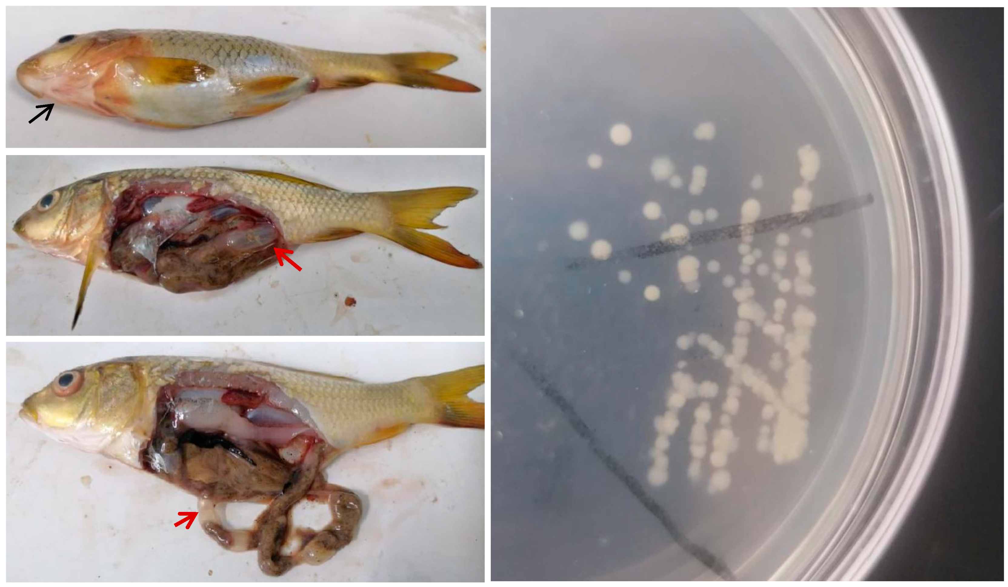
Figure 2.
Symptoms on the epithelial surface and inside the body of experimentally infected fish, showing hemorrhage on body surface, abdominal distention, ascites, and flatulence in the intestine. A1, A2, A. veronii infection, B1, B2, A. hydrophila infection, C1, C2, P. shigelloides infection, D1, D2, C. freundii infection, E1, E2, mixed infection.
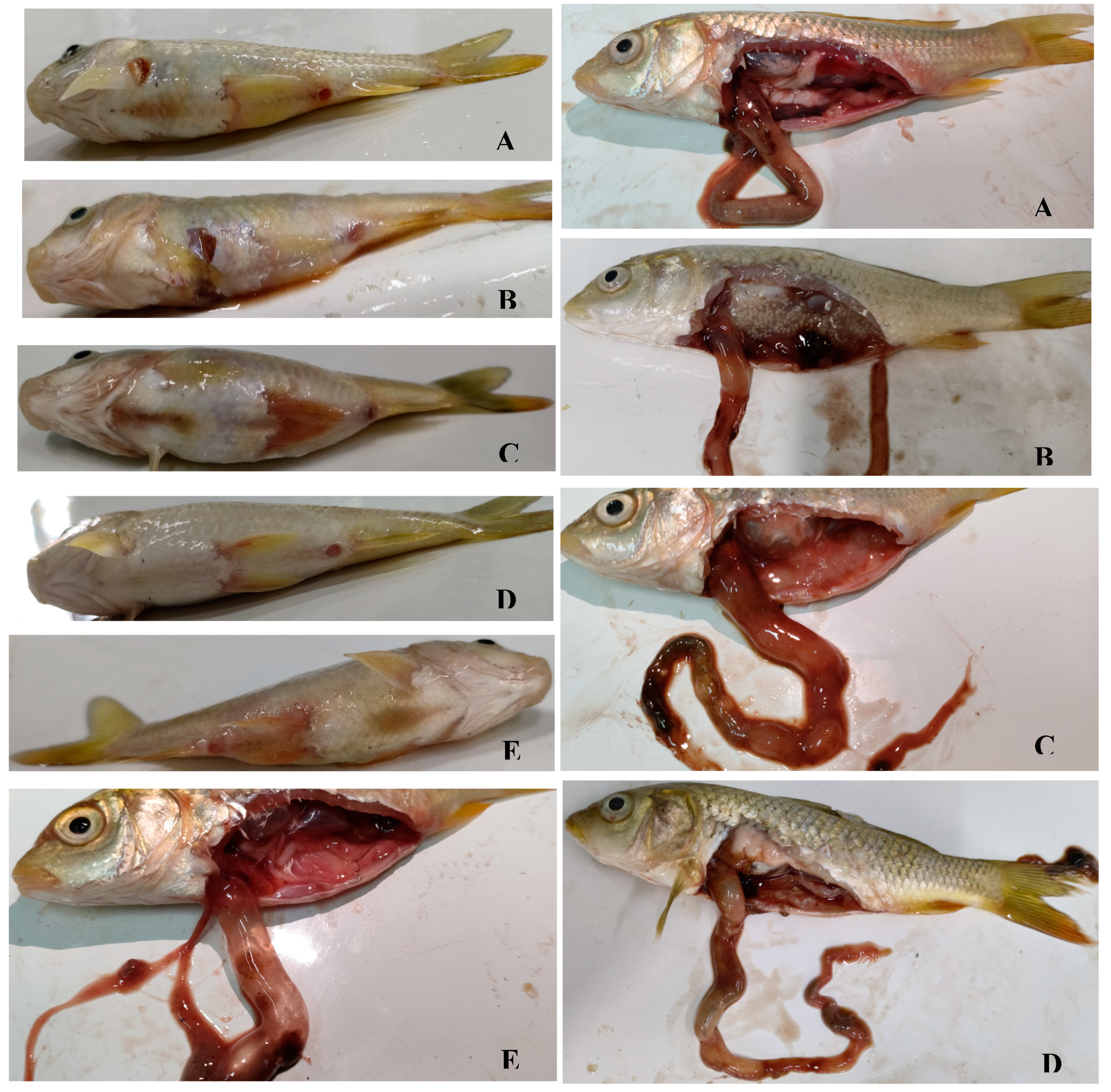

Table 1.
Biochemical Characteristics of isolated strains.
| Test | P. shigelloides | C. freundii | Test | A. veronii | A.hydropha |
| H2S production | - | + | Glucose (gas production) | + | + |
| Phenylalanine deaminase | - | - | Sucrose | + | + |
| Gluconate | - | - | Mannose | + | + |
| Indole reaction | + | - | Indole reaction | + | + |
| Voges–Proskauer | - | - | Voges–Proskauer | + | + |
| Citrate | - | + | Arabinose | - | + |
| Motility | + | + | Arginine dihydrolase | + | + |
| Glucose (gas production) | - | + | Inositol | - | - |
| Lysine decarboxylase | + | - | Lysine | + | + |
| Ornithine decarboxylase | + | - | Unsalted peptone water | + | + |
| Raffinose | - | + | 3% NaCI peptone water | + | + |
| Sorbitol | - | + | 6% NaCI peptone water | + | + |
| Adonitol | - | - | 8% NaCI peptone water | - | - |
| Xylose | - | + | 10% NaCI peptone water | - | - |
| Urease | - | + |
Note: “+” is masculine and “-” is feminine.
Table 2.
Cumulative mortality of experimentally infected fish by isolates.
| Group | Concentration (CFU) | Fish | Dead fish number on day after challenge | Accumulative mortality | LD50 value (CFU) | ||||||||||||||
| 1 | 2 | 3 | 4 | 5 | 6 | 7 | 8 | 9 | 10 | 11 | 12 | 13 | 14 | 15 | |||||
| Control | 0 | 10 | 0 | 0 | 0 | 0 | 0 | 0 | 0 | 0 | 0 | 0 | 0 | 0 | 0 | 0 | 0 | 0% | |
| P. shigelloides | 1.0×10⁷ | 10 | 0 | 0 | 0 | 0 | 4 | 1 | 1 | 3 | 1 | 0 | 0 | 0 | 0 | 0 | 0 | 100% | 4.74×10⁴ |
| 1.0×10⁶ | 10 | 0 | 0 | 0 | 0 | 2 | 1 | 0 | 3 | 0 | 2 | 0 | 0 | 1 | 0 | 0 | 90% | ||
| 1.0×10⁵ | 10 | 0 | 0 | 0 | 0 | 0 | 0 | 0 | 3 | 1 | 2 | 0 | 1 | 0 | 0 | 0 | 70% | ||
| 1.0×10⁴ | 10 | 0 | 0 | 0 | 0 | 0 | 0 | 0 | 1 | 0 | 0 | 0 | 1 | 0 | 0 | 0 | 20% | ||
| C. freundii | 1.0×10⁷ | 10 | 0 | 0 | 0 | 0 | 4 | 2 | 3 | 1 | 0 | 0 | 0 | 0 | 0 | 0 | 0 | 100% | 1.95×10⁴ |
| 1.0×10⁶ | 10 | 0 | 0 | 0 | 0 | 1 | 1 | 2 | 3 | 1 | 1 | 1 | 0 | 0 | 0 | 0 | 100% | ||
| 1.0×10⁵ | 10 | 0 | 0 | 0 | 0 | 0 | 1 | 0 | 1 | 5 | 1 | 1 | 0 | 0 | 0 | 0 | 90% | ||
| 1.0×10⁴ | 10 | 0 | 0 | 0 | 0 | 0 | 0 | 0 | 0 | 3 | 0 | 0 | 0 | 0 | 0 | 0 | 30% | ||
| A. veronii | 1.0×10⁷ | 10 | 2 | 4 | 3 | 0 | 0 | 0 | 0 | 0 | 0 | 0 | 0 | 0 | 0 | 0 | 0 | 90% | 5.12×10⁴ |
| 1.0×10⁶ | 10 | 1 | 0 | 0 | 0 | 4 | 2 | 0 | 1 | 0 | 0 | 0 | 1 | 0 | 0 | 0 | 90% | ||
| 1.0×10⁵ | 10 | 1 | 0 | 0 | 0 | 0 | 1 | 0 | 2 | 1 | 0 | 0 | 2 | 0 | 0 | 0 | 70% | ||
| 1.0×10⁴ | 10 | 0 | 0 | 0 | 0 | 0 | 1 | 0 | 0 | 1 | 0 | 0 | 0 | 0 | 0 | 0 | 20% | ||
| A. hydrophila | 1.0×10⁷ | 10 | 1 | 0 | 0 | 0 | 3 | 1 | 2 | 0 | 0 | 1 | 0 | 2 | 0 | 0 | 0 | 100% | 1.53×10⁵ |
| 1.0×10⁶ | 10 | 0 | 0 | 0 | 0 | 1 | 0 | 1 | 1 | 4 | 0 | 1 | 0 | 0 | 0 | 0 | 80% | ||
| 1.0×10⁵ | 10 | 0 | 0 | 0 | 0 | 0 | 0 | 0 | 0 | 1 | 0 | 1 | 2 | 0 | 0 | 0 | 40% | ||
| 1.0×10⁴ | 10 | 0 | 0 | 0 | 0 | 0 | 0 | 0 | 0 | 0 | 0 | 0 | 1 | 0 | 0 | 0 | 10% | ||
| Mixed infection | 1.0×10⁷ | 10 | 0 | 0 | 0 | 0 | 3 | 1 | 1 | 2 | 2 | 1 | 0 | 0 | 0 | 0 | 0 | 100% | 5.41×10⁴ |
| 1.0×10⁶ | 10 | 0 | 0 | 0 | 0 | 1 | 1 | 0 | 1 | 2 | 1 | 2 | 0 | 1 | 0 | 0 | 90% | ||
| 1.0×10⁵ | 10 | 0 | 0 | 0 | 0 | 1 | 0 | 0 | 1 | 0 | 0 | 1 | 1 | 0 | 1 | 0 | 50% | ||
| 1.0×10⁴ | 10 | 0 | 0 | 0 | 0 | 0 | 0 | 0 | 0 | 0 | 1 | 2 | 0 | 0 | 0 | 0 | 30% | ||
Table 3.
Susceptibility of bacterial isolates to antibiotics.
| Antibiotic | Drug concentration (μg/ disc) | Inhibition zone diameter (mm) | |||
| A.veronii | A.hydrophila | C. freundii | P.shigelloides | ||
| Streptomycin | 10 | 19S | 15S | 15S | 15S |
| Enrofloxacin | 10 | 40S | 23S | 10R | 30S |
| Florfenicol | 30 | 30S | 32S | 0R | 28S |
| Gentamicin | 10 | 21S | 19S | 20S | 19S |
| Kanamycin | 30 | 20S | 20S | 15I | 18S |
| Neomycin | 30 | 17S | 18S | 17I | 18S |
| Tetracycline | 30 | 11R | 25S | 0R | 21S |
| Norfloxacin | 10 | 34S | 23S | 14I | 27S |
| Co-trimoxazole | 23.75/1.25 | 19S | 20S | 0R | 13I |
| Ceftizoxime | 30 | 44S | 40S | 32S | 37S |
| Ampicillin | 10 | 10R | 10R | 14I | 0R |
Note: “S” is sensitive, “I” is intermediate, and “R” is resistant.
Disclaimer/Publisher’s Note: The statements, opinions and data contained in all publications are solely those of the individual author(s) and contributor(s) and not of MDPI and/or the editor(s). MDPI and/or the editor(s) disclaim responsibility for any injury to people or property resulting from any ideas, methods, instructions or products referred to in the content. |
© 2025 by the authors. Licensee MDPI, Basel, Switzerland. This article is an open access article distributed under the terms and conditions of the Creative Commons Attribution (CC BY) license (http://creativecommons.org/licenses/by/4.0/).
Copyright: This open access article is published under a Creative Commons CC BY 4.0 license, which permit the free download, distribution, and reuse, provided that the author and preprint are cited in any reuse.


