Preprint
Article

This version is not peer-reviewed.

Efficacy of Prostate Biopsies via Transperineal and Transrectal Routes for Significant Prostate Cancer Detection: A Multicenter Paired-Matched Study

A peer-reviewed article of this preprint also exists.

Submitted:

05 January 2025

Posted:

07 January 2025

You are already at the latest version

Abstract

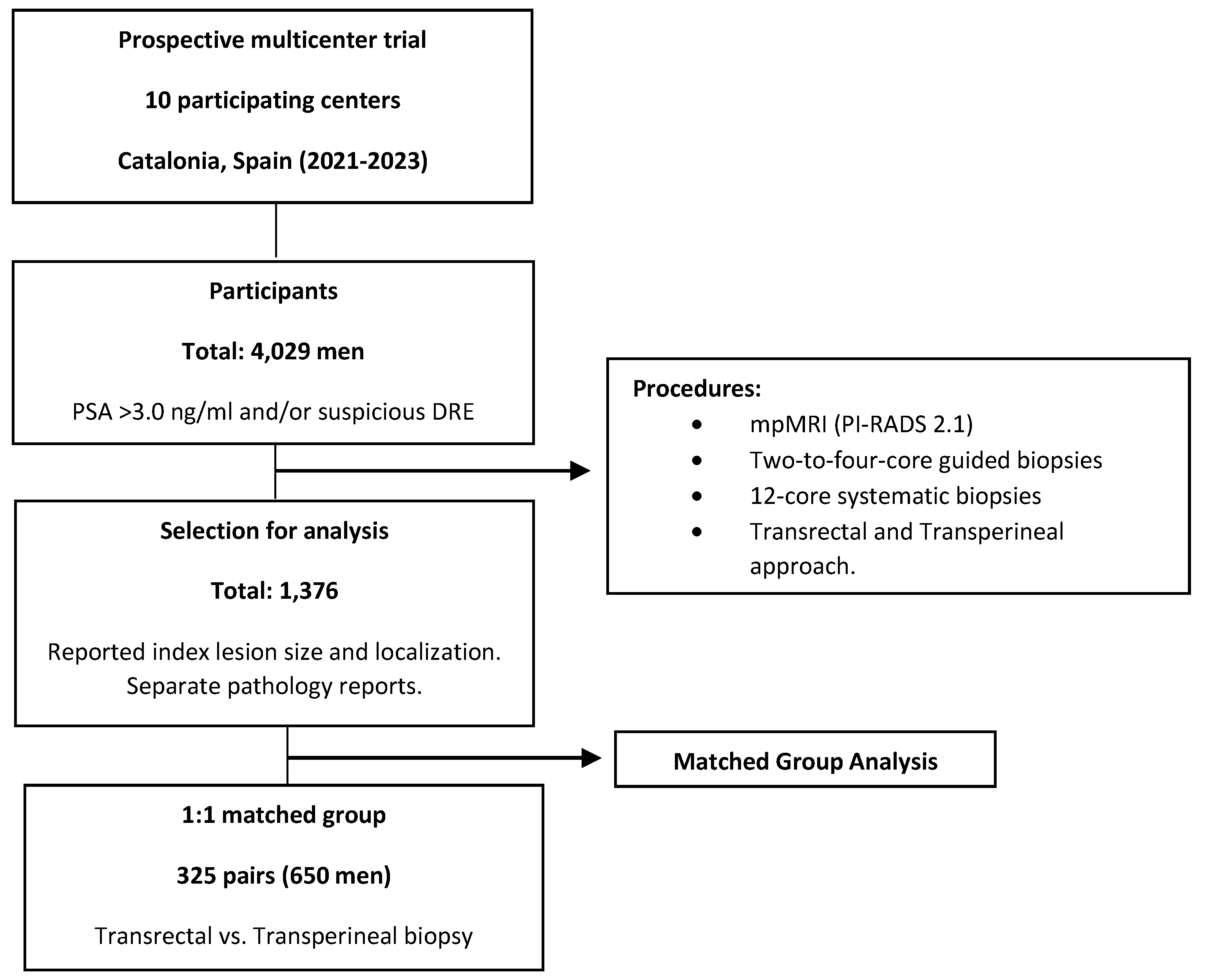
Objective: To compare the efficacy of transrectal and transperineal prostate-guided biopsies to magnetic resonance imaging (MRI) index lesions in detecting clinically significant prostate cancer (csPCa), and to evaluate the role of systematic biopsies. Methods: This prospective and multicenter trial, conducted in the early detection program of csPCa of Catalonia (Spain) between 2021 and 2023, involved 4,029 men suspected of ha-ving PCa who underwent multiparametric MRI followed by guided and systematic biopsies. From this cohort, 1,376 men with reported size and localization of index lesions were selected. A matched group of 325 pairs of men subjected to transrectal and transperineal biopsy was chosen to account for confounding variables. We compared csPCa detection rates at index lesions and systematic biopsies, as well as by lesion localization. Results: Transperineal and transrectal biopsies detected csPCa in 49.5% vs. 40.6% overall (p = 0.027), 44.6% vs. 30.8% at index lesions (p = 0.001), and 24.3% vs. 35.1% at systematic biopsies (p = 0.003). CsPCa detection rates were higher in transperineal biopsies across all index lesion localizations, with significant increases in anterior zone (47.8% vs. 20.8% at mid-base, p = 0.039; 52.9% vs. 24.2% at apex, p = 0.024) and central zone (33.3% vs. 5.9%, p = 0.003). CsPCa detected only in systematic biopsies was 10.5% in transrectal biopsies and 4.9% in transperineal biopsies (p = 0.012). Conclusions: Targeted biopsies via the transperineal route showed higher csPCa detection rates than transrectal biopsies, parti-cularly for anterior and apical lesions, with systematic biopsies showing reduced utility.

Keywords: 
;  ;  ;  ;  ;  

1. Introduction

Prostate cancer (PCa) is the second leading cause of mortality among men [1]. The introduction of Multiparametric Magnetic Resonance Imaging (MRI) has significantly improved early PCa detection, especially clinically significant PCa (csPCa), thereby reducing morbidity and mortality through timely intervention [2]. Confirming the diagnosis of PCa typically involves a prostate biopsy, with techniques such as MRI-ultrasound (MRI-US) fusion-guided biopsy being employed to reduce false negatives [3]. However, the widespread global use of the transrectal approach presents considerable risks, including severe complications such as prostatitis, sepsis, and rectal bleeding [5]. In this scenario, Transperineal Prostate Biopsy (TPB) has emerged as an alternative that overcomes some limitations of Transrectal Prostate Biopsy (TRB) and increases the safety profile of the prostate biopsy procedure [6]. Concerns regarding infection risks and the prevalence of multidrug-resistant bacterial strains have led the European Association of Urology (EAU) to recommend a shift towards the transperineal route [7]. Reports indicate sepsis rates as low as zero with the transperineal approach [8], leading international groups to advocate for discontinuing the use of TRB as soon as possible [9]. However, a randomized trial conducted in the U.S., involving men undergoing either transrectal or transperineal prostate biopsy under local anesthesia, found complication rates of 2.6% and 2.7%, respectively. Importantly, no participants developed sepsis in either group [10]. The transperineal approach offers benefits, particularly in terms of anterior and apical sampling, where the far field suffers from degraded TRB resolution, making visualization challenging [11]. A study analyzing a modified transrectal-guided sextant biopsy, including anteriorly directed cores at the prostate apex, revealed the identification of 17% of tumors in the anterior and apical zones [12]. This critical consideration is currently being evaluated in the ongoing TRANSLATE trial, which randomizes men suspected of having PCa to undergo MRI-targeted and systematic TRUS-guided biopsy via transrectal or transperineal routes, with the endpoint of diagnosing csPCa [13]. The targeted biopsy of index lesions, areas of highest oncological suspicion, has proven to be highly effective, capable of identifying more than 95% of csPCa. This highlights the importance of focusing evaluation on these critical areas [14]. In the era of MRI-targeted biopsy, controversy continues over the effectiveness of TPB and TRB. However, only a few studies have assessed the detection rates of PCa and csPCa between these two approaches.
We hypothesize that the transperineal route for prostate biopsies is more effective than the transrectal route in targeting the index lesion for csPCa detection in men suspected of having PCa. Our first objective is to analyze the efficacy of guided biopsies to the index lesion for detecting csPCa in a matched paired group to avoid the influence of confounding variables. Secondly, we aim to compare the additional role of systematic biopsies in detecting csPCa according to the biopsy route. Finally, we seek to determine if specific localizations of index lesions benefit from either the transrectal or transperineal biopsy route.

2. Materials and Methods

2.1. Design, Setting, and Participants

This is an ad hoc analysis of a prospective, multicenter trial conducted as part of the csPCa opportunistic screening program in Catalonia (Spain), between 2021 and 2023, in ten participating centers. The study involved 4,029 men with serum prostate-specific antigen (PSA) higher than 3.0 ng/mL and/or suspicious digital rectal examination (DRE), who underwent multiparametric magnetic resonance imaging (mpMRI), reported using the Prostate Imaging-Reporting and Data System v. 2.1, followed by two-to four-core guided biopsies and/or 12-core systematic biopsies. From this cohort, 1,376 men were selected since index lesion size and localization was reported as well as its pathology and that of systematic biopsies separately. A 1:1 matched group of 325 pairs of men subjected to transrectal and transperineal biopsy was finally selected to account for confounding variables. The patient selection is represented in Figure 1. The study received approval from the review board of the Vall d´Hebron Hospital coordinator center (PRAG-02/2021). All study participants signed written consent.

2.2. PCa Diagnostic Approach

Men suspected of having PCa were initially selected from urologists at the primary health setting due to a serum PSA testing higher than 3.0 ng/mL and/or a suspicious DRE and referred to the nearest participant centers. The MRI systems had magnetic field strengths of 1.5 Tesla in four centers and 3.0 Tesla in six centers, where expert radiologist reported using the PI-RADS v2.1 [15]. For all prostate biopsies, MRI-TRUS image fusion was performed, utilizing cognitive techniques in five centers and software techniques in five centers. Lesions with the highest PI-RADS scores and size were defined as MRI index lesions [14]. Biopsies were carried out transrectally in four centers and transperineally in six centers. Each suspected lesion underwent 2-to 4 targeted cores in addition to a 12-core systematic biopsy. Experienced operators conducted the biopsies in each center, and experienced uropathologists analyzed the biopsy materials, using the International Society of Urologic Pathology grade groups (ISUP-GG). Clinically insignificant PCa (iPCa) was defined with an ISUP-GG of 1, while csPCa was considered when the ISUP-GG was 2 or higher [16].

2.3. Variables in the Study

Age (years), serum Prostate Specific Antigen (PSA) (ng/mL), Digital Rectal Exam (DRE) characteristics (normal vs. suspicious), type of prostate biopsy (initial vs. repeated), first degree of PCa family history (no vs. yes), MRI prostate volume (mL), PI-RADS score (2-5), Tesla (1.5 vs. 3.0), number of suspicious lesions (1-3), length of index lesion (mm), posterior-anterior localization of index lesion (peripheral, central, fibromuscular anterior zone), cranio-caudal localization of index lesion (mid-base vs. apex), type of fusion TRUS-MRI images (cognitive vs. software), type of biopsy approach (transrectal vs. transperineal). PCa, csPCa, and iPCa detected in targeted biopsy of the index lesion and systematic biopsy were outcome variables.

2.4. Statistical Analysis

Participating centers provided anonymized datasets which were harmonized for statistical analysis. The study complied with Standards of Reporting for MRI-Targeted Biopsy Studies (START) guidelines (16). Medians and interquartile ranges (25th–75th percentile) described quantitative variables, while numbers and percentages described qualitative ones. Pearson's chi-square and Mann-Whitney U tests compared qualitative and quantitative variables, respectively. Binary logistic regression identified independent predictors for csPCa, accounting for the prostate biopsy route. A 1:1 matching group was selected based on the biopsy route to normalize effects of confounding variables using the R package matching v4.10, a multivariate and propensity score matching software with automated balance optimization (R Foundation for Statistical Computing, Vienna, Austria). Significant differences were at p < 0.05; p-values from 0.05 to 0.1 suggested trends. This analysis was conducted with the Statistical Package for the Social Sciences (version 29.0, IBM Corp., Armonk, NY, USA)

3. Results

3.1. Characteristics of Cohort Study

The characteristics of the cohort study are summarized in Table 1. The median age was 68 years (IQR: 62-73), and the median PSA was 7.2 ng/mL (IQR: 5.3-10.8). Abnormal DRE findings were present in 25% of cases, repeated biopsies in 27.4%, and a family history of PCa in 10.8%. The median MRI prostate volume was 52 mL (IQR: 38-73), and the PSA density was 0.14 ng/mL/mL (IQR: 0.09-0.13). A 3 Tesla MRI was performed in 46.4% of cases, with two suspicious lesions identified in 23.9% and three in 3.8%. The PI-RADS score for the index lesion was 2 in 15.1% of cases, 3 in 20.3%, 4 in 43.3%, and 5 in 21.7%. The median index lesion length was 11 mm (IQR: 0.7-15). The index lesion was in the peripheral zone in 64.8% of cases, the central/transitional zone in 14.2%, and the fibromuscular anterior zone in 21%. Regarding the cranio-caudal axis, the index lesion was located at the mid-base in 59.2% of cases and at the apex in 40.8%. Software TRUS-MRI fusion images for targeted biopsies were used in 55.7% of cases. The transperineal route was used in 59.8% of cases, and the transrectal route in 40.2%. Overall, csPCa was detected in 41.3% of targeted biopsies of the index lesion and in 29.7% of systematic biopsies, while iPCa was detected in 13.1% and 18.5% of cases, respectively.

3.2. Binary Logistic Regression for Searching Independent Predictive Variables of csPCa, Selection of a Matched Group to Avoid Confounders, and Characteristics of Paired Groups

Odds ratios (OR) and 95% confidence intervals (CI) of the potential independent predictive variables for csPCa are presented in Table 2. It was found that age, serum PSA, DRE, type of biopsy, prostate volume, Tesla, type of TRUS-MRI fusion images, and PI-RADS were independent predictive variables for csPCa. To avoid the influence of these confounders for csPCa detection and normalize the overall influence of the prostate biopsy route according to the localization of the index lesion, a 1:1 matched group of 325 pairs of participants (650 cases) was selected. Table 3 summarizes the comparison of all variables included in the matched study group. All characteristics of participants, MRI, and biopsy techniques were statistically similar respect to the peripheral localization vs. other localization of index lesions and the outcome variables csPCa and iPCa. The index lesion was localized in the peripheral zone in 49.2% of men who underwent transrectal biopsy while 50.8% of those who underwent the transperineal route, p = 0.793. Central/transitional zone localization was observed in 21.2% and 10.2%, respectively, p = 0.032, and fibromuscular anterior zone localization was observed in 13.2% and 22.8%, respectively, p = 0.028. Regarding the cranio-caudal axis, 48% of index lesions occupied the mid-base in men who underwent TRB while 63.4% in those subjected to TPB; index lesions occupied the apex in 52% and 36.6%, respectively, p < 0.001. The overall detection of PCa in guided biopsies of the index lesion and systematic biopsy conducted through the transrectal route was 58.5%, while 67.1% when the biopsy was conducted through the transperineal route, p = 0.028.

3.3. Overall Efficacy of Systematic Biopsies According to the Prostate Biopsy Route

The overall detection rate of PCa in systematic biopsies when the transrectal route was utilized was 52.9%, while 48% when the transperineal route was used, p = 0.239. Regarding csPCa detection, these rates were 35.1% and 24.3%, respectively, p = 0.003; and for iPCa detection, the rates were 17.8% and 23.7%, respectively, p = 0.082. We note that 10.5% of csPCa were only detected in systematic biopsies when the transrectal route was conducted, while 4.9% were detected when the transperineal route was used, p = 0.012 (Table 4).

3.4. Overall Efficacy of Guided Biopsies to the Index Lesion According to the Biopsy Route and Localizations

The overall detection rates of PCa in the index lesion for the transrectal and transperineal routes were 43.1% and 61.5%, respectively, p < 0.001. CsPCa detection rates were 30.8% and 44.6%, respectively, p < 0.001; and iPCa detection rates were 12% and 16.9%, respectively, p = 0.094, as shown in Table 3.
The rate of csPCa detection in the recodified localizations of index lesions regarding the posterior-anterior and cranio-caudal axes and the prostate biopsy route are summarized in Table 5. We noted non-significant differences between both biopsy routes when the index lesion was localized in the peripheral zone, either in the mid-base or apex. TPB detected more csPCa than TRB in the central and mid-base zones, 33.3% and 5.9%, respectively, p = 0.003. In the anterior and mid-base zones, csPCa detection rates were 47.8% and 20.8%, respectively, p = 0.039. In the anterior and apical zones, these rates were 44.6% and 24.2%, respectively, p = 0.024.

4. Discussion

Technological advancements in prostate MRI have significantly impacted the diagnostic landscape, leading to higher detection rates of csPCa [18]. However, direct comparisons of the effectiveness of TPB versus TRB, especially with MRI/US fusion techniques, remain scarce. Our study addressed this gap with a comprehensive analysis of csPCa detection rates across TPB and TRB in the MRI era.
The recently published PREVENT Randomized Trial found no significant differences in csPCa detection between transperineal and transrectal routes, although transperineal biopsies avoided infectious complications [19]. Rai et al. conducted a similar meta-analysis, demonstrating higher csPCa detection with the transperineal approach compared to the transrectal approach [20]. Zattoni et al. compared csPCa detection in men with PI-RADS scores of 3-5 who underwent MRI/US fusion biopsy via the transperineal route in 3,307 and transrectal route in 1,936. They found a significantly higher detection rate for TPB, identifying this method as an independent predictor of csPCa with detection rates of 49.1% compared to 35.2% detected in TRB [21]. Koparal et al. conducted a multicenter study comparing csPCa detection in TPB and TRB. They matched 276 men undergoing TPB with 508 patients undergoing TRB by age, DRE, PSA density, and PI-RADS score. Their findings indicated that both MRI-targeted TPB and 12-core systematic TPB were significantly superior to TRB in detection rates of 27.5% vs. 19.5%, and 24.6% vs. 16.3%, respectively [22]. Diamand et al. recently published a multicentric European study in which they also reported higher detection rates using MRI-guided biopsy in the transperineal approach [23]. Kaneko et al., in a single-center pair-matched study, reported csPCa detection rates of 56% vs. 49%, which were significantly higher for TPB compared to TRB. When combining systematic and targeted biopsies, similar detection rates were observed: 59% for TPB and 60% for TRB [25]. In our multicenter pair-matched study, we observed csPCa detection rates of 30.8% and 44.6% in the index lesion for the transrectal and transperineal routes, respectively. For combined systematic and targeted biopsies, detection rates were 40.6% for TRB and 49.5% for TPB, which showed a significant difference. TPB exhibited significantly higher csPCa detection rates than TRB, particularly for index lesion targeting. The results obtained in our series are consistent with some of the previously described articles [19,20,21,22,23].
Detecting csPCa requires both systematic and targeted biopsies due to its multifocal nature and the risk of missing lesions with MRI [24]. The 2017 PROMIS trial showed that systematic biopsies alone have a 48% sensitivity for csPCa but combining them with targeted biopsies increases sensitivity to 93% [26]. The PRECISION study found that targeted biopsies detected csPCa in 38% of cases, compared to 26% with systematic biopsies alone [27]. The 2018 MRI-First study reported similar sensitivities for both methods [28]. The 2019 PAIREDCAP trial confirmed the necessity of using both biopsy types, as each method alone fails to detect all csPCa cases [29]. The GÖTEBORG-2 trial indicated that using transrectal MRI-targeted biopsies instead of systematic biopsies for patients with high PSA levels halved the risk of over-diagnosis but delayed the identification of some intermediate-risk tumors [30]. Few studies compare the effectiveness of systematic biopsies alone between transrectal and transperineal routes in prostate fusion biopsies. In our study, systematic biopsies alone via the transrectal route had higher csPCa detection rates (10.5%) compared to the transperineal route (4.9%). We consider this finding relevant, and it is not generally detailed in the published series that compare fusion biopsies between both approach.
Our analysis also revealed that lesion location influences diagnostic outcomes. Apical and anterior prostate lesions were more effectively sampled in TPB due to better access, while TRB provided superior sampling of posterior lesions due to probe proximity and needle trajectory. No significant differences were noted in csPCa detection between biopsy approaches for peripheral zone lesions. However, TPB was more effective in detecting csPCa in central and mid-base zones compared to TRB, with detection rates of 33.3% and 5.9%, respectively. Additionally, in the anterior and mid-base zones, detection rates were 47.8% and 20.8%, respectively, which were significant. For anterior and apical zones, the rates of csPCa detection were higher in TPB, with a 44.6% detection rate compared to 24.2% in TRB. This differential impact based on lesion location was further evidenced in subgroup analyses, showing higher csPCa detection rates for anterior and apical lesions with TPB. Consistent with our findings, meta-analysis published by Uleri et al. found the transperineal approach superior for detecting csPCa in anterior and apical prostate tumors and equivalent for peripheral tumors. It demonstrated more precision for smaller anterior tumors than the transrectal method [31].
While our study benefits from a multicenter design and large sample size, it is not without limitations. The retrospective nature of some data could introduce inherent selection biases, which we attempted to minimize using a matched-cohort design. Differences in biopsy protocols across various institutions could have generated variability in PCa detection, representing a limitation in this study. Although all centers followed current practice guidelines and terminology, the lack of centralized review adds heterogeneity to the interpretation of MRIs and the analysis of biopsies due to the involvement of multiple physicians. However, all MRI analyses were conducted by specialized genitourinary radiologists using version 2.1 of PI-RADS, and biopsy specimens were analyzed by experienced uropathologists at all centers. Our matched study was insufficient regarding certain locations, as differences were observed in some of them, and it was performed for peripheral locations versus other locations. Another potential limitation of our study is not incorporating the results of secondary biopsied targeted lesions.
Future research should focus on conducting large prospective and randomized trials to determine the optimal biopsy approach in different clinical scenarios. These studies should avoid the influence of confounders with especial attention to the localization and size of targeted lesions.

5. Conclusions

Targeted biopsies of index lesions performed via the transperineal route demonstrated higher detection rates of csPCa than those performed via the transrectal route. This improvement in the efficacy of targeted biopsies via the transperineal route was especially notable for lesions located in the anterior and apical zones. The efficacy of systematic biopsies decreased when the transperineal route was utilized and especially in detecting csPCa only in them.

Author Contributions

Juan Morote and Nahuel Paesano had full access to all the data in the study and takes responsibility for the integrity of the data and the accuracy of the data analysis. Study concept and design: Paesano, Morote. Acquisition of data: Paesano, Picola, Muñoz-Rodriguez, Ruiz-Plazas, Muñoz-Rivero, Celma, García-de Manuel, Abascal, Servian. Analysis and interpretation of data: Morote. Drafting of the manuscript: Morote. Critical revision of the manuscript for important intellectual content: Paesano, Abascal, Servian, Trilla. Statistical analysis: Morote. Obtaining funding: Morote. Administrative, technical, or material support: None. Supervision: Morote, Trilla.

Funding

This research was supported by the Instituto de Salut Carlos III (SP) and the European Union (PI20/01666).

Institutional Review Board Statement

This study was approved by the ethics committee of the coordinating center (PRAG20/002).

Informed Consent Statement

Written informed consent was obtained from all participants.

Data Availability Statement

The data presented in this study are available on request from the corresponding author.

Conflicts of Interest

The authors certifies that all conflicts of interest, including specific financial interests and relationships and affiliations relevant to the subject matter or materials discussed in the manuscript (eg, employment/affiliation, grants or funding, consultancies, honoraria, stock ownership or options, expert testimony, royalties, or patents filed, received, or pending), are the following: None.

Equal Contribution as Last Authors

Enrique Trilla and Juan Morote.

Abbreviations

The following abbreviations are used in this manuscript:
PCa Prostate Cancer
csPCa Clinically Significant Prostate Cancer
iPCa Insignificant Prostate Cancer
MRI Magnetic Resonance Imaging
TRB Transrectal Biopsy
TPB Transperineal Biopsy
PSA Prostate-Specific Antigen
PI-RADS Prostate Imaging-Reporting and Data System
DRE Digital Rectal Examination
mpMRI Multiparametric Magnetic Resonance Imaging
ISUP-GG International Society of Urologic Pathology Grade Groups
EAU European Association of Urology
START Standards of Reporting for MRI-Targeted Biopsy Studies

References

  1. Siegel, R.; Miller, K.; Wagle, N.; Jemal, A. Cancer statistics, 2023. CA. Cancer J. Clin. 2023, 73, 17–48. [Google Scholar] [CrossRef] [PubMed]
  2. World Health Organization. Guide de Cancer Early Diagnosis. [Internet]. Geneva: World Health Organization; 2017. Available from: https://apps.who.int/iris/bitstream/handle/10665/254500/9789241511940-eng.pdf?
  3. Pepe, P.; Garufi, A.; Priolo, G.; Pennisi, M. Transperineal Versus Transrectal MRI/TRUS Fusion Targeted.
  4. Biopsy: Detection Rate of Clinically Significant Prostate Cancer. Clin. Genitourin. Cancer. 2017, 15, 33–36. [CrossRef] [PubMed]
  5. Derin, O.; Fonseca, L.; Sanchez-Salas, R.; Roberts, M. Infectious complications of prostate biopsy: winning battles but not war. World J. Urol. 2020, 38, 2743–2753. [Google Scholar] [CrossRef] [PubMed]
  6. Jacewicz, M.; Günzel, K.; Rud, E.; Sandbæk, G. ; MegheliA; Busch, J. ; et al. Antibiotic prophylaxis versus no antibiotic prophylaxis in transperineal prostate biopsies (NORAPP): a randomised, open-label, non-inferiority trial. Lancet Infect Dis 2022, 22, 1465–1471. [Google Scholar]
  7. Mottet, N.; van den Bergh, R.; Briers, E.; Van den Broeck De Santis, M.; Lam, T.; et al. EAU-EANM-ESTRO- ESUR-SIOG guidelines on prostate cancer—2020 update. Part 1: screening, diagnosis, and local treatment with curative intent. Eur. Urol. 2021, 79, 243–262. [Google Scholar] [CrossRef]
  8. Loeb, S.; Vellekoop, A.; Ahmed, H.; Catto, J.; Emberton, M.; Nam, R.; et al. Systematic review of complications of prostate biopsy. Eur Urol. 2013, 64, 876–892. [Google Scholar] [CrossRef]
  9. Grummet, J.; Gorin, M.; Popert, R.; Lamb, A.; Moore, C.; Briers, E.; et al. “TREXIT 2020”: why the time to abandon transrectal prostate biopsy starts now. Prostate Cancer Prostatic Dis. 2020, 23, 62–65. [Google Scholar] [CrossRef]
  10. Stefanova, V.; Buckley, R.; Flax, S.; Hajek, D.; Tunis, A.; Lai, E.; et al. Transperineal Prostate Biopsies Using Local Anesthesia: Experience with 1,287 Patients. Prostate Cancer Detection Rate, Complications and Patient Tolerability. J. Urol. 2019, 201, 1121–1126. [Google Scholar] [CrossRef]
  11. Takashima, R.; Egawa, S.; Kuwao, S.; Baba, S. Anterior distribution of Stage T1c nonpalpable tumors in radical prostatectomy specimens. Urology. 2002, 59, 692–697. [Google Scholar] [CrossRef]
  12. Wright, J.; Ellis, W. Improved prostate cancer detection with anterior apical prostate biopsies. Urol. Oncol. 2006, 24, 492–495. [Google Scholar] [CrossRef]
  13. Bryant, R.; Yamamoto, H.; Eddy, B.; Kommu, S.; Omer, A.; Leslie, T.; et al. Protocol for the TRANSLATE prospective, multicentre, randomised clinical trial of prostate biopsy technique. BJU Int. 2023, 131, 694–704. [Google Scholar] [CrossRef] [PubMed]
  14. Paesano, N.; Catalá, V.; Tcholakian, L.; Alomar, X.; Trilla, E.; Morote, J.; et al. The effectiveness of mapping-targeted biopsies on the index lesion in transperineal prostate biopsies. Int. Braz. J. Urol. 2024, 50, 296–308. [Google Scholar] [CrossRef] [PubMed]
  15. Turkbey, B.; Rosenkrantz, A.; Haider, H.; Padhani, A.; Villeirs, G.; Macura, K.; et al. Prostate Imaging Reporting and Data System Version 2.1: 2019 Update of Prostate Imaging Reporting and Data System Version 2. Eur. Urol. 2019, 76, 340–351. [Google Scholar] [CrossRef] [PubMed]
  16. Epstein, J.; Zelefsky, M.; Sjoberg, D.; Nelson, J.; Egevad, L.; Vickers, A.; et al. A Contemporary Prostate Cancer Grading System: A Validated Alternative to the Gleason Score. Eur. Urol. 2016, 69, 428–435. [Google Scholar] [CrossRef]
  17. Moore, C.M.; Kasivisvanathan, V.; Gill, I.S.; Emberton, M.; Klotz, L.; Simon, R.; et al. Standards of reporting for MRI-targeted biopsy studies (START) of the prostate: recommendations from an International Working Group. Eur. Urol. 2013, 64, 544–552. [Google Scholar] [CrossRef]
  18. Petov, V.; Azilgareeva, C.; Shpikina, A.; Morozov, A.; Kozlov, V.; Rodler, S.; et al. Robot-Assisted Magnetic Resonance Imaging-Targeted versus Systematic Prostate Biopsy; Systematic Review and Meta-Analysis. Cancers (Basel). 2023, 15, 1181. [Google Scholar] [CrossRef]
  19. Hu, J.C.; Assel, M.; Allaf, M.E.; et al. Transperineal Versus Transrectal Magnetic Resonance Imaging-targeted and Systematic Prostate Biopsy to Prevent Infectious Complications: The PREVENT Randomized Trial. Eur. Urol. 2024, 86, 61–68. [Google Scholar] [CrossRef]
  20. Rai, B.; Mayerhofer, C.; Somani, B.; Kallidonis, P.; Nagele Udo Tokas, T. Magnetic Resonance Imaging/Ultrasound Fusion-guided Transperineal Versus Magnetic Resonance Imaging/Ultrasound Fusion-guided Transrectal Prostate Biopsy-A Systematic Review. Eur. Urol. Oncol. 2021, 4, 904–913. [Google Scholar] [CrossRef]
  21. Zattoni, F.; Marra, G.; Kasivisvanathan, V.; Grummet, J.; Olivier, J.; Moon, D.; et al. The Detection of Prostate Cancer with Magnetic Resonance Imaging-Targeted Prostate Biopsies is Superior with the Transperineal vs the Transrectal Approach. A European Association of Urology-Young Academic Urologists Prostate Cancer Working Group Multi-Institutional Study. J. Urol. 2022, 208, 830–837. [Google Scholar]
  22. Koparal, M.; Sözen, T.; Karşıyakalı, N.; Aslan, G.; Akdoğan, B.; Şahin, B.; et al. Comparison of transperineal and transrectal targeted prostate biopsy using Mahalanobis distance matching within propensity score caliper method: A multicenter study of Turkish Urooncology Association. Prostate. 2022, 82, 425–432. [Google Scholar] [CrossRef]
  23. Diamand, R.; Guenzel, K.; Mjaess, G.; Lefebvre, Y.; Ferriero, M.; Simone, G.; et al. Transperineal or Transrectal Magnetic Resonance Imaging-targeted Biopsy for Prostate Cancer Detection. Eur. Urol. Focus. 2024. [Google Scholar]
  24. Kaneko, M.; Medina, L.; Lenon, M.; Aron, M.; Gill, I.; Abreu, A.; et al. Transperineal vs transrectal magnetic resonance and ultrasound image fusion prostate biopsy: a pair-matched comparison. Sci. Rep. 2023, 13, 13457. [Google Scholar] [CrossRef] [PubMed]
  25. Morote, J.; Picola, N.; Muñoz-Rodriguez, J.; Paesano, N.; Celma, A.; Servian, P.; et al. A Diagnostic Accuracy Study of Targeted and Systematic Biopsies to Detect Clinically Significant Prostate Cancer, including a Model for the Partial Omission of Systematic Biopsies. Cancers (Basel). 2023, 15, 4543. [Google Scholar] [CrossRef] [PubMed]
  26. Ahmed, H.; El-Shater Bosaily, A.; Brown, L.; Gabe, R.; Kaplan, R.; Parmar, M.; et al. Diagnostic accuracy of multi-parametric MRI and TRUS biopsy in prostate cancer (Promis): a paired validating confirmatory study. Lancet. 2017, 389, 815–822. [Google Scholar] [CrossRef] [PubMed]
  27. Kasivisvanathan, V.; Rannikko, A.S.; Borghi, M.; Panebianco, V.; Vaarala, M.; Briganti, A.; et al. MRI-Targeted or Standard Biopsy for Prostate-Cancer Diagnosis. N. Engl. J. Med. 2018, 378, 1767–1777. [Google Scholar] [CrossRef] [PubMed]
  28. Rouvière, O.; Puech, P.; Renard-Penna, R.; Claudon, M.; Roy, C.; Colombel, M.; et al. Use of prostate systematic and targeted biopsy on the basis of multiparametric MRI in biopsy-naive patients (MRI-FIRST): A prospective, multicentre, paired diagnostic study. Lancet Oncol. 2018, 20, 100–109. [Google Scholar] [CrossRef]
  29. Elkhoury, F.; Felker, E.; Kwan, L.; Sisk, A.; Delfin, M.; Marks, L.; et al. Comparison of Targeted vs. Systematic Prostate Biopsy in Men Who Are Biopsy Naive: The Prospective Assessment of Image Registration in the Diagnosis of Prostate Cancer (PAIREDCAP) Study. JAMA Surg. 2019, 154, 811–818. [Google Scholar] [CrossRef]
  30. Hugosson, J.; Månsson, M.; Wallström, J.; Axcrona, U.; Carlsson, S.; Egevad, L.; et al. Prostate cancer screening with PSA and MRI followed by targeted biopsy only. N Engl. J.Med. 2022, 387, 2126–2137. [Google Scholar] [CrossRef]
  31. Uleri, A.; Baboudjian, M.; Tedde, A.; Palou, J.; Basile, G.; Briganti, A.; et al. Is There an Impact of Transperineal Versus Transrectal Magnetic Resonance Imaging-targeted Biopsy in Clinically Significant Prostate Cancer Detection Rate? A Systematic Review and Meta-analysis. Eur. Urol. Oncol. 2023, 6, 621–628. [Google Scholar] [CrossRef]
Figure 1. Flowchart of the patient selection strategy.
Preprints 145278 g001
Table 1. Characteristics of the study population.
Characteristic Measurement
Number of men 1,376
Age, years, median (IQR) 68 (62-73)
Serum PSA, ng/ml, median (IQR) 7.2 (5.3-10.8)
Abnormal DRE, n (%) 344 (25.0)
Repeated prostate biopsy, n (%) 377 (27.4)
PCa family history, n (%) 149 (10.8)
Prostate volume, cc, median (IQR) 52 (38-73)
PSA density, ng/ml/cc, median (IQR) 0.14 (0.09-0.13)
3 Tesla mpMRI, n (%) 638 (46.4)
Suspicious lesions, n (%)
1
2
1,376 (100)
2 329 (23.9)
3 52 (3.8)
Index lesion PI-RADS score, n (%)
2
355 (15.1)
3
449 (20.3)
4 960 (43.3)
5 468 (21.7)
Index lesion length, mm, median (IQR) 11 (0.7-15)
Postero-anterior index lesion localization, n (%)
Peripheral zone 891 (64.8)
Central/transitional zone 196 (14.2)
Anterior (fibromuscular) zone 289 (21.0)
Craniocaudal index lesion localization, n (%)
Mid-Base 815 (59.2)
Apex 561 (40.8)
Software image TRUS-MRI fusion biopsy, n (%) 767 (55.7)
Transperineal route, n (%) 823 (59.8)
Overall PCa detection, n (%) 867 (63.0)
sPCa 652 (47.4)
iPCa 215 (15.6)
PCa detected at index lesion biopsy, n (%) 749 (54.4)
sPCa 568 (41.3)
iPCa 180 (13.1)
PCa detected at systematic biopsies, n (%) 662 (48.1)
sPCa 408 (29.7)
iPCa 254 (18.5)
IQR =interquartile range; PSA =prostate-specific antigen; DRE =digital rectal examination; PCa =prostate cancer; PI-RADS =Prostate imaging-report and data system; sPCa =significant PCa; iPCa =insignificant PCa.
Table 2. Logistic regression analysis for searching independent predictive variables of csPCa detection in index lesion, apart from the prostate biopsy route.
Predictive variable Odds ratio (95% CI) p Value
Age, Ref. one year 1.054 (1,035-1.073)
-
< 0.001
Serum PSA, Ref. one ng/mL 1.035 (1.013-1.057) 0.001
DRE, Ref. normal 1.581 (1.152-2.170) 0.005
Type of biopsy, Ref. initial 0.627 (0.460-0.857) 0.003
PCa family history, Ref. no 1.472 (0.952-2.275) 0.082
Prostate volume, Ref. one mL 0.978 (0.972-0.983) < 0.001
Tesla, Ref. 1.5 1.296 (1.221-2.728) < 0.001
Number of suspicious lesions, Ref. 1 1.021 (0.943-1.167) 0.671
Length of index lesion, Ref. one mm 1.003 (0.981-1.026) 0.783
PI-RADS score of index lesion, Ref. 2 3.938 (3.201-4.845) < 0.001
Postero-anterior localization of index lesion, Ref. PZ 0,888 (0.777-1.015) 0.081
Craniocaudal localization of index lesion, Ref, mid-base 0.907 (0.804-1.022) 0.110
Type of guided biopsy, Ref. software 1.911 (1.439-2.539) < 0.001
CI =confidence interval; PSA =prostate-specific antigen; DRE =digital rectal examination; PCa =prostate cancer; PI-RADS =prostate imaging-report and data system; PZ =peripheral zone.
Table 3. Compared characteristics of the matched selected groups based on the prostate biopsy route.
Characteristic Transrectal Transperineal p Value
Number of men 325 325 -
Age, years, median (IQR) 67 (62-73) 67 (63-72) 1.000
Serum PSA, ng/ml, median (IQR) 6.9 (4.9-9.6) 6.9 (5.2-9.9) 0.998
Abnormal DRE, n (%) 65 (20.0) 68 (20.0) 1.000
Repeated prostate biopsy, n (%) 89 (27.4) 86 (26.5) 0.754
PCa family history, n (%) 39 (12.0) 43 (13.2) 0.838
Prostate volume, cc, median (IQR) 50 (38-68) 50 (38-68) 1.000
PSA density, ng/ml/cc, median (IQR) 0.14 (0.10-0.14) 0.14 (0.10-0.13) 0.999
3 Tesla mpMRI, n (%) 169 (52.0) 164 (50.5) 0.892
Suspicious lesions, n (%)
1
2
375(100) 375 (100) 1.000
2 81 (21.6) 87 (23.2) 0.894
3 14 (3.7) 16 (4.3) 0.905
Index lesion PI-RADS score, n (%)
2
3 (0.9) 3 (0.9) 1.000
3
72 (22-2) 72 (22-2) 1.000
4 193 (59.4) 193 (59.4) 1.000
5 57 (15.5) 57 (15.5) 1.000
Index lesion length, mm, median (IQR) 10 (0.7-14) 10 (7-14) 1.000
Posteroanterior index lesion localization, n (%)
Peripheral zone 213 (49.2) 220 (50.8) 0.793
Central (central and transitional) zone 69 (21.2) 33 (10.2) 0.032
Anterior (fibromuscular) zone 43 (13.2) 72 (22.2) 0.028
Craniocaudal index lesion localization, n (%)
Mid-base 156 (48.0) 206 (63.4) < 0.001
Apex 169 (52.0) 119 (36.6) < 0.001
Software image TRUS-MRI fusion biopsy, n (%) 164 (49.2) 158 (48.6) 0.897
Overall PCa detection, n (%) 190 (58.5) 218 (67.1) 0.028
sPCa 132 (40.6) 161 (49.5) 0.027
iPCa 58 (17.8) 57 (17.5) 0.997
PCa detected at index lesion biopsy, n (%) 140 (43.1) 200 (61.5) < 0.001
sPCa 100 (30.8) 145 (44.6) < 0.001
iPCa 39 (12.0) 55 (16.9) 0.094
PCa detected at systematic biopsies, n (%) 172 (52.9) 156 (48.0) 0.239
sPCa 114 (35.1) 79 (24.3) 0.003
iPCa 58 (17.8) 77 (23.7) 0.082
IQR =interquartile range; PSA =prostate-specific antigen; DRE =digital rectal examination; PCa =prostate cancer; PI-RADS =Prostate imaging-report and data system; sPCa =significant PCa; iPCa =insignificant PCa.
Table 4. The effectiveness of systematic biopsies and targeted biopsies on the index lesion for detecting overall PCa, csPCa, and iPCa, based on the prostate biopsy route, in the matched selected groups.
Type of PCa Systematic biopsies Targeted biopsies
TR route (n = 325) TP route
(n = 325)
p Value TR route
(n = 325)
TP route
(n = 325)
p Value
csPCa, n (%) 144 (35.1) 79 (24.3) 0.003 100 (30.8) 145 (44.6) < 0.001
iPCa, n (%) 58 (17.8) 77 (23.7) 0.082 39 (12.0) 55 (16.9) 0.094
Overall PCa, n (%) 172 (52.9) 156 (48.0) 0.239 140 (43.1) 200 (61.5) < 0.001
n =number; PCa = prostate cancer; csPCa =clinically significant PCa; iPCa = insignificant PCa; TR =transrectal route; TP = transperineal route. Inhibitors.
Table 5. Detection of csPCa on targeted biopsies of index lesion according to its localization and the prostate biopsy route.
Localization of
index lesion
csPCa detection Transrectal Transperineal p Value
PZ-MB, n (%) 102/217 (47.0) 36/81 (44.4) 66/136 (48.5) 0.577
CZ-MB, n (%) 11/75 (14.7) 3/51 (5.9) 8/24 (33.3) 0.003
AZ-MB, n (%) 27/70 (38.6) 5/24 (20.8) 22/46(47.8) 0.039
PZ-AP, n (%) 79/221 (35.7) 48/136 (35.3) 31/85 (36.5) 0.886
AZ-AP, n (%) 26/67 (37.7) 8/33 (24.2) 18/34 (52.9) 0.024
All localizations, n (%) 245/650 (37.7) 100/325 (30.8) 145/325 (44.6) > 0.001
TR =transrectal; TP =transperineal; PZ =peripheral zone; CZ =central zone; AZ =anterior zone; MB =mid-base; AP =apical.
Disclaimer/Publisher’s Note: The statements, opinions and data contained in all publications are solely those of the individual author(s) and contributor(s) and not of MDPI and/or the editor(s). MDPI and/or the editor(s) disclaim responsibility for any injury to people or property resulting from any ideas, methods, instructions or products referred to in the content.
Copyright: This open access article is published under a Creative Commons CC BY 4.0 license, which permit the free download, distribution, and reuse, provided that the author and preprint are cited in any reuse.
Prerpints.org logo

Preprints.org is a free preprint server supported by MDPI in Basel, Switzerland.

Subscribe

Disclaimer

Terms of Use

Privacy Policy

Privacy Settings

© 2026 MDPI (Basel, Switzerland) unless otherwise stated