Preprint
Article

This version is not peer-reviewed.

Metagenomic Characterization of Microbiome Taxa Associated with Coral Reef Communities in North Area of Tabuk Region Saudia Arabia

A peer-reviewed article of this preprint also exists.

Submitted:

30 December 2024

Posted:

03 January 2025

You are already at the latest version

Abstract

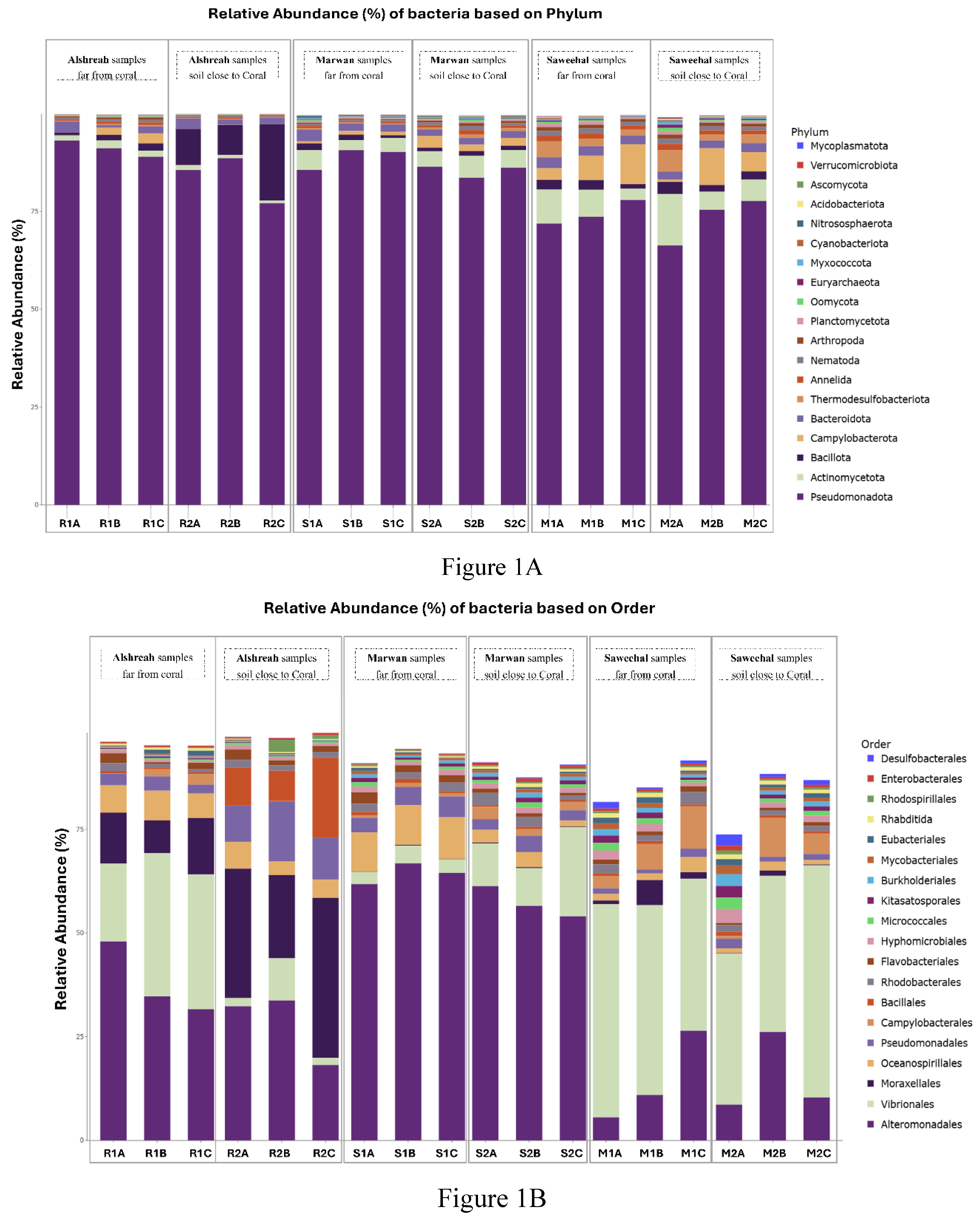
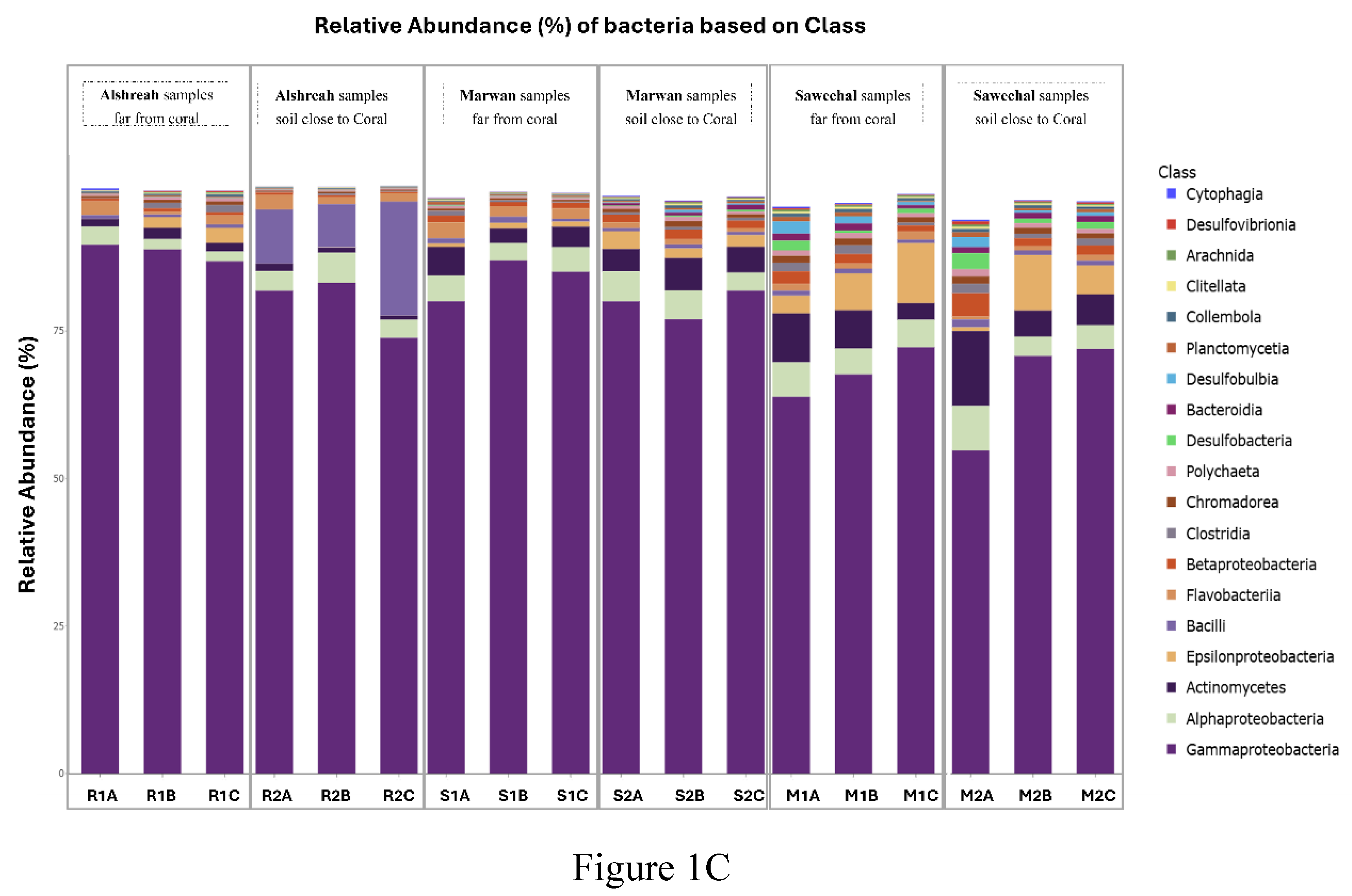
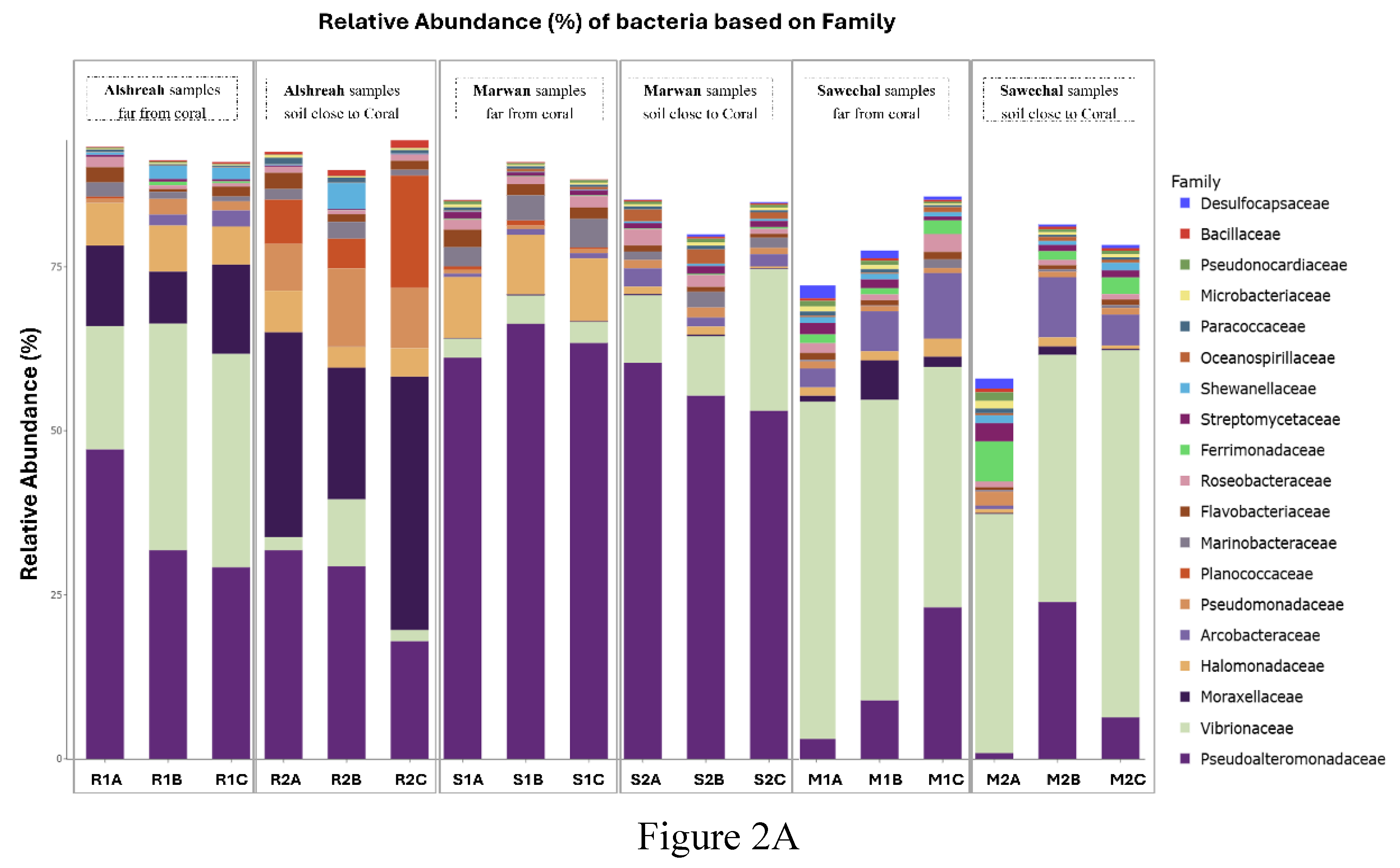
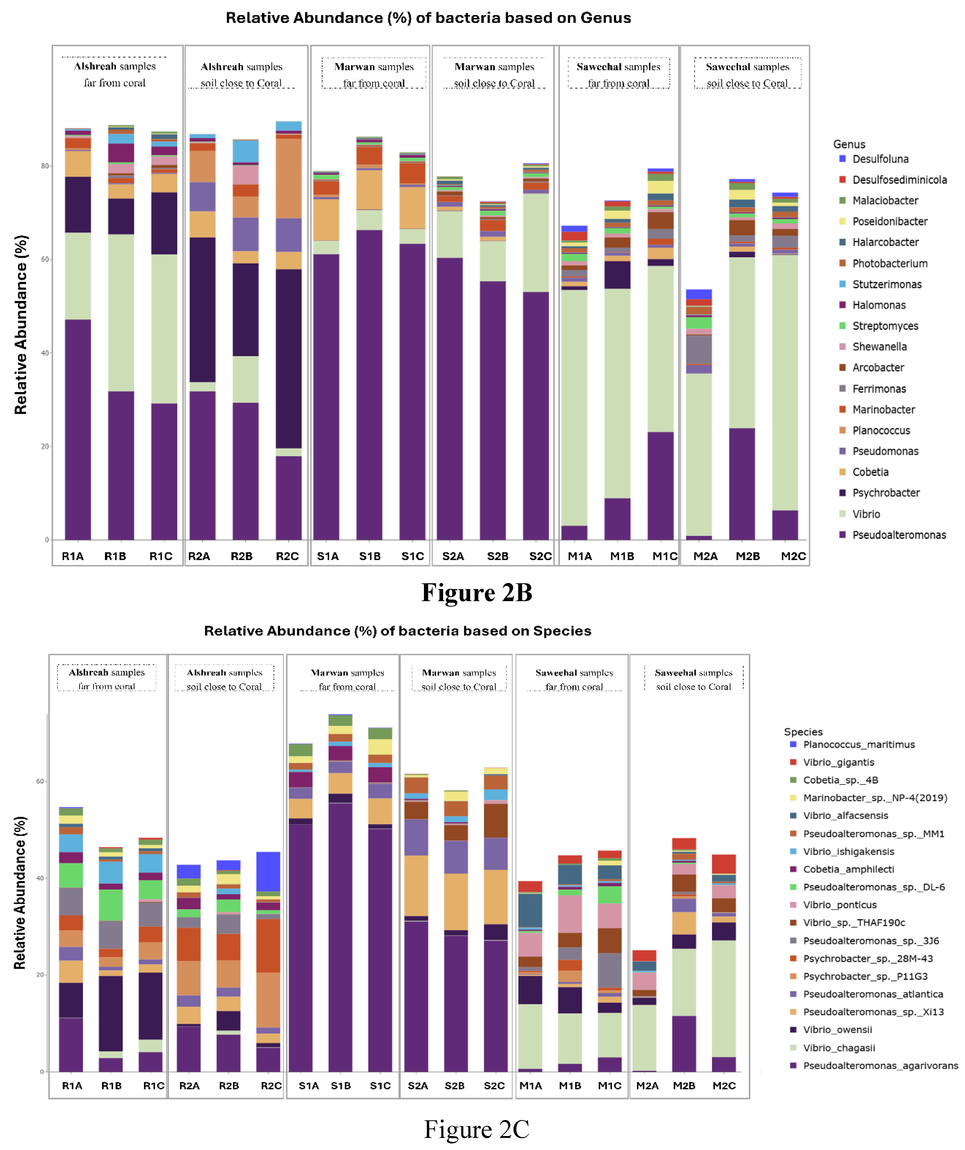
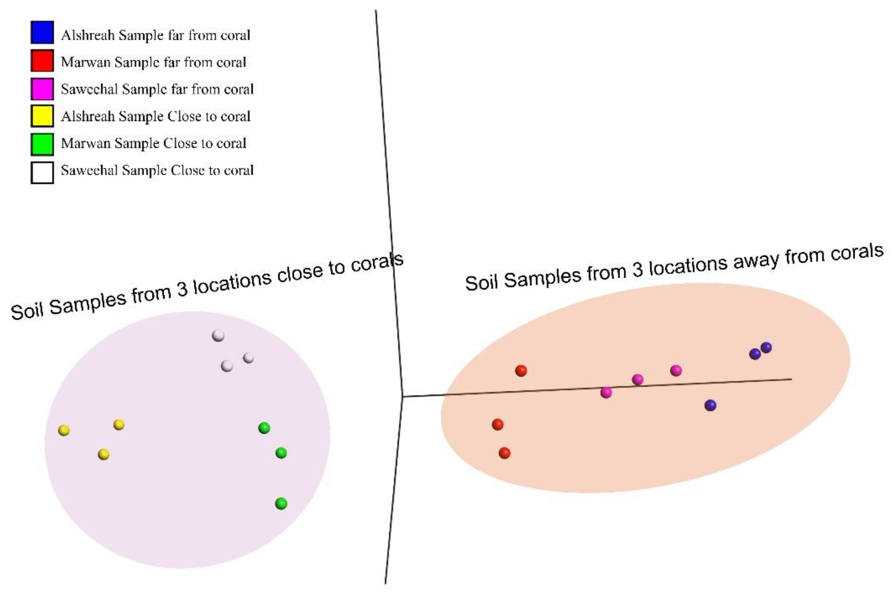
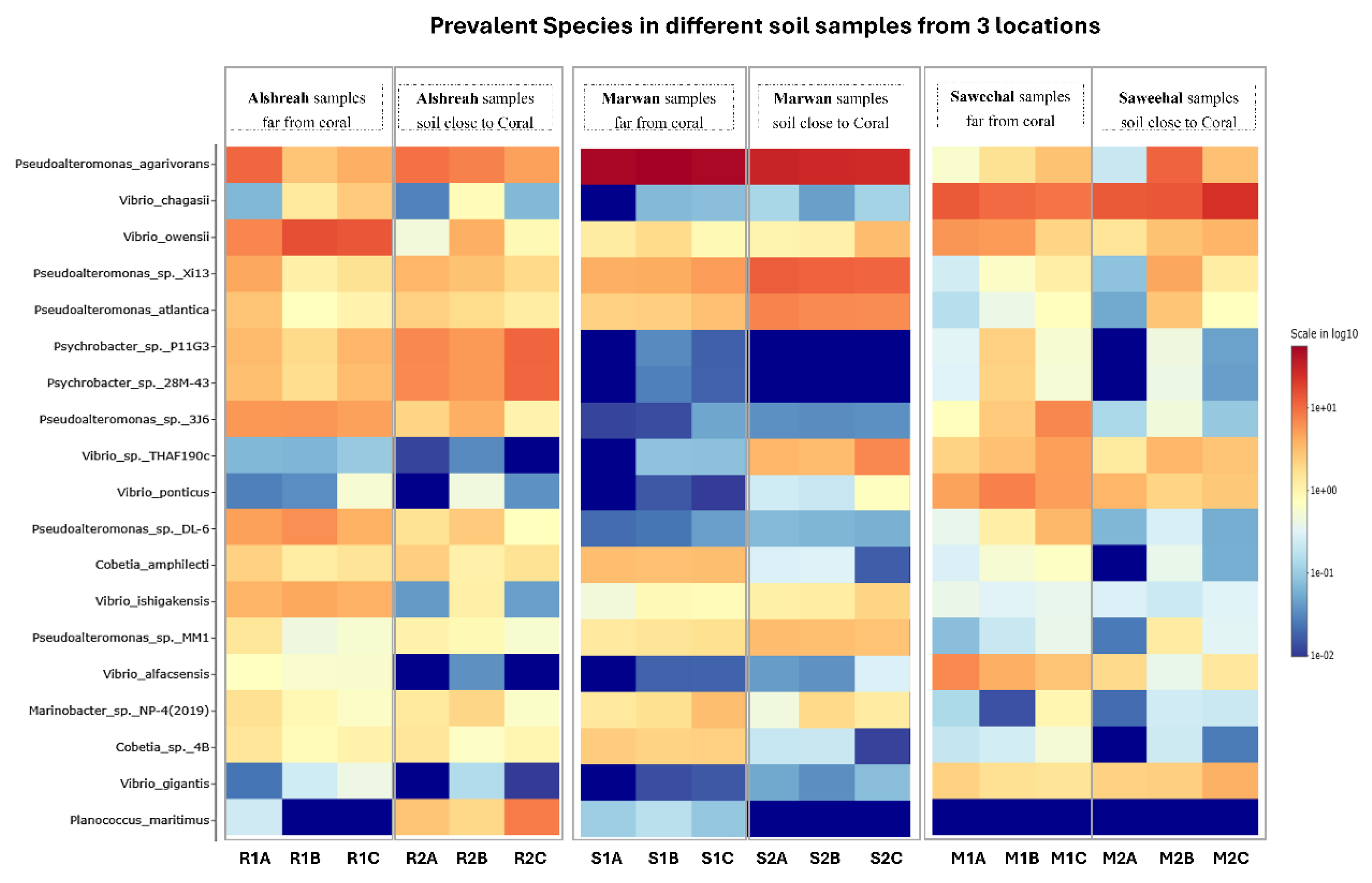
The coral microbiome is highly related to the overall health and the survival and proliferation of coral reefs. As the Red Sea's unique physiochemical characteristics, such a significant north-south temperature and salinity gradient, make it a very intriguing research system. However, the Red Sea is rather isolated, with a very diversified ecosystem rich in coral communities, its makeup of the coral-associated microbiome remains little understood. Therefore, comprehending the makeup and dispersion of the endogenous microbiome associated with coral is crucial for understanding how the coral microbiome coexists and interacts, as well as its contribution to temperature tolerance and resistance against possible pathogens. Here we investigate, metgenomic sequencing targeting the 16S rRNA was performed using DNAs from the sediment samples to identify the coral microbi-ome and to understand the dynamics of microbial taxa and genes in the surface mucous layer (SML) microbiome of the coral communities in three distinct areas close to and far from coral communi-ties in the Red Sea. These findings highlighted the genomic array of the microbiome in three areas around and beneath the coral communities, an revealed distinct bacterial communities in each group, where Pseudoalteromonas agarivorans (30%), Vibrio owensii (11%), and Pseudoalteromonas sp. Xi13 (10%) were the most predominant species in samples closer to coral (a coral-associated mi-crobiome), with the domination of Pseudoalteromonas_agarivorans and Vibrio_owensii in Alshreah samples distant from coral, while Pseudoalteromonas_sp._Xi13 was more abundant in closer sam-ples.Moreover, Proteobacteria such as Pseudoalteromonas, Pseudomonas and Cyanobacteria were the most prevalent phyla of coral microbiome. Further in Saweehal showed highest diversity far from corals 52.8% and in Alshreah close (7.35%), than at Marwan ( 1.75%). The microbial community was less diversified in the samples from Alshreah Far (5.99%) and Marwan Far (1.75%), which had comparatively lower values for all indices. Also, Vibrio species were the most prevalent microor-ganisms in the coral mucus, and the prevalence of these bacteria is significantly higher than those found in the surrounding saltwater. These findings reveal that there is a notable difference in mi-crobial diversity across the various settings and locales, revealing that geographic variables and coral closeness affect the diversity of microbial communities. There were significant differences in microbial community composition regarding the proximity to coral. Besides, there were strong pos-itive correlations between genera Pseudoalteromonas and Vibrio in close-to-coral environments, suggesting that these bacteria may play a synergistic role in immuring coral, rising its tolerance to-wards environmental stress and overall coral health.

Keywords: 
;  ;  ;  ;  ;  ;  ;  

1. Introduction

Coral reefs are the globe's most diversified symbiotic ecosystem. Corals coexist in a complex, multipartite symbiosis with a variety of bacteria from different kingdoms, some of which are linked to essential processes like climate change adaptation [1]. Further its significant in oceans corals function as metaorganisms reliant on dynamic multipartite symbioses with various microbes. Homeostasis is maintained within this intricate system by these interkingdom interactions between the multicellular eukaryotic coral host and its accompanying microbiota, which have supported its resilience for more than 500 million years [2,3,4]. Associations within the metaorganism comprise a large diversity of viruses, prokaryotes, and microeukaryotes that collectively are termed the coral holobiont [5,6,7,8]. Chief among the holobiont microbes, the primary endosymbiotic dinoflagellate of the family Symbiodiniaceae provides the bulk of the required nutritional needs to their coral hosts [9,10]. In addition, an increasing body of evidence is unravelling the key roles particular bacterial species in specific and general prokaryotic communities play in maintaining holobiont fitness, potentially via exchanging essential metabolites, recycling nutrient, and providing protection against pathogenic microbe [11]. In the Anthropocene era, climate change disrupts these symbiotic relationships, leading to dysbiosis that is characterized by the overgrowth of opportunistic and putatively pathogenic microbes and results in a compromised coral immune system, inevitably causing the onset of coral bleaching and/or disease [12]. Most coral microbiome work has been exclusively focused on either endosymbiotic algae or bacteria, while ignoring the other, largely underexplored members of the coral microbiomes due to difficulties associated with studying their role in the holobiont. However, our comprehension of the nature and functional relevance of intricate symbiotic connections within corals is limited by information gaps and technological difficulties. Despite the fact that corals collectively contain one-third of all marine bacterial phyla, known bacterial symbionts and antagonists of corals only make up a small portion of this diversity. These taxa cluster into specific genera, indicating that these bacteria were able to acquire a niche within the holobiont through selective evolutionary mechanisms.
Diverse bacterial symbionts associated with corals
Corals harbor a diverse bacterial microbiome [13], spanning 39 phyla [14], over one-third of the bacterial phyla identified in seawater [15]. A component of these coral-associated bacterial communities is believed to enhance the health and resilience of corals. [16,17,18]. In the extensive array of bacterial phyla linked to corals, Proteobacteria, Bacteroidetes, Cyanobacteria, and Firmicutes rank as some of the most prevalent, as determined by the 16S rRNA gene phylogeny analysis of 21,100 sequences obtained from a public database. [14]. Furthermore, the majority of cultivable bacteria were found to belong to the phyla Proteobacteria, Firmicutes, Bacteroidetes, and Actinobacteria in a recent metaanalysis of 3055 bacterial isolates from 52 coral investigations. [19]. Environmental stresses like ocean warming may cause the collective reef microbiome to react quickly, which might ultimately result in reef microbialization. Reef microbialization is marked by a change in both abundance and biomass favoring microbes, particularly a transition towards a pathogenic assemblage that has the potential to cause significant declines. [20]. Coral-associated bacteria inhabit several compartments within the coral, such as the SML, tissues, gastric cavity, and skeleton, [12,21,22]. Unique physiochemical characteristics and environmental gradients, such as pollution [23,24], significantly influence the microbial composition inside these compartments [21,25,26]. The bacterial makeup differs among these several niche compartments, with certain bacteria favoring the colonization of certain compartments. Bacteria from the families Chloroflexi, Sphingobacterium, Roseobacter, and Pseudoalteromonas were exclusively identified in the SML [25], whilst Endozoicomonas were located among aggregates in coral tissues [27,28]. According to Ritchie and Smith (2004), this niche specificity implies that some bacteria are tailored to the local microenvironment inside the coral colony, which eventually results in distinct interactions with the host within each milieu. Diverse bacterial populations have been seen in the coral skeleton, surpassing those found in the coral tissue or the SML [21].
Function of Coral -associated Microbiome in coral life and healthy:
The Coral–Symbiodiniaceae symbiosis is the driving force of the holobiont, and this symbiosis interaction can shape the whole coral life and development. The symbiotic association between corals and Symbiodiniaceae facilitated the formation of the reef (calcium carbonate skeleton) through reciprocal nutrient [29].. The symbiosis depends on mutual metabolite exchanges, wherein Symbiodinaceae provide surplus photosynthetically produced dissolved organic matter to the coral host in return for access to inorganic nutrients and CO2 produced during respiration [9,30,31]. The transfer of organic photosynthates, such as glucose, by Symbiodiniaceae is energetically enough for the host to fulfill 100% of its respiratory needs [11,32]. While corals may assimilate ammonium for nitrogen acquisition, the Symbiodiniaceae primarily facilitate the absorption of inorganic nitrogen in the forms of nitrate and ammonium [33]. A fraction of this nitrogen is exchanged with the coral host as dissolved organic nitrogen (e.g., amino acids)[34,35,36].
Role of Nitrogen- fixing microbiome associated with corals:
Nitrogen- fixing microbiome are very common in coral [1]. Diazotrophs are frequently linked to coral tissues [5,33,37] especially during early life stages (larvae and juveniles) [38], suggesting the potential significance of nitrogen fixation within the coral holobiont. Communities of ammonia-oxidizing bacteria and archaea may partially oxidize ammonium produced by nitrogen fixation. Similarly, denitrifying bacteria have been documented in corals [39]. Rädecker et al. (2022) have more recently documented the close connection between coral bleaching and disruptions in nitrogen cycle [40]. Nonetheless, the molecular processes linking the nitrogen-related activities of these microbial communities remain mainly unidentified. Like all phytoplankton, Symbiodiniaceae form associations with bacteria that affect their physiology and the availability of nutrients [41]. Numerous Symbiodiniaceae cultures and other phytoplankton lineages share members of the Rhodobacteraceae family, which has been demonstrated to be crucial in supplying phytoplankton with vital nutrients, hormones, and cofactors [42].
Coral associated Microbiome in Red Sea :
Microorganisms are essential in several reef processes, encompassing primary production and the cycling of nutrients and organic waste [43]. Microbes are pervasive symbionts of eukaryotic species, supplying the host with nutrition, facilitating chemical cycle, and offering defense roles [44]. Because of its unique physiochemical characteristics, including a significant north-south temperature and salinity gradient, the Red Sea is an especially intriguing research system. While the southern and western parts of the Red Sea are still completely unknown, the majority of research has been done in the center and northern sections[16,44,45]. In spite of the Red Sea's enormous coral variety, Pocillopora verrucosa, Dipsastraea spp., Pleuractis granulosa, and Stylophora pistillata were the most researched corals. The most common bacterial families were Rhodobacteraceae and Vibrionaceae, whereas bacteria from the class Gammaproteobacteria dominated microbial diversity [44,46]. The dominant north-south environmental conditions do seem to be correlated with the microbial population in the water column. For instance, the cyanobacteria Synechococcus and heterotrophic picoplankton are often more prevalent in the warmer, less salinized waters of the south. Conversely, the microorganisms linked to corals appear to be preserved across the Red Sea and several other regions globally [45,46]. Numerous coral species in the Red Sea have Endozoicomonas bacteria, a phenomenon also documented globally. Red Sea corals exhibit many microbial-based illnesses, such as white syndromes, skeletal eroding band, black band disease, and growth abnormalities; nevertheless, these occurrences are seldom in Red Sea waters [47,48,49,50]. Consequently, although significant climatic extremes influence free-living microbial communities in the Red Sea, the microorganisms in strictly regulated symbiotic habitats seem to be preserved, but strain-level and genotype specialization remain subjects of ongoing investigation.
Aim:
In this study we aim to investigate the coral-associated bacterial taxa and gene forming clusters of microbiomes and their distribution along the environmental gradient in the studied sites. Further Identify the most prevalent bacterial taxa in proximity to coral communities and at further distances to enhance the understanding of the molecular processes behind coral–microbiota interactions related to temperature tolerance and potential pathogens causing diseases.

2. Materials and Methods

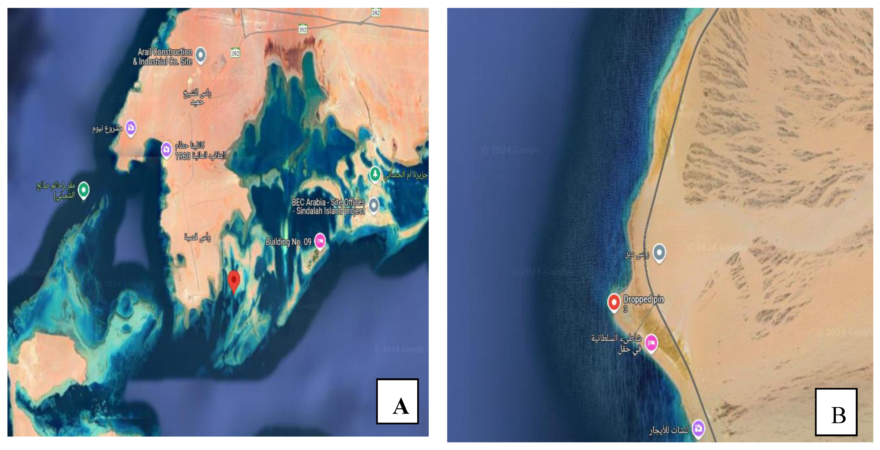
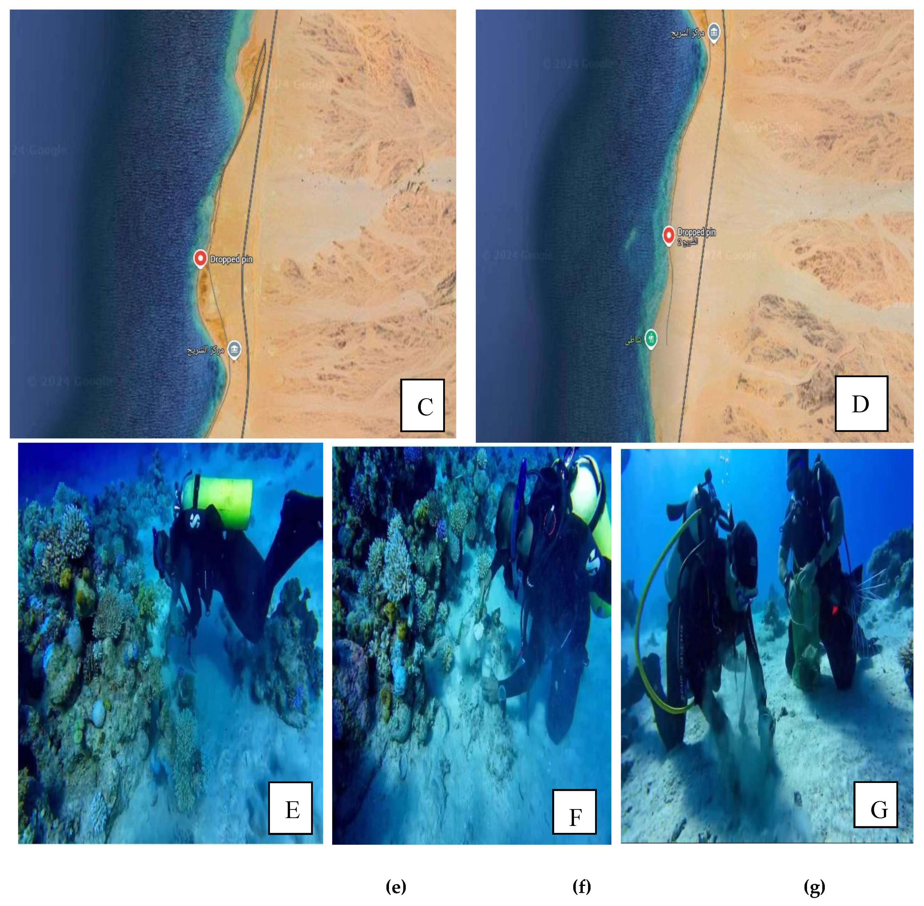
2.1. Study Location and Sampling

Samples of soil were taken from ten different places along the shore of three distinct sites in Red Sea Alshreah, Saweehal, and Marwan. Figure 1 displays the research area’s location on a map (Alshreah, Saweehal, and Marwan). Three duplicate soil samples were gathered from two habitats at each location, obtained far from coral and samples taken near to coral from different ten distinct sites in the selected areas. The coordination of the selected site were 28°01'52.3"N 34°39'22.1"E, for Saweehal, 29°01'33.7"N 34°50'50.4"E, for Marwan, and 29°06'41.7"N 34°52'32.0"E, for Alshreah 1, 29°05'22.7"N 34°52'24.4"E, for Alshreah 2. The selected samples were collected at different depth from 10 m to 20 m in depth. The samples were collected in sterile plastic containers and transported on ice to the University of Tabuk, Saudi Arabia’s Faculty of Science’s Biodiversity Genomics Unit. After being stored at −80 °C for DNA extraction and quantification, the samples were sent to the Genome Life Science Company in India for metagenomics and DNA sequencing.

2.2. Isolation and Quantitative Analysis of DNA

DNA was extracted from the soil samples in accordance with the manufacturer's instructions, utilizing an Alexgen Soil DNA Extraction Kit (CAT No. AG-SD50). The extracted DNA was then quantified using a Qubit® 4.0 fluorometer. The 16S rRNA gene was amplified using universal primers 8F and 1492R, and the ampli-cons that were produced were seen using 1.8% (w/v) agarose gel electrophoresis.

2.3. Setting up the Library

Following the manufacturer’s guidelines, libraries for paired-end sequencing were constructed utilizing a DNA Library Kit from Twist Bioscience for Illumina® (CAT No./ID 104119). Initially, 50 ng of DNA was enzymatically sheared into smaller fragments, which were then prepared for adapter ligation through A-tailing and end repair. The ends of the DNA fragments were fitted with an Illumina-specific adapter to facilitate the binding of the sequencing primers, PCR amplification, and library formation. High-fidelity PCR amplification was performed using a HiFi-PCR MasterMix (Takara, Kusatsu, Japan) to maximize yield. The quantity and quality of the amplified libraries were evaluated using an Agilent TapeStation (4150) system (Agilent Technologies, Waldbronn, Germany) with High-Sensitivity (D1000) Screen-Tape®, following the manufacturer’s instructions.

2.4. Cluster Generation and Sequencing

After analyzing the TapeStation profile and determining the Qubit concentration, the library was placed into an Illumina NovaSeq 6000 system to generate clusters and sequence them. Paired-end sequencing was employed to sequence template fragments in both directions. Molecules from the library hybridized to corresponding adaptor oligonucleotides on the paired-end flow cell. Sequencing from the opposite end of the fragment was made possible by the adaptor design, which enabled selective cleavage of forward strands following reverse strand re-synthesis.

2.5. Data Generation

To isolate individual samples, raw sequence data produced by the NovaSeq6000 platform was demultiplexed. Prior to de novo assembly, adaptor sequences and low-quality reads (QV < 20) were eliminated from the dataset by quality filtering. MEGAHIT v1.2.9 [51,52], a specialist metagenome assembler made to handle big and complicated metagenomic datasets, was then used to put the cleaned reads together.

2.6. Gene Prediction

Using Prodigal (v2.6.3) [53,54] in the metagenome gene prediction mode, gene prediction was executed on the assembled scaffolds. Following that, the projected gene sequences were used for functional and taxonomic studies.
Metagenomic Sequencing and Analysis
Low-quality and single-ended metagenomic reads (length <50 bp or with a quality value (Q-score) <20) were removed using Sickle (v1.33) to ensure high-quality input data [55]. Subsequently, Multiple_Megahit program was used to assemble contigs and scaffolds for each sample, using default parameters and setting a minimum contig size of 300 bp. MetaGene-predicted open reading frames (ORFs) with a length ≥ 100 bp were translated into amino acid sequences. To construct a non-redundant gene catalog, gene sequences were clustered with identity ≥ 0.9 and coverage ≥ 0.9 using CD-HIT to eliminate redundancy.
Using SOAPaligner, gene abundance was computed against the non-redundant gene catalog after it was mapped from high-quality reads. Taxonomic and functional annotations used BLASTP (BLAST v2.2.28+) to search the NR database with an e-value ≤ 1e−5 and the KEGG database.
Taxonomic Annotation
The taxonomy of metagenomic reads was assigned using Kaiju, a fast and sensitive classifier designed for metagenomes [56]. Kaiju relies on the Burrows–Wheeler transform algorithm to identify MEMs at the protein level within a reference database comprised of annotated protein-coding genes. The database comprised complete microbial genomes from NCBI RefSeq, as well as the microbial subset of the NCBI NR database, optionally including fungi and microbial eukaryotes.
The reads are translated into all six possible reading frames and searched against the reference database for MEMs. Taxonomic assignments were done using the lowest common ancestor (LCA) approach within the taxonomic hierarchy for reads that match multiple sequences. This experiment used the standalone version of Kaiju with the following parameters: Database: NR, Sequence low complexity filter: ON, Run mode: Greedy, Minimum match length: 11 amino acids, Minimum match score: 75 and Allowed mismatches: 5. The generated sequences were subsequently analyzed using Kaiju to provide high-quality taxonomic annotation.
Alpha Diversity Analysis
Alpha diversity metrics, which quantify species richness and evenness within a sample, were calculated from OTU abundance data based on Kaiju classifications. For alpha diversity estimation, the phyloseq R package (v1.48.0) was used, applying the estimate_richness function [57]. This analysis included indices such as Chao1, ACE, Shannon, Simpson, Inverse Simpson, and Fisher, providing a detailed understanding of microbial diversity.
Statistical Analysis and Data Visualization
All statistical analyses were carried out in R (v4.3.2), using various packages for data manipulation and visualization. Used packages and their versions were as follows: tidyverse (v2.0.0) for data manipulation [58], ggplot2 (v3.4.4) for making publication-quality plots [59], corrplot (v0.92) for visualization of correlation matrices, ComplexHeatmap (v2.18.0) for making intricate heatmaps [60]. All analyses were conducted within the environment of R to ensure reproducibility and accuracy.

3. Results

Study Site Characteristics

The 60 soil samples used in this investigation came from three different sites in the north area of tabuk region, at different depth from 10 meter to 20 meter in depth. The geographic coordinates of these habitat show that they were geographically dispersed. With median values of pH 7.2 and 8.3 respectively. There were also differences in temperature, of 28.5 °C (range: 28.4 °C, to 29.8 °C). Further the turbidity range were from 11 to 16, and significant variations were observed in the salinity ranged between 43.0 to 44.4. The An overview of the features of the study site is given in Table 1,
Taxonomic Composition
Metagenomic sequencing identified a total of 12,299 operational taxonomic units (OTUs), which were classified into 73 phyla, 149 classes, 322 orders, 736 families, and 2,545 genera. At the phylum level, Pseudomonadota dominated the microbial communities across all samples, followed by Actinomycetota and Bacillota. The most abundant orders were Alteromonadales, followed by Vibrionales and Moraxellales. At the class level, Gammaproteobacteria, Alphaproteobacteria, and Actinobacteria were the most prevalent. The dominant families included Pseudoalteromonadaceae, followed by Vibrionaceae and Moraxellaceae. At the genus level, Pseudomonas, Vibrio, and Psychrobacter exhibited the highest relative abundances. Finally, the most abundant species were Pseudoalteromonas agarivorans, Vibrio chagasii, Vibrio owensii, and Pseudoalteromonas sp. Xi13. The relative abundances of dominant bacterial taxa at different taxonomic levels are visually represented by phylum (Figure 1A), order (Figure 1B), class (Figure 1C), family (Figure 2A), genus (Figure 2B), and species (Figure 2C).
Dominant Microbial Genera per Sample
Alshreah Samples Far from Coral (R1A, R1B, R1C): The dominant genus in these samples is Pseudoalteromonas, averaging 36% of the microbial population. Other notable genera include Vibrio at 28% and Psychrobacter at 11%. These genera reflect a marine-associated microbiome characteristic of areas further from coral influence.
Alshreah Samples Close to Coral (R2A, R2B, R2C): Psychrobacter dominates these samples, accounting for about 29%, followed by Pseudoalteromonas (26%) and Vibrio (8%). The change in dominance reflects that the microbial communities are responding to proximity to coral, which could affect nutrient cycling.
Saweehal Samples Away from Coral (S1A, S1B, S1C): Pseudoalteromonas leads again, averaging 63%. Vibrio contributes about 3%, and Cobetia 9%. These results indicate a microbiome very much affected by the open-water environment.
Saweehal Samples Close to Coral (S2A, S2B, S2C): The community is slightly diversified but dominated by Pseudoalteromonas at 57%, and minor contributions from Vibrio 8% and Cobetia 7%. Such an indication shows coral influence, possibly contributing to the higher presence of symbiotic or coral-associated microbes.
Marwan Samples Far from Coral (M1A, M1B, M1C): The dominant genus is Vibrio at an average of 44%, followed by Pseudoalteromonas at 13% and Psychrobacter at 2%. This shows a different microbial profile probably affected by other environmental factors.
Marwan Samples Close to Coral (M2A, M2B, M2C): Vibrio still dominates at 42%, while Pseudoalteromonas accounts for 10% and Psychrobacter 2%. The data suggest that although proximity to coral alters the community structure, Vibrio is still a resilient genus under these conditions.
Table 1. Distribution of top 5 Genera Across Six Sample Classes. The table provides the average relative abundance (as a percentage) of the five most dominant bacterial genera across six sample classes obtained from three different locations (Alshreah, Saweehal, and Marwan). At each location samples are taken from two environments: samples taken far from coral and samples taken close to coral in 3 replicates.
Genus Alshreah Saweehal Marwan
R1A-R1C (%)
Far from Corals
R2A-R2C (%)
Close to Corals
S1A-S1C (%)
Far from Corals
S2A-S2C (%)
Close to Corals
M1A-M1C (%)
Far from Corals
M2A-M2C (%)
Close to Corals
Pseudoalteromonas 36 26 63 57 13 10
Vibrio 28 8 3 8 44 42
Psychrobacter 11 29 ~1 ~1 2 2
Cobetia 4 4 9 7 3 2
Pseudomonas ~1 7 ~1 ~1 3 4
Analysis of Bacterial Communities in Coral Proximity and Distances
To understand the relationships of samples based on bacterial community composition, PCA (Principal Component Analysis) and pairwise correlation analyses were performed by using species prevalence data. The obtained results showed significant clustering patterns as samples closer to corals formed distinct clusters separate from those farther away (Figure 3). These clusters thereby indicate how proximity to corals influences the bacterial communities perhaps due to ecological or environmental factors forcing microbial associations. The PCA in Figure 2 shows that samples closer to coral were more similar to each other (FDR-corrected p-value = 7 × 10⁻⁵).
Further analysis with 2D Hierarchical clustering corroborated the PCA results. Samples differentiated using the distance from coral presented separated bacterial genera (Table 2). Dominant species in samples closer to coral included Pseudoalteromonas_agarivorans (30%), Vibrio_owensii (11%), and Pseudoalteromonas_sp._Xi13 (10%), indicating a coral-associated microbiome. Conversely, samples farther from coral were dominated by Pseudoalteromonas_sp._DL-6 (35%), Vibrio_chagasii (13%), and Psychrobacter_sp._P11G3 (11%).
In every group, unique populations of bacteria were present. For instance, in Alshreah samples far from coral, the Pseudoalteromonas_agarivorans and Vibrio_owensii were dominant, while the near-coral samples showed higher relative abundance of Pseudoalteromonas_sp._Xi13. Likewise, Saweehal samples that are far from coral had dominance by Pseudoalteromonas_atlantica and Cobetia_sp._cqz5-12. Such difference points out how the environment in association with coral dictates microbial communities.Pairwise correlation analysis revealed significant associations among bacterial species. Strong positive correlations were observed between genera Pseudoalteromonas and Vibrio in close-to-coral environments, suggesting that these bacteria may play a synergistic role in nutrient cycling or coral health. In contrast, genera like Psychrobacter and Cobetia were predominant in nutrient-sparse conditions of open-water environments.The heatmap (Figure 4) visually supports these findings: samples closer to coral are associated with higher microbial diversity, while samples farther away are more homogenous. PCA plots in Figure 3 also depict the spatial separation of clusters with distinct community structures, indicating different community structures that are dependent on proximity to coral (FDR-corrected p-value = 5 × 10⁻¹⁵).The relative abundance of the top 10 bacterial species (Table 2) was then analyzed across samples from Alshreah, Saweehal, and Marwan, distinguishing between those collected far from the coral and those close to it. There were significant differences in microbial community composition in relation to the proximity to coral.
Microbial Alpha Diversity
To estimate microbial diversity in each sample, we applied a series of alpha diversity indices. Observed species richness is the number of different OTUs that exist and displayed large variability among samples, being as low as 3069 OTUs to as high as 12,217 OTUs. The species richness estimators, Chao1 and ACE, supported these findings and revealed that the samples from Alshreah (R1A, R1B, and R1C) and Saweehal (S1A, S1B, and S1C) possessed the highest diversity, especially in Saweehal Far with 52.28% and Alshreah Close with 7.35%.
To account for both species’ richness and evenness, we further calculated the Shannon and Simpson indices. These indices exhibited a generally moderate to high diversity along the samples with the highest marked diversity being recorded at Alshreah Close (7.35%). The Fisher's alpha index that estimates species richness showed an agreement in the trend with the abovementioned indices and highlighted samples from Saweehal Far and Alshreah Close as the ones most diverse in microbial communities while at Marwan Far showed diversity metrics at lower ranges every time.In contrast, samples from Marwan Far (1.75%) and Alshreah Far (5.99%) had relatively lower values for all indices, indicating a less diverse microbial community. These results indicate significant variation in microbial diversity among the different locations and environments, which suggests that proximity to coral and geographical factors influence microbial community structure. Table 3 presents the alpha diversity metrics that were observed for the samples analyzed.
Clusters of Orthologous Groups (COG) Functional Annotation
To explain the functional profiles of protein clusters in samples from different proximity to coral, COG functional annotation was performed. A hierarchical classification of COG functions (Supplementary File 1) was established based on the average TPM (Transcripts Per Million) values of clustered proteins assigned to each COG category across the far-from-coral and close-to-coral samples.
COG Functional Categories in Far-from-Coral Samples
Of the top 20 COG categories in the far-from-coral samples, the most abundant functional annotations were:
COG0840-Methyl-accepting chemotaxis protein: This category was present prominently in all the far-from-coral samples, averaging 6,000–7,147 TPM, which means that this category might be responsible for adaptation and movement of microorganisms according to environmental stimuli.
COG0841-Cation/multidrug efflux pump: This functional class showed the highest average TPM values across the far-from-coral samples, ranging from 5,358 to 7,147. It may reflect involvement in efflux functions and, as such, might be responsible for contributing to resistance in microbial communities of nutrient-limited or stress environments.
COG0642-Signal transduction histidine kinase: COG group with function related to signal transduction and mechanisms responding to the environment; all have high TPM values; this average is 5,051–5,533. The presence of such proteins might be indicative of microbes' adaptation to changing conditions.
COG1629-Outer membrane receptor proteins, primarily Fe transport: In high abundance in the far-from-coral samples (average TPM = 4,200–4,533), these proteins can presumably participate in nutrient acquisition in particular, iron, since iron is a limiting element for marine environments.
COG Functional Categories in Close-to-Coral Samples
In close-to-coral samples, the following categories of COG were dominant:
  • COG0841-Cation/multidrug efflux pump: The category of COG0841, like far-from-coral samples, dominated close-to-coral samples with TPM ranging from 4,165 to 7,543. These efflux pumps could be contributing toward cellular integrity and environmental responses possibly in association with defense mechanisms mediated by coral-associated microbes.
  • COG0840: Methyl-accepting chemotaxis protein: Proteins in this category were also highly represented (average TPM = 4,338–6,524), which reflects their participation in microbial responses to chemical gradients and signaling in the coral-associated environment.
  • COG0642-Signal transduction histidine kinase: This category showed a high level of abundance (average TPM = 4,220–5,166) in coral-associated samples, indicating active regulation of the microbial signaling pathways in the presence of the coral ecosystem.
  • COG1012-NAD-dependent aldehyde dehydrogenases: These metabolism and detoxification enzymes were significantly enriched in the near-coral samples (average TPM = 4,347–4,649), reflecting the metabolic shifts microbes might undergo in response to coral-driven nutrient environments.
  • COG0243-Anaerobic dehydrogenases, usually selenocysteine-containing: This functional category has TPM values of about 4,124, and this would reflect the microbial adaptation to oxygen-limited environments that could be a reflection of anaerobic or microaerophilic conditions near the coral.
The most abundant functional categories between the two environments were COG0841 (Cation/multidrug efflux pump) and COG0840 (Methyl-accepting chemotaxis protein), indicating their key role in microbial survival and adaptation to different environmental conditions. Other important functions include signal transduction (COG0642), which plays a role in the response to environmental stimuli, and iron transport (COG1629), potentially crucial for nutrient acquisition in both environments.
These results give a broad view of the functional roles of microbial communities in both far-from-coral and close-to-coral samples, with key biological processes such as stress response, nutrient transport, and environmental signaling.

4. Discussion REFERENCE 61-92 ADDED

Coral reefs are complex ecosystems that are essential to preserving the health of the seas. However, a number of recent studies offer strong evidence that interactions between microbes and corals are not always consistent throughout the colony and are instead influenced by a variety of factors, such as the physical environment, life history stage, host physiological characteristics, and location within the coral substructure (tissue, gastric cavity, mucus, and skeleton). Numerous studies have demonstrated that coral microbial communities are frequently species-specific and that microbial communities within a species can be extremely stable across biogeographies and environmental conditions. Microbes are essential to several reef activities, including primary production and the cycling of nutrients and organic waste. Microbes are pervasive symbionts of eukaryotic species, supplying the host with nutrition, facilitating chemical cycle, and offering defense roles. They have an effect on the coral's surface area and total accretion, but they also have an impact on community dynamics, coral reproduction, reef species diversity and prosperity, and structural topography [61]. Common coral associates known as ciliates are believed to top-down regulate particular microbial populations and opportunistically feed on coral-associated bacteria. Corals are disease resistant because they have inherent immune system, corals mucus produce antibiotic and utilize cellular phagocytic defense mechanism to eliminate the infectious bacteria and defend against pathogens [62].The Red Sea has some unusual features that may make microbial processes and interactions unique there. These include very high temperatures, very salinity (from very little freshwater input), very high solar irradiation all year round, and a large amount of dust from the nearby desserts that comes in through the Aeolian process. Despite the Red Sea's exceptional biotic conditions, research into the sea has hitherto been hindered by political and practical hurdles. The majority of early studies were carried out in the Gulf of Aqaba, a tiny, shallow outflow in the Red Sea's northeast (Fig. 4.1). Research in this domain has yielded significant insights on Red Sea microorganisms, particularly concerning possible disease-associated bacteria of reef creatures, Corallasia halofolliculina [45,46,47,48]. In the Gulf of Aqaba, the microorganisms that live in the sand were also seen to change when the sediment depth changed. Although the richness of bacterial communities is similar in surface (0–2 cm), intermediate (2–6 cm), and deeper (6–12 cm) sands, there are taxonomic alterations associated with sediment depth [46]. Rhodobacteraceae sequences were found in the more aerobic upper layers of sediment, whereas acidobacteriales predominated in the lower and intermediate layers of sediment, perhaps because of the low oxygen levels [47]. From investigations of additional reef sand environments, bacteria from these same families were found [48]. So far, Red Sea sediment investigations have not incorporated archaea in their techniques, but sequences associated with the archaea, and notably the ammonia oxidizer Nitrosopumilus maritimus, seem to be common in reef sands [63]. Diseases on coral reefs are caused by a number of factors, including red tide outbreaks, increased seawater depth, excessive solar radiation, rising seawater temperatures, and the presence of human waste [64]. The results of our metagenomic study on coral reef have provided baseline information on the prevalence of coral diseases that are having an impact on reefs in northern region of Red Sea of Tabuk, Saudi Arabia. The study's findings unequivocally demonstrate that illness frequency varied greatly among reefs. Disease development may be influenced by both biotic and abiotic factors. Biologic diseases are caused by pathogenic microorganisms, which include bacteria, fungi, and viruses. Abiotic illnesses can arise as a result of environmental stresses that are either naturally occurring or man-made, such as alterations in the surrounding environment or exposure to pollutants. The metagenomic analysis of the bacterial community structures among different locations of Northern Red Sea,Tabuk,K.S.A identified were a total of 12,299 operational taxonomic units (OTUs), which were classified into 73 phyla, 149 classes, 322 orders, 736 families, and 2,545 genera. Our study demonstrated prevalence three top most of phylum, Pseudomonadota, followed by Actinomycetota and Bacillota across all samples collected from three different sites Alshreah, Saweehal and Marwan of northern red sea coral reef of Tabuk. The five most prevalent bacterial genera among six sample classes collected from three distinct sites (Alshreah, Saweehal, and Marwan) are shown in table 1 along with their average relative abundance (as a percentage). Samples collected in three repetitions from two habitats at each location, obtained far from coral and samples taken near to coral. Samples of Alshreah that are far from coral (R1A, R1B, and R1C): With an average of 36% of the microbial population, Pseudoalteromonas is the most prevalent genus in these samples. Other noteworthy genera are Psychrobacter (11%), and Vibrio (28%). The marine-associated microbiome found in regions farther away from coral impact is reflected in these taxa table 1. These results are quite similar with the previous studies done in the Bohai, Yellow, South, and East China Seas have shown that the high abundance of Pseudomonadota and Plancytomycetes could be due to the environmental eutrophication [65,66].
Soil samples of Alshreah Near Coral (R2A, R2B, R2C): With almost 29% of the Psychrobacter predominates, followed by Pseudoalteromonas (26%), and Vibrio (8%). The shift in dominance indicates how the microbial communities react to coral proximity, which may have an impact on the cycle of nutrients. Planctomycetota have significant biogeochemical functions in anaerobic ammonium oxidation [67], methane oxidation [68], and carbon recycling [69],whereas pseudomonadota is essential for the breakdown of organic nitrogen in sediments [70,71]. Since both phyla are chemotrophic bacteria, their high abundance may be a result of the environment's high relative chemical concentration. Saweehal soil samples away from Coral With an average of 63%, pseudoalteromonas is once again in the lead. About 3% comes from Vibrio and 9% from Cobetia. These findings suggest that the open-water environment has a significant impact on the microbiota. Whereas soil samples from same site from the vicinity of coral reef accounts Pseudoalteromonas for 57% of the total, with Vibrio and Cobetia contributing just 8% and 7%, respectively table 1. These results demonstrate the effect of corals, which may be one reason why symbiotic or coral-associated bacteria are more prevalent. According to current research, there are bacterial species present that are assumed to play advantageous functions for the deep Red Sea corals, such as those related to the nitrogen and carbon cycles e.g., E. fistula, Dendrophyllia sp [72].According to the earlier studies members of the Pseudomonadaceae and Endozoicomonadaceae families have a high abundance of genes encoding proteins involved in host-symbiont identification and colonization [61].
Previous studies also suggest other characteristics, such as the breakdown of host-derived taurine, which was commonly observed in strains of Cobetia and Halomonas (Halomonadaceae), and antiviral protection in some Endozoicomonas strains. The widespread occurrence of these bacterial species linked to various coral taxa raises the possibility that they are significantly corroborated for coral probiotic thermal protective role, metabolism and health [73,74].Some studies provided an information with evidence that some members of this taxon play a key role in the sulfur cycle in corals [75]. Metagenomic profiling revealed that some other samples had a variety of metabolic sources, while around half of the samples indicated possible concentration of high sulfate and nitrite reducers and dehalogenating bacteria. The dominance of Alteromonas and Pseudoalteromonas, are known to be important in the metabolism of dimethyl-sulfoniopropionate (DMSP), may be the only explanation for the high concentration of sulfate reducers [76].Elevated levels of DMSP production in Northern Red Sea corals and nearby waters may be indicated by the high abundance of certain taxa that can prove to be good Beneficial Microorganisms for Corals (BMC) candidates, and found the genes involved in the degradation of dimethylsulfoniopropionate (DMSP) in the genomes of Endozoicomonas, verified that some bacteria can use DMSP as a carbon source [77]. Psychrobacter Phenylpyruvicus is a nonmotile, gram-negative, catalase- and oxidase-positive bacterium that is easily isolated from saltwater and other typical marine settings. It may be involved in the degradation of dimethylsulfoniopropionate (DMSP), a significant osmolyte of algae. When DMSP is broken down, dimethylsulfide is released into the atmosphere, where it may affect the weather by promoting the production of clouds. Although P. pacifiensis may also be found in subsea floor sediments and at sea bottoms of up to 6000 meters, most soil habitats cannot support Psychobacter species unless they are exposed to constant low temperatures and sporadic freezing [77].
The bile acid derivatives from marine Psychrobacter sp. were described as an example of marine-derived antibacterial steroids in previously conducted research [78]. Both the coral and the endosymbiotic dinoflagellates create DMSP, which is then broken down by related bacteria to yield acrylate, dimethylsulfide (DMS), and dimethyl sulfoxide (DMSO). These compounds may play a part in antioxidant capacity, and osmoregulation [79]. Therefore, increased DMSP production might indicate the ability to endure the extreme salinity and heat anomalies that describes distinctive nature of the northern Red Sea. In our studies different species of vibrio bacteria such as vibrio owensii, vibrio chagasii, and vibrio ponticus are observed in table 2. Species of vibrio owansii OCN002 has been recognised as first bacteria from Hawaii's reefs that causes chronic montipora white syndrome (cMWS), a tissue-loss disease found on corals throughout the Hawaiian archipelago. Another bacterial species of Pseudoalteromonas piratica strain OCN003 causes Chronic MWS to acute MWS (aMWS) [89]. Pseudoalteromonas agarivorans species produces most common metalloproteinase collagenolytic protease which degrade collagen, is the primary pathogen affecting the coral reef [68].Some species of Pseudoaltromonas exhibit extracellular antibacterial, compounds produced by Pseudoalteromonas spp. linked to certain coral species may protect against invading pathogens. Similarly other studies have reported a higher prevalence of Pseudoalteromonas phylotypes in Acropora millepora that are resistant to Vibrio sp. infection. Additionally, certain strains of P.alteromonas species are known to fix nitrogen through dinitrogen, and they may then transfer fixed nitrogen to the algal endosymbiosis linked to P. damicornis larvae [81].Furthermore, the Red Sea possesses a high level of coral endemism, but research on endemic species is still in its early stages. There is a chance to investigate endemic corals that could have unique characteristics, as we were unable to locate any relevant publications about the bacterial communities of Red Sea endemic corals. These particular characteristics and possible special bacterial partners may be especially intriguing if linked to the endemic corals' ability to adjust to environmental gradients in the Red Sea through the flexibility and plasticity of their microbiomes [82]. Additionally, it has been reported that Vibrios microbe can produce enzymes to break down polycyclic aromatic hydrocarbons to produce antibiotic substances, and provide essential polyunsaturated fatty acids. Chitin is the second most abundant polymer and the primary source of amino sugars in the oceans [83]. Salini vibrio sp., a member of the Vibrionaceae family, was part of a probiotic consortium that helped coral bleaching recover and protect against mortality, according to Santoro and colleagues [84]. Because of its antagonistic activity against two well-known coral diseases, Vibrio coralliilyticus and V. alginolyticus, this Salinivibrio sp. was chosen to be a part of the consortium of beneficial microorganisms for corals (BMCs). The distinct functions of particular genera within the same bacterial family in the coral holobiont can be explained by the substantial differences in genomes between non-pathogenic and pathogenic members of the Vibrionaceae family [61].The studies indicates that while the precise function of various bacterial phylotypes in Red Sea corals is yet to be fully understood, they may play crucial roles in holobiont fitness. The study found distinct bacterial communities in each group, with Pseudoalteromonas_agarivorans and Vibrio_owensii dominating in Alshreah samples distant from coral, while Pseudoalteromonas_sp._Xi13 was more abundant in closer samples. Most of the microorganisms found in coral mucus are Vibrio strains, and the quantities of these bacteria are significantly greater than those found in the surrounding saltwater. The majority of Vibrio strains, including V. mediterranei, V. coralliilyticus, V. harveyi, and V. alginolyticus, are cause behind coral bleaching, have been shown to induce illnesses in marine organisms when their mucus separates from the coral [85]. In coral reef environments, Some Vibrio species are essential for the transfer and circulation of a variety of materials such as chitin, alginate, and agar and energy as these vibrio genera has tendency to consume an extensive array of carbon substrates. It was shown that coral symbiotic microorganisms Proteobacteria such as Pseudoalteromonas, Pseudomonas and Cyanobacteria where the most prevalent phyla of coral microorganisms are crucial markers of coral health in maintaining material transformation, and biogeochemical cycles.
In habitats around coral, strong positive relationships between the species Pseudoalteromonas and Vibrio were found, indicating that these bacteria could work in concert to support coral health or nutrient cycling. Therefore, corals and the nearby saltwater could be the best places to research Vibrio's spread and pathogenicity as well as find new species and enzymes. Furthermore, figure 4 depicts heat map of bacterial abundance across all samples, through a 2D-hierarchical clustering heatmap. The heatmap's rows represent the various bacterial species, while the columns show the samples that have been grouped together according to their site and closeness to the coral. The Shannon and Simpson indices were also computed to take species richness and evenness into consideration. The highest diversity has been identified in Saweehal far from corals (S1A, S1B, and S1C) 52.8% and in Alshreah close (7.35%) was observed, while at Marwan far every time showed lower ranges of microbial community diversity ie; 1.75%. The microbial community was less diversified in the samples from Alshreah Far (5.99%) and Marwan Far (1.75%), which had comparatively lower values for all indices. According to these findings, there is a notable difference in microbial diversity across the various settings and locales, revealing that geographic variables and coral closeness affect the diversity of microbial communities Table 3. The more prevalent microbial community of corals from inner and outer reefs zones found in the soil of various coral reef settings is consistent with the alphaproteobacterium Candidatus having a higher relative abundance in the surface mucus layer (SML) microbiome of corals around the coral reef zone.
The taxonomic diversity measures support the idea that the microbial communities in the coral's immediate closest environment influence the coral SML microbiome. However, both at the taxonomic and functional levels, the local environment (i.e., inner reefs vs outer reefs) and the coral host versus soil simultaneously determine the microbial community structure (i.e., relative abundances of sequences) in Northern Red Sea reef system [Pollock., et al 2018]. Aligning query gene or protein sequences with COG database sequences COG functional annotation was used to describe the functional patterns of protein clusters in samples that were at varying distances from coral. The average TPM (Transcripts Per Million) values of clustered proteins allocated to each COG category across the far-from-coral and close-to-coral samples were used to create a hierarchical categorization of COG functions (Supplementary File 1). With an average of 6,000–7,147 TPM, Methyl-accepting Chemotaxis Protein was found in all of the distant coral samples. This suggests that this category may be in charge of microorganisms' ability to adapt and migrate in response to environmental stimuli. While Methyl-accepting chemotaxis proteins in coral associated environment were highly represented, depicts their involvement in microbial responses to chemical gradients and signaling.
The multidrug/cation efflux pump, with high average TPM levels, may contribute to microbial resistance in stressful or nutrient-limited settings, as indicated by far-from-coral. Similar to far from coral cation/Multidrug Efflux Pump category dominates close-to-coral samples, supporting environmental reactions and cellular integrity through defensive mechanisms mediated by coral-associated bacteria. Signal transduction histidine kinase, has high TPM levels of 5,051–5,533, suggesting microorganisms have adapted to their far coral environment. Whereas signal transduction histidine kinase abundance in coral-associated samples indicates that the coral ecosystem actively regulates microbial signal transduction pathways and adapts a broad range of environmental cues. Histidine kinase (HK) autophosphorylating response regulator protein enable the cells to detect and react external stimuli which thrive as a molecular switch that regulates variety of effector activities with adaptable conserved domains that may be easily changed to the unique requirements of various systems [86]. Outer membrane receptor proteins, abundant in far-from-coral samples with TPM average 4200-4533, may be involved in nutrient acquisition, particularly iron, as iron is a limiting element for marine habitats [87].
Soil around the coral reefs are rich in metabolism, with detoxifying enzymes like NAD-dependent aldehyde dehydrogenases undergoing metabolic changes based on coral-driven food conditions.Anaerobic dehydrogenases, including selenocysteine, exhibit microbial adaptability to oxygen-limited conditions, possibly due to anaerobic or microaerophilic conditions near coral, as indicated by TPM values of around 4,124. To summarize, the most prevalent COG in both distant and close coral reef environments rely on cation/multidrug efflux pump, methyl-accepting chemotaxis protein, iron transport, and signal transduction for microbial survival, environmental adaptability, and nutrition absorption [87].The shedding of carbon-rich mucus by corals may result in chemical hotspots, While, the diffusion of methyl-accepting compounds away from the coral surface into the surrounding saltwater creates microscale chemical gradients that extend into the nearby ocean [88]. The findings of methyl chemotaxis pattern suggest that bacteria connected to the surface of the coral had noticeably more chemotactic capabilities then bacteria away from the coral. Chemotaxis may serve as a behavioural filter that determines the makeup of microbial communities inside particular coral microniches [89].
It is unclear what environmental conditions lead to the development of planktonic blooms and how the microbial communities interact with one another. To determine the rhythmic pattern (diurnal, monthly, or seasonal) of microbial composition and track changes in these patterns in response to human activities, extensive time-series investigations of Red Sea microbiota across multiple niches are required. Coastal areas of the Red Sea have seen a sharp increase in pollution in recent decades as a result of growing industries and urbanization. Industrial and anthropogenic pollutants discharged directly into the marine environment have the potential to alter the makeup of microbial communities. Phylogenetic analysis of cultured microbial communities isolated from sediments in industrial zones along the Egyptian Red Sea coast was carried out by Mustafa et al. [90].They investigated distinct microbial communities, mainly oil/hydrocarbon degrading bacteria and a number of human pathogens, including known Vibrio and Clostridium species. According to the study, alterations in the profile of the microbial population may serve as a gauge for the effects of human activity on marine habitats.
Modelling and comprehending the microbial dynamics of an environment requires the detection of spatiotemporal patterns in the microbiome. Large datasets of the ocean microbiome and environmental characteristics have been produced in recent years by an increasing number of time-series studies of the ocean microbiota in order to provide ecological insights into the stability and dynamics of microbial communities over time [91].In order to estimate the distribution pattern of bacteria and their genes in distinct niches, upcoming Red Sea metagenomic research should develop computational models of the microbiota that take into consideration a variety of environmental parameters. The Red Sea may serve as a sophisticated model for researching the possible effects of climate change on future seas due to its high year-round temperatures and relative isolation from other oceans [92].Further microbiome structure and diversity are crucial markers of coral health in maintaining material transformation, and biogeochemical cycles. When integrated and juxtaposed with upcoming Red Sea microbiome data, the information gathered from these investigations may provide a very sensitive indicator of environmental shifts and worldwide wellness.

5. Conclusions

The present study emphasized the Microbiome taxa and gene forming of the microbiome in three distinct locations surrounding and beneath the coral communities. The structural makeup of the microbial communities varied significantly according to the closeness to the coral. This study revealed that Pseudoalteromonas agarivorans, Vibrio owensii, and Pseudoalteromonas sp. Xi13 are the most predominant species in samples closer to coral. Moreover, Vibrio species were the most prevalent microorganisms in the coral mucus. Furthermore, in close-to-coral habitats, there were strong positive associations between the genera Vibrio and Pseudoalteromonas, indicating that these bacteria may work in concert to support coral health or nutrient cycling

Supplementary Materials

The following supporting information can be downloaded at the website of this paper posted on Preprints.org.

Funding

“This research was funded by Deanship of Research and Graduate Studies at University of Tabuk for funding this work through Research no.0144-1444-S.

Acknowledgments

This article is derived from a research grant funded by the Research, Development, and Innovation Authority (RDIA) - Kingdom of Saudi Arabia with grant number (13445-Tabuk-2023-UT-R-3-1-SE).The authors extend their appreciation to the Deanship of Research and Graduate Studies at University of Tabuk for funding this work through Research no.0144-1444-S.

Conflicts of Interest

The authors declare no conflicts of interest.

References

  1. Mohamed, A.R.; Ochsenkühn, M.A.; Kazlak, A.M.; Moustafa, A.; Amin, S.A. The coral microbiome: towards an understanding of the molecular mechanisms of coral–microbiota interactions. FEMS Microbiology 2023, 1–26. [Google Scholar] [CrossRef]
  2. Jaspers, C.; Fraune, S.; Arnold, A.E.; et al. Resolving structure and function of metaorganisms through a holistic framework combining reductionist and integrative approaches. Zoology 2019, 133, 81–87. [Google Scholar] [CrossRef] [PubMed]
  3. Robbins, S.J.; Singleton, C.M.; Chan, C.X.; et al. A genomic view of the reef building coral Porites lutea and its microbial symbionts. Nat Microbiol 2019, 4, 2090–100. [Google Scholar] [CrossRef]
  4. Peixoto, R.S.; Sweet, M.; Villela, H.D.M.; et al. Coral probiotics: premise, promise, prospects. Annu Rev Anim Biosci 2021, 9, 265–88. [Google Scholar] [CrossRef] [PubMed]
  5. Rohwer, F.; Seguritan, V.; Azam, F.; et al. Diversity and distribution of coral-associated bacteria. Mar Ecol Prog Ser 2002, 243, 1–10. [Google Scholar] [CrossRef]
  6. Rosenberg, E.; Koren, O.; Reshef, L.; et al. The role of microorganisms in coral health, disease and evolution. Nat Rev Microbiol 2007, 5, 355–62. [Google Scholar] [CrossRef]
  7. Rosenberg, E.; Zilber-Rosenberg, I. The hologenome concept of evolution after 10 years. Microbiome 2018, 6, 78. [Google Scholar] [CrossRef]
  8. Zilber-Rosenberg, I.; Rosenberg, E. Microbial-driven genetic variation in holobionts. FEMS Microbiol Rev 2021, 45, fuab022. [Google Scholar] [CrossRef] [PubMed]
  9. Muscatine, L. The role of symbiotic algae in carbon and energy flux in reef corals. In: Dubinsky Z (ed.), Ecosystems of the World. Amsterdam: Elsevier, 1990,75–87.
  10. Morris, L.A.; Voolstra, C.R.; Quigley, K.M.; et al. Nutrient availability and metabolism affect the stability of coral–Symbiodiniaceae symbioses. Trends Microbiol 2019, 27, 678–89. [Google Scholar] [CrossRef]
  11. Bourne, D.G.; Morrow, K.M.; Webster, N.S. Insights into the coral microbiome: underpinning the health and resilience of reef ecosystems. Annu Rev Microbiol 2016, 70, 317–40. [Google Scholar] [CrossRef] [PubMed]
  12. van Oppen, M.J.H.; Blackall, L.L. Coral microbiome dynamics, functions and design in a changing world. Nat Rev Microbiol 2019, 17, 557–67. [Google Scholar] [CrossRef] [PubMed]
  13. Blackall, L.L.; Wilson, B.; van Oppen, M.J. Coral—the world’smost diverse symbiotic ecosystem. Mol Ecol 2015, 24, 5330–47. [Google Scholar] [CrossRef] [PubMed]
  14. Huggett, M.J.; Apprill, A. Coral microbiome database: integration of sequences reveals high diversity and relatedness of coral associated microbes. Environ Microbiol Rep 2019, 11, 372–85. [Google Scholar] [CrossRef] [PubMed]
  15. Chen, J.; Guo, Y.; Jia, Y.; et al. Diversity, function and evolution of marine microbe genomes. bioRxiv 2021. [CrossRef]
  16. Ziegler, M.; Grupstra, C.G.B.; Barreto, MM.; et al. Coral bacterial community structure responds to environmental change in a host-specific manner. Nat Commun 2019, 10, 3092. [Google Scholar] [CrossRef] [PubMed]
  17. Voolstra, C.R.; Ziegler, M. Adapting with microbial help: microbiome flexibility facilitates rapid responses to environmental change. BioEssays 2020, 42, 2000004. [Google Scholar] [CrossRef] [PubMed]
  18. Meunier, V.; Geissler, L.; Bonnet, S.; et al. Microbes support enhanced nitrogen requirements of coral holobionts in a high CO2 environment. Mol Ecol 2021, 30, 5888–99. [Google Scholar] [CrossRef]
  19. Sweet, M.; Villela, H.; Keller-Costa, T.; et al. Insights into the cultured bacterial fraction of corals. m Systems 2021, 6, e0124920. [Google Scholar] [CrossRef]
  20. Haas, A.; Fairoz, M.; Kelly, L.; et al. Global microbialization of coral reefs. Nat Microbiol 2016, 1, 16042. [Google Scholar] [CrossRef]
  21. Pollock, F.J.; McMinds, R.; Smith, S.; et al. Coral-associated bacteria demonstrate phylosymbiosis and cophylogeny. Nat Commun 2018, 9, 4921. [Google Scholar] [CrossRef] [PubMed]
  22. Vanwonterghem, I.; Webster, N.S. Coral reef microorganisms in a changing climate. iScience 2020, 23, 100972. [Google Scholar] [CrossRef] [PubMed]
  23. Wangpraseurt, D.; Jacques, S.L.; Petrie, T.; et al. Monte Carlo modeling of photon propagation reveals highly scattering coral tissue. Front Plant Sci 2016, 7, 1404. [Google Scholar] [CrossRef] [PubMed]
  24. Pernice, M.; Raina, J.B.; Radecker, N.; et al. Down to the bone: the role of overlooked endolithic microbiomes in reef coral health. ISME J 2020, 14, 325–34. [Google Scholar] [CrossRef]
  25. Sweet, M.J.; Croquer, A.; Bythell, J.C. Bacterial assemblages differ between compartments within the coral holobiont. Coral Reefs 2010, 30, 39–52. [Google Scholar] [CrossRef]
  26. Leite, D.C.A.; Salles, J.F.; Calderon, E.N.; et al. Coral bacterial-core abundance and network complexity as proxies for anthropogenic pollution. Front Microbiol 2018, 9, 833. [Google Scholar] [CrossRef]
  27. Neave, M.J.; Michell, C.T.; Apprill, A.; et al. Endozoicomonas genomes reveal functional adaptation and plasticity in bacterial strains symbiotically associated with diverse marine hosts. Sci Rep 2017, 7, 40579. [Google Scholar] [CrossRef] [PubMed]
  28. Matthew J. Neave, Amy Apprill, Greta Aeby, Sou Miyake, and Christian R. Voolstra. Microbial Communities of Red Sea Coral Reefs, Coral reef of the World 11, Christian R. Voolstra Michael L. Berumen. Springer, Nature Switzerland AG 2019, Vol 11; 53-68.
  29. Pogoreutz, C.; Voolstra, C.R.; Rädecker, N.; et al. The coral holobiont highlights the dependence of cnidarian animal hosts on their associated microbes. In: Bosch TCG, Hadfield, M.G.; (eds). Cellular Dialogues in the Holobiont. Boca Raton: CRC Press, 2020.
  30. Falkowski, P.G.; Dubinsky, Z.; Muscatine, L.; et al. Population control in symbiotic corals. Bioscience 1993, 43, 606–11. [Google Scholar] [CrossRef]
  31. Cunning, R.; Muller, E.B.; Gates, R.D.; et al. A dynamic bioenergetic model for coral–Symbiodinium symbioses and coral bleaching as an alternate stable state. J Theor Biol 2017, 431, 49–62. [Google Scholar] [CrossRef] [PubMed]
  32. Muscatine, L.; Porter, J.W. Reef corals: mutualistic symbioses adapted to nutrient-poor environments. Bioscience 1977, 27, 454–60. [Google Scholar] [CrossRef]
  33. Lema, K.A.; Willis, B.L.; Bourne, D.G. Corals form characteristic associations with symbiotic nitrogen-fixing bacteria. Appl Environ Microbiol 2012, 78, 3136–44. [Google Scholar] [CrossRef]
  34. Wang, J.; Douglas, A. Essential amino acid synthesis and nitrogen recycling in an alga–invertebrate symbiosis. Mar Biol 1999, 135, 219–22. [Google Scholar] [CrossRef]
  35. Yellowlees, D.; Rees, T.A.V.; Leggat, W. Metabolic interactions between algal symbionts and invertebrate hosts. Plant Cell Environ 2008, 31, 679–94. [Google Scholar] [CrossRef]
  36. Reynaud, S.; Martinez, P.; Houlbrèque, F.; et al. Effect of light and feeding on the nitrogen isotopic composition of a zooxanthellate coral: role of nitrogen recycling. Mar Ecol Prog Ser 2009, 392, 103–10. [Google Scholar] [CrossRef]
  37. Olson, N.D.; Lesser, M.P. Diazotrophic diversity in the Caribbean coral, Montastraea cavernosa. Arch Microbiol 2013, 195, 853–9. [Google Scholar] [CrossRef]
  38. Lema, K.A.; Bourne, D.G.; Willis, B.L. Onset and establishment of diazotrophs and other bacterial associates in the early life history stages of the coral Acropora millepora. Mol Ecol 2014, 23, 4682–95. [Google Scholar] [CrossRef]
  39. Kimes, N.E.; Van Nostrand, J.D.; Weil, E.; et al. Microbial functional structure of Montastraea faveolata, an important Caribbean reef building coral, differs between healthy and yellow-band diseased colonies. Environ Microbiol 2010, 12, 541–56. [Google Scholar] [CrossRef] [PubMed]
  40. Rädecker, N.; Pogoreutz, C.; Gegner, H.M.; et al. Heat stress reduces the contribution of diazotrophs to coral holobiont nitrogen cycling. ISME J 2022, 16, 1110–8. [Google Scholar] [CrossRef]
  41. Seymour, J.R.; Amin, S.A.; Raina, J.B.; et al. Zooming in on the phycosphere: the ecological interface for phytoplankton–bacteria relationships. Nat Microbiol 2017, 2, 17065. [Google Scholar] [CrossRef] [PubMed]
  42. Cirri, E.; Pohnert, G. Algae–bacteria interactions that balance the planktonic microbiome. New Phytol 2019, 223, 100–6. [Google Scholar] [CrossRef]
  43. Delgadillo-Ordoñez, N.; Raimundo, I.; Barno, A.R.; Osman, E.O.; Villela, H.; Bennett-Smith, M.; Voolstra, C.R.; Benzoni, F.; Peixoto, R.S. Red Sea Atlas of Coral-Associated Bacteria Highlights Common Microbiome Members and Their Distribution across Environmental Gradients. Micro 2022, 10, 2340. [Google Scholar] [CrossRef] [PubMed]
  44. Osman, E.O.; Suggett, D.J.; Voolstra, C.R.; Pettay, D.T.; Clark, D.R.; Pogoreutz, C.; Sampayo, E.M.; Warner, M.E.; Smith, D.J. Coral Microbiome composition along the northern Red Sea suggests high plasticity of bacterial and specificity of endosymbiotic dinoflagellate communities. Microbiome 2020, 8, 8. [Google Scholar]
  45. Ziegler, M.; Roik, A.; Porter, A.; Zubier, K.; Mudarris, M.S.; Ormond, R.; Voolstra, C.R. Coral microbial community dynamics in response to anthropogenic impacts near a major city in the Central Red Sea. Mar Pollut Bull 2016, 105, 629–640. [Google Scholar] [CrossRef] [PubMed]
  46. Antony, C.P.; Peixoto, R.S. Unlocking the genomic potential of Red Sea coral probiotics Inês Raimundo, Phillipe M. Rosado, Adam R. Barno. Scientifc Reports 2024, 14, 14514. [Google Scholar]
  47. Winkler, R.; Antonius, A.; Abigail Renegar, D. The skeleton eroding band disease on coral reefs of Aqaba, Red Sea. Mar Ecol 2004, 25, 129–144. [Google Scholar] [CrossRef]
  48. Roik, A.; Röthig, T.; Roder, C.; Ziegler, M.; Kremb, S.G.; Voolstra, C.R. Year-long monitoring of Physico-chemical and biological variables provide a comparative baseline of coral reef functioning in the Central Red Sea. PLoS One 2016, 11, e0163939. [Google Scholar] [CrossRef]
  49. Röthig, T.; Ochsenkühn, M.A.; Roik, A.; van der Merwe, R.; Voolstra, C.R. Long-term salinity tolerance is accompanied by major restructuring of the coral bacterial microbiome. Mol Ecol 2016, 25, 1308–1323. [Google Scholar] [CrossRef]
  50. Röthig, T.; Roik, A.; Yum, L.K.; Voolstra, C.R. Distinct bacterial microbiomes associate with the Deep-Sea coral Eguchipsammia fistula from the Red Sea and from aquaria settings. Front Mar Sci 2017, 4, 259. [Google Scholar] [CrossRef]
  51. Isaza, D.F.G.; Cramp, R.L.; Franklin, C.E. Living in polluted waters: A meta-analysis of the effects of nitrate and interactions with other environmental stressors on freshwater taxa. Environ. Pollut. 2020, 261, 114091. [Google Scholar] [CrossRef] [PubMed]
  52. Li, D.; Liu, C.M.; Luo, R.; Sadakane, K.; Lam, T.W. MEGAHIT: An ultra-fast single-node solution for large and complex metagenomics assembly via succinct de Bruijn graph. Bioinformatics 2015, 31, 1674–1676. [Google Scholar] [CrossRef] [PubMed]
  53. Newton, R.J.; Jones, S.E.; Eiler, A.; McMahon, K.D.; Bertilsson, S. A guide to the natural history of freshwater lake bacteria. Microbiol. Mol. Biol. Rev. 2011, 75, 14–49. [Google Scholar] [CrossRef]
  54. Hyatt, D.; Chen, G.L.; Locascio, P.F.; Land, M.L.; Larimer, F.W.; Hauser, L.J. Prodigal: Prokaryotic gene recognition and translation initiation site identification. BMC Bioinform. 2010, 11, 119. [Google Scholar] [CrossRef]
  55. Wu, Y.-W.; Simmons, B.A.; Singer, S.W. MaxBin 2.0: an automated binning algorithm to recover genomes from multiple metagenomic datasets. Bioinformatics, 2016, 32, 605–607. [Google Scholar] [CrossRef] [PubMed]
  56. Menzel, P.; et al. Fast and sensitive taxonomic classification for metagenomics with Kaiju. Nat. Commun. 2016, 7, 11257. [Google Scholar] [CrossRef] [PubMed]
  57. McMurdie, P.J.; Holmes, S. phyloseq: An R Package for Reproducible Interactive Analysis and Graphics of Microbiome Census Data. PLoS ONE 2013, 8, e61217. [Google Scholar] [CrossRef]
  58. Wickham, W.H.; Averick, M.; Bryan, J.; Chang, W.; McGowan, L.D.; et al. Welcome to the Tidyverse. Journal of Open Source Software, 2019, 4, 1686. [Google Scholar] [CrossRef]
  59. Wickham, H. (2016) ggplot2: Elegant Graphics for Data Analysis. Springer-Verlag, New York. [CrossRef]
  60. Gu, Z.; Eils, R.; Schlesner, M. Complex heatmaps reveal patterns and correlations in multidimensional genomic data. Bioinformatics 2016, 32, 2847–2849. [Google Scholar] [CrossRef]
  61. Sweet, M.; Villela, H.; Keller-Costa, T.; Costa, R.; Romano, S.; Bourne, D.G.; Cárdenas, A.; Huggett, M.J.; Kerwin, A.H.; Kuek, F.; et al. Insights into the cultured bacterial fraction of corals. mSystems. 2021, 6, e01249-20. [Google Scholar] [CrossRef]
  62. Pollock, F.J.; McMinds, R.; Smith, S.; et al. Coral-associated bacteria demonstrate phylosymbiosis and cophylogeny. Nat Commun. 2018, 9, 4921. [Google Scholar] [CrossRef] [PubMed]
  63. Sato, Y.; Civiello, M.; Bell, S.C.; et al. Integrated approach to understanding the onset and pathogenesis of black band disease in corals. Environ. Microbiol. 2016, 18, 752–756. [Google Scholar] [CrossRef] [PubMed]
  64. Moriarty, T.; Leggat, W.; Huggett, M. J.; Ainsworth, T. Coral disease causes, consequences, and risk within coral restoration. Trends in Microbiology, 2020, 28, 793–807. [Google Scholar] [CrossRef] [PubMed]
  65. Zhang, J.; Chen, M.; Huang, J.; Guo, X.; Zhang, Y.; Liu, D.; Wu, R.; He, H.; Wang, J. Diversity of the microbial community and cultivable protease-producing bacteria in the sediments of the Bohai Sea, Yellow Sea and South China Sea. PLoS ONE 2019, 14, e0215328. [Google Scholar] [CrossRef]
  66. Ye, Q.; Wu, Y.; Zhu, Z.; Wang, X.; Li, Z.; Zhang, J. Bacterial diversity in the surface sediments of the hypoxic zone near the Changjiang Estuary and in the East China Sea. MicrobiologyOpen 2016, 5, 323–339. [Google Scholar] [CrossRef]
  67. Chung, H.W.; Liu, C.C.; Chiu, Y.S.; Liu, J.T. Spatiotemporal variation of Gaoping River plume observed by Formosat-2 high resolution imagery. J. Mar. Syst. 2014, 132, 28–37. [Google Scholar] [CrossRef]
  68. Bhattacharyya, A.; Haldar, A.; Bhattacharyya, M.; Ghosh, A. Anthropogenic influence shapes the distribution of antibiotic resistant bacteria (ARB) in the sediment of Sundarban estuary in India. Sci. Total Environ. 2019, 647, 1626–1639. [Google Scholar] [CrossRef]
  69. Wang, Y.; Qian, P.Y. Conservative fragments in bacterial 16S rRNA genes and primer design for 16S ribosomal DNA amplicons in metagenomic studies. PLoS ONE 2009, 4, e7401. [Google Scholar] [CrossRef]
  70. Zhou, M.-Y.; Wang, G.-L.; Li, D.; Zhao, D.-L.; Qin, Q.-L.; Chen, X.-L.; Chen, B.; Zhou, B.-C.; Zhang, X.-Y.; Zhang, Y.-Z. Diversity of both the cultivable protease-producing bacteria and bacterial extracellular proteases in the coastal sediments of King George Island, Antarctica. PLoS ONE 2013, 8, e79668. [Google Scholar] [CrossRef]
  71. Zhang, X.Y.; Han, X.X.; Chen, X.L.; Dang, H.Y.; Xie, B.B.; Qin, Q.L.; Shi, M.; Zhou, B.C.; Zhang, Y.Z. Diversity of cultivable protease-producing bacteria in sediments of Jiaozhou Bay, China. Front. Microbiol. 2015, 6, 1021. [Google Scholar] [CrossRef]
  72. Röthig, T.; Yum, L.K.; Kremb, S.G.; Roik, A.; Voolstra, C.R. Microbial community composition of deep-sea corals from the Red Sea provides insight into functional adaption to a unique environment. Sci. Rep. 2017, 7, 44714. [Google Scholar] [CrossRef]
  73. Neave, M.J.; Michell, C.T.; Apprill, A.; Voolstra, C.R. Endozoicomonas genomes reveal functional adaptation and plasticity in bacterial strains symbiotically associated with diverse marine hosts. Sci. Rep. 2017, 7, 40579. [Google Scholar] [CrossRef] [PubMed]
  74. Rosado, P.M.; Leite, D.C.A.; Duarte, G.A.S.; Chaloub, R.M.; Jospin, G.; da Rocha, U.N.; Saraiva, J.P.; Dini-Andreote, F.; Eisen, J.A.; Bourne, D.G.; et al. Marine probiotics: Increasing coral resistance to bleaching through microbiome manipulation. ISME J. 2019, 13, 921–936. [Google Scholar] [CrossRef]
  75. Tandon, K.; Lu, C.-Y.; Chiang, P.-W.; Wada, N.; Yang, S.-H.; Chan, Y.-F.; Chen, P.-Y.; Chang, H.-Y.; Chiou, Y.-J.; Chou, M.-S.; et al. Comparative genomics: Dominant coral-bacterium Endozoicomonas acroporae metabolizes dimethylsulfoniopropionate (DMSP). ISME J. 2020, 14, 1290–1303. [Google Scholar] [CrossRef] [PubMed]
  76. Hopkins, F.E.; Bell, T.G.; Yang, M.; Suggett, D.J.; Steinke, M. Air exposure of coral is a significant source of dimethylsulfide (DMS) to the atmosphere. Sci Rep. 2016, 6, 36031. [Google Scholar] [CrossRef]
  77. Peixoto, R.S.; Rosado, P.M.; de Assis Leite, D.C.; Rosado, A.S.; Bourne, D.G. Beneficial Microorganisms for Corals (BMC): Proposed mechanisms for coral health and resilience. Front. Microbiol. 2017, 8, 341. [Google Scholar] [CrossRef] [PubMed]
  78. Li, H.; Shinde, P.B.; Lee, *!!! REPLACE !!!*; et al. Bile acid derivatives from a sponge-associated bacterium Psychrobacter sp. Arch. Pharm. Res. 2009, 32, 857–862. [Google Scholar] [CrossRef]
  79. Gardner, S.G.; Nielsen, D.A.; Laczka, O.; Shimmon, R.; Beltran, V.H.; Ralph, P.J.; et al. Dimethylsulfoniopropionate, superoxide dismutase and glutathione as stress response indicators in three corals under short-term hyposalinity stress. Proc R Soc B Biol Sci. 2016, 283, 20152418. [Google Scholar] [CrossRef] [PubMed]
  80. Shore-Maggio, A.; Aeby, G.S.; Callahan, S.M. Influence of salinity and sedimentation on Vibrio infection of the Hawaiian Coral Montipora capitata. Dis Aquat Organ. 2018, 128, 63–71. [Google Scholar] [CrossRef] [PubMed]
  81. Ceh, J.; Kilburn, M.R.; Cliff, J.B.; Raina, J-B.B.; Van Keulen, M.; Bourne, D.G. Nutrient cycling in early coral life stages: Pocillopora damicornis larvae provide their algal symbiont (Symbiodinium) with nitrogen acquired from bacterial associates. Ecol Evol. 2013, 3, 2393–400. [Google Scholar] [CrossRef]
  82. van Oppen, M.J.H.; Blackall, L.L. Coral microbiome dynamics, functions and design in a changing world. Nat. Rev. Microbiol. 2019, 17, 557–567. [Google Scholar] [CrossRef] [PubMed]
  83. Raimundo, I.; Silva, R.; Meunier, L.; Valente, S.M.; Lago-Lestón, A.; Keller-Costa, T.; Costa, R. Functional metagenomics reveals differential chitin degradation and utilization features across free-living and host-associated marine microbiomes. Microbiome 2021, 9, 43. [Google Scholar] [CrossRef]
  84. Santoro, E.P.; Borges, R.M.; Espinoza, J.L.; Freire, M.; Messias, C.S.M.A.; Villela, H.D.M.; Pereira, L.M.; Vilela, C.L.S.; Rosado, J.G.; Cardoso, P.M.; et al. Coral microbiome manipulation elicits metabolic and genetic restructuring to mitigate heat stress and evade mortality. Sci. Adv. 2021, 7, eabg3088. [Google Scholar] [CrossRef]
  85. Huang, J.; Zeng, B.; Liu, D.; Wu, R.; Zhang, J.; Liao, B.; He, H.; Bian, F. Classification and Structural Insight into Vibriolysin-Like Proteases of Vibrio Pathogenicity. Microb. Pathog. 2018, 117, 335–340. [Google Scholar] [CrossRef] [PubMed]
  86. Kabbara, S.; Hérivaux, A.; Dugé de Bernonville, T.; Courdavault, V, .; Clastre, M, .; Gastebois., A.; et al. Diversity and evolution of sensor histidine kinases in eukaryotes. Genome Biology Evolut. 2019, 11, 86–108. [Google Scholar] [CrossRef] [PubMed]
  87. Zhang, Y.; Ip, J.C.; Xie, J.Y.; Yeung, Y.H.; Sun, Y.; Qiu, J.W. Host-symbiont transcriptomic changes during natural bleaching and recovery in the leaf coral Pavona decussata. Sci. Total Environ. 2021, 806, 150656. [Google Scholar] [CrossRef] [PubMed]
  88. Wild, C.; Holger, W.; Huettel, M. Influence of coral mucus on nutrient fluxes in carbonate sands. Mar Ecol Prog Ser. 2005, 287, 87–98. [Google Scholar] [CrossRef]
  89. Dennis, P.G.; Seymour, J.; Kumbun, K.; Tyson, G.W. Diverse populations of lake water bacteria exhibit chemotaxis towards inorganic nutrients. ISME J. 2013, 7, 1661–1664. [Google Scholar] [CrossRef]
  90. Mustafa, G.A.; Abd-Elgawad, A.; Abdel-Haleem, A.M.; Siam, R. Egypt's Red Sea coast: phylogenetic analysis of cultured microbial consortia in industrialized sites. Frontiers in Microbiology 2014, 5. [Google Scholar] [CrossRef]
  91. Giovannoni, S.J.; Vergin, K.L. Seasonality in Ocean Microbial Communities. Science 2012, 335, 671–676. [Google Scholar] [CrossRef]
  92. Faust, K.; Lahti, L.; Gonze, D.; de Vos, W.M.; Raes, J. Metagenomics meets time series analysis: unraveling microbial community dynamics. Current Opinion in Microbiology. 2015, 25, 56–66. [Google Scholar] [CrossRef]
Figure 1. (A) A map showing location of the sampling sites within Sweahle. (B) Locations of the sampling sites within Marwan, (C) Location of the sampling site in Alshearh 1 and (D) Location of the sampling site in Alshearh 2. € & (F) Represent the sampling method close to Coral communities and (G) Sampling methods far away from Coral.
Preprints 144642 g001Preprints 144642 g002
Figure 1. Relative abundance of dominant bacterial phyla, orders and class in soil samples from 3 different location close and far from corals.
Preprints 144642 g003Preprints 144642 g004
Figure 2. Relative abundance of dominant bacterial family, genus and species in soil samples from 3 different location close and far from corals.
Preprints 144642 g005Preprints 144642 g006
Figure 3. PCA of Bacterial Community Composition. This figure depicts how samples cluster by bacterial community composition using PCA. The samples are color-coded according to their distance from coral (far-from-coral vs. close-to-coral) and grouped according to location (Alshreah, Saweehal, and Marwan).
Preprints 144642 g007
Figure 4. Heatmap of Bacterial Abundance Across Samples. Figure 2 shows the top bacterial species' relative abundance across all samples, through a 2D-hierarchical clustering heatmap. In the heatmap, rows represent the different bacterial species, whereas the columns correspond to the samples grouped together based on their proximity to the coral and location.
Preprints 144642 g008
Table 1. showing different water parameters recorded in the selected sites prior sampling of the soil samples.
Water parameters Average in site 1 Average in site 2 Average in site 3 Average in site 4 Average in site 4 Average in site 5 Average in site 6 Average in site 7 Average in site 8 Average in site 9
pH 8.1 8.3 8.0 7.3 8.3 8.4 7.2 8.1 8.4 8.3
Temperature˚C 28.7 28,4 29.7 29.3 28.0 29.8 29.4 29.5 28.8 29.6
Turbidity (NTU) 13 14 12 13 13 11 14 15 14 16
DO (mg/l) 3,36 3.54 3.31 3.44 3.72 3.46 3.33 3.34 3.52 3.50
Salinity ppt 44.5 43.3 43,6 44.4 43.0 44.0 44.3 44.2 43.47 44.2
Table 2. The table presents the relative abundance (%) of the top 10 bacterial species across samples collected from three different locations (Alshreah, Saweehal, and Marwan) under two environmental conditions: far from coral and close to coral. Columns for "Far-from-Coral (%)" and "Close-to-Coral (%)" provide aggregated averages for the respective environmental conditions across all locations. The table highlights variations in species composition influenced by coral proximity.
Species Alshreah Far (%) Alshreah Close (%) Saweehal Far (%) Saweehal Close (%) Marwan Far (%) Marwan Close (%) Far-from-Coral (%) Close-to-Coral (%)
Pseudoalteromonas agarivorans 5.99 7.35 52.28 28.77 1.75 4.94 20.00 13.6
Vibrio chagasii 1.35 0.31 0.05 0.1 10.98 17.2 4.12 5.87
Vibrio owensii 12.24 1.8 1.34 1.79 4.49 2.73 6.02 2.10
Pseudoalteromonas sp. Xi13 2.47 2.84 4.55 11.84 0.71 1.97 2.57 5.55
Pseudoalteromonas atlantica 1.55 1.82 2.58 6.93 0.45 1.2 1.52 3.31
Psychrobacter sp. P11G3 2.96 7.98 0.02 0 1.07 0.17 1.35 2.71
Psychrobacter sp. 28M-43 2.72 7.86 0.02 0 1.03 0.16 1.25 2.67
Pseudoalteromonas sp. 3J6 5.46 2.4 0.03 0.04 3.5 0.24 2.99 0.89
Vibrio sp. THAF190c 0.08 0.02 0.06 4.63 3.45 2.59 1.19 2.41
Vibrio ponticus 0.21 0.17 0.01 0.39 5.93 2.81 2.05 1.12
Table 3. Alpha Diversity Indices for each sample set.
Sample ID Observed Chao1 ACE Shannon Simpson Fisher
Alshreah Far (R1A, R1B, R1C) 5.99 7.35 52.28 28.77 1.75 4.94
Alshreah Close (R2A, R2B, R2C) 5.46 2.4 2.57 5.55 2.99 0.89
Saweehal Far (S1A, S1B, S1C) 52.28 1.75 0.31 2.12 5.87 0.89
Saweehal Close (S2A, S2B, S2C) 4.94 3.31 1.52 5.55 2.77 0.88
Marwan Far (M1A, M1B, M1C) 1.75 4.12 1.75 4.94 2.94 2
Marwan Close (M2A, M2B, M2C) 4.94 1.75 4.94 2.1 0.95 1.1
Disclaimer/Publisher’s Note: The statements, opinions and data contained in all publications are solely those of the individual author(s) and contributor(s) and not of MDPI and/or the editor(s). MDPI and/or the editor(s) disclaim responsibility for any injury to people or property resulting from any ideas, methods, instructions or products referred to in the content.
Copyright: This open access article is published under a Creative Commons CC BY 4.0 license, which permit the free download, distribution, and reuse, provided that the author and preprint are cited in any reuse.
Prerpints.org logo

Preprints.org is a free preprint server supported by MDPI in Basel, Switzerland.

Subscribe

Disclaimer

Terms of Use

Privacy Policy

Privacy Settings

© 2025 MDPI (Basel, Switzerland) unless otherwise stated